1. Introduction
Plankton is ubiquitous in our waters. From freshwater to seas and oceans, plankton represents the backbone of the aquatic food chain, as well as being a major actor in the bioregulation of local and global climate (
Behrenfeld et al. 2005). The intimate connection between plankton and its local environment is apparent. Hence, the response of aquatic microorganisms to external perturbations can be used to make useful inference (
Pastore et al. 2019).
Our objective is to combine a model organism with an image processing pipeline to produce robust and reliable assay for long-term monitoring chemicals in the aquatic environment. To determine the suitability of Stentor as a biosensor, we measure the morphological and dynamic responses to a variety of industrial chemicals and concentrations. We are particularly interested in sub-lethal doses, as lethal doses are trivial to determine (i.e., no movement).
In this paper we propose a system comprising a detector and a set of algorithms capable of establishing a link between external perturbations and morphological and behavioral modifications of plankton. The paper is organized as follows. In the first section we describe the experimental setup used for acquiring the videos of swimming plankton. Next, we discuss a feature engineering step, necessary to establish a relationship between the external perturbation and changes in the organism, as well as the algorithms applied for detecting plankton response to chemicals. Finally, we show the preliminary results obtained using the butylparaben and draw some conclusions.
2. Methodology
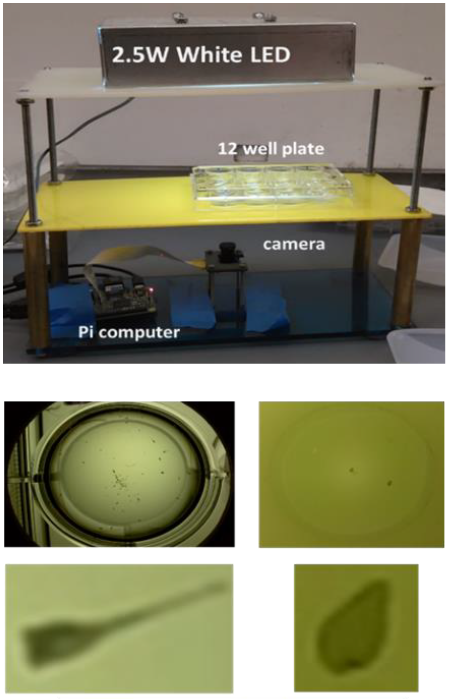
We used the single cells ciliate Stentor coeruleus as our test organism. The organisms were obtained from a commercial vendor (Carolina Biological Supply Company, Burlington, NC). For our preliminary work, we exposed the organisms to increasing concentration of butylparaben. We adopted a manual acquisition method that imaged our test organism in a single well of a 12 well tissue culture plate filled with a mixture of sterile spring water and butylparaben (
Figure 1). We imaged Stentor using a 5 Megapixel camera module (Arducam) with an adjustable lens connected to a Raspberry Pi computer that captures 1080p resolution color video (1920x1080) at 30 frames per second. Eleven serial dilutions of butylparaben in sterilized spring water were prepared, from 971 ppm to 0.9 ppm. The wells were filled with live Stentor coeruleus hand-pipetted from the stock container. Each well contained an average of 10 Stentor in 0.5 mL of spring water. Each diluted butylparaben solution was added to one of 11 wells in the plate, resulting in a final concentration from 971 ppm to 1 ppb. In the twelfth well, 0.5 mL of sterile spring water was added for use as a control. The completed plate was swirled on a horizontal surface in a figure-eight pattern to mix the solution, then placed on the imaging platform (
Figure 1). A one-minute video was captured for each well, starting with well #1 (the highest chemical concentration) and concluding with well #12 (no-chemical, control). All the Stentor in well #1 to #4 died within minutes of chemical introduction, establishing the highest non-lethal dose as 61 ppm.
3. Image processing
The loaded wells contained a considerable amount of contamination (mostly algae), for it was too laborious to hand select individual Stentor for the chemical trials. Thus, videos required additional image processing to distinguish Stentor from the algae. Stentor were floating in a flat-bottom 17 mm deep clear polystyrene well and imaged from below. The tall vertical walls of the well optically obscured the perimeter of the well (
Figure 1, middle row left) which often results in sample occlusion since Stentor tend to swim around the perimeter. The large diameter of the well reduced the resolution of each Stentor, contributing to the difficulty of distinguishing Stentor from algae. The imaging processing pipeline consisted of detection, tracking, identification, feature extraction and analysis. The detection process converted the color image of each frame to a gray image, then converted the gray image to a binary quantized image. A tracking method placed a bounding box around each object in the binary image and measured the cell area by counting the number of “on” pixels in the bounding box. For each tracked object in each frame, the (x, y) center, height, width and area were stored. An identification method assigned a unique identification label (ID) to each object in a frame and maintained that ID across frames as the object moves. Images of algae were manually removed from the pipeline after the identification process.
4. Morphological features
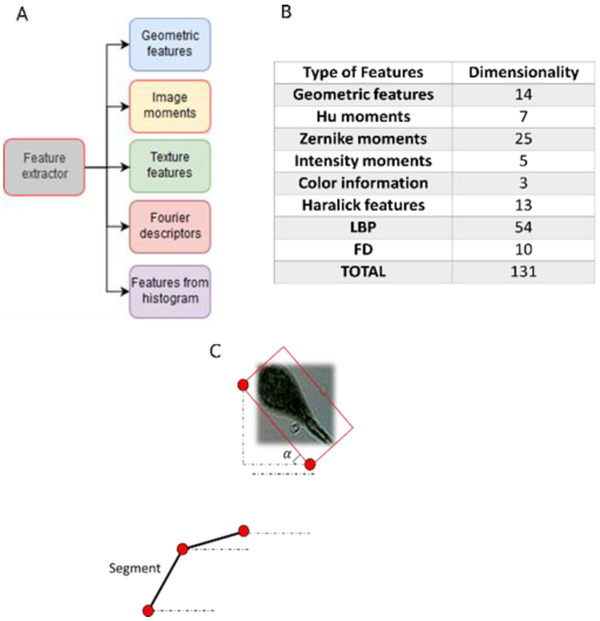
Five classes of morphological features (
Figure 2A) were extracted from the cropped plankton cell images captured in each frame of video. The classes consisted of 131 features (
Figure 2B) (
Vito P. Pastore et al. 2019). Each group of features capture different characteristics of the image. Shape-based features describe geometric aspects of the cell. Moments-based features have been shown to be useful in image classification and shape retrieval. Texture shape features include Haralick descriptors, Local Binary Patterns (LBP) and gray scale histograms. Fourier Descriptors (FD), extracted from the contours of the image, have been shown to be useful in differentiating shapes.
5. Motion based features
We extracted four features from the Stentor reconstructed tracks. The average speed and body angle (
Figure 2C) were extracted each frame. The body angle is defined as the angle that the major axis of the minimum fitting bounding box makes relative to the x axis (
Figure 2C). A significant change in body angle indicates a change in direction. A segment length is the distance traveled between two body angle changes. The turning rate is number of turns occurring over the duration of a track.
6. Multi-Target Tracking
Measuring the dynamics of multiple Stentor presents a multi-object tracking challenge (
Bolme et al. 2010). The lack of unique identifiable features makes tracking difficult when Stentor cross paths. The presence of background noise (e.g., algae) creates cluttered space to search. Tracking involves detection followed by data association; linking the detections between two successive frames. Supervised learning is a useful technique detecting organisms. However, supervised learning requires a substantial training set and the large set of videos and resulting frames in our experiments makes manual annotation difficult.
To address this challenge, we implemented a semi-supervised annotation strategy. On the first frame, the tracker placed bounding boxes around each detected Stentor. For each successive frame, there were two possible outcomes: the tracker correctly tracked the Stentor, or the tracker incorrectly acquired another object (another Stentor or algae). When the latter outcome occurred, we manually correct the position.
The tracking training set was used to train an machine learning method that simultaneously tracks the Stentor and adapts to its changing appearance (
Sujoy Kumar Biswas et al. 2019). The method uses random perturbation of Stentor images from the training set to learn a filter to recognize Stentor.
The tracking method produced a bounding box for each Stentor for each frame, along with a unique ID. The tracking output was used to identify motion patterns response of Stentor to each test chemical.
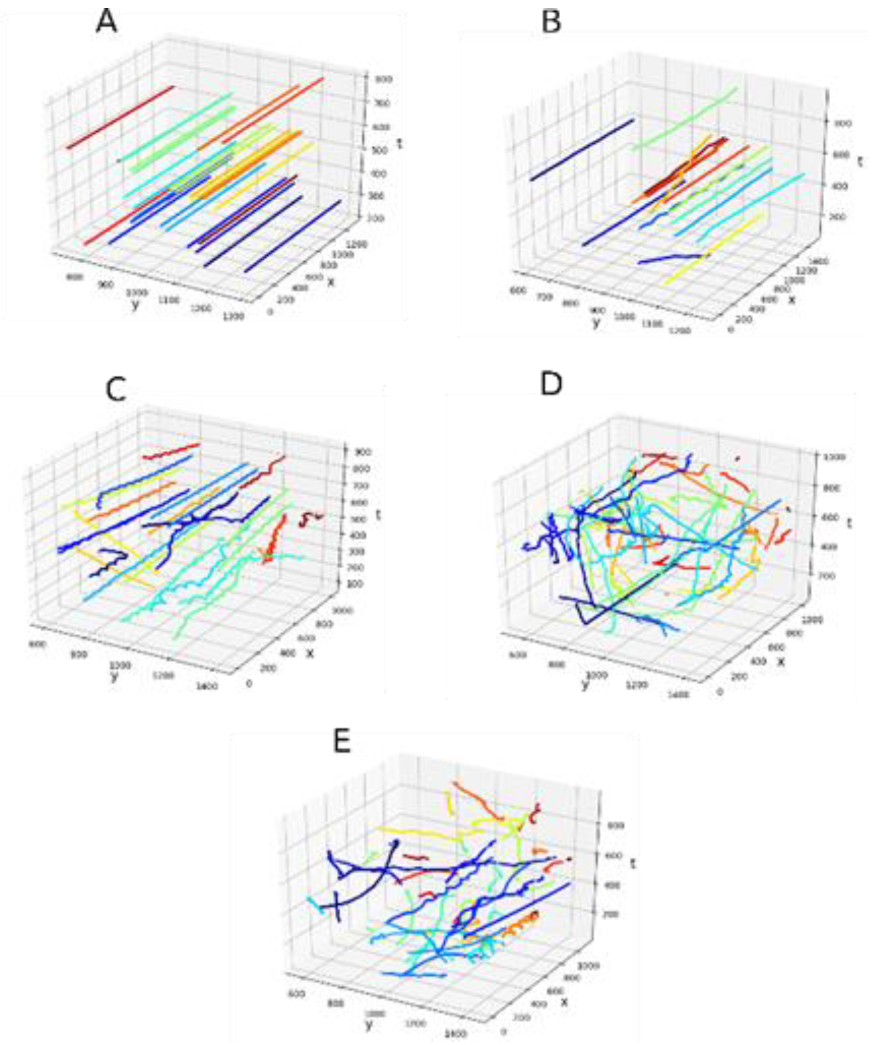
Figure 3E displays the tracks of the Stentor when they swim in the natural medium (control, no chemical). One can observe the random movement of the Stentor, indicative of food searching. However, the frequent changes in directions is noticeably subdued with the increasing dosage of the chemicals as evident in
Figure 2 panels B-E. Ultimately the Stentor stop moving and die at toxic chemical concentrations. This is reflected by linear and straight tracks indicating the Stentor are barely moving or dead.
7. Morphological-dynamical analysis
We exposed Stentor to increasing doses of butylparaben.
Figure 3A and 3B show that Stentor have mostly been incapacitated by the first two concentrations of chemical. As the chemical concentration is reduced, the swimming behavior resembles the controls (
Figure 3E). We used the features described in
Section 3 to visualize in the feature space of the Stentor under five chemical concentrations. We performed a Principal Component Analysis (PCA) and presented the results in Figure 4A. The PCA is computed using behavioral and morphological features, listed in
Figure 2. The Stentor exposed to the highest concentrations (971 ppm, 121 ppm and 60.7 ppm) can be distinguished from the other conditions. When the concentration is sufficiently low (3.8 ppm) the correspondent cluster become close to the control cluster, while still being well separable. These results suggest that Stentor morphological and dynamical difference can be distinguished at sub-lethal chemical concentrations.
Dynamic analysis was conducted on motion features for each chemical concentration and control. Figure 4B shows the average turning rate of all the Stentor in well at four chemical concentrations and control. Figure 4C shows the corresponding average segment length. Figure 4D shows the average speed. Exposure to sub-lethal concentrations of butylparaben impaired Stentor movement, reducing their speed and turning frequency. This will have significant consequences to their survival as it adversely impact their ability to hunt food and avoid predators.
8. Conclusions
Our preliminary results show high sensitivity of Stentor coeruleus to low non-lethal doses of butylparaben. We demonstrated that the introduction of the chemical creates cell morphological and dynamic modifications in the feature space. High chemical concentrations kill or render them nearly motionless while sublethal doses adversely impact speed, turning rate and trajectory. The results suggest that the Stentor coeruleus is a good model organism for a chemical biosensor. Combining Stentor with a computer vision pipeline that implement feature detection and analysis may provide an automated method to monitor sublethal chemical toxicity in the aquatic environment. Further analysis is necessary to determine if Stentor response can be used to discriminate among common chemical pollutants found in fresh water.