

Volume-Based Stratification of Lumbar Foraminal Stenosis: A Single-Center Cohort Integrating MRI/CT Morphometrics with Stepwise Interventional, Minimally Invasive and Decompression–Stabilization Surgery

 ,
, 
Abstract
1. Introduction
Explicit Hypothesis and Objectives for the End of the Introduction
2. Materials and Methods
2.1. Study Design, Objectives, and Reporting Framework
2.2. Setting and Data Sources
2.2.1. Case Identification, Sampling Strategy, and Interventional Durability Analysis
Cohort A: Historical Surgical Cohort
Cohort B: Asymptomatic MRI Morphometric Reference Cohort
Cohort C: Interventional Therapy Cohort
Cohort D: Algorithm-Guided Surgical LFS Cohort
2.3. Ethics, Consent, and Confidentiality
2.4. Cohort Architecture and Independence
- Historical surgical cohort (2017–2018; n = 351): Used to describe primary procedure distribution, revision incidence, and structural causes of persistent/recurrent symptoms.
- Asymptomatic morphometric MRI reference cohort (2017–2023; n = 500): Used to establish level- and side-specific normative foraminal canal morphometrics and nerve–foramen spatial relationships.
- Interventional therapy cohort (2017–2023; n = 463): Patients with chronic vertebrogenic pain and/or radiculopathy without dominant central canal stenosis beyond Schizas A/B, treated with staged diagnostic blocks and interventions to quantify response magnitude, durability, and escalation to surgery.
- Algorithm-guided surgical LFS cohort (2017–2023; n = 256): Comparative cohort of surgically treated LFS patients selected and managed using the prespecified volume- and stability-guided algorithm, with standardized morphometric and PROM follow-up.
2.5. Eligibility Criteria
2.5.1. Algorithm-Guided Surgical Cohort (Primary Evaluative Cohort; n = 256)
2.5.2. Asymptomatic Reference Cohort (n = 500)
2.5.3. Interventional Cohort (n = 463)
2.6. Clinical Assessment and Outcome Measures
2.7. Imaging Acquisition, Grading, and Standardization
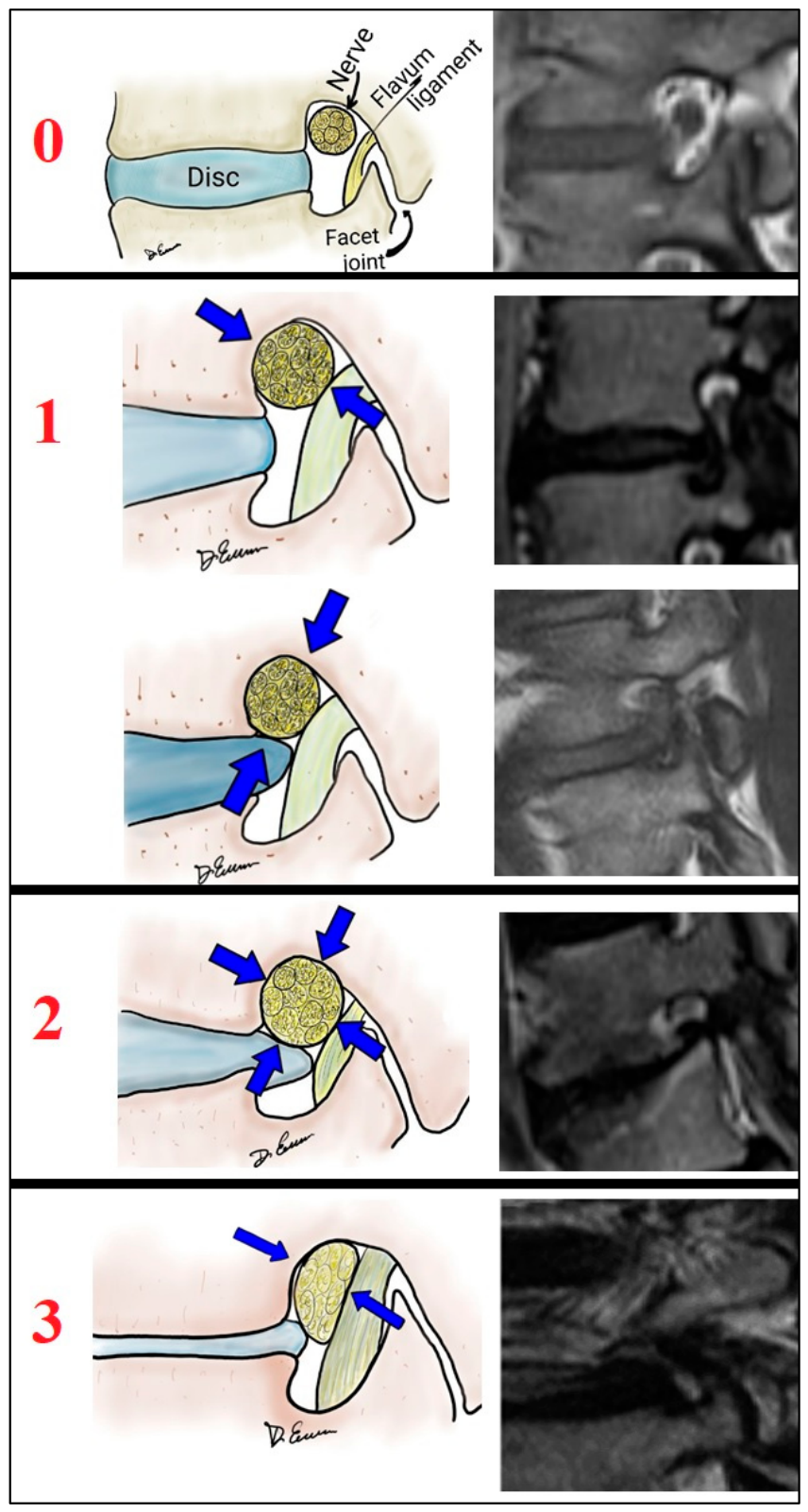
- Foraminal stenosis: Lee classification [1] MRI grade 0–3.
- Central canal morphology: Schizas grade (A/B eligible for cohorts C and D; C/D excluded from D).
- Disk degeneration: Pfirrmann.
- Facet degeneration: Fujiwara.
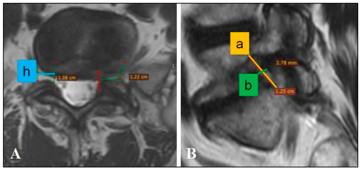
2.8. Foraminal Volumetry: Measurement Workflow
- Superior–inferior boundaries: inferior border of the pedicle above and superior border of the pedicle below.
- Anterior boundary: posterolateral vertebral body/disk/osteophyte complex.
- Posterior boundary: superior articular process/facet complex and ligamentous elements visible on MRI.
- −
- Grade 0 (normal) is where fat surrounds the nerve root circumferentially.
- −
- Grade 1 is a mild degree of stenosis characterized by obliteration of the perineural tissue surrounding the nerve root transversely or vertically.
- −
- Grade 2 means a moderate degree in which there is a marked narrowing of the foramen in width and height, although without morphological changes in the nerve root.
- −
- Grade 3 is characterized either by the destruction of the nerve root or by morphological changes.
2.8.1. Plane Selection and Measurement Points
- Identify the disk level on sagittal T2.
- Select the axial slice (or oblique-axial reformat) passing through the maximum visualization of the exiting nerve root within the foramen.
- Confirm that the slice lies between pedicles (to avoid the far-lateral extraforaminal region).
2.8.2. Elliptical-Cylinder Volume Model
- Medial landmark: medial pedicle border at the transition between the lateral recess and the entrance of the neural foramen.
- Anterior reference: posterior vertebral body/disco-osteophyte line at the index level.
- Posterior reference: anterior surface of the superior articular process/facet complex.
- Lateral landmark: lateral cortical margin of the pedicle or the point where the exiting root leaves the osseous foraminal corridor.
- Measurement axis: medial-to-lateral foraminal depth along the expected trajectory of the exiting nerve root, avoiding far-lateral extraforaminal tissue.
2.8.3. Nerve–Foramen Spatial Relationship (Occupancy)
- The exiting nerve root was measured at the same plane using two orthogonal diameters (an, bn) to approximate nerve cross-sectional area as an ellipse.
- Nerve “segment length” (h) was set equal to the measured foraminal h to maintain geometric compatibility.
2.8.4. Measurement Quality Control and Reproducibility
2.9. Stability Assessment
2.9.1. Radiographic Assessment
2.9.2. Instability Criteria (Reproducible Thresholds)
2.10. Interventional Therapy Protocol (Cohort C)
- Cannula length 100–150 mm; active tip 5–10 mm.
- Lesioning temperature 80 °C for 90 s.
- Levels treated were selected according to positive diagnostic blocks.
2.11. Surgical Techniques and Group Definitions (Cohort D)
2.11.1. Group 1: Minimally Invasive/Indirect Decompression (n = 119)
2.11.2. Group 2: Direct Decompression vs. Decompression–Stabilization (n = 137)
2.12. Follow-Up Schedule, Imaging Timing, and Safety Endpoints
2.13. Statistical Analysis Plan
- Kruskal–Wallis with Dunn post hoc and Holm correction (≥3 groups).
- Mann–Whitney U (2 groups).
- χ2 or Fisher exact (categorical).
- Wilcoxon signed-rank (paired).
- Friedman with Conover–Iman post hoc and Holm correction (≥3 time points).
- Multivariable linear regression for continuous outcomes (ODI, VAS, SF-36 changes).
- Multivariable logistic regression for binary outcomes (MacNab success; reoperation).
- Covariates included age, sex, index level, Pfirrmann grade, Fujiwara grade, baseline PROMs, Lee grade [1], foraminal volume/occupancy, and instability status. Model diagnostics included assessment of collinearity and goodness-of-fit.
2.14. Missing Data Handling
3. Results
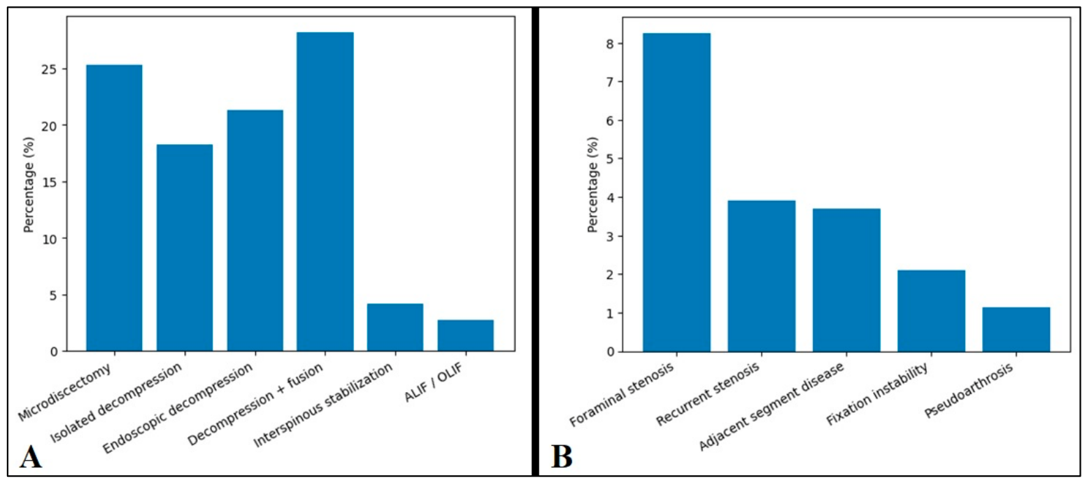
3.1. Retrospective Analysis of Surgical Outcomes and Causes of Treatment Dissatisfaction
- Foraminal canal stenosis: 8.26%.
- Recurrent foraminal stenosis: 3.9%.
- Adjacent segment disease: 3.7%.
- Fixation instability: 2.1%.
- Pseudoarthrosis: 1.13%.
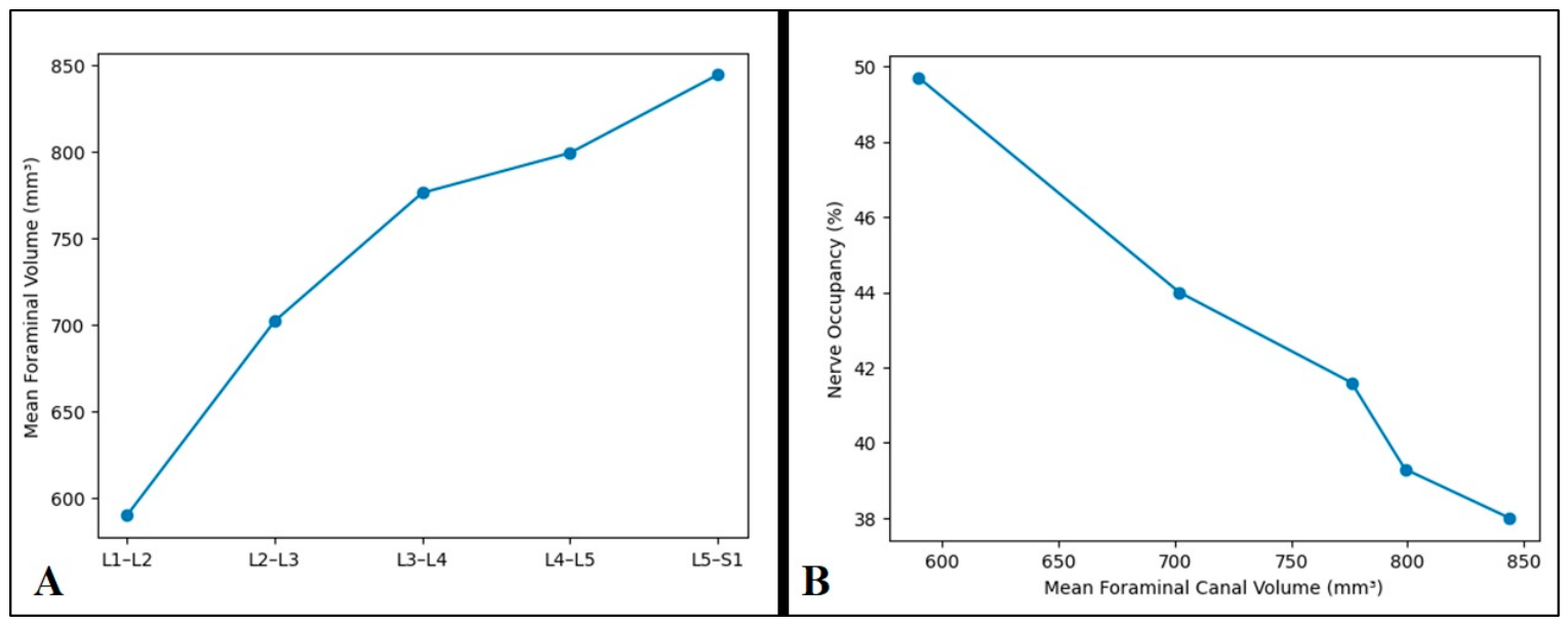
3.2. MRI Morphometric Screening of the Lumbar Foraminal Canal in Asymptomatic Adults
3.3. Outcomes of Interventional Therapy in Chronic Vertebrogenic Pain (n = 463)
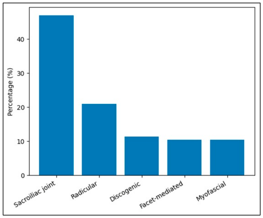
- Sacroiliac joint-related pain: 46.9%.
- Radicular pain: 20.9%.
- Discogenic pain: 11.4%.
- Facet-mediated pain: 10.4%.
- Myofascial pain: 10.4%.
- Radiculopathy regressed in 44.7% of patients.
- Facet-mediated pain improved in 44.0%.
- Median pain relief duration was 3–6 months.
3.4. Surgical Outcomes in Algorithm-Guided Foraminal Stenosis (n = 256)
3.5. Comparative Outcomes: Endoscopic Decompression vs. ALIF
3.5.1. Pain and Functional Recovery
3.5.2. Quality of Life
3.6. Comparative Outcomes: Microsurgical Decompression vs. TLIF
3.7. Validation of the Volume-Based Diagnostic and Therapeutic Algorithm
4. Discussion
4.1. Decompression Alone vs. Decompression Plus Fusion: Stability Remains the Hinge Variable
4.2. Indirect Decompression Literature After 36: Why “Reducibility” and Failure-to-Indirect-Decompress Matter
4.3. Long-Horizon Tradeoffs: Adjacent Segment Pathology and Why Avoiding Fusion Maximalism Remains Rational
4.4. Interventional Durability and the Role of Structured Temporization
4.5. Limitations
5. Conclusions
Author Contributions
Funding
Institutional Review Board Statement
Informed Consent Statement
Data Availability Statement
Conflicts of Interest
References
- Lee, S.; Lee, J.W.; Yeom, J.S.; Kim, K.J.; Kim, H.J.; Chung, S.K.; Kang, H.S. A practical MRI grading system for lumbar foraminal stenosis. AJR Am. J. Roentgenol. 2010, 194, 1095–1098. [Google Scholar] [CrossRef] [PubMed]
- Schizas, C.; Theumann, N.; Burn, A.; Tansey, R.; Wardlaw, D.; Smith, F.W.; Kulik, G. Qualitative grading of severity of lumbar spinal stenosis based on the morphology of the dural sac on magnetic resonance images. Spine 2010, 35, 1919–1924. [Google Scholar] [CrossRef] [PubMed]
- Pfirrmann, C.W.; Metzdorf, A.; Zanetti, M.; Hodler, J.; Boos, N. Magnetic resonance classification of lumbar intervertebral disc degeneration. Spine 2001, 26, 1873–1878. [Google Scholar] [CrossRef] [PubMed]
- Fujiwara, A.; Tamai, K.; Yamato, M.; An, H.S.; Yoshida, H.; Saotome, K.; Kurihashi, A. The relationship between facet joint osteoarthritis and disc degeneration of the lumbar spine: An MRI study. Eur. Spine J. 1999, 8, 396–401. [Google Scholar] [CrossRef] [PubMed]
- Cohen, S.P.; Bhaskar, A.; Bhatia, A.; Buvanendran, A.; Deer, T.; Garg, S.; Hooten, W.M.; Hurley, R.W.; Kennedy, D.J.; McLean, B.C.; et al. Consensus practice guidelines on interventions for lumbar facet joint pain from a multispecialty, international working group. Reg. Anesth. Pain Med. 2020, 45, 424–467. [Google Scholar] [CrossRef] [PubMed]
- Cohen, S.P.; Kapural, L.; Kohan, L.; Li, S.; Hurley, R.W.; Vallejo, R.; Eshraghi, Y.; Dinakar, P.; Durbhakula, S.; Beall, D.P.; et al. Cooled radiofrequency ablation versus standard medical management for chronic sacroiliac joint pain: A multicenter, randomized comparative effectiveness study. Reg. Anesth. Pain Med. 2024, 49, 184–191. [Google Scholar] [PubMed]
- Giordan, E.; Billeci, D.; Del Verme, J.; Varrassi, G.; Coluzzi, F. Endoscopic Transforaminal Lumbar Foraminotomy: A Systematic Review and Meta-Analysis. Pain Ther. 2021, 10, 1481–1495. [Google Scholar] [CrossRef] [PubMed]
- Cofano, F.; Langella, F.; Petrone, S.; Baroncini, A.; Cecchinato, R.; Redaelli, A.; Garbossa, D.; Berjano, P. Clinical and radiographic performance of indirect foraminal decompression with anterior retroperitoneal lumbar approach for interbody fusion (ALIF). Clin. Neurol. Neurosurg. 2021, 209, 106946. [Google Scholar] [CrossRef] [PubMed]
- Försth, P.; Ólafsson, G.; Carlsson, T.; Frost, A.; Borgström, F.; Fritzell, P.; Öhagen, P.; Michaëlsson, K.; Sandén, B. A Randomized, Controlled Trial of Fusion Surgery for Lumbar Spinal Stenosis. N. Engl. J. Med. 2016, 374, 1413–1423. [Google Scholar] [CrossRef] [PubMed]
- Ghogawala, Z.; Dziura, J.; Butler, W.E.; Dai, F.; Terrin, N.; Magge, S.N.; Coumans, J.V.; Harrington, J.F.; Amin-Hanjani, S.; Schwartz, J.S.; et al. Laminectomy plus Fusion versus Laminectomy Alone for Lumbar Spondylolisthesis. N. Engl. J. Med. 2016, 374, 1424–1434. [Google Scholar] [CrossRef] [PubMed]
- Okuda, S.; Yamashita, T.; Matsumoto, T.; Nagamoto, Y.; Sugiura, T.; Takahashi, Y.; Maeno, T.; Iwasaki, M. Adjacent Segment Disease After Posterior Lumbar Interbody Fusion: A Case Series of 1000 Patients. Glob. Spine J. 2018, 8, 722. [Google Scholar] [CrossRef]
- Nurmukhametov, R.M.; Klimov, V.; Medetbek, A.; Kudryakov, S.A.; Dosanov, M.; Alekseevna Guseva, A.; Ruslanovich Baigushev, P.; Arturovich Kerimov, T.; Montemurro, N. Comparative Clinical and Volumetric Outcomes of Contemporary Surgical Techniques for Lumbar Foraminal Stenosis: A Retrospective Cohort Study. Surgeries 2025, 6, 91. [Google Scholar] [CrossRef]
- Nurmukhametov, R.; Encarnacion Ramirez, M.D.J.; Dosanov, M.; Medetbek, A.; Kudryakov, S.; Wisam Alsaed, L.; Chmutin, G.; Reyes Soto, G.; Ntalaja Mukengeshay, J.; Mpoyi Chérubin, T.; et al. Quantifying Lumbar Foraminal Volumetric Dimensions: Normative Data and Implications for Stenosis—Part 2 of a Comprehensive Series. Med. Sci. 2024, 12, 34. [Google Scholar] [CrossRef]
- Nurmukhametov, R.; Encarnacion Ramirez, M.D.J.; Dosanov, M.; Medetbek, A.; Kudryakov, S.; Reyes Soto, G.; Ponce Espinoza, C.B.; Mukengeshay, J.N.; Mpoyi Cherubin, T.; Nikolenko, V.; et al. Exploring Pathways for Pain Relief in Treatment and Management of Lumbar Foraminal Stenosis: A Review of the Literature. Brain Sci. 2024, 14, 740. [Google Scholar] [CrossRef] [PubMed]
- Montemurro, N.; Zotti, N.; Guercini, J.; De Carolis, G.; Leoni, C.; Marotta, R.; Tomei, R.; Baggiani, A.; Paolicchi, A.; Lazzini, S.; et al. Value-based healthcare in management of chronic back pain: A multidisciplinary- and lean-based approach. Surg. Neurol. Int. 2024, 15, 348. [Google Scholar] [CrossRef] [PubMed]
- Nurmukhametov, R.; Medetbek, A.; Ramirez, M.E.; Afsar, A.; Sharif, S.; Montemurro, N. Factors affecting return to work following endoscopic lumbar foraminal stenosis surgery: A single-center series. Surg. Neurol. Int. 2023, 14, 408. [Google Scholar] [CrossRef] [PubMed]
- Ramirez, M.D.J.E.; Chmutin, G.; Nurmukhametov, R.; Soto, G.R.; Kannan, S.; Piavchenko, G.; Nikolenko, V.; Efe, I.E.; Romero, A.R.; Mukengeshay, J.N.; et al. Integrating Augmented Reality in Spine Surgery: Redefining Precision with New Technologies. Brain Sci. 2024, 14, 645. [Google Scholar] [CrossRef]
- Nurmukhametov, R.; Dosanov, M.; Encarnacion, M.D.J.; Barrientos, R.; Matos, Y.; Alyokhin, A.I.; Baez, I.P.; Efe, I.E.; Restrepo, M.; Chavda, V.; et al. Transforaminal Fusion Using Physiologically Integrated Titanium Cages with a Novel Design in Patients with Degenerative Spinal Disorders: A Pilot Study. Surgeries 2022, 3, 175–184. [Google Scholar] [CrossRef]
- Zhu, K.; Yan, S.; Guo, S.; Tong, J.; Li, C.; Tan, J.; Wan, W. Morphological changes of contralateral intervertebral foramen induced by cage insertion orientation after unilateral transforaminal lumbar interbody fusion. J. Orthop. Surg. Res. 2019, 14, 79. [Google Scholar] [CrossRef] [PubMed]
- Kim, J.H.; Kim, H.S.; Kapoor, A.; Adsul, N.; Kim, K.J.; Choi, S.H.; Jang, J.S.; Jang, I.T.; Oh, S.H. Feasibility of full endoscopic spine surgery in patients over the age of 70 years with degenerative lumbar spine disease. Neurospine 2018, 15, 131–137. [Google Scholar] [CrossRef] [PubMed]
- Park, M.K.; Son, S.K.; Park, W.W.; Choi, S.H.; Jung, D.Y.; Kim, D.H.U. Unilateral biportal endosccopy for decompression of extraforaminal stenosis at the lumbosacral junction: Surgical techniques and clinical outcomes outcomes. Neurospine 2021, 18, 871–879. [Google Scholar] [PubMed]
- Vande Kerckhove, M.; d’Astorg, H.; Ramos-Pascual, S.; Saffarini, M.; Fiere, V.; Szadkowski, M. SPINE: High heterogeneity and no significant differences in clinical outcomes of endoscopic foraminotomy vs fusion for lumbar foraminal stenosis: A meta-analysis. EFORT Open Rev. 2023, 8, 73–89. [Google Scholar] [CrossRef] [PubMed]
- Lewandrowski, K.U.; Fiorelli, R.K.A.; Pereira, M.G.; Abraham, I.; Alfaro Pachicano, H.H.; Elfar, J.C.; Alhammoud, A.; Landgraeber, S.; Oertel, J.; Hellinger, S.; et al. Polytomous Rasch Analyses of Surgeons’ Decision-Making on Choice of Procedure in Endoscopic Lumbar Spinal Stenosis Decompression Surgeries. Int. J. Spine Surg. 2024, 18, 164–177. [Google Scholar] [CrossRef] [PubMed]
- Brodeur, P.G.; Medina Perez, G.; Hartnett, D.A.; McDonald, C.L.; Gil, J.A.; Cruz, A.I., Jr.; Kuris, E.O. Surgeon Volume and Social Disparity are Associated with Postoperative Complications After Lumbar Fusion. World Neurosurg. 2022, 163, e162–e176. [Google Scholar] [CrossRef] [PubMed]
- Fitzgerald, A.; McCool, R.; Carr, E.; Miller, P.; Reddish, K.; Lohr, C.C.; Annoni, E.; Lawrence, B. A systematic review of bone graft products used in lumbar interbody fusion procedures for degenerative disc disease. N. Am. Spine Soc. J. 2024, 5, 100579. [Google Scholar]
- Kim, J.; Kim, S.; Song, I. Octacalcium phosphate, a promising bone substitute material: A narrative review. J. Yeungnam Med. Sci. 2024, 41, 4–12. [Google Scholar] [CrossRef] [PubMed]
- Lewandrowski, K.U.; Elfar, J.C.; Li, Z.M.; Burkhardt, B.W.; Lorio, M.P.; Winkler, P.A.; Oertel, J.M.; Telfeian, A.E.; Dowling, Á.; Vargas, R.A.A.; et al. The Changing Environment in Postgraduate Education in Orthopedic Surgery and Neurosurgery and Its Impact on Technology-Driven Targeted Interventional and Surgical Pain Management: Perspectives from Europe, Latin America, Asia, and The United States. J. Pers. Med. 2023, 13, 852. [Google Scholar] [CrossRef] [PubMed]
- Nurmukhametov, R.M.; Dzhumabekovich Abakirov, M.; Anatolyevich Kudryakov, S.; Kaskirbayevich Dosanov, M.; Nuriddinov, D.; Beis Zhanchivdorj, B.; Borja Cevallos, K.S.; Yadigerovich Bozo, I.; Martinez Mateo, A.L.; Montemurro, N. Gene-Activated Octacalcium Phosphate (OCP/VEGF) Versus Autologous Bone Graft for Single-Level TLIF in Degenerative Lumbar Stenosis. Surgeries 2026, 7, 29. [Google Scholar] [CrossRef]
- Sallent, I.; Capella-Monsonís, H.; Procter, P.; Bozo, I.Y.; Deev, R.V.; Zubov, D.; Vasyliev, R.; Perale, G.; Pertici, G.; Baker, J.; et al. The Few Who Made It: Commercially and Clinically Successful Innovative Bone Grafts. Front. Bioeng. Biotechnol. 2020, 8, 952. [Google Scholar] [CrossRef] [PubMed]
- Deev, R.V.; Drobyshev, A.Y.; Bozo, I.Y.; Isaev, A.A. Ordinary and Activated Bone Grafts: Applied Classification and the Main Features. BioMed Res. Int. 2015, 2015, 365050. [Google Scholar] [CrossRef] [PubMed]
- Yamada, K.; Matsuda, H.; Nabeta, M.; Habunaga, H.; Suzuki, A.; Nakamura, H. Clinical outcomes of microscopic decompression for degenerative lumbar foraminal stenosis: A comparison between patients with and without degenerative lumbar scoliosis. Eur. Spine J. 2011, 20, 947–953. [Google Scholar] [PubMed]
- Ramirez, M.D.J.E.; Nurmukhametov, R.; Bernard, E.; Peralta, I.; Efe, I.E. A Low-Cost Three-Dimensional Printed Retractor for Transforaminal Lumbar Interbody Fusion. Cureus 2022, 14, e24185. [Google Scholar] [PubMed]
- Montemurro, N.; Pierozzi, E.; Inchingolo, A.M.; Pahwa, B.; De Carlo, A.; Palermo, A.; Scarola, R.; Dipalma, G.; Corsalini, M.; Inchingolo, A.D.; et al. New biograft solution, growth factors and bone regenerative approaches in neurosurgery, dentistry, and orthopedics: A review. Eur. Rev. Med. Pharmacol. Sci. 2023, 27, 7653–7664. [Google Scholar] [CrossRef] [PubMed]
- Bozo, I.Y.; Drobyshev, A.Y.; Redko, N.A.; Komlev, V.S.; Isaev, A.A.; Deev, R.V. Bringing a Gene-Activated Bone Substitute into Clinical Practice: From Bench to Bedside. Front. Bioeng. Biotechnol. 2021, 9, 599300. [Google Scholar] [CrossRef] [PubMed]
- Tan, G.H.; Goss, B.G.; Thorpe, P.J.; Williams, R.P. CT-based classification of long spinal allograft fusion. Eur. Spine J. 2007, 16, 1875–1881. [Google Scholar] [CrossRef] [PubMed]
- Shimizu, T.; Fujibayashi, S.; Otsuki, B.; Murata, K.; Matsuda, S. Indirect decompression via oblique lateral interbody fusion for severe degenerative lumbar spinal stenosis: A comparative study with direct decompression transforaminal/posterior lumbar interbody fusion. Spine J. 2021, 21, 963–971. [Google Scholar] [CrossRef] [PubMed]
- Fujibayashi, S.; Hynes, R.A.; Otsuki, B.; Kimura, H.; Takemoto, M.; Matsuda, S. Effect of indirect neural decompression through oblique lateral interbody fusion for degenerative lumbar disease. Spine 2015, 40, E175–E182. [Google Scholar] [CrossRef] [PubMed]
- Walker, C.T.; Xu, D.S.; Cole, T.S.; Alhilali, L.M.; Godzik, J.; Angel Estrada, S.; Pedro Giraldo, J.; Wewel, J.T.; Morgan, C.D.; Zhou, J.J.; et al. Predictors of indirect neural decompression in minimally invasive transpsoas lateral lumbar interbody fusion. J. Neurosurg. Spine 2021, 35, 80–90. [Google Scholar] [CrossRef] [PubMed]
- Yingsakmongkol, W.; Jitpakdee, K.; Kerr, S.; Limthongkul, W.; Kotheeranurak, V.; Singhatanadgige, W. Successful Criteria for Indirect Decompression with Lateral Lumbar Interbody Fusion. Neurospine 2022, 19, 805–815. [Google Scholar] [CrossRef] [PubMed]
- Tseng, S.C.; Lin, Y.H.; Wu, Y.C.; Shih, C.M.; Chen, K.H.; Lee, C.H.; Pan, C.C. Indirect decompression via oblique lumbar interbody fusion is sufficient for treatment of lumbar foraminal stenosis. Front. Surg. 2022, 9, 911514. [Google Scholar] [CrossRef] [PubMed]
- Li, J.; Xu, T.Z.; Zhang, N.; Chen, Q.X.; Li, F.C. Predictors for second-stage posterior direct decompression after lateral lumbar interbody fusion: A review of five hundred fifty-seven patients in the past five years. Int. Orthop. 2022, 46, 1101–1109. [Google Scholar] [PubMed]
- Hattori, S.; Matsutani, S. Three Cases of Indirect Decompression Failure Following Oblique Lumbar Interbody Fusion Requiring Early Direct Posterior Decompression: Analysis of Etiologies and Literature Review. Cureus 2025, 17, e90798. [Google Scholar] [CrossRef] [PubMed]
- Patel, K.; Harikar, M.M.; Venkataram, T.; Chavda, V.; Montemurro, N.; Assefi, M.; Hussain, N.; Yamamoto, V.; Kateb, B.; Lewandrowski, K.U.; et al. Is Minimally Invasive Spinal Surgery Superior to Endoscopic Spine Surgery in Postoperative Radiologic Outcomes of Lumbar Spine Degenerative Disease? A Systematic Review. J. Neurol. Surg. A Cent. Eur. Neurosurg. 2024, 85, 182–191. [Google Scholar] [PubMed]
- Abraham, I.; Lewandrowski, K.U.; Elfar, J.C.; Li, Z.M.; Fiorelli, R.K.A.; Pereira, M.G.; Lorio, M.P.; Burkhardt, B.W.; Oertel, J.M.; Winkler, P.A.; et al. Randomized Clinical Trials and Observational Tribulations: Providing Clinical Evidence for Personalized Surgical Pain Management Care Models. J. Pers. Med. 2023, 13, 1044. [Google Scholar] [CrossRef] [PubMed]
- Weinstein, J.N.; Tosteson, T.D.; Lurie, J.D.; Tosteson, A.N.; Blood, E.; Hanscom, B.; Herkowitz, H.; Cammisa, F.; Albert, T.; Boden, S.D.; et al. Surgical versus nonsurgical therapy for lumbar spinal stenosis. N. Engl. J. Med. 2008, 358, 794–810. [Google Scholar] [CrossRef] [PubMed]
- Weinstein, J.N.; Lurie, J.D.; Tosteson, T.D.; Hanscom, B.; Tosteson, A.N.; Blood, E.A.; Birkmeyer, N.J.; Hilibrand, A.S.; Herkowitz, H.; Cammisa, F.P.; et al. Surgical versus nonsurgical treatment for lumbar degenerative spondylolisthesis. N. Engl. J. Med. 2007, 356, 2257–2270. [Google Scholar] [CrossRef] [PubMed]
- Park, P.; Garton, H.J.; Gala, V.C.; Hoff, J.T.; McGillicuddy, J.E. Adjacent segment disease after lumbar or lumbosacral fusion: Review of the literature. Spine 2004, 29, 1938–1944. [Google Scholar] [CrossRef] [PubMed]
- Harrop, J.S.; Youssef, J.A.; Maltenfort, M.; Vorwald, P.; Jabbour, P.; Bono, C.M.; Goldfarb, N.; Vaccaro, A.R.; Hilibrand, A.S. Lumbar adjacent segment degeneration and disease after arthrodesis and total disc arthroplasty. Spine 2008, 33, 1701–1707. [Google Scholar] [CrossRef] [PubMed]
- Hilibrand, A.S.; Robbins, M. Adjacent segment degeneration and adjacent segment disease: The consequences of spinal fusion? Spine J. 2004, 4, 190S–194S. [Google Scholar] [CrossRef] [PubMed]
- Ghiselli, G.; Wang, J.C.; Bhatia, N.N.; Hsu, W.K.; Dawson, E.G. Adjacent segment degeneration in the lumbar spine. J. Bone Jt. Surg. Am. 2004, 86, 1497–1503. [Google Scholar] [CrossRef]
- Ahsan, M.K.; Hossain, M.R.; Khan, M.S.I.; Zaman, N.; Ahmed, N.; Montemurro, N.; Chaurasia, B. Lumbar revision microdiscectomy in patients with recurrent lumbar disc herniation: A single-center prospective series. Surg. Neurol. Int. 2020, 11, 404. [Google Scholar] [CrossRef] [PubMed]
- Zigler, J.E.; Glenn, J.; Delamarter, R.B. Five-year adjacent-level degenerative changes in patients with single-level disease treated using lumbar total disc replacement with ProDisc-L versus circumferential fusion. J. Neurosurg. Spine 2012, 17, 504–511. [Google Scholar] [CrossRef] [PubMed]
- Lewandrowski, K.U.; Dowling, Á.; Lee, J.H.; Burkhardt, B.W.; Alves, Ó.L.; Parajón, A.; Ramirez, J.F.; Montemurro, N.; Vaccaro, A.R.; Lorio, M.P. Transforaminal Endoscopic Lumbar Decompression: Defining Its Scope and Limitations. Int. J. Spine Surg. 2026, 11, 8888. [Google Scholar]
- Janjua, M.I.; Abbasi, S.; Idrees, A.; Yaqoob, E.; Montemurro, N.; Chaurasia, B.; Javed, S. Benefits and Drawbacks of Teleneurosurgery in Low- and Middle-Income Countries: A Scoping Review. Acta Neurol. Taiwan 2025, 34, 189–195. [Google Scholar] [CrossRef]
- Lewandrowski, K.U.; Alfaro Pachicano, H.H.; Alvim Fiorelli, R.K.; Elfar, J.C.; Landgraeber, S.; Oertel, J.; Hellinger, S.; Dowling, Á.; De Carvalho, P.S.T.; Ramos, M.R.F.; et al. Comparative Analysis of Learning Curve, Complexity, Psychological Stress, and Work Relative Value Units for CPT 62380 Endoscopic Lumbar Spinal Decompression vs. Traditional Lumbar Spine Surgeries: A Paired Rasch Survey Study. Int. J. Spine Surg. 2024, 18, 138–151. [Google Scholar] [CrossRef] [PubMed]





| Parameter | Interobserver ICC | 95% CI | Intraobserver ICC | 95% CI |
|---|---|---|---|---|
| Foraminal long-axis diameter, a | 0.92 | 0.89–0.95 | 0.95 | 0.92–0.97 |
| Foraminal short-axis diameter, b | 0.90 | 0.86–0.93 | 0.94 | 0.90–0.96 |
| Foraminal depth, h | 0.87 | 0.82–0.91 | 0.91 | 0.87–0.94 |
| Foraminal volume | 0.91 | 0.88–0.94 | 0.95 | 0.92–0.97 |
| Nerve volume proxy | 0.86 | 0.80–0.90 | 0.90 | 0.85–0.93 |
| Nerve occupancy | 0.88 | 0.83–0.92 | 0.92 | 0.88–0.95 |
Disclaimer/Publisher’s Note: The statements, opinions and data contained in all publications are solely those of the individual author(s) and contributor(s) and not of MDPI and/or the editor(s). MDPI and/or the editor(s) disclaim responsibility for any injury to people or property resulting from any ideas, methods, instructions or products referred to in the content. |
© 2026 by the authors. Published by MDPI on behalf of the Swiss Federation of Clinical Neuro-Societies. Licensee MDPI, Basel, Switzerland. This article is an open access article distributed under the terms and conditions of the Creative Commons Attribution (CC BY) license.
Share and Cite
Nurmukhametov, R.M.; Dzhumabekovich Abakirov, M.; Anatolyevich Kudryakov, S.; Martinez Mateo, A.L.; Taveras, J.L.; Peralta Baez, I.; Kaskirbayevich Dosanov, M.; Montemurro, N. Volume-Based Stratification of Lumbar Foraminal Stenosis: A Single-Center Cohort Integrating MRI/CT Morphometrics with Stepwise Interventional, Minimally Invasive and Decompression–Stabilization Surgery. Clin. Transl. Neurosci. 2026, 10, 20. https://doi.org/10.3390/ctn10030020
Nurmukhametov RM, Dzhumabekovich Abakirov M, Anatolyevich Kudryakov S, Martinez Mateo AL, Taveras JL, Peralta Baez I, Kaskirbayevich Dosanov M, Montemurro N. Volume-Based Stratification of Lumbar Foraminal Stenosis: A Single-Center Cohort Integrating MRI/CT Morphometrics with Stepwise Interventional, Minimally Invasive and Decompression–Stabilization Surgery. Clinical and Translational Neuroscience. 2026; 10(3):20. https://doi.org/10.3390/ctn10030020
Chicago/Turabian StyleNurmukhametov, Renat Madekhatovich, Medetbek Dzhumabekovich Abakirov, Stepan Anatolyevich Kudryakov, Alberto Luis Martinez Mateo, Jonathan Lara Taveras, Ismael Peralta Baez, Medet Kaskirbayevich Dosanov, and Nicola Montemurro. 2026. "Volume-Based Stratification of Lumbar Foraminal Stenosis: A Single-Center Cohort Integrating MRI/CT Morphometrics with Stepwise Interventional, Minimally Invasive and Decompression–Stabilization Surgery" Clinical and Translational Neuroscience 10, no. 3: 20. https://doi.org/10.3390/ctn10030020
APA StyleNurmukhametov, R. M., Dzhumabekovich Abakirov, M., Anatolyevich Kudryakov, S., Martinez Mateo, A. L., Taveras, J. L., Peralta Baez, I., Kaskirbayevich Dosanov, M., & Montemurro, N. (2026). Volume-Based Stratification of Lumbar Foraminal Stenosis: A Single-Center Cohort Integrating MRI/CT Morphometrics with Stepwise Interventional, Minimally Invasive and Decompression–Stabilization Surgery. Clinical and Translational Neuroscience, 10(3), 20. https://doi.org/10.3390/ctn10030020