Article

Colloidal Hydroxyapatite Nanoparticles as a Carrier for Cannabinol: The Effect on Model Langmuir Monolayers and Bilayer Lipid Membranes


1
Faculty of Chemistry, University of Warsaw, Pasteur 1, 02-093 Warsaw, Poland
2
Biotech Evolution, Mokra 3C, 05-092 Łomianki, Poland
*
Authors to whom correspondence should be addressed.
Colloids Interfaces 2026, 10(3), 33; https://doi.org/10.3390/colloids10030033
Submission received: 17 February 2026 / Revised: 10 April 2026 / Accepted: 22 April 2026 / Published: 24 April 2026

Abstract

Cannabinol (CBN) is a highly lipophilic phytocannabinoid whose biomedical application is limited by poor water solubility. In this study, colloidal hydroxyapatite nanoparticles (nHAp) were evaluated as a carrier for CBN, and their effect on model lipid membranes was investigated. Interactions between CBN and lipids were examined using Langmuir monolayers and lipid bilayers (black lipid membranes, BLMs). Langmuir monolayer studies revealed strong interactions between CBN and lipids, resulting in changes in isotherms, compressibility, and monolayer stability. BLM measurements indicated that delivery of CBN via nHAp modifies the electrical properties and stability of the lipid bilayer, suggesting alterations in membrane organization and permeability. These results demonstrate that hydroxyapatite nanoparticles can effectively serve as a carrier for cannabinol while modulating its interactions with lipid membranes.

1. Introduction

Hydroxyapatite (nHAp, Ca10(PO4)6(OH)2) is one of the most widely used biomaterials in medicine and bioengineering due to its excellent biocompatibility, bioactivity, and ability to form surfaces that promote adsorption and interactions with various biologically active molecules [1,2,3]. In recent years, increasing attention has been paid to the functionalization of hydroxyapatite with organic compounds to obtain hybrid nanostructures with tailored biological activity. In particular, modification of hydroxyapatite with bioactive molecules such as cannabinoids offers new opportunities for the development of multifunctional nanocarriers with potential applications in drug delivery and controlled interactions with biological membranes [4,5].
Cannabinol (CBN) is a phytocannabinoid present in Cannabis sativa, formed primarily through the oxidation of Δ9-tetrahydrocannabinol (THC) during plant maturation or storage. Unlike THC, CBN does not exhibit strong psychoactive effects, which makes it an attractive candidate for biomedical applications [6,7,8,9]. Numerous studies have demonstrated that CBN possesses a broad spectrum of biological activities, including anticancer, antibacterial, anti-inflammatory, analgesic, and sedative effects [10,11]. In vitro investigations have shown that CBN can inhibit cell proliferation, induce apoptosis, and cause cell cycle arrest in various cancer cell lines, such as glioma, liver cancer, breast cancer, and acute leukemia, through modulation of signaling pathways including ERK1/2 and AKT, as well as regulation of genes involved in cell cycle control and apoptosis [12,13]. In addition, CBN exhibits antibacterial activity against Gram-positive strains, including methicillin-resistant Staphylococcus aureus (MRSA), highlighting its potential for antimicrobial applications [14]. Its anti-inflammatory, analgesic, and sedative properties further support its relevance for therapeutic use [15,16,17,18].
Due to its lipophilic character and structural similarity to cholesterol, CBN readily interacts with lipid assemblies and can incorporate into lipid bilayers [19,20,21]. Consequently, understanding how CBN-loaded nanocarriers interact with membrane structures is crucial for evaluating their biological performance. Studies employing model lipid systems, such as Langmuir monolayers and free-standing black lipid membranes (BLMs), provide valuable insight into the mechanisms governing nanocarrier–membrane interactions and their influence on membrane organization, fluidity, and phase behavior.
Given the increasing interest in hydroxyapatite-based nanocarriers functionalized with cannabinoids, a detailed investigation of the interactions between nHAp–CBN nanostructures and lipid membranes is of particular importance. This study aimed to characterize cannabinol-modified hydroxyapatite nanocarriers and to investigate their interactions with biomimetic lipid membranes using Langmuir monolayers and black lipid membrane (BLM) models, with a focus on their impact on lipid organization and membrane physicochemical properties.

2. Materials and Methods

2.1. Chemicals

The nanostructural hydroxyapatite was fabricated from inorganic salts such as calcium nitrate tetrahydrate, Ca(NO3)2∙4H2O, and diammonium hydrogen phosphate (NH4)2HPO4, both of analytical grade, with the use of 25% aqueous solution as the precipitating agent. All chemicals were supplied by Chempur, Piekary Śląskie, Poland. Deionized water with a resistivity of 18.2 MΩ cm at 25 °C was obtained using the Milli-Q ultra-pure water filtering system from Merck (Merck, Warsaw, Poland). Cannabinol (CBN) isolate was obtained as a gift from Biotech Evolution. Asolectin from soybean (average molecular weight 758 g/mol) was supplied by Merck, Darmstadt, Germany.

2.2. Synthesis of Hydroxyapatite

Hydroxyapatite was synthesized using calcium nitrate tetrahydrate (Ca(NO3)2·4H2O) and ammonium hydrogen phosphate ((NH4)2HPO4) as precursors. A total of 119.9 mg of Ca(NO3)2·4H2O was dissolved in 16.67 mL of distilled water, while 136.7 mg of (NH4)2HPO4 was dissolved in 27.33 mL of distilled water in separate beakers. The pH of both solutions was adjusted to 12 by the addition of a 25% aqueous ammonia solution, and the pH value was verified using indicator paper. The calcium nitrate solution was stirred on a magnetic stirrer at 35 °C. Subsequently, the ammonium hydrogen phosphate solution was added dropwise using a burette under continuous stirring. The formation of a white precipitate was observed during the addition. Stirring was continued for 3 h at 35 °C. After completion of the reaction, the suspension was sealed with Parafilm and aged at room temperature for one week. Following the aging process, the precipitate was collected by centrifugation at 4500 rpm for 5 min. The supernatant was discarded, and the precipitate was washed several times with distilled water to remove residual ammonia. The final product was suspended in distilled water for further use. After 6 weeks of storage in distilled water, SEM analysis confirmed that prolonged suspension did not affect the shape or morphology of the structures.

2.3. Modification of Hydroxyapatite

The nHAp_CBN suspension was prepared using previously synthesized hydroxyapatite. The nHAp suspension was centrifuged to separate water from the precipitate, and the supernatant was then discarded. The removal of water was necessary to enable the subsequent adsorption of CBN, which is hydrophobic. Subsequently, the appropriate amount of CBN was weighed, dissolved in ethanol, and added to the dry hydroxyapatite carrier. The mixture was left for 1 h to equilibrate the adsorption of CBN onto the nHAp structure. Based on previous studies, this duration was sufficient to reach adsorption equilibrium. After this period, the mixture was centrifuged, and the resulting precipitate was transferred to a vial containing distilled water. The remaining ethanol fractions were retained for analysis by HPLC and UV–Vis spectroscopy to determine the content of unadsorbed cannabinol.

2.4. Methods

The morphology was investigated using Scanning Electron Microscopy Crossbeam 350, Zeiss, Stuttgart, Germany. For DLS and Zeta potential measurements, the Malvern Zetasizer Nano ZS (Malvern, UK) was used (DLS and Zeta potential measurements were carried out on four independent samples, with 10 measurements recorded for each sample). Zeta potential was measured in distilled water and was calculated using the Smoluchowski equation.
The modification of the hydroxyapatite pores was characterized by FTIR spectroscopy with a Nicolet 8700 Spectrometer, Fisher Scientific (Pittsburgh, PA, USA). The analysis with Differential Scanning Calorimetry was performed using a DSCQ20.
In this work, a validated in-house HPLC method was used for the qualification and quantification of cannabinol. The Kanuer Azura HPLC system equipped with a DAD MWD 2.1 L detector, an Azura pump P 6.1 L, an autosampler 6.1 L, a column thermostat CT 2.1 and the column Eurospher II C18P 100-3, 100 × 4.6 mm, (KNAUER Wissenschaftliche Geräte GmbH, Berlin, Germany) was used. The aqueous eluent was acidified with phosphoric acid to pH 2.2. Acetonitrile was used as the second mobile phase. The absorption line at 228 nm was monitored. The UV-vis spectra were obtained using a SHIMADZU UV-2401 PC (Kyoto, Japan). HPLC measurements were done from 5 different syntheses.

2.5. Interactions of the Nanocarrier with Biomimetic Lipid Membranes

In the presented studies, the lipid asolectin was used for Langmuir monolayers on an aqueous subphase and free-standing bilayer lipid membranes separating two aqueous solutions. The selection of asolectin was deliberate due to its biomimetic character and its ability to form stable and homogeneous lipid films at the air–water interface as well as in the form of BLMs. As a natural mixture of phospholipids, containing mainly phosphatidylcholine, phosphatidylethanolamine, and phosphatidylinositol, asolectin accurately reflects the lipid complexity of biological membranes, which is particularly important in studies of interactions with carriers of bioactive compounds. The use of asolectin enables the formation of a model membrane system with properties similar to those of natural cell membranes, especially in terms of lipid fluidity, organization, and the presence of functional groups. Despite its heterogeneous composition, the application of asolectin is justified in studies of an applicative nature, where the objective is to mimic the behavior of real biological membranes rather than to analyze a single lipid component.

2.6. Langmuir Films

Langmuir membranes were prepared using a KSV NIMA L & LB Langmuir trough. Prior to each measurement, all elements of the Langmuir trough were thoroughly cleaned to remove surface contaminants. The trough and barriers were cleaned with methanol and chloroform, while the platinum Wilhelmy plate was cleaned by flaming with a gas burner. The trough was then filled with the subphase solution, and a lipid solution in chloroform at a concentration of 2 mg/mL was spread onto the surface using a microsyringe. Before starting each experiment, a 10 min waiting period was allowed to ensure complete evaporation of the solvent from the monolayer surface. Measurements were carried out at a barrier compression speed of 20 mm/min until a surface pressure of 30 mN/m was reached [22,23]. This surface pressure corresponds to the pressure present in biological cell membranes. Langmuir and CPD isotherms were measured in three independent replicates.

2.7. Bilayer Lipid Membranes

Planar bilayer lipid membranes (BLMs) were formed by using a two-compartment vessel with a 0.5 mm thick Teflon septum separating the compartments. The membranes were formed in a 1.0 mm round aperture drilled in this septum. Both compartments were filled with 0.1 M KCl or 0.1 M KCl containing 5 mg of nHAp or nHAp_CBN aqueous solution (5 mL). A small amount (typically less than 2 μL), but sufficient for the formation of a BLM, of lipid solution made of asolectin (2 mg, Sigma Aldrich, Waltham, MA, USA) dissolved in n-decane, was spread on the aperture using a pipette. The lipid film formation was initially optically via the stereomicroscope in a reflected light, right-angle arrangement. As the bilayer formation proceeded, colored interference fringes and Newton circles appeared, followed by black bilayer regions as self-assembly continued, expanding finally over the whole aperture. Paralleling the visual observations, the formation of the membrane was monitored by recording the membrane capacitance with a four-electrode (Ag, AgCl) benchtop potentiostat/galvanostat and a four-electrode capacitance meter described previously [24] (KSP Instr., Warsaw, Poland). All other electrochemical experiments on BLMs were performed with the same instrument using the same four-electrode configuration, two on each side of the membrane. The bilayer lipid membrane (BLM) formation was considered complete when the membrane capacitance attained stable values with simultaneous total blackening of the hole of the septum. Typically, the membrane formation was achieved within 600 s. The bilayer lipid membrane thickness was calculated as a mean value of at least five consecutive BLM formations. The same procedure was used for BLMs modified with a controlled amount of CBN.

3. Results and Discussion

3.1. Physicochemical Characterization of the Obtained Conjugate

A key stage of this study involved the quantitative and qualitative characterization of the obtained composite. The results obtained using a range of analytical techniques are presented below.

3.1.1. Morphological and Colloidal Characterization of Hydroxyapatite Nanoparticles

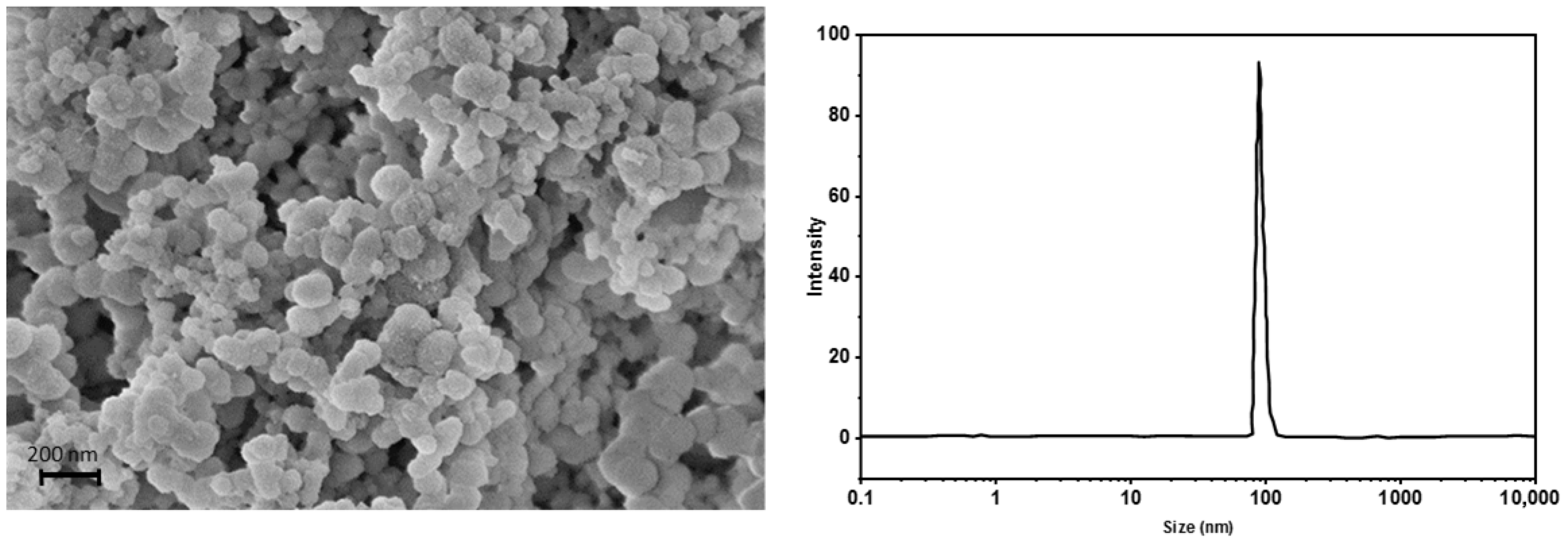
The morphology of hydroxyapatite (nHAp) particles plays a crucial role in their suitability for biomedical applications. SEM analysis of nano-hydroxyapatite powder (Figure 1) showed particle diameters ranging from approximately 85 to 140 nm [25]. Larger agglomerates, formed during drying, were also observed, while the particles exhibited a near-spherical morphology with a relatively uniform size distribution.
To characterize particle behavior in a colloidal suspension, CBN-modified hydroxyapatite was analyzed using dynamic light scattering (DLS) and zeta potential measurements. The analyses were performed immediately after dispersion in water and after 20 s of sonication. Prior to sonication, the average hydrodynamic particle size was 219 ± 32 nm, which decreased to 108 ± 19 nm following sonication, indicating effective deagglomeration. Small agglomerates of about 500 nm were occasionally detected but accounted for only 0.8% of the total particle population.
The zeta potential of the suspension was –9 ± 1.6 mV, indicating relatively low electrostatic stabilization of the system [26]. Such a value may suggest a tendency for particle aggregation over time, particularly in media containing electrolytes or biomolecules that can screen surface charges. Therefore, the colloidal stability of HAp_CBN may be influenced by the surrounding medium, and potential aggregation in physiologically relevant environments should be taken into consideration when interpreting their interactions with lipid membranes. Our carrier is stable for up to 6 weeks of storage in the refrigerator, which was confirmed by DLS and Zeta potential measurements.
In the context of potential medical applications, where HAp_CBN may act as a carrier for CBN and other therapeutic agents (e.g., anticancer drugs), the presence of additional drug molecules could further influence the colloidal stability of the system. In particular, hydrophilic drugs may adsorb onto the surface of hydroxyapatite particles, which could contribute to additional stabilization of the particle structure and modify interparticle interactions.

3.1.2. Qualitative Analysis—IR Spectroscopy

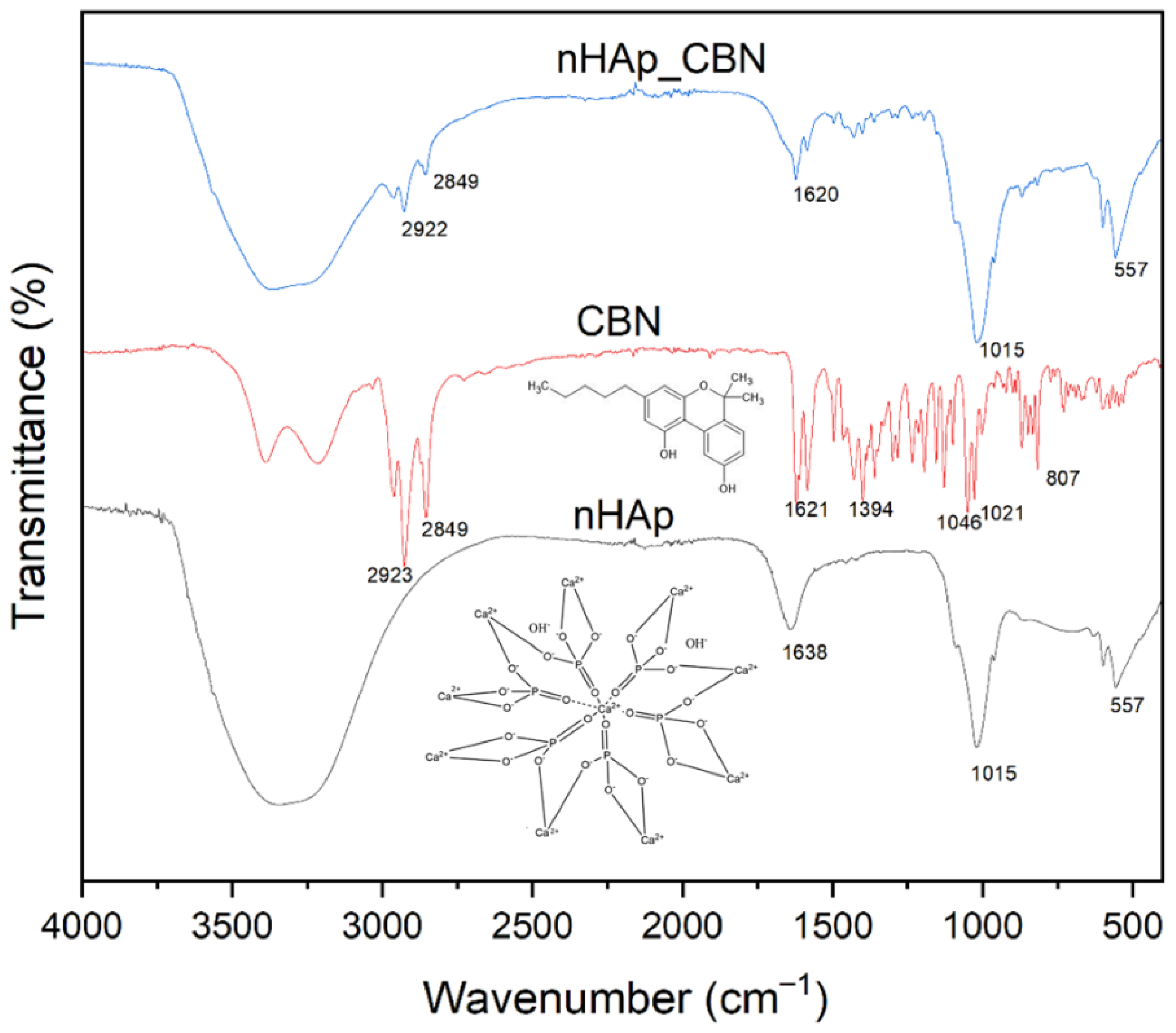
The IR spectra presented in Figure 2 for hydroxyapatite, cannabinol (CBN), and CBN adsorbed within the carrier structure enable a detailed analysis of the functional groups present. In the spectrum of pure nanohydroxyapatite, a characteristic band at 1638 cm−1 is observed, which is attributed to the bending vibrations of H–O–H bonds of adsorbed water molecules [27]. The presence of bands at wavenumbers of 1015 cm−1 and 557 cm−1 corresponds to vibrations characteristic of phosphate groups (PO43−), which are typical of hydroxyapatite and widely used for its identification [28,29,30]. The CBN spectrum exhibits characteristic bands in the range of 2955–2849 cm−1, assigned to C–H stretching vibrations in aliphatic groups. The band at 1621 cm−1 corresponds to C=C stretching vibrations in alicyclic rings. Stretching vibrations of the C–O–C bond of the ether group are observed at a wavenumber of 1046 cm−1, while CH2 deformation vibrations occur at 1394 cm−1 [31,32,33]. In the spectrum recorded for the nHap_CBN conjugate, changes in the intensity of selected bands were observed, particularly in the region around 1620 cm−1. The presence of bands in the range of 2922–2849 cm−1, corresponding to C–H vibrations, further confirms the presence of CBN within the carrier structure. The decreased intensity of these bands results from the relatively low content of cannabinol compared to the total mass of hydroxyapatite. At the same time, no significant changes were observed in the intensity of the band associated with phosphate group vibrations at 1015 cm−1, which confirms the dominant contribution of hydroxyapatite in the structure of the obtained conjugate.

3.1.3. High-Performance Liquid Chromatography (HPLC) Analysis

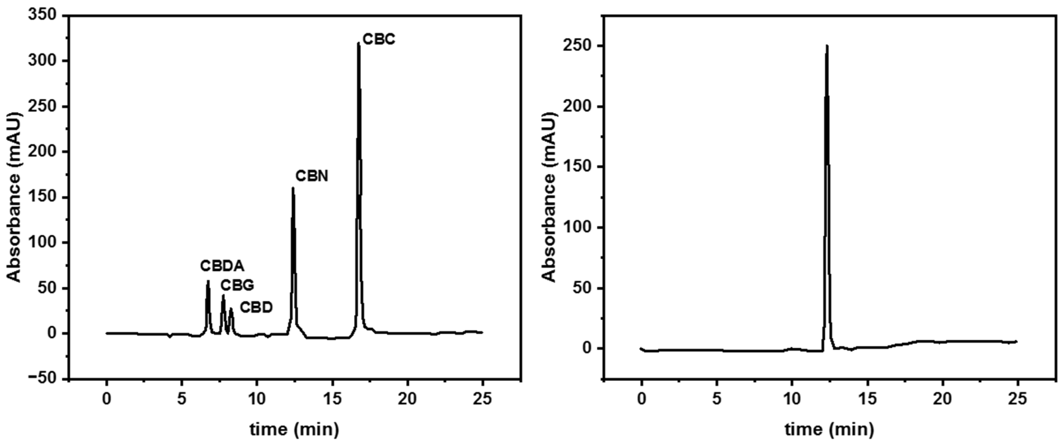
For the identification of cannabinoids based on their retention times, a mixture of these compounds in ethanol was prepared and subsequently analyzed by high-performance liquid chromatography (HPLC) using a UV–Vis detector.
Figure 3 presents the chromatogram obtained for the cannabinoid mixture. Determination of the retention times at which individual compounds eluted from the chromatographic column enabled their unambiguous identification in subsequent experiments.
A corresponding analysis was performed for the ethanolic solution remaining after the adsorption of cannabinol within the hydroxyapatite (nHAp) structure. Analysis of retention times confirmed the presence of cannabinol (CBN) not bound to the carrier, with a retention time of 12.38 min [34].
Based on the chromatograms, the concentration of cannabinol in solution before and after adsorption was determined, allowing estimation of the depletion of CBN from the solution and, consequently, the amount of the compound adsorbed within the pores of hydroxyapatite. The amount of adsorbed cannabinol was determined to be 13.2 wt % relative to the mass of the carrier.

3.1.4. Thermal Analysis

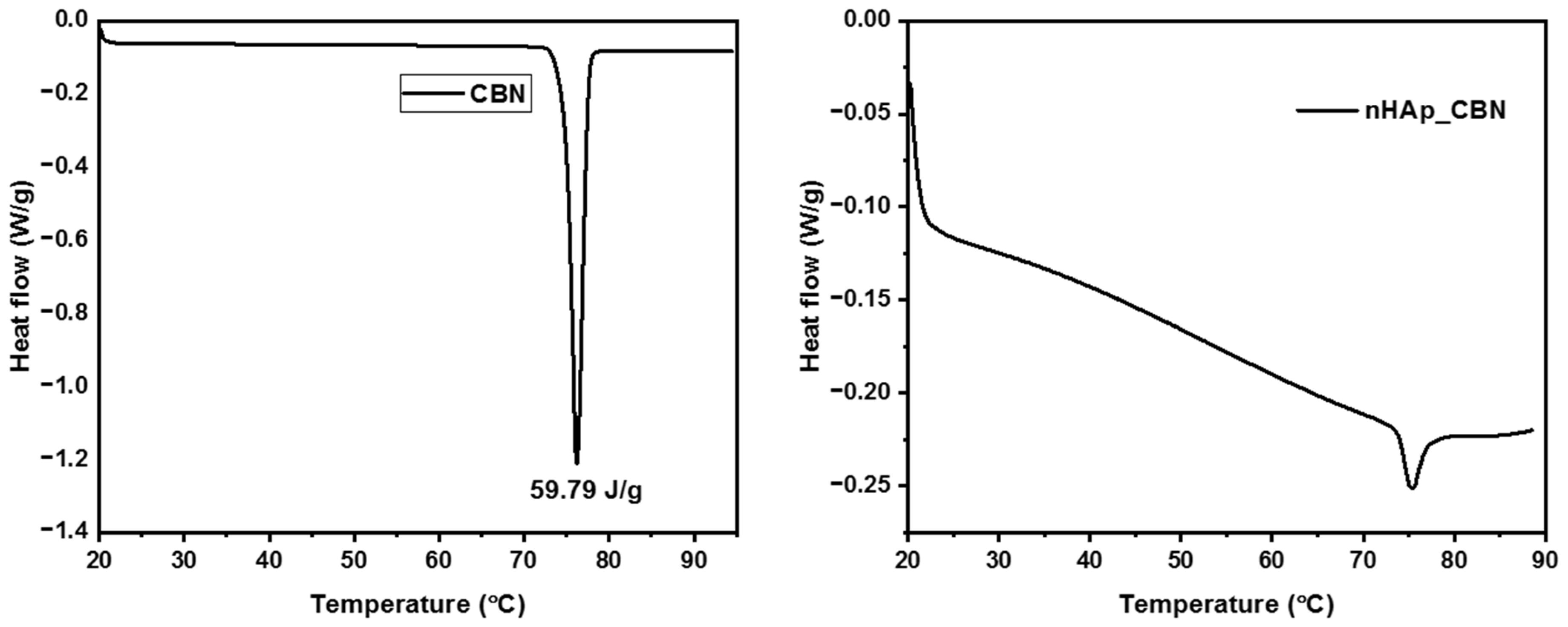
Dried samples of hydroxyapatite, CBN, and the cannabinoid-modified carrier were analyzed using differential scanning calorimetry (DSC). Hydroxyapatite is a material with a stable structure, and thus, the DSC thermogram of the unmodified compound is not expected to exhibit any peaks associated with phase transitions. In contrast, for pure cannabinol and the cannabinoid incorporated into the carrier, characteristic endothermic peaks corresponding to the melting of the active substance can be observed. In DSC thermograms, changes in heat flow appear as peaks, with the area under each peak corresponding to the enthalpy change (ΔH) associated with the thermal transition.
The recorded thermogram for pure hydroxyapatite shows no distinct peaks in the temperature range of 40–90 °C, confirming the stability of hydroxyapatite within this interval and indicating the absence of phase transitions. For pure CBN, a clear peak was observed at 75.85 °C (Figure 4), with a peak area of 59.79 J/g, corresponding to the enthalpy of the phase transition. After conversion, this corresponds to a value of 18.56 kJ/mol, consistent with literature data [35].
For hydroxyapatite modified with CBN, a peak was also observed at 75.44 °C, but with a significantly smaller area. By comparing the enthalpy change of pure CBN with that obtained for the CBN-modified hydroxyapatite, the amount of adsorbed cannabinol can be estimated. This value (about 5.3%), however, is lower than that obtained from HPLC analysis. The DSC signal of CBN in the nHAp_CBN composite may originate predominantly from cannabinoid molecules located on the surface or in shallow pores, whereas CBN confined deep within the hydroxyapatite pores may not undergo a detectable phase transition due to strong surface interactions and restricted molecular mobility. Nevertheless, the recorded DSC thermogram still allows a qualitative confirmation of the presence of cannabinol in hydroxyapatite [36].

3.1.5. UV-VIS Spectrometry

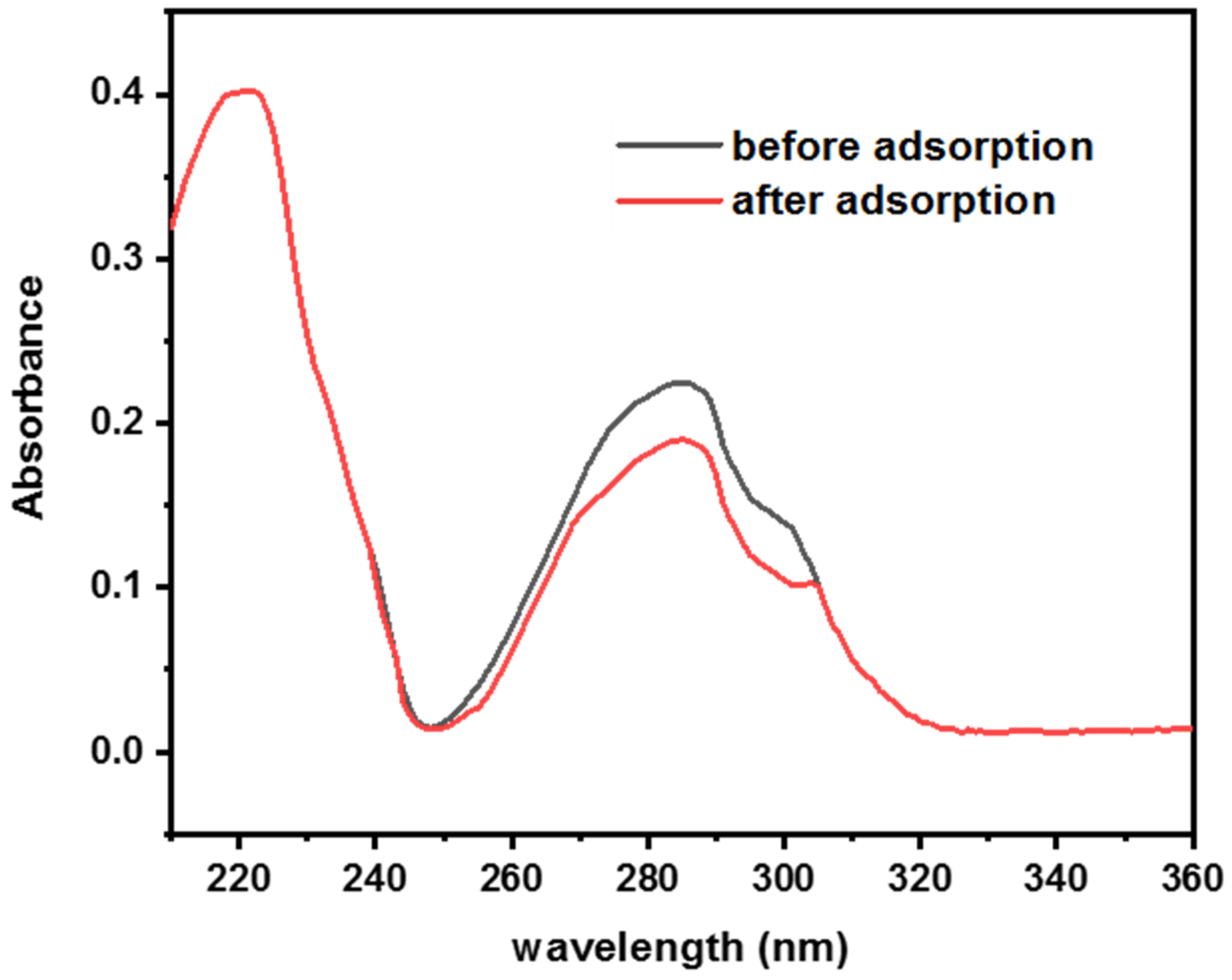
The UV–Vis spectrum of CBN exhibits a strong absorption band in the 280–290 nm range, characteristic of its aromatic and conjugated π system, with additional weaker bands at shorter wavelengths. These features make UV–Vis spectroscopy suitable for the quantitative determination of CBN in solution. The CBN content in the conjugate was estimated based on absorbance spectra recorded before and after the adsorption equilibrium of the compound onto hydroxyapatite was reached. Adsorption resulted in a decrease in absorbance, with the maximum absorbance of cannabinol observed at approximately 285 nm (Figure 5) [37]. Furthermore, by constructing a calibration curve from spectra of a series of standard CBN solutions, the exact content of cannabinol in the conjugate was determined to be 12.3 wt %. Notably, HPLC analysis yielded a comparable value of 13.2 wt %, confirming the consistency of both analytical methods. The release of cannabinol from the carrier in the octane/water system is presented in the Supporting Information.

3.2. Analysis of Nanostructure Effects on Biomimetic Membranes

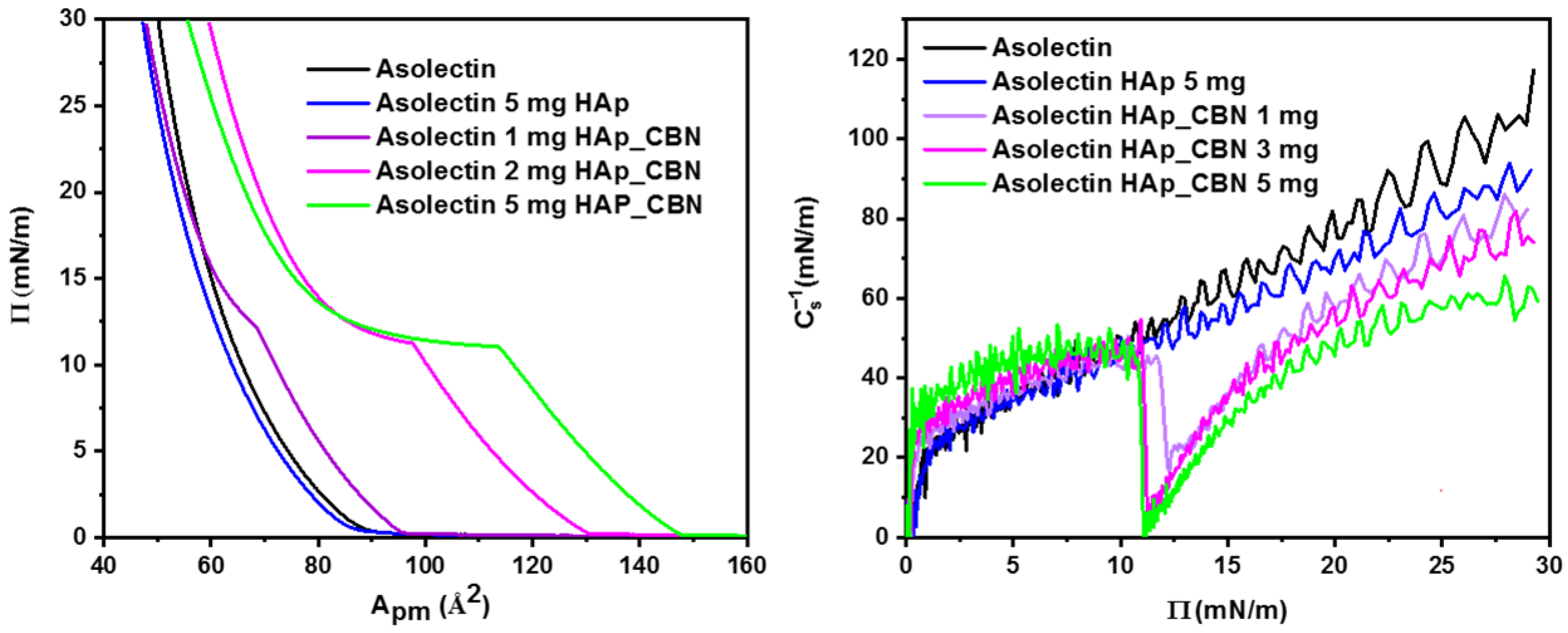
After evaluation of the physicochemical characteristics of the obtained materials, we now turn to the investigation of the interactions between hydroxyapatite, cannabinol, and the air–water interface. The surface pressure–area isotherms presented in Figure 6 were recorded on subphases consisting of pure water, a hydroxyapatite suspension at a concentration of 5 mg/L, and a suspension of hydroxyapatite modified with cannabinol at concentrations ranging from 1 to 5 mg/L. A comparison of the isotherms obtained for asolectin on water and for asolectin on a subphase containing 5 mg of hydroxyapatite (unmodified with CBN) revealed no significant changes in the shape of the curves, their slopes, or the molecular area values of the monolayer. This indicates that the carrier itself does not affect the organization of asolectin within the lipid film, which is a desirable effect, as the carrier should be inert and should not alter membrane properties.
The amount of hydroxyapatite used (5 mg) was selected based on observations from previous studies conducted on hydroxyapatite combined with the cannabinoids CBD and CBG (manuscript under review). It was shown that the use of higher cannabinoid concentrations, and, in particular, larger amounts of the nHAp_CBN carrier, leads to difficulties in monolayer formation due to significant changes in membrane fluidity. Therefore, in the membrane–nHAp interaction studies, the same maximum carrier concentration was applied as in the subsequent membrane–nHAp_CBN interaction experiments (Langmuir and BLM studies).
Based on the recorded isotherms, the compressibility modulus was also calculated (according to Equation (1)), and its values as a function of surface pressure are presented in Figure 6 (right panel).
C s = 1 A ( dA d π ) T , p
The results confirm the absence of significant deviations in Cs−1 values upon the addition of pure hydroxyapatite to the subphase. The Cs−1 value is approximately 105 mN/m for asolectin on water and is only about 10% lower in the presence of nHAp.
Pronounced changes in the isotherms are observed only after the addition of hydroxyapatite modified with cannabinol. Even at a concentration of 1 mg of nHAp_CBN, the isotherm shape is altered, and a collapse in the compressibility modulus is observed at a surface pressure of approximately 11–12 mN/m, decreasing to about 20 mN/m, which indicates a change in the monolayer phase state. Analysis of the isotherm also reveals differences in the slope of the curve above and below this surface pressure (11 mN/m), further confirming the observed changes in the phase behavior and fluidity of the lipid film induced by the presence of the cannabinoid on the carrier nanoparticles. Further increases in the amount of the cannabinol-modified carrier intensify these effects, with phase transitions in both the isotherms and compressibility modulus plots becoming increasingly pronounced, clearly demonstrating the significant impact of CBN on the organization of asolectin molecules within the monolayer. The preliminary observations of cannabinol interactions were sufficiently compelling to warrant further studies using black lipid membrane (BLM) bilayer systems.

3.2.1. CBN Present in the BLM and Langmuir Monolayer-Forming Solution—Effect on the Physicochemical Behavior

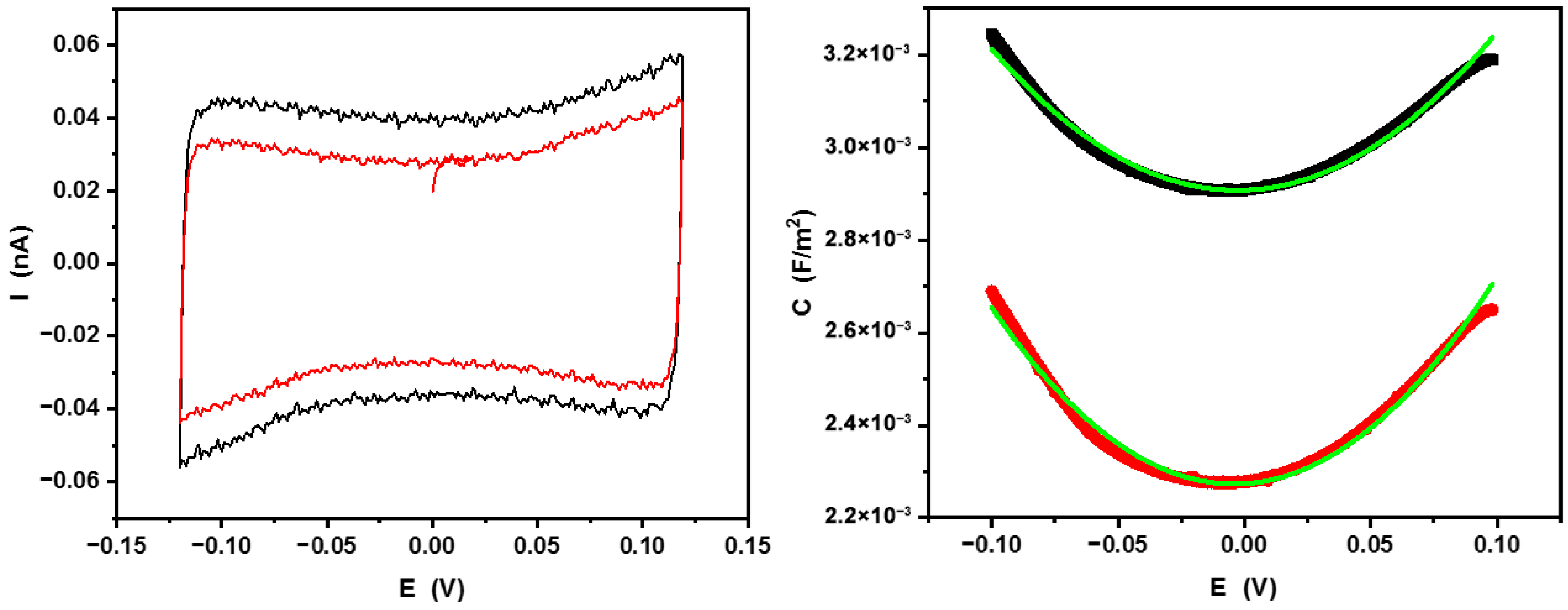
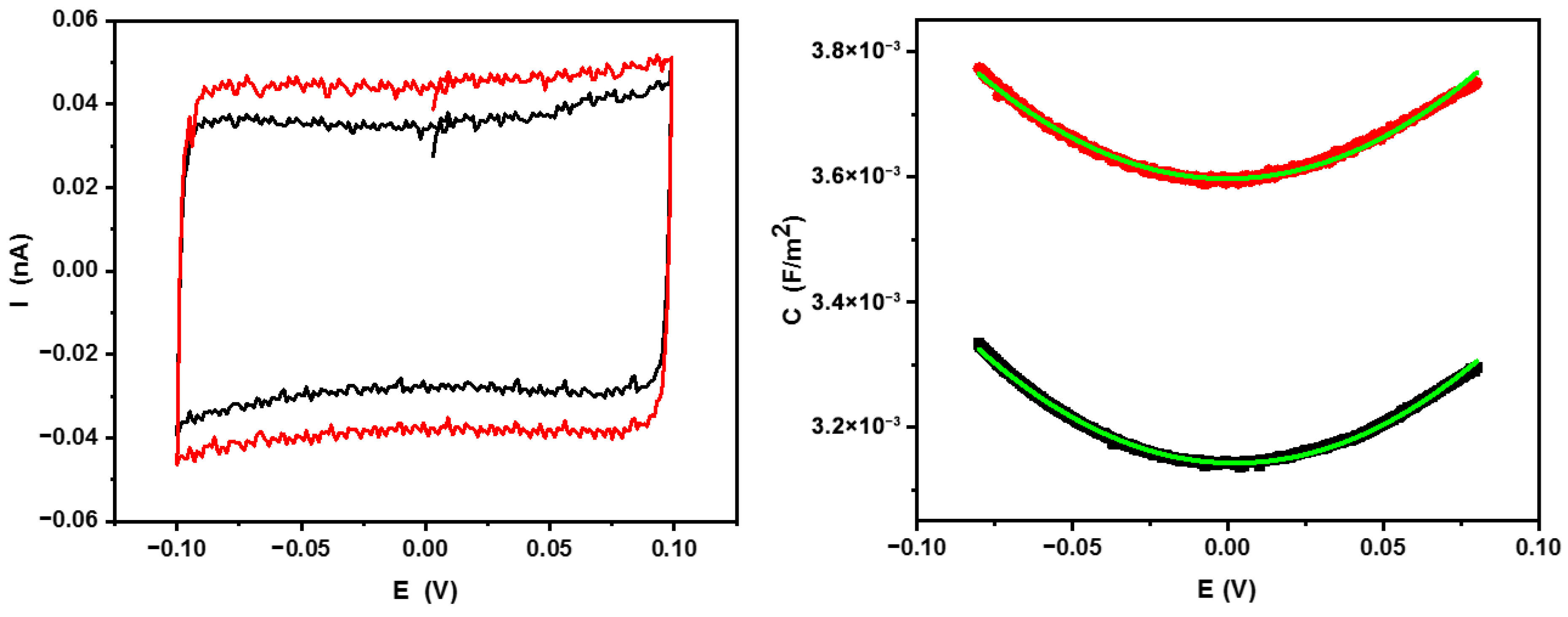
BLMs. To visualize the possible influence of CBN on both biomembrane mimetic systems, cyclic voltammetry and capacitance measurements were performed on free-standing BLMs separating two aqueous 0.1 M KCl solutions. The bilayers were formed either from pristine asolectin in decane solution (2 mg/mL) or from the same solution containing 0.01 M CBN. Assuming that the final content of CBN in the BLM would be the same as in the forming solution, after bilayer formation, we performed CV and capacitance measurements. Please note that due to the fragility of BLMs, to avoid their breakdown, the polarization could not exceed ±120 mV. The results are shown in Figure 7.
From these results, it is obvious that the presence of CBN in the forming solution of asolectin led to a decrease in membrane capacitance of ca. 20%. Assuming the BLM behavior as a “soft” dielectric capacitor, one can conclude that the incorporation of CBN into the lipid bilayer increased its thickness. This conclusion can be drawn solely under the proviso that the dielectric permittivity of the BLM was not affected by the presence of CBN. Moreover, to evaluate the potential effects of CBN alone or nHAp_CBN on bilayer physical properties, we used the electrostriction method [38], in which the free-standing BLM capacitance, C, is plotted against linearly changing potential, E, applied across the membrane. To avoid BLM breakdown, we used the potential range of ±80 mV. Due to the non-linear dependence of BLM specific capacitance C on the applied voltage E, the electrostriction equation takes the form [39,40,41].
C = C 0 + β E 2
where C0 is the specific capacitance (capacitance per area of BLM), assessed from the minimum of the capacitance–potential plot, assuming the BLM area equal to the area of the opening in the septum, and β is the so-called electrostriction coefficient, inversely proportional to the elasticity modulus of the BLM (Young’s modulus). The same equation was also used to fit the C(E) plots shown in Figure 9 (right panel)later in the text. Upon close inspection of the effect of potential on BLM capacitance, one can observe that the CBN-containing BLM is slightly “softer” (red curve is a somewhat deeper parabola with β larger than in the absence of CBN) than that of a pristine asolectin BLM, suggesting slightly greater elasticity of a BLM containing CBN.

3.2.2. Langmuir Monolayers

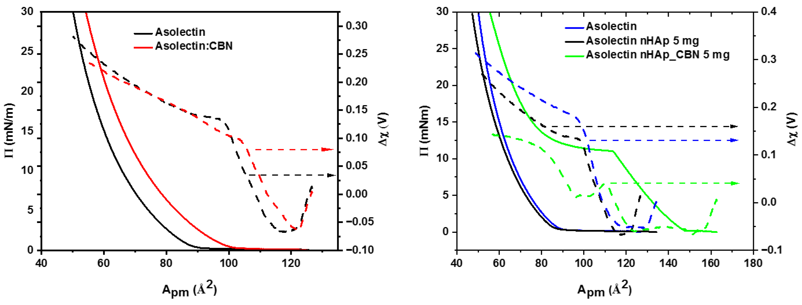
To gain better insight into the possible localization of CBN molecules in the lipid layer, we used the Langmuir technique again, coupled with surface potential (contact potential difference, CPD) monitoring, in an attempt to differentiate the effects of CBN molecules on various parts of the asolectin monolayer. The resultant graphs are shown in Figure 8. Langmuir monolayers were formed on the aqueous subphase from a monolayer-forming chloroform solution (10 μL) containing 0.01 M cannabinol (Figure 8, left panel, red curves). For comparison, Langmuir monolayers were also formed from pristine asolectin (Figure 8, left panel, black curves; compare also Figure 6, black curve). From what can be elucidated from Figure 8 (left panel), in the presence of a small amount (0.01 M) of CBN in the monolayer-forming solution, the same as was used for BLM, the pressure–area isotherm for asolectin monolayers is shifted toward a larger area by ca. 15 Å2 per lipid molecule. Furthermore, its slope is somewhat less steep, indicating a disorganizing influence of hydrophobic cannabinol molecules on the organization of the lipid monolayer (our unpublished results show that CBN does not form a Langmuir monolayer).
Let us consider the information that can be extracted from the CPD isotherms, which are also shown in this figure. The CPD value can be used to assess the value of the so-called “effective” normal component of dipole moments (μ) [42] of molecules forming the monolayer:
Δ χ = μ A ε 0 ε r + ψ e d l = 1 A ε 0 ( μ 1 ε 1 + μ 2 ε 2 + μ 3 ε 3 ) + ψ e d l
where Δχ is the contact potential, CPD; A is the area per molecule at a given contact potential; and εr and εo are the relative dielectric permittivity of a monolayer and the permittivity of free space, respectively. According to the Demchak–Fort model [43,44,45], the normal component of the dipole moment contains the contributions due to the reorientation of water molecules in the presence of the monolayer, μ 1 ε 1 , the normal component from the hydrophilic headgroups at the interface, μ 2 ε 2 , and the normal component of the dipole moment from the hydrophobic tails of the monolayer-forming molecules, μ 3 ε 3 , whereas ψ e d l is the contribution from the electric double layer formed between the charged headgroups and the counterions in the subphase. All these contributions can be treated separately. Because the monolayers are formed on the pure aqueous subphase, we assume a fixed and almost null contribution from the electric double layer. Moreover, in our opinion, the first two contributions should remain unaffected by the presence of highly hydrophobic CBN in the monolayer. Therefore, in further considerations, we focus only on the component related to the orientation of the hydrophobic tails of lipid molecules. As is evident in Figure 8 (left panel), paralleling the changes in the Langmuir isotherms, the contact potential isotherm threshold also shifts toward a larger area per lipid molecule, pointing to an earlier ordering of dipoles that is not yet detectable by an increase in surface pressure. However, the CPD value at ca. 100 Å2 is by ca. 50 mV smaller than that for a pristine asolectin monolayer at the same area. Then it gradually monotonically to attain a value similar to that of a pure asolectin monolayer. Therefore, taking into account the Langmuir isotherm profiles together with the CPD results shown in Figure 8 (left panel), we attribute the increase in BLM thickness and the elastic behavior (shown in Figure 7) to the localization of cannabinol molecules in the hydrophobic region of both the Langmuir monolayer and bilayer lipid membrane.

3.2.3. nHAp_CBN Nanocarriers in Aqueous Phase—Effect on BLMs and Langmuir Monolayers

Further studies examined the influence of CBN adsorbed on the nHAp (5 mg/L) colloidal suspension present on both sides of the pristine asolectin bilayer lipid membrane. This configuration more closely approximates the target system in which the nanocarriers, after administration, would interact with cellular membranes. The obtained results are shown in Figure 9. To our surprise, the response was different from that in the case of CBN already present in the BLM-forming solution (and inside the formed BLM; Figure 7).
Figure 9. (Left panel): Cyclic voltammograms of pristine asolectin bilayer lipid membrane with 0.1 M KCl aqueous solutions of pure 5 mg/L nHAp with no CBN adsorbed (black curve), and pristine asolectin BLM in contact with 5 mg/L nHAp_CBN in 0.1 M KCl present on both sides of the membrane (red curve). (Right panel): BLM capacitance vs. polarization potential curves of the same bilayers (color coded as in A). No CBN was present in the bilayer-forming solution. These graphs are representative of at least five measurements on independently formed BLMs. Fits seen in Figure 9 (right panel) correspond to an R-squared value of R2 = 0.995. Cyclic voltammograms recorded at a scan rate of 10 mV/s.
Colloids 10 00033 g009
As can be inferred from this figure, the membrane capacitance increased by ca.13% in the presence of nHAp_CBN in 0.1 M KCl aqueous electrolyte on both sides of a pristine BLM. This observation implies that the BLMs separating solutions containing colloidal hydroxyapatite loaded with CBN are thinner than those in the absence of nHAp_CBN. However, this interpretation should be taken with care because it can also be a result of either an increase in the BLM area or an increase in its dielectric permittivity due to the partial penetration of nHAp_CBN nanocarriers into the BLM. From these experiments, we cannot point toward the main cause of the observed results; nevertheless, they prove a significant effect of the carrier with CBN on the BLM behavior.
To gain deeper insight into the possible interaction mechanism, we returned to the Langmuir isotherm together with the surface potential studies in the case of the highest, 5 mg/L concentration of carriers. The results are shown in Figure 8 (right panel), including also the effect of nHAp without adsorbed CBN (right panel, black curves) for comparison purposes.
In the case of this system, there is a significant difference because the Langmuir film lacks a CBN admixture. Therefore, nHAp, as well as nHAP_CBN present in the aqueous subphase, can interact only with the hydrophilic part of the organized monomolecular film. As can be seen in the case of nHAp present in the subphase, the surface pressure isotherm of asolectin is similar to that of lipid molecules spread on a clean water subphase. Similarly, the CPD isotherm in the presence of nHAp in the subphase parallels the pattern of contact potential in the absence of cannabinol.
It is evident from the data presented in Figure 8 (right panel) that the presence of nHAp nanocarriers loaded with CBN molecules triggers a drastic change not only in the Langmuir isotherm, as was discussed above (c.f., Figure 6, left panel), but also in the contact potential profile over the whole compression regime. In contrast, the presence of 5 mg of the unmodified carrier in the subphase led to a more “fluid” shape of the isotherm and a larger rise of the contact potential values (Figure 8, right panel, dark blue curves, left and right Y axes) as compared to the pure subphase, and the addition of 5 mg nHAp modified with CBN resulted in the pronounced changes also in the CPD profile (Figure 8 (right panel), green curve, right Y axis). Firstly, the initial increase in its value is almost twofold smaller, but it also starts at a much lower area per molecule than its associated isotherm. In the surface pressure regime around 11 mN/m, where the isotherm shows a behavior typically assigned to a monolayer “phase transition” (molecules disordering) induced by the 5 mg nHAp_CBN, the surface potential value also stopped its increase until ca. 95 Å2 area per molecule is reached as monolayer compression proceeds, and then it rises again to attain almost a plateau of ca. 120 mV compared with ca. 300 mV for the unmodified 5 mg nHAp. We are inclined to assign these changes to the strong hydrophobic interactions of CBN present on the surface of nHAp, disordering at low surface pressure range of the alignment of hydrophobic lipid tails (between 110 Å2 and 90 Å2 per molecule). This is reflected by a plateau on the surface pressure isotherm and a CPD drop and plateau. Finally, as the barriers move closer, the surface pressure increases, but the organization of lipid dipoles involved in the interactions with nHAp_CBN cannot attain their alignment similar to that of asolectin alone or asolectin with nHAp in the aqueous subphase. This causes a lowering of the final CPD value from ca. 300 mV to ca. 120 mV (Figure 8, right panel).

3.2.4. Effect of nHAp_CBN on Pre-Formed Langmuir Monolayers

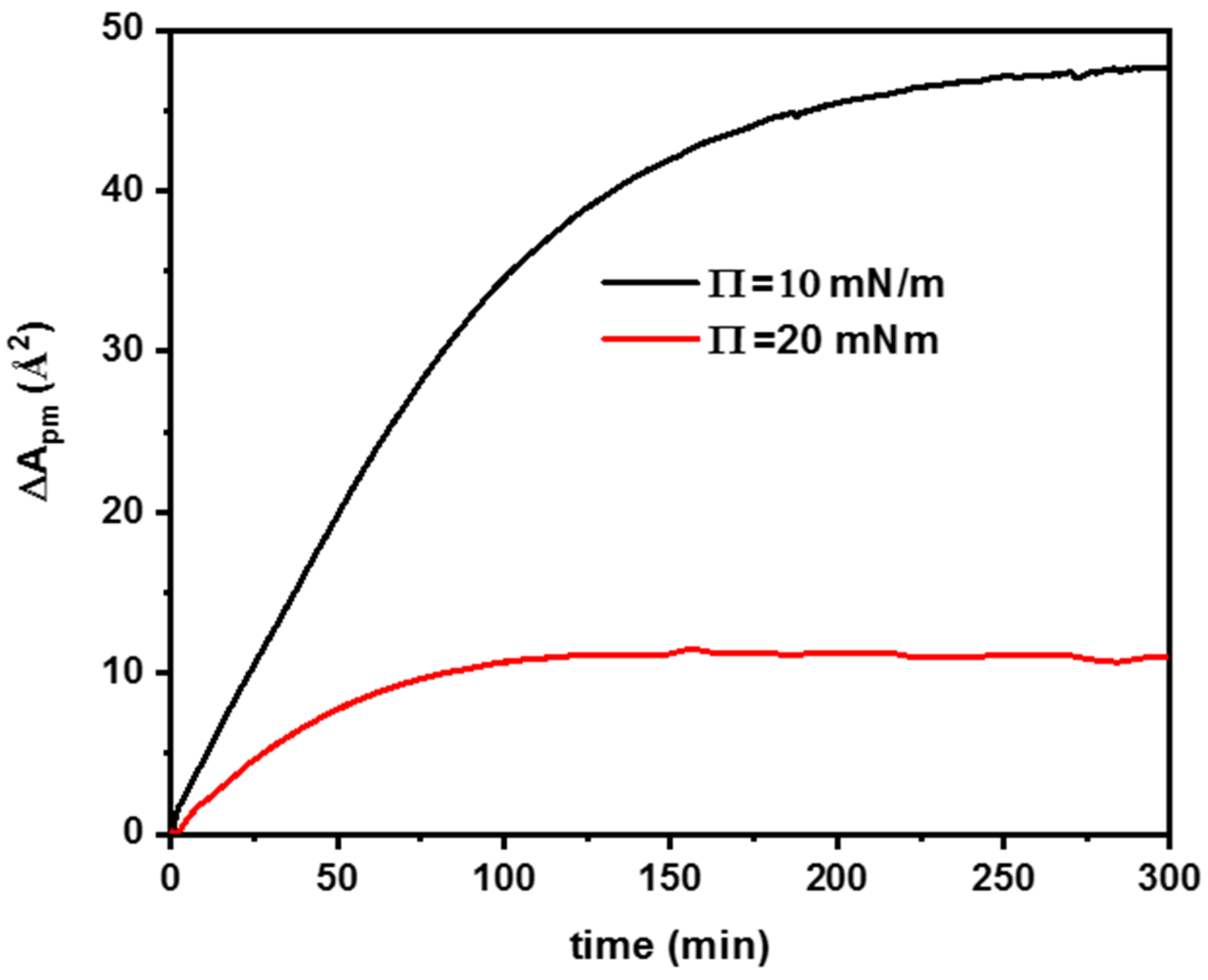
The effect of CBN-modified hydroxyapatite (nHAp_CBN) on Langmuir films and BLM layers was previously investigated by introducing the nanostructures directly into the subphase during film formation (vide supra). This study demonstrated a strong interaction between CBN and asolectin at low surface pressures. Here, the interaction of nHAp_CBN with pre-formed membranes is examined. Monolayers were prepared at surface pressures of 10 and 20 mN/m, chosen due to pronounced changes observed within this region of the isotherm, followed by the injection of an nHAp_CBN suspension into the subphase (Figure 6, left panel). The resulting changes in molecular area, while keeping constant surface pressures of 10 and 20 mN/m, were monitored to evaluate interactions between the nanoparticles and the already organized lipid film (Figure 10). The extent of the observed effects strongly depended on the surface pressure, indicating that CBN interactions are influenced by the degree of asolectin layer ordering. At a surface pressure of 10 mN/m, the addition of CBN-containing nanostructures resulted in an increase in molecular area of approximately 50 Å2 (consistent with the data presented in Figure 6, left panel, comparison of the green and black isotherms), suggesting substantial adsorption of the cannabinoid into the lipid layer. This behavior can be attributed to the relatively low degree of membrane ordering, where asolectin molecules may adopt orientations partially parallel to the water surface, exposing hydrophobic regions toward the subphase. Such a configuration facilitates the penetration of cannabinol molecules between lipid molecules, leading to monolayer expansion. Moreover, it further enables hydrogen-bond formation between the CBN hydroxyl group and the polar headgroups of asolectin (–P=O), as suggested in [21], thereby improving cannabinol penetration into the Langmuir monolayer. In contrast, at a surface pressure of 20 mN/m, where lipid packing is more compact and molecules are predominantly oriented perpendicular to the subphase surface, the interaction is markedly weaker, as reflected by a molecular area change of only about 12 Å2. Under these conditions, the hydrophilic headgroups of asolectin form an effective barrier that restricts the access of CBN incorporated within the nHAp structure to the hydrophobic lipid tails.

4. Conclusions

In this work, a nanohydroxyapatite-cannabinol (nHAp_CBN) conjugate was successfully prepared and thoroughly characterized in terms of its structural, physicochemical, and interfacial properties. SEM analysis confirmed that the starting nano-hydroxyapatite consisted of near-spherical particles with diameters in the range of 85–140 nm, exhibiting a relatively uniform size distribution, while larger agglomerates were formed during the drying process. The colloidal behavior of CBN-modified hydroxyapatite was further characterized using DLS measurements, which demonstrated good dispersion stability in an aqueous environment, particularly after sonication. In this case, the hydrodynamic diameters decreased from approximately 219 nm to 108 nm. The obtained size distribution and colloidal stability indicate that the suspension is suitable for further formulation. Spectroscopic analyses provided clear evidence for the incorporation of cannabinol into the hydroxyapatite structure. Quantitative determination using UV-Vis spectroscopy and HPLC yielded consistent results, indicating a CBN content of 12.3 wt % and 13.2 wt %, respectively. Differential scanning calorimetry corroborated these findings by revealing characteristic endothermic transitions associated with cannabinol in the conjugate, confirming the presence of the active compound and indicating its incorporation within the inorganic carrier. Interfacial investigations using Langmuir monolayers and bilayer lipid membranes provided insight into the interactions of nHAp and nHAp_CBN with model biological membranes. CBN-modified hydroxyapatite significantly affected the surface pressure–area isotherms, indicating its ability to interact with lipid films at the air–liquid interface. The presence of cannabinol further modified these interactions, influencing monolayer packing and stability in a concentration-dependent manner. Complementary BLM studies demonstrated that the nHAp_CBN system is capable of interacting with lipid bilayers, suggesting a potential impact on membrane structure and properties.
Overall, the results establish a clear relationship between the physicochemical characteristics of the nHAp_CBN conjugate and its interfacial activity toward lipid model systems. Importantly, a key achievement of this study is the successful incorporation of the highly hydrophobic drug cannabinol into a hydrophilic and biocompatible nanohydroxyapatite carrier, resulting in a stable hybrid system dispersible in aqueous media. This demonstrates the ability of nHAp to act as an effective inorganic platform for the delivery of poorly water-soluble bioactive compounds.
The combined structural, spectroscopic, thermal, and interfacial analyses highlight the potential of cannabinol-modified hydroxyapatite as a functional hybrid material for further studies in drug delivery or nanoparticle–membrane interactions, particularly in the context of overcoming solubility limitations of hydrophobic therapeutics.

Supplementary Materials

The following supporting information can be downloaded at: https://www.mdpi.com/article/10.3390/colloids10030033/s1, S1. Release of Cannabinol in the Octane/Water System; Figure S1. Time-dependent release profile of CBN from hydroxyapatite in the octane/water system.

Author Contributions

Conceptualization, P.K. and D.N.; methodology, E.Ś. and D.N.; formal analysis, J.T. and D.N.; investigation, E.Ś., J.T., D.K., P.K. and D.N.; data curation, J.T., E.Ś., D.N. and P.K.; writing—original draft preparation, P.K. and D.N.; writing—review and editing, J.T., D.N. and P.K.; supervision, D.N. and P.K.; project administration, P.K. and D.N.; funding acquisition, P.K. All authors have read and agreed to the published version of the manuscript.

Funding

This research received no external funding.

Data Availability Statement

Data is contained within the article or Supplementary Materials.

Acknowledgments

We would like to thank Biotech Evolution for providing the CBN isolate. SEM measurements were performed at the Core Facility of the Faculty of Chemistry, University of Warsaw.

Conflicts of Interest

The authors declare no conflicts of interest.

References

  1. Kavasi, R.-M.; Coelho, C.C.; Platania, V.; Quadros, P.A.; Chatzinikolaidou, M. In Vitro Biocompatibility Assessment of Nano-Hydroxyapatite. Nanomaterials 2021, 11, 1152. [Google Scholar] [CrossRef] [Scilit] [PubMed]
  2. Ferraris, S.; Yamaguchi, S.; Barbani, N.; Cazzola, M.; Cristallini, C.; Miola, M.; Vernè, E.; Spriano, S. Bioactive Materials: In Vitro Investigation of Different Mechanisms of Hydroxyapatite Precipitation. Acta Biomater. 2020, 102, 468–480. [Google Scholar] [CrossRef] [Scilit]
  3. Moreno, E.C.; Kresak, M.; Hay, D.I. Adsorption of Molecules of Biological Interest onto Hydroxyapatite. Calcif. Tissue Int. 1984, 36, 48–59. [Google Scholar] [CrossRef] [Scilit]
  4. Turon, P.; Del Valle, L.; Alemán, C.; Puiggalí, J. Biodegradable and Biocompatible Systems Based on Hydroxyapatite Nanoparticles. Appl. Sci. 2017, 7, 60. [Google Scholar] [CrossRef] [Scilit]
  5. Biedrzycka, A.; Skwarek, E. Composites of Hydroxyapatite and Their Application in Adsorption, Medicine and as Catalysts. Adv. Colloid Interface Sci. 2024, 334, 103308. [Google Scholar] [CrossRef] [Scilit]
  6. Maioli, C.; Mattoteia, D.; Amin, H.I.M.; Minassi, A.; Caprioglio, D. Cannabinol: History, Syntheses, and Biological Profile of the Greatest “Minor” Cannabinoid. Plants 2022, 11, 2896. [Google Scholar] [CrossRef] [Scilit]
  7. McAllister, S.D.; Soroceanu, L.; Desprez, P.-Y. The Antitumor Activity of Plant-Derived Non-Psychoactive Cannabinoids. J. Neuroimmune Pharmacol. 2015, 10, 255–267. [Google Scholar] [CrossRef] [Scilit] [PubMed]
  8. Martínez, V.; Iriondo De-Hond, A.; Borrelli, F.; Capasso, R.; Del Castillo, M.D.; Abalo, R. Cannabidiol and Other Non-Psychoactive Cannabinoids for Prevention and Treatment of Gastrointestinal Disorders: Useful Nutraceuticals? Int. J. Mol. Sci. 2020, 21, 3067. [Google Scholar] [CrossRef] [Scilit] [PubMed]
  9. Thakur, G.A.; Duclos, R.I.; Makriyannis, A. Natural Cannabinoids: Templates for Drug Discovery. Life Sci. 2005, 78, 454–466. [Google Scholar] [CrossRef] [Scilit]
  10. Zhong, N.; Li, D.; Wang, B.; Kovalchuk, O.; Kovalchuk, I. Cannabinol Inhibits Cell Growth and Triggers Cell Cycle Arrest and Apoptosis in Cancer Cells. Biocatal. Agric. Biotechnol. 2023, 48, 102627. [Google Scholar] [CrossRef] [Scilit]
  11. Rana, L.; Harwansh, R.K.; Deshmukh, R. Recent Updates on Phytopharmaceuticals-Based Novel Phytosomal Systems and Their Clinical Trial Status: A Translational Perspective. Crit. Rev. Ther. Drug Carr. Syst. 2025, 42, 1–54. [Google Scholar] [CrossRef] [Scilit]
  12. Kadriya, A.; Forbes-Robertson, S.; Falah, M. The Anticancer Activity of Cannabinol (CBN) and Cannabigerol (CBG) on Acute Myeloid Leukemia Cells. Molecules 2024, 29, 5970. [Google Scholar] [CrossRef] [Scilit] [PubMed]
  13. Yaman, Z.; Aydemir, S.; Uğur, Y.; Demirtaş, S.; Şentürk, H.; Şalva, E.; Erdoğan, S. Cytotoxic and Pro-Apoptotic Effects of Cold-Pressed Hemp Seed Oil on Lung Cells: Cannabinoid and Fatty Acid Profiling via LC–MS/MS and GC–FID. ChemistrySelect 2025, 10, e04822. [Google Scholar] [CrossRef] [Scilit]
  14. Russo, E.B.; Marcu, J. Cannabis Pharmacology: The Usual Suspects and a Few Promising Leads. In Advances in Pharmacology; Elsevier: Amsterdam, The Netherlands, 2017; Volume 80, pp. 67–134. [Google Scholar] [CrossRef] [Scilit]
  15. Solano-Orrala, D.; Silva-Cullishpuma, D.A.; Díaz-Cruces, E.; Gómez-López, V.M.; Toro-Mendoza, J.; Gomez d’Ayala, G.; Troconis, J.; Narváez-Muñoz, C.; Alexis, F.; Mercader-Ros, M.T.; et al. Exploring the Potential of Nonpsychoactive Cannabinoids in the Development of Materials for Biomedical and Sports Applications. ACS Appl. Bio Mater. 2024, 7, 8177–8202. [Google Scholar] [CrossRef] [Scilit] [PubMed]
  16. Taudul, J.; Celej, J.; Żelechowska-Matysiak, K.; Kępińska, D.; Majkowska-Pilip, A.; Strawski, M.; Krysiński, P.; Nieciecka, D. Tailored Iron Oxide Nanoparticles as Potential Cannabinoid Carriers for Anti-Cancer Treatment. Biomolecules 2025, 15, 230. [Google Scholar] [CrossRef] [Scilit] [PubMed]
  17. Velasco, G.; Hernández-Tiedra, S.; Dávila, D.; Lorente, M. The Use of Cannabinoids as Anticancer Agents. Prog. Neuro-Psychopharmacol. Biol. Psychiatry 2016, 64, 259–266. [Google Scholar] [CrossRef] [Scilit]
  18. Velasco, G.; Sánchez, C.; Guzmán, M. Anticancer Mechanisms of Cannabinoids. Curr. Oncol. 2016, 23, 23–32. [Google Scholar] [CrossRef] [Scilit]
  19. Hillard, C.J.; Harris, R.A.; Bloom, A.S. Effects of the Cannabinoids on Physical Properties of Brain Membranes and Phospholipid Vesicles: Fluorescence Studies. J. Pharmacol. Exp. Ther. 1985, 232, 579–588. [Google Scholar] [CrossRef] [Scilit]
  20. Lawrence, D.K.; Gill, E.W. The Effects of Δ1-Tetrahydrocannabinol and Other Cannabinoids on Spin-Labeled Liposomes and Their Relationship to Mechanisms of General Anesthesia. Mol. Pharmacol. 1975, 11, 595–602. [Google Scholar] [CrossRef] [Scilit]
  21. Muta, T.; Khetan, R.; Song, Y.; Garg, S. Optimising Cannabidiol Delivery: Improving Water Solubility and Permeability Through Phospholipid Complexation. Int. J. Mol. Sci. 2025, 26, 2647. [Google Scholar] [CrossRef] [Scilit]
  22. Söderlund, T.; Alakoskela, J.-M.I.; Pakkanen, A.L.; Kinnunen, P.K.J. Comparison of the Effects of Surface Tension and Osmotic Pressure on the Interfacial Hydration of a Fluid Phospholipid Bilayer. Biophys. J. 2003, 85, 2333–2341. [Google Scholar] [CrossRef] [Scilit]
  23. Marsh, D. Lateral Pressure in Membranes. Biochim. Biophys. Acta (BBA) Rev. Biomembr. 1996, 1286, 183–223. [Google Scholar] [CrossRef] [Scilit]
  24. Kalinowski, S.; Figaszewski, Z. A Four-Electrode Potentiostat-Galvanostat for Studies of Bilayer Lipid Membranes. Meas. Sci. Technol. 1995, 6, 1050–1055. [Google Scholar] [CrossRef] [Scilit]
  25. Aljamhan, A.S.; Alrefeai, M.H.; Alhabdan, A.; Alhusseini, S.A.; Farooq, I.; Vohra, F.; Naseem, M.; Alkhudhairy, F. Influence of ER-CR-YSGG Laser and Photodynamic Therapy on the Dentin Bond Integrity of Nano-Hydroxyapatite Containing Resin Dentin Adhesive: SEM-EDX, Micro-Raman, Micro-Tensile, and FTIR Evaluation. Polymers 2021, 13, 1903. [Google Scholar] [CrossRef] [Scilit]
  26. Uskoković, V.; Odsinada, R.; Djordjevic, S.; Habelitz, S. Dynamic Light Scattering and Zeta Potential of Colloidal Mixtures of Amelogenin and Hydroxyapatite in Calcium and Phosphate Rich Ionic Milieus. Arch. Oral Biol. 2011, 56, 521–532. [Google Scholar] [CrossRef] [Scilit]
  27. Belbasi, Z.; Petr, J.; Hrbac, J.; Jirovsky, D. HPLC Method for Novel Cannabinoid Separation with Electrochemical Detection on Boron-Doped Diamond Electrode. J. Pharm. Biomed. Anal. 2025, 262, 116875. [Google Scholar] [CrossRef] [Scilit]
  28. Šoptrajanov, B. Very Low H–O–H Bending Frequencies. I. Overview and Infrared Spectra of NiKPO4·H2O and Its Deuterated Analogues. J. Mol. Struct. 2000, 555, 21–30. [Google Scholar] [CrossRef] [Scilit]
  29. Venkatesan, J.; Lowe, B.; Manivasagan, P.; Kang, K.-H.; Chalisserry, E.; Anil, S.; Kim, D.; Kim, S.-K. Isolation and Characterization of Nano-Hydroxyapatite from Salmon Fish Bone. Materials 2015, 8, 5426–5439. [Google Scholar] [CrossRef] [Scilit] [PubMed]
  30. Poinern, G.E.; Brundavanam, R.K.; Mondinos, N.; Jiang, Z.-T. Synthesis and Characterisation of Nanohydroxyapatite Using an Ultrasound Assisted Method. Ultrason. Sonochem. 2009, 16, 469–474. [Google Scholar] [CrossRef] [Scilit] [PubMed]
  31. Jastrzębski, W.; Sitarz, M.; Rokita, M.; Bułat, K. Infrared Spectroscopy of Different Phosphates Structures. Spectrochim. Acta Part A Mol. Biomol. Spectrosc. 2011, 79, 722–727. [Google Scholar] [CrossRef] [Scilit]
  32. Hidayati, N.; Saefumillah, A.; Cahyana, A.H. Simple Isolation Method of Cannabinol from Cannabis Sativa to Produce Secondary Reference Standard Analysis Material. IOP Conf. Ser. Mater. Sci. Eng. 2020, 902, 012063. [Google Scholar] [CrossRef] [Scilit]
  33. Thanakorncharoenwit, M.; Sukmak, P.; Faikhruea, K.; Yukhet, P.; Pongkorpsakol, P.; Aonbangkhen, C.; Khotavivattana, T.; Vilaivan, T. A Facile One-Pot Iodine-Mediated Synthesis of Cannabinol and Derivatives, and Their Intestinal Epithelial Barrier Protective Effect. Tetrahedron 2024, 165, 134189. [Google Scholar] [CrossRef] [Scilit]
  34. Li, H.; Chang, S.-L.; Chang, T.-R.; You, Y.; Wang, X.-D.; Wang, L.-W.; Yuan, X.-F.; Tan, M.-H.; Wang, P.-D.; Xu, P.-W.; et al. Inclusion Complexes of Cannabidiol with β-Cyclodextrin and Its Derivative: Physicochemical Properties, Water Solubility, and Antioxidant Activity. J. Mol. Liq. 2021, 334, 116070. [Google Scholar] [CrossRef] [Scilit]
  35. Stinchcomb, A.L.; Valiveti, S.; Hammell, D.C.; Ramsey, D.R. Human Skin Permeation of Δ8-Tetrahydrocannabinol, Cannabidiol and Cannabinol. J. Pharm. Pharmacol. 2004, 56, 291–297. [Google Scholar] [CrossRef] [Scilit] [PubMed]
  36. Haro Mares, N.B.; Döller, S.C.; Wissel, T.; Hoffmann, M.; Vogel, M.; Buntkowsky, G. Structures and Dynamics of Complex Guest Molecules in Confinement, Revealed by Solid-State NMR, Molecular Dynamics, and Calorimetry. Molecules 2024, 29, 1669. [Google Scholar] [CrossRef] [Scilit] [PubMed]
  37. Vacek, J.; Vostalova, J.; Papouskova, B.; Skarupova, D.; Kos, M.; Kabelac, M.; Storch, J. Antioxidant Function of Phytocannabinoids: Molecular Basis of Their Stability and Cytoprotective Properties under UV-Irradiation. Free Radic. Biol. Med. 2021, 164, 258–270. [Google Scholar] [CrossRef] [Scilit] [PubMed]
  38. Hianik, T.; Šnejdárková, M.; Rehák, M. Electrostriction of Biotin-Streptavidin-Modified Bilayer Lipid Membranes on a Metal Support. Bioelectrochem. Bioenerg. 1996, 39, 235–239. [Google Scholar] [CrossRef] [Scilit]
  39. White, S.H.; Chang, W. Voltage Dependence of the Capacitance and Area of Black Lipid Membranes. Biophys. J. 1981, 36, 449–453. [Google Scholar] [CrossRef] [Scilit]
  40. Taylor, G.J.; Venkatesan, G.A.; Collier, C.P.; Sarles, S.A. Direct in Situ Measurement of Specific Capacitance, Monolayer Tension, and Bilayer Tension in a Droplet Interface Bilayer. Soft Matter 2015, 11, 7592–7605. [Google Scholar] [CrossRef] [Scilit]
  41. Gross, L.C.M.; Heron, A.J.; Baca, S.C.; Wallace, M.I. Determining Membrane Capacitance by Dynamic Control of Droplet Interface Bilayer Area. Langmuir 2011, 27, 14335–14342. [Google Scholar] [CrossRef] [Scilit]
  42. El Abed, A.I. Experimental Validation of the Helmoltz Equation for the Surface Potential of Langmuir Monolayers. Phys. Rev. E 2009, 80, 041601. [Google Scholar] [CrossRef] [Scilit]
  43. Cavalli, A.; Dynarowicz-Łątka, P.; Oliveira, O.N., Jr.; Feitosa, E. Using an Effective Surface Charge to Explain Surface Potentials of Langmuir Monolayers from Dialkyldimethylammonium Halides with the Gouy–Chapman Theory. Chem. Phys. Lett. 2001, 338, 88–94. [Google Scholar] [CrossRef] [Scilit]
  44. Demchak, R.J.; Fort, T. Surface Dipole Moments of Close-Packed Un-Ionized Monolayers at the Air-Water Interface. J. Colloid Interface Sci. 1974, 46, 191–202. [Google Scholar] [CrossRef] [Scilit]
  45. Nieciecka, D.; Królikowska, A.; Joniec, A.; Krysinski, P. Partitioning of Doxorubicin into Langmuir and Langmuir–Blodgett Biomimetic Mixed Monolayers: Electrochemical and Spectroscopic Studies. J. Electroanal. Chem. 2013, 710, 59–69. [Google Scholar] [CrossRef] [Scilit]
Figure 1. (Left panel): SEM image of nano-hydroxyapatite powder; (right panel): DLS size distribution of CBN-modified hydroxyapatite after sonication.
Colloids 10 00033 g001
Figure 2. IR spectra of hydroxyapatite (nHAp), cannabinol (CBN), and cannabinol incorporated into the hydroxyapatite structure (nHAp_CBN).
Colloids 10 00033 g002
Figure 3. (Left panel): HPLC chromatogram of the cannabinoid mixture with compound identification; (Right panel): HPLC chromatogram of the ethanolic solution containing unbound cannabinol after adsorption on hydroxyapatite.
Colloids 10 00033 g003
Figure 4. DSC curves of pure CBN (left panel) and CBN-modified nHAp (right panel).
Colloids 10 00033 g004
Figure 5. UV–Vis spectrum of an ethanolic CBN solution before and after adsorption on hydroxyapatite.
Colloids 10 00033 g005
Figure 6. (Left panel): Langmuir isotherms; (right panel): compressibility coefficient; the composition of the lipid phase and the subphase is indicated in the legend (nHAp_CBN denotes the amount of hydroxyapatite modified with CBN in the subphase).
Colloids 10 00033 g006
Figure 7. (Left panel): Cyclic voltammograms of pristine asolectin bilayer lipid membrane (black curve) and asolectin BLM formed from a solution containing asolectin and 0.01 M cannabinol (red curve). (Right panel): BLM capacitance vs. polarization potential curves of the same bilayers (color code as in the left panel). These graphs are representative of at least five measurements on independently formed BLMs. Both experiments were conducted in 0.1 M KCl aqueous solutions on both sides of the BLM. Fits seen in Figure 7 (right panel) correspond to an R-squared value of R2 = 0.985, for fitting parameters C0 = 2.9 × 10⁻³ and β = 3.2 × 10⁻² (pristine asolectin) and C0 = 2.3 × 10⁻³ and β = 4.1 × 10⁻²; see text for details. Cyclic voltammograms recorded at a scan rate of 10 mV/s.
Colloids 10 00033 g007
Figure 8. Langmuir isotherms (π–A) and corresponding surface potential (ΔV–A) changes. (Left panel): asolectin monolayer (black curve) and asolectin containing 0.01 M CBN (red curve). (Right panel): asolectin monolayer on subphases of varying composition, as indicated in the legend.
Colloids 10 00033 g008
Figure 10. Changes in the molecular area (ΔApm) of asolectin monolayers at surface pressures of 10 and 20 mN/m after injection of nHAp_CBN into the subphase.
Colloids 10 00033 g010
Disclaimer/Publisher’s Note: The statements, opinions and data contained in all publications are solely those of the individual author(s) and contributor(s) and not of MDPI and/or the editor(s). MDPI and/or the editor(s) disclaim responsibility for any injury to people or property resulting from any ideas, methods, instructions or products referred to in the content.