Article

Glass-Forming Ionic Liquid Crystal Gold–Carbon Nanocomposites with Ultrafast Optical Nonlinearity Sign Reversal

1
Institute of Physics of National Academy of Sciences of Ukraine, 03680 Kyiv, Ukraine
2
Laboratoire de Physique des Lasers, Atomes et Molecules (PhLAM) Faculté des Sciences et Technologies, L’Université de Lille, Sis 42, Rue Paul Duez, 59800 Lille, France
3
Forschungszentrum Jülich GmbH, Institute of Bioelectronics (IBI-3), 52428 Jülich, Germany
4
V.I. Vernadsky Institute of General and Inorganic Chemistry of National Academy of Sciences of Ukraine, 03142 Kyiv, Ukraine
5
Department of Physics and Engineering Physics, Central Connecticut State University, New Britain, CT 06050, USA
*
Author to whom correspondence should be addressed.
J. Compos. Sci. 2025, 9(9), 472; https://doi.org/10.3390/jcs9090472
Submission received: 17 June 2025 / Revised: 11 August 2025 / Accepted: 16 August 2025 / Published: 2 September 2025
(This article belongs to the Special Issue Recent Progress in Hybrid Composites)

Abstract

The development of new types of nanocomposites capable of manipulating light is critical for various modern photonics applications. Recently, we proposed the use of overlooked glass-forming ionic liquid crystals made of cadmium octanoate containing gold, carbon, or both carbon and gold nanoparticles as promising optical and nonlinear optical materials. These were characterized using nanosecond laser pulses at a wavelength of 532 nm. In this paper, femtosecond radiation at different wavelengths (600 nm and 800 nm) is employed to study ultrafast electronic nonlinear optical processes in mesomorphic glass nanocomposites. The observed nonlinear optical response probed at the femtosecond time scale dramatically differs from that at the nanosecond time scale reported previously. The intensity-dependent effective nonlinear absorption coefficient of all studied samples remains positive due to the dominant reverse saturable absorption effect, while the nonlinear refractive index exhibits a sign reversal depending on the intensity and wavelength of laser pulses. The strategy for producing glass-forming ionic liquid crystal gold–carbon nanocomposites with an ultrafast nonlinear optical response is of high interest for modern applications in advanced photonics, and it can also be applied to other types of glass-forming metal alkanoates and nanomaterials.

Graphical Abstract

1. Introduction

A recent review of post-2000 nonlinear optical materials emphasized the importance of designing and characterizing new multifunctional nanocomposites for future progress in nonlinear optical science and technology [1]. Traditionally, metal nanoparticles dispersed in rigid or liquid media were used to produce materials exhibiting a strong nonlinear optical response [2,3,4]. Nanoparticles embedded in a matrix represent a special type of broad guest–host system for photonics applications [5,6,7,8]. If nanoparticles are dispersed in liquid crystals, the tunability of liquid crystals offers an advantage of the reconfigurability of optical and nonlinear optical properties of such composite materials [9,10,11,12].
Even though nanoparticles are considered highly promising nonlinear optical materials, their aggregation in conventional liquid and liquid crystal matrices is a serious challenge that hampers their practical applications. The use of rigid glass matrices for embedding nanoparticles is a natural choice to overcome aggregation-related issues. Transparent inorganic isotropic glasses are commonly used [13,14,15]. In addition, anisotropic organic glasses are gaining popularity [16,17]. Among these, glass-forming liquid crystals are of special interest because they combine the order of a liquid crystal state with the rigidity of glass, which can lead to interesting photonics applications [18,19,20].
Ionic liquid crystals made of metal alkanoates can form liquid crystal glass that exhibits long-term stability [21,22,23]. The combination of liquid crystal template synthesis [24] with the excellent glass-forming properties of mesomorphic metal alkanoates was successfully applied to produce metal-alkanoate-based liquid crystal glass samples containing semiconductor nanoparticles [25,26], metal nanoparticles [27,28], and a variety of core–shell nanoparticles [29,30,31]. The designed liquid crystal glass nanocomposites exhibit a fast and strong nonlinear optical response due to various nonlinear optical mechanisms [25,26,27,28,29,30,31]. In our previous publication, to achieve better control over the nonlinear optical properties of such materials, we proposed using two types of nanoparticles (gold and carbon) dispersed simultaneously in a liquid crystal glass matrix of cadmium octanoate [32]. The choice of gold [33,34,35,36] and carbon nanodopants [37] was dictated by their strong nonlinear optical response, as reported in many publications ([33,34,35,36,37] and references therein). Recently, gold nanoparticles were used to enhance the femtosecond nonlinear optical response of sodium borate oxide glasses [33]. Bimetallic gold–silver nanoparticles improved the third-order optical nonlinearity of lithium zinc calcium fluoroborate glasses [34], and gold nanostars demonstrated tunable nonlinear absorption [36]. The combination of two types of nanoparticles in a liquid crystal glass resulted in an unusual nonlinear optical response of nanocomposites [32]. In addition to varying the magnitude of the nonlinear optical parameters, it was even possible to change the sign of the effective nonlinear absorption coefficient [32]. The observed unusual nonlinear optical properties were associated with strong interactions between gold and carbon nanoparticles in a glassy material, modifying the nonlinear optical response of the nanocomposite [32], in line with recent reports on the enhanced nonlinear optical response of nanohybrids [38,39,40,41]. It is important to note that using two types of nanoparticles and/or nanohybrids to create nonlinear optical nanocomposites is very promising because such nanocomposites often exhibit a synergistic nonlinear optical response, as reported in a growing number of publications [42,43,44,45]. In general, crystalline and ceramic nanocomposites are considered key materials for next-generation nonlinear optical technologies [46,47,48]. Therefore, detailed studies of new nanocomposites have both scientific and applied value, as they advance fundamental science and its applications, and can result in new cutting-edge technologies [1,46,47,48]. In this paper, we report the unusual ultrafast nonlinear optical properties of often-overlooked nanocomposites made of glass-forming ionic liquid crystals combined with gold and carbon nanoparticles. These nanocomposites were proposed in our recent paper, where mesomorphic cadmium octanoate was used as a host material for nanodopants [32]. The mesomorphic behavior of several cadmium alkanoates (dodecanoate and longer alkyl chains) exhibiting “crystal–smectic liquid crystal” phase transitions at elevated temperatures (>368 K) was reported back in the late seventies [49]. Paper [49] reported temperatures of phase transitions for several homologues of C d C n H 2 n + 1 C O O 2 with n = 11,13,15,17. The liquid crystal properties of cadmium alkanoates, including salts with shorter alkyl chains (n = 5,7,9,11,13,15,17), were discussed much later using several complementary techniques, such as differential thermal analysis, polarizing optical microscopy, small-angle X-ray scattering, and IR spectroscopy [50]. In particular, polycrystalline powders of cadmium octanoate melt with the formation of a smectic A mesophase at 371 K, and the mesophase turns into an isotropic liquid around 441 K [50]. Upon cooling from a mesophase, cadmium octanoate easily vitrifies with the formation of a liquid crystal glass stable at room temperature. This fact was used in follow-up papers for the creation of liquid crystal glass made of cadmium octanoate and containing semiconductor [51] and metal nanoparticles [32,52]. In addition to using cadmium alkanoates for the template synthesis of nanomaterials and the fabrication of glass nanocomposites for photonics [32,52], cadmium alkanoates were used to control the polymerization process [53], for the controlled synthesis of CdSe nanoplatelets [54], and for the elucidation of their formation mechanisms [55,56].
The results presented in [32] were obtained using nanosecond laser pulses. For a better understanding of the mechanisms underlying the nonlinear optical response of liquid crystal glass nanocomposites containing gold and carbon nanoparticles, it is important to perform optical and nonlinear optical measurements using shorter, i.e., picosecond and femtosecond, laser pulses. The results obtained using nanosecond laser pulses are difficult to interpret because of the presence of thermal effects [57]. The aim of this article is to study ultrafast electronic nonlinearities without the influence of thermal effects. The nonlinear optical properties of cadmium octanoate glass containing gold nanoparticles, carbon nanoparticles, and the combination of both gold and carbon nanoparticles, studied with femtosecond laser pulses, have not been described in the literature. In addition, the use of different wavelengths makes it possible to determine how materials respond to excitation in regions far from their absorption peaks, which is important for optimizing materials for specific photonics applications. To highlight the role of the interaction between gold and carbon nanoparticles in forming the nonlinear optical properties of the proposed nanocomposites, we also study their fluorescence emission spectra.

2. Materials and Methods

In this paper, we study mesomorphic glass-forming cadmium octanoate Cd+2(C7H15COO)2 (abbreviated as CdC8) containing gold nanoparticles (4% mol.), carbon nanoparticles (2% weight), or both carbon (2% weight) and gold (4% mol.) nanoparticles simultaneously. A detailed description of the liquid crystalline and glass-forming properties of cadmium octanoate was reported in previous publications [50,51]. The preparation and detailed structural characterization of the studied glass nanocomposites using various complementary techniques, including X-ray scattering, scanning electron microscopy, and transmission electron microscopy, were described in our previous papers [32,52]. The average diameter of spherical gold nanoparticles is 15 nm, and the average size of carbon nanoparticles is 6 nm.
The glassy samples containing nanoparticles were sandwiched between quartz substrates. The thickness of the studied glass nanocomposites, along with their linear extinction coefficients α0 measured at wavelengths of 600 and 800 nm, is shown in Table 1.
The photoluminescence characteristics of the studied samples were analyzed using an Edinburgh FS5 Spectrofluorometer. A picosecond pulsed diode laser with an output wavelength of 375 nm was employed to measure time-resolved photoluminescence.
Z-scan experiments were performed on these samples using a Gaussian laser beam generated by femtosecond laser Mira-900F with a Legend HE optical amplifier, at wavelengths of 800 nm and 600 nm, with a repetition rate of 1 kHz and pulse durations of 176 fs and 100 fs, respectively. Z-scan measurements were conducted using both closed-aperture (CA) and open-aperture (OA) schemes [58,59,60]. For the closed-aperture configuration, the normalized transmission in the far field is given by Equation (1):
T C A z = 1 1 + 4 x x 2 + 1 2 Φ 0 + 4 x 2 + 1 3 Φ 0 2
where x = z/z0, z is the longitudinal distance from the focal point, z0 is the Rayleigh range of the beam, L is the thickness of the sample, Leff = (1 − exp(−α0L))/α0 is the effective length of the medium, I0 is the peak intensity, n2 is the nonlinear index of refraction, and ΔΦ0 = kn2I0Leff is the phase change due to nonlinear refraction [61].
Alternatively, for the open-aperture z-scan technique, the transmittance can be approximated by Equation (2):
T O A z = 1 β I 0 L e f f 2 2 1 + x 2 × 1 + I 0 I s 1 + x 2
where β is the nonlinear absorption coefficient and Is is the saturation intensity of the medium [62].
The nonlinear refractive index n 2 and the nonlinear absorption coefficient β are related to the third-order optical susceptibility χ 3 , according to Equations (3)–(5), where n 0 is a linear refractive index [63]:
R e χ 3 e s u = n 0 2 ε 0 c 2 10 4 π n 2 c m 2 / W
I m χ 3 e s u = n 0 2 λ ε 0 c 2 4 π 2 10 2 β c m / W
χ 3 = R e χ 3 2 + I m χ 3 2
The reported experiments were carried out at room temperature (293 K) and normal atmospheric pressure (~105 Pa).

3. Results and Discussion

3.1. Linear Optical Properties: Light Absorption and Fluorescence

The UV–visible absorption spectra of the nanocomposites are shown in Figure 1. Optical absorbance, defined as the common logarithm of the inverse transmittance, is plotted against the wavelength. The glassy cadmium octanoate containing gold nanoparticles (solid curve 1) exhibits a strong absorption peak due to the surface plasmon resonance (SPR) at 542 nm, which is expected for gold nanoparticles [1,2,3,4]. The cadmium octanoate doped with carbon nanoparticles is characterized by a pronounced absorption band in the UV region with a peak around 368 nm (Figure 1, dashed curve 2), typically associated with the n-π* transitions of C=O groups on their surface [64]. The glass sample containing both gold and carbon nanoparticles exhibits a surface plasmon resonance peak and strong absorption in the UV region (Figure 1, dotted curve 3).
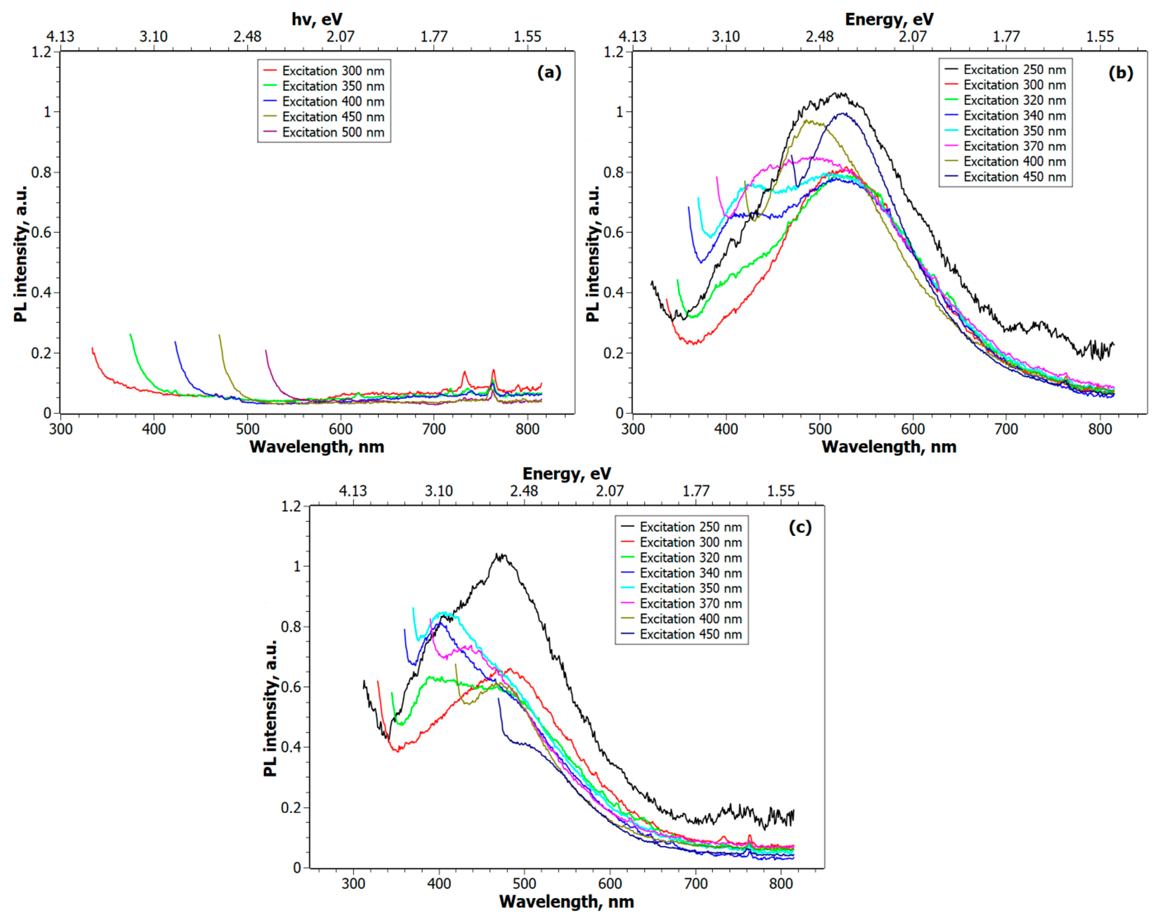
Even though the three studied samples absorb light in the UV–visible range, their luminescence properties differ substantially. The photoluminescence spectra of the nanocomposites obtained at ~20 °C are shown in Figure 2. As seen, for the CdC8 + Au NPs sample (Figure 2a), no luminescence is observed. The luminescence of bulk gold materials is known to be very weak, with a quantum yield on the order of 10−10, due to numerous nonradiative channels of excited-state relaxation [65]. In the case of gold nanomaterials with sizes greater than 5 nm, their luminescence quantum yield is still rather weak, on the order of 10−7 [66]. In the present paper, we were unable to detect the luminescence due to gold nanoparticles dispersed in liquid crystal glass (Figure 2a). By reducing the size of gold nanoparticles to create gold nanoclusters less than 2 nm, it is possible to achieve much stronger luminescence, as discussed in recent reviews [67,68]. The photoluminescence spectra for the CdC8 + C QDs sample are shown in Figure 2b. It was found that under excitation at a wavelength of 250 nm, three bands are present in the luminescence spectrum, with peaks centered around 406 nm, 490 nm, and 523 nm. The change in the excitation wavelength affects their intensity and maximum shift. The band in the region of 406 nm increases its intensity with an increase in the wavelength of the excitation radiation from 300 nm to 370 nm, and its maximum shifts towards the long-wave region of the spectrum. However, it disappears at 400 nm excitation. The band at 490 nm is amplified, with the effect visible at 370 nm excitation. The emission band at 523 nm does not change significantly when excited between 300 and 370 nm; however, it increases sharply at 450 nm and decreases sharply at 500 nm.
For the CdC8 + Au NPs + C QDs sample, the luminescence spectra shown in Figure 2c were obtained. Under excitation with a wavelength of 250 nm, two luminescence bands are observed at 405 nm and 470 nm. With increasing excitation wavelength, the first band does not appear at 300 nm but reappears at 320 nm, with a maximum of 390 nm. Further increasing the excitation wavelength shifts this band’s peak towards the longer-wavelength region of the spectrum, with its intensity increasing and reaching a maximum at 350 nm excitation. A further increase in the excitation wavelength leads to a decrease in its intensity. The band in the region of 470 nm remains almost unchanged with increasing excitation wavelength.
The observed emission bands of the two samples, containing either carbon nanoparticles or a mixture of both carbon and gold nanoparticles, can be associated with the characteristic emission bands of carbon nanoparticles reported in the literature [69]. The change in intensity depending on the excitation wavelength and the maximum shift towards the long-wavelength region of the spectrum indicates a heterogeneous distribution of NPs by size, which can result in different quantum confinement states. As shown in [70], one possible reason for the luminescence of C QDs is the radiation recombination of excitons on the surface of the NPs. Functional groups on the surface of C QDs can also cause luminescence. As reported in [71], surface transitions have different probabilities at different excitation wavelengths due to various functional groups on the C QDs surface. The fact that each transition plays a dominant role at the corresponding excitation wavelength leads to a shift in the maxima of the radiation spectra. At the same time, in the CdC8 + Au NPs + C QDs sample, the presence of Au slightly affected the luminescence spectrum. Passivation of the surface by the gold shell can exclude certain surface transitions from the luminescence process, which correspondingly affects the emission spectra of the sample.
In addition to the stationary emission spectra shown in Figure 2, the fluorescence decay curves of the studied samples, excited by a picosecond laser at a wavelength of 375 nm, are compiled in Figure 3. The results were obtained by scanning the relaxation of luminescence bands in the time domain for the studied nanocomposites at two wavelengths identified in Figure 2b,c. In the case of the CdC8 + C QDs sample, the time dependence of signals at wavelengths of 420 nm and 520 nm was measured (Figure 3a). For the CdC8 + Au NPs + C QDs sample, the measured signals were detected at wavelengths of 390 nm and 468 nm (Figure 3b). The solid red and cyan curves show the result of approximating the data with a multiexponential function (6):
R t = B 1 e t τ 1 + B 2 e t τ 2 + B 3 e t τ 3
where τ1, τ2, and τ3 are the relaxation times.
The data approximation using Equation (6) shown in Figure 3 indicates a multiexponential decrease in lifetime and suggests that fluorescence occurs because of the recombination of multi-excitons, although other processes cannot be excluded. The 420 nm emission band in CdC8 + C QDs exhibits carrier relaxation with three characteristic times: 0.58 ns, 3.18 ns, and 8.74 ns. For the 520 nm band, these times slightly increase to 0.71 ns, 3.43 ns, and 9.54 ns. A similar trend is observed in the CdC8 + Au NPs + C QDs sample. For the 390 nm band, the relaxation times are 0.35 ns, 3.20 ns, and 11.77 ns. For the 468 nm band, the τ1 and τ2 components increase to 0.77 ns and 3.79 ns, while the τ3 component changes only slightly to 11.68 ns.
The results of applying Equation (6) to the curves shown in Figure 3, yielding the characteristic relaxation times of the studied samples, are presented in Table 2.
The measured linear optical absorption and emission spectra of the studied samples generally agree with the existing literature on gold and carbon nanoparticles [1,2,3,4,72,73,74]. An important comment should be mentioned. The fact that glassy cadmium octanoate containing gold nanoparticles does not exhibit luminescence, whereas the presence of the same gold nanoparticles affects the emission spectra of cadmium octanoate containing carbon nanoparticles, as shown in Figure 2b,c and Figure 3a,b, supports our earlier assumption about the existence of a small fraction of carbon and gold nanoparticles in very close proximity. This leads to substantial interactions between carbon and gold nanoparticles that, in some cases, can even form nanohybrids [32]. The presented results indicate that such interactions modify the linear optical properties of the studied samples. A natural question is whether the nonlinear optical response of the studied samples is also affected and to what degree. In our previous paper, we conducted nonlinear optical measurements using a Z-scan method by exciting the samples with nanosecond laser pulses. To deepen our understanding of the nonlinear optical properties of the proposed unconventional liquid crystal glass nanocomposites, it is important to perform Z-scan measurements using femtosecond laser pulses. The laser beam diameter was 3.5 mm at 800 nm and 3.1 mm at 600 nm. For both wavelengths, the aperture size was 1.4 mm in a CA Z-scan configuration. A 25 cm focal length lens focused the beam.

3.2. Nonlinear Optical Properties at λ = 800 nm

The use of femtosecond laser pulses at two different wavelengths (800 nm and 600 nm) allowed for probing the nonlinear optical response of the studied samples, both near and far from the optical resonance. This section describes the results obtained using femtosecond laser pulses at a wavelength of 800 nm.
The results of measurements of the nonlinear absorption using the open-aperture (OA) Z-scan scheme for the CdC8 + Au NPs sample are shown in Figure 4a. We found that the most accurate approximation of experimental data is achieved using Equation (2), which accounts for both saturable absorption and two-photon absorption effects [62]. We believe the observed phenomenon combines two effects: two-photon absorption (2PA), which dominates at higher intensities (near the focal point), and weak saturable absorption, which occurs at lower intensities (far from the focal point). Away from the focal point, there is a slight brightening or absorption saturation, and as we approach the focus, two-photon absorption begins to dominate. Single-photon transitions at a quantum energy of 1.55 eV (800 nm) are possible near one of the active points (X) of the ~1.7 eV Brillouin band. By saturating the lowest energy levels of the s-p band, a brightening effect is produced. As the focus is approached and light intensity increases significantly, the mechanism of two-photon absorption begins to dominate, which is possible at the second optically active point (L) of the Brillouin zone (~2.4 eV).
The nonlinear refractive index (n2) of the sample was evaluated using a closed-aperture (CA) Z-scan scheme. The corresponding data and approximation curves are shown in Figure 4b. They indicate the self-defocusing effect (n2 < 0), which remains unchanged with varying laser intensity. Even with femtosecond excitation, several mechanisms can contribute to the observed nonlinear optical response. These include the nonlinear polarization of bound electrons under the action of a strong applied electric field (the optical Kerr effect), which has a typical femtosecond-scale response time (~1–100 fs) [63]. Additionally, the contribution from hot electrons, specifically nonequilibrium electron heating, cannot be excluded. The response time of optical nonlinearity due to hot electrons includes a fast excitation time (~1–100 fs), followed by slower electron–electron (100–500 fs) and electron–phonon (0.5–2 ps) relaxations [75].
Thus, the observed nonlinear behavior of the nonlinear refraction has an electronic origin. The role of the host material is to provide a suitable medium for dispersing nanoparticles and avoiding their aggregation. In addition, the presence of the host material can affect the measured nonlinear optical response through the local field factor, nonlinear light scattering, and thermal nonlinear optical response (at a substantially longer, i.e., picosecond–nanosecond, timescale). Under femtosecond laser excitation, the thermal nonlinearity of a host material does not contribute to the measured optical nonlinearities. According to the existing literature, for focused laser beams, the response time of thermal nonlinearity is much longer than the duration of femtosecond pulses [57]. Its rise time is in the picosecond–nanosecond range, whereas the relaxation time is in the microsecond range [22,57,63]. As a result, during the femtosecond excitation pulse, thermal nonlinearity does not have enough time to develop; thus, it does not affect the measured values of the nonlinear refractive indices of the studied samples. In this case, thermal nonlinearity can only affect the measured values of the nonlinear refractive index through cumulative thermal effects. As reported in several papers, cumulative thermal effects can appear if femtosecond pulses excite a sample at high frequencies, around 10 kHz or higher [57,61]. In the present paper, the femtosecond pulse frequency is 1 kHz, corresponding to a 1 ms time interval between two sequential femtosecond pulses. Because the characteristic relaxation time of thermal nonlinearity measured for glass-forming metal alkanoates is shorter than 100 µs [22], thermal effects do not accumulate from pulse to pulse. There is also indirect evidence for the absence of thermal effects, as discussed below. The presence of thermal effects leads to the broadening and asymmetry of the measured Z-scan [57,61]. We did not observe this in our experiments. Additionally, in the case of thermal nonlinearity, there must be a correlation between the measured nonlinear absorption coefficients and nonlinear refractive indices (i.e., stronger absorption should lead to a stronger thermal effect). We did not observe this either. As a result, our findings indicate that the observed nonlinear optical response is ultrafast, originating from electronic and electron–phonon interactions.
The nonlinear absorption coefficient (βeff), the imaginary part of the third-order susceptibility (Imχ(3)), the nonlinear refractive index (n2), and the real part of the third-order susceptibility (Reχ(3)) for the CdC8 + Au NPs sample, calculated from the data obtained, are compiled in Table 3. The meaning of the physical quantities is explained in the preceding section.
The experimental data and the best-fit curves obtained by the OA Z-scan method for the CdC8 + C QDs sample are shown in Figure 5a. Similar to the previous sample of CdC8 + Au NPs, different mechanisms of nonlinear absorption are observed. At low intensities of laser radiation, nonlinear brightening dominates, and as the sample approaches the focal point where the intensity increases, the two-photon absorption mechanism reported in [76,77] for carbon nanoparticles begins to dominate.
The data obtained in the CA Z-scan scheme of Figure 5b are different from the previous sample. At peak intensities of 160.84 GW/cm2 and 368.46 GW/cm2 at the focal point, self-defocusing (n2 < 0) is observed. However, at higher peak intensities of 438.65 GW/cm2 and 584.86 GW/cm2, the sign of the nonlinear refractive index changes to positive.
The nonlinear optical parameters calculated from the data obtained for the CdC8 + Au NPs sample are compiled in Table 4.
For the CdC8 + Au NPs + C QDs sample, where both Au NPs and C QDs are present, using the OA Z-scan scheme, the data shown in Figure 6a were obtained. Their approximation, as with the previous samples, was performed by applying Equation (2), which considers two mechanisms of nonlinear absorption. The nonlinear refraction effect observed using the CA Z-scan scheme is shown in Figure 6b. Similar to the CdC8 + C QDs sample, a self-defocusing effect (n2 < 0) is observed at lower laser intensities (163.76 GW/cm2 and 204.70 GW/cm2). However, at higher laser intensities (365.54 GW/cm2 and 584.86 GW/cm2), the sample exhibits self-focusing properties (n2 > 0).
The nonlinear optical parameters obtained for the CdC8 + Au NPs + C QDs sample are compiled in Table 5. The presence of two types of nanoparticles in glassy cadmium octanoate modifies the magnitude of the nonlinear absorption coefficient and the nonlinear refractive index of the studied sample.

3.3. Nonlinear Optical Properties at λ = 600 nm

Even though femtosecond laser pulses with a wavelength of 600 nm also excite the studied samples in the off-resonance region, this wavelength is closer to their resonance frequency than 800 nm (Figure 1). Therefore, the expected nonlinear optical response measured at 600 nm can differ from that observed at 800 nm.
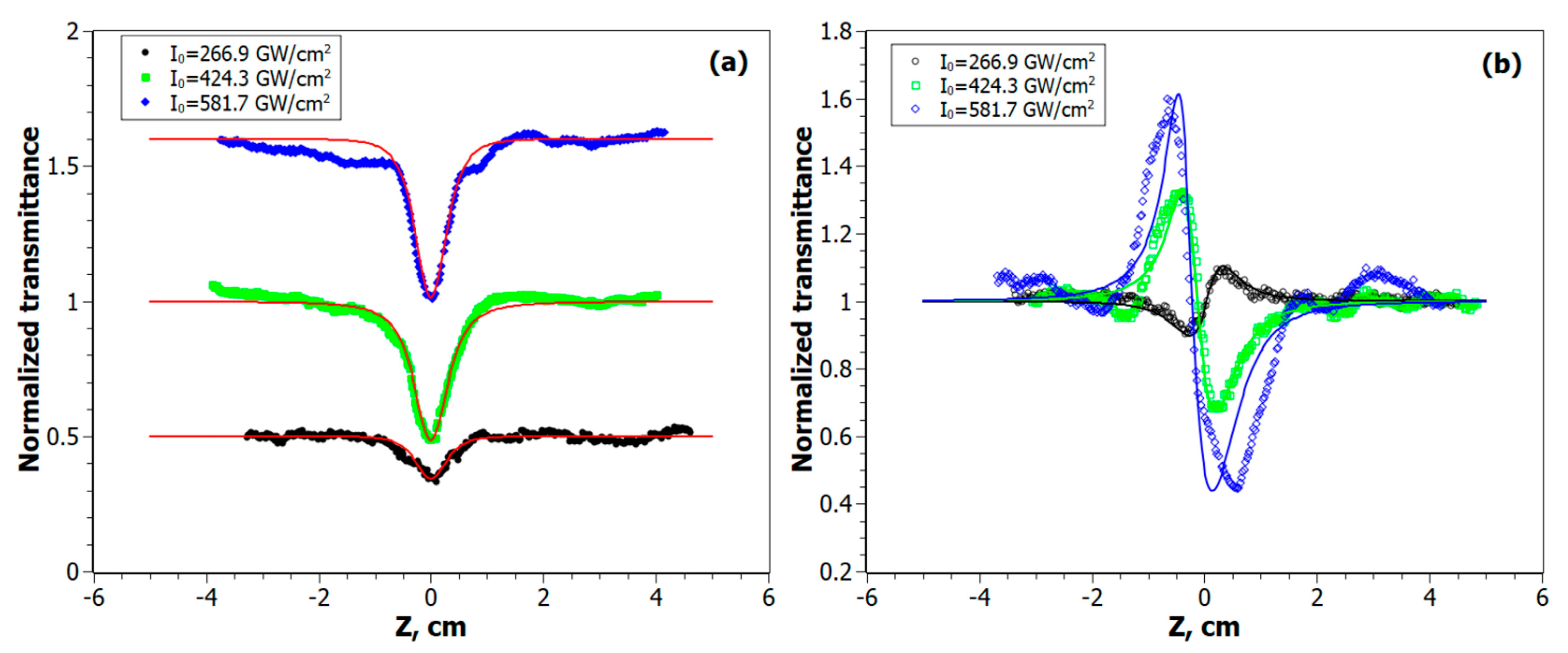
The data obtained using the OA Z-scan scheme for the CdC8 + Au NPs sample are shown in Figure 7a. The character of the nonlinear absorption does not change; two mechanisms are present: absorption saturation at a distance far from the focal point and two-photon absorption in the vicinity of the focal point. For the CA Z-scan results shown in Figure 7b, the behavior is noticeably different from that measured at 600 nm. At a relatively lower peak intensity of I0 = 266.9 GW/cm2, we observe self-focusing (n2 > 0), while at higher values of I0, the sample exhibits self-defocusing properties (n2 < 0).
The nonlinear optical parameters of the CdC8 + Au NPs nanocomposite obtained at a wavelength of 600 nm are presented in Table 6.
The Z-scan results for the CdC8 + C QDs sample measured at 600 nm are shown in Figure 8. Similar to the sample with gold nanoparticles (CdC8 + Au NPs), according to the OA Z-scan data in Figure 8a, the mechanism of absorption saturation at a distance far from the focal point changes to two-photon absorption when approaching the focus. In the case of the nonlinear refraction experiments using CA Z-scan (Figure 8b), the phenomenon of self-defocusing is observed, i.e., n2 has a negative sign. Contrary to the case of excitation at 800 nm, changing the intensity of the laser radiation does not change the sign of the refractive index, which remains negative (Figure 8b).
The nonlinear optical parameters of the CdC8 + C QDs sample, obtained using the Z-scan measurement results (Figure 8), are presented in Table 7.
The OA Z-scan results for the nanocomposite containing both Au NPs and C QDs are shown in Figure 9a. Similar to the previous samples, two mechanisms of nonlinear absorption are observed. Similar to the case of excitation at 600 nm, the CA Z-scan measurements detected an interesting change in the sign of the nonlinear refractive index when negative n2 at lower intensities becomes positive (n2 > 0) at higher intensities (Figure 9b).
The nonlinear optical parameters of the CdC8 + Au NPs + C QDs sample, obtained at a wavelength of 600 nm, are listed in Table 8.
The presented results indicate a general feature of the studied unconventional liquid crystal glass nanocomposites: both the nonlinear refractive index and the nonlinear absorption coefficient are intensity-dependent (Table 3, Table 4, Table 5, Table 6, Table 7 and Table 8 and Figure 4, Figure 5, Figure 6, Figure 7, Figure 8 and Figure 9). Conventional third-order nonlinear optical materials are characterized by constant values of n2 and β. Therefore, the obtained values should be treated as effective nonlinear optical parameters. It should be noted that we do not consider higher-order (i.e., fifth-order) optical nonlinearities. The presence of higher-order optical nonlinearities leads to characteristic changes in the shape of Z-scan curves, which can become very asymmetric. As a rule, such higher-order effects can appear in some materials at light intensities well above 100 GW/cm2, contributing 1–10% to the observed nonlinear optical response [78]. Curve fitting involving higher-order nonlinearities also requires substantial modifications. In this paper, the nearly symmetrical Z-scan curves obtained were analyzed using the formalism of the dominant third-order nonlinear optical response.
In general, the intensity-dependent extinction coefficient α I can be expressed as a sum (7):
α I = α 0 1 + I I S + β I + α s ( I )
where the first term accounts for the absorption saturation effect in the region far from the focal point, the second term describes the effective two-photon absorption process, and the third term α s ( I ) accounts for the effect of nonlinear scattering of light [32,79]. There are also reports discussing the possibility of the saturation of a two-photon absorption process, thus making the coefficient β intensity-dependent, i.e., β ( I ) [80,81,82,83]. In this paper, the simultaneous presence of several processes represented by Equation (7) results in an intensity dependence of the evaluated nonlinear absorption coefficient β of the studied samples.
The observed intensity-dependent nonlinear refraction in the studied samples is another interesting effect worth mentioning because it also depends on the wavelength of excitation and the type of nanocomposites. Glassy cadmium octanoate containing gold nanoparticles exhibits an intensity-dependent negative nonlinear refractive index n 2 at 800 nm. Interestingly, when measured at 600 nm, the same material shows a positive sign of n 2 at lower light intensity and a negative sign of n 2 at higher intensities. Glassy cadmium octanoate doped with carbon nanoparticles exhibits the opposite behavior: an intensity-dependent negative n 2 at 600 nm, and a negative sign of n 2 at lower light intensities, followed by a positive n 2 at higher intensities evaluated at 800 nm. When both gold and carbon nanoparticles are dispersed in a liquid crystal glass, the sample exhibits a similar trend at both 600 nm and 800 nm wavelengths: an intensity-dependent negative n 2 at lower light intensities, followed by a positive n 2 at higher intensities.
The results indicate that the studied nanocomposites can be considered for ultra-fast optical limiting. At lower light intensities, ground state bleaching or absorption saturation occurs, as represented by the first term of Equation (7). At higher intensities, two-photon absorption dominates, as indicated by the second term of Equation (7). It should be noted that designing such optical limiters requires knowledge of the intensity dependence of the nonlinear absorption coefficient to compute the intensity of light passing through the sample. In general, the intensity of light I propagating through a sample along the z-direction changes according to Equation (8):
d I d z = α I I
where α I is given by Equation (7) [83]. As a result, by knowing α I , the light intensity and the corresponding optical limiter transmittance as a function of intensity can be computed. Thus, the reported values of the nonlinear optical parameters of the designed liquid crystal glass doped with nanoparticles have practical value and can benefit the design of ultrafast optical limiters. Similarly, the intensity-dependent nonlinear refractive index, with the possibility of its sign reversal, offers additional ways to control light, which can be leveraged for advanced photonics applications.
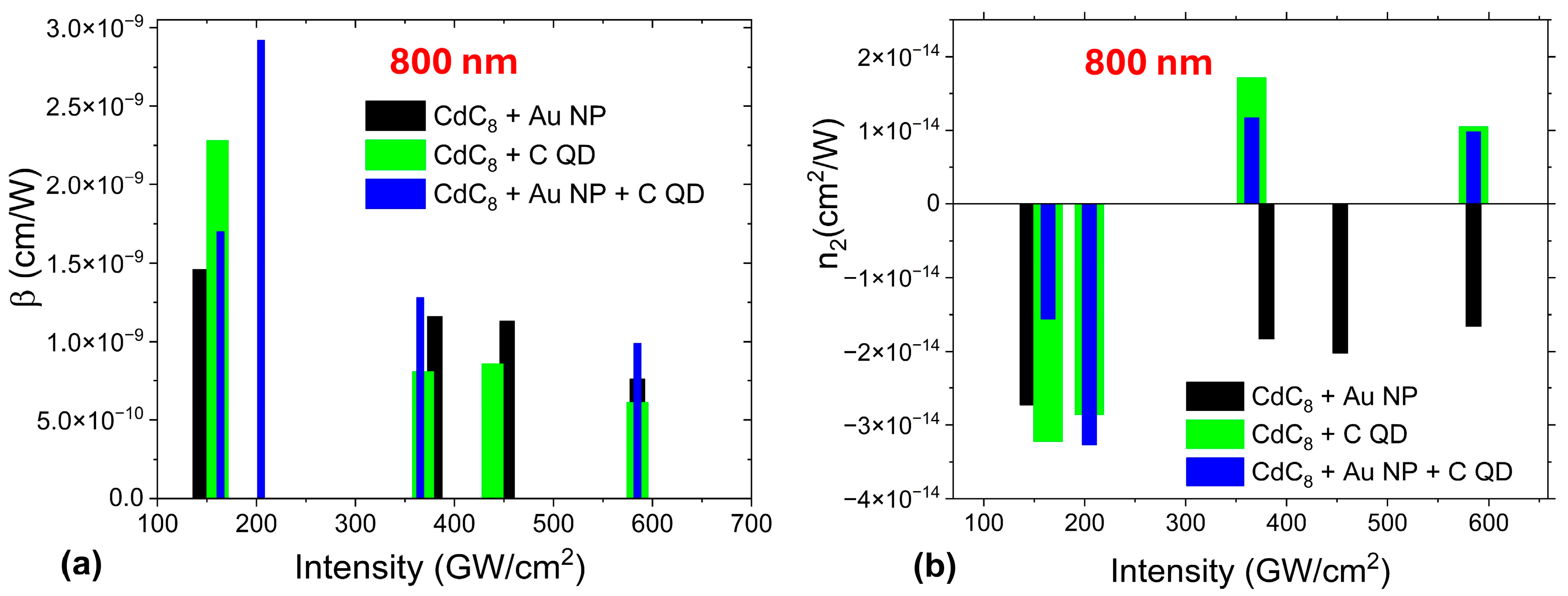
The obtained values for the nonlinear optical parameters of the studied materials generally overlap with reported data for nanocomposites under similar excitation conditions. Direct comparison is challenging due to numerous factors, including material composition, pulse duration, intensity, and central wavelength, all of which can influence the results. For greater detail, readers are referred to several comprehensive reviews compiling existing results [1,2,3,4,11]. The nonlinear absorption coefficients of all the studied samples are positive and decrease with higher light intensities (Figure 10a and Figure 11a). Their dependence on light intensity is caused by the interplay between absorption saturation at lower light intensities and the two-photon absorption process that dominates at higher light intensities.
The intensity-dependent nonlinear refractive index of the studied samples can be either positive or negative, depending on their composition and the wavelength and intensity of light (Figure 10b and Figure 11b). In the case of off-resonance excitation (i.e., at a wavelength of 800 nm, Figure 10b), the nonlinear refractive index n2 of cadmium octanoate glass containing gold nanoparticles is negative over the entire intensity range (100–600 GW/cm2). In contrast, the samples with carbon nanoparticles or a combination of both carbon and gold nanoparticles exhibit a sign reversal of n2 from negative at lower intensities to positive at higher light intensities (Figure 10b). The sign reversal of n2 depends on the wavelength of the excitation. At a shorter wavelength (600 nm), liquid crystal glass containing gold nanoparticles has a positive n2 at lower intensities and a negative n2 at higher intensities (Figure 11b). The same glass containing carbon nanoparticles is characterized by a negative nonlinear refractive index at both lower and higher light intensities (Figure 11b). A glass sample with both carbon and gold nanoparticles changes the sign of n2 from negative at lower intensities to positive at higher light intensities (Figure 11b).

4. Conclusions

The proposed unconventional nanocomposites made of cadmium octanoate liquid crystal glass, containing gold nanoparticles, carbon nanoparticles, and a combination of gold and carbon nanoparticles, exhibit a rich nonlinear optical response when probed with femtosecond laser pulses. Both the nonlinear absorption coefficient and the nonlinear refractive index of the studied glass nanocomposites are intensity-dependent and are affected by the sample composition and the wavelength of the excitation laser pulse, as shown in Figure 10 and Figure 11.
The physical mechanisms responsible for this interesting behavior include intrinsic electronic nonlinearities of gold and carbon nanoparticles, which involve both intraband and interband transition contributions, as well as the hot electron contribution, along with the local field enhancement effect. These are major nonlinear optical mechanisms typical for nanocomposite materials containing metal and carbon nanoparticles, especially under excitation by femtosecond laser pulses [13]. Interband transitions result in photon absorption with energies of 2.0–2.4 eV (corresponding to wavelengths from 520 nm to 600 nm), leading to valence band–conduction band (d–sp) electron transitions. Under femtosecond excitation, light absorption due to interband transitions can dominate over absorption due to surface plasmon resonance. In carbon nanodots, interband absorption can also occur. Light absorption due to intraband transitions within the conduction band is typical for gold nanoparticles, caused by nearly free electrons in the sp conduction band. Both intraband and interband transitions can result in high electronic temperatures, leading to the hot electron effect. In addition, in the case of two types of nanoparticles in a liquid crystal glass matrix, both the value and the sign of the nonlinear refractive index can be affected by interactions between nanoparticles, as indirectly inferred from time-resolved luminescence experiments. It is important to emphasize the role of the nonlinear light scattering effect, which can contribute to the intensity dependence of both the nonlinear absorption coefficient and the nonlinear refractive index [79]. The existence of multiple nonlinear optical mechanisms makes the theoretical analysis of the obtained results a very difficult task, a solution to which will be sought in future publications.
The proposed unconventional glass nanocomposites can benefit the rapidly developing photonics and nanotechnology applications. Moreover, given the ease of fabrication, low cost, and long-term stability of the studied samples, the developed strategy to design liquid crystal glass made of metal alkanoates can be applied to produce a variety of multifunctional materials.

Author Contributions

Conceptualization, V.R., G.K., S.B., T.M., and Y.G.; methodology, V.R., G.K., S.B., D.Z., A.T., G.Y., and Y.G.; software, A.T., V.R., and D.Z.; validation, V.R., A.T., D.Z., and G.Y., formal analysis, V.R., A.T., D.Z., G.K., S.B., G.Y., T.M., and Y.G.; investigation, V.R., A.T., D.Z., G.K., S.B., G.Y., T.M., and Y.G.; resources, G.K., T.M., and Y.G.; data curation, V.R., S.B., G.K., and Y.G.; writing—original draft preparation, V.R., A.T., S.B., D.Z., G.K., G.Y., and T.M.; writing—review and editing, V.R., G.K., and Y.G.; supervision, V.R., G.K., and Y.G.; project administration, V.R., G.K., T.M., and Y.G.; funding acquisition, V.R., G.K., T.M., and Y.G. All authors have read and agreed to the published version of the manuscript.

Funding

This research was funded by the National Academy of Sciences of Ukraine (projects No. 1.4.B/219, 3/20-H, B/197, N. 6 (6541230), and No. 0123U100832), Le fonds spécial Solidarité Ukraine du programme PAUSE, and by the CSU—AAUP Faculty Research Grant.

Data Availability Statement

The original contributions presented in this study are included in the article. Further inquiries can be directed to the corresponding author.

Acknowledgments

D. Zhulai greatly appreciates a research grant from the German National Academy of Sciences Leopoldina. Y. Garbovskiy acknowledges funding from the CSU—AAUP Faculty Research Grant. The authors acknowledge funding from NASU via project No. 0123U100832. The authors are grateful for the technical support of this work from the Femtosecond Laser Complex of the National Academy of Sciences of Ukraine, which assisted in the femtosecond studies, and the Department of Biological Systems of the Institute of Physics of the National Academy of Sciences of Ukraine, which assisted in the luminescence studies.

Conflicts of Interest

Author Dmytro Zhulai was employed by the company Forschungszentrum Jülich GmbH, Institute of Bioelectronics (IBI-3). The remaining authors declare that the research was conducted in the absence of any commercial or financial relationships that could be construed as a potential conflict of interest.

References

  1. Vermeulen, N.; Espinosa, D.; Ball, A.; Ballato, J.M.; Boucaud, P.; Boudebs, G.; Campos, C.L.A.V.; Dragic, P.D.; Gomes, A.; Huttunen, M.J.; et al. Post-2000 nonlinear optical materials and measurements: Data tables and best practices. J. Phys. Photonics 2023, 5, 035001. [Google Scholar] [CrossRef]
  2. Palpant, B. Third order nonlinear optical response of metal nanoparticles. In Nonlinear Optical Properties of Matter; Papadopoulos, M.G., Andrzej, J.S., Jerzy, L., Eds.; Springer: Berlin/Heidelberg, Germany, 2006. [Google Scholar]
  3. Zhang, Y.-X.; Wang, Y.-H. Nonlinear optical properties of metal nanoparticles: A review. RSC Adv. 2017, 7, 45129–45144. [Google Scholar] [CrossRef]
  4. Ganeev, R.A. Nonlinear Optical Refraction and Absorption of Media. In Nonlinear Optical Properties of Materials; Springer: Dordrecht, Germany, 2013. [Google Scholar]
  5. Nath, S.; Puthukkudi, A.; Mohapatra, J.; Biswal, B.P. Covalent Organic Frameworks as Emerging Nonlinear Optical Materials. Angew. Chem. 2023, 135, e202218974. [Google Scholar] [CrossRef]
  6. Zhang, L.; Li, H.; Yang, Y.; Yang, D.; Cui, Y.; Qian, G. Aligned chromophores in a host–guest MOF crystal for switchable polarized nonlinear optical response. J. Mater. Chem. C 2022, 10, 14915–14920. [Google Scholar] [CrossRef]
  7. Mitus, A.C.; Saphiannikova, M.; Radosz, W.; Toshchevikov, V.; Pawlik, G. Modeling of Nonlinear Optical Phenomena in Host-Guest Systems Using Bond Fluctuation Monte Carlo Model: A Review. Materials 2021, 14, 1454. [Google Scholar] [CrossRef]
  8. Chen, Y.-B.; Zhang, C.; Luo, D.; Chen, R.; Zhang, J.; Chen, S.; Fang, W. Host-Guest Chemistry and Nonlinear Optical Behaviors of Aluminum Molecular Rings. Chin. J. Chem. 2024, 42, 2581. [Google Scholar] [CrossRef]
  9. Khoo, I.C. Nonlinear optics, active plasmonic and tunable metamaterials with liquid crystals. Prog. Quantum Electron. 2014, 38, 77–117. [Google Scholar] [CrossRef]
  10. Xu, J.; Yang, R.; Fan, Y.; Fu, Q.; Zhang, F. A Review of Tunable Electromagnetic Metamaterials with Anisotropic Liquid Crystals. Front. Phys. 2021, 9, 633104. [Google Scholar] [CrossRef]
  11. Si, G.; Zhao, Y.; Leong, E.S.P.; Liu, Y.J. Liquid-Crystal-Enabled Active Plasmonics: A Review. Materials 2014, 7, 1296–1317. [Google Scholar] [CrossRef] [PubMed]
  12. Zhang, X.; Xu, Y.; Valenzuela, C.; Zhang, X.; Wang, L.; Feng, W.; Li, Q. Liquid crystal-templated chiral nanomaterials: From chiral plasmonics to circularly polarized luminescence. Light. Sci. Appl. 2022, 11, 223. [Google Scholar] [CrossRef] [PubMed]
  13. Yamane, M.; Asahara, Y. Glasses for Photonics; Cambridge University: New York, NY, USA, 2000. [Google Scholar]
  14. Petzold, U. Optical Glass: A High-Tech Base Material as Key Enabler for Photonics. In Advances in Glass Science and Technology; Sglavo, V., Ed.; IntechOpen: London, UK, 2018. [Google Scholar] [CrossRef]
  15. Vasconcelos, H.C. Optical Nonlinearities in Glasses’. In Nonlinear Optics—Nonlinear Nanophotonics and Novel Materials for Nonlinear Optics; IntechOpen: London, UK, 2022. [Google Scholar] [CrossRef]
  16. Chen, H.M.P.; Ou, J.J.; Chen, S.H. Glassy liquid crystals as self-organized films for robust optoelectronic devices. In Nanoscience with Liquid Crystals; Springer International Publishing: Berlin/Heidelberg, Germany, 2014; pp. 179–208. [Google Scholar]
  17. Gujral, A.; Yu, L.; Ediger, M.D. Anisotropic organic glasses. Curr. Opin. Solid State Mater. Sci. 2018, 22, 49–57. [Google Scholar] [CrossRef]
  18. Bunning, T.J.; Mather, P.T.; Hood, P.J.; Barnes, W. Mid-wavelength IR (MWIR) polarizers from glassy cholesteric liquid crystals. Liq. Cryst. 1999, 26, 557–565. [Google Scholar] [CrossRef]
  19. Zhou, J.Y.; Wu, C.G.; Jia, Y.G. Preparation and optical properties of linear polysiloxane-based glassy cholesteric liquid crystals with green reflected color. Liq. Cryst. 2021, 48, 215–222. [Google Scholar] [CrossRef]
  20. Hu, G.; Kelly, S.M.; Kitney, S.P.; Harrison, W.; Lambert, B. Novel nematic and glassy liquid crystalline oligomers as electroluminescent organic semiconductors. Liq. Cryst. 2021, 48, 626–640. [Google Scholar] [CrossRef]
  21. Casado, F.J.M.; Riesco, M.R.; Yélamos, M.I.R.; Arenas, A.S.; Cheda, J.A.R. The role of calorimetry in the structural study of mesophases and their glass states. J. Therm. Anal. Calorim. 2012, 108, 399–413. [Google Scholar] [CrossRef]
  22. Klimusheva, G.; Mirnaya, T.; Garbovskiy, Y. Versatile nonlinear-optical materials based on mesomorphic metal alkanoates: Design, properties, and applications. Liq. Cryst. Rev. 2015, 3, 28–57. [Google Scholar] [CrossRef]
  23. Martínez-Casado, F.J.; Ramos-Riesco, M.; Rodríguez-Cheda, J.A.R.; Cucinotta, F.; Fernández-Martínez, A.; Garrido, L.; Matesanz, E.; Marchese, L. Short lead(II) soaps: From weakly fluorescent crystals to strongly phosphorescent and structurally varied vitreous phases. A thermal, structural and spectroscopic study. J. Mater. Chem. C 2014, 2, 9489–9496. [Google Scholar] [CrossRef]
  24. Nagaraj, M. Liquid Crystals Templating. Crystals 2020, 10, 648. [Google Scholar] [CrossRef]
  25. Lyashchova, A.; Fedorenko, D.; Garbovskiy, Y.; Klimusheva, G.; Mirnaya, T.; Asaula, V. Strong thermal optical nonlinearity causes by CdSe nanoparticles synthesized in smectic ionic liquid crystals. Liq. Cryst. 2013, 40, 1377. [Google Scholar] [CrossRef]
  26. Zhulai, D.; Koval’chuk, A.; Bugaychuk, S.; Klimusheva, G.; Mirnaya, T.; Vitusevich, S. Photoconductivity of ionic thermotropic liquid crystal with semiconductor nanoparticles. J. Mol. Liq. 2018, 267, 405–410. [Google Scholar] [CrossRef]
  27. Rudenko, V.; Garbosvkiy, Y.; Klimusheva, G.; Mirnaya, T. Intensity dependent nonlinear absorption coefficients and nonlinear refractive indices of glass-forming ionic liquid crystals doped with gold and silver nanoparticles. J. Mol. Liq. 2018, 267, 56–60. [Google Scholar] [CrossRef]
  28. Rudenko, V.; Garbovskiy, Y.; Klimusheva, G.; Mirnaya, T.; Yaremchuk, G. Enhanced optical nonlinearity of the “nonlinear host–nonlinear guest” glassy nanocomposites made of the mesomorphic cobalt octanoate and noble metal nanoparticles. J. Opt. Soc. Am. B 2016, 33, 648–655. [Google Scholar] [CrossRef]
  29. Rudenko, V.; Tolochko, A.; Zhulai, D.; Klimusheva, G.; Mirnaya, T.; Yaremchuk, G.; Asaula, V. Nonlinear optical properties of metal alkanoate composites with hybrid core/shell nanoparticles. Appl. Nanosci. 2018, 8, 823–829. [Google Scholar] [CrossRef]
  30. Rudenko, V.; Tolochko, A.; Bugaychuk, S.; Zhulai, D.; Klimusheva, G.; Yaremchuk, G.; Mirnaya, T.; Garbovskiy, Y. Probing Optical Nonlinearities of Unconventional Glass Nanocomposites Made of Ionic Liquid Crystals and Bimetallic Nanoparticles. Nanomaterials 2022, 12, 924. [Google Scholar] [CrossRef]
  31. Rudenko, V.; Tolochko, A.; Bugaychuk, S.; Zhulai, D.; Klimusheva, G.; Yaremchuk, G.; Mirnaya, T.; Garbovskiy, Y. Intensity-dependent optical nonlinearities of composite materials made of ionic liquid crystal glass and bimetallic nanoparticles. Liq. Cryst. 2023, 50, 174–180. [Google Scholar] [CrossRef]
  32. Rudenko, V.; Tolochko, A.; Bugaychuk, S.; Zhulai, D.; Klimusheva, G.; Yaremchuk, G.; Mirnaya, T.; Garbovskiy, Y. Modifying optical nonlinearities of ionic liquid crystal glass by adding gold and carbon nanoparticles. J. Mol. Liq. 2024, 393, 123641. [Google Scholar] [CrossRef]
  33. Gurushantha, K.; Jagannath, G.; Kolavekar, S.; Pramod, A.; Almuqrin, A.H.; Sayyed, M.; Murthy, N.L.; Rao, S.V.; Keshavamurthy, K. Gold nanoparticles enhanced femtosecond nonlinear optical properties of sodium borate oxide glasses. Infrared Phys. Technol. 2023, 131, 104663. [Google Scholar] [CrossRef]
  34. Jagannathan, A.; Rajaramakrishna, R.; Gangareddy, J.; Rajashekara, K.; Soma, V.R.; Kaewkhao, J.; Kothan, S.; Surzhikova, D.P. Third order nonlinear optical properties of lithium zinc calcium fluoroborate glasses embedded with Au–Ag nanoparticles. Opt. Mater. 2024, 149, 115013. [Google Scholar] [CrossRef]
  35. Hernández-Acosta, M.A.; Torres-Torres, C.; Bornacelli, J.; García-Merino, J.A.; Can-Uc, B.; Rangel-Rojo, R.; Oliver, A. A comprehensive study of the nonlinear optical response exhibited by ion-implanted silica plates with Au and Pt nanostructures. Results Opt. 2021, 4, 100098. [Google Scholar] [CrossRef]
  36. Chen, S.; Niu, R.; Gao, Y.; Li, Z.; Liu, K.; Wang, Y.; Song, Y.; Zhang, X. Tunable nonlinear absorption of gold nanostars and application as a saturable absorber. Opt. Laser Technol. 2024, 168, 109873. [Google Scholar] [CrossRef]
  37. Tan, Z.; Dong, J.; Liu, Y.; Luo, Q.; Li, Z.; Yun, T.; Jiang, T.; Cheng, X.; Huang, D. Nonlinear optics of graphitic carbon allotropes: From 0D to 3D. Nanoscale 2025, 17, 1171–1212. [Google Scholar] [CrossRef]
  38. Yadav, R.K.; Aneesh, J.; Sharma, R.; Abhiramnath, P.; Maji, T.K.; Omar, G.J.; Mishra, A.K.; Karmakar, D.; Adarsh, K.V. Designing Hybrids of Graphene Oxide and Gold Nanoparticles for Nonlinear Optical Response. Phys. Rev. Appl. 2018, 9, 044043. [Google Scholar] [CrossRef]
  39. Biswas, S.; Kole, A.K.; Tiwary, C.S.; Kumbhakar, P. Enhanced Nonlinear Optical Properties of Graphene Oxide–Silver Nanocomposites Measured by Z-Scan Technique. RSC Adv. 2016, 6, 10319–10325. [Google Scholar] [CrossRef]
  40. Abhijith, T.; Edappadikkunnummal, S.; Suthar, R.; Thomas, S.; Karak, S. Au–WS2 Nanohybrids with Enhanced Optical Nonlinearity for Optical Limiting Applications. ACS Appl. Nano Mater. 2023, 6, 2327. [Google Scholar] [CrossRef]
  41. Ren, J.; Yang, P.; Wang, A.; Zhu, W.; Shang, D.; Song, Y. Synergistic promoted nonlinear optical effects in polyaniline nanohybrids covalently functionalized with tin porphyrin. Colloids Surf. A Physicochem. Eng. Asp. 2022, 650, 129588. [Google Scholar] [CrossRef]
  42. Salah, A.; Hassab-Elnaby, S.; Ramadan, M.A. Boosting the nonlinear optical absorption of graphene oxide, and gold nanorods by tailoring graphene oxide-gold nanorods hybrids. SN Appl. Sci. 2023, 5, 288. [Google Scholar] [CrossRef]
  43. Zhao, X.; Wang, F.; Wu, J.; Zhu, B.; Gu, Y. Synthesis and Third-Order Nonlinear Synergistic Effect of ZrO2/RGO Composites. Nanomaterials 2021, 11, 2741. [Google Scholar] [CrossRef]
  44. Saravanan, M. Nonlinear and Optical Limiting Properties of Graphene–Ferrite Nanocomposites: A Review. Phys. Status Solidi RRL 2024, 18, 2400105. [Google Scholar] [CrossRef]
  45. Zhu, B.; Wang, F.; Cao, Y.; Wang, C.; Wang, J.; Gu, Y. Nonlinear optical enhancement induced by synergistic effect of graphene nanosheets and CdS nanocrystals. Appl. Phys. Lett. 2016, 108, 252106. [Google Scholar] [CrossRef]
  46. Yan, Z.; Fan, J.; Pan, S.; Zhang, M. Recent advances in rational structure design for nonlinear optical crystals: Leveraging advantageous templates. Chem. Soc. Rev. 2024, 53, 6568–6599. [Google Scholar] [CrossRef]
  47. Zhang, J.-X.; Zhou, S.-H.; Wu, X.-T.; Lin, H.; Zhu, Q.-L. Nonbonding Electron Inversion-Driven Structural Engineering: Synergistic Enhancement of Linear and Nonlinear Optical Properties. Angew. Chem. Int. Ed. 2025, 64, e202506658. [Google Scholar] [CrossRef]
  48. Feng, X.; Qiu, J.; Zhou, S. Synergistic effect in nonlinear response in glass ceramics. J. Am. Ceram. Soc. 2024, 107, 1936–1948. [Google Scholar] [CrossRef]
  49. Konkoly-Thege, I.; Ruff, I.; Adeosun, S.O.; Sime, S.J. Properties of molten carboxylates. Part 6. A quantitative differential thermal analysis study of phase transitions in some zinc and cadmium carboxylates. Thermochim. Acta 1978, 24, 89–96. [Google Scholar] [CrossRef]
  50. Asaula, V.M.; Myrna, T.A.; Yaremchuk, H.H.; Tolochko, A.S. Mesomorphic and glass formed properties of gomologies series of cadmium alcanoates. Ukr. Chem. J. 2011, 77, 24–27. Available online: http://jnas.nbuv.gov.ua/article/UJRN-0000895786 (accessed on 1 August 2025).
  51. Klimusheva, G.; Dmitruk, I.; Mirnaya, T.; Tolochko, A.; Bugaychuk, S.; Naumenko, A.; Asaula, V. Monodispersity and ordering of semiconductor quantum dots synthesised in ionic liquid crystalline phase of cadmium alkanoates. Liq. Cryst. 2013, 40, 980–988. [Google Scholar] [CrossRef]
  52. Zhulai, D.; Boichuk, N.; Pustovyi, D.; Kovalchuk, O.; Garbovskiy, Y.; Klimusheva, G.; Mirnaya, T.; Yaremchuk, G.; Vitusevich, S. Synthesis of Carbon and Gold Nanoparticles in Ionic Liquid Crystals: Structural Properties and Electrical Behavior for Electro-Optical Sensors. Nano Sel. 2025, e202400063. [Google Scholar] [CrossRef]
  53. Nakashima, S.; Tatewaki, Y.; Okada, S.; Nagakura, H.; Shindo, A.; Mikura, C.; Shiga, K.; Terakawa, K.; Yamada, M. Polymerization of a divalent metal salt of an unsaturated carboxylic acid stimulated by ion exchange with a metal salt of fatty acids. Polym. J. 2016, 48, 855–858. [Google Scholar] [CrossRef]
  54. Jiang, Y.; Ojo, W.-S.; Mahler, B.; Xu, X.; Abécassis, B.; Dubertret, B. Synthesis of CdSe Nanoplatelets without Short-Chain Ligands: Implication for Their Growth Mechanisms. ACS Omega 2018, 3, 6199–6205. [Google Scholar] [CrossRef]
  55. van der Bok, J.C.; Prins, P.T.; Montanarella, F.; Maaskant, D.N.; Brzesowsky, F.A.; van der Sluijs, M.M.; Salzmann, B.B.V.; Rabouw, F.T.; Petukhov, A.V.; Donega, C.D.M.; et al. In Situ Optical and X-ray Spectroscopy Reveals Evolution toward Mature CdSe Nanoplatelets by Synergetic Action of Myristate and Acetate Ligands. J. Am. Chem. Soc. 2022, 144, 8096–8105. [Google Scholar] [CrossRef] [PubMed]
  56. Wagnon, B.; Valleix, R.; Abécassis, B. Cadmium Carboxylate Preparation Method Impacts the Synthesis of CdSe Nanoplatelets through Differences in Dissolution Kinetics. Chem. Mater. 2025, 37, 4291–4300. [Google Scholar] [CrossRef]
  57. Perumbilavil, S.; Sandeep, C.S.; Matham, M.V. Unconventional z-scan signatures in water arising from the interplay of repetition rate dependent electronic and thermal nonlinearities. J. Mol. Liq. 2025, 425, 127181. [Google Scholar] [CrossRef]
  58. Sheik-Bahai, M.; Said, A.A.; Wei, T.; Hagan, D.J.; Van Stryland, E.W. Sensitive measurement of optical nonlinearities using a single beam. IEEE J. Quantum Electron. 1990, 26, 760–769. [Google Scholar] [CrossRef]
  59. Sheik-Bahai, M.; Said, A.A.; Van Stryland, E.W. High-sensitivity, single-beam n2 measurements. Opt. Lett. 1989, 14, 955. [Google Scholar] [CrossRef]
  60. Chapple, P.B.; Staromlynska, J.; Hermann, J.A.; Mckay, T.J.; Mcduff, R.G. Single-Beam Z-Scan: Measurement Techniques and Analysis. J. Nonlinear Opt. Phys. Mater. 1997, 6, 251. [Google Scholar] [CrossRef]
  61. Kwak, C.H.; Lee, Y.L.; Kim, S.G. Analysis of asymmetric Z-scan measurement for large optical nonlinearities in an amorphous As2S3 thin film. J. Opt. Soc. Am. B 1999, 16, 600–604. [Google Scholar] [CrossRef]
  62. Rout, A.; Boltaev, G.S.; Ganeev, R.A.; Fu, Y.; Maurya, S.K.; Kim, V.V.; Rao, K.S.; Guo, C. Nonlinear Optical Studies of Gold Nanoparticle Films. Nanomaterials 2019, 9, 291. [Google Scholar] [CrossRef]
  63. Boyd, R. Nonlinear Optics, 2nd ed.; Academic Press: San Diego, CA, USA, 2003. [Google Scholar]
  64. Mehta, V.N.; Jha, S.; Singhal, R.K.; Kailasa, S.K. Preparation of multicolor emitting carbon dots for HeLa cell imaging. New J. Chem. 2014, 38, 6152–6160. [Google Scholar] [CrossRef]
  65. Mooradian, A. Photoluminescence of Metals. Phys. Rev. Lett. 1969, 22, 185. [Google Scholar] [CrossRef]
  66. Cheng, Y.; Lu, G.; He, Y.; Shen, H.; Zhao, J.; Xia, K.; Gong, Q. Luminescence Quantum Yields of Gold Nanoparticles Varying with Excitation. Nanoscale 2016, 8, 2188–2194. [Google Scholar] [CrossRef] [PubMed]
  67. Zheng, J.; Zhou, C.; Yu, M.; Liu, J. Different sized luminescent gold nanoparticles. Nanoscale 2012, 4, 4073–4083. [Google Scholar] [CrossRef] [PubMed]
  68. Zhang, G.; Fang, B.; Peng, J.; Deng, S.; Hu, L.; Lai, W. Luminescent gold nanoclusters from synthesis to sensing: A comprehensive review. Chem. Eng. J. 2025, 503, 158294. [Google Scholar] [CrossRef]
  69. Surendran, P.; Lakshmanan, A.; Vinitha, G.; Ramalingam, G.; Rameshkumar, P. Facile preparation of high fluorescent carbon quantum dots from orange waste peels for nonlinear optical applications. Luminescence 2019, 35, 196–202. [Google Scholar] [CrossRef]
  70. Sun, Y.-P.; Zhou, B.; Lin, Y.; Wang, W.; Fernando, K.S.; Pathak, P.; Meziani, M.J.; Harruff, B.A.; Wang, X.; Wang, H. Quantum-Sized Carbon Dots for Bright and Colorful Photoluminescence. J. Am. Chem. Soc. 2006, 128, 7756–7757. [Google Scholar] [CrossRef]
  71. Mehta, V.N.; Jha, S.; Basu, H.; Singhal, R.K.; Kailasa, S.K. One-step hydrothermal approach to fabricate carbon dots from apple juice for imaging of mycobacterium and fungal cells. Sens. Actuators B Chem. 2015, 213, 434–443. [Google Scholar] [CrossRef]
  72. Wang, L.; Li, W.; Li, M.; Su, Q.; Li, Z.; Pen, D.; Wu, M. Ultrastable Amine, Sulfo Cofunctionalized Graphene Quantum Dots with High Two-Photon Fluorescence for Cellular Imaging. ACS Sustain. Chem. Eng. 2018, 6, 4711. [Google Scholar]
  73. Yan, Y.; Gong, J.; Chen, J.; Zeng, Z.; Huan, W.; Pu, K.; Liu, J.; Chen, P. Recent Advances on Graphene Quantum Dots: From Chemistry and Physics to Applications. Adv. Mater. 2019, 31, e1808283. [Google Scholar] [CrossRef]
  74. Liu, J.; Li, R.; Yang, B. Carbon Dots: New Type of Carbon-Based Nanomaterial with Wide Applications. ACS Cent. Sci. 2020, 6, 2179–2195. [Google Scholar] [CrossRef]
  75. Link, S.; Burda, C.; Wang, Z.L.; El-Sayed, M.A. Electron dynamics in gold and gold–silver alloy nanoparticles: The influence of a nonequilibrium electron distribution and the size dependence of the electron–phonon relaxation. J. Chem. Phys. 1999, 111, 1255. [Google Scholar] [CrossRef]
  76. Orooji, Y.; Gol, H.G.; Jaleh, B.; Vaziri, M.R.R.; Eslamipanah, M. Large Optical Nonlinearity of the Activated Carbon Nanoparticles Prepared by Laser Ablation. Nanomaterials 2021, 11, 737. [Google Scholar] [CrossRef]
  77. Santos, C.I.M.; Mariz, I.F.A.; Pinto, S.N.; Gonçalves, G.; Bdikin, I.; Marques, P.A.A.P.; Neves, M.G.P.M.S.; Martinho, J.M.G.; Maçôas, E.M.S. Selective two-photon absorption in carbon dots: A piece of the photoluminescence emission puzzle. Nanoscale 2018, 10, 12505–12514. [Google Scholar] [CrossRef]
  78. e Silva, N.J.B.; Brito, F.d.C.d.M.; Baltar, H.T.; Magalhães, J.L.; Viana, V.G.F.; Santos, F.E.P.; Garcia, H.A. Third- and fifth-order optical nonlinearities of norbixin. Results Opt. 2022, 6, 100205. [Google Scholar] [CrossRef]
  79. Liberman, V.; Sworin, M.; Kingsborough, R.P.; Geurtsen, G.P.; Rothschild, M. Nonlinear bleaching, absorption, and scattering of 532-nm-irradiated plasmonic nanoparticles. J. Appl. Phys. 2013, 113, 053107. [Google Scholar] [CrossRef]
  80. Wang, K.; Long, H.; Fu, M.; Yang, G.; Lu, P. Intensity-dependent reversal of nonlinearity sign in a gold nanoparticle array. Opt. Lett. 2010, 35, 1560–1562. [Google Scholar] [CrossRef]
  81. Dong, N.; Li, Y.; Zhang, S.; McEvoy, N.; Gatensby, R.; Duesberg, G.S.; Wang, J. Saturation of Two-Photon Absorption in Layered Transition Metal Dichalcogenides: Experiment and Theory. ACS Photonics 2018, 5, 1558–1565. [Google Scholar] [CrossRef]
  82. Mandal, D.; Shrivastava, M.; Sharma, S.; Poonia, A.K.; Marik, S.; Singh, R.P.; Adarsh, K.V. Band Edge Carrier-Induced Sign Reversal of an Ultrafast Nonlinear Optical Response in Few-Layer ReS2 Nanoflakes. ACS Appl. Nano Mater. 2022, 5, 5479–5486. [Google Scholar] [CrossRef]
  83. Sutherland, R.L. Handbook of Nonlinear Optics, 2nd ed.; CRC Press: Boca Raton, FL, USA, 2003. [Google Scholar] [CrossRef]
Figure 1. Optical absorption spectra of the studied nanocomposites: (1)—CdC8 + Au NPs, (2)—CdC8 + C QDs, and (3)—CdC8 + Au NPs + C QDs.
Jcs 09 00472 g001
Figure 2. Fluorescence emission spectra of the studied nanocomposites: (a) CdC8 + Au NPs, (b) CdC8 + C QDs, and (c) CdC8 + Au NPs + C QDs. The measurements were taken at different excitation wavelengths, gradually increasing from 373 nm to 450 nm.
Jcs 09 00472 g002
Figure 3. Fluorescence decay (lifetime) curves of the nanocomposites: (a) signals are measured at 420 nm and 520 nm for the CdC8 + C QDs sample, and (b) signals are detected at 390 nm and 468 nm for the CdC8 + Au NPs + C QDs sample. The red and cyan solid lines are theoretical fits using a multiexponential Equation (6).
Jcs 09 00472 g003
Figure 4. Z-scan curves of the normalized transmittance measured using a central wavelength of 800 nm for the CdC8 + Au NPs sample: (a) open-aperture scheme; (b) closed-aperture scheme. Solid lines are theoretical fits to the experimental data.
Jcs 09 00472 g004
Figure 5. Z-scan curves of the normalized transmittance measured using a central wavelength of 800 nm for the CdC8 + C QDs sample: (a) open-aperture scheme; (b) closed-aperture scheme. Solid lines are theoretical fits to the experimental data.
Jcs 09 00472 g005
Figure 6. Z-scan curves of the normalized transmittance for the CdC8 + Au NPs + C QDs sample: (a) open-aperture scheme, (b) closed-aperture scheme. Measurements were performed using femtosecond pulses with a central wavelength of 800 nm. Solid lines are theoretical fits to the experimental data.
Jcs 09 00472 g006
Figure 7. Z-scan curves of the normalized transmittance for the CdC8 + Au NPs sample: (a) open-aperture scheme; (b) closed-aperture scheme. The measurements were performed at 600 nm. Solid lines represent theoretical fits to the experimental data.
Jcs 09 00472 g007
Figure 8. Z-scan curves of the normalized transmittance for the CdC8 + C QDs sample: (a) open-aperture scheme; (b) closed-aperture scheme. The measurements were carried out at 600 nm. Solid lines are theoretical fits to the experimental data.
Jcs 09 00472 g008
Figure 9. Z-scan curves of the normalized transmittance for the CdC8 + Au NPs + C QDs sample: (a) open-aperture scheme; (b) closed-aperture scheme. The measurements were performed at 600 nm. Solid lines represent theoretical fits to the experimental data.
Jcs 09 00472 g009
Figure 10. Nonlinear optical parameters evaluated at a wavelength of 800 nm. (a) The dependence of the nonlinear absorption coefficient of the studied nanocomposites on the intensity of light. (b) The dependence of the nonlinear refractive index of the studied nanocomposites on the intensity of light.
Jcs 09 00472 g010
Figure 11. Nonlinear optical parameters evaluated at a wavelength of 600 nm. (a) The dependence of the nonlinear absorption coefficient of the studied nanocomposites on the intensity of light. (b) The dependence of the nonlinear refractive index of the studied nanocomposites on the intensity of light.
Jcs 09 00472 g011
Table 1. Thickness and linear extinction coefficients of the studied nanocomposite samples.
SampleThickness L, µmα0 (800 nm), cm−1α0 (600 nm), cm−1
CdC8 + Au NPs55.0419.56568.34
CdC8 + C QDs43.8126.58206.93
CdC8 + Au NPs + C QDs29.0204.71290.62
Table 2. Characteristic relaxation times of the studied nanocomposites: CdC8 + C QDs and CdC8 + Au NPs + C QDs.
SampleEmission Wavelength, nmτ1, nsτ2, nsτ3, ns
CdC8 + C QDs4200.58 ± 0.023.18 ± 0.028.74 ± 0.02
5200.71 ± 0.023.43 ± 0.029.54 ± 0.02
CdC8 + Au NPs + C QDs3900.35 ± 0.023.20 ± 0.0211.77 ± 0.02
4680.77 ± 0.023.79 ± 0.0211.68 ± 0.02
Table 3. The calculated nonlinear optical coefficients of the CdC8 + Au NPs nanocomposite at 800 nm.
I0, GW/cm2β, cm/WIs, GW/cm2n2, cm2/WReχ(3), esuImχ(3), esuχ(3), esu
143.291.46 × 10−9630−2.73 × 10⁻¹⁴−1.17 × 10−125.29 × 10−131.28 × 10⁻¹²
380.161.16 × 10−9730−1.83 × 10⁻¹⁴−0.78 × 10−124.21 × 10−130.89 × 10⁻¹²
453.271.13 × 10−9780−2.02 × 10⁻¹⁴−0.86 × 10−124.10 × 10−130.96 × 10⁻¹²
584.860.76 × 10−9800−1.66 × 10⁻¹⁴−0.71 × 10−122.76 × 10−130.76 × 10⁻¹²
Table 4. The calculated nonlinear optical coefficients of the CdC8 + C QDs nanocomposite at 800 nm.
I0, GW/cm2β, cm/WIs, GW/cm2n2, cm2/WReχ(3), esuImχ(3), esuχ(3), esu
160.842.28 × 10−9140−3.22 × 10⁻¹⁴−1.38 × 10−120.83 × 10−121.61 × 10⁻¹²
368.460.81 × 10−9258−2.86 × 10⁻¹⁴−1.22 × 10−120.29 × 10−121.26 × 10⁻¹²
438.650.86 × 10−92501.72 × 10−140.73 × 10−120.31 × 10−120.80 × 10⁻¹²
584.860.61 × 10−93601.05 × 10−140.45 × 10−120.22 × 10−120.50 × 10⁻¹²
Table 5. The calculated nonlinear optical coefficients of the CdC8 + Au NPs + C QDs nanocomposite at 800 nm.
I0, GW/cm2β, cm/WIs, GW/cm2n2, cm2/WReχ(3), esuImχ(3), esuχ(3), esu
163.761.70 × 10−9240−1.56 × 10⁻¹⁴−0.67 × 10−120.62 × 10−120.91 × 10⁻¹²
204.702.92 × 10−9170−3.27 × 10⁻¹⁴−1.40 × 10−121.06 × 10−121.75 × 10⁻¹²
365.541.28 × 10−92951.17 × 10−140.50 × 10−120.46 × 10−120.68 × 10⁻¹²
584.860.99 × 10−94500.98 × 10−140.42 × 10−120.36 × 10−120.55 × 10⁻¹²
Table 6. The calculated nonlinear optical coefficients of the CdC8 + Au NPs nanocomposite at 600 nm.
I0, GW/cm2ΔTβ, cm/WIs, GW/cm2n2, cm2/WReχ(3), esuImχ(3), esuχ(3), esu
266.90.1721.25 × 10−96701.32 × 10−140.57 × 10−120.34 × 10−120.66 × 10⁻¹²
424.3−0.6401.37 × 10−9750−3.1 × 10⁻¹⁴−1.33 × 10−120.37 × 10−121.38 × 10⁻¹²
581.7−1.1161.11 × 10−9750−3.94 × 10⁻¹⁴−1.69 × 10−120.29 × 10−121.71 × 10⁻¹²
Table 7. The obtained nonlinear optical coefficients of the CdC8 + C QDs nanocomposite at 600 nm.
I0, GW/cm2ΔTβ, cm/WIs, GW/cm2n2, cm2/WReχ(3), esuImχ(3), esuχ(3), esu
342.18−0.9150.8 × 10−9300−3.21 × 10⁻¹⁴−1.37 × 10−120.22 × 10−121.39 × 10⁻¹²
479.05−0.0920.62 × 10−9400−0.23 × 10⁻¹⁴−0.10 × 10−120.17 × 10−120.20 × 10⁻¹²
615.92−0.5730.66 × 10−9800−1.12 × 10⁻¹⁴−0.48 × 10−120.18 × 10−120.51 × 10⁻¹²
Table 8. The obtained nonlinear optical coefficients of the CdC8 + Au NPs + C QDs nanocomposite at 600 nm.
I0, GW/cm2ΔTβ, cm/WIs, GW/cm2n2, cm2/WReχ(3), esuImχ(3), esuχ(3), esu
314.80−0.3741.46 × 10−9295−2.1 × 10⁻¹⁴−8.96 × 10−133.97 × 10−139.8 × 10⁻¹³
479.050.4721.24 × 10−93901.74 × 10−147.43 × 10−133.37 × 10−138.16 × 10⁻¹³
615.920.7260.98 × 10−94502.08 × 10−148.89 × 10−132.67 × 10−139.28 × 10⁻¹³
Disclaimer/Publisher’s Note: The statements, opinions and data contained in all publications are solely those of the individual author(s) and contributor(s) and not of MDPI and/or the editor(s). MDPI and/or the editor(s) disclaim responsibility for any injury to people or property resulting from any ideas, methods, instructions or products referred to in the content.