Article

Construction of pH-Responsive Drug Carrier Based on Molecularly Imprinted Polymers for Controlled Capecitabine Release

1
College of Chemistry, Jilin University, Changchun 130012, China
2
School of Pharmaceutical Sciences, Jilin University, Changchun 130021, China
*
Author to whom correspondence should be addressed.
J. Compos. Sci. 2025, 9(8), 421; https://doi.org/10.3390/jcs9080421
Submission received: 2 July 2025 / Revised: 1 August 2025 / Accepted: 4 August 2025 / Published: 6 August 2025
(This article belongs to the Special Issue Functional Composites: Fabrication, Properties and Applications)

Abstract

In this study, a pH-responsive molecularly imprinted polymer (MIP) drug carrier was developed utilizing boric acid-functionalized mesoporous silica nanoparticles (MSNs) as the substrate. The carrier was engineered for controlled drug release, with capecitabine (CAPE) being selected as the template molecule due to its structural characteristics and clinical relevance. In vitro drug release studies demonstrated the pH-responsive release behaviors of the fabricated carrier, highlighting its promising applicability in the controlled release of pharmaceutical compounds containing cis-diols, particularly for site-specific therapy where pH variations serve as physiological triggers.

1. Introduction

At present, tumors are one of the most significant health challenges globally. Chemotherapy is a commonly used method that is supplemented with chemotherapeutic drugs such as capecitabine (CAPE) during the treatment process [1]. However, traditional chemotherapeutic drugs affect not only tumor cells but also healthy cells, easily causing toxic/side effects [2,3,4]. Therefore, it is of great importance to develop a reasonable strategy to reduce adverse effects on normal tissues/cells during tumor therapy. Nowadays, drug carriers have shown satisfactory advantages in the field of tumor treatment for loading/releasing drugs. Thus, the development of drug carriers represents a critical strategy for minimizing chemotherapy-induced side effects/toxicity.
Molecularly imprinted polymers (MIPs) are widely used as drug carriers for the release of drugs [5]. The cavities of MIPs with high selectivity are achieved by template molecules, cross-linkers, and functional monomers, which have tailor-made binding sites to the template both in shape and size [6,7,8,9]. MIPs display properties including high specificity, good stability, and easy preparation, which makes them regarded as an ideal drug carrier [10]. Moreover, as one of the substrates of MIPs, mesoporous silica nanoparticles (MSNs) possess various advantages due to their orderly mesoporous structure, exceptionally high specific surface area, large pore volume, biocompatibility, and modifiable surfaces [11,12], which makes them an attractive substrate in preparing drug carriers. However, unmodified MIPs lack the capability of controlled drug release, which might result in toxic/side effects [13]. Accordingly, it is necessary to design MIPs properly as a suitable drug carrier.
Due to the slightly acidic tumor microenvironment, it is meaningful to develop a pH-responsive MIP carrier for releasing drugs. As one of the boric acid groups with pH-responsivity, boric acid can covalently interact with cis-diol-containing compounds to form ester rings under alkaline conditions, whereas ester rings can be hydrolyzed in an acidic environment [14,15]. Therefore, it is a good choice to use boric acid as the functional monomer for constructing pH-responsive MIPs [16]. More interestingly, CAPE is the chemotherapeutic drug containing cis-diols, which can be loaded and released under the change in pH values through the utilization of boric acid-functionalized MIPs.
The application of MIPs in releasing CAPE has been reported by Liu’s [17,18,19] and Jia’s [20] groups, respectively. Nevertheless, the lack of property of pH-responsive drug release might lead to limited therapeutic efficiency in the acidic tumor microenvironment. In addition, MSN has not been employed as the substrate in preparing an MIP drug carrier. Thus, the release of CAPE based on MIPs with pH-responsivity utilizing MSN as the substrate remains to be undeveloped. In this work, a pH-responsive MIP drug carrier was prepared utilizing MSN as the substrate, 4-formylphenylboric acid (4-FPBA) as the functional monomer, and CAPE as the drug template (Scheme 1). In vitro drug release studies demonstrated the controlled release behavior of CAPE in the simulated tumor microenvironment and normal bioliquids, which established an experimental foundation for pH-responsive CAPE release. This result implies that the prepared MIP carrier may hold promise for reducing adverse effects on healthy tissues/cells in tumor therapy due to the property of pH-responsive drug release.

2. Materials and Methods

Chemicals and reagents, apparatus and measurements, the preparation of amino-functionalized MSN (NMSN), the preparation of non-imprinted polymers (NIPs) based on BMSN (BMSN@NIPs), the verification of boric acid functionalization of boric-functionalized MSN (BMSN), the establishment of calibration curves of CAPE solution, and the determination of the drug loading concentrations are shown in the Supporting Information.

2.1. Preparation of BMSN@MIP and BMSN@MIP/CAPE

The preparation of MIPs based on BMSN (BMSN@MIP) and CAPE-loaded MIP (BMSN@MIP/CAPE) were carried out referring to the previous literature with some modification [21,22]. Firstly, 100 mg NMSN was dispersed in 10 mL ethanol. Then, 100 mg 4-FPBA and 80 mg NaBH3CN were added into the mixture, accompanied by stirring for 24 h. The BMSN, obtained by centrifugation, was sequentially washed with ethanol and water three times and dried for later use. For the preparation of BMSN@MIP/CAPE, 20 mg BMSN was dispersed in 20 mL phosphate-buffered saline (PBS, 0.01 M, pH 7.4), after which 20 mg CAPE was added into the mixture. After the incubation, the resulting BMSN@MIP/CAPE was separated by centrifugation and then washed with PBS. Subsequently, for the preparation of BMSN@MIP, BMSN@MIP/CAPE was dispersed in 10 mL ethanol, and then 0.175 mL of NH3·H2O and 2.5 mL precursor (consisting of 56 μL TEOS and 25 mL ethanol) were added with a 30 min reaction period. The obtained BMSN@MIP was washed with 0.1 M HAc to remove the template, and the HAc solution was replaced every 3 h until no template was detected.

2.2. Optimization of Imprinting Time in Synthesis of BMSN@MIP

In order to optimize the affinity of BMSN@MIP toward CAPE, the imprinting time from 20 to 100 min was investigated to evaluate the optimal imprinting time, which was determined by the imprinting factor (IF) values. Generally, 0.1 mg mL−1 CAPE solution (in 0.01 M PBS, pH 7.4) was incubated with 10 mg BMSN@MIP or BMSN@NIP for 24 h, respectively, and the mixture was obtained by centrifugation. The amount of the unloaded CAPE in the supernatant was then calculated using a UV-vis spectrometer at a wavelength of 304 nm. The IF values were calculated using the following formula [23]:
IF   =   Q MIP Q NIP
where QMIP and QNIP (mg g−1) represent the amount of CAPE loaded by BMSN@MIP and BMSN@NIP, respectively.

2.3. Evaluation of Encapsulation Efficiency

In order to determine the encapsulation efficiency (EE) of BMSN@MIP, the drug concentration (0.01, 0.03, 0.05, 0.07, 0.09 mg mL−1) and incubating time (12, 16, 20, 24, 28 h) in the preparation process of BMSN@MIP/CAPE were selected as the optimization parameters. The EE of BMSN@MIP was firstly evaluated by incubating 10 mg BMSN@MIP with different concentrations of the CAPE solution for a specific time. After centrifugation, the collected supernatant exhibited a characteristic UV absorption peak centered at a wavelength of 304 nm.
The EE of BMSN@MIP was then explored by incubating 10 mg BMSN@MIP with a specific concentration of the CAPE solution for different times (12, 16, 20, 24, 28 h) and the absorbance of CAPE was measured at 304 nm. The EE was calculated to identify the effective drug concentration and incubating time in preparing BMSN@MIP/CAPE, which was determined to be the following [24]:
EE   ( % )   =   Weight   of   encapsulated   CAPE Total   weight   of   added   CAPE × 100 %

2.4. In Vitro Drug Release Studies

To determine the release behavior of CAPE, the tumor microenvironment and normal bioliquids were simulated by utilizing PBS (pH 6.0 and 7.4), respectively. BMSN@MIP was incubated with 0.03 mg mL−1 CAPE for 20 h to prepare BMSN@MIP/CAPE, and 10 mg BMSN@MIP/CAPE was then dispersed in the release medium (0.01 M PBS, pH 6.0, and 7.4). A total of 5 mL of the release medium was collected and analyzed at predetermined intervals (0.5, 1, 2, 4, 6, 8, 10, 12, and 24 h), with immediate replenishment using equal volumes of fresh medium. The absorbance of CAPE in the supernatant at specific time intervals was determined using a UV-vis spectrometer at a wavelength of 304 nm, and then the concentration of CAPE was measured. Thereafter, the cumulative release percentage of CAPE was calculated using the following formula [25]:
Drug   release   percentage   ( % )   =   Cumulative   weight   of   released   CAPE Total   weight   of   loaded   CAPE × 100 %
In order to explore the kinetics of CAPE release, the following kinetic models were utilized including the Higuchi model and Korsmeyer-Peppas (K-P) model [26,27]:
The Higuchi model is presented by the following equation:
Q t   =   k H t 1 / 2
where t (h) is the drug release time, Qt (mg) is the amount of drug release at time t, and kH is the Higuchi constant.
K-P model is displayed as follows:
M t M   =   k K-P t n
where Mt/M is the fractional drug released, kK-P represents the release percentage constant, and n is the release index. The value of n indicates the mechanisms related to the drug release from particles (i.e., n ≤ 0.45 implies that the drug release follows the Fickian diffusion mechanism; n ranging from 0.45 to 0.89 represents the anomalous transport, non-Fickian diffusion; n = 0.89 exhibits the zero-order release; while n > 0.89 describes the super case II transport) [28]. The appropriate model of drug release was determined according to the values of the coefficient of correlation (R2) and n [27].

3. Results and Discussion

3.1. Characterization

The FT-IR spectra (Figure 1A) of MSN, NMSN, and BMSN exhibit absorption peaks of the Si-O bond at 1081 cm−1 (Si-O), 795 cm−1 and 456 cm−1 (Si-O-Si), and 950 cm−1 (Si-OH), which display the presence of characteristic bonds of MSN [29]. The absorption peak of NMSN at 2962 cm−1 corresponds to the C-N stretching vibration, and the absorption band between 3700 and 3300 cm−1 is attributed to the N-H stretching vibration [30]. Moreover, the peak of BMSN at 1462 cm−1 is an indication of the presence of a B-O bond [29]. In addition, the spectrum of CAPE displays multiple absorption peaks of 3224 cm−1 (O-H), 2864 cm−1 (C-H), 1715 cm−1 (C=O), 1611 cm−1 (C=N), 1341 cm−1 (C-F), 1248 cm−1 (C-O), 1113 cm−1 (C-O-C), and 1047 cm−1 (C-N) [31]. Characteristic peaks at 3224 cm−1, 2864 cm−1, 1341 cm−1, and 1047 cm−1 of BMSN@MIP/CAPE suggest the successful functionalization of -B(OH)2 and the absorption of CAPE.
Compared with the UV-vis spectrum of NMSN (Figure 1B), the characteristic absorption peak of 260 nm suggests the successful functionalization of -B(OH)2 in BMSN. In addition, two representative absorption peaks at 304 nm and 282 nm were observed in the UV-vis spectra of BMSN@MIP and CAPE (Figure 1B), respectively. The slight shift in the absorption peak between CAPE in BMSN@MIP and free CAPE may be attributed to the coverage of the imprinted layer [32].
The crystal structure of MSN and NMSN is then explored by small-angle X-ray diffraction (XRD) from 2θ = 0–4°. As can be seen in Figure 1C, three diffraction peaks at 2θ = 0.23°, 0.52°, and 2.16° exist in the XRD patterns of MSN and NMSN, showing that -NH2 modification hardly changes the crystal structure of MSN [33]. Moreover, as shown in Figure 1C, CAPE exhibits characteristic diffraction peaks at 2θ = 5.12° and 20.14° [34], and a broad peak in the 2θ range of 15–30° appears in the curve of BMSN@MIP/CAPE, which indicates that CAPE was loaded by BMSN@MIP compared with that of free CAPE [35]. Subsequently, XPS patterns of NMSN and BMSN were determined and are shown in Figure 1D. The presence of N 1s and B 1s confirms the successful modification of -NH2 and functionalization of the -B(OH)2 of NMSN and BMSN (Figure S1), which is consist with the results of the verification of the boric acid functionalization of BMSN (Figure S3).
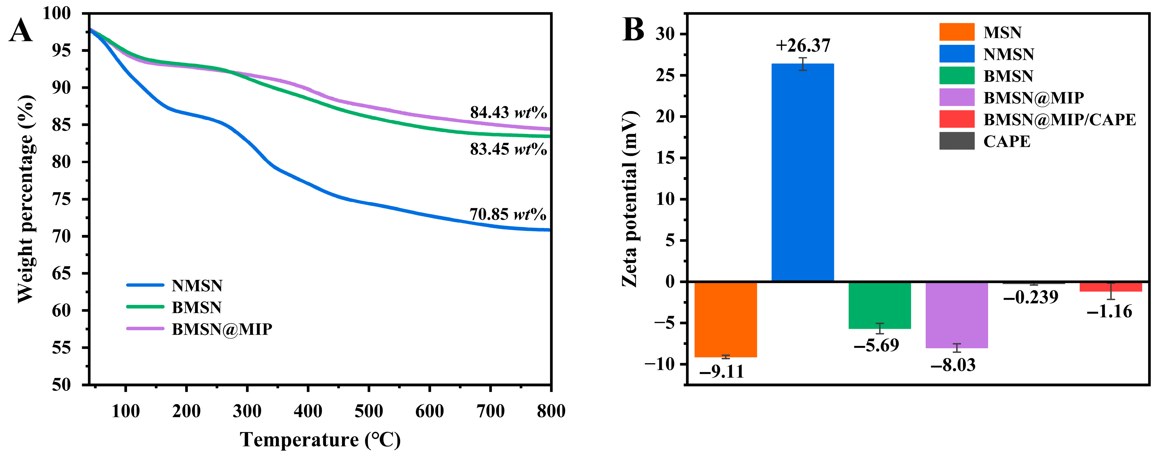
In addition, thermogravimetric analysis (TGA) was carried out to verify the thermal stability of NMSN, BMSN, and BMSN@MIP. As shown in Figure 2A, NMSN, BMSN, and BMSN@MIP exhibit good thermal stability with the remaining weight percentages of 70.85 wt%, 83.45 wt%, and 84.43 wt% at 800 °C, respectively. The weight loss of NMSN from 0 to 100 °C is mainly caused by the evaporation of ethanol and water adsorbed in the preparation process [36], and the decrease in weight from 300 to 600 °C of BMSN@MIP may be owing to the decomposition of the imprinting layer [37].
The zeta potentials of MSN, NMSN, BMSN, BMSN@MIP, BMSN@MIP/CAPE, and CAPE are demonstrated in Figure 2B. The zeta potential of MSN is −9.11 mV, which is ascribed to the negatively charged Si-OH groups [38]. The presence of the positively charged -NH2 group results in the zeta potential of NMSN as +26.37 mV, indicating the successful modification of -NH2 [39]. Meanwhile, BMSN exhibits a negative zeta potential of −5.69 mV, which confirms the existence of -B(OH)2 in BMSN and indicates the functionalization of the boric acid group [40]. The SEM image (Figure S2) of BMSN@MIP displays the regular spherical morphology with an average size of 69.51 nm.

3.2. Optimization of Imprinting Time in Synthesis of BMSN@MIP

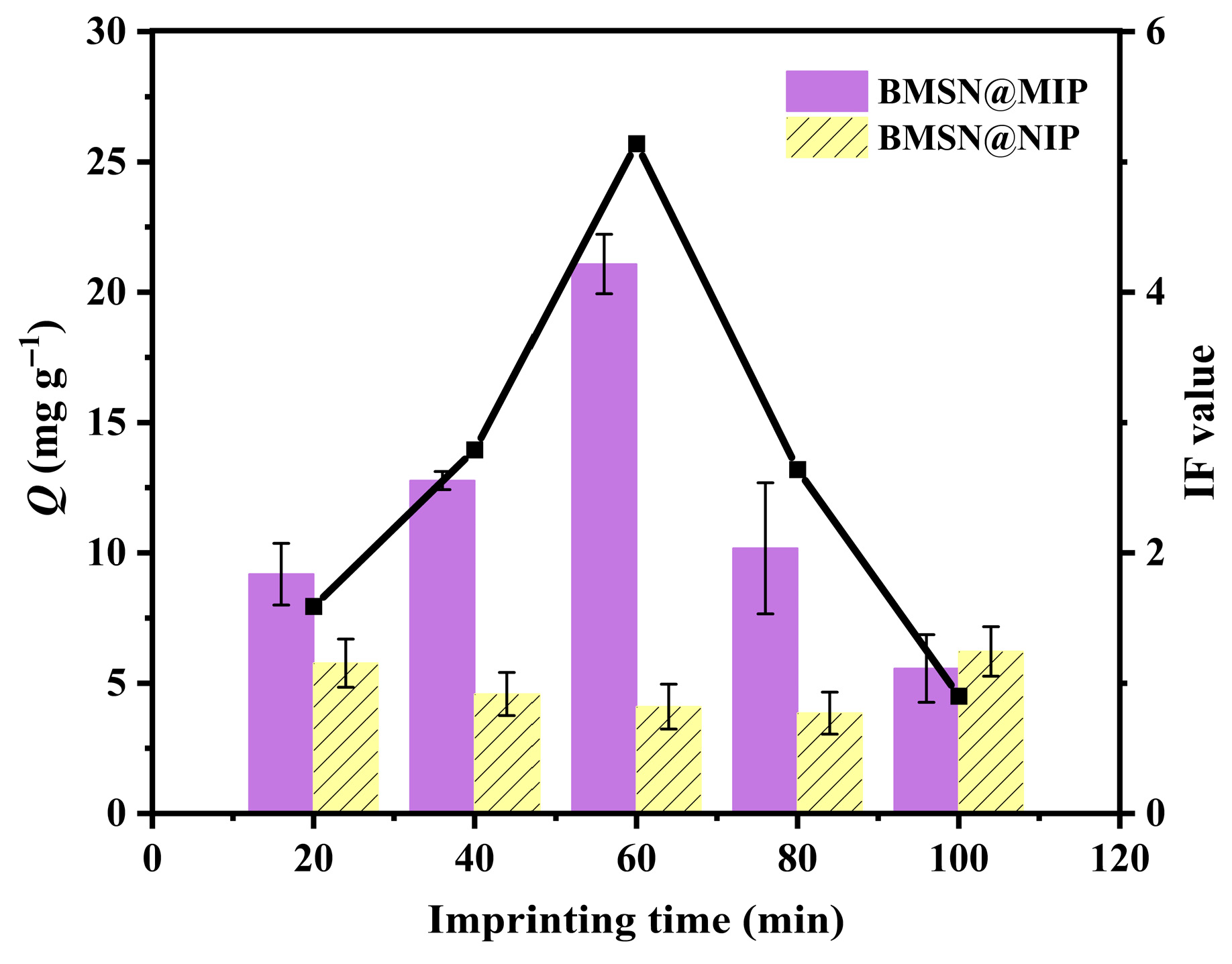
As previously reported, the imprinting time plays a significant role in controlling the thickness of the imprinted layer of MIPs [23], which affects the adsorption capacity of MIPs toward templates [41]. As shown in Figure 3 and Table 1, the highest IF value (5.14) of BMSN@MIP was obtained at the imprinting time of 60 min, which reveals the satisfactory affinity of BMSN@MIP toward CAPE [42]. Thus, the imprinting time of 60 min was utilized in the preparation process of BMSN@MIP.

3.3. Evaluation of Encapsulation Efficiency

Encapsulation efficiency serves as a critical parameter for evaluating the drug loading performance of drug carriers [43], and the drug concentration/incubating time are key factors in determining the encapsulation capacity [5,19]. As illustrated in Figure 4A and Table S1, the prepared BMSN@MIP/CAPE exhibits the highest EE value of 40.82% under the CAPE concentration of 0.03 mg mL−1, which was then identified as the optimal drug concentration. EE values of BMSN@MIP/CAPE at different incubating times are displayed in Figure 4B, which indicates the highest EE value of 40.98% under 20 h [44]. Thus, the optimal drug concentration and incubating time were selected to be 0.03 mg mL−1 and 20 h in the preparation of BMSN@MIP/CAPE, respectively.

3.4. In Vitro Drug Release Studies

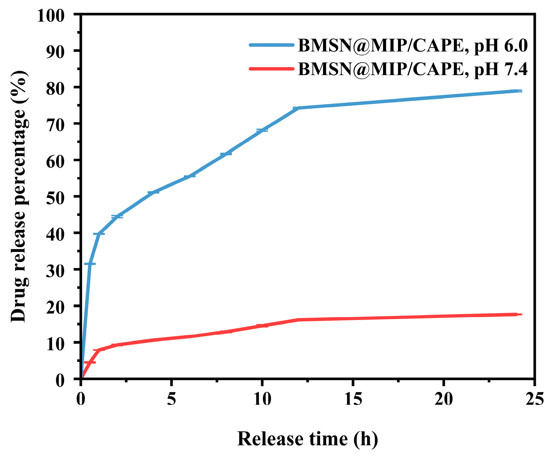
In vitro drug release behaviors were investigated in the simulated tumor microenvironment (pH 6.0) and normal bioliquids (pH 7.4). As illustrated in Figure 5, the cumulative drug release percentage of CAPE is 17.69% at pH 7.4 and 78.90% at pH 6.0. The relatively high CAPE release percentage under the weakly acidic environment compared with that of the normal environment is attributed to the hydrolysis of the borate ester bonds, which are formed between -B(OH)2 in BMSN@MIP and cis-diols in CAPE [19].
Furthermore, the Higuchi and K-P models were employed to explore the drug release kinetics models by fitting the experimental data of CAPE release behaviors (Table 2) [45]. Specifically, the Higuchi model was utilized to describe the drug diffusion and the K-P model was used to represent the drug release mechanisms as a semi-empirical function [10]. As can be found in Table 2, the K-P model is better fitted to describe the release behavior of CAPE than the Higuchi model on the basis of the R2 values, and the exponent n of the K-P model indicates that the release of CAPE is driven by Fickian diffusion [28,46]. The observed kinetics aligning with Fickian diffusion further supports the diffusion-limited release behavior under the tested conditions, confirming the rate-limiting step in the diffusion of CAPE from the carrier to the surrounding medium, while erosion or swelling of the polymer plays comparatively minor roles.

4. Conclusions

In this study, pH-responsive MIPs were constructed utilizing MSN as the substrate, 4-FPBA as the functional monomer, and CAPE as the drug template. An imprinting time of 60 min was chosen to prepare BMSN@MIP on the basis of the IF value of 5.14. In addition, during the process of preparing BMSN@MIP/CAPE, a drug concentration of 0.03 mg mL−1 and incubating time of 20 h were evaluated to obtain the optimal EE values. In addition, in vitro drug release studies exhibit cumulative drug release percentages of 78.90% in the simulated tumor microenvironment (pH 6.0) and 17.69% in the normal bioliquids (pH 7.4), respectively, implying controlled drug release. Accordingly, BMSN@MIP displays a key role as a pH-responsive drug carrier and offers the possibility for reducing toxic/side effects due to its pH-responsive drug release property, which could be achieved in further investigation.

Supplementary Materials

The following supporting information can be downloaded at: https://www.mdpi.com/article/10.3390/jcs9080421/s1. Figure S1. High-resolution XPS pattern of B 1 s of BMSN; Figure S2: SEM image of BMSN@MIP; Figure S3: Verification of boric acid functionalization of BMSN; Figure S4: Calibration curves of CAPE solution at (A) pH 6.0 and (B) pH 7.4; Table S1: EE values of BMSN@MIP/CAPE under different CAPE concentrations and incubation times. References [47,48] are cited in Supplementary Materials.

Author Contributions

Methodology, Z.G. and T.H.; formal analysis, Z.G., T.H., and Y.L.; investigation, Z.G., T.H., and Y.L.; data curation, G.X.; writing—original draft preparation, Z.G., T.H., and Y.L.; writing—review and editing, Q.J.; supervision, Q.J.; project administration, Q.J.; funding acquisition, Q.J. All authors have read and agreed to the published version of the manuscript.

Funding

This work is financially supported by the Open Project of State Key Laboratory of Supramolecular Structure and Materials, Jilin University, China (No. sklssm2024018).

Data Availability Statement

Data are contained within the article and Supplementary Materials.

Conflicts of Interest

The authors declare no conflicts of interest.

References

  1. Peer, D.; Karp, J.M.; Hong, S.; Farokhzad, O.C.; Margalit, R.; Langer, R. Nanocarriers as an emerging platform for cancer therapy. Nat. Nanotech. 2007, 2, 751–760. [Google Scholar] [CrossRef]
  2. Wong, H.L.; Bendayan, R.; Rauth, A.M.; Li, Y.Q.; Wu, X.Y. Chemotherapy with anticancer drugs encapsulated in solid lipid nanoparticles. Adv. Drug Deliver. Rev. 2007, 59, 491–504. [Google Scholar] [CrossRef]
  3. Durán, J.D.G.; Arias, J.L.; Gallardo, V.; Delgado, A.V. Magnetic colloids as drug vehicles. J. Pharm. Sci. 2008, 97, 2948–2983. [Google Scholar] [CrossRef]
  4. Arias, J.L. Novel strategies to improve the anticancer action of 5-fluorouracil by using drug delivery systems. Molecules 2008, 13, 2340. [Google Scholar] [CrossRef]
  5. Guo, Z.M.; Xu, W.H.; Xu, G.X.; Jia, Q. An updated overview of MOF@MIP in drug carrier and drug analysis: Construction, application and prospective. TrAC Trends Anal. Chem. 2023, 167, 117275. [Google Scholar] [CrossRef]
  6. Li, P.F.; Liu, Z. Glycan-specific molecularly imprinted polymers towards cancer diagnostics: Merits, applications, and future perspectives. Chem. Soc. Rev. 2024, 53, 1870–1891. [Google Scholar] [CrossRef]
  7. Takeuchi, T.; Kitayama, Y.; Sasao, R.; Yamada, T.; Toh, K.; Matsumoto, Y.; Kataoka, K. Molecularly imprinted nanogels acquire stealth in situ by cloaking themselves with native dysopsonic proteins. Angew. Chem. Int. Ed. 2017, 56, 7088–7092. [Google Scholar] [CrossRef]
  8. Xu, S.X.; He, H.; Liu, Z. Molecularly imprinted nanogels acquire stealth in situ by cloaking themselves with native dysopsonic proteins. Chemisity 2022, 40, 635–650. [Google Scholar] [CrossRef]
  9. Xing, R.R.; Wang, S.S.; Bie, Z.Y.; He, H.; Liu, Z. Preparation of molecularly imprinted polymers specific to glycoproteins, glycans and monosaccharides via boronate affinity controllable-oriented surface imprinting. Nat. Protoc. 2017, 12, 964–987. [Google Scholar] [CrossRef] [PubMed]
  10. Han, S.; Song, Y.Z.; Liu, S.W.; Zhao, L.; Sun, R.N. Dual responsive molecularly imprinted polymers based on UiO-66-DOX for selective targeting tumor cells and controlled drug release. Eur. Polym. J. 2022, 171, 111219. [Google Scholar] [CrossRef]
  11. Slowing, I.I.; Vivero-Escoto, J.L.; Wu, C.W.; Lin, V.S.Y. Mesoporous silica nanoparticles as controlled release drug delivery and gene transfection carriers. Adv. Drug Deliver. Rev. 2008, 60, 1278–1288. [Google Scholar] [CrossRef]
  12. Gkiliopoulos, D.; Bikiaris, D.; Efstathiadis, D.; Triantafyllidis, K. Glassy and rubbery epoxy composites with mesoporous silica. J. Compos. Sci. 2023, 7, 243. [Google Scholar] [CrossRef]
  13. Xu, S.X.; Wang, L.S.; Liu, Z. Molecularly imprinted polymer nanoparticles: An emerging versatile platform for cancer therapy. Angew. Chem. Int. Ed. 2021, 60, 3858–3869. [Google Scholar] [CrossRef] [PubMed]
  14. Li, D.J.; Chen, Y.; Liu, Z. Boronate affinity materials for separation and molecular recognition: Structure, properties and applications. Chem. Soc. Rev. 2015, 44, 8097–8123. [Google Scholar] [CrossRef]
  15. Liu, Z.; He, H. Synthesis and applications of boronate affinity materials: From class selectivity to biomimetic specificity. Accounts Chem. Res. 2017, 50, 2185–2193. [Google Scholar] [CrossRef]
  16. Guo, W.J.; Ji, J.Y.; Shi, Y.Y.; Li, N.; Yan, Z.C.; Du, Q.Z. Recent advances in composite materials integrating molecularly imprinted polymers for targeted drug delivery systems. Eur. Polym. J. 2025, 228, 113825. [Google Scholar] [CrossRef]
  17. Zhang, L.P.; Mo, C.E.; Huang, Y.P.; Liu, Z.S. Preparation of liquid crystalline molecularly imprinted polymer coated metal organic framework for capecitabine delivery. Part. Part. Syst. Charact. 2019, 36, 1800355. [Google Scholar] [CrossRef]
  18. Mo, C.E.; Chai, M.H.; Zhang, L.P.; Ran, R.X.; Huang, Y.P.; Liu, Z.S. Floating molecularly imprinted polymers based on liquid crystalline and polyhedral oligomeric silsesquioxanes for capecitabine sustained release. Int. J. Pharmaceut. 2019, 557, 293–303. [Google Scholar] [CrossRef]
  19. Yuan, F.F.; Zhang, R.R.; Ma, X.L.; Yang, J.; Huang, Y.P.; Liu, Z.S. Cooperation effect of 4-vinylbenzeneboronic acid/methacrylic acid on affinity of capecitabine imprinted polymer for drug carrier. Eur. J. Pharm. Sci. 2020, 154, 105476. [Google Scholar] [CrossRef]
  20. Guo, Z.M.; Zheng, H.J.; Ma, J.T.; Xu, G.X.; Jia, Q. Design of pH-responsive molecularly imprinted polymer as a carrier for controlled and sustainable capecitabine release. Anal. Chim. Acta. 2024, 1317, 342881. [Google Scholar] [CrossRef] [PubMed]
  21. Du, J.; He, M.F.; Wang, X.M.; Fan, H.; Wei, Y.M. Facile preparation of boronic acid-functionalized magnetic nanoparticles with a high capacity and their use in the enrichment of cis-diol-containing compounds from plasma. Biomed. Chromatogr. 2015, 29, 312–320. [Google Scholar] [CrossRef] [PubMed]
  22. Ren, X.H.; Wang, H.Y.; Li, S.; He, X.W.; Li, W.Y.; Zhang, Y.K. Preparation of glycan-oriented imprinted polymer coating Gd-doped silicon nanoparticles for targeting cancer Tn antigens and dual-modal cell imaging via boronate-affinity surface imprinting. Talanta 2020, 223, 121706. [Google Scholar] [CrossRef]
  23. Luo, Z.D.; Tian, M.F.; Ahmad, N.; Xie, Y.X.; Xu, C.G.; Liu, J.; Zhao, C.J.; Li, C.Y. A surface multiple imprinting layers membrane with well-oriented recognition sites for selective separation of chlorogenic acid from Ficus carica L. Food Chem. 2024, 433, 137347. [Google Scholar] [CrossRef]
  24. Lu, H.F.; Xu, S.X.; Guo, Z.C.; Zhao, M.H.; Liu, Z. Redox-responsive molecularly imprinted nanoparticles for targeted intracellular delivery of protein toward cancer therapy. ACS Nano 2021, 15, 18214–18225. [Google Scholar] [CrossRef]
  25. Karimi, S.; Rasuli, H.; Mohammadi, R. Facile preparation of pH-sensitive biocompatible alginate beads havening layered double hydroxide supported metal-organic framework for controlled release from doxorubicin to breast cancer cells. Int. J. Biol. Macromol. 2023, 234, 123538. [Google Scholar] [CrossRef]
  26. Costa, P.; Lobo, J.M.S. Modeling and comparison of dissolution profiles. Eur. J. Pharm. Sci. 2001, 13, 123–133. [Google Scholar] [CrossRef] [PubMed]
  27. Rehman, U.; Sarfraz, R.M.; Mahmood, A.; Akbar, S.; Altyar, A.E.; Khinkar, R.M.; Gad, H.A. pH-Responsive hydrogels for the delivery of capecitabine: Development, optimization and pharmacokinetic studies. Gels 2022, 8, 775. [Google Scholar] [CrossRef] [PubMed]
  28. Dash, S.; Murthy, P.N.; Nath, L.; Chowdhury, P. Kinetic modeling on drug release from controlled drug delivery systems. Acta Pol. Pharm. 2010, 67, 217–223. [Google Scholar]
  29. Erdem, B.; Azko, Ç. B-SBA-15-SO3H: A versatile mesoporous catalyst. J. Porous Mater. 2017, 24, 1237–1246. [Google Scholar] [CrossRef]
  30. D’ Souza, L.; Devi, P.; Divya Shridhar, M.P.; Naik, C.G. Use of fourier transform infrared (FTIR) spectroscopy to study cadmium-induced changes in padina tetrastromatica (hauck). Anal. Chem. Insights 2008, 3, 135–143. [Google Scholar] [CrossRef]
  31. Cipurković, A.; Horozić, E.; Ljubijankić, N.; Odobašić, A.; Galijašević, S.; Saletović, M. Synthesis and spectral characterization of Fe (II) and Mn (II) complexes with oral fluorouracil prodrug capecitabine. J. Chem. 2017, 10, 1381–1390. [Google Scholar] [CrossRef]
  32. Rezaei, F.; Ashraf, N.; Zohuri, G.H. A smart electrochemical sensor based upon hydrophilic core–shell molecularly imprinted polymer for determination of L-tryptophan. Microchem. J. 2023, 185, 108260. [Google Scholar] [CrossRef]
  33. Lu, Q.Y.; Gao, F.; Komarneni, S.; Mallouk, T.E. Ordered SBA-15 nanorod arrays inside a porous alumina membrane mallouk. J. Am. Chem. Soc. 2004, 126, 8650–8651. [Google Scholar] [CrossRef]
  34. Narayan, R.; Gadag, S.; Cheruku, S.P.; Raichur, A.M.; Day, C.M.; Garg, S.; Manandhar, S.; Pai, K.S.R.; Suresh, A.; Mehta, C.H.; et al. Chitosan-glucuronic acid conjugate coated mesoporous silica nanoparticles: A smart pH-responsive and receptor-targeted system for colorectal cancer therapy. Carbohyd. Polym. 2021, 261, 117893. [Google Scholar] [CrossRef]
  35. Popova, M.; Trendafilova, I.; Zgureva, D.; Kalvachev, Y.; Boycheva, S.; Tušar, N.N.; Szegedi, A. Polymer-coated mesoporous silica nanoparticles for controlled release of the prodrug sulfasalazine. J. Drug Deliver. Sci. Tec. 2018, 44, 415–420. [Google Scholar] [CrossRef]
  36. Delpiano, G.R.; Casula, M.F.; Piludu, M.; Corpino, R.; Ricci, P.C.; María, V.R.; Sanjust, E.; Monduzzi, M.; Salis, A. Assembly of multicomponent nano-bioconjugates composed of mesoporous silica nanoparticles, proteins, and gold nanoparticles. ACS Omega 2019, 4, 11044–11052. [Google Scholar] [CrossRef]
  37. Tian, M.J.; Yu, K.; Li, L.Y.; Wang, Y.F.; Guo, L.P.; Zhang, Z.L.; Lu, Y.X.; Li, L. Fabrication of dual-template molecularly imprinted mesoporous silica for simultaneous rapid and efficient detection of bisphenol A and diethylstilbestrol in environmental water samples. Anal. Methods 2019, 11, 4761–4768. [Google Scholar] [CrossRef]
  38. Feng, Y.S.; Qin, Y.T.; Zhuang, J.; Ma, Y.J.; He, X.W.; Li, W.Y.; Zhang, Y.K. Mitochondria-targeted thymidylate synthase inhibitor based on fluorescent molecularly imprinted polymers for tumor antimetabolic therapy. ACS Appl. Mater. Interfaces 2023, 15, 31139–31149. [Google Scholar] [CrossRef] [PubMed]
  39. Sapino, S.; Ugazio, E.; Gastaldi, L.; Miletto, I.; Berlier, G.; Zonari, D.; Oliaro-Bosso, S. Mesoporous silica as topical nanocarriers for quercetin: Characterization and in vitro studies. Eur. J. Pharm. Biopharm. 2015, 89, 116–125. [Google Scholar] [CrossRef]
  40. Popova, M.D.; Szegedi, Á.; Kolev, I.N.; Mihály, J.; Tzankov, B.S.; Momekov, G.T.; Lambov, N.G.; Yoncheva, K.P. Carboxylic modified spherical mesoporous silicas as drug delivery carriers. Int. J. Pharm. 2012, 436, 778–785. [Google Scholar] [CrossRef] [PubMed]
  41. Zhu, W.T.; Peng, H.L.; Luo, M.; Yu, N.X.; Xiong, H.; Wang, R.H.; Li, Y.B. Zipper-like magnetic molecularly imprinted microspheres for on/off-switchable recognition and extraction of 17β-estradiol from food samples. Food Chem. 2018, 261, 87–95. [Google Scholar] [CrossRef] [PubMed]
  42. Chen, L.X.; Wang, X.Y.; Lu, W.H.; Wu, X.Q.; Li, J.H. Molecular imprinting: Perspectives and applications. Chem. Soc. Rev. 2016, 45, 2137–2211. [Google Scholar] [CrossRef]
  43. Dwi, H.; Fuad, M.K.A.; Khairul, A.; Parsaoran, S.; Meida, M.S. In vitro evaluation of curcumin encapsulation in gum arabic dispersions under different environments. Molecules 2022, 27, 3855. [Google Scholar] [CrossRef]
  44. Ma, Y.J.; Zhao, H.Q.; Ma, Q.Y.; Cheng, D.W.; Zhang, Y.F.; Wang, W.X.; Wang, J.; Sun, J.F. Development of chitosan/potato peel polyphenols nanoparticles driven extended-release antioxidant films based on potato starch. Food Packag. Shelf Life 2022, 31, 100793. [Google Scholar] [CrossRef]
  45. Roya, S.; Somayeh, A.; Bahareh, H. pH-sensitive molecularly imprinted polymer based on graphene oxide for stimuli actuated controlled release of curcumin. J. Alloys Compd. 2020, 857, 157603. [Google Scholar] [CrossRef]
  46. Gao, Y.K.; He, Y.; Zhang, H.T.; Zhang, Y.X.; Gao, T.B.; Wang, J.H.; Wang, S.L. Zwitterion-functionalized mesoporous silica nanoparticles for enhancing oral delivery of protein drugs by overcoming multiple gastrointestinal barriers. J. Colloid. Interface Sci. 2021, 582, 364–375. [Google Scholar] [CrossRef]
  47. Das, D.; Yang, Y.; O’Brien, J.; Brezan, D.; Nimesh, S.; Bernatchez, S.; Hill, M.; Sayari, A.; Vincent, R.; Kumarathasan, P. Synthesis and physicochemical characterization of mesoporous SiO2 nanoparticles. J. Nanomater. 2014, 2014, 1–12. [Google Scholar] [CrossRef]
  48. Lu, C.C.; Li, H.Y.; Wang, H.Y.; Liu, Z. Probing the interactions between boronic acids and cis-diol-containing biomolecules by affinity capillary electrophoresis. Anal. Chem. 2013, 85, 2361–2369. [Google Scholar] [CrossRef]
Scheme 1. Schematic illustration of synthetic process of BMSN@MIP.
Jcs 09 00421 sch001
Figure 1. (A) FT-IR spectra of MSN, NMSN, BMSN, BMSN@MIP/CAPE, and CAPE; (B) UV-vis spectra of NMSN, BMSN, BMSN@MIP, and CAPE; (C) XRD patterns of MSN, NMSN, BMSN@MIP, BMSN@MIP/CAPE, and CAPE; (D) XPS patterns of NMSN and BMSN.
Jcs 09 00421 g001
Figure 2. (A) TGA profiles of NMSN, BMSN, BMSN@MIP, and BMSN@MIP/CAPE; (B) zeta potentials of MSN, NMSN, BMSN, BMSN@MIP, BMSN@MIP/CAPE, and CAPE.
Jcs 09 00421 g002
Figure 3. Optimization of imprinting time in synthesis of BMSN@MIP.
Jcs 09 00421 g003
Figure 4. EE values of BMSN@MIP under different (A) CAPE concentrations and (B) incubating times.
Jcs 09 00421 g004
Figure 5. Drug release profiles of CAPE in PBS (0.01 M, pH 6.0, and 7.4).
Jcs 09 00421 g005
Table 1. Optimization of the imprinting time in the synthesis process of BMSN@MIP.
Imprinting Time (min)IF
201.59
402.79
605.14
802.64
1000.90
Table 2. Kinetic parameters of CAPE release.
Drug Release Kinetic ModelsParametersBMSN@MIP/CAPE
at pH 6.0at pH 7.4
HiguchikH (h−1)20.694.15
R20.690.97
Korsmeyer-PeppaskK-P (hn)37.945.26
n0.240.41
R20.990.99
Disclaimer/Publisher’s Note: The statements, opinions and data contained in all publications are solely those of the individual author(s) and contributor(s) and not of MDPI and/or the editor(s). MDPI and/or the editor(s) disclaim responsibility for any injury to people or property resulting from any ideas, methods, instructions or products referred to in the content.