Article

Finite Element Analysis of Strain-Mediated Direct Magnetoelectric Coupling in Multiferroic Nanocomposites for Material Jetting Fabrication of Tunable Devices

Multifunctional Electronic Materials & Devices Research Lab (MeMDRL), Department of Electrical and Computer Engineering, The University of Texas at San Antonio, San Antonio, TX 78249, USA
*
Author to whom correspondence should be addressed.
J. Compos. Sci. 2025, 9(5), 228; https://doi.org/10.3390/jcs9050228
Submission received: 1 March 2025 / Revised: 21 April 2025 / Accepted: 26 April 2025 / Published: 1 May 2025
(This article belongs to the Section Composites Applications)

Abstract

:
Magnetoelectric composites enable strain-mediated coupling between magnetic and electric fields, supporting applications in sensors, actuators, and tunable devices. This study presents a finite element modeling framework for simulating the direct magnetoelectric effect in core–shell and layered nanocomposites fabricated by material jetting (inkjet printing). The model incorporates nonlinear magnetostrictive behavior of cobalt ferrite nanoparticles and size-dependent piezoelectric properties of barium titanate, allowing efficient simulation of complex interfacial strain transfer. Results show a strong dependence of coupling on field orientation, particle arrangement, and interfacial geometry. Simulations of printed droplet geometries, including coffee ring droplet morphologies, reveal enhanced performance through increased surface area and directional alignment. These findings highlight the potential of material jetting for customizable, high-performance magnetoelectric devices and provide a foundation for simulation-guided design.

1. Introduction

The strain-mediated direct magnetoelectric (ME) effect arises when a composite material electrically polarizes under applied magnetic fields through mechanical coupling of magnetostrictive and piezoelectric phases [1]. This cross-coupling of electronic and magnetic properties is sought for use in radio frequency (RF) and microwave components [2], power electronics [3], sensors [4], and memory devices [5]. The strain-mediated ME coupling coefficient, α M E , quantifies the conversion of magnetic to electric energy in a ME material:
α M E = δ E δ H = 1 t δ V δ H
where δ E is the change in electric field, δ H is the change in applied magnetic field, δ V is the generated output voltage, and t is the corresponding sample thickness of the ME structure. Experimental evaluation of α M E is usually performed at room temperature by applying a small ac magnetic field (typically~1 Oe) and a dc bias magnetic field while measuring the generated voltage across the sample [6]. A composite’s highest α M E value is found at a specific dc bias field due to the nonlinear responses of magnetostrictive materials [7]. Traditionally, α M E is represented with units of m V c m   O e and typical values range from tens to hundreds of m V c m   O e for composites at non-resonant conditions, while values for operation near electromechanical resonance can achieve more than 10× the non-resonant efficiency [8]. Important parameters for ME composite behavior are magnetostrictive, interfacial, and piezoelectric properties and the connectivity of the constituent phases [9].
ME materials research followed the discovery of the ME effect in Cr2O3 in the 1960s [10] with efforts to overcome the weak coupling in single-phase materials by engineering composites [11]. By the late 1970s, Newnham and colleagues introduced the composite connectivity nomenclature (e.g., 0-3, 2-2, 1-3) to describe dimensional continuity of active and passive phases in piezoelectric composites [12]. This system has since been widely adopted across composite materials science, including ME composites [13]. In the 1990s–2000s, advances in micromechanics models [14], thin-film deposition techniques [15], phase-field [16], and finite element analysis (FEA) [17] improved prediction and optimization of ME responses. In the 2010s, density functional theory (DFT) provided atomic-level insights into interfacial coupling [18], and more recently, machine learning has been applied to the discovery and design of ME composites [19]. Although FEA modeling is limited in capturing atomistic phenomena and can be computationally intensive relative to phase-field or first-principles methods, it remains a quantitatively robust tool for resolving mesoscale electromechanical interactions and enabling predictive design of ME architectures tailored for integration into functional electronic systems [20,21,22,23].
The common constituent materials studied in ME composites are C o F e 2 O 4 (CFO), Terfenol-D, N i F e 2 O 4 , F e 3 O 4 , FeCoSiB alloy for the magnetostrictive phase [24] and B a T i O 3 (BTO), P b ( Z r , T i ) O 3 (PZT), Polyvinylidene fluoride (PVDF) for the piezoelectric phase [25]. While the magnetic phase is usually a metal alloy or oxides, the piezoelectric phase has the option of polymers (e.g., PVDF) or inorganic materials (e.g., BTO, PZT).
Fabrication of ME composites has been demonstrated by many techniques at several size scales, including solid-state reaction [26], sol-gel deposition [27], pulsed laser deposition [28], magnetron sputtering [29], electrospinning [30], and spark plasma sintering of bulk composites [31]. Additive manufacturing (AM) methods have gained attention in recent decades for the potential advantages in rapid prototyping, design freedom, material/energy efficiency, on-demand customization/fabrication, and new material possibilities [32]. Material jetting (MJ) of electronic materials is an emerging AM technique that deposits fluids containing functional material, and includes inkjet printing (IJP), aerosol spray, and direct ink writing. One advantage of MJ over other AM methods is its ability to deposit multiple materials (metals, organics, and inorganics) using the same equipment, fitting the need of electronic device fabrication for patterned insulators, conductors, and functional layers. Core–shell nanocomposites of CFO cores and barium titanate BTO shells have been studied for their interesting dielectric and ME properties towards diverse applications including biomedicine and energy harvesting [33,34]. This work focuses on MJ of ME composite materials consisting of CFO and BTO by seeking a novel approach to modeling strain-mediated ME composites, with particular focus on 0-3 connected core–shell nanocomposites and layered 2-2 structures fabricated by MJ AM.
FEA simulation of strain transfer, interfacial coupling, and nanoscale effects in ME composites provides insights into material behavior and guides design optimization [35,36,37,38]. One simplified approach for strain-mediated FEA ME composites is to homogenize the material by defining a representative elementary volume (REV) [39]. This approach agreed with an analytic model of a macroscopic cylindrical rod, and enabled model geometries without analytical solutions. However, this approach does not capture the local magnetostrictive and piezoelectric effects. Important theoretical studies on polymer-based ME composites provide insight into FEA modeling and materials design optimization [40,41]. Additional work has demonstrated that PVDF-based composites offer enhanced flexibility and biocompatibility, while PZT-based laminates provide superior ME coefficients, guiding the design of efficient ME devices for sensors and energy harvesters [42]. Other FEA approaches that model distinct phases in 2-2 connected composites include analysis of layered composites with asymmetric geometries by incorporating nonlinear materials models [43]. This approach showed good agreement with experimental data in literature and was used to optimize the performance of the ME composite based on modulus ratio, bias field, frequency of applied ac field, and volume fraction of the ferromagnetic material. These approaches leverage the simple geometry of 2-2 connected composites’ interfaces. Capturing the strain-mediated ME effect in FEA models in 0-3 connected composites is challenging due to the more complex interface geometry and is addressed in this work by solving magnetostrictive behavior in individual particles and then applying this mechanical pressure to a separate piezoelectric model.
Our motivation to develop FEA of ME composites includes investigation and development of devices based on these materials in the context of MJ AM. A critical aspect of this effort is understanding the nanoscale interactions of functional nanocomposites or nanoparticles, where inherent magneto-mechanical-electrical coupling effects manifest at the ~100 nm scale. FEA modeling aims to capture these localized interactions and size effects, enabling the precise study of field distributions, stress-strain behaviors, and ME coupling at the nanoscale, which are otherwise difficult to measure experimentally. Additional challenges, addressed by this work, include scaling, i.e., a composite comprised of nanoparticles with the overall dimensions approaching the micro to millimeter scale will inevitably require large numbers of meshing elements, making computations increasingly complex and resource intensive.

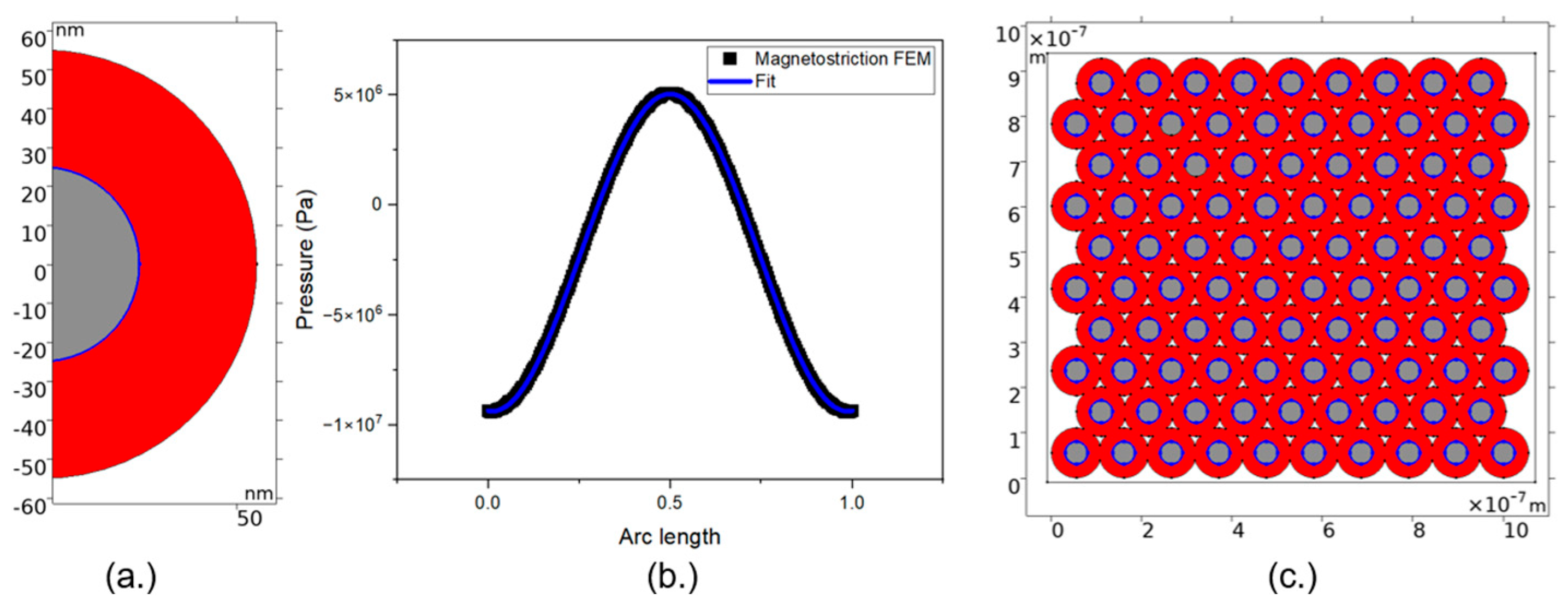
2. Magnetoelectric Numerical Model

Calculations of strain-mediated ME coupling coefficients require determining three factors: (1) deformations induced in the magnetostrictive phase by the magnetic field, (2) how this deformation transfers to the piezoelectric phase, and (3) the resulting electric field generated in the piezoelectric materials. This work employs a two-stage approach for computational efficiency. The first stage models the magnetostrictive stress and displacement in the magnetic phase (e.g., a single core–shell particle) to yield forces and displacements at the magnetostrictive–piezoelectric interface. These outputs are then used in the second stage to model the piezoelectric phase response (e.g., an assembly of core–shell particles in various configurations). This method enables detailed analysis of magnetostrictive–piezoelectric interfaces and accommodates complex, variable connectivity patterns in larger geometries.
Figure 1 shows an example of model domains and boundary conditions for the magnetostrictive model alongside an image of the model mesh. A magnetic field is simulated by modeling a coil with uniform current distribution.
Using triangular (2D) and tetrahedral (3D) mesh at the magnetic–piezoelectric interface in ME composites is an established approach for modeling strain-mediated coupling in FEA simulations [44]. Domain walls typically form at scales of 10–50 nm, and recent studies have demonstrated that resolutions between 1–10 nm are adequate for capturing the key physics of ME coupling [45]. This resolution effectively models nanoscale heterogeneity influencing ME behavior while maintaining a balance with computational efficiency.
A non-linear magnetostrictive material is simulated by Equation (2).
ε m s = 3 2 λ s M s 2 d e v ( M M )
where ε m s is the magnetostrictive strain, λ s is the magnetostrictive coefficient, M s is the saturation magnetization, and M is magnetization. The term d e v ( M M ) in Equation (2) is the deviatoric part of the outer product (or tensor product) of the magnetization vector M with itself. The non-linear magnetization is modeled in terms of the so-called effective field, H e f f .
M = M s L H e f f H e f f | H e f f |
where L is the Langevin function, represented as follows:
L = coth ( 3 χ m H e f f M s ) M s 3 χ m H e f f
where χ m is magnetic susceptibility. We define the effective field as follows:
H e f f = H + 3 λ S μ 0 M s 2 S e d M
where H is the magnetic field, μ 0 is the magnetic permeability, and S e d is the deviatoric stress tensor, defined as follows:
S e d = d e v ( C   ε )
where C is the stiffness matrix and ε is the elastic strain [46].
Our FEA employs a structured mesh with refined elements near the CFO-BTO interface to capture strain transfer effects accurately. Mesh independence tests were conducted to ensure computational accuracy while maintaining efficiency. Boundary conditions were selected to replicate real-world constraints: the magnetostrictive phase was subjected to a uniform magnetic field applied via a coil model, while the piezoelectric phase was modeled with a free boundary condition to reflect the flexible nature of the inkjet-printed structure. The mesh is constructed to maximize the elements along this interface. This allows for sub-nanometer-sized meshing elements while keeping the model computation time to a few seconds, enabling multidimensional parametric sweeps. Figure 2 illustrates the boundary conditions and how simulated magnetostrictive stress data are analyzed with a non-linear fit (Figure 2b) and applied to magnetostrictive–piezoelectric interface boundaries in the piezoelectric model (Figure 2c).
This approach was extended to inkjet-printed (IJP) geometries, where picoliter-scale droplets are precisely deposited onto a substrate to form structured composite films. The typical footprint of an IJP droplet on a substrate is around 10–50 μm, and the size and surface of the dried IJP NP film is governed by the ink’s particle loading percent, NP morphology, solvent system, ink/substate interaction, surface roughness, and evaporation conditions [47].
The physical and mechanical properties (density, stiffness tensor) for CFO used in our FEA simulations are based on experimentally measured values obtained from [48]. The saturation magnetization (Ms), magnetostrictive coefficient (λ), and magnetic susceptibility were obtained by the non-linear magnetostrictive material model fits to experimental data from [49]. Regarding size dependance of material properties, several considerations are made. At small sizes (<20 nm), CFO NPs exhibit reduced saturation magnetization due to surface spin disorder and transition to superparamagnetic behavior with negligible coercivity [50]. For BTO NPs, size reduction impacts ferroelectricity and dielectric properties non-linearly, with a critical size range around 0.8–1.1 μm where dielectric permittivity peaks, while ferroelectricity generally decreases, and crystalline structure remains influential even at nanoscale dimensions [51]. Table 1 summarizes the range in values of piezoelectric coupling coefficients and dielectric permittivity found in literature by material processing and form. The stiffness of BTO ceramics remains relatively consistent across different grain sizes [52]. Three materials from Table 1 were used to define the material properties of the piezoelectric material phase of the FEA models—conventional ceramic, single crystal, and thin film—thereby incorporating size effects for both the magnetostrictive and piezoelectric phases.
FEA simulations typically assume perfect strain transfer across idealized interfaces, largely due to the complexity of incorporating interfacial defects and imperfections that can degrade coupling efficiency [53,54]. Techniques like phase-field models and first-principles calculations offer more precise representations of interfacial physics [55,56]
Simulation experiments were organized to first develop three non-linear magnetostrictive models that accurately capture the behavior of CFO NPs. After satisfactory comparison of these material models to experimental data, models of ME composites were developed to investigate piezoelectric response.
Table 1. Comparison of piezoelectric and dielectric properties of various forms and processing methods of BTO.
BaTiO3 FormPiezoelectric Coupling
Coefficient (pC/N)
Dielectric
Constant
K
Reference
d33d31d15
Single crystal85.6−34.5392130Zgonik et al. [57]
Conventional ceramic190−34.52701700Jaffe et al. [58]
Microwave-sintered ceramic260--3300Shen et al. [59]
Spark plasma-sintered ceramic, 100 nm domain416---Shen et al. [59]
Epitaxial film (10 nm) by PLD<2---Kelley et al. [60]
Spin coat film (300 nm) sintered at 900 °C---~600Bajac et al. [61]
Spray pyrolysis thin film---20–25Golego et al. [62]

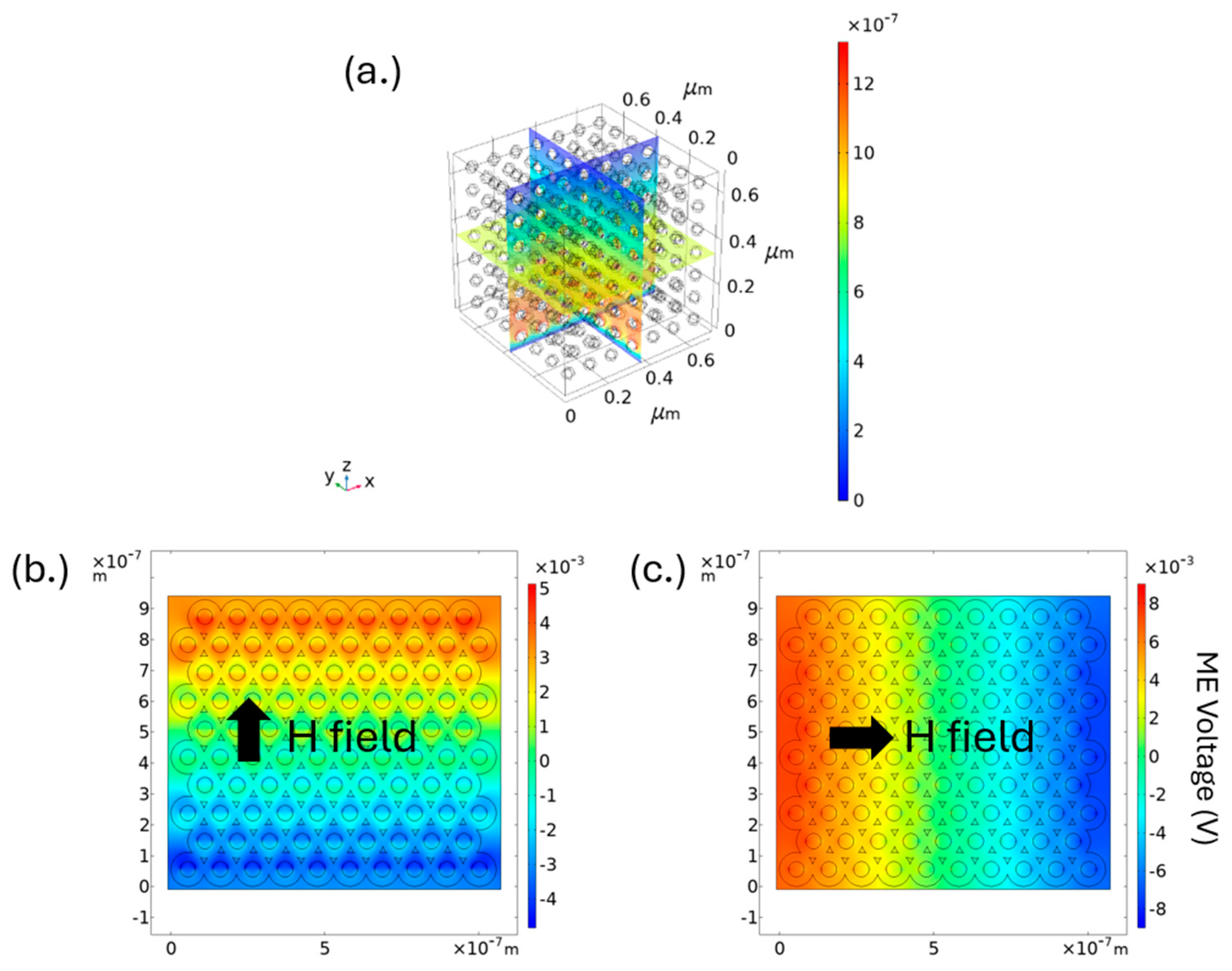
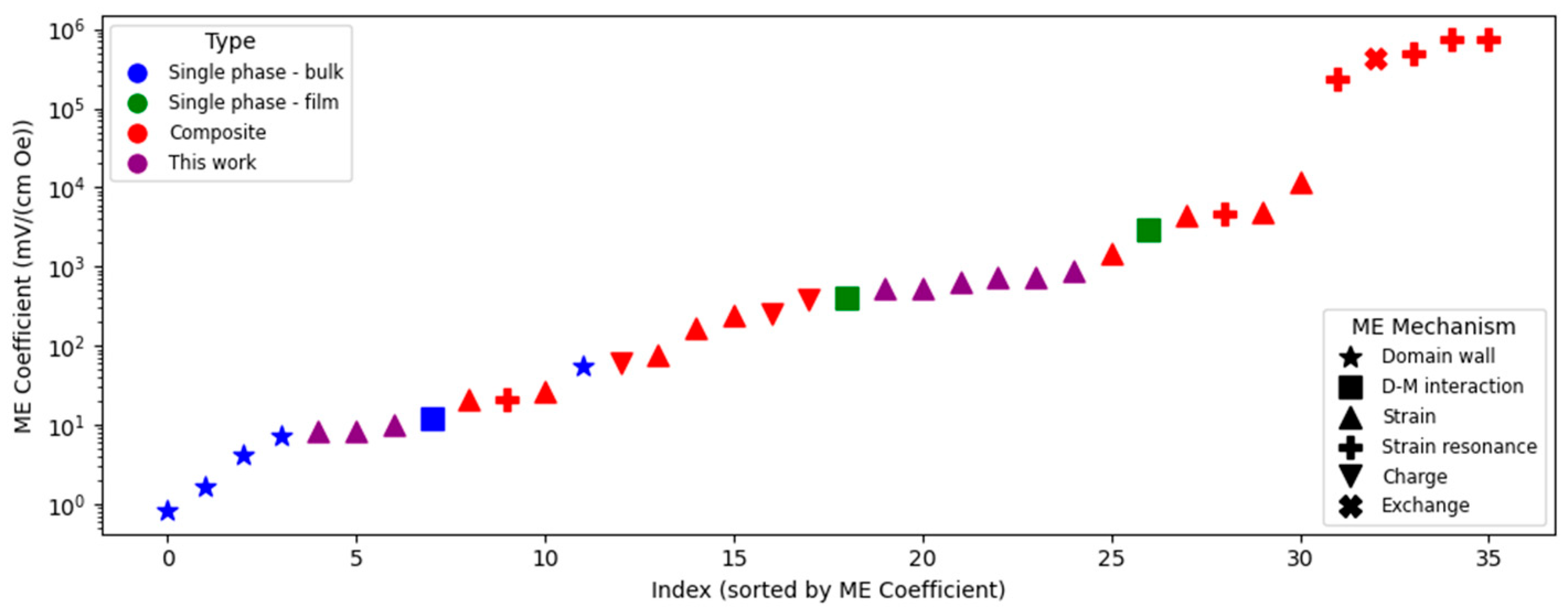
3. Results and Discussion

Figure 3 shows magnetostriction results compared to experimental data, demonstrating how the model effectively captures the nonlinear response. Magnetostrictive material models were developed based on the response of three different nanoparticle (NP) sizes relevant to inkjet printing (IJP) fabrication. NP size influences magnetic properties, with superparamagnetic behavior observed for NPs below approximately 8 nm. NP size is a critical consideration for IJP, as NPs must be sufficiently small to reliably pass through microfluidic channels. Figure 3b plots the resulting stresses developed along the core–shell interface for the three material models shown in Figure 3a. Both shear and normal stresses are symmetrical for the spherical NP, with maximal normal stresses at points parallel to the applied field, and maximal shear stresses at approximately 45° to the applied field. Figure 3c and Figure 3d illustrate the stress and strain, respectively, of the spherical core–shell model. The mechanical material properties of the BTO shell are a critical parameter in these results.
The magnetostrictive pressure (Pa) at the interface of magnetic:piezoelectric phases is obtained as illustrated in Figure 2. A fit of this data is input as boundary conditions in a piezoelectric model to investigate the ME response of different nanocomposites. This allows for larger and more complex geometries to be explored. Figure 4 shows 2D plots of the generated ME voltage of a 0-3 connected core–shell composite comparing in-plane and out-of-plane magnetic fields. The in-plane ME voltage is about 40% higher than the out-of-plane model due to substrate clamping effects and the piezoelectric tensor of the BTO phase.
FEA simulations conducted in this study provide insights into the behavior of ME 0-3 composite materials under different magnetic and electric field orientations. The goal of this study is to set up a multiphysics model that sufficiently incorporates the constraints of nanomaterial ink synthesis/formulation and MJ AM in a computationally efficient way to enable multifactor parameter sweeps. Once confirmed, such a model will then be useful for device design optimization. To show a baseline model for validation, symmetry is exploited by using 2D models wherever possible. Figure 4 illustrates a comparison of 2D and 3D periodic geometries, where adding an unnecessary third dimension increases degrees of freedom and computation time.
The ME coupling coefficient, α M E , is sensitive to the relative orientation of the applied magnetic fields, illustrated by the ME voltage shown in Figure 4. When the magnetic field is aligned parallel to the composite’s magnetostrictive phase, the induced strain is maximized, resulting in a significant deformation that transfers efficiently to the piezoelectric phase. This alignment leads to the highest observed values of α M E . Conversely, when the fields are misaligned, the strain-induced polarization in the piezoelectric phase is reduced, resulting in a lower α M E .
This orientation dependence is consistent with theoretical predictions and experimental observations in the literature, supporting the accuracy of the FEA model [63,64]. The strong nonlinear behavior observed in the simulations is primarily attributed to the magnetostrictive properties of the cobalt ferrite phase, which exhibits a non-linear response to the applied magnetic field. This non-linearity is particularly pronounced at higher magnetic field strengths, where the material begins to saturate, leading to a diminishing return in strain generation. Two-dimensional FEA studies comparing the generated piezoelectric voltage of different particle arrangements morphologies and the level of sintering (modeled as overlapping neighboring particles) suggest that the critical parameter for composite geometry is the interfacial surface area between the core and shell. This finding supports the concept that leveraging an increased surface-to-volume ratio with decreasing core–shell nanoparticle size will lead to enhanced coupling.
The capabilities of IJP as an AM technique were also investigated through simulation experiments. IJP offers precise control over material deposition, enabling the fabrication of structures with highly controlled particle distributions and phase orientations. The simulations suggest that IJP can be effectively used to create ME composites with tailored properties by optimizing the deposition patterns and controlling the alignment of magnetic particles during the printing process. This could lead to the fabrication of devices with enhanced ME coupling tailored to specific applications.
The drying process of an IJP droplet significantly influences the final NP film morphology. A common phenomenon observed in nanoinks is the formation of ‘coffee ring’ patterns, where nanoparticles accumulate at the edges of the drying droplet due to solvent evaporation dynamics. When the contact line pins to the substrate and does not move during the evaporation process, inhomogeneous evaporation across the drop surface drives a capillary flow towards the contact line, leading to an accumulation of particles along the contact line edge. The coffee ring effect will result in a large ridge along the edge of the deposited pattern with little or no NPs in the interior of the pattern. The alternative to the coffee ring pattern occurs when the ink’s contact line does not pin during the evaporation process and retracts towards the interior. Here NPs are deposited along the interior of the pattern, leading to a mound of NPs at the center of the geometry. Hybrid evaporation modes can occur, so-called “slip stick”, where the contact line will alternatively pin and retract during the evaporation process, resulting in concentric coffee rings. There are strategies to mitigate and leverage ink evaporation phenomena to achieve desired film morphology. The coffee ring effect often results due to the desire for optimal substrate wetting and pinning for pattern fidelity. Understanding the role IJP droplet geometry has on magnetostriction and ME properties and resulting devices is lacking in the research literature. FEA models using experimentally supported magnetostrictive materials are applied to coffee ring geometries observed from IJP.
FEA parametric sweeps for the coffee ring base and wall width (radial direction), and the ring thickness (vertical direction) were performed, with results shown in Figure 5. Figure 5a shows the coffee ring geometry cross-section with model parameters in red. Analyzed results for the models included total magnetostrictive displacement per active volume, which was compared to inform impact of coffee ring on magnetostrictive performance. Figure 5b plots the normalized displacements against changing coffee ring width and thickness. The resulting surface displacement (blue line in Figure 5a, plotted in Figure 5c) is calculated to serve as input for piezoelectric studies. Uniform films with the same droplet radius and thickness were computed for comparison shown in Figure 5d. The simulated applied field is in the vertical direction (perpendicular to substrate).
Results shown in Figure 5d illustrate that coffee ring geometries are consistently more efficient at converting magnetic energy into mechanical deformation than films with comparable radii and thicknesses. This result can be understood by Equation (3), in that geometries that are maximal in length parallel to the field will exhibit greater magnetostrictive deformation. This is supported by plotting the total displacement along the coffee ring surface (Figure 5c), where displacement is maximal at the ring’s peak. Parametric studies of the ring geometry’s total displacement per active volume material in Figure 5b show relatively tall, narrow rings had the highest relative magnetostrictive displacements. These findings show that magnetostrictive ‘coffee rings’ on size scales feasible for IJP may be promising for out-of-plane magnetic field functionalities.
The findings from the results in Figure 6 were used to simulate IJP ME structures of CFO ring arrays encased in piezoelectric ceramic BTO, shown in Figure 6. Figure 6a, illustrates the 2-2 ME composite with a 5 × 5 array of magnetostrictive rings embedded within the BTO disc (cutaway shows ring cross-section). Figure 6b illustrates the ME voltage distribution, with the inset showing a cross-sectional view of the simulated device. Figure 6c plots the simulated ME voltage by piezoelectric BTO layer thickness. Results show negative values for ME voltage due to negative magnetostrictive coupling coefficient for CFO, resulting in contraction along the applied magnetic field. Results in Figure 6b are useful for understanding the interaction between adjacent rings and the film thickness. Figure 6c illustrates how this modeling approach can be applied to investigate optimal design considerations including specific constraints of IJP, in this case showing the piezoelectric voltage approaches an asymptotic limit with increasing layer thickness.
The results shown in Figure 6 highlight the use of this modular approach to modeling the strain-mediated magnetoelectric effect, by allowing for computationally efficient scenarios for computing parametric sweeps of design factors. Calculated ME coupling coefficients from the above model agree with those found in the literature [65]. Table 2 summarizes FEA calculations for α M E .
ME coupling coefficients for CFO-BTO composites typically range from a few to several hundred mV/cm Oe, depending on composition, structure, and fabrication method. Recent studies have reported values spanning from coupling coefficients around 2.55 mV/cm Oe for 0-3 type composites [66] to 325.8 mV/cm Oe for 2D nanocomposites prepared using novel techniques such as supercritical CO2 methods [67]. Other notable research includes 74 mV/cm Oe for BFO-CFO bulk heterojunctions on flexible substrates [68] and up to 52.06 mV/cm Oe for CFO-modified composites with optimized particle concentrations [69]. FEA calculated coupling in this work overestimates the coupling due to the ideal assumptions of the model. Non-ideal material structure and phase interface will tend to reduce the experimentally measured coupling. Comparison to experimental values suggests single-crystal thin films make up the piezoelectric phase [70,71]. Our FEA simulations of CFO-BTO ceramic composites resulted in ME coupling coefficients from 2 to 899.7 mV/cm Oe. This range is due to the impact of the size effect on piezoelectric phase properties.
The FEA results highlight the achievable performance of these ME composite materials and emphasize the importance of process optimization to reach as close as possible to ideal assumptions. Figure 7 compares FEA calculations of the ME coupling coefficients from this work to several experimentally measured coefficients from varied materials categorized by material type and coupling mechanism.
Figure 7 highlights how composites generally have greater coupling than single-phase materials, except for D-M interaction single-phase materials, and how operation at resonance greatly increases coupling. Within composite materials, there are important design considerations regarding choice of constituent phases. Ceramics offer the greatest potential coupling but require higher processing temperatures, may be toxic due to lead (Pb) content, and can be more brittle compared to polymer-based composites.
Strong mechanical coupling at the magnetic–piezoelectric interface is crucial for strain-mediated ME composite performance, as it determines the efficiency of strain transmission between the phases. Assuming perfect strain transfer across the interface is an assumed simplification in FEA modeling of ME composites, but real-world factors such as lattice mismatches, cracks, voids, weak bonding, and interdiffusion can reduce strain transmission efficiency and ME coupling. FEA research has explored more realistic scenarios by assigning interfacial transfer coefficients to quantify mechanical coupling efficiency [77]. Critical challenges persist in capturing interfacial defects and bonding quality, with experimental reviews highlighting discrepancies between epitaxial (idealized) and polycrystalline (realistic) interface models [78,79]. Current paradigms emphasize defect-aware interfacial parameterization and multiphysics validation to bridge simulation-experiment gaps in strain-mediated coupling [80,81].
IJP introduces more factors that influence the final ME properties of the composite. One critical aspect is ink formulation, where nanoparticle dispersion and solvent selection impact the uniformity of deposition. A high nanoparticle loading fraction enhances magnetostrictive response but may lead to aggregation, reducing effective strain transfer. The drying process further affects the final morphology: ‘Coffee ring’ effects can create inhomogeneous nanoparticle distributions, altering the local strain environment. Studies have shown that controlled drying conditions, such as substrate heating and solvent engineering, can mitigate these effects and improve material homogeneity [82]. Additionally, MJ methods including IJP tend to have sub-micron particle size requirements, which is a challenge for refining the properties of the piezoelectric phase due to size effects. Post-deposition processing strategies for phase densification, sintering, grain growth, and poling are critical [83,84]. Furthermore, the presence of interfacial defects due to AM must be considered, as porosity and layer misalignment can lead to reduced ME coupling. Research has shown that optimized processes like UV–ozone and plasma treatment can enhance adhesion and quality in IJP, potentially improving interface bonding [85]. IJP provides a unique advantage over conventional fabrication methods by enabling direct-write deposition of functional materials with high spatial resolution. However, its effectiveness for producing ME composites must be evaluated against other techniques such as sol-gel deposition, electrospinning, and spark plasma sintering (SPS). Sol-gel deposition offers excellent chemical control and phase purity but requires high-temperature annealing, which may be incompatible with flexible substrates. Electrospinning can create highly aligned nanofiber-based ME composites with tunable connectivity but suffer from scalability limitations. SPS enables dense, bulk ME composites with minimal porosity but requires expensive processing equipment and is less adaptable for thin-film applications. Compared to these methods, IJP provides a balance of rapid prototyping, material efficiency, and multi-material compatibility, making it a promising approach for next generation ME device fabrication. Future work will explore how processing parameters—such as droplet spacing, drying kinetics, and ink formulation—can be optimized to enhance the ME response of IJP composites, along with studies to investigate hybrid approaches that integrate IJP layers with sintered or electro-spun structures could enhance ME coupling while leveraging the advantages of each technique.

4. Conclusions

This study presents a modular, multiphysics FEA framework to simulate strain-mediated ME coupling in 0-3 and 2-2 connected composites, including structures relevant to IJP additive manufacturing. By integrating experimentally validated material models and applying them to geometries derived from IJP droplet behavior, including coffee ring morphologies, this work advances predictive modeling of ME devices at the micro- and nanoscale.
The simulations reveal that ME coupling strength is highly sensitive to magnetic field orientation, nanoparticle distribution, and interfacial surface area. Additionally, parametric sweeps suggest that leveraging geometric effects such as ring-like morphologies can enhance magnetostrictive deformation, pointing toward new design rules for optimizing ME performance in printed electronics.
These findings underscore the viability of IJP for creating ME composite structures with tunable properties, bridging the gap between nanoscale material behavior and macroscale device performance. Future work will focus on incorporating interfacial imperfections, thermal effects, and long-term material stability into the FEA model, as well as validating simulated structures through experimental fabrication and characterization. The modeling approach developed here establishes a foundation for iterative design of the next generation of magnetoelectric sensors, actuators, and memory components.

Author Contributions

Investigation, formal analysis, and original draft preparation, by W.P.F.; validation, review, and editing, by W.P.F. and S.G.; methodology and supervision, by A.S.B.; conceptualization and funding acquisition, by R.G. All authors have read and agreed to the published version of the manuscript.

Funding

This material is based upon research supported by, or in part by, the U.S. Department of the Navy under award number N00014-21-1-2857.

Data Availability Statement

The original contributions presented in this study are included in the article. Further inquiries can be directed to the corresponding author(s).

Acknowledgments

The hybrid inkjet printing equipment was supported by DURIP program under Office of Naval Research Grant N00014-16-1-3096. R.G. acknowledges partial support of U.S. Department of Energy/National Nuclear Security Administration Minority Serving Institutions Partnership Program under Award No. DE-NA0004003.

Conflicts of Interest

The authors declare no conflict of interest.

References

  1. Van Run, A.; Terrell, D.R.; Scholing, J.H. An in situ grown eutectic magnetoelectric composite material: Part 2 physical properties. J. Mater. Sci. 1974, 9, 1710–1714. [Google Scholar] [CrossRef]
  2. Nan, T.; Zhou, Z.; Liu, M.; Yang, X.; Gao, Y.; Assaf, B.A.; Lin, H.; Velu, S.; Wang, X.; Luo, H.; et al. Quantification of strain and charge co-mediated magnetoelectric coupling on ultra-thin Permalloy/PMN-PT interface. Sci. Rep. 2014, 4, 3688. [Google Scholar] [CrossRef] [PubMed]
  3. Leung, C.M.; Li, J.; Viehland, D.; Zhuang, X. A review on applications of magnetoelectric composites: From heterostructural uncooled magnetic sensors, energy harvesters to highly efficient power converters. J. Phys. D Appl. Phys. 2018, 51, 263002. [Google Scholar] [CrossRef]
  4. Marauska, S.; Jahns, R.; Greve, H.; Quandt, E.; Knöchel, R.; Wagner, B. MEMS magnetic field sensor based on magnetoelectric composites. J. Micromechanics Microengineering 2012, 22, 065024. [Google Scholar] [CrossRef]
  5. Bibes, M.; Barthélémy, A. Towards a magnetoelectric memory. Nat. Mater. 2008, 7, 425–426. [Google Scholar] [CrossRef]
  6. Amiri, M.H.; Dehsari, H.S.; Asadi, K. Magnetoelectric coupling coefficient in multiferroic capacitors: Fact vs. Artifacts. J. Appl. Phys. 2022, 132, 164102. [Google Scholar] [CrossRef]
  7. Liu, Y.X.; Wan, J.G.; Liu, J.-M.; Nan, C.W. Effect of magnetic bias field on magnetoelectric coupling in magnetoelectric composites. J. Appl. Phys. 2003, 94, 5118–5122. [Google Scholar] [CrossRef]
  8. Gupta, R.; Kotnala, R.K. A review on current status and mechanisms of room-temperature magnetoelectric coupling in multiferroics for device applications. J. Mater. Sci. 2022, 57, 12710–12737. [Google Scholar] [CrossRef]
  9. Corral-Flores, V.; Bueno-Baques, D.; Carrillo-Flores, D.; Matutes-Aquino, J.A. Enhanced magnetoelectric effect in core-shell particulate composites. J. Appl. Phys. 2006, 99, 08J503. [Google Scholar] [CrossRef]
  10. Dzyaloshinskii, I.E. On the magneto-electrical effects in antiferromagnets. Sov. Phys. JETP 1960, 10, 628–629. [Google Scholar]
  11. Van Suchtelen, J. Product properties: A new application of composite materials. Phillips Res. Rep. 1972, 27, 28–37. [Google Scholar]
  12. Newnham, R.; Skinner, D.; Cross, L. Connectivity and piezoelectric-pyroelectric composites. Mater. Res. Bull. 1978, 13, 525–536. [Google Scholar] [CrossRef]
  13. Nan, C.-W.; Bichurin, M.; Dong, S.; Viehland, D.; Srinivasan, G. Multiferroic magnetoelectric composites: Historical perspective, status, and future directions. J. Appl. Phys. 2008, 103, 031101. [Google Scholar] [CrossRef]
  14. Getman, I. Magnetoelectric composite materials: Theoretical approach to determine their properties. Ferroelectrics 1994, 162, 45–50. [Google Scholar] [CrossRef]
  15. Mori, K.; Wuttig, M. Magnetoelectric coupling in Terfenol-D/polyvinylidenedifluoride composites. Appl. Phys. Lett. 2002, 81, 100–101. [Google Scholar] [CrossRef]
  16. Zhang, J.X.; Li, Y.L.; Schlom, D.G.; Chen, L.Q.; Zavaliche, F.; Ramesh, R.; Jia, Q.X. Phase-field model for epitaxial ferroelectric and magnetic nanocomposite thin films. Appl. Phys. Lett. 2007, 90, 052909. [Google Scholar] [CrossRef]
  17. Liu, G.; Nan, C.-W.; Cai, N.; Lin, Y. Calculations of giant magnetoelectric effect in multiferroic composites of rare-earth-iron alloys and PZT by finite element method. Int. J. Solids Struct. 2004, 41, 4423–4434. [Google Scholar] [CrossRef]
  18. Yang, Q.; Tao, L.; Jiang, Z.; Zhou, Y.; Tsymbal, E.Y.; Alexandrov, V. Magnetoelectric Effect at the Ni/HfO2 Interface Induced by Ferroelectric Polarization. Phys. Rev. Appl. 2019, 12, 024044. [Google Scholar] [CrossRef]
  19. Zhu, W.; Yang, C.; Huang, B.; Guo, Y.; Xie, L.; Zhang, Y.; Wang, J. Predicting and optimizing coupling effect in magnetoelectric multi-phase composites based on machine learning algorithm. Compos. Struct. 2021, 271, 114175. [Google Scholar] [CrossRef]
  20. Talleb, H.; Ren, Z. Finite element modeling of magnetoelectric laminate composites in considering nonlinear and load effects for energy harvesting. J. Alloys Compd. 2014, 615, 65–74. [Google Scholar] [CrossRef]
  21. Nan, C.W.; Li, M.; Huang, J.H. Calculations of giant magnetoelectric effects in ferroic composites of rare-earth–iron alloys and ferroelectric polymers. Phys. Rev. B 2001, 63, 144415. [Google Scholar] [CrossRef]
  22. Nan, C.-W.; Cai, N.; Shi, Z.; Zhai, J.; Liu, G.; Lin, Y. Large magnetoelectric response in multiferroic polymer-based composites. Phys. Rev. B Condens. Matter Mater. Phys. 2005, 71, 014102. [Google Scholar] [CrossRef]
  23. Cai, N.; Zhai, J.; Nan, C.-W.; Lin, Y.; Shi, Z. Dielectric, ferroelectric, magnetic, and magnetoelectric properties of multiferroic laminated composites. Phys. Rev. B 2003, 68, 224103. [Google Scholar] [CrossRef]
  24. Liang, X.; Chen, H.; Sun, N.X. Magnetoelectric materials and devices. APL Mater. 2021, 9, 041114. [Google Scholar] [CrossRef]
  25. Patil, D.R.; Kumar, A.; Ryu, J. Recent progress in devices based on magnetoelectric composite thin films. Sensors 2021, 21, 8012. [Google Scholar] [CrossRef]
  26. Henrichs, L.F.; Mu, X.; Scherer, T.; Gerhards, U.; Schuppler, S.; Nagel, P.; Merz, M.; Kübel, C.; Fawey, M.H.; Hansen, T.C.; et al. First-time synthesis of a magnetoelectric core–shell composite via conventional solid-state reaction. Nanoscale 2020, 12, 15677–15686. [Google Scholar] [CrossRef]
  27. Wan, J.G.; Wang, X.W.; Wu, Y.J.; Zeng, M.; Wang, Y.; Jiang, H.; Zhou, W.Q.; Wang, G.H.; Liu, J.M. Magnetoelectric CoFe2O4–Pb (Zr, Ti) O3 composite thin films derived by a sol-gel process. Appl. Phys. Lett. 2005, 86, 122501. [Google Scholar] [CrossRef]
  28. Hohenberger, S.; Lazenka, V.; Selle, S.; Patzig, C.; Temst, K.; Lorenz, M. Magnetoelectric coupling in epitaxial multiferroic BiFeO3–BaTiO3 composite thin films. Phys. Status Solidi B 2020, 257, 1900613. [Google Scholar] [CrossRef]
  29. Jahns, R.; Piorra, A.; Lage, E.; Kirchhof, C.; Meyners, D.; Gugat, J.L.; Krantz, M.; Gerken, M.; Knöchel, R.; Quandt, E. Giant magnetoelectric effect in thin-film composites. J. Am. Ceram. Soc. 2013, 96, 1673–1681. [Google Scholar] [CrossRef]
  30. Nam, C.; Na, Y.; Park, S.C.; Kim, H.; Jeong, C.K.; Hwang, G.-T.; Park, K.-I. Energy harvesting of fully-flexible magnetoelectric composites using a piezoelectric P (VDF-TrFE) and magnetostrictive CoFe 2 O 4 nanofiber. J. Mater. Chem. A 2023, 11, 559–568. [Google Scholar] [CrossRef]
  31. Jiang, Q.; Shen, Z.; Zhou, J.; Shi, Z.; Nan, C.-W. Magnetoelectric composites of nickel ferrite and lead zirconnate titanate prepared by spark plasma sintering. J. Eur. Ceram. Soc. 2007, 27, 279–284. [Google Scholar] [CrossRef]
  32. Divakaran, N.; Das, J.P.; Mohanty, S.; Ramadoss, A.; Nayak, S.K. Comprehensive review on various additive manufacturing techniques and its implementation in electronic devices. J. Manuf. Syst. 2022, 62, 477–502. [Google Scholar] [CrossRef]
  33. Betal, S.; Saha, A.K.; Ortega, E.; Dutta, M.; Ramasubramanian, A.K.; Bhalla, A.S.; Guo, R. Core-shell magnetoelectric nanorobot–A remotely controlled probe for targeted cell manipulation. Sci. Rep. 2018, 8, 1755. [Google Scholar] [CrossRef] [PubMed]
  34. Murali, N.; Mukherjee, D.; Das, S.B.; Dhillon, A.K.; Siddhanta, S.; Mallick, D.; Betal, S. Magnetoelectric Core–Shell Nanoparticle–Based Wearable Hybrid Energy Harvesters for Biomedical Applications. Adv. Eng. Mater. 2023, 25, 2301061. [Google Scholar] [CrossRef]
  35. Pradhan, D.K.; Kumari, S.; Rack, P.D.; Kumar, A. Applications of strain-coupled Magnetoelectric composites. Encycl. Smart Mater. 2021, 2021. [Google Scholar]
  36. Avakian, A.; Gellmann, R.; Ricoeur, A. Nonlinear modeling and finite element simulation of magnetoelectric coupling and residual stress in multiferroic composites. Acta Mech. 2015, 226, 2789–2806. [Google Scholar] [CrossRef]
  37. Labusch, M.; Etier, M.; Lupascu, D.C.; Schröder, J.; Keip, M.-A. Product properties of a two-phase magneto-electric composite: Synthesis and numerical modeling. Comput. Mech. 2014, 54, 71–83. [Google Scholar] [CrossRef]
  38. Liu, Y.X.; Wan, J.G.; Liu, J.-M.; Nan, C.W. Numerical modeling of magnetoelectric effect in a composite structure. J. Appl. Phys. 2003, 94, 5111–5117. [Google Scholar] [CrossRef]
  39. Do, T.A.; Talleb, H.; Gensbittel, A.; Ren, Z. Homogenization of magnetoelectric 0–3 type composites by 3-D multiphysics finite-element modeling. IEEE Trans. Magn. 2019, 55, 7401004. [Google Scholar] [CrossRef]
  40. Fernández, C.L.; Pereira, N.; Lanceros-Mendez, S.; Martins, P. Evaluation and optimization of the magnetoelectric response of CoFe2O4/poly (vinylidene fluoride) composite spheres by computer simulation. Compos. Sci. Technol. 2017, 146, 119–130. [Google Scholar] [CrossRef]
  41. Fernández, C.L.; Pereira, N.; Martins, P.; Lanceros-Méndez, S. Theoretical design of high-performance polymer-based magnetoelectric of fibrilar structures. Compos. Sci. Technol. 2018, 155, 126–136. [Google Scholar] [CrossRef]
  42. Martins, P.; Fernandez, C.; Silva, D.; Lanceros-Méndez, S. Theoretical optimization of magnetoelectric multilayer laminates. Compos. Sci. Technol. 2021, 204, 108642. [Google Scholar] [CrossRef]
  43. Sudersan, S.; Maniprakash, S.; Arockiarajan, A. Nonlinear magnetoelectric effect in unsymmetric laminated composites. Smart Mater. Struct. 2018, 27, 125005. [Google Scholar] [CrossRef]
  44. Yu, N.; Wu, X.; Liu, X.; Li, R.; Zhang, H.; Gao, L. Hybrid mesh for magnetotelluric forward modeling based on the finite element method. Sci. Rep. 2023, 13, 532. [Google Scholar] [CrossRef]
  45. Kish, L.L.; Thaler, A.; Lee, M.; Zakrzewski, A.V.; Reig-I-Plessis, D.; Wolin, B.A.; Wang, X.; Littrell, K.C.; Budakian, R.; Zhou, H.D.; et al. Domain wall patterning and giant response functions in ferrimagnetic spinels. Adv. Sci. 2021, 8, 2101402. [Google Scholar] [CrossRef]
  46. Liu, X.; Zheng, X. A nonlinear constitutive model for magnetostrictive materials. Acta Mech. Sin. 2005, 21, 278–285. [Google Scholar] [CrossRef]
  47. Beedasy, V.; Smith, P.J. Printed electronics as prepared by inkjet printing. Materials 2020, 13, 704. [Google Scholar] [CrossRef]
  48. Kumar, S.; Meena, S.K.; Jain, R. Structural and elastic studies of cobalt ferrites nanoparticles. Int. J. Adv. Eng. Technol. 2021, 12, 32–37. [Google Scholar]
  49. Mohaideen, K.K.; Joy, P. Influence of initial particle size on the magnetostriction of sintered cobalt ferrite derived from nanocrystalline powders. J. Magn. Magn. Mater. 2013, 346, 96–102. [Google Scholar] [CrossRef]
  50. Stein, C.R.; Bezerra, M.T.S.; Holanda, G.H.A.; André-Filho, J.; Morais, P.C. Structural and magnetic properties of cobalt ferrite nanoparticles synthesized by co-precipitation at increasing temperatures. AIP Adv. 2018, 8, 056303. [Google Scholar] [CrossRef]
  51. Zheng, P.; Zhang, J.; Tan, Y.; Wang, C. Grain-size effects on dielectric and piezoelectric properties of poled BaTiO3 ceramics. Acta Mater. 2012, 60, 5022–5030. [Google Scholar] [CrossRef]
  52. McNeal, M.P.; Jang, S.-J.; Newnham, R.E. The effect of grain and particle size on the microwave properties of barium titanate (BaTiO3). J. Appl. Phys. 1998, 83, 3288–3297. [Google Scholar] [CrossRef]
  53. Jayachandran, K.P.; Guedes, J.M.; Rodrigues, H.C. Homogenization method for microscopic characterization of the composite magnetoelectric multiferroics. Sci. Rep. 2020, 10, 1276. [Google Scholar] [CrossRef] [PubMed]
  54. Bichurin, M.; Sokolov, O.; Ivanov, S.; Ivasheva, E.; Leontiev, V.; Lobekin, V.; Semenov, G. Modeling the Magnetoelectric Composites in a Wide Frequency Range. Materials 2023, 16, 5813. [Google Scholar] [CrossRef]
  55. Wu, P.; Ma, X.; Zhang, J.; Chen, L. Phase-field model of multiferroic composites: Domain structures of ferroelectric particles embedded in a ferromagnetic matrix. Philos. Mag. 2010, 90, 125–140. [Google Scholar] [CrossRef]
  56. Reher, M.; Spaldin, N.A.; Weber, S.F. First-principles study of structural, electronic, and magnetic properties at the (0001) Cr2O3−(111) Pt interface. Phys. Rev. Res. 2024, 6, 033263. [Google Scholar] [CrossRef]
  57. Zgonik, M.; Bernasconi, P.; Duelli, M.; Schlesser, R.; Günter, P.; Garrett, M.H.; Rytz, D.; Zhu, Y.; Wu, X. Dielectric, elastic, piezoelectric, electro-optic, and elasto-optic tensors of BaTiO3 crystals. Phys. Rev. B 1994, 50, 5941. [Google Scholar] [CrossRef]
  58. Jaffe, B.E.; Cook, W.R.; Jaffe, H.J. The piezoelectric effect in ceramics. In Piezoelectric ceramics; Academic Press: Boston, MA, USA, 1971; pp. 7–21. [Google Scholar]
  59. Shen, Z.-Y.; Li, J.-F. Enhancement of piezoelectric constant d33 in BaTiO3 ceramics due to nano-domain structure. J. Ceram. Soc. Jpn. 2010, 118, 940–943. [Google Scholar] [CrossRef]
  60. Kelley, K.P.; Yilmaz, D.E.; Collins, L.; Sharma, Y.; Lee, H.N.; Akbarian, D.; Van Duin, A.C.T.; Ganesh, P.; Vasudevan, R.K. Thickness and strain dependence of piezoelectric coefficient in BaTiO3 thin films. Phys. Rev. Mater. 2020, 4, 024407. [Google Scholar] [CrossRef]
  61. Bajac, B.; Vukmirovic, J.; Tripković, Đ.; Djurdjic, E.; Stanojev, J.; Cvejic, Z.; Skoric, B.; Srdić, V.V. Structural characterization and dielectric properties of BaTiO3 thin films obtained by spin coating. Process. Appl. Ceram. 2014, 8, 219–224. [Google Scholar] [CrossRef]
  62. Golego, N.; Studenikin, S.A.; Cocivera, M. Properties of dielectric BaTiO3 thin films prepared by spray pyrolysis. Chem. Mater. 1998, 10, 2000–2005. [Google Scholar] [CrossRef]
  63. Kukhar, V.G.; A Pertsev, N.; Kholkin, A.L. Thermodynamic theory of strain-mediated direct magnetoelectric effect in multiferroicfilm–substrate hybrids. Nanotechnology 2010, 21, 265701. [Google Scholar] [CrossRef] [PubMed]
  64. Begué, A.; Ciria, M. Strain-mediated giant magnetoelectric coupling in a crystalline multiferroic heterostructure. ACS Appl. Mater. Interfaces 2021, 13, 6778–6784. [Google Scholar] [CrossRef] [PubMed]
  65. Nizamov, T.R.; Amirov, A.A.; Kuznetsova, T.O.; Dorofievich, I.V.; Bordyuzhin, I.G.; Zhukov, D.G.; Ivanova, A.V.; Gabashvili, A.N.; Tabachkova, N.Y.; Tepanov, A.A.; et al. Synthesis and Functional Characterization of CoxFe3− xO4-BaTiO3 Magnetoelectric Nanocomposites for Biomedical Applications. Nanomaterials 2023, 13, 811. [Google Scholar] [CrossRef]
  66. Wang, L.; Lu, Q.; Lv, X.; Su, Y. Magnetoelectric coupling property of 0-3 type CoFe2O4-BaTiO3 nanocomposites. Ceram. Int. 2023, 49, 16363–16369. [Google Scholar] [CrossRef]
  67. Gao, B.; Xu, S.; Xu, Q. CO2-Induced Spin-Lattice Coupling for Strong Magnetoelectric Materials. Adv. Sci. 2024, 11, 2303692. [Google Scholar] [CrossRef]
  68. Amrillah, T.; Bitla, Y.; Shin, K.; Yang, T.; Hsieh, Y.-H.; Chiou, Y.-Y.; Liu, H.-J.; Do, T.H.; Su, D.; Chen, Y.-C.; et al. Flexible multiferroic bulk heterojunction with giant magnetoelectric coupling via van der Waals epitaxy. Acs. Nano. 2017, 11, 6122–6130. [Google Scholar] [CrossRef]
  69. Sun, G.; Lan, M.; Zhang, Y.; Zhong, S.; Li, H.; Chen, C.; Ren, K.; Ding, Y.; Deng, X.; Gao, R.; et al. Enhanced magnetoelectric coupling performance in CoFe2O4@ BaTiO3 multiferroic liquid by tuning the CoFe2O4 morphology. Mater. Today Commun. 2023, 37, 107423. [Google Scholar] [CrossRef]
  70. Betal, S.; Bhalla, A.S.; Guo, R. Interface studies in heterostructured core-shell magnetoelectric nanocomposites. Ferroelectrics 2018, 534, 89–94. [Google Scholar] [CrossRef]
  71. Kaviraj, P.; Pramanik, R.; Arockiarajan, A. Influence of individual phases and temperature on properties of CoFe2O4-BaTiO3 magnetoelectric core-shell nanocomposites. Ceram. Int. 2019, 45, 12344–12352. [Google Scholar] [CrossRef]
  72. Kaviraj, P.; Pramanik, R.; Arockiarajan, A. Magnetoelectric composites: Applications, coupling mechanisms, and future directions. Nanomaterials 2020, 10, 2072. [Google Scholar] [CrossRef] [PubMed]
  73. Ortega, N.; Kumar, A.; Scott, J.F.; Katiyar, R.S. Multifunctional magnetoelectric materials for device applications. J. Phys. Condens. Matter 2015, 27, 504002. [Google Scholar] [CrossRef] [PubMed]
  74. Cheng, Y.; Peng, B.; Hu, Z.; Zhou, Z.; Liu, M. Recent development and status of magnetoelectric materials and devices. Phys. Lett. A 2018, 382, 3018–3025. [Google Scholar] [CrossRef]
  75. Chu, Z.; PourhosseiniAsl, M.; Dong, S. Review of multi-layered magnetoelectric composite materials and devices applications. J. Phys. D Appl. Phys. 2018, 51, 243001. [Google Scholar] [CrossRef]
  76. Wang, Y.; Hu, J.; Lin, Y.; Nan, C.-W. Multiferroic magnetoelectric composite nanostructures. NPG Asia Mater. 2010, 2, 61–68. [Google Scholar] [CrossRef]
  77. Wang, Y.; Su, Y.; Li, J.; Weng, G.J. A theory of magnetoelectric coupling with interface effects and aspect-ratio dependence in piezoelectric-piezomagnetic composites. J. Appl. Phys. 2015, 117, 164106. [Google Scholar] [CrossRef]
  78. Pesquera, D.; Khestanova, E.; Ghidini, M.; Zhang, S.; Rooney, A.P.; Maccherozzi, F.; Riego, P.; Farokhipoor, S.; Kim, J.; Moya, X.; et al. Large magnetoelectric coupling in multiferroic oxide heterostructures assembled via epitaxial lift-off. Nat. Commun. 2020, 11, 3190. [Google Scholar] [CrossRef]
  79. Hu, J.-M.; Nan, C.-W. Opportunities and challenges for magnetoelectric devices. APL Mater. 2019, 7, 080905. [Google Scholar] [CrossRef]
  80. Ding, Z.; Li, P.; Qin, Z.; Huang, W.; Zhao, P.; Zhou, D.; Meng, X.; Sato, Y.S.; Dong, H. Strain-mediated defect engineering toward rapid atomic migration in Fe–Al diffusion couples. Nano Lett. 2024, 24, 12171–12178. [Google Scholar] [CrossRef]
  81. Pak, S.; Lee, J.; Lee, Y.W.; Jang, A.R.; Ahn, S.; Ma, K.Y.; Kim, J.M. Strain-mediated interlayer coupling effects on the excitonic behaviors in an epitaxially grown MoS2/WS2 van der Waals heterobilayer. Nano Lett. 2017, 17, 5634–5640. [Google Scholar] [CrossRef]
  82. Rey, M.; Walter, J.; Harrer, J.; Perez, C.M.; Chiera, S.; Nair, S.; Ickler, M.; Fuchs, A.; Michaud, M.; Uttinger, M.J.; et al. Versatile strategy for homogeneous drying patterns of dispersed particles. Nat. Commun. 2022, 13, 2840. [Google Scholar] [CrossRef] [PubMed]
  83. Dadkhah, M.; Tulliani, J.-M.; Saboori, A.; Iuliano, L. Additive manufacturing of ceramics: Advances, challenges, and outlook. J. Eur. Ceram. Soc. 2023, 43, 6635–6664. [Google Scholar] [CrossRef]
  84. Travitzky, N.; Bonet, A.; Dermeik, B.; Fey, T.; Filbert-Demut, I.; Schlier, L.; Schlordt, T.; Greil, P. Additive manufacturing of ceramic-based materials. Adv. Eng. Mater. 2014, 16, 729–754. [Google Scholar] [CrossRef]
  85. Verding, P.; Mary Joy, R.; Reenaers, D.; Kumar, R.S.N.; Rouzbahani, R.; Jeunen, E.; Thomas, S.; Desta, D.; Boyen, H.G.; Pobedinskas, P.; et al. The influence of UV–ozone, O2 plasma, and CF4 plasma treatment on the droplet-based deposition of diamond nanoparticles. ACS Appl. Mater. Interfaces 2023, 16, 1719–1726. [Google Scholar] [CrossRef]
Figure 1. Magnetostriction model geometry with labeled material domains and boundary conditions (left). FEA mesh of the core, shell, and surrounding air domains used in the magnetostriction mode (right).
Jcs 09 00228 g001
Figure 2. Boundary conditions for strain transfer from the magnetostrictive to the piezoelectric phases are as follows: (a) The interface surface (blue) where pressure due to magnetostriction is simulated. (b) Plot of pressure (Pa) vs. core–shell interface length, with a non-linear fit y ( x ) = y 0 + A   s i n ( π x x c w ) , where y 0 ,   A ,     x c , and w represent offset, amplitude, phase shift, and period, respectively. The R-squared value for the fit exceeds 0.9999. (c) The non-linear pressure fit is used as boundary conditions for the piezoelectric model.
Jcs 09 00228 g002
Figure 3. (a) Comparison of FEA and experimental magnetostriction (ppm) for three different CFO nanoparticle sizes, 4, 40, and 60 nm; experimental data from [49]. (b) FEA results for the shear (dotted lines) and normal stress (solid line) for 4 (red), 40 (blue), and 60 (green) CFO nanoparticles calculated along the core–shell interface. (c,d) show colormaps of the stress (Pa) and strain, respectively, for spherical core–shell geometry.
Jcs 09 00228 g003
Figure 4. (a) FEA model of a 3D square-packed 0-3 connected ME composite with 2D colormap slices of magnetostrictive-induced piezoelectric voltage. Piezoelectric voltage in a 0-3 core–shell composite model for magnetic field orientations (b) perpendicular and (c) parallel to the substrate.
Jcs 09 00228 g004
Figure 5. (a) Cross-section geometry for FEA model of CFO magnetostrictive coffee ring structure. (b) Surface plot shows normalized total displacement as a function of the coffee ring’s width (radial direction) and thickness (vertical direction) Arrows in axis caption emphasize that decreasing the width of the ring while increasing the vertical thickness results in greater magnetostrictive stress. (c) Representative model output plotting the total displacement (um) along the simulated coffee ring surface. (d) Normalized plot of total displacement comparing simulated coffee rings to circular films occupying the same substrate surface area.
Jcs 09 00228 g005
Figure 6. (a) FEA model geometry of magnetostrictive CFO ring array (black) embedded in piezoelectric BTO layer (white). (b) Resulting ME piezo-voltage from magnetostriction of the CFO rings. (c) Parametric sweep for ME voltage (V) for increasing piezoelectric layer thickness.
Jcs 09 00228 g006
Figure 7. Comparison of FEA ME coupling coefficients to range of values found in the literature [72,73,74,75,76]. ME coefficients are categorized by single-phase bulk (blue), single-phase film (green), composite (red) and FEA results from this work (purple). Single-phase ME mechanisms shown are domain wall and Dzyaloshinskii–Moriya (D-M) interactions. Composite ME mechanisms shown are strain, strain resonance, charge and exchange interactions.
Jcs 09 00228 g007
Table 2. FEA calculations for ME coupling coefficients.
Composite ConnectivityCFO NP
Size (nm)
α M E ( m V c m   O e )
CeramicSingle
Crystal
Thin
Film
0-340899.7652.59.9
60744.7540.18.2
4696.95017.6
2-240336.998.33
6026760.62.3
4247.956.32.2
Disclaimer/Publisher’s Note: The statements, opinions and data contained in all publications are solely those of the individual author(s) and contributor(s) and not of MDPI and/or the editor(s). MDPI and/or the editor(s) disclaim responsibility for any injury to people or property resulting from any ideas, methods, instructions or products referred to in the content.

