Article

An Investigation into Fe3O4 Nanoparticle-Based Composites for Enhanced Electromagnetic Radiation Shielding

1
Department of “General Physics”, Institute of Energy and Mechanical Engineering, Satbayev University, 22a Satpaev Str., Almaty 050013, Kazakhstan
2
Faculty of Physics and Technology, Al Farabi Kazakh National University, 71 al-Farabi Ave., Almaty 050040, Kazakhstan
*
Authors to whom correspondence should be addressed.
J. Compos. Sci. 2025, 9(5), 226; https://doi.org/10.3390/jcs9050226
Submission received: 9 March 2025 / Revised: 24 April 2025 / Accepted: 29 April 2025 / Published: 30 April 2025
(This article belongs to the Section Composites Applications)

Abstract

:
In both fundamental and applied scientific exploration, nanostructured protective materials have garnered substantial interest owing to their multifaceted utilization in the fields of medicine, pharmaceuticals, and electronics, among others. This study investigated the evolution of cutting-edge materials for electromagnetic radiation attenuation, with a specific emphasis on the incorporation of superparamagnetic magnetite nanoparticles, Fe3O4, into composite systems. The nanoparticles were generated through chemical condensation, meticulously adjusting the proportions of iron salts, specifically FeSO4·7H2O and FeCl3·6H2O, in conjunction with a 25% aqueous solution of ammonia, NH4OH·H2O. This study examined the intricate details of the crystalline structure, the precise composition of phases, and the intricate physicochemical attributes of these synthesized Fe3O4 nanoparticles. The analysis was conducted employing a suite of advanced techniques, including scanning electron microscopy (SEM), transmission electron microscopy (TEM), X-ray diffraction (XRD), and energy-dispersive analysis (EDAX). The key findings of this research suggest that the magnetic nanoparticles generated through chemical condensation have an average size between 10 and 11 nm. This size was determined using BET surface area measurements, which were precise to within 0.1 nm. Moreover, this study demonstrated that incorporating superparamagnetic nanoparticles into composite materials significantly reduces microwave radiation. In particular, an optimal concentration of 0.25% by weight leads to a maximum decrease of 21.7 dB in cement specimens measuring 10 mm in thickness. Moreover, a critical threshold concentration of 0.5 weight percent is established, beyond which the interactions of nanoparticles inhibit the process of remagnetization. These investigations demonstrate that it is feasible to pursue a route towards the development of highly effective electromagnetic shielding materials tailored to specific requirements for diverse applications.

1. Introduction

In the contemporary era of rapid technological advancements, where electromagnetic radiation is extensively employed in the GHz range, the issue of electromagnetic interference (EMI) has become a matter of significant concern [1]. EMI has the capacity to cause disruptions to critical equipment and systems across various sectors, including the medical, industrial, commercial, and military sectors, presenting risks to safety, operational effectiveness, and data integrity [2,3]. In healthcare, for instance, EMI has the potential to disrupt delicate diagnostic and therapeutic instruments such as magnetic resonance imaging machines and cardiac pacemakers, posing a serious threat to patient well-being [4,5]. In industrial and commercial environments, EMI can lead to malfunctions in automated systems, communication networks, and electronic gadgets, resulting in substantial economic losses and increased safety hazards [6,7]. The military sector also faces risks, as EMI can compromise the operation of radar systems, navigational equipment, and communications devices, potentially compromising national security. These challenges underscore the critical need for innovative shielding materials capable of mitigating EMI in a wide range of applications, thereby safeguarding critical infrastructure and ensuring the reliability of essential systems [8,9].
The effectiveness of EMI shielding is heavily reliant on the interplay between the protective material and electromagnetic radiation [10]. One crucial mechanism at work is reflection, which occurs when there is an impedance mismatch between the shield and the incident wave. This discrepancy causes a fraction of the electromagnetic energy to bounce back from the surface of the shield, resulting in reflectivity losses [11,12]. These losses are primarily due to the interaction between mobile charge carriers within the shielding material and the electromagnetic field and can be quantified by calculating the ratio of the material’s resistance to its inherent impedance [13,14].
Permittivity is a crucial property that significantly affects a material’s capacity to attenuate electromagnetic radiation. Its significance becomes particularly pronounced in the development of sophisticated shielding materials, specifically in the context of nanotechnology and the production of hybrid composites [15,16].
Recent research has focused on the incorporation of advanced nanomaterials, such as graphene, carbon nanotubes, and magnetite Fe3O4, into polymer matrices to significantly enhance their EMI shielding properties. These nanoscale materials possess remarkable electrical, magnetic, and mechanical characteristics, making them ideal candidates for developing highly effective shielding materials. Graphene and carbon nanotubes, for instance, exhibit high electrical conductivity and a vast surface area, which facilitate improved charge carrier mobility and increased reflection losses [17,18,19,20].
The Fe3O4 nanoparticles, in contrast, confer magnetic permeability, facilitating the efficient absorption of electromagnetic radiation through magnetic dipole interactions. Incorporating these nanomaterials into polymeric matrices offers a promising route to developing advanced shielding materials with enhanced performance [21,22,23].
The integration of nanoparticles into polymer matrices gives rise to novel hybrid composites with tailored electromagnetic properties. For example, polymer composites incorporating graphene exhibit exceptional shielding capabilities owing to their unique permittivity and conductivity, allowing for both the reflection and absorption of electromagnetic radiation [24,25,26]. Similarly, Fe3O4/polymer composites take advantage of the superparamagnetic properties of magnetite nanoparticles, resulting in enhanced absorption losses, particularly in the microwave spectrum. These developments highlight the potential of nanomaterials to address the growing demand for lightweight, flexible, and highly effective EMI shielding solutions in applications such as wearable electronics, aerospace engineering, and telecommunication systems.
This study investigated the process of electromagnetic shielding to enhance the protective properties of a composite construction material. Two different synthesis methods were employed for the preparation of magnetite (Fe3O4): chemical precipitation and solution combustion. The aim of this research was to examine the complex dynamics of how various chemical synthesis routes affect the particle size of the resulting magnetite nanoparticles and their shielding performance. Specifically, this study focused on analyzing the phase composition, purity level, crystallite size, and magnetic properties of the synthesized compound, as well as the protective performance of the resulting composite building materials, while maintaining their mechanical integrity.

2. Materials and Methods

2.1. Synthesis of Magnetite Nanoparticles with Chemical Condensation Method

Magnetite nanoparticles were synthesized through a process known as chemical condensation, or co-precipitation [27,28]. To synthesize these nanoparticles, we used the following equipment and materials: a thermometer, an analytical balance, a heated magnetic stirrer, an oven for drying, a stand, a separating funnel, and heat-resistant flasks with a capacity of one liter. Additionally, we employed highly pure chemicals such as iron sulfate (FeSO4·7H2O), iron trichloride (FeCl3·6H2O), and a 25% ammonia solution (NH4OH·H2O). The process of synthesis commenced with the preparation of two aqueous solutions containing iron salts. Specifically, an aqueous solution of ferrous sulfate was prepared by dissolving 18.064 g of FeSO4·7H2O in 325 milliliters of distilled water, resulting in a solution with a concentration of 0.2 moles per liter. Similarly, an aqueous solution comprising iron trichloride was obtained by dissolving 28.11 g of FeCl3·6H2O in the same amount of distilled H2O, yielding a solution with a molarity of 0.32 moles per liter. The prepared solutions were subsequently transferred to a thermally resistant container, where a magnetic stir bar was employed to thoroughly agitate the contents. Throughout this process, the temperature of the solution was maintained at 50 degrees Celsius. Thereafter, 200 milliliters of a 25% solution of ammonia (NH4OH⋯H2O) was added to the mixture in a series of drops using a separatory funnel. This process took approximately 20 s to complete. Following the addition of all the ammonia solution, the mixture was stirred for an additional 20 min, allowing the reaction to fully complete the formation of magnetite nanoparticles.
The formation of magnetite nanoparticles occurs through the chemical reaction below:
FeSO4·7H2O + 2FeCl3·6H2O + 8NH3·H2O → Fe3O4↓ + 6NH4Cl + (NH4)2SO4 + 23H2O
The resulting precipitate was subjected to filtration and washing with distilled water, until the pH level reached a neutral point. Thereafter, the material was dried at a temperature of 70 degrees Celsius within a drying chamber until all the moisture had been expelled.
The results showed that this process resulted in the production of up to 12.3 g of magnetite nanoparticles (Fe3O4) within the final product.

2.2. Synthesis of Magnetite Nanoparticles Using the Liquid-Phase Combustion Method

The synthesis of magnetic nanoparticles of magnetite (Fe3O4) was accomplished through a meticulously designed liquid-phase combustion procedure, employing a judicious selection of fuel and oxidizing agents in varying proportions. The initial materials employed in this process were analytically pure ferric nitrate (Fe(NO3)3·9H2O) and citric acid (C6H8O7·6H2O), which were not subjected to any additional purification steps. The precise ratio of fuel to oxidizer significantly influenced both the pH level of the initial solution and the dispersive properties of the final product, with three distinct combinations of ferric-nitrate-to-citric acid ratios being employed: 1:1, 1.5:1, and 2:1. This liquid-phase combustion reaction was a result of a chemical interaction between citric acid and ferric nitrate, as represented by the chemical equation below:
54Fe(NO3)3·9H2O + 46C6H8O7·H2O → 18Fe3O4 + 276CO2 + 716H2O + 81N2
In each experiment, an accurately measured quantity of ferrous nitrate, Fe(NO3)3·9H2O, and citric acid, C6H8O7·6H2O, in a molar ratio of 1:1, 1.5:1, or 2:1, was dissolved in 30 milliliters of distilled water. The mixture was thoroughly stirred for a period of twenty minutes to ensure homogeneity. The resulting solution was then transferred into a flask with a flat base, which was sealed with a rubber stopper containing two tubes: one for introducing argon gas and the other for evacuating any gases produced during the reaction. Initially, the flask was flushed with argon to remove any traces of oxygen, which, under atmospheric conditions, could interfere with the formation of Fe2O3 nanoparticles. Maintaining an argon atmosphere throughout the process was essential to preserve the properties of the product during cooling, as Fe2O3 nanoparticles are highly reactive and can easily react with oxygen at elevated temperatures if not protected. To initiate the liquid-phase combustion process, a beaker containing a homogeneous solution of iron(III) nitrate, citric acid, and distilled water was placed in a sand bath and heated. At a temperature of 150 °C for the 1:1 mixture and a temperature of 190 °C for the mixtures of 1:1.5 and 1:2 molar ratios, the intense evaporation of water occurred. Following the evaporation of water and the achievement of a temperature of approximately 390 °C, a self-ignition process commenced, culminating in the formation of a final product manifesting as a dark, finely dispersed powder.

2.3. Device Characterization

The morphological features of the samples were analyzed using scanning electron microscopy (SEM), specifically the FEI Quanta 200i 3D model (FEI Company, Hillsboro, OR, USA). The crystal structure of magnetite nanoparticles was studied using X-ray diffraction analysis on the MiniFlex 300/600. Optical images of magnetite nanoparticles were obtained using a Leica DM 600 M microscope (Leica Microsystems GmbH, Wetzlar, Germany). The structural and morphological characteristics of the magnetite nanoparticles were explored using a transmission electron microscope (TEM), specifically the JEOL JEM-1011 manufactured in Tokyo, Japan. The specific surface area of the samples was determined with the thermal desorption method for inert gases, employing the SORBTOMETR-M device. The relative magnetic permeability of magnetite nanoparticles was evaluated through calculations based on measurements of their magnetic moments and the intensity of the magnetic field inside them. To explore the shielding properties of the manufactured specimens, a sophisticated system for measuring the transmission and reflection coefficients was employed. This system, designated as SNA 0.01–18, is based on a meticulous process of separating incident and reflected waves, followed by an equally meticulous analysis of their intensities [28,29,30,31]. The measurement module incorporates a 6P-23M horn antenna, which enables both the transmission and reception of microwaves within the frequency ranges of 0.7–2 GHz and 2–17 GHz, respectively. This configuration allows for a comprehensive analysis of the shielding capabilities of the materials under investigation.

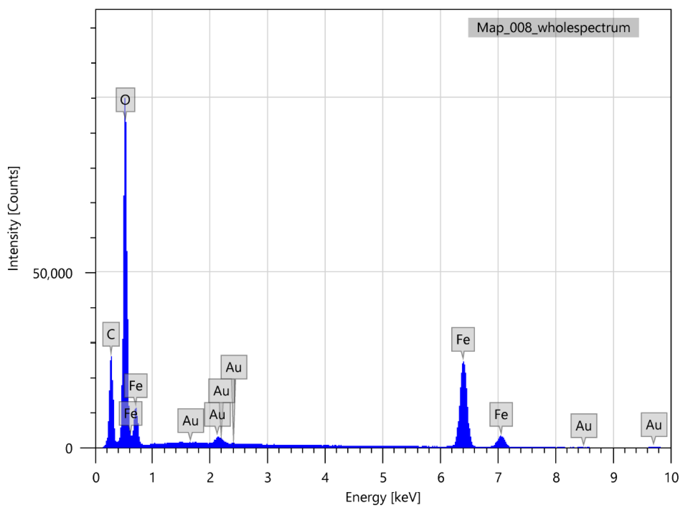
3. Results and Discussion

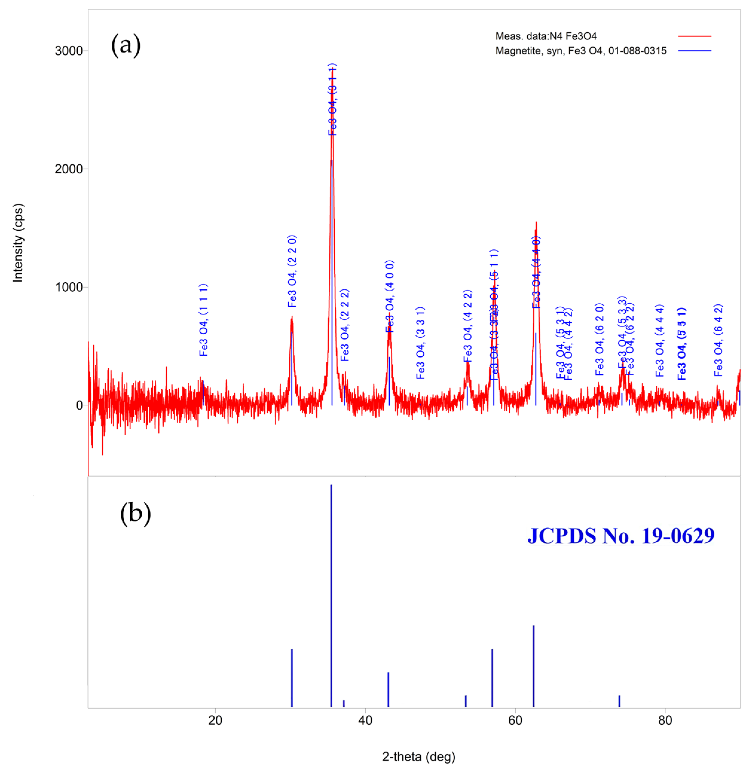
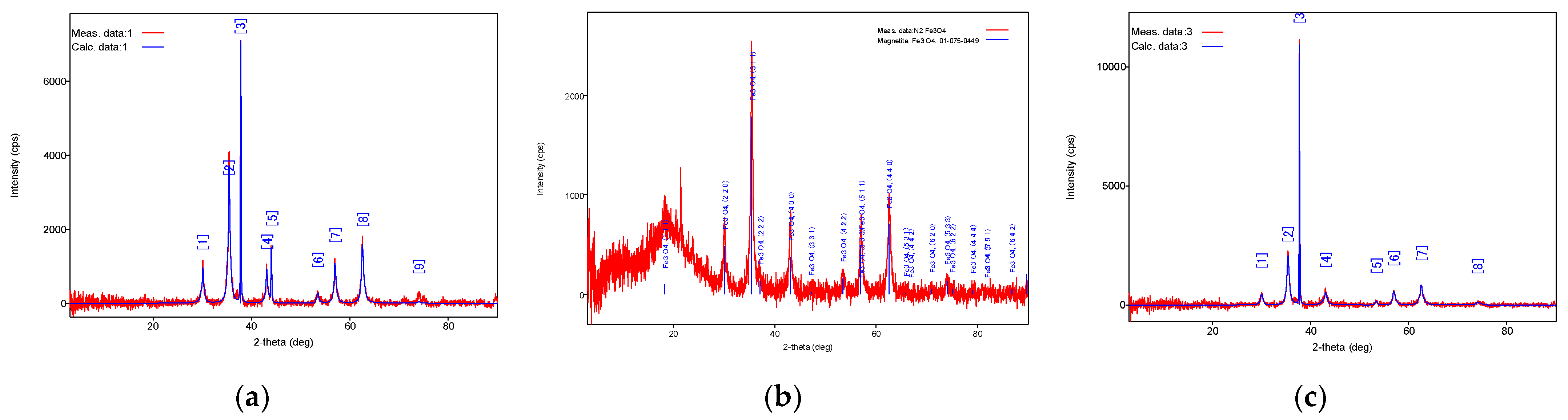
3.1. Chemical Condensation Method (CCM)

These investigations showed that the samples of magnetite nanoparticles obtained through chemical condensation exhibit a spinel crystal structure. Table 1 presents the results of the X-ray phase analysis. Figure 1 illustrates the results of the X-ray structural analysis.
The findings of the X-ray diffraction analysis reveal that the specimen is composed of a single magnetite phase (Fe3O4), featuring nanoparticles with a non-stoichiometric structure. This is evident from the reduction in the crystal lattice parameter. It is plausible that the sample also contains trace amounts of hematite (Fe2O3), although this cannot be definitively ascertained due to their shared cubic symmetry. The diffraction pattern obtained from the sample exhibits a background characteristic of iron-based compounds when utilizing copper radiation. Additionally, there are crystalline phases manifested through diffraction lines. Furthermore, an amorphous phase is indicated by a halo peaking at 18.8 degrees. Employing the Scherrer formula, the average size of iron oxide crystallites was calculated and found to be approximately 13 nm.
The obtained magnetite nanoparticles were examined using a microscope, as depicted in Figure 2. These nanomagnetites exhibit a characteristic iron-black hue with a discernible metallic sheen that remains visible at all magnification levels. This property would not be discernible through electron microscopy, which necessitates the preliminary drying of nanoparticles. One of the notable benefits of magnetite nanoparticles lies in their readiness for immediate use without the requirement for prior drying before incorporating shielding materials. This makes them ideal for immediate application following chemical synthesis. Prior to this study, the nanoparticles were washed with distilled water, followed by drying at a temperature of 70 degrees Celsius under a neutral atmosphere for a duration of 48 h.
In this investigation, TEM was employed to investigate the dimensions, structural arrangement, and morphological features of the materials. We focused on magnetite nanoparticles, which are depicted in Figure 3a–c. The results indicate that the average diameter of the spherical nanoparticles measures from 16 to 125 nm. Moreover, agglomerations or clusters of nanoparticles were observed, potentially arising from the process of the TEM preparation. When developing composite materials, it becomes essential to consider the dispersion of particle sizes within the powder employed as an active component. Figure 3d presents a histogram of the final distribution of particle sizes of the powder obtained by measuring 30 randomly selected nanoparticles. The analysis revealed a bimodal distribution of particles with distinct dimensions.
The specific surface area of nanoparticles represents a crucial parameter that significantly influences their reactivity. Through the measurement of this characteristic, it becomes possible to determine the average size of these nanoparticles.
The analysis of magnetite nanoparticles produced through chemical condensation, employing the Brunauer–Emmet–Teller method, revealed that their specific surface area is equivalent to 131.88 square meters per gram. These data allow us to calculate the average size of magnetite nanoparticles using the following mathematical formula:
D = 6 ρ S
where S is the specific surface area, m2/g; ρ is the theoretical density, g/m3.
The magnetic moment values of magnetite nanoparticle samples were measured using a 14 Tesla cryogenic vibrating sample magnetometer. The results are shown in Figure 4. These measurements demonstrate that magnetite nanoparticles undergo a transition to a superparamagnetic state, as indicated by the absence of hysteresis in their magnetization curve when subjected to an external field. This transition can be explained by the formation of a single-domain structure within the nanoparticles, which leads to uniform magnetization throughout the entire volume.

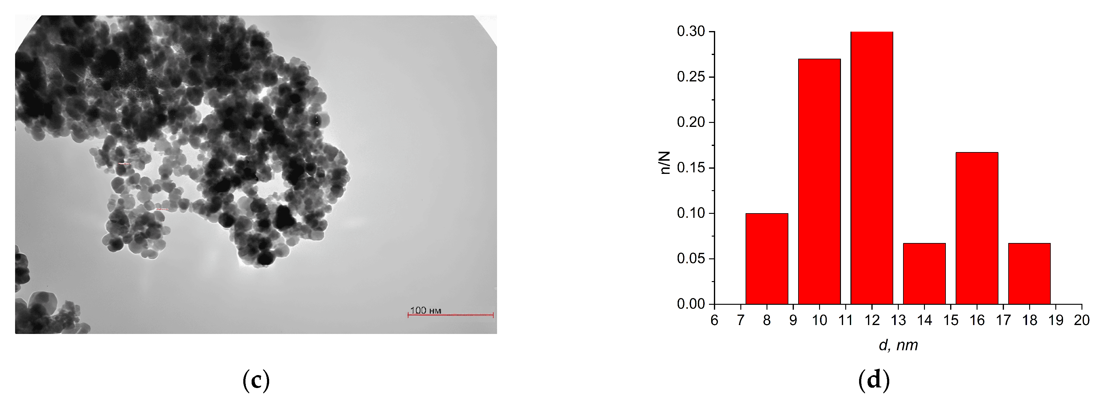
3.2. Liquid-Phase Combustion Method

To elucidate the crystal structure of magnetite nanoparticles, X-ray diffraction analysis was employed (see Figure 5). The impact of manipulating the ratio of fuel to oxidizer concentration on the crystal structure and the size of crystallites was explored.
The findings of the X-ray diffraction analysis reveal that the crystalline structure of the samples is composed of magnetite, Fe3O4. The dimensions of the crystallites are influenced by variations in the proportion of fuel and oxidizer concentrations.
The morphology and structure of magnetite nanoparticles synthesized using the solution combustion method were studied using SEM. Figure 6 shows the SEM images of the samples. These investigations showed that during the synthesis process, magnetite nanoparticles tend to agglomerate and form layered structures. The amorphous phase dominates the samples, especially in aggregates larger than 100 nm. To separate these aggregates and ensure uniform mixing and distribution of magnetite nanoparticles in the shielding material, we conducted a preliminary ultrasound treatment of nanoparticle powders in an aqueous suspension of nanomagnetites.
For clarity, the elemental composition was analyzed using EDS spectra of Fe(NO3)/C6H8O7 in a 1:1 molar ratio (Figure 7). Distinct emission lines of Fe-Kα (6.40 keV) and O-Kα (0.53 keV) confirm the formation of Fe3O4, with elemental ratios (Fe:O = 0.75:1.00) corresponding to the theoretical stoichiometry of magnetite (Fe3O4; Fe:O = 0.75). The absence of nitrogen signals (expected at 0.39 keV for Kα) indicates complete decomposition of the precursor during the combustion synthesis.
The specific surface area of the synthesized magnetite nanoparticle powders was determined using the solution combustion method. Table 2 presents the results of the specific surface analysis, accompanied by calculations of the average particle size obtained at various stoichiometric ratios of fuel and oxidant.
The findings of the specific surface analysis using the BET method indicate that at a 1:1 ratio, the specific surface area is 72.203 square meters per gram. At a 1:1.5 ratio, this value decreases to 22.240 square meters per gram, while at a 1:2 ratio, it further decreases to 9.204 square meters per gram.
It was established that the structural characteristics of the synthesized nanoparticles depend on the proportion of fuel and oxidant employed in the synthesis procedure. Three specific combinations of iron nitrate and citric acid (1:1, 1.5:1, and 2:1) were examined.
The X-ray diffraction analysis revealed that the initial proportion significantly affects the formation of Fe3O4 nanoparticles. The structural features and morphology of these nanoparticles were investigated using optical and scanning electron microscopy. Scanning electron microscopy demonstrated that Fe3O4 nanoparticles have a tendency to aggregate during synthesis, forming agglomerates with layered structures. Amorphous phases predominate in the samples, especially in clusters exceeding 100 nm in size. The analysis of the specific surface area, conducted using the BET method, revealed that for a 1:1 molar ratio, the value of the specific surface area is 72.203 square meters per gram. In the case of a 1.5:1 molar ratio, this value is 22.24 square meters per gram, while for a 2:1 molar ratio, it is 9.204 square meters per gram. Based on these data, we were able to calculate the average dimensions of magnetite nanoparticles synthesized via the solution combustion technique. In our calculations, we assumed that these nanoparticles possessed a spherical shape. The average diameter of Fe3O4 nanoparticles was determined to be 16 nm for a 1:1 molar ratio, 51 nm for 1.5:1, and 125 nm for 2.0:1.

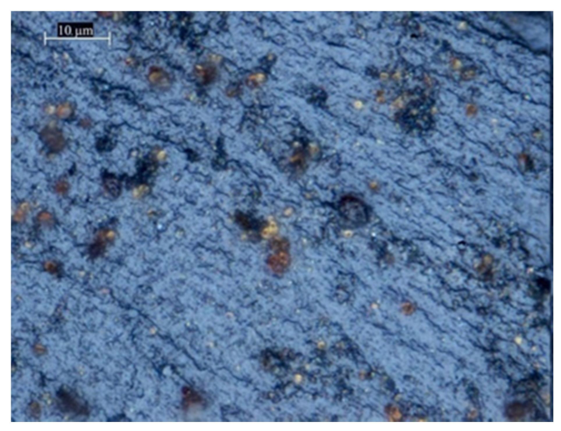
3.3. Investigation of the Shielding Characteristics of Cement-Based Materials Incorporating Magnetite Nanoparticles Synthesized Through Chemical Precipitation

The instrument employed in this study is capable of measuring in the frequency ranges of 0.7–2 GHz and 2–17 GHz, with the aid of additional converter modules. The operation of this device is controlled using a computer via Scalar Network Analyzer software. All data are processed and visualized in graphical form.
Microwave attenuation measurements were employed to determine the transmission of electromagnetic waves through the specimen, providing insights into its overall attenuation characteristics. Furthermore, additional measurements were conducted using a 3 mm thick metal plate and a calibrated reference sample to comprehensively understand the shielding properties, encompassing both reflective and absorptive components.
The results of transmission coefficient measurements for the control sample (without magnetite nanoparticles) and the cement-based composite material containing 0.25 percent magnetite nanoparticles are presented in Figure 8, depicted in graphical form.
As can be seen from the graph, at a frequency range from 0.7 to 2 GHz, the addition of magnetite nanoparticles weakens the overall microwave background. The concentration of 0.25% magnetite nanoparticles makes it possible to achieve maximum attenuation of the transmission coefficient to 21.7 dB.
Figure 9 shows the results of measurements of the transmission coefficient for a cement-based composite material with a content of 0.5% and 1% magnetite nanoparticles in the frequency range of 0.7–2 GHZ.
The graph clearly shows that within the frequency spectrum ranging from 0.7 to 2 GHz, the addition of magnetite nanomaterials leads to a reduction in the overall microwave background level.
At a concentration of 0.25% magnetite nanoparticles, the maximum attenuation of the transmission coefficient is achieved, that is, 21.7 dB. Increasing the concentration to 0.5% does not significantly alter the overall attenuation value.
When 0.5% of magnetite nanoparticles are present, the transmission coefficient attenuation ranges from 16.787 dB at 0.75 GHz to 19.915 dB at 1.55 GHz.
Further increasing the concentration of nanoparticles up to 1% in the material results in a decrease in the transmission coefficient. This suggests that the maximum shielding effect is achieved when the concentration is 0.5%. It can be deduced that concentrations exceeding 0.5% may lead to oversaturation and a subsequent degradation of the composite material’s shielding properties.
Figure 9 illustrates the findings obtained through the measurement of the absorption coefficient using a metal plate on cement-based composites with magnetite nanoparticle concentrations of 0.5% and 1%. The measurements were conducted within the frequency range of 0.7–2 GHz.
It was shown that the addition of magnetite nanoparticles synthesized through the CCM process significantly enhances the shielding properties of concrete. When the concentration of nanoparticles reaches 0.25%, an optimum attenuation level of 21.7 dB is achieved at a frequency of 1.55 GHz.
By manipulating the concentration of these nanoparticles, it becomes feasible to selectively attenuate specific frequency ranges. For example, with a nanoparticle concentration of 0.5%, an attenuation of 17 dB at 11 GHz can be obtained. Furthermore, the best attenuation over a broad range extending from 9 GHz to 13 GHz is also achievable at this concentration level.
At a concentration of magnetite nanoparticles of 1%, a microwave attenuation of 16.4 dB is achieved at a frequency of 8 GHz. Within the frequency range from 7 to 8.5 GHz and from 13 to 14 GHz, specimens containing 1% magnetite exhibit a higher attenuation of microwave radiation compared to other specimens.
The average shielding effectiveness resulting from the addition of magnetite consistently remains below 1 dB. This is primarily due to a magnetite concentration of no more than 1 wt.% being added. Such a low additive content allows for the mechanical integrity of cement-based composites to be retained while providing improved electromagnetic shielding properties [28]. Thus, in comparison to traditional shielding materials which necessitate a higher filler concentration (3–5% by weight), the reduced threshold of Fe3O4 content (0.25–0.5% by weight) presents a substantial advantage in terms of the economic viability, manufacturability, and structural robustness of the mechanical characteristics.
It should be noted that Fe3O4 nanoparticles have magnetic losses due to their high magnetic permeability, which allows for effective interaction with the magnetic component of electromagnetic waves. In the frequency range of 0.7–2 GHz, ferromagnetic resonance plays a key role in the absorption of microwave radiation [32]. This resonance occurs when the frequency of the applied field matches the frequency of the natural processes within the magnetic dipoles within the particles. Due to the size of the Fe3O4 particles that we synthesized, this magnetic resonance can occur in the 0.7–2 GHz range, leading to increased energy dissipation.
This suggests that adjusting the concentration of magnetite nanoparticles allows for the customization of shielding for specific frequencies. The measurements of reflection using a metallic plate indicate that varying concentrations of nanomagnetite significantly affect the absorption spectrum of microwave radiation.
The most promising results in terms of microwave absorption were obtained with the sample containing 0.25% magnetite, which outperformed all the other specimens. It is noteworthy that the reflection coefficient maintained a relatively steady state despite fluctuations in the nanoparticle concentration. This observation suggests that both the scattering and absorption processes significantly contribute to the reduction in microwave radiation intensity.

3.4. Study of the Shielding Properties of Cement Stone with Additives of Magnetite Nanoparticles Synthesized with Liquid-Phase Combustion

As mentioned earlier, this device allows for measurements to be made in the frequency ranges from 0.7 to 2 GHz and from 2 to 17 GHz. The test samples were prepared with a content of 0.5% and 1% of magnetite nanoparticles.
This measurement method is similar to that previously described for samples containing magnetite nanoparticles obtained using the CCM. For clarity, the graphs are presented in comparison with the base sample without any magnetite nanoparticle additives. Figure 10 depicts the findings of investigations into the transmission coefficient of cement-based composites incorporating magnetite nanoparticle additives produced through the process of liquid-phase synthesis. These findings suggest that magnetite nanoparticles synthesized using the solution combustion method significantly impact the shielding capabilities of cement. Increasing the concentration up to 1%, as observed in magnetite nanoparticles generated using the CCM, results in the deterioration of shielding properties.
Notably, the overall attenuation background at 0.5% is significantly lower for composite materials incorporating magnetite nanoparticles fabricated through solution combustion compared to those incorporating magnetite nanoparticles created using the CC method. The optimal shielding performance for cement stone incorporating nanoparticles synthesized using the solution combustion approach was 14.4 dB, while for specimens containing magnetite nanoparticles manufactured using the CCM and having the same magnetite concentration and frequency, the shielding efficacy was 16.8 dB at the same frequency. At a frequency of 1.55 GHz, the attenuation coefficient of composite material samples prepared using cement and magnetite nanoparticles via the CCM was 13.5 dB, while the corresponding coefficient for cement stone containing nanoparticles obtained through the solution combustion method was 19.9 dB.

4. Conclusions

Nanoscale magnetic particles exhibiting superparamagnetic behavior were synthesized via two distinct approaches: chemical condensation and liquid-phase combustion. Utilizing transmission electron microscopy, it was determined that the mean diameter of the nanoparticles generated through chemical condensation amounted to 11 nm. The surface area-to-volume ratio of these nanoparticles was subsequently measured using the BET method.
Based on these findings, the average diameters of nanoparticles produced via chemical condensation were calculated as D = 10 ± 0.1 nm. In contrast, for nanoparticles obtained using liquid-phase combustion, the average diameters were determined to range from 16 to 125 nm depending on the component ratio.
Unlike conventional shielding methods that use a high concentration of additives, which can lead to brittleness, our research demonstrates the possibility of creating microwave shielding using a low concentration of Fe3O4 additives. This has significant advantages in terms of the cost, manufacturing process, and structural integrity of the resulting composite material.
This study explored the impact of superparamagnetic particles within composite materials, with varying concentrations, on mitigating microwave radiation.
The optimal concentration of magnetite nanoparticles within cement stone, which achieves the maximum attenuation of microwave radiation, was identified as 0.25 weight percent. For samples measuring 10 mm in thickness, the reduction in radiation amounted to an impressive 21.7 dB. It was revealed that samples containing nanoscale magnetite at a concentration of 0.5 weight percent exhibited the widest range of frequencies for the most significant reduction in microwave intensity.

Author Contributions

Conceptualization, A.L. and D.A.; methodology, B.K.; software, D.Y.; validation, A.L., D.A., and B.K.; formal analysis, D.Y.; investigation, B.K.; resources, A.L.; data curation, A.L.; writing—original draft preparation, A.L. and D.A.; writing—review and editing, D.Y.; visualization, B.K.; supervision, D.Y.; project administration, A.L. and D.Y.; funding acquisition, A.L. All authors have read and agreed to the published version of the manuscript.

Funding

This work was supported by the Ministry of Science and Higher Education of the Republic of Kazakhstan (Grant No. AP19679690).

Data Availability Statement

Data are contained within the article.

Conflicts of Interest

The authors declare no conflicts of interest.

References

  1. Goubau, G.; Schwering, F. On the Guided Propagation of Electromagnetic Wave Beams. IRE Trans. Antennas Propag. 1961, 9, 248–256. [Google Scholar] [CrossRef]
  2. Lee, J.; Kang, H.; Shin, B.G.; Song, Y.J. Cement Composites with Carbon Fiber for Electromagnetic Interference Shielding Applications. Carbon 2024, 220, 118861. [Google Scholar] [CrossRef]
  3. Taghipour, N.; Whitworth, G.L.; Othonos, A.; Dalmases, M.; Pradhan, S.; Wang, Y.; Kumar, G.; Konstantatos, G. Low-Threshold, Highly Stable Colloidal Quantum Dot Short-Wave Infrared Laser Enabled by Suppression of Trap-Assisted Auger Recombination. Adv. Mater. 2022, 34, 2107532. [Google Scholar] [CrossRef]
  4. Lapinsky, S.E.; Easty, A.C. Electromagnetic Interference in Critical Care. J. Crit. Care 2006, 21, 267–270. [Google Scholar] [CrossRef] [PubMed]
  5. Wanasinghe, D.; Aslani, F.; Ma, G.; Habibi, D. Review of Polymer Composites with Diverse Nanofillers for Electromagnetic Interference Shielding. Nanomaterials 2020, 10, 541. [Google Scholar] [CrossRef]
  6. Jia, X.; Li, Y.; Shen, B.; Zheng, W. Evaluation, Fabrication and Dynamic Performance Regulation of Green EMI-Shielding Materials with Low Reflectivity: A Review. Compos. Part B Eng. 2022, 233, 109652. [Google Scholar] [CrossRef]
  7. Sankaran, S.; Deshmukh, K.; Ahamed, M.B.; Khadheer Pasha, S.K. Recent Advances in Electromagnetic Interference Shielding Properties of Metal and Carbon Filler Reinforced Flexible Polymer Composites: A Review. Compos. Part A Appl. Sci. Manuf. 2018, 114, 49–71. [Google Scholar] [CrossRef]
  8. Rubežienė, V.; Varnaitė-Žuravliova, S.; Sankauskaitė, A.; Pupeikė, J.; Ragulis, P.; Abraitienė, A. The Impact of Structural Variations and Coating Techniques on the Microwave Properties of Woven Fabrics Coated with PEDOT:PSS Composition. Polymers 2023, 15, 4224. [Google Scholar] [CrossRef]
  9. Shi, Y.; Wu, M.; Ge, S.; Li, J.; Alshammari, A.S.; Luo, J.; Amin, M.A.; Qiu, H.; Jiang, J.; Asiri, Y.M.; et al. Advanced Functional Electromagnetic Shielding Materials: A Review Based on Micro-Nano Structure Interface Control of Biomass Cell Walls. Nano-Micro Lett. 2025, 17, 3. [Google Scholar] [CrossRef]
  10. Wanasinghe, D.; Aslani, F.; Ma, G.; Habibi, D. Advancements in Electromagnetic Interference Shielding Cementitious Composites. Constr. Build. Mater. 2020, 231, 117116. [Google Scholar] [CrossRef]
  11. Peng, M.; Qin, F. Clarification of Basic Concepts for Electromagnetic Interference Shielding Effectiveness. J. Appl. Phys. 2021, 130, 225108. [Google Scholar] [CrossRef]
  12. Lan, C.; Zou, L.; Wang, N.; Qiu, Y.; Ma, Y. Multi-Reflection-Enhanced Electromagnetic Interference Shielding Performance of Conductive Nanocomposite Coatings on Fabrics. J. Colloid Interface Sci. 2021, 590, 467–475. [Google Scholar] [CrossRef] [PubMed]
  13. Gao, S.; Yang, S.-H.; Wang, H.-Y.; Wang, G.-S.; Yin, P.-G. Excellent Electromagnetic Wave Absorbing Properties of Two-Dimensional Carbon-Based Nanocomposite Supported by Transition Metal Carbides Fe3C. Carbon 2020, 162, 438–444. [Google Scholar] [CrossRef]
  14. Kaidar, B.; Imash, A.; Smagulova, G.; Keneshbekova, A.; Kazhdanbekov, R.; Yensep, E.; Akalim, D.; Lesbayev, A. Magnetite-Incorporated 1D Carbon Nanostructure Hybrids for Electromagnetic Interference Shielding. Nanomaterials 2024, 14, 1291. [Google Scholar] [CrossRef] [PubMed]
  15. Ji, X.; Chen, F.; Liu, J.; Ma, Q.; Qiao, R.; Xu, Z. Synthesis of MWCNTs/Si3N4 Nanocomposites Via Click Chemistry and Electromagnetic Wave-Absorption Properties. J. Electron. Mater. 2025, 54, 1228–1244. [Google Scholar] [CrossRef]
  16. Liu, Y.; Zhang, N.; Wang, Y.; Zong, M.; Zhu, Y. Construction of Polymer-Derived Double Transition Metal Compound/Carbon Matrix Nanocomposites for Efficient Electromagnetic Wave Absorber. Ceram. Int. 2024, 50, 45416–45426. [Google Scholar] [CrossRef]
  17. Xia, X.; Xiao, Q. Electromagnetic Interference Shielding of 2D Transition Metal Carbide (MXene)/Metal Ion Composites. Nanomaterials 2021, 11, 2929. [Google Scholar] [CrossRef]
  18. Li, Y.; Zhang, D.; Zhou, B.; He, C.; Wang, B.; Feng, Y.; Liu, C. Synergistically Enhancing Electromagnetic Interference Shielding Performance and Thermal Conductivity of Polyvinylidene Fluoride-Based Lamellar Film with MXene and Graphene. Compos. Part A Appl. Sci. Manuf. 2022, 157, 106945. [Google Scholar] [CrossRef]
  19. Cheng, M.; Xu, X.; Zhang, H.; Kong, S.; Feng, Z.; Meng, F.; Wang, Y. Self-Assembling, Flexible and Stable Aramid Nanofiber/Polypyrrole/MXene Composite Film for Efficient Electromagnetic Interference Shielding, Dual-driven Heating and Infrared Thermal Camouflage. Chem. Eng. J. 2025, 505, 159112. [Google Scholar] [CrossRef]
  20. Katheria, A.; Das, P.; Nayak, J.; Roy, B.; Pal, A.; Biswas, S.; Das, N.C. MXene and Fe3O4 Decorated G-C3N4 Incorporated High Flexible Hybrid Polymer Composite for Enhanced Electrical Conductivity, EMI Shielding and Thermal Conductivity. Next Mater. 2025, 6, 100292. [Google Scholar] [CrossRef]
  21. Oraby, H.; Tantawy, H.R.; Correa-Duarte, M.A.; Darwish, M.; Elsaidy, A.; Naeem, I.; Senna, M.H. Tuning Electro-Magnetic Interference Shielding Efficiency of Customized Polyurethane Composite Foams Taking Advantage of RGO/Fe3O4 Hybrid Nanocomposites. Nanomaterials 2022, 12, 2805. [Google Scholar] [CrossRef]
  22. Zhao, Y.; Zhang, Y.; Li, R.; Wang, Z.; Lou, Z.; Li, Y. Facile Synthesis of Ultralight and Porous Melamine-Formaldehyde (MF) Resin-Derived Magnetic Graphite-Like C3N4/Carbon Foam with Electromagnetic Wave Absorption Behavior. Crystals 2020, 10, 656. [Google Scholar] [CrossRef]
  23. Michałowska, A.; Kudelski, A. The First Silver-Based Plasmonic Nanomaterial for Shell-Isolated Nanoparticle-Enhanced Raman Spectroscopy with Magnetic Properties. Molecules 2022, 27, 3081. [Google Scholar] [CrossRef]
  24. Yang, C.; Li, K.; Hu, M.; Li, X.; Li, M.; Hu, X.; Li, Y.; Huang, Z.; Meng, G. Flexible ZrO2/ZrB2/C Nanofiber Felt with Enhanced Microwave Absorption and Ultralow Thermal Conductivity. J. Mater. 2025, 11, 100988. [Google Scholar] [CrossRef]
  25. Wu, F.; Hu, P.; Hu, F.; Tian, Z.; Tang, J.; Zhang, P.; Pan, L.; Barsoum, M.W.; Cai, L.; Sun, Z. Multifunctional MXene/C Aerogels for Enhanced Microwave Absorption and Thermal Insulation. Nano-Micro Lett. 2023, 15, 194. [Google Scholar] [CrossRef]
  26. Hou, T.; Wang, B.; Jia, Z.; Wu, H.; Lan, D.; Huang, Z.; Feng, A.; Ma, M.; Wu, G. A Review of Metal Oxide-Related Microwave Absorbing Materials from the Dimension and Morphology Perspective. J. Mater. Sci. Mater. Electron. 2019, 30, 10961–10984. [Google Scholar] [CrossRef]
  27. Lesbayev, A.B.; Smagulova, G.T.; Kim, S.; Prikhod’ko, N.G.; Manakov, S.M.; Guseinov, N.; Mansurov, Z.A. Solution-Combustion Synthesis and Characterization of Fe3O4 Nanoparticles. Int. J. Self-Propagating High-Temp. Synth. 2018, 27, 195–197. [Google Scholar] [CrossRef]
  28. Lesbayev, A.; Elouadi, B.; Borbotko, T.; Manakov, S.; Smagulova, G.; Boiprav, O.; Prikhodko, N. Influence of Magnetite Nanoparticles on Mechanical and Shielding Properties of Concrete. Eurasian Chem. J. 2017, 19, 223. [Google Scholar] [CrossRef]
  29. Boiprav, O.V.; Bogush, V.A. Lightweight microwave absorbers with loop-like surface irregularities based on foiled materials for anechoic chambers. MOMENTO 2024, 1–16. [Google Scholar] [CrossRef]
  30. Boiprav, O.V.; Bogush, V.A. Advanced Layered Flexible Radio-Absorbing Materials Based on Powdered Charcoal. Inorg. Mater. Appl. Res. 2024, 15, 280–288. [Google Scholar] [CrossRef]
  31. Hasanov, M. The development of a piezoelectric defect detection device through mathematical modeling applied to polymer composite materials. New Mater. Compd. Appl. 2024, 8, 62–74. [Google Scholar] [CrossRef]
  32. Wang, L.; Wang, Y.; Liu, D.; Wang, Y.; Zhang, X.; Wen, G.; Zhang, X.; Huang, X. Carbon-Coated Octahedral Fe3O4/Fe2O3 Nanocomposites for Enhanced Electromagnetic Wave Absorption. ACS Appl. Nano Mater. 2025, 8, 2952–2964. [Google Scholar] [CrossRef]
Figure 1. X-ray diffraction pattern: (a) magnetite nanoparticles obtained with the CCM; (b) standard PDF cards Fe3O4: JCPDS No. 19-0629.
Jcs 09 00226 g001
Figure 2. Optical images of magnetite nanoparticles with a magnification of 10 μm.
Jcs 09 00226 g002
Figure 3. TEM images of magnetite nanoparticles (ac) obtained with the CCM and size distribution (d).
Jcs 09 00226 g003aJcs 09 00226 g003b
Figure 4. Magnetic hysteresis of magnetite nanoparticles at ambient temperature.
Jcs 09 00226 g004
Figure 5. (ac) X-ray diffraction patterns of magnetite nanoparticles prepared using the solution combustion method with fuel-to-oxidizer ratios of 1:1, 1:1.5, and 1:2, respectively.
Jcs 09 00226 g005
Figure 6. (ac) SEM images of magnetite nanoparticles prepared using the solution combustion method with fuel-to-oxidizer ratios of 1:1, 1:1.5, and 1:2, respectively.
Jcs 09 00226 g006
Figure 7. Elemental composition using EDS spectra of Fe(NO3)/C6H8O7 (1:1).
Jcs 09 00226 g007
Figure 8. The transmission coefficient for a standard sample and a composite material based on cement with 0.25 percent magnetite nanoparticles was measured in the frequency range from 0.7 to 2 GHz.
Jcs 09 00226 g008
Figure 9. The transmission coefficient for a composite material based on cement incorporating 0.5–1% magnetite nanoparticles was determined to be in the frequency range of 0.7–2 GHz.
Jcs 09 00226 g009
Figure 10. The transmission coefficient of a reference sample devoid of nanomagnetic iron oxide nanoparticles and a specimen incorporating 0.5 percent by weight of magnetite nanoparticles within the frequency spectrum spanning 0.7–2 GHz.
Jcs 09 00226 g010
Table 1. X-ray diffractometric data of magnetite nanoparticles obtained with the CCM.
2-Theta (deg)D (ang.)Height (cps)FWHM (deg)Int. I (cps deg)Phase Name
18.50(4)4.791(11)71(24)0.65(14)65(14)Fe3O4(111)
30.08(3)2.969(3)481(63)0.58(3)354(22)Fe3O4(2.2.0)
35.521(14)2.5252(9)1876(125)0.569(13)1530(25)Fe3O4(3.1.1)
43.21(4)2.0922(18)474(63)0.56(4)364(21)Fe3O4(4.0.0)
53.60(10)1.708(3)201(41)0.72(10)220(23)Fe3O4(4.2.2)
57.15(4)1.6104(10)701(76)0.61(3)641(20)Fe3O4(5.1.1)
62.69(3)1.4809(6)1008(92)0.68(2)943(24)Fe3O4(4.4.0)
71.30(7)1.3217(12)66(23)0.9(4)101(24)Fe3O4(6.2.0)
74.16(12)1.2776(18)214(42)0.78(19)326(30)Fe3O4(5.3.3)
Table 2. Relationship between particle size and various fuel-to-oxidant ratios.
Stoichiometric RatioLXRD, nmSBET (m2/g)DBET, nmCarbon Content, %
Fe(NO3)/C6H8O7, 1:12072.20316 ± 124.37
Fe(NO3)/C6H8O7, 1:1.51822.24051 ± 232.70
Fe(NO3)/C6H8O7, 1:2139.204125 ± 438.06
Disclaimer/Publisher’s Note: The statements, opinions and data contained in all publications are solely those of the individual author(s) and contributor(s) and not of MDPI and/or the editor(s). MDPI and/or the editor(s) disclaim responsibility for any injury to people or property resulting from any ideas, methods, instructions or products referred to in the content.

