Article

Preparation and Photoelectric Properties of Nanostructured Native Oxide of Gallium Monoselenide with Applications in Gas Sensors

1
Faculty of Physics and Engineering, Moldova State University, 60 Alexei Mateevici Str., MD-2009 Chisinau, Moldova
2
Faculty of Electrical Engineering, Czech Technical University in Prague, Technická 1902/2, Praha 6, 166 27 Prague, Czech Republic
3
Faculty of Physics, Alexandru Ioan Cuza University of Iasi, Bulevardul Carol I, Nr. 11, 700506 Iasi, Romania
4
Science Research Department, Institute of Interdisciplinary Research, Research Center in Environmental Sciences for the North-Eastern Romanian Region (CERNESIM), Alexandru Ioan Cuza University of Iasi, Bulevardul Carol I, Nr. 11, 700506 Iasi, Romania
*
Authors to whom correspondence should be addressed.
J. Compos. Sci. 2025, 9(4), 194; https://doi.org/10.3390/jcs9040194
Submission received: 12 February 2025 / Revised: 11 April 2025 / Accepted: 16 April 2025 / Published: 19 April 2025

Abstract

:
Using the Bridgman technique, GaSe single crystals were obtained which were mechanically split into plane-parallel plates with a wide range of thicknesses. By heat treatment in air at 820 °C and 900 °C, for 30 min and 6 h, micro- and nanocomposite layers of Ga2Se3–Ga2O3 and β–Ga2O3 (native oxide) with surfaces made of nanowires/nanoribbons were obtained. The obtained composite Ga2Se3–Ga2O3 and nanostructured β–Ga2O3 are semiconductor materials with band gaps of 2.21 eV and 4.60 eV (gallium oxide) and photosensitivity bands in the green–red and ultraviolet-C regions that peaked at 590 nm and 262 nm. For an applied voltage of 50 V, the dark current in the photodetector based on the nanostructured β–Ga2O3 layer was of 8.0 × 10⁻¹³ A and increased to 9.5 × 10⁻⁸ A upon 200 s excitation with 254 nm-wavelength radiation with a power density of 15 mW/cm2. The increase and decrease in the photocurrent are described by an exponential function with time constants of τ1r = 0.92 s, τ2r = 14.0 s, τ1d = 2.18 s, τ2d = 24 s, τ1r = 0.88 s, τ2r = 12.2 s, τ1d = 1.69 s, and τ2d = 16.3 s, respectively, for the photodetector based on the Ga2Se3–Ga2S3–GaSe composite. Photoresistors based on the obtained Ga2Se3–Ga2O3 composite and nanostructured β–Ga2O3 layers show photosensitivity bands in the spectral range of electronic absorption bands of ozone in the same green–red and ultraviolet-C regions, and can serve as ozone sensors (detectors).

1. Introduction

GaSe (gallium monoselenide) is a typical representative of the lamellar metal (Ga and In) chalcogenides group. It consists of elementary layered packings of the Se-Ga-Ga-Se type with predominantly covalent bonds inside them and weak polarization bonds (of Van der Waals type) between them. In addition, its C6 symmetry axis is perpendicular to the surface of the stratified packings [1,2,3]. Four polymorphs of these single crystals (β, γ, ε, and σ) are currently being studied [4,5]. In GaSe single crystals grown by the Bridgman–Stockbarger technique, the ε polytype (P-6m2 space group, point group D3h), with no inversion center, appears to be prevalent [4,6].
The weak bonds between the elementary packings allow researchers, through mechanical splitting, to obtain plane-parallel lamellae with thicknesses that are close to the unit cells’ size (~10 Å) and display atomically smooth surfaces with a relatively low surface state density (≤1010 cm−2) [7,8]. Being a p–type semiconductor, with a band gap of ~2.0 eV and pronounced anisotropy of its mechanical and optical properties, GaSe is a promising material for THz electronics, nonlinear optics, optoelectronics, and, especially, 2D devices [9,10,11]. Despite the fact that the valence bonds on the surface of elementary GaSe packings are closed, as demonstrated in references [12,13,14], under normal atmospheric conditions, as well as at high temperatures, an oxide layer that is predominantly composed of gallium oxide, Ga2O3 (native oxide), is formed on the surface of GaSe [12,13,14]. Several crystal modifications of gallium oxide have been reported: α–, β–, γ–, δ–, and ε–Ga2O3 [15]. Among these polymorphs, β–Ga2O3 with a monoclinic crystal lattice (space group C2/m) exhibits high thermodynamic stability [16]. It is an n–type semiconductor with a band gap of 4.5–4.9 eV, displaying photoluminescence emission in the violet–blue region and high photosensitivity upon UV-C (ultraviolet-C) excitation in the wavelength range of (210–280) nm [17,18,19].
The current interest in UV-C photodetectors is driven by their wide area of application, including in fire sensors, photodetectors for space communications, and UV monitoring of the atmospheric ozone layer, as well as in military applications such as missile and supersonic aircraft detection.
Since ozone (O3) is known to be an extremely toxic gas and a powerful oxidizing agent (stronger than oxygen), there is an acute need for ozone-specific sensors, which are used for the protection of high-power electrical installations as well as the controlling of technological processes in the chemical, pharmaceutical, and food industries. The main characteristic of ozone, which determines its special physical and chemical properties, is the large amount of energy emitted upon its decomposition (~24 kcal/mol). Under the influence of UV radiation, ozone dissociates into molecular and atomic oxygen, upon which it the releases the listed amount of energy. Further, ozone is a highly explosive gas, due to the heat that is released and its rapid volume expansion upon combustion [20]. In atmospheric ozone detection, optical methods, in which the intensity of the absorption band is measured and the Bouguer–Lambert–Beer (B-L-B) equation (relating absorbance and concentration) is used, are the most common ones [21]. In [22], the development of an ozone sensor based on amorphous β–Ga2O3 layers decorated with IGZO (indium gallium zinc oxide) nanoparticles was reported. It was established that its sensitivity depends on the IGZO nanoparticle size. In addition, in [23], the influence of ozone on the fluorescence of some biological samples was demonstrated.
In this work, the technological conditions for obtaining (by heat treatment of single-crystal GaSe plates in air) nanostructured β–Ga2O3 layers on p–GaSe substrate are studied, together with the chemical composition, surface morphology and photoconductivity of β–Ga2O3; based on these studies, the elaboration of experimental samples of ozone sensors was carried out.

2. Materials and Methods

Gallium monoselenide was synthesized from its elemental components, Ga (6N) and Se (5N), which were placed in stoichiometric amounts in a two-zone (zone I and zone II) furnace whose axis was tilted at ~30° to the horizontal, at a temperature of 1300 °C for 12 h.
Figure 1 schematically shows the experimental setup used for the compound synthesis Figure 1a, as well as a diagram of the temperature distribution along the length of the two-zone furnace used for the synthesis (1) and growth (2) of GaSe single crystals Figure 1b.
At this temperature (1300 °C), the Ga vapor pressure does not exceed 2.0 atm (1370 Torr), while that of selenium (Se) vapor at ~1000 °C is higher than 4 atm, a pressure that cannot be supported by usual quartz ampoules. For this reason, the synthesis of the GaSe compound and the obtaining of single crystals with a mass of ~25 g were carried out in quartz ampoules with an internal diameter of 14 mm and a length of ~25 cm. After pumping down the ampoule with the material subjected to synthesis to a residual gas pressure of 10−5 Torr and sealing the ampoule, it was placed in the furnace, so that a portion of ~10 cm length was outside the furnace. The temperature in the ampoule sector containing the material was slowly increased at a rate of ~80–100 °C/h, up to a temperature of 1300 °C. Starting at a temperature of ~300 °C, selenium condensation occured in the “cold” sector of the ampoule. At the synthesis temperature of GaSe, the temperature of the selenium sector of the ampoule was stabilized with an air flow at a temperature of ~600 °C. In this thermal regime, after the complete combination of Se with Ga, the ampoule was inserted for ~10 h, along its entire length, into the first zone (I) of the furnace, at a temperature of 1300 °C. Homogenization of the melt was achieved by rotating and vibrating the ampoule for ~8 h.
In this way, using the Bridgman–Stockbarger method, single crystals with a diameter of 14 mm and a length of 3 cm were obtained.
The furnace with the synthesized compound was moved to a vertical position and the temperature in the second zone (II) of the furnace was set at 870 °C, while, in the first zone, it slowly decreased (over 6 h) to ~1050 °C. In this way, a temperature gradient of ~45 °C/cm was established over the ~4 cm length between zones I and II. The ampoule containing the synthesized material was passed through this temperature gradient at a speed of ~0.5 mm/h.
In this way, p–type GaSe single crystals with hole concentrations of p~1014 cm−3 and mobility of µ ≈ 25 cm2/(V·s) were obtained. Plane-parallel plates with a surface area of ~14 × 20 mm2 and thicknesses in the range of 50–150 µm were split from single crystalline ingots.
The influence of temperature on the growth process of β–Ga2O3 crystallites has been investigated in several works [24,25,26]. Studies on the formation and growth kinetics of β–Ga2O3 microcrystallites on the surface of GaSe wafers at temperatures of 750 °C and 900 °C have shown that, at 750 °C, crystallization seeds of Ga2O3 are emphasized on the surface of GaSe wafers, while, at 900 °C, the size of β–Ga2O3 crystallites is that of hundreds of nanowires [26]. The growth rate of β–Ga2O3 crystallites, due to gallium oxidation from the vapor phase in the temperature range of 750–800 °C, is equal to 0.7–1.0 µm/h, and, as demonstrated in reference [21], at a temperature of 900 °C, it is ~5 µm/h.
The GaSe plates with a thickness of ~100 µm were thermally oxidized in air at 820 °C for 30 min, followed by 900 °C for 6 h. The surfaces of plates that were heat-treated for 30 min were covered with a gray-colored layer, while, after 6 h of oxidation at 900 °C, the plates surfaces were covered with a white microgranular layer, which intensely diffused the incident light. As a result of this technological procedure, the plates thickness increases by ~8–10%, from 100 µm to ~110 µm. The thicknesses of the layers formed by the heat treatment of the GaSe plates were determined by microscopic measurements along the cleavage edge. For the manufacture of GaSe photoreceptors, plates with a thickness of ~50 µm and a surface area of ~5 × 8 mm2 were used, on the surface of which two In strips were deposited by vacuum (~10−6 Torr) evaporation at a distance of ~2 mm from each other. Photoresistors were also manufactured from β–Ga2O3 and nanostructured β–Ga2O3/Ga2Se3–GaSe composites with a surface area of ~8 × 12 mm2 and a thickness of 100–150 µm. After deposition, one of the plates that had two In strips on its surface, at a distance of 4–6 mm from each other, was subjected to a heat treatment at ~820 °C for 30 min. The crystal structure of the synthesized materials was studied by X-ray diffraction (XRD) technique, using a Rigaku Ultima IV diffractometer with λCuKα = 1.54060 Å wavelength radiation. The experimental data were analyzed using the afferent PDXL software suite. The measured XRD patterns were compared with the standard JCPDS data. The surface morphology of the layer formed on the surfaces of GaSe plates after the heat treatment at 900 °C was analyzed using images recorded with a ZEISS Ultra Plus Scanning Electron Microscope (SEM). This equipment was completed with an energy-dispersive X-ray spectroscopy (EDXS) module, which allowed the determination of the surface elemental composition of the synthesized samples. The chemical composition of the primary material (GaSe single crystals) and the nanostructured β–Ga2O3 layers was identified using combined diffusion spectra recorded with a Micro-Raman spectrometer (WiTec Alpha 300 Ra, Ulm, Germany) upon first harmonic excitation (λ = 532 nm) of a Nd:YAG laser. Diffuse reflectance spectra of the surface of the microstructured layers were measured using a Specord M-40 spectrophotometer (spectral energy resolution of 0.5 meV) equipped with accessories for diffuse reflectance measurements at an angle of 90° to the incident beam. The photocurrent through photoresistors was recorded with a V7-30 type electrometer-voltmeter. The photoresponse spectra were recorded by means of a specialized photometric setup that included a high-power MDR-2 monochromator with diffraction gratings (600 and 1200 mm−1). A DVS-25 hydrogen-deuterium lamp and a DKS3-1000 Xe arc lamp were used as UV radiation sources, while, for the visible region, a 50 W wolfram vapor lamp with an inert gas was used.
The ozone-enriched atmosphere was generated with a 30 W BUV-30 optical quartz tubular lamp, with a diameter of 18 mm and a length of 60 cm, in which the electric discharge takes place in a Hg/Ar vapor mixture at a pressure of 10−1–10−2 Torr that is placed inside a 50 cm diameter and 100 cm long glass tube. The glass tube was sealed at both ends with quartz windows by using a technical resin gasket, through which electrodes were passed to supply the lamp with Hg vapor. As a radiation source with a wavelength of 254 nm, a 25 W H2/D2 vapor lamp was used. In the red spectral region, the 635 nm-wavelength radiation of a laser diode was used as a radiation source. The tube with/without air/O3 atmosphere was placed at the entrance slit of a MDR-2 monochromator, whose exit radiation was recorded with a β–Ga2O3 photoresistor (for wavelength λ = 254 nm) and a Ga2Se3/Ga2O3-based photoresistor (for λ = 638 nm); to establish the stationary regime of the air/O3 composition, the radiation at the exit slit of the monochromator was recorded with a Si-based photoelement. The generated photocurrent (by the receiver) was recorded as a function of time, when the probe radiation was passed through both atmospheric air and air/O3 column.

3. Results

3.1. Crystal Structure and Material Composition

The chemical composition of the GaSe single crystals and the material obtained by heat treatment in air at temperatures of 820 °C and 900 °C for 30 min and 6 h, respectively, was determined by the analysis of XRD patterns and Raman spectra.
Figure 2a shows the XRD diagram (X-ray wavelength λCuKα = 1.54060 Å), in the 2θ angular range from 10° to 90°, of GaSe single crystals obtained by the Bridgman technique. In this range of Bragg diffraction angles, lines with maxima located at 22.213°, 33.591°, 45.660°, 57.921°, 71.178°, and 85.360° are clearly visible; according to (ICDD-JCPDS) PDF card no. 01-078-1927, these lines can be indexed as X-ray reflections from (004), (006), (008), (0010), (207), and (0014) crystal planes of ε–GaSe (hexagonal lattice, space group P63/mmc (194), with parameters a = 3.750 Å, c = 15.995 Å, and γ = 120.0°).
Figure 2b illustrates the XRD patterns of the material obtained by heat treatment in air, at a temperature of 820 °C, of the single crystal ε–GaSe plates. It can be observed that, along with the diffraction lines located at 2θ angles equal to 22.22 and 57.89°, which correspond to the intense lines in Figure 2a and are identified as reflections of a hexagonal ε–GaSe phase, the lines at 28.43, 47.27, 55.80, 57.70, 76.14, and 88.62° are clearly visible, which, according to the PDF card no. 760975, can be ascribed to the (002), (150), (370), ( 2 ¯ 46), and ( 4 ¯ 46) reflections of the monoclinic Ga2Se3 lattice with parameters a = 6.660 Å, b = 11.65 Å, c = 6.649 Å, and β = 108.84°. We also mention the good coincidence of the Ga2Se3 (crystallites) diffraction lines in the XRD pattern of the composite obtained by heat treatment in air at a temperature of 820 °C for 30 min (Figure 2b) with the X-ray reflections of the Ga2Se3 granular thin films studied in references [27,28,29]. At the same time, the lines at 2θ = 33.48, 48.59, 69.11, 70.79, and 84.43° are emphasized, which corresponds to the XRD lines labeled 3, 10, 21, 22, and 29 that were obtained from the XRD diagram of the material obtained by heat treatment at 900 °C for 6 h (Figure 2c). Therefore, the heat treatment in air, at 820 °C, of GaSe single crystals leads to the formation of a composite of GaSe, Ga2Se3, and β–Ga2O3 crystallites.
As a result of the heat treatment in air at a temperature of 900 °C, a gray micro-corrugated layer is formed on the surface of the GaSe plates, which, upon increasing the treatment duration from 0.5 h to 6 h, turns into a microgranular layer with a white color. The XRD diagram of the material obtained by the thermal oxidation of the GaSe plates with a thickness d ≤ 100 µm is presented in Figure 2c; the diffraction lines in this diagram have been identified using gallium oxide PDF card no. 43-1012. According to the card, the XRD lines lying between 30° and 90° correspond to the X-ray diffraction produced by crystallographic plane assemblies of monoclinic β–Ga2O3 (space group C2/m) with the lattice parameters a = 12.23 Å, b = 5.04 Å, c = 5.80 Å, and β = 103.7°. It can be seen that there is a good correlation between the XRD diagram in Figure 2c and that of β–Ga2O3 nanowire assemblies studied in references [30,31]. Since the diffraction peaks of the ε–GaSe hexagonal lattice are missing in the XRD diagram shown in Figure 2c, one can assume that, following the heat treatment (at 900 °C, for 6 h) in air, the complete oxidation of the single crystalline GaSe plates with thicknesses below 100 µm took place. At the same time, in the XRD diagram in Figure 2c, three low-intensity lines at 2θ angles of 47.27°(8), 55.80°(13), and 88.62°(30), also present in Figure 2b, are emphasized. These lines were interpreted as characteristic reflections of Ga2Se3 (monoclinic lattice).
The elemental composition of the material obtained by the thermal oxidation of ε–GaSe plates was studied using its EDXS spectra. The thickness (d) of the layer analyzed by the EDXS technique (penetrated by the electron beam) can be approximated using the Kanaya–Okayama empirical formula [32]:
d µ m = 0.0276 ρ A Z e f Z e f 0.889 E 0 1.67
where E0 denotes the electron energy, in keV (E0~10 keV), while Zef—the effective atomic number, A(Zef)—the mass number, and ρ—the density in g/cm3 for Ga. When Zef = 31, A(Zef) = 69.72, and ρ = 5.86 g/cm3, the thickness of the β–Ga2O3 layer formed by the heat treatment at 900 °C for 6 h is found to be of ~0.5 µm. Figure 3 shows the EDXS spectra of, and the concentration of chemical elements in, the material obtained by the thermal oxidation of single crystalline GaSe plates at temperatures of 820 °C and 900 °C for 30 min and 6 h, respectively.
As can be seen from these two spectrograms, in the surface layer of the sample that was heat treated at 820 °C for 30 min, when penetrated by the 7 KeV electron beam, there is a concentration of ~1.24 at.% Se and ~9 at.% C. The low Se concentration in the layer penetrated by the electron beam is also caused by the fact that, at this temperature, the intense oxidation of Ga2Se3 crystallites occurs in air [32]. The carbon was most likely absorbed (as carbon dioxide, CO2) onto the sample surface from the atmosphere. The β–Ga2O3 compound is known to exhibit photocatalytic activity in the reduction of carbon dioxide to carbon monoxide (CO) and oxygen [33]. The properties of β–Ga2O3 thin films, as mentioned in [34], are affected by the presence of carbon dioxide. In [35], the influence of heat treatment on the dynamics of surface carbon contamination is studied, and it is found that, at a temperature of 800 °C, the concentration of contaminated carbon on the surface of the studied material decreases by approximately 70%.
According to previous studies [13,14,36], the thermal oxidation of GaSe layers in air can be described by the following relations [37]:
GaSe + 1/4O2 → 1/3Ga2Se3 + 1/6Ga2O3
Ga2Se3 + 3/2O2 → Ga2O3 + 3Se
Therefore, one can admit that the sample obtained at 820 °C contains, together with the β–Ga2O3 native oxide, a small amount (~1.0 at.%) of nanostructured Ga2Se3. In reference [36], the phase transformations in GaSe single crystals during the heat treatment in air at temperatures in the range of 400–900 °C were thoroughly studied and it was well established that the Ga2Se3 phase is formed at the temperature of 650 °C. According to Equation (2), at the heat treatment temperature of 900 °C, this phase changes into β–Ga2O3.
The experimental results presented in Figure 3 can be interpreted if we assume that the material obtained at 820 °C contains 29.83% β–Ga2O3 and the native oxide layer was absorbed from an atmosphere of 8.90% CO2. At high temperatures, the dissociation of the CO2 molecule into carbon monoxide and oxygen (which is released into the atmosphere) occurs:
CO2 → CO + 1/2O2
As can be seen from the EDXS diagram, the material obtained by heat treatment at 900 °C for 6 h contains less carbon (5.36 at.%), while, for the formation of the Ga2O3 compound from 54.87 at.% atomic oxygen, 36.58 at.% Ga will be required from the total of 39.46 at.% Ga contained in the sample.
As can be seen from Figure 2b, as a result of heat treatment in air at a temperature of 900 °C for 6 h, several compounds of gallium with oxygen are formed on the sample surface. If one admits that, following heat treatment in air at 900 °C for 6 h, GaSe → Ga2Se3β–Ga2O3 transformations take place, then, as can be seen from the table in Figure 2b, the nanostructured β–Ga2O3 layer is enriched with ~2.9% nanodispersed Ga. We mention that the presence of C in the EDSX spectra is due to carbon that is absorbed into the sample surface from the annealing atmosphere. At the same time, in the EDXS spectrum of the β–Ga2O3 layer formed by the long-term (~6 h) heat treatment in air of GaSe plates at a temperature of 900 °C, traces of Cu impurities (~0.3 at.%) are emphasized (Figure 2b). Copper exists as an impurity with a concentration of ≤10−4 at.% in the gallium that is used in the synthesis of GaSe single crystals. The fact that the presence of Cu atoms is not registered (measurement accuracy ≤0.01%) in the EDXS spectrum of the material obtained by heat treatment at a temperature of 820 °C for ~30 min indicates that Cu atoms accumulate by thermal diffusion from the GaSe plate into the oxide layer (β–Ga2O3) during its formation at a temperature of 900 °C. This process is stimulated both by the temperature of β–Ga2O3 compound formation and by the existence of subnanometer interlayer spaces (gaps) of ~0.386 nm [38] between the elementary planar packings. As mentioned above, in the material obtained at 900 °C (Figure 2c), along with the diffraction lines of β–Ga2O3 (monoclinic lattice), three low-intensity lines are also emphasized, indicating the presence of Ga2Se3 crystallites. Since the EDXS spectra of this material do not reveal the presence of Se atoms, we can assume that, in the surface layer of the sample with a thickness of d ≈ 0.5 μm, which was obtained by 6 h heat treatment at 900 °C, complete oxidation of the Ga2Se3 crystallites also occurs.

Raman Spectra

The chemical composition of the layer formed on the surfaces of the GaSe plates that were subjected to heat treatment in air at temperatures of 820 °C and 900 °C was studied using micro-Raman spectroscopy. Figure 4 shows Raman spectra recorded from the surface of the GaSe plates that were heat treated in air at 820 °C for 30 min and at 900 °C for 6 h, respectively.
In the Raman spectrum of the material obtained by heat treatment in air at a temperature of 820 °C for 30 min, in the wavenumber range between 100 and 500 cm−1, two bands with a broad contour and maxima located at 156 and 296 cm−1 are emphasized, as well as several low-intensity peaks at 201, 230, and 254 cm−1. The Raman band peaks at 156, 296, 230, and 254 cm−1 are well interpreted in the works [36,39], respectively, as vibration modes of α–Ga2Se3. The poorly outlined bands with maxima at 201 and 341 cm−1 are interpreted in references [40,41] as Raman scattering produced by β–Ga2O3 single crystals. Since the spectrum in Figure 4a contains only Raman bands of Ga2Se3 and Ga2O3 compounds, the ~15% oxygen surplus in the ~0.5 µm thick layer penetrated by the electron beam (EDXS spectrum in Figure 3a) can be considered as oxygen absorbed into the Ga2Se3–Ga2O3 composite layer. Long-term (6 h) heat treatment in air leads to compositional changes in the surface layers of GaSe plates. In Figure 4b, a Raman spectrogram of the material obtained after 6 h of heat treatment in air at a temperature of 900 °C is presented. As can be observed from this figure, the band at 201 cm−1 is enhanced and, at the same time, several bands with moderate intensities positioned at 349, 416, 475, 656, and 746 cm−1 and bands with low intensities at 115, 146, 169, 327, and 630 cm−1 are shown to appear; these bands can be attributed to Ga–O vibrations in GaO4 tetrahedra, as well as to GaO4 tetrahedra inside the β–Ga2O3 unit cell [42]. The low-intensity bands with maxima at 254 and 296 cm−1 correspond to the intermediate phase Ga2Se3 [39], which is formed in the oxidation process of GaSe single crystals.

3.2. Surface Morphology of Ga2Se3–Ga2O3 Composite Layers and β–Ga2O3

As mentioned above, at temperatures t ≥ 650 °C (in air), structural transformations occur in ε–GaSe plates with a hexagonal crystal lattice, which change to monoclinic Ga2Se3. Since the parameter a of the Ga2Se3 lattice is ~2 times greater than that of hexagonal GaSe, this transformation (GaSe → Ga2Se3) leads to the undulation of the sample surface, as can be seen from Figure 5a. At the same time, on the corrugated surface of the sample, nanowire receptacles can be observed (Figure 5b).
As a result of the long-term heat treatment (at 900 °C for 6 h), the replacement of selenium by oxygen ions takes place and a layer of β–Ga2O3 nanowires/nanoribbons is formed (Figure 5c). Since the ionic radius of the O2− is equal to ~1.46 Å, and that of the Se ion is ~2.02 Å, upon the formation of β–Ga2O3 native oxide through the substitution of selenium ion in Ga2Se3 with oxygen, a surface micro-granulation will occur, leading to intense diffuse scattering of the incident light.
The average crystallite sizes in the formations resulting from heat treatment in air at 900 °C for 6 h, in the crystallographic planes (202) and (403), were approximated from the analysis of the line contour for the XRD peaks located at 2θ = 38.40°and 64.50° (Figure 2b), which was conducted using the Debye–Scherrer equation:
d = 0.94 λ β c o s θ
where λ is the X-ray wavelength and β-line broadening at half-maximum intensity (FWHM), in radians. Taking into account the angular broadening of the XRD lines at 2θ = 38.40°and 64.50°, equal to 9 × 10⁻³ rad and 1.12 × 10⁻² rad, the obtained crystallite sizes are determined to be 16.3 nm and 14.6 nm, respectively. As can be seen from the SEM image (Figure 5c), as a result of the heat treatment (at 900 °C for 6 h) in air of GaSe plates, the resulting β–Ga2O3 nanoformations (nanowires, nanosheets) reach a length of ~7 µm. These formations are needle-shaped, with thicknesses ranging from 10 to 80 nm (Figure 5d). We note that the nanowires obtained by oxidation of the Ga–Ga2O3 mixture [43], as well as those obtained by the CVD (chemical vapor deposition) method [2], have a thickness of 30–50 nm. In additions, the crystallite sizes in the β–Ga2O3 thin films prepared by r.f. (radio frequency) magnetron sputtering are in the range of 9–11 nm [25].

3.3. Absorption Band Edge Analysis

The absorption threshold of ε–GaSe single crystals is well studied in [44,45,46,47] and was found to be determined by the presence of direct and indirect exciton bands. Since the binding energy of the electron–hole pair (the excitonic Rydberg) in GaSe is ∼20 meV, direct excitons at room temperature are partially ionized.
Figure 6a shows the transmission spectrum of a GaSe plate with a thickness of ~0.8 µm. In the fundamental absorption edge region, an absorption band with a minimum centered at 616 nm (2.013 eV) is emphasized. This band is well studied and identified as an n = 1 exciton absorption band [47,48]. The binding energy of the electron–hole pair (exciton), determined from the analysis of low-temperature absorption spectra using the Wannier–Mott exciton approximation, is 20.6 meV [48]. The low intensity of this band is caused by the fact that excitons at room temperature (thermal energy kT = 24.5 meV; k—Boltzmann constant) are partially ionized. Taking into account the magnitude of the n = 1 exciton binding energy, the direct band gap at room temperature for ε–GaSe crystals is 2.033 eV. If the binding energy of the electron–hole pair in gallium selenide is taken into account, then the band gap of GaSe single crystals at room temperature is 2.033 eV. The direct band gap in GaSe crystals was determined in references [46,49], and was found to be equal to ~2.020 eV. In the spectral region of 600–400 nm, the transmittance of the ~2.8 µm thick GaSe plate was found to decrease ~10 times, from 0.27 to 0.03, which corresponds to an absorption coefficient of ~3.5 × 104 cm−1 and 3.3 × 105 cm−1, respectively. The optical transmittance minimum, located at 616 nm (2.013 eV) (Figure 6a-curve 2), corresponds to the absorption with the formation of n = 1 excitons at room temperature. The structural and compositional defects that occur as a result of the heat treatment in air at 820 °C for 30 min lead to a shift of the absorption edge to shorter wavelengths (~570 nm, 2.18 eV); at the same time, these defects are able to screen the excitonic bonds (Figure 6b). Figure 6a (inset) shows the (αhν)2 = f() dependence from which, by extrapolation to αhν → 0, the direct band gap of this material was determined to be equal to 2.21 eV. The light absorption in the vicinity of the fundamental absorption threshold of Ga2Se3 crystals was well studied in [50,51], where it was established that this is a semiconductor compound that exhibits both indirect optical transitions with a band gap equal to 1.95 eV and direct optical transitions with a band gap of ~2.30 eV. Therefore, both the structural transformation GaSe→Ga2Se3 and the oxidation of GaSe/Ga2Se3 crystallites with the formation of β–Ga2O3 crystallites lead to the formation of composites of Ga2Se3 and β–Ga2O3 crystallites, with direct band gaps of 2.21 eV. The optical band gap in Ga2Se3 crystals and thin films was determined in [52,53] to be equal to 2.10 eV. The long-term (~6 h) heat treatment was found to determine a decrease in the optical transparency of these plates, together with an intense diffuse scattering of the incident light. The fundamental absorption edge of this sample was analyzed from the diffuse reflectance spectrum (Figure 6c) using the Kubelka–Munk function [54]:
F(Rd) = (1 − Rd )2/(2Rd) = α/S
where Rd represents the diffuse reflectance, α is the absorption coefficient, and S is the diffusion coefficient (radiation scattering factor), which does not depend on the wavelength λ for scattering grain sizes greater than λ [55,56]. The band gap of the β–Ga2O3 layer, determined by extrapolating the (α/S·)2 curve to zero absorption, as seen in Figure 6c, is equal to 4.60 eV. This value is in good agreement with that of the band gap of β–Ga2O3 single crystals and nanostructured thin films [57,58,59].

3.4. Photoresponse of Photoresistors Based on Untreated and Air-Heated Single Crystalline GaSe Plates

Figure 7 shows the spectral dependence of the photosensitivity (photocurrent divided by the number of absorbed incident photons) in GaSe single crystals and GaSe plates subjected to heat treatment in air at 820 °C for 30 min (GaSe–Ga2Se3/Ga2O3 composite) and at 900 °C for 6 h (nanostructured β–Ga2O3).
As can be seen from Figure 7a (curve 1), GaSe single crystals are materials that display high photosensitivity in the visible region. In this figure, a band with a maximum at 616 nm (2.01 eV) is emphasized, which correlates well with the respective exciton absorption bands of the material (Figure 6a).
The lifetime of n = 1 excitons in GaSe, at a temperature of 1.6 K, is ~2 × 10⁻¹¹ s [48,60]. At room temperature (293 K), due to thermal ionization, this parameter is much smaller. As mentioned above, the room temperature thermal energy is equal to 24.5 meV, while the binding energy of the electron–hole pair is 20.4 meV. During their lifetime, excitons are annihilated by photon emission or, as a result of thermal ionization, form non-equilibrium charge carriers, engendering photosensitivity bands with a maximum at 616 nm.
At the same time, the photocurrent through the sample monotonically increases in the wavelength range of 560–400 nm. This increase in photoresponse is determined by the low density of recombination centers of nonequilibrium charge carriers at the sample surface [8] and the increase in the absorption coefficient together with the energy of the incident photons (Figure 6a, curve 1). As can be seen from the XRD patterns, as a result of high-temperature (~820 °C) heat treatment in air, the transformation of hexagonal GaSe single crystalline layers into monoclinic Ga2Se3 with a small amount of Ga2O3 takes place. Figure 7a (curve 2) shows the dependence of the photosensitivity on the incident wavelength of the photoresistor made on the basis of Ga2Se3–Ga2O3 composite. This sample exhibits maximum photosensitivity in the vicinity of a 590 nm wavelength (2.10 eV), which correlates well with the direct band gap in Ga2Se3 crystals, as determined using the transmission spectrum (Figure 6a, curve 2).
The photoelectric properties of Ga2Se3 single crystals with ordered vacancies are well studied in [39,61], where the observed photoconductivity (PC) spectrum at room temperature consisted of a broad contour band covering the energy range 1.6 eV–2.6 eV, with a maximum at 1.970 eV. The low-energy tail of the PC band contour, lying in the photon energy region between 1.90 and 1.60 eV, is interpreted as a process of generating nonequilibrium charge carriers by means of impurity states [36,56].
The β–Ga2O3 is a n-type semiconductor [62] and an emerging wide-band-gap transparent conductive oxide [63,64] in which acceptor energy states can be generated by suitable doping [65]; these states can effectively drive the electrical conductivity of thin films. The resistivity of Ga2O3 films obtained by PLD (pulsed laser ablation) in an oxygen atmosphere of 10−1–10−4 Pa is of the order of 102–103 Ω, while the resistivity of the β–Ga2O3 films obtained in a nitrogen atmosphere is one/two orders of magnitude higher, up to 104–105 Ω. As can be seen from the EDXS diagrams (Figure 3b), the β–Ga2O3 film obtained by 6 h of heat treatment in air at a temperature of 900 °C contains ~0.31 at.% Cu. At this temperature, Cu could form Cu2O/CuO oxides or act as a dopant of β–Ga2O3 nanowire/nanosheet layers. Characteristic of Cu2O crystals is the presence of an intense X-ray diffraction line at 2θ = 36.6° [66]. The location of this line in the actual XRD diagram (Figure 2c) could be between lines 4 (2θ = 35.18°) and 5 (2θ = 37.39°), but the presence of the X-ray diffraction line is not evident in the figure. Further, the Raman spectra of the β–Ga2O3 layer (Figure 3b) do not contain the characteristic intense line of Cu2O crystallites located at 218 cm−1 [66]. The ionic radius of O2− and Ga2− is equal to 1.40 Å and 0.62 Å, respectively, while that of Cu1+ is 0.91 Å [67]. For this reason, Cu diffusion in β–Ga2O3 is preferentially achieved through oxygen vacancies. As can be seen from the XRD diagram (Figure 3a,b), in the material obtained by heat treatment at 820 °C for 30 min, the presence of Cu is not emphasized, while, in the layer penetrated by the electron beam, there is a surplus of ~15% oxygen, which involves the existence of a certain concentration of oxygen vacancies. In the layer obtained by heat treatment at 900 °C for 6 h at 0.31% at. of Cu, an oxygen deficit is attested, which implies the presence of oxygen vacancies in the β–Ga2O3 layer. These considerations suggest that Cu diffusion in the β–Ga2O3 occurs, most likely via oxygen vacancies. Copper is the preferred dopant for generating acceptor levels which, at low concentrations, effectively compensate for the donors, transforming the β–Ga2O3 layers into a semi-insulating material [68,69,70].
Nanostructured β–Ga2O3 plates exhibit low room-temperature electrical conductivity (~10−9–10−10 Ω−1cm−1). As mentioned in references [19,71,72], photoreceptors based on nanostructured β–Ga2O3 are characterized by low dark currents (10−12–10−13 A). The dark current and the sensitivity bandwidth are the basic parameters that determine the signal–noise ratio and the sensitivity threshold of the photoreceptor, respectively.
The photocurrent through the resistor made on the basis of β–Ga2O3 plates, obtained by the heat treatment of GaSe plates with a thickness of 140 µm in air at a temperature of 900 °C, upon excitation with a UV-C radiation with a maximum at 250 nm and a power density of 2 mW/cm2 (Il), is more than two orders of magnitude (Il/It ≈ 102) higher than the dark current (It). In Figure 7b, the spectral distribution of the photosensitivity of this photoresistor is shown. As can be seen, the photosensitivity spectrum is represented by a broad contour band covering the UV-C spectral range, which exhibits wavelengths shorter than 320 nm and a poorly contoured band with maximum at ~262 nm (4.73 eV), which is blueshifted by 0.13 eV compared to the band gap determined from the diffuse reflectance spectrum (Figure 6c). We note that the maximum of the spectral sensitivity band of the nanostructured β–Ga2O3 photoresistor of GaSxSe1−x solid solution plates (with x = 0.17) that were obtained by heat treatment in a water vapor-enriched atmosphere, which were previously studied in [73], corresponds to the wavelength of 246 nm (5.04 eV). The maximum of the photosensitivity bands of the β–Ga2O3 layers, as demonstrated in reference [34], varies depending on their structure and the dopant used. Therefore, the photosensitivity band of nanostructured β–Ga2O3 layers covers the wavelength range below 320 nm, with a maximum at 260 nm, while the photosensitivity band of the nanostructured Ga2Se3–Ga2O3–GaSe composite is located in the green–orange region, with a maximum at 590 nm. In recent years, the photoelectric properties of nanostructured β–Ga2O3 thin films have been intensively studied for applications in solar-blind photodetectors, as they have a spectral sensitivity in the UV-C region (200–290 nm) and display a photosensitivity maximum in the wavelength range of 235–260 nm [72,74,75,76]. The absorption threshold of β–Ga2O3 crystals consists of two subbands, one with an optical density of 0.5 in the spectral range of 250–280 nm, and another with a higher optical density (~2.0) at wavelengths λ < 250 nm [19]. An analogously structured absorption spectrum is shown by nanostructured β–Ga2O3 thin films [77]. The photoconductivity band of β–Ga2O3 single crystals, studied in [19], covers the spectral range between 250 and 345 nm, with a maximum at ~275 nm, and is strongly influenced by the density of the surface recombination states of the material. In β–Ga2O3 thin films, the generation of non-equilibrium charge carriers in the UV-C band at wavelengths λ ≤ 255 nm predominates [72]. As demonstrated in [78], the photoconductivity of β–Ga2O3 thin films is strongly influenced by their thermal treatment in a normal atmosphere.
Figure 8 shows the time dependence of the photocurrent upon irradiation of the photoreceptor based on a β–Ga2O3 nanostructured layer (a) and that based on the Ga2Se3–Ga2O3–GaSe composite (b). As can be seen from curve 1, upon irradiation with 254 nm wavelength radiation with a flux density of 15 μW, the saturation regime (photocurrent of 95 nA) is reached after t ≤ 400 s. The time constant at the beginning of the illumination was 60 s. For an applied voltage of 50 V, the dark current is 8 × 10⁻¹³ A. The photocurrent–dark current ratio is ~105 (~5 orders of magnitude). In [38], the characteristics of a photodetector with sensitivity in the UV-C region, made on the basis of a polycrystalline β–Ga2O3 layer, are studied; upon irradiation with 254 nm and ~30 µW/cm2 radiation, for an applied voltage of 20 V, the ratio It/I0 ≈ 103 (It—photocurrent, I0—dark current of 2 × 10⁻¹⁰ A) was obtained. For a photodetector based on β–Ga2O3 nanoformations, this ratio was ~104 [74]. The dark current (Il) through the photoresistor based on the Ga2Se3–Ga2O3–GaSe composite, to which the voltage of 10 V is applied, was ~2 × 10⁻¹⁰ A and increased with a time constant of 20 s upon irradiation with a 637 nm wavelength radiation beam with a power density of 15 mW/cm2 3.3 × 10⁻⁸ A.
For this photodetector, under illumination with a 638 nm wavelength radiation with a power density of 15 mW/cm2 and an applied voltage of 10 V, the ratio It/I0 is ~1.6 × 10². As can be seen from Figure 8a,b, the saturation photocurrent (curve 1) is obtained after ~150 s and ~40 s for photodetectors based on β–Ga2O3 nanoformations and a Ga2Se3–Ga2O3–GaSe composite, respectively. The photocurrent–time characteristic [I1(t)] can be described by the following relationship [79]:
I 1 = I 0 1 A e x p t τ 1 B e x p t τ 2
where I0 denotes the saturation photocurrent, A and B are dimensionless constants, t represents the time, and τ1 and τ2 are the relaxation times characteristic of the band-to-band recombination and charge carrier transfer through jumps from the capture levels, respectively. By fitting the experimental results to Formula (7), we obtain τ 1 r = 0.92 s, τ 2 r = 14.09 s, and τ 1 d = 1.8 s, τ 2 d = 24 s for the β–Ga2O3-based photodetector, and τ 1 r = 0.88 s, τ 2 r = 12.2 s, and τ 1 d = 1.6 s, τ 2 d = 16.3 s for the photoreceptor based on the Ga2Se3–Ga2O3–GaSe composite, where indices r and d indicate the photocurrent increase (illumination turned on) and decrease (illumination turned off) regimes, respectively. We note that the above time constant values are comparable to those of the receptors based on β–Ga2O3 thin films that were studied in references [80,81].
UV-C radiation receptors have been developed based on β–Ga2O3 thin films deposited on SiO2 substrate by r.f. sputtering [74]. The illuminated–dark ratio (ratio of the current when illuminated with a radiation beam with λ = 254 nm to the dark current) varies within the limits of 104–105 and depends on the manufacturing technology used. In the time interval of ~20 s, the photocurrent decreases by ~3.5 orders of magnitude. The authors of the works [72,74] developed a technology for manufacturing flexible photodetectors for UV-C radiation that are based on β–Ga2O3 nanowire layers. The photocurrent in the receiver with Ag electrodes reaches a saturation value in ~20 s. In the same time interval, the photocurrent decreases by ~5 orders of magnitude, with time constants of ~50 ms at a flux density (λ = 254 nm) of 20 µW/cm2 being observed for the photoreceptor with thin films of β–Ga2O3/Ga–Ga2O3 composite [72].
In the wavelength range of 220–1000 nm, four electronic absorption bands of O3 (Hartley, Huggins, Chappuis, and Wulf) that have a vibrational structure are clearly visible [82,83]. Among these, the Hartley band, covering the spectral range of 230–300 nm with a maximum at 255 nm, and the Chappuis band in the visible region, lying in the range between 550 and 700 nm with maximum at ~590 nm, are the most frequently used for ozone detection in the atmosphere. As can be seen in Figure 7b, the photosensitivity band of the photoresistor based on β–Ga2O3 oxide correlates well with the electronic absorption band of O3 (Hartley band). Besides, the contour of the photosensitivity band of the photoresistor based on the Ga2Se3–Ga2O3–GaSe composite (Figure 7a, curve 2) fits perfectly into the green–red absorption band of ozone, with a maximum located at 590 nm. This good coincidence indicates that layers of β–Ga2O3 nanowire assemblies and Ga2Se3–Ga2O3–GaSe nanocomposite are suitable for use as atmospheric photoreceptors in optical instruments for detecting atmospheric ozone using the Hartley and Chappuis bands, respectively.
The absorption coefficient in the center of the Hartley band (λ ≈ 250 nm) reached ~120 cm−1, while, in the visible region band located at ~600 nm, it only reached ~0.06 cm−1. The time-dependence of the photocurrent generated by 254 nm wavelength radiation crossing the ozone-enriched air column, with a length of 80 cm, is shown in Figure 8a (curve 2). The ozone was generated by the radiation provided by a BUV-30 Hg/Ar lamp with a power of 30 W. As can be seen from the comparison of curves (1) and (2) in Figure 8a, in the case of the O3-enriched air column with a length of 80 cm, the photocurrent induced by 254 nm radiation decreased by ~2 times, from 93 nA to 43 nA. The photocurrent generated by the photodetector made on the basis of the Ga2Se3–Ga2O3–GaSe composite, upon excitation with 637 nm wavelength radiation passed through the O3-enriched air column with a length of 3 m (Figure 8b, curve 2), decreased by ~10% compared to the photocurrent generated by the light passing through the normal atmosphere (from 33 nA in the air/O3 atmosphere to 14 nA). Knowing the currents generated by photoresistors under illumination with radiation passing through a column of length l, in the normal atmosphere (I0) and in an O3-enriched atmosphere (I1), using the Lambert–Beer law [84]:
I 1 I 0 = e x p A c l
the absorption coefficient of the ozone layer in the environment, α = Ac, where c is the ozone concentration and A is a constant that is not dependent on c, can be determined. Therefore, for photocurrents of 90 nA and 43 nA (Figure 7a), when 254 nm wavelength radiation is passed through an 80 cm long column of normal air and air/O3, respectively, we obtain Ac = 9.2 × 10⁻³ cm−1. And for measurements of the relative concentration in the ozone absorption band (Chappuis band, Figure 7b), we obtain Acl = 2.8 × 10⁻³, with a ratio of the constant A being equal to ~3.3 for measurements in the Hartley and Chappuis bands, respectively.

4. Conclusions

Through heat treatment in air at a temperature of 820 °C for 30 min, compositional changes can be made to occur in GaSe plates, with a material consisting of Ga2Se3 nanocrystallites with a small amount of β–Ga2O3 native oxide being obtained as a result. The band gap of this material is equal to ~2.1 eV. Upon increasing the heat treatment temperature to 900 °C and the duration up to 6 h, GaSe plates with thicknesses up to 100 µm are completely oxidized, forming layers of nanostructured β–Ga2O3 with a band gap of 4.6 eV.
The Ga2Se3β–Ga2O3 nanocomposite exhibits photosensitivity in the green–red spectral region. The photosensitivity band of this material falls within the visible absorption band of ozone (O3), with a maximum at 590 nm.
The Ga2Se3/β–Ga2O3 layers obtained by heat treatment in air at 820 °C for 30 min and the β–Ga2O3 layers obtained by heat treatment in air at 900 °C for 6 h can serve as atmospheric O3 sensors, based on their absorption bands in the UV–C and the green–red regions.

Author Contributions

Conceptualization, V.S., L.L., A.M. and M.C.; software, A.C.; validation, S.G.; investigation, V.S., A.M. and M.C.; writing—original draft preparation, V.S., M.C. and L.L.; writing—review and editing, L.L.; visualization, A.C. and S.G.; supervision, M.C.; project administration, V.S. All authors have read and agreed to the published version of the manuscript.

Funding

This research was funded by the Ministry of Education and Research of the Republic of Moldova within the subprogramme no. 011210, “Advanced physical methods and UAV-based technologies for complex monitoring, evaluation and modeling” and the international project within the NATO Science for Peace and Security (SPS) program “Advanced technologies for physical resilience of critical infrastructures” (APRIORI), no. SPS MYP G6140.

Data Availability Statement

The original contributions presented in this study are included in the article. Further inquiries can be directed to the corresponding author(s).

Acknowledgments

The authors thank the Ministry of Education and research of the Republic of Moldova for the support provided through the mentioned funding instrument/mechanism.

Conflicts of Interest

The authors declare no conflicts of interest.

References

  1. Fritz, H. Structural Chemistry of Layer-Type Phases; Springer Science & Business Media: Dordrecht, The Netherlands, 1976. [Google Scholar]
  2. Schlueter, M. The electronic structure of GaSe. Nuovo Cim. B 1973, 13, 313–360. [Google Scholar] [CrossRef]
  3. Plucinski, L.; Johnson, R.L.; Kowalski, B.J.; Kopalko, K.; Orlowski, B.A.; Kovalyuk, Z.D.; Lashkarev, G.V. Electronic band structure of GaSe (0001): Angle-resolved photoemission and ab initio theory. Phy. Rev. B 2003, 68, 125304. [Google Scholar] [CrossRef]
  4. Wieting, T.J.; Verble, J.L. Interlayer bonding and the latratament termicice vibrations of β–GaSe. Phy. Rev. B 1972, 5, 1473–1479. [Google Scholar] [CrossRef]
  5. Jellinek, F.; Hahn, H. Zur polytypie des galliummonoselenids, gase. Z. Naturforsch. B 1961, 16, 713–715. [Google Scholar] [CrossRef]
  6. Lang, O.; Schlaf, R.; Tomm, Y.; Pettenkofer, C.; Jaegermann, W. Single crystalline GaSe/WSe2 heterointerfaces grown by van der Waals epitaxy. I. Growth conditions. J. Appl. Phys. 1994, 75, 7805–7813. [Google Scholar]
  7. Lei, S.; Ge, L.; Liu, Z.; Najmaei, S.; Shi, G.; You, G.; Lou, J.; Vajtai, R.; Ajayan, P.M. Synthesis and photoresponse of large GaSe atomic layers. Nano Lett. 2013, 13, 2777–2781. [Google Scholar] [CrossRef] [PubMed]
  8. Williams, R.H.; McEvoy, A.J. Electron emission studies from GaSe surfaces. J. Vac. Sci. Technol. 1972, 9, 867–870. [Google Scholar] [CrossRef]
  9. Le Toullec, R.; Balkanski, M.; Besson, J.M.; Kuhn, A. Optical absorption edge of a new GaSe polytype. Phys. Lett. A 1975, 55, 245–246. [Google Scholar] [CrossRef]
  10. Gan, X.-T.; Zhao, C.-Y.; Hu, S.-Q.; Wang, T.; Song, Y.; Li, J.; Zhao, Q.-H.; Jie, W.-Q.; Zhao, J.-L. Microwatratament termics continuous-wave pumped second harmonic generation in few-and mono-layer GaSe. Light Sci. Appl. 2018, 7, 17126. [Google Scholar] [CrossRef]
  11. Afaneh, T.; Fryer, A.; Xin, Y.; Hyde, R.H.; Kapuruge, N.; Gutiérrez, H.R. Large-area growth and stability of monolayer gallium monochalcogenides for optoelectronic devices. ACS Appl. Nano Mater. 2020, 3, 7879–7887. [Google Scholar] [CrossRef]
  12. Drapak, S.I.; Gavrilyuk, S.V.; Kovalyuk, Z.D.; Litvin, O.S. Native oxide emerging of the cleavage surface of gallium selenide due to prolonged storage. Semiconductors 2008, 42, 414–421. [Google Scholar] [CrossRef]
  13. Beechem, T.; Kowalski, B.; Brumbach, M.; McDonald, A.E.; Spataru, C.D.; Howell, S.W.; Ohta, T.; Pask, J.A.; Kalugin, N. Oxidation of ultrathin GaSe. Appl. Phys. Lett. 2015, 107, 173103. [Google Scholar] [CrossRef]
  14. Filippo, E.; Siciliano, M.; Genga, A.; Micocci, G.; Tepore, A.; Siciliano, T. Single crystalline β–Ga2O3 nanowires synthesized by thermal oxidation of GaSe layer. Mater. Res. Bull. 2013, 48, 1741–1744. [Google Scholar] [CrossRef]
  15. Xue, W.; Faizan, M.; Na, G.; He, X.; Fu, Y.H.; Zhang, L. Discovery of new polymorphs of gallium oxides with particle swarm optimization-based structure searches. Adv. Electron. Mater. 2020, 6, 2000119. [Google Scholar]
  16. Masataka, H.; Shizuo, F. Gallium Oxide: Materials Properties, Crystal Growth, and Devices; Springer: Berlin/Heidelberg, Germany, 2020; Volume 293. [Google Scholar]
  17. Kanika, A.; Mukesh, K. Sputratament termicered-growth of high-temperature seed-layer assisted β–Ga2O3 thin film on silicon-substrate for cost-effective solar-blind photodetector application. ECS J. Solid State Sci. Technol. 2020, 9, 065013. [Google Scholar]
  18. Hyunsu, K.; Jin, C.; Park, S.; Lee, W.I.; Lee, C. Structure and luminescence properties of thermally nitrided Ga2O3 nanowires. Mater. Res. Bull. 2013, 48, 613–617. [Google Scholar]
  19. Tippins, H.H. Optical absorption and photoconductivity in the band edge of β−Ga2O3. Phys. Rev. 1965, 140, A316. [Google Scholar] [CrossRef]
  20. Berale, E.; Zapan, M. Organic Chemistry; Tehnica: Bucharest, Romania, 1977; p. 680. [Google Scholar]
  21. Washenfelder, R.A.; Wagner, N.L.; Dube, W.P.; Brown, S.S. Measurement of atmospheric ozone by cavity ring-down spectroscopy. Environ. Sci. Technol. 2011, 45, 2938–2944. [Google Scholar] [CrossRef]
  22. Sui, Y.; Liang, H.; Chen, Q.; Huo, W.; Du, X.; Mei, Z. Room-temperature ozone sensing capability of IGZO-decorated amorphous Ga2O3 films. ACS Appl. Mater. Interfaces 2020, 12, 8929–8934. [Google Scholar] [CrossRef]
  23. Garner, A.L.; St Croix, C.M.; Pitt, B.R.; Leikauf, G.D.; Ando, S.; Koide, K. Specific fluorogenic probes for ozone in biological and atmospheric samples. Nat. Chem. 2009, 1, 316–321. [Google Scholar] [CrossRef]
  24. Alema, F.; Hertog, B.; Osinsky, A.; Mukhopadhyay, P.; Toporkov, M.; Schoenfeld, W.V. Fast growth rate of epitaxial β–Ga2O3 by close coupled showerhead MOCVD. J. Cryst. Growth 2017, 475, 77–82. [Google Scholar] [CrossRef]
  25. Makeswaran, N.; Battu, A.K.; Deemer, E.; Ramana, C.V. Crystal growth and structure–property optimization of thermally annealed nanocrystalline Ga2O3 films. Cryst. Growth Des. 2020, 20, 2893–2903. [Google Scholar] [CrossRef]
  26. Galván, C.; Galván, M.; Arias-Cerón, J.S.; López-Luna, E.; Vilchis, H.; Sánchez-R, V.M. Structural and Raman studies of Ga2O3 obtained on GaAs substrate. Mater. Sci. Semicond. Process. 2016, 41, 513–518. [Google Scholar] [CrossRef]
  27. Lübbers, D.; Leute, V. The crystal structure of β-Ga2Se3. J. Solid State Chem. 1982, 43, 339–345. [Google Scholar] [CrossRef]
  28. Kathryn, G.; (Kees) de Groot, C.H.; Gurnani, C.; Hector, A.L.; Huang, R.; Jura, M.; Levason, W.; Reid, G. Telluroether and selenoether complexes as single source reagents for low pressure chemical vapor deposition of crystalline Ga2Te3 and Ga2Se3 thin films. Chem. Mater. 2013, 25, 1829–1836. [Google Scholar]
  29. Kanaya, K.; Okayama, S. Penetration and energy-loss theory of electrons in solid targets. J. Phys. D Appl. Phys. 1972, 5, 43–58. [Google Scholar] [CrossRef]
  30. Dai, L.; Chen, X.; Zhang, X.N.; Jin, A.Z.; Zhou, T.; Hu, B.Q.; Zhang, Z. Growth and optical characterization of Ga2O3 nanobelts and nanosheets. J. Appl. Phys. 2002, 92, 1062–1064. [Google Scholar] [CrossRef]
  31. Weng, T.-F.; Ho, M.-S.; Chandrasekar, S.; Babu, B.; Chung, P.-F. VLS growth of pure and Au decorated β–Ga2O3 nanowires for room temperature CO gas sensor and resistive memory applications. Appl. Surf. Sci. 2020, 533, 147476. [Google Scholar] [CrossRef]
  32. Kojima, N.; Akira, Y.; Kiyoshi, T.; Tamotsu, O.; Makoto, K.; Koki, S. Photoinduced oxidation of epitaxial Ga2Se3 grown by molecular beam epitaxy. Jpn. J. Appl. Phys. 1993, 32, L887. [Google Scholar] [CrossRef]
  33. Ryota, I.; Masato, A.; Akiyo, O.; Yuma, K.; Yu, K.; Muneaki, Y.; Tetsuo, T.; Tomoko, Y. Photocatalytic activity of Ga2O3 supported on Al2O3 for water splitting and CO2 reduction. ACS Omega 2019, 4, 5451–5458. [Google Scholar]
  34. Guenter, W.; Baldini, M.; Gogova, D.; Schmidbauer, M.; Schewski, R.; Albrecht, M.; Galazka, Z.; Klimm, D.; Fornari, R. Homoepitaxial growth of β–Ga2O3 layers by metal-organic vapor phase epitaxy. Phys. Status Solidi A 2014, 211, 27–33. [Google Scholar]
  35. Navarro-Quezada, A.; Galazka, Z.; Alamé, S.; Skuridina, D.; Vogt, P.; Esser, N. Surface properties of annealed semiconducting β–Ga2O3 (1 0 0) single crystals for epitaxy. Appl. Surf. Sci. 2015, 349, 368–373. [Google Scholar] [CrossRef]
  36. Siciliano, T.; Tepore, M.; Genga, A.; Micocci, G.; Siciliano, M.; Tepore, A. Thermal oxidation of amorphous GaSe thin films. Vacuum 2013, 92, 65–69. [Google Scholar] [CrossRef]
  37. Balitskii, O.A.; Savchyn, V.P. Thermodynamic study of AIIIBVI compounds oxidation. Mater. Sci. Semicond. Process. 2004, 7, 55–58. [Google Scholar] [CrossRef]
  38. Jouanne, M.; Julien, C.; Balkanski, M. Polarization studies of polar phonons of InSe. Phys. Status Solidi B 1987, 144, K147–K150. [Google Scholar] [CrossRef]
  39. Bletskan, D.I.; Kabatsii, V.N.; Kranjčec, M. Photoelectric properties of ordered-vacancy Ga2Se3 single crystals. Inorg. Mater. 2010, 46, 1290–1295. [Google Scholar] [CrossRef]
  40. Zhengwei, C.; Wang, X.; Saito, K.; Tanaka, T.; Nishio, M.; Guo, Q. The impact of growth temperature on the structural and optical properties of catalyst-free β–Ga2O3 nanostructures. Mater. Res. Express 2016, 3, 025003. [Google Scholar]
  41. Chun, H.J.; Young, S.C.; Seung, Y.B.; Hee, W.S.; Su, J.H.; Jeunghee, P.; Hyunik, Y. Controlled structure of gallium oxide nanowires. J. Phys. Chem. B 2003, 107, 9042–9046. [Google Scholar] [CrossRef]
  42. Dohy, D.; Lucazeau, G.; Revcolevschi, A. Raman spectra and valence force field of single-crystalline β–Ga2O3. J. Solid State Chem. 1982, 45, 180–192. [Google Scholar] [CrossRef]
  43. Cai, K.F.; Shen, S.; Yan, C.; Bateman, S. Preparation, characterization and formation mechanism of gallium oxide nanowires. Curr. Appl. Phys. 2008, 8, 363–366. [Google Scholar] [CrossRef]
  44. Zhirko, Y.I.; Zharkov, I.P. Investigation of some mechanisms for formation of exciton absorption bands in layered semiconductor n–InSe and p–GaSe crystals. Semicond. Phys. Quantum Electron. Optoelectron. 2003, 6, 134–140. [Google Scholar] [CrossRef]
  45. Kuroda, T.; Yamauchi, Y.; Minami, F. Spin beats of anisotropic excitons in GaSe. J. Lumin. 2000, 87, 213–215. [Google Scholar] [CrossRef]
  46. Le Toullec, R.; Piccioli, N.; Chervin, J.C. Optical properties of the band-edge exciton in GaSe crystals at 10 K. Phys. Rev. B 1980, 22, 6162–6170. [Google Scholar] [CrossRef]
  47. Zalamai, V.V.; Syrbu, N.; Stamov, I.G.; Beril, I. Wannier–Mott excitons in GaSe single crystals. J. Opt. 2020, 22, 085402. [Google Scholar] [CrossRef]
  48. Mooser, E.; Schlüter, M. The band-gap excitons in gallium selenide. Il Nuovo Cimento B 1973, 18, 164–208. [Google Scholar] [CrossRef]
  49. Gauthier, M.; Polian, A.; Besson, J.M.; Chevy, A. Optical properties of gallium selenide under high pressure. Phys. Rev. B 1989, 40, 3837. [Google Scholar] [CrossRef] [PubMed]
  50. Takumi, M.; Koshio, Y.; Nagata, K. X-Ray, Raman and Photoluminescence Study of Vacancy Ordered β–Ga2Se3 under High Pressure. Phys. Status Solidi B 1999, 211, 123–129. [Google Scholar] [CrossRef]
  51. Yoon, C.-S.; Park, K.-H.; Kim, D.-T.; Park, T.-Y.; Jin, M.-S.; Oh, S.-K.; Kim, W.-T. Optical properties of Ga2Se3 and Ga2Se3: Co2+ single crystals. J. Phys. Chem. Solids 2001, 62, 1131–1137. [Google Scholar] [CrossRef]
  52. Springford, M. The luminescence characteristics of some group III-VI compounds. Proc. Phys. Soc. 1963, 82, 1020. [Google Scholar] [CrossRef]
  53. Okamoto, T.; Nobuaki, K.; Akira, Y.; Makoto, K.; Kiyoshi, T.; Yoshio, N.; Osamu, N. Optical anisotropy of vacancy-ordered Ga2Se3 grown by molecular beam epitaxy. Jpn. J. Appl. Phys. 1992, 31, L143. [Google Scholar] [CrossRef]
  54. Boldish, S.I.; White, W.B. Optical band gaps of selected ternary sulfide minerals. Am. Min. 1998, 83, 865–871. [Google Scholar] [CrossRef]
  55. Murphy, A.B. Modified Kubelka–Munk model for calculation of the reflectance of coatings with optically-rough surfaces. J. Phys. D Appl. Phys. 2006, 39, 3571. [Google Scholar] [CrossRef]
  56. Bizarro, M.; Rodil, S.E. Physicochemical characterization of photocatalytic materials. In Photocatalytic Semiconductors: Synthesis, Characterization, and Environmental Applications; Springer International Publishing: Cham, Switzerland, 2015; pp. 103–153. [Google Scholar]
  57. Varley, J.B.; Weber, J.R.; Janotti, A.; Van de Walle, C.G. Oxygen vacancies and donor impurities in β–Ga2O3. Appl. Phys. Lett. 2010, 97, 142106. [Google Scholar] [CrossRef]
  58. Du, X.; Zhao, L.; Caina, L.; Weiguang, W.; Mingxian, W.; Xianjin, F.; Hongdi, X.; Jin, M. Preparation and characterization of Sn–doped β–Ga2O3 homoepitaxial films by MOCVD. J. Mater. Sci. 2015, 50, 3252–3257. [Google Scholar] [CrossRef]
  59. Nieto-Caballero, F.G.; García-Salgado, G.; Díaz-Becerril, T.; Rosendo-Andrés, E.; Juárez-Santiesteban, H.; Gracia-Jiménez, M.; Tovar-Corona1, A.; Peña- Rosas, U.; Peña-Sierra, R. β–Ga2O3 Particles Formed of a Complex Organic by Electrolysis. Int. J. Electrochem. Sci. 2015, 10, 9742–9750. [Google Scholar] [CrossRef]
  60. Razbirin, B.; Mushinskii, V.; Karaman, M.; Starukhin, A.; Gamarts, E. Optical alignment of excitons in GaSe. Izv. Akad. Nauk SSSR Ser. Fiz. 1976, 40, 1872–1875. [Google Scholar]
  61. Abdal-Rahman, M.; El Shaikh, H.A. Photoelectric properties of single crystals. J. Phys. D Appl. Phys. 1996, 29, 889. [Google Scholar] [CrossRef]
  62. Sasaki, K.; Masataka, H.; Akito, K.; Takekazu, M.; Shigenobu, Y. MBE grown Ga2O3 and its power device applications. J. Cryst. Growth 2013, 378, 591–595. [Google Scholar] [CrossRef]
  63. Orita, M.; Hiromichi, O.; Masahiro, H.; Hideo, H. Deep-ultraviolet transparent conductive β–Ga2O3 thin films. Appl. Phys. Lett. 2000, 77, 4166–4168. [Google Scholar] [CrossRef]
  64. Zhang, F.; Li, H.; Guo, Q. Structural and Electrical Properties of Ga2O3 Films Deposited under Different Atmospheres by Pulsed Laser Deposition. J. Electron. Mater. 2018, 47, 6635–6640. [Google Scholar] [CrossRef]
  65. Wong, M.H.; Lin, C.-H.; Akito, K.; Shigenobu, Y.; Hisashi, M.; Yoshinao, K.; Masataka, H. Acceptor doping of β–Ga2O3 by Mg and N ion implantations. Appl. Phys. Lett. 2018, 113, 102103. [Google Scholar] [CrossRef]
  66. Gao, H.; Junying, Z.; Min, L.; Kejia, L.; Dong, G.; Yue, Z. Evaluating the electric property of different crystal faces and enhancing the Raman scattering of Cu2O microcrystal by depositing Ag on the surface. Curr. Appl. Phys. 2013, 13, 935–939. [Google Scholar] [CrossRef]
  67. Shannon, R.D. Revised Effective Ionic Radii and Systematic Studies of Interatomic Distances in Halides and Chalcogenides. Acta Crystallogr. 1976, A32, 751–767. [Google Scholar] [CrossRef]
  68. Gailin, G.; Li, G. Microstructure and properties of Cu-doped β–Ga2O3 rod prepared with liquid metallic gallium. J. Eur. Ceram. Soc. 2025, 45, 116916. [Google Scholar]
  69. Babu, A.; Rao, N.M. Effect of copper substitution on the structural, optical, and magnetic properties of β–Ga2O3 powders. Appl. Phys. A 2025, 131, 172. [Google Scholar] [CrossRef]
  70. Neal, A.T.; Mou, S.; Rafique, S.; Zhao, H.; Ahmadi, E.; Speck, J.S.; Stevens, K.T.; Blevins, J.D.; Thomson, D.B.; Moser, N.; et al. Donors and deep acceptors in β–Ga2O3. Appl. Phys. Lett. 2018, 113, 062101. [Google Scholar] [CrossRef]
  71. Du, J.; Jie, X.; Chen, G.; Hao, L.; Pengyu, L.; Huiying, H.; Jingjing, D.; Zhiyuan, Z.; Hua, G. Highly sensitive and ultrafast deep UV photodetector based on a β–Ga2O3 nanowire network grown by CVD. J. Phys. D Appl. Phys. 2016, 49, 425105. [Google Scholar] [CrossRef]
  72. Sui, Y.; Huili, L.; Wenxing, H.; Yan, W.; Zengxia, M. A flexible and transparent β–Ga2O3 solar-blind ultraviolet photodetector on mica. J. Phys. D Appl. Phys. 2020, 53, 504001. [Google Scholar] [CrossRef]
  73. Veaceslav, S.; Caraman, M.; Qiu, H.; Tjardts, T.; Sereacov, A.; Aktas, C.; Adelung, R.; Lupan, O. Photodetector Based on β–Ga2O3 Nanowires on GaSxSe1-X Solid Solution Substrate. In International Conference on Nanotechnologies and Biomedical Engineering; Springer: Berlin/Heidelberg, Germany, 2023. [Google Scholar]
  74. Wang, S.; Hanlin, S.; Zhe, W.; Xiaohui, Z.; Goran, U.; Daoyou, G.; Jingqin, S.; Peigang, L.; Aiping, L.; Chaorong, L.; et al. In situ synthesis of monoclinic β–Ga2O3 nanowires on flexible substrate and solar-blind photodetector. J. Alloys Compd. 2019, 787, 133–139. [Google Scholar] [CrossRef]
  75. Cui, S.-J.; Mei, Z.-X.; Hou, Y.-N.; Chen, Q.-S.; Liang, H.-L.; Zhang, Y.-H.; Huo, W.-X.; Du, X.-L. Enhanced photoresponse performance in Ga/Ga2O3 nanocomposite solar-blind ultraviolet photodetectors. Chin. Phys. B 2018, 27, 067301. [Google Scholar] [CrossRef]
  76. Wang, Q.; Jian, C.; Pan, H.; Mingkai, L.; Yinmei, L.; Homewood, K.P.; Gang, C.; Hong, C.; Yunbin, H. Influence of growth temperature on the characteristics of β–Ga2O3 epitaxial films and related solar-blind photodetectors. Appl. Sur. Sci. 2019, 489, 101–109. [Google Scholar] [CrossRef]
  77. Jangir, R.; Tapas, G.; Pragya, T.; Porwal, S.; Himanshu, S.; Rai, S.K.; Khattak, B.Q.; Oak, S.M. Synthesis and characterization of β–Ga2O3 nanostructures grown on GaAs substrates. Appl. Sur. Sci. 2011, 257, 9323–9328. [Google Scholar] [CrossRef]
  78. Zhou, C.; Liu, K.; Xing, C.; Jiaheng, F.; Jialin, Y.; Zhenzhong, Z.; Lei, L.; Yang, X.; Dezhen, S. Performance improvement of amorphous Ga2O3 ultraviolet photodetector by annealing under oxygen atmosphere. J. Alloys Compd. 2020, 840, 155585. [Google Scholar] [CrossRef]
  79. Seeger, K. Semiconductor Physics; Springer: Berlin, Germany, 1973. [Google Scholar]
  80. Qu, Y.; Wu, Z.; Ai, M.; Guo, D.; An, Y.; Yang, H.; Li, L.; Tang, W. Enhanced Ga2O3/SiC ultraviolet photodetector with graphene top electrodes. J. Alloys Compd. 2016, 680, 247–251. [Google Scholar] [CrossRef]
  81. Yoon, Y.; Kim, S.; Lee, I.G.; Cho, B.J.; Hwang, W.S. Electrical and photocurrent properties of a polycrystalline Sn-doped β–Ga2O3 thin film. Mat. Sci. Semicon. Process. 2021, 121, 105430. [Google Scholar] [CrossRef]
  82. Banwell, C.N. Fundamentals of Molecular Spectroscopy; McGraw-Hill Book Company: New York, NY, USA, 1983; p. 107. [Google Scholar]
  83. Herzberg, G. Electronic Spectra and Electronic Structure of Polyatomic Molecules. In Molecular Spectra and Molecular Structure III; Van Nostrand-Reinhold: New York, NY, USA, 1966; p. 617. [Google Scholar]
  84. Mosorov, V. The Lambert-Beer law in time domain form and its application. Appl. Radiat. Isot. 2017, 128, 1–5. [Google Scholar] [CrossRef]
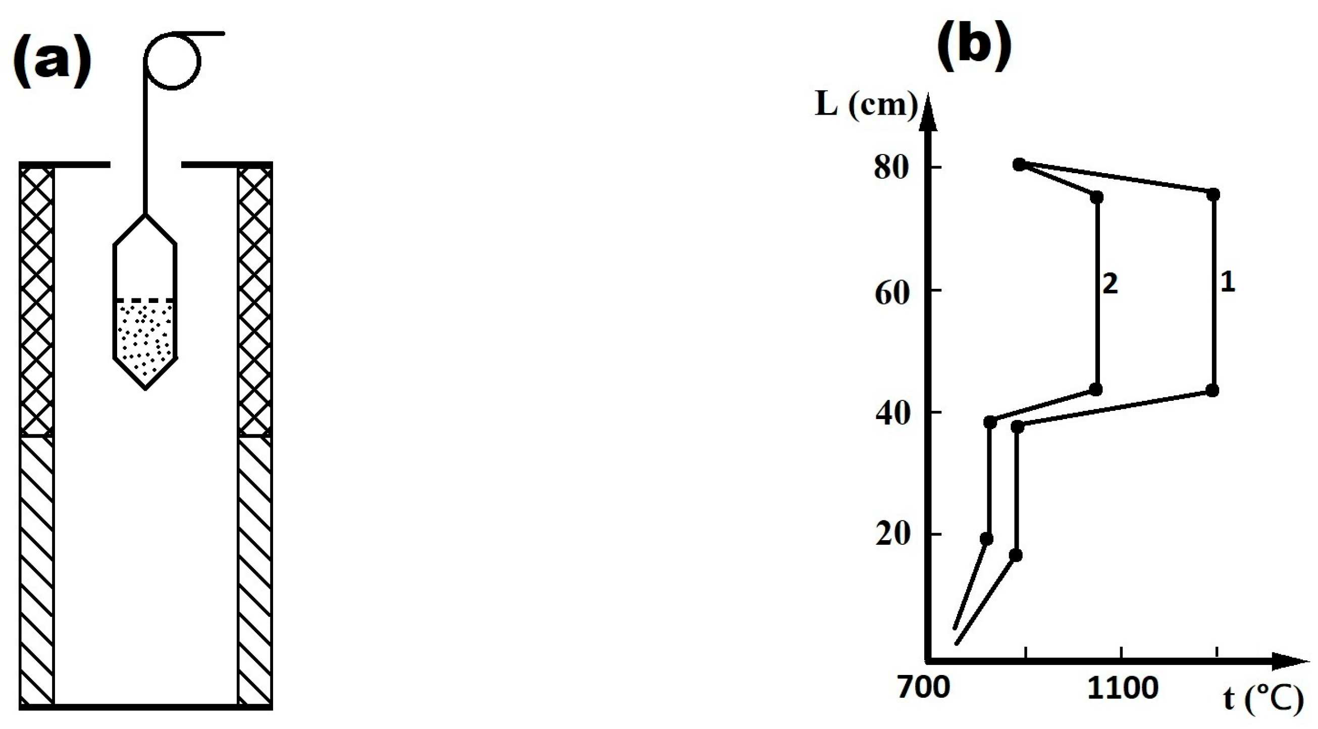
Figure 1. Experimental module for compound synthesis (a) and diagram of temperature distribution along the furnace length for the growth of GaSe single crystals (b).
Jcs 09 00194 g001
Figure 2. XRD plots of GaSe single crystals grown by Bridgman technique: (a) GaSe–Ga2Se–Ga2O3 composite formed by heat treatment (6 h at 820 °C), in air, of single-crystalline GaSe plates (b) and β–Ga2O3 crystals obtained by 6 h heat treatment at 900 °C, in air, of GaSe single crystals with peak indexing: 1—(401); 2—(002); 3—(111); 4—(111); 5—(401); 6—(311); 7—(202); 8—(600); 9—(112); 10—(510); 15—(313); 20—(020); 22—(022); 28—(314) (c).
Jcs 09 00194 g002
Figure 3. EDXS spectra for the surface of single crystalline GaSe plate subjected to heat treatment in air, at 820 °C for 30 min (a) and at 900 °C for 6 h (b).
Jcs 09 00194 g003
Figure 4. Raman spectra for the surface of GaSe plates subjected to heat treatment in air at 820 °C for 30 min (a) and at 900 °C for 6 h (1—115 cm−1; 2—146 cm−1; 3—169 cm−1; 4—201 cm−1; 5—254 cm−1; 6—296 cm−1; 7—327 cm−1; 8—349 cm−1; 9—416 cm−1; 10—475 cm−1; 11—630 cm−1; 12—656 cm−1; 13—746 cm−1) (b).
Jcs 09 00194 g004
Figure 5. SEM images of ε–GaSe (0001) surface after heat treatment in air at 820 °C for 30 min (a), micro- and nanoformations of α–Ga2Se3/β–Ga2O3 composite (b) and the nanostructuring of β–Ga2O3 layer obtained by heat treatment at 900 °C for 6 h (c,d).
Jcs 09 00194 g005aJcs 09 00194 g005b
Figure 6. Transmission spectrum of the ~2.8 µm thick single crystalline GaSe plate before (curve 1) and after heat treatment in air at 820 °C for 30 min (curve 2); inset determining direct band gap of this material (a); diffuse reflectance spectrum of the β–Ga2O3 layer formed on GaSe plate surfaces by heat treatment in air at 900 °C for 6 h (b); absorption edge of β–Ga2O3 layer (c).
Jcs 09 00194 g006
Figure 7. Photosensitivity of untreated GaSe single crystals (curve 1) and of semiconductor obtained by heat treatment of ~140 µm thick GaSe plate in air at 820 °C for 30 min (curve 2) (a) and at 900 °C for 6 h (b).
Jcs 09 00194 g007
Figure 8. Time dependence of the photocurrent for the photoresistor made of a layer of β–Ga2O3 nanowires/nanoribbons for U = 50 V, under illumination with 254 nm wavelength radiation (power density of 15 µW/cm2), in normal atmosphere (curve 1) and O3-enriched atmosphere (curve 2), air column length of 80 cm (a); time-dependent photocurrent for the photoresistor based on the Ga2Se3–Ga2O3 composite under illumination with 638 nm wavelength and 15 mW/cm2 radiation, applied voltage of 10 V, in normal atmosphere (curve 1) and an O3-enriched atmosphere (curve 2) and air column length of 3 m (b).
Jcs 09 00194 g008
Disclaimer/Publisher’s Note: The statements, opinions and data contained in all publications are solely those of the individual author(s) and contributor(s) and not of MDPI and/or the editor(s). MDPI and/or the editor(s) disclaim responsibility for any injury to people or property resulting from any ideas, methods, instructions or products referred to in the content.