Article

Graphene Oxide–Antibiotic Coatings with Improved Resistance to Microbial Colonization for Arthroplasty Implants

by
Gheorghe Iosub
1,
Adelina-Gabriela Niculescu
2,3,
Valentina Grumezescu
4,
Gabriela Dorcioman
4,
Oana Gherasim
4,
Valentin Crăciun
4,
Dragoș Mihai Rădulescu
1,*,
Alexandru Mihai Grumezescu
2,3,
Miruna Silvia Stan
5,
Sorin Constantinescu
1,
Alina Maria Holban
6 and
Adrian-Radu Rădulescu
1
1
Faculty of Medicine, Carol Davila University of Medicine and Pharmacy, 050474 Bucharest, Romania
2
Department of Science and Engineering of Oxide Materials and Nanomaterials, National University of Science and Technology POLITEHNICA Bucharest, 011061 Bucharest, Romania
3
Research Institute of the University of Bucharest—ICUB, University of Bucharest, 050663 Bucharest, Romania
4
Lasers Department, National Institute for Laser, Plasma and Radiation Physics, 077125 Magurele, Romania
5
Department of Biochemistry and Molecular Biology, Faculty of Biology, University of Bucharest, 050095 Bucharest, Romania
6
Department of Botany and Microbiology, Faculty of Biology, University of Bucharest, 050095 Bucharest, Romania
*
Author to whom correspondence should be addressed.
J. Compos. Sci. 2025, 9(2), 82; https://doi.org/10.3390/jcs9020082
Submission received: 19 December 2024 / Revised: 23 January 2025 / Accepted: 26 January 2025 / Published: 10 February 2025
(This article belongs to the Special Issue Advances in Laser Fabrication of Composites)

Abstract

:
In this study, we investigated the biocompatibility and antibacterial efficiency of hydroxyapatite/graphene oxide/ceftazidime (HAp/GO/CFZ) coatings obtained by the Matrix-Assisted Pulsed Laser Evaporation (MAPLE) technique for arthroplasty implants. The coatings were evaluated for their ability to inhibit biofilm formation by model opportunistic pathogens, specifically Gram-positive Staphylococcus aureus and Gram-negative Escherichia coli, for 24, 48, and 72 h. A significant reduction in the biofilm formation was demonstrated by coating surfaces, which led to a diminution of approximately 4 logs in the CFU/mL values compared to controls. These findings suggested that HAp/GO/CFZ coatings have the potential to prevent infections associated with arthroplasty implants, thereby improving patient outcomes and implant longevity.

1. Introduction

Arthroplasty is an orthopedic surgical intervention consisting of removing the articular surfaces or the joints, aiming to replace them with specific implants that restore joint function and eliminate pain [1]. Arthroplasty is generally recommended for patients who have suffered severe joint damage caused by joint disorders like osteoarthritis, trauma, inflammatory arthritis, and osteoarticular infections, which often lead to severe pain, limited joint function, and considerable deterioration in patients’ quality of life [2]. The most common arthroplasties include hip [3], knee [4], elbow [5], ankle [6], wrist [7], and shoulder [8] interventions. Although it is a common surgical procedure, arthroplasty can also lead to complications that may require repeated surgery and patients who undergo a much slower recovery process. Thus, it has been observed that bone fractures can occur at the implantation site, both during surgery and post-operatively, but also implant fractures can occur, leading to implant failure. Other complications may include infections (which may impair wound healing, as well as the success of the surgery), instability, deformities, continuous pain, and aseptic loosening of the implant [9,10,11,12,13]. In this context, clinicians and researchers should explore better the arthroplasty implant options to withstand and impede post-surgical complications. One of the most promising research directions is the development of bioactive coatings with adequate mechanical and biological properties.
Hydroxyapatite (HAp) has been widely explored for applications related to the reconstruction, regeneration, and replacement of hard tissues, especially due to its biocompatibility, non-immunogenicity, bioactivity, and osteoconductivity [14,15]. Hydroxyapatite coatings have been demonstrated to promote the osseointegration of metallic implants and facilitate subsequent bone regeneration, especially by developing HAp-based composite or hybrid coatings [16,17,18]. Combining hydroxyapatite with biopolymers, carbon-based nanomaterials, metal oxide nanoparticles, and natural or synthetic antimicrobial agents has been proposed and validated as a remarkable alternative for generating better-performing implant coatings [14,19,20,21,22].
Graphene oxide (GO) is a multifaceted material that has attracted attention for biomedical applications owing to its unique characteristics, including a highly specific surface area, excellent mechanical properties, and ability to be easily functionalized [23,24,25,26]. GO has been successfully used for implants’ coating to improve their properties and limit the corrosion of the base material by preventing the adsorption and migration of corrosive species on the material’s surface and increasing its corrosion resistance [27,28,29]. GO has also received great attention in tissue engineering and regenerative medicine due to its appealing features like electrical conductivity, mechanical resistance, and antimicrobial effects. It has been shown that GO coatings have promoted and supported bone formation around orthopedic and dental implants by enhancing cell adhesion, proliferation, and differentiation, thus helping the implants to integrate [30,31,32,33]. Owing to its intrinsic antimicrobial activity, GO can be used in coatings to prevent microbial adhesion and limit biofilm formation. By intercepting periprosthetic infections, these two properties can reduce post-operative complications and improve implant functionality [14,24].
At this stage, the study of graphene and graphene oxide presents certain challenges that need to be overcome. It has been shown that GO exhibits in vivo dose-dependent toxicity through the formation of reactive oxygen species (ROS), which cause cytotoxicity. Eliminating graphene oxide from the body remains a challenge and a subject for further study. Although this material has some limitations, GO-based composites exhibit impressive potential for biomedical applications, including drug delivery, tissue engineering, and antimicrobial therapy [14]. In this regard, combining GO with HAp holds great promise for developing advanced composites with synergistic properties.
Various coating techniques, such as chemical vapor deposition, electrochemical deposition, physical deposition, microarc oxidation, sol-gel, and plasma spraying, are employed for coating the metallic implants used in arthroplasty. There are also some physical vapor-deposition techniques, including magnetron sputtering, cathodic arc deposition, electron beam deposition, pulsed laser deposition, and close field magnetron sputter ion plating, that have been used to improve the properties of implants and coating materials by increasing their corrosion resistance, mechanical properties, biocompatibility, and functional performance [21].
One particularly appealing method for developing nanostructured coatings is the Matrix-Assisted Pulsed Laser Evaporation (MAPLE) process. The MAPLE technique has been extensively explored for biomedical applications to obtain coatings that improve biocompatibility and confer superior properties to the biomaterials and devices on which they are deposited [34,35]. It has also been validated in tissue engineering and regenerative medicine, thanks to the tissue-mimicking microstructure and functionality of developed coatings, as well as in the fabrication of biosensors. Moreover, being a non-contact and contamination-free process that does not damage the composition and structure of deposited compounds, MAPLE is considered ideal for generating uniform organic–inorganic thin layers or bioactive coatings with controlled thickness [36,37,38,39,40,41].
In this context, this study aims to highlight the potential of the MAPLE technique in developing drug-loaded HAp/GO coatings for titanium surfaces. The work also intends to evaluate the bioactivity and antimicrobial efficiency of proposed composites, finally proving that the GO-based coatings improved the surfaces of arthroplasty implants, which would further reflect on patient outcomes.

2. Materials and Methods

2.1. Materials

Commercial hydroxyapatite nanopowder (<200 nm particle size (BET), ≥97% purity), Sigma–Aldrich/Merck (Darmstadt, Germany) was the main component in the HAp/GO/CFZ composites. Graphene oxide (GO, powder, 4–10% edge-oxidized), ceftazidime hydrate (CFZ, C22H22N6O7S2 × xH2O), and dimethyl sulfoxide (DMSO, C2H6OS) were also purchased from Sigma–Aldrich (Merck Group, Darmstadt, Germany). Double-side polished (1 0 0) silicon (Si, 1 cm2 area) and titanium (Ti, 12 mm diameter, 0.2 mm thickness) substrates were provided by a local supplier.
For the biological and microbiological tests, most chemicals were purchased from Sigma–Aldrich (Merck Group, Darmstadt, Germany), otherwise the provider was accordingly mentioned during protocols. The American Type Culture Collection (ATCC®, Manassas, VA, USA) provided the hFOB 1.19 (ATCC® CRL-11372) cell line, but also the Staphylococcus aureus (S. aureus, ATCC® 25923) and Escherichia coli (E. coli, ATCC® 25922) strains.

2.2. Methods

2.2.1. Experimental Conditions of the MAPLE Process

For MAPLE targets, the HAp:GO:CFZ powdery mixture (5:1:0.5 weight ratio) was suspended in DMSO (2.6%), then as-obtained solutions were poured into a pre-cooled copper target holder that was subsequently immersed in liquid nitrogen for 30 min.
The obtaining of HAp/GO/CFZ coatings was conducted using a KrF* COMPexPro 205 laser source (λ = 248 nm and τFWHM = 25 ns) from Lambda Physik/Coherent (Göttingen, Germany) that operated at a repetition rate of 13 Hz. A laser beam homogenizer was used to improve the energy distribution of the laser spot. The laser fluence was within the range of 300–500 mJ/cm2, whereas the laser spot area was set to 22 mm2. All depositions were conducted at room temperature into a background pressure of 1 Pa at a target–substrate separation distance of 5 cm by applying 70,000 laser pulses. During the deposition, the target was kept at a temperature of ∼173 K by active liquid nitrogen cooling. The coatings were deposited onto (1 0 0) Si and Ti substrates for physicochemical analyses and biological assays, respectively, while a control set of coatings was prepared by dropcast on the double-side polished silicon.

2.2.2. Physicochemical Investigation

Scanning electron microscopy (SEM) images were collected using the secondary electron beam (20 kV acceleration voltage) of an InspectS50 FEI equipment (Thermo Fisher Scientific, Hillsboro, OR, USA) accessorized with an energy-dispersive X-ray spectroscopy (EDS) detector (Thermo Fisher Scientific, Hillsboro, OR, USA).
The compositional analysis was performed using an IRTracer-100 system (Shimadzu Europa GmbH, Duisburg, Germany). All scans were recorded in the 400–4000 cm−1 wavenumber range (4 cm−1 resolution) in the attenuated total reflection (ATR-FTIR) mode, and then they were processed with the LabSolutions IR version 2.2. software (Shimadzu).
X-ray diffraction (XRD) patterns were acquired using an Empyrean diffractometer (Malvern Panalytical, Almelo, The Netherlands), working with a Cu X-ray tube (45 kV, 40 mA) in a grazing-incidence geometry (parallel beam obtained with a mirror in the incident beam). The acquired patterns were analyzed using the HighScore Plus version 5.2 software (Malvern Panalytical, Almelo, The Netherlands) and the International Centre for Diffraction Data (ICDD®) 2022 database (Newtown Square, PA, USA).

2.2.3. Assessment of In Vitro Biocompatibility

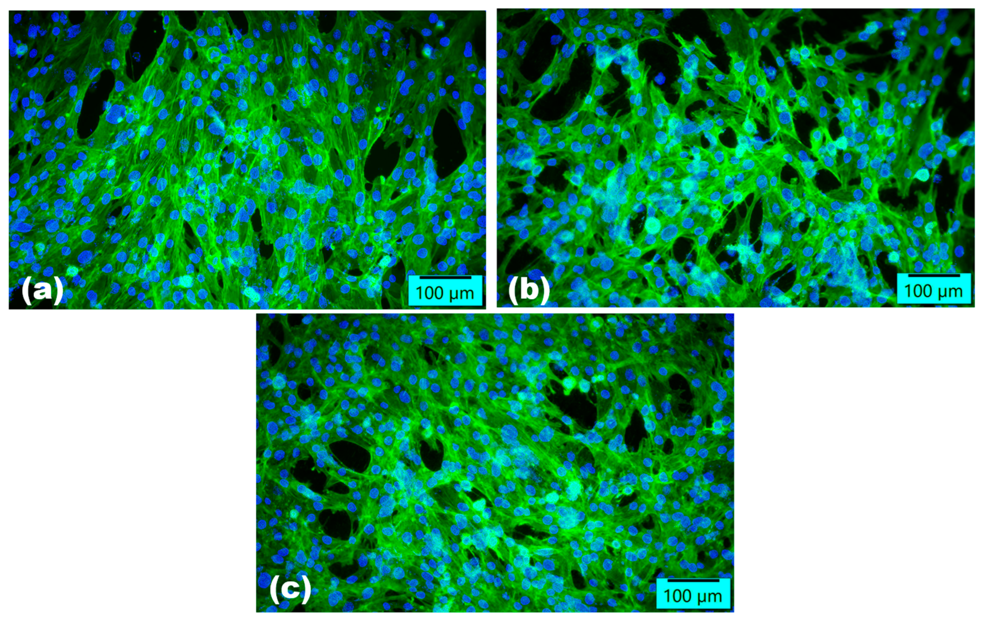
Human osteoblast hFOB 1.19 cells were grown at 34 °C under a humidified atmosphere with 5% CO2 in Dulbecco Modified Eagle’s Medium/Ham’s F-12 medium (1:1) without phenol red (Sigma–Aldrich/Merck, Darmstadt, Germany), supplemented with 10% fetal bovine serum (FBS), 2.5 mM of L-glutamine (Sigma-Aldrich), and 0.3 mg·mL−1 of G418 antibiotic (Sigma-Aldrich) with 10% FBS (Gibco/Life Technologies, Waltham, OR, USA) at 37 °C in a humidified atmosphere with 5% CO2. The cells were seeded at a density of 2 × 10⁴ cells/well on the tissue culture plastic surface of a 96-well plate (Control specimens) or on the top of the tested samples (uncoated surfaces—Reference specimens, and HAp/GO/CFZ-coated surfaces), which were previously sterilized under UV light. After 72 h of incubation in standard conditions, the biocompatibility tests were performed as previously described by Pirusca et al. [42]. The MTT test, based on the reduction of 3-(4,5-dimethylthiazol-2-yl)-2,5-diphenyltetrazolium bromide (MTT) within metabolically active cells, was used for the measurement of cellular viability, while the cell membrane integrity was assessed by quantifying the lactate dehydrogenase (LDH) release with the Cytotoxicity Detection KitPLUS (Roche, Basel, Switzerland). The cell morphology was visualized on the Olympus IX71 fluorescence microscope (Olympus, Tokyo, Japan) by labeling the actin filaments with phalloidin conjugated with fluorescein isothiocyanate (FITC) and cell nuclei with 4′,6-diamidino-2-phenylindole (DAPI) after the cells were fixed and permeabilized.

2.2.4. Microbiological Assay

To evaluate the antibacterial effectiveness of obtained coatings, we tested them against monospecific biofilms formed by the Gram-positive Staphylococcus aureus and the Gram-negative Escherichia coli. The biofilm formation was observed on coatings’ surfaces at different time intervals (24, 48, and 72 h).
In detail, both UV-sterilized uncoated (control) and nanocoated specimens were placed in sterile 24-well plates containing 1 mL of Luria–Bertani (LB) broth (Thermo Fisher Scientific), then inoculated with 10 μL of a 0.5 McFarland standard density microbial suspension (1.5 × 10⁸ CFU/mL). The plates were incubated at 37 °C for 24 h. Following incubation, the culture media were removed, and the samples were washed with sterile phosphate-buffered saline (PBS, Sigma–Aldrich/Merck, Darmstadt, Germany). The samples were then transferred to new sterile plates with fresh LB broth and incubated at 37 °C for 24/48/72 h. After incubation, the samples were gently washed with PBS and transferred into 1.5 mL centrifuge tubes containing sterile PBS. All specimens were vortexed for 20 s and sonicated for 10 s to detach the biofilm cells, producing cell suspensions. Serial 10-fold dilutions were made from the biofilm-embedded microbial cells, and the resulting PBS dilutions were seeded on LB agar plates (Thermo Fisher Scientific, Hillsboro, OR, USA) to determine the colony-forming units (CFU/mL) using a viable cell count assay.

3. Results

To investigate the compositional integrity of obtained materials and identify the optimal laser fluence for coatings’ processing, comparative infrared studies were conducted between the dropcast (DC, reference) and the MAPLE-coated samples (Figure 1). Compared to the DC specimen, a significant reduction of relevant absorption bands was observed for samples processed with minimal and middle laser fluence values. This observation was correlated with the poor and nonstoichiometric transfer of composite material that occurred at 300 mJ/cm2 and 400 mJ/cm2.
FT-IR analysis mainly provides information about the functional properties that correlate with the functional composition and structure of composite coatings. The broadband with maximal peak at ~3400 cm−1 was assigned to the O–H stretching vibrations within hydroxyl groups corresponding to GO. The band detected at ~1700 cm−1 was consigned to the carbonyl group, vibrations at ~1240 cm−1 indicated the stretching of C–O–C groups, and O–H deformation band was identified at ~1410 cm−1, while C–O stretching vibration was identified at ~1060 cm−1 [23,27,43,44]. Regarding the presence of HAp, the bands were assigned as follows: ~3750 cm−1 (OH– stretch) ~1090, and 1025 cm−1 (P–O asymmetric stretching within PO43−); ~962 cm−1 (P–O symmetric stretching within PO43−); ~630 cm−1 (OH libration); ~600 and ~570 cm−1 (O–P–O deformation within PO43−) [45]. Besides N–H bending noticed at ~1600 cm−1, other CFZ-originating carbon-containing moieties (~1750 and ~1700 cm−1 for C=O and carboxamide groups, respectively) could have been overlapped by the more abundant GO-originating functions [46,47]. In terms of preserved stoichiometry and laser transfer efficiency, optimal results were found for materials processed with the highest laser fluence of 500 mJ/cm2.
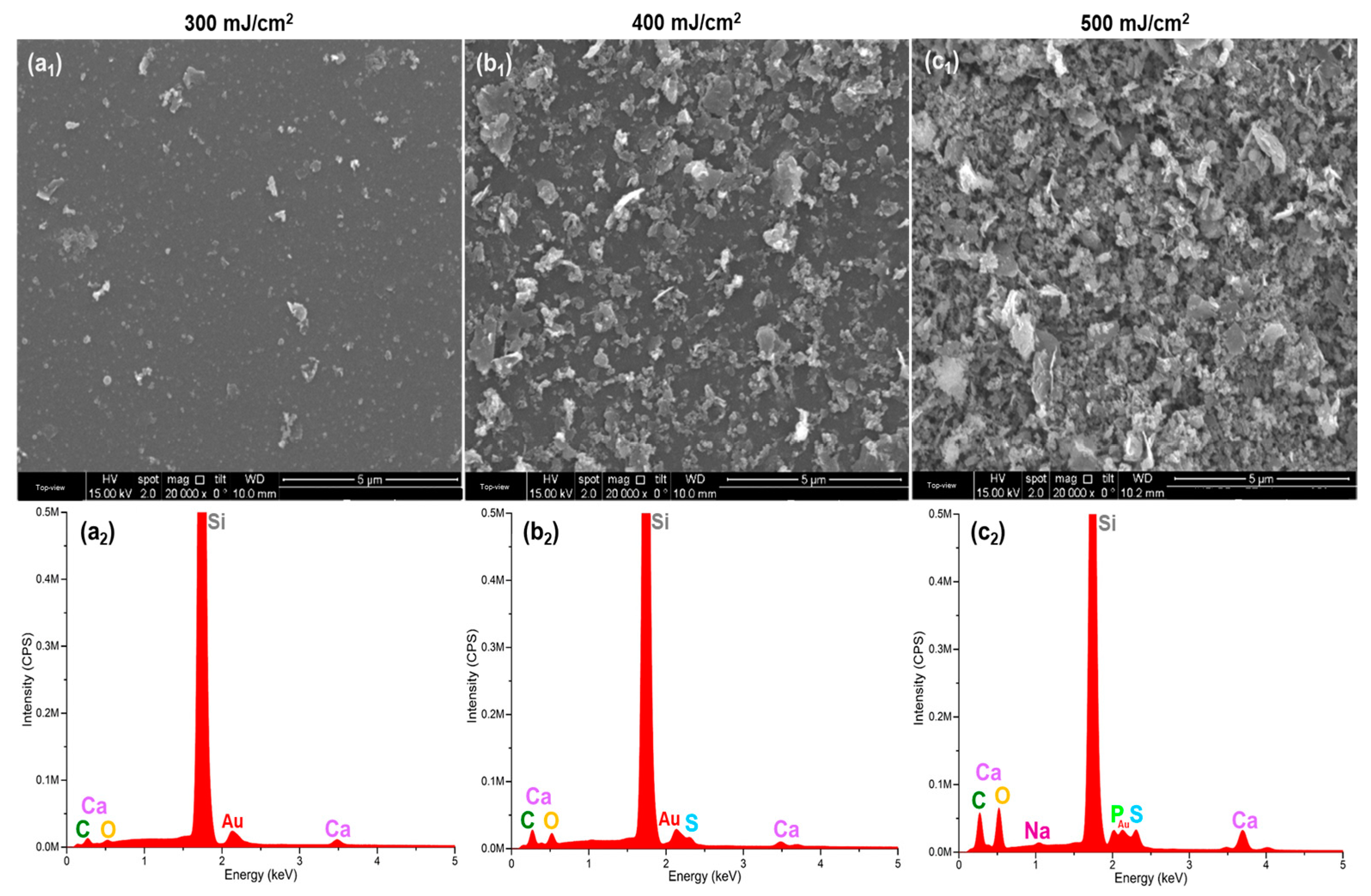
Compliant with the IR data, a more efficient transfer of the composite material was achieved by increasing the laser fluence, as evidenced both at microstructural (Figure 2a1–c1) and compositional (Figure 2a2–c2) levels. The presence of hydroxyapatite-based spherical nanostructured aggregates was observed on all surfaces, regardless of the laser fluence, but a significant increase in the amount of inorganic phase was noticed for coatings processed at 500 mJ/cm2. A similar behavior was observed for the graphene-based nanomaterial, which ranged from a few scattered flakes (Figure 2a1) to the full coverage of the substrate (Figure 2c1). This outcome was also supported by the EDS analysis, as collected spectra evidenced the presence of HAp-originating Ca (3.6–4.2 keV) and P (~2 keV) [28,30], GO-originating C (~0.3 keV) [31,32], and antibiotic-originating (S and Na) elements in the case of HAp/GO/CFZ materials processed at 500 mJ/cm2 (Figure 2c2).
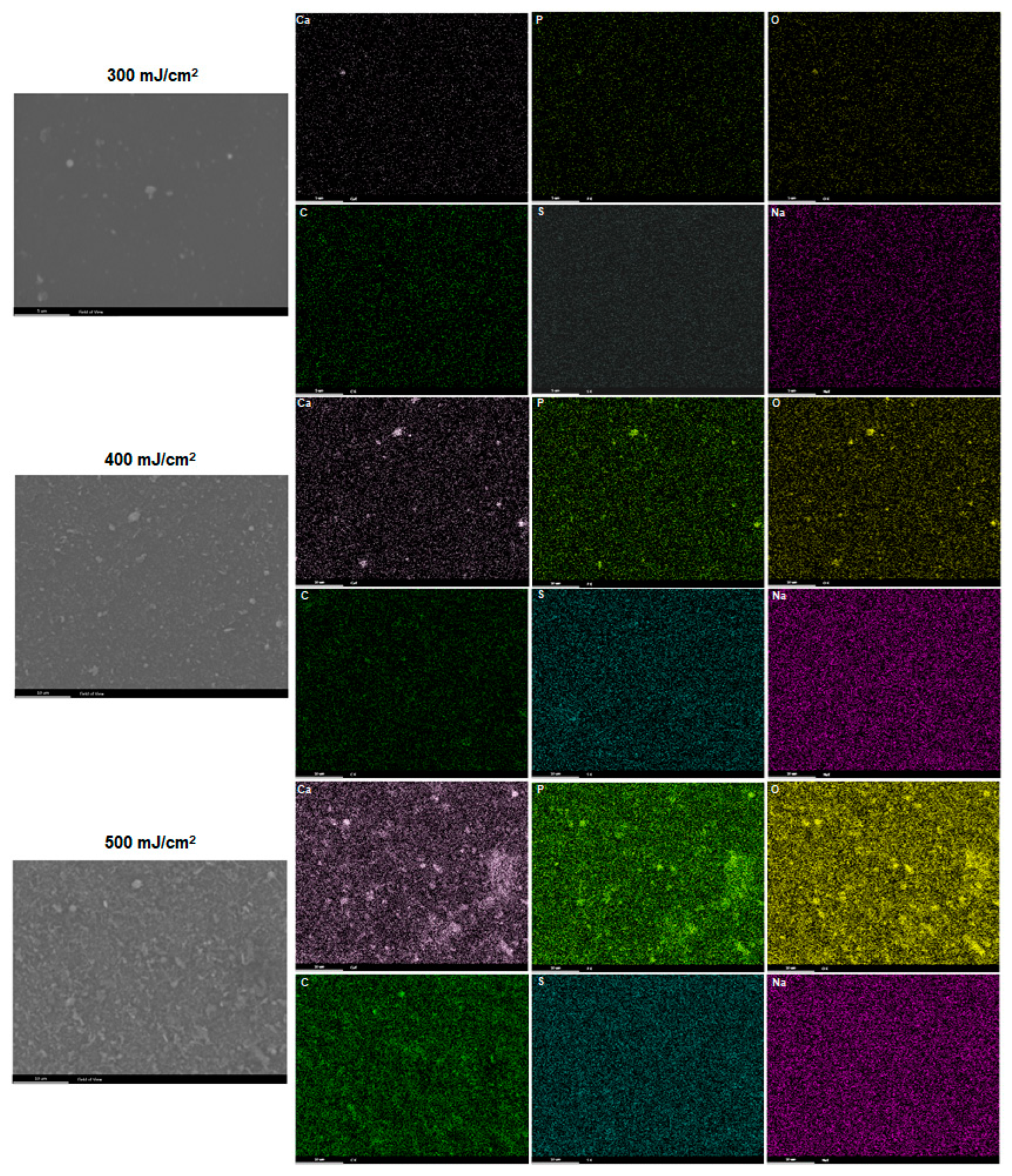
Compliant with FT-IR results and SEM images, the EDS maps (Figure 3) revealed a preserved composition and enhanced elemental distribution with increasing laser fluence. More precisely, the most effective transfer of HAp aggregates (enriched in Ca, P, and O elements) embedded within a uniform carbonaceous layer (GO-originating C and O) was observed for coatings processed at 500 mJ/cm2. Also, the surface distribution of CFZ-originating elements (S and Na) was more prominent in this case, confirming the successful use of this laser fluence for the efficient transfer of HAp/GO/CFZ composite.
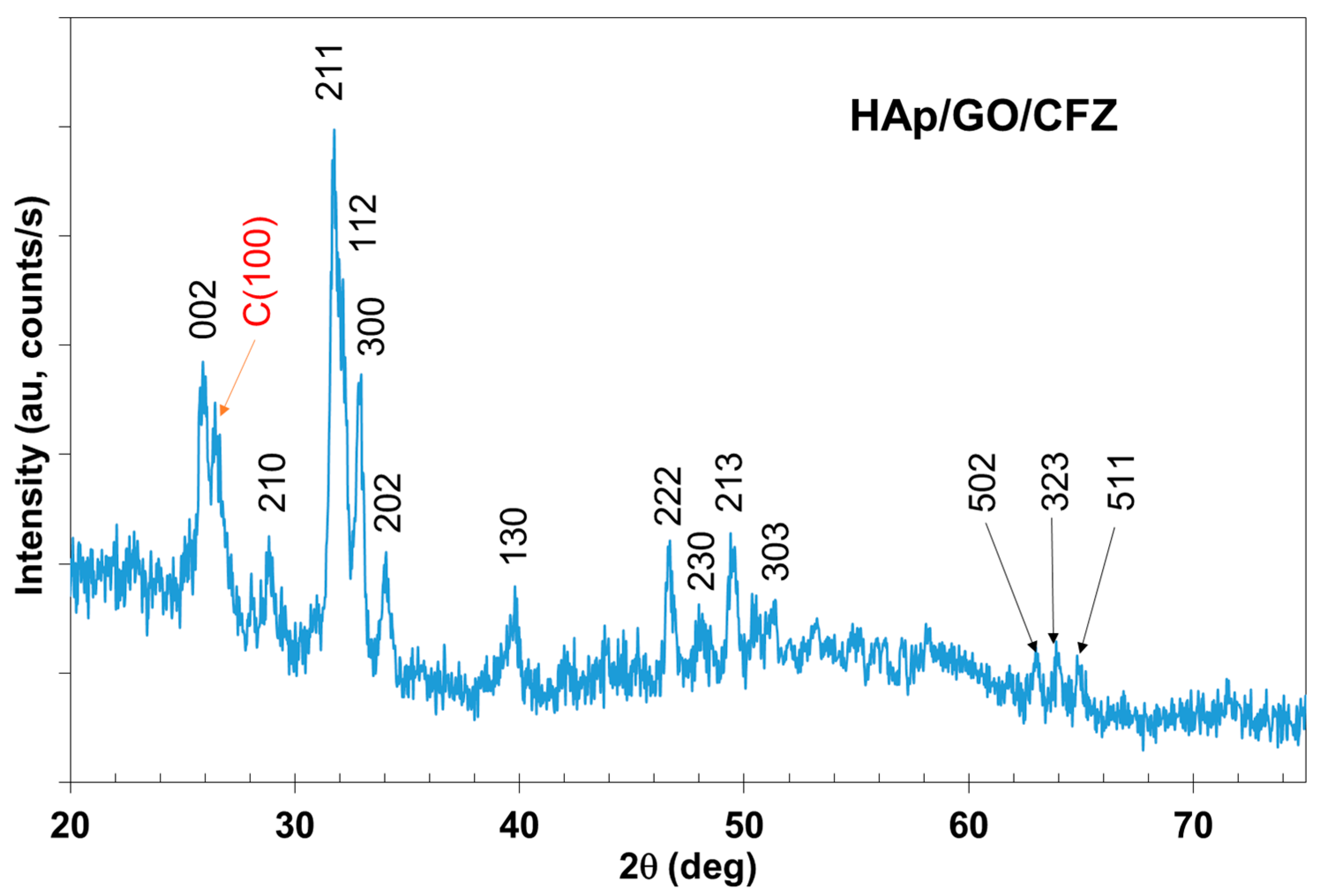
The indexed patterns correspond to synthetic hydroxylapatite reference pattern 04-013-6613 (ICDD® 2022 database). The deposited layer is polycrystalline, with grain sizes estimated from the Williamson–Hall plot and Line profile analysis of around 10 nm. A peak corresponding to carbon (reference pattern 04-016-4291) was also observed (Figure 4).
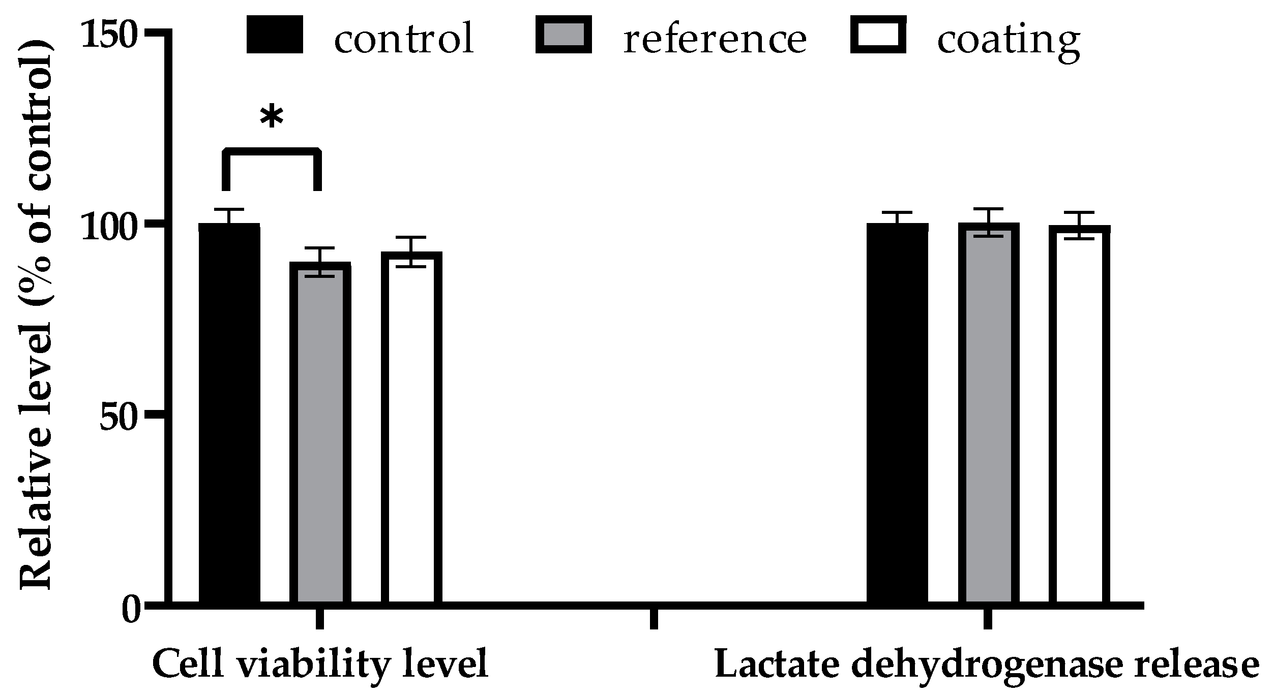
Figure 5 reveals the biocompatibility of the MAPLE-processed HAp/GO/CFZ sample (500 mJ/cm2) in the presence of human osteoblasts, the level of cell viability being almost similar to that of control (wells without any sample) and higher than that obtained in the case of the reference sample (uncoated surface). The lack of toxicity was confirmed by the absence of LDH release above the control level, showing that the osteoblasts’ membranes remained intact during contact with the HAp/GO/CFZ sample’s surface. In addition, the fluorescence staining of actin filaments in hFOB osteoblasts (Figure 6) revealed almost similar cell densities for both control and coated samples after 72 h of incubation. Many cell-to-cell junctions established by numerous lamellipodia and filopodia can be observed, which indicate good cell adhesion and migration on the coated surface, with the osteoblasts having a structured organization of actin cytoskeleton. In contrast, fewer cells were noticed in the reference sample, which suggests that the developed HAp/GO/CFZ coating has a very important role in cell adhesion, migration, and proliferation throughout its composition and properties.
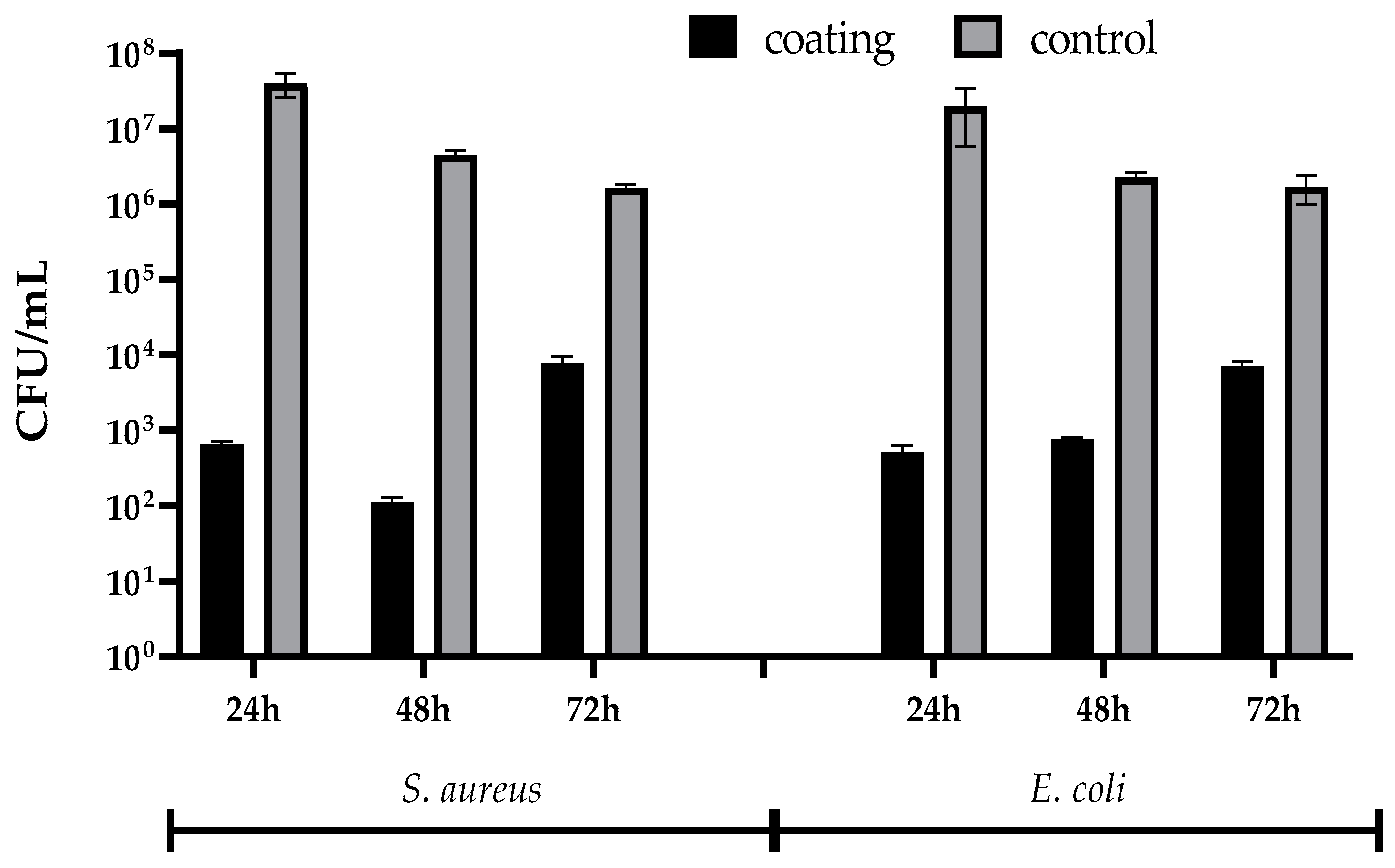
The microbiological results (Figure 7) demonstrate the ability of HAp/GO/CFZ coatings to intercept the opportunistic colonization of medical biomaterials (titanium). At the 24 h mark, biofilm formation is significantly lower on the HAp/GO/CFZ-coated surfaces compared to the control surfaces for both S. aureus and E. coli. Specifically, S. aureus shows a bacterial formulation of approximately 102 CFU/mL on the HAp/GO/CFZ surface, which is 4 logs lower than that of the titanium control (around 106 CFU/mL). For E. coli, biofilm formation is similarly reduced by the HAp/GO/CFZ-coated surfaces, with values of around 102 CFU/mL, compared to 106 CFU/mL on uncoated titanium-(control surfaces). This indicates that the HAp/GO/CFZ treatment significantly inhibits early biofilm formation for both bacteria.
After 48 h, the antibiofilm activity of HAp/GO/CFZ continues to be evident. The biofilm formation ability of S. aureus remains significantly reduced on HAp/GO/CFZ-coated surfaces, with approximately 103 CFU/mL values, compared to 107 CFU/mL on the uncoated titanium control. E. coli biofilm also shows a substantial inhibition, with biofilm-forming cells at about 103 CFU/mL on HAp/GO/CFZ surfaces versus 106 CFU/mL on control surfaces. This suggests that the antibiofilm effect of HAp/GO/CFZ is sustained over time and is effective against both types of bacteria.
By the 72 h mark, there is still a clear distinction in biofilm formation between the HAp/GO/CFZ-coated surfaces and the controls. S. aureus shows a biofilm-forming ability of approximately 104 CFU/mL on HAp/GO/CFZ surfaces, while the -control surfaces exhibit around 107 CFU/mL. E. coli population on HAp/GO/CFZ surfaces is about 104 CFU/mL, whereas the controls are slightly lower than previous time points, still around 106 CFU/mL. This consistent reduction highlights the prolonged effectiveness of HAp/GO/CFZ in preventing biofilm accumulation.
Overall, the HAp/GO/CFZ treatment determined strong antibiofilm activity against both S. aureus and E. coli by significantly reducing the formation and development of bacterial biofilms compared to uncoated titanium surfaces at all tested time points (24, 48, and 72 h) (Figure 7). This inhibitory effect is most pronounced in the early colonization stages (24 h) and remains substantial throughout the 72 h, indicating immediate and sustained antibiofilm properties. Therefore, the use of HAp/GO/CFZ coatings could be highly beneficial in applications where biofilm formation poses a significant problem, such as medical implants.

4. Discussion

Laser-processed coatings provide an attractive and application-tuned strategy to improve implantable biomaterials and devices’ surface characteristics (biomechanics, thermochemical stability, corrosion behavior) and modulate interface cellular events [48,49]. HAp-based coatings loading antimicrobial agents exhibit impressive therapeutic potential for hard tissue applications by boosting the osseointegration of metallic surfaces, enhancing subsequent bone regeneration, and exerting local antibiotherapy [14,50]. Being suitable for the stable, stoichiometric and uniform transfer of organics, the MAPLE technique provides indisputable versatility in fabricating biocompatible coatings with local therapeutic outcomes (immunomodulation, antimicrobial activity, antitumor action).
Achieving the unaltered and efficient transfer composites by MAPLE processing is a challenging aspect related to the final characteristics and functional outcomes of coated biomaterials and generally requires precise parametrization. Compositional and microstructural studies, and even comparative application-related evaluation, are employed for fabricating optimized coatings, with the laser fluence parameter (i.e., distribution of laser energy over target area) being mostly investigated [51]. In our study, HAp/GO/CFZ coatings were obtained by MAPLE processing, and the investigations revealed their suitable physical, chemical, and (micro)biological properties for improving arthroplasty implants.
HAp/GO/CFZ (5:1:0.5 weight ratio) composites were processed by MAPLE using different laser fluences (300, 400, and 500 mJ/cm2), then complementary compositional (FT-IR, EDS) and microstructural (SEM) studies were performed. FT-IR analysis (Figure 1), confirming the transfer of composite coatings through specific functional groups when compared to the reference mixture (dropcast), evidenced better laser transfer efficiency with increasing the laser fluence. Complementary EDS spectra (Figure 2a2–c2) confirmed the enhanced material transfer for higher laser fluences, with the 500 mJ/cm2 resulting in the most efficient transfer of organics (predominant presence of GO-/CFZ-originating elements, like C, S, and Na). Compliant with previous observations, EDS maps (Figure 3) demonstrated uniform and improved elemental distribution for the highest laser fluence value, while XRD data (Figure 4) confirmed the stoichiometric transfer of coatings obtained at 500 mJ/cm2. Moreover, SEM observations (Figure 2a1–c1) evidenced the formation of composite coatings following MAPLE processing, with an increased transfer efficiency for the highest laser fluence, which resulted in the uniform and full coverage of the substrate by a nanostructured composite coating. Taken together, our results demonstrated that the 500 mJ/cm2 laser fluence was optimal for the efficient MAPLE fabrication of uniform and nanostructured HAp/GO/CFZ coatings with preserved composition and stoichiometry.
The current findings are compliant with previous studies that reported the potential of the MAPLE technique for obtaining uniform coatings based on GO and/or HAp for improving biomedical implant surfaces. Besides its intrinsic antimicrobial activity (electrostatic-guided inhibition and inactivation of pathogens, size-related mechanical damage and chemical oxidation, and intracellular oxidative impairment) [52,53], GO can also act as a potentiating agent for natural or commercial antimicrobials. Thus, nanostructured GO-based coatings have been priorly evaluated for their ability to provide local antimicrobial effects. Nanocomposites based on biodegradable polyesters (polylactic acid or polycaprolactone), GO, and synthetic antibiotics (cephalosporins or carbapenems, respectively) have been validated for the biocompatible and antibiofilm surface modification of metallic implants [24]. GO, combined with stearate-conjugated silver nanoparticles, has also been proven successful for the local modulation of biofilm-associated periprosthetic infections [50]. Embedding growth factors within MAPLE-processed HAp-based coatings loading cephalosporin [14] or aminoglycoside antibiotics [50] has been reported as an effective strategy to simultaneously boost the osseointegration potential of metallic biomaterials and prevent or limit peri-prosthetic infections in bone-related applications. Developing HAp-based nanocomposites for loading growth factors has also been considered a promising approach for modulating the cellular response of orthopedical metallic implants, resulting in effective nanocoatings that support and promote the bone repair process.
Concerning the biological properties of the newly developed coatings, excellent biocompatibility was noticed with respect to human osteoblasts. After 72 h of standard incubation, HAp/GO/CFZ-coated titanium supported the cellular viability and did not induce cytotoxic effects, the results being superior when compared to the uncoated specimens (Figure 5). Compliant with the quantitative data, the HAp/GO/CFZ coatings were observed to play an essential role in cell adhesion, migration, and proliferation (Figure 6).
In addition, the biofilm formation ability of Gram-positive (S. aureus) and Gram-negative (E. coli) strains was significantly lower on the HAp/GO/CFZ-coated titanium compared to the uncoated surfaces (Figure 7). Important decreases of ~ 4 logs in bacterial populations were evidenced during the early stages of biofilm development (24 h), regardless of the tested pathogens. For S. aureus, the same inhibitory efficiency was evidenced after 48 h, while a slightly reduced activity (~ 3 logs) was observed after 72 h. Extended testing times led to lower antibiofilm effects against E. coli, with still important inhibitory action of ~3 logs and ~2 logs after 48 and 72 h, respectively. Altogether, microbiological results demonstrated the sustained antibiofilm efficiency of HAp/GO/CFZ coatings.
Thus, based on previous experience and current biological and microbiological results, it can be concluded that the chosen materials for this study have provided a desirable synergy toward achieving biocompatible and potent antimicrobial platforms for preventing infections associated with arthroplasty implants. The synergy between GO, HAp, and antimicrobial agents has also been demonstrated in several studies focused on different biomedical applications, with recent validation in wound care management [21] (in combination with zirconia and polylactic acid), hydrogels for reducing implant-associated friction [19] (in combination with cyclodextrin-based pseudopolyrotaxane), antibacterial coatings for titanium implants [36] (in combination with zinc oxide), bone tissue engineering [37] (in combination with agarose), and osteogenic differentiation in medicine and clinical dentistry [38] (in combination with polycaprolactone).
Previous studies concerning arthroplasty implant coatings have also validated the success of various materials and the feasibility of other deposition techniques. For example, Martinez et al. [36] have employed a high-velocity oxygen fuel spray (HVOF) method for preparing hydroxyapatite/graphene oxide/zinc oxide coatings with bioactive and antibacterial properties. Alternatively, Ryu and colleagues [39,40] have proposed the utilization of direct energy deposition (DED) technology for mimicking the porous structure of natural bones and applying titanium porous coatings onto CoCr alloy-based implants. A different approach was reported by Gabor et al. [54]. The authors have suggested the utilization of physical vapor deposition and microarc oxidation for fabricating hybrid Ti and ZrTi coatings that cover metallic Ti-6Al-4V bone implants.

5. Conclusions

To summarize, the results presented in this study demonstrate the significant antibiofilm activity of HAp/GO/CFZ coatings obtained by the MAPLE technique. Based on experimental outcomes, it can be stated that HAp/GO/CFZ-coated surfaces significantly diminished biofilm formation by both Gram-positive S. aureus and Gram-negative E. coli, a reduction that was consistent across all tested time points (24, 48, and 72 h). Thus, the proposed nanocomposite exhibits immediate and sustained antibacterial properties due to the synergistic action of constituent materials. In more detail, the combination of GO, HAp, and ceftazidime can collectively disrupt bacterial adhesion and biofilm development, acting as a highly efficient antimicrobial coating for implant surfaces. Moreover, the structural and compositional analyses revealed desirable physicochemical properties, confirming the uniform deposition and preserved material properties of the coatings, thus validating the effectiveness of the MAPLE technique for such applications.
To conclude, these findings indicate the high potential of HAp/GO/CFZ coatings in preventing infections associated with arthroplasty implants, rendering them promising for further improving patient outcomes and extending implant longevity. Nonetheless, in-depth research is further needed to optimize the coating composition and explore its efficacy in in vivo models, eventually moving toward clinical application of these materials.

Author Contributions

Data curation, V.G. and D.M.R.; Formal analysis, G.I., A.-G.N., V.G., G.D., O.G., V.C., D.M.R., A.M.G., M.S.S., S.C. and A.M.H.; Investigation, G.I., A.-G.N., V.G., G.D., O.G., V.C., D.M.R., A.M.G., M.S.S., S.C., A.M.H. and A.-R.R.; Methodology, A.M.G. and A.-R.R.; Writing—original draft, G.I., A.-G.N., V.G., G.D., O.G., V.C., D.M.R., A.M.G., M.S.S., S.C., A.M.H. and A.-R.R.; Writing—review and editing, A.-G.N., A.M.G., A.M.H. and A.-R.R. All authors have read and agreed to the published version of the manuscript.

Funding

This research received no external funding.

Data Availability Statement

The original contributions presented in this study are included in the article. Further inquiries can be directed at the corresponding author.

Conflicts of Interest

The authors declare no conflicts of interest.

References

  1. Gao, J.; Xing, D.; Dong, S.; Lin, J. The Primary Total Knee Arthroplasty: A Global Analysis. J. Orthop. Surg. Res. 2020, 15, 190. [Google Scholar] [CrossRef] [PubMed]
  2. Arthroplasty—Advanced Techniques and Future Perspectives; IntechOpen: Rijeka, Croatia, 2023.
  3. Colibazzi, V.; Coladonato, A.; Zanazzo, M.; Romanini, E. Evidence Based Rehabilitation After Hip Arthroplasty. HIP Int. 2020, 30, 20–29. [Google Scholar] [CrossRef]
  4. Konnyu, K.J.B.; Thoma, L.M.; Cao, W.; Aaron, R.K.; Panagiotou, O.A.; Bhuma, M.R.B.; Adam, G.P.M.; Balk, E.M.; Pinto, D. Rehabilitation for Total Knee Arthroplasty: A Systematic Review. Am. J. Phys. Med. Rehabil. 2022, 102, 19–33. [Google Scholar] [CrossRef] [PubMed]
  5. Samdanis, V.; Manoharan, G.; Jordan, R.W.; Watts, A.C.; Jenkins, P.; Kulkarni, R.; Thomas, M.; Rangan, A.; Hay, S.M. Indications and Outcome in Total Elbow Arthroplasty: A Systematic Review. Shoulder Elb. 2019, 12, 353–361. [Google Scholar] [CrossRef]
  6. McKenna, B.J.; Cook, J.; Cook, E.A.; Crafton, J.; Knabel, M.; Swenson, E.; Miner, S.; Manning, E.; Basile, P. Total Ankle Arthroplasty Survivorship: A Meta-Analysis. J. Foot Ankle Surg. 2020, 59, 1040–1048. [Google Scholar] [CrossRef] [PubMed]
  7. Boeckstyns, M.E.H.; Herzberg, G. Complications After Total Wrist Arthroplasty. J. Hand Surg. 2024, 49, 177–187. [Google Scholar] [CrossRef]
  8. Leafblad, N.; Asghar, E.; Tashjian, R.Z. Innovations in Shoulder Arthroplasty. J. Clin. Med. 2022, 11, 2799. [Google Scholar] [CrossRef] [PubMed]
  9. Docter, S.; Philpott, H.T.; Godkin, L.; Bryant, D.; Somerville, L.; Jennings, M.; Marsh, J.; Lanting, B. Comparison of Intra and Post-Operative Complication Rates Among Surgical Approaches in Total Hip Arthroplasty: A Systematic Review and Meta-Analysis. J. Orthop. 2020, 20, 310–325. [Google Scholar] [CrossRef] [PubMed]
  10. Goldenberg, B.T.; Samuelsen, B.T.; Spratt, J.D.; Dornan, G.J.; Millett, P.J. Complications and Implant Survivorship Following Primary Reverse Total Shoulder Arthroplasty in Patients Younger than 65 years: A Systematic Review. J. Shoulder Elb. Surg. 2020, 29, 1703–1711. [Google Scholar] [CrossRef]
  11. Hermus, J.; Voesenek, J.; van Gansewinkel, E.; Witlox, M.; Poeze, M.; Arts, J. Complications Following Total Ankle Arthroplasty: A Systematic Literature Review and Meta-Analysis. Foot Ankle Surg. 2022, 28, 1183–1193. [Google Scholar] [CrossRef] [PubMed]
  12. Júnior, D.J.E.; e Castro, A.D.A.; Fonseca, E.K.U.N.; Baptista, E.; Padial, M.B.; Rosemberg, L.A. Main Complications of Hip Arthroplasty: Pictorial Essay. Radiol. Bras. 2020, 53, 56–62. [Google Scholar] [CrossRef]
  13. Porrino, J.; Wang, A.; Moats, A.; Mulcahy, H.; Kani, K. Prosthetic Joint Infections: Diagnosis, Management, and Complications of the Two-Stage Replacement Arthroplasty. Skelet. Radiol. 2020, 49, 847–859. [Google Scholar] [CrossRef]
  14. Florea, D.A.; Grumezescu, V.; Bîrcă, A.C.; Vasile, B.Ș.; Iosif, A.; Chircov, C.; Stan, M.S.; Grumezescu, A.M.; Andronescu, E.; Chifiriuc, M.C. Bioactive Hydroxyapatite-Magnesium Phosphate Coatings Deposited by MAPLE for Preventing Infection and Promoting Orthopedic Implants Osteointegration. Materials 2022, 15, 7337. [Google Scholar] [CrossRef] [PubMed]
  15. Aijaz, M.; Ahmad, M.; Ansari, M.A.; Ahmad, S.; Kumar, A. Tools and Techniques Used for the Development of Scaffold for Bone Tissue Regeneration: A Detailed Review. Bioint. Res. App. Chem. 2024, 14, 123. [Google Scholar] [CrossRef]
  16. Burduşel, A.-C.; Sarchizian, D.; Niculescu, A.-G.; Holban, A.M.; Popescu, R.-C.; Truşcă, R.; Andronescu, E. Synthesis and Characterization of Hydroxyapatite–Zinc Oxide Nanocomposites Incorporating Rosemary and Thyme Essential Oils for Enhanced Bone Regeneration and Antimicrobial Activity. Rom. J. Morphol. Embryol. 2024, 65, 409–419. [Google Scholar] [CrossRef] [PubMed]
  17. Es-Saddik, M.; Laasri, S.; Bensemlali, M.; Hariti, N.; Laghzizil, A.; Taha, M. Experimental and Finite Element Study of the Mechanical Behavior of Hydroxyapatite/Tricalcium Phosphate/Alumina Biocomposite. Bioint. Res. Appl. Chem. 2024, 14, 37. [Google Scholar] [CrossRef]
  18. Buradagunta, R.S.; Madiga, J.; Dumpala, R. Producing Calcium Deficient Nano-hydroxyapatite from Silver pomfret Fish Bones for Biomedical Applications. Lett. Appl. NanoBioSci. 2024, 13, 130. [Google Scholar] [CrossRef]
  19. Fu, X.-K.; Cao, H.-B.; An, Y.-L.; Zhou, H.-D.; Shi, Y.-P.; Hou, G.-L.; Ha, W. Bioinspired Hydroxyapatite Coating Infiltrated with a Graphene Oxide Hybrid Supramolecular Hydrogel Orchestrates Antibacterial and Self-Lubricating Performance. ACS Appl. Mater. Interfaces 2022, 14, 31702–31714. [Google Scholar] [CrossRef]
  20. Stan, M.S.; Nica, I.C.; Dinischiotu, A.; Grumezescu, V.; Stoica, A.E.; Holban, A.M.; Grumezescu, A. Novel Coatings Based on Nanostructured Cefepime-Functionalized Magnetite for Implantable Devices. Mater. Proc. 2020, 2, 19. [Google Scholar] [CrossRef]
  21. Al-Wafi, R.; Mansour, S.; AlHammad, M.; Ahmed, M. Biological Response, Antibacterial Properties of ZrO2/Hydroxyapatite/Graphene Oxide Encapsulated into Nanofibrous Scaffolds of Polylactic Acid for Wound Healing Applications. Int. J. Pharm. 2021, 601, 120517. [Google Scholar] [CrossRef]
  22. Zulkarnain, N.A.; Ramli, A.; Kadir, N.H.A. Optimization on Characterization and Physicochemical of Non-Calcined and Calcined Nanocrystallite of Cuttlefish (Sepia officinalis) Bones Powder for Sustainable Green Technology Applications. Biointerface Res. Appl. Chem. 2024, 14, 58. [Google Scholar] [CrossRef]
  23. He, D.; Peng, Z.; Gong, W.; Luo, Y.; Zhao, P.; Kong, L. Mechanism of a Green Graphene Oxide Reduction with Reusable Potassium Carbonate. RSC Adv. 2015, 5, 11966–11972. [Google Scholar] [CrossRef]
  24. Constantinescu, S.; Gherasim, O.; Dorcioman, G.; Grumezescu, V.; Iosub, G.; Niculescu, A.-G.; Moldoveanu, E.-T.; Rădulescu, D.M.; Grumezescu, A.M. MAPLE-Prepared Graphene Oxide-Based Coatings for Improved Orthopedic Screws used in Knee Interventions. Rom. J. Morphol. Embryol. 2024, 65, 433–442. [Google Scholar] [CrossRef] [PubMed]
  25. Darmawan, I.; Wannasen, L.; Pinitsoontorn, S. Synthesis, Characterization, and Electrochemical Properties of Reduced Graphene Oxide Produced from Sugarcane Bagasse for Supercapacitor Applications. Biointerface Res. Appl. Chem. 2024, 14, 109. [Google Scholar] [CrossRef]
  26. Debbarma, J.; Debnath, R.; Saha, M. Graphene from Sugarcane Bagasse for Nonenzymatic Electrochemical Determination of Glucose. Lett. Appl. NanoBioSci. 2023, 12, 115. [Google Scholar] [CrossRef]
  27. Zhou, K.; Zhu, Y.; Yang, X.; Luo, J.; Li, C.; Luan, S. A Novel Hydrogen Peroxide Biosensor based on Au–Graphene–HRP–Chitosan Biocomposites. Electrochim. Acta 2010, 55, 3055–3060. [Google Scholar] [CrossRef]
  28. Feng, P.; Niu, M.; Gao, C.; Peng, S.; Shuai, C. A Novel Two-Step Sintering for Nano-Hydroxyapatite Scaffolds for Bone Tissue Engineering. Sci. Rep. 2014, 4, 5599. [Google Scholar] [CrossRef]
  29. Basavarajappa, N.S.; Niranjan, K.; Shilpa, K.C. Computation of Degree-Based Numerical Descriptors of Porous Graphene. Lett. Appl. NanoBioSci. 2023, 12, 117. [Google Scholar] [CrossRef]
  30. Predoi, D.; Iconaru, S.L.; Predoi, M.V.; Motelica-Heino, M.; Guegan, R.; Buton, N. Evaluation of Antibacterial Activity of Zinc-Doped Hydroxyapatite Colloids and Dispersion Stability Using Ultrasounds. Nanomaterials 2019, 9, 515. [Google Scholar] [CrossRef]
  31. Kalil, H.; Maher, S.; Bose, T.; Bayachou, M. Manganese Oxide/Hemin-Functionalized Graphene as a Platform for Peroxynitrite Sensing. J. Electrochem. Soc. 2018, 165, G3133–G3140. [Google Scholar] [CrossRef]
  32. Xavier, J.R.; Vinodhini, S.P. Fabrication of Reduced Graphene Oxide Encapsulated MnO2/MnS2 Nanocomposite for High Performance Electrochemical Devices. J. Porous Mater. 2023, 30, 1897–1910. [Google Scholar] [CrossRef]
  33. Horst, D.J.; Vieira, R.A.; García-García, A.; Moreno-Barcenas, A. X-MoS2/GO/Graphene Heterostructures and Doping Effects: KPFM Based Study of Surface Potential. Bioint. Res. App. Chem. 2024, 14, 118. [Google Scholar] [CrossRef]
  34. Alfe, M.; Minopoli, G.; Tartaglia, M.; Gargiulo, V.; Ausanio, G. Biocompatible Hybrid Graphenic Thin Coatings on Flexible Substrates through Matrix-Assisted Pulsed Laser Evaporation (MAPLE). ACS Appl. Mater. Interfaces 2024, 16, 38956–38967. [Google Scholar] [CrossRef]
  35. Ciocîlteu, M.V.; Mocanu, A.G.; Biță, A.; Manda, C.V.; Nicolicescu, C.; Rău, G.; Belu, I.; Pîrvu, A.S.; Balasoiu, M.; Nănescu, V.; et al. Development of Hybrid Implantable Local Release Systems Based on PLGA Nanoparticles with Applications in Bone Diseases. Polymers 2024, 16, 3064. [Google Scholar] [CrossRef] [PubMed]
  36. Martinez, G.; Henao, J.; Mandujano-González, V.; Giraldo-Betancur, A.; Forero-Sossa, P.A.; Corona-Castuera, J.; Rivera-Gil, M.A.; Poblano-Salas, C. Exploring Hydroxyapatite/Graphene Oxide/Zinc Oxide Composite Powders for the Preparation of Bioactive-Antibacterial Coatings by HVOF. Appl. Phys. A 2022, 128, 813. [Google Scholar] [CrossRef]
  37. Khosalim, I.P.; Zhang, Y.Y.; Yiu, C.K.Y.; Wong, H.M. Synthesis of a Graphene Oxide/Agarose/Hydroxyapatite Biomaterial with the Evaluation of Antibacterial Activity and Initial Cell Attachment. Sci. Rep. 2022, 12, 1971. [Google Scholar] [CrossRef] [PubMed]
  38. Daulbayev, C.; Sultanov, F.; Korobeinyk, A.V.; Yeleuov, M.; Taurbekov, A.; Bakbolat, B.; Umirzakov, A.; Baimenov, A.; Daulbayev, O. Effect of Graphene Oxide/Hydroxyapatite Nanocomposite on Osteogenic Differentiation and Antimicrobial Activity. Surf. Inter. 2021, 28, 101683. [Google Scholar] [CrossRef]
  39. Ryu, D.J.; Ban, H.Y.; Jung, E.Y.; Sonn, C.-H.; Hong, D.H.; Ahmad, S.; Gweon, B.; Lim, D.; Wang, J.H. Osteo-Compatibility of 3D Titanium Porous Coating Applied by Direct Energy Deposition (DED) for a Cementless Total Knee Arthroplasty Implant: In Vitro and In Vivo Study. J. Clin. Med. 2020, 9, 478. [Google Scholar] [CrossRef] [PubMed]
  40. Ryu, D.J.; Jung, A.; Ban, H.Y.; Kwak, T.Y.; Shin, E.J.; Gweon, B.; Lim, D.; Wang, J.H. Enhanced Osseointegration Through Direct Energy Deposition Porous Coating for Cementless Orthopedic Implant Fixation. Sci. Rep. 2021, 11, 22317. [Google Scholar] [CrossRef] [PubMed]
  41. Spirescu, V.A.; Niculescu, A.-G.; Slave, Ș.; Bîrcă, A.C.; Dorcioman, G.; Grumezescu, V.; Holban, A.M.; Oprea, O.-C.; Vasile, B.Ș.; Grumezescu, A.M.; et al. Anti-Biofilm Coatings Based on Chitosan and Lysozyme Functionalized Magnetite Nanoparticles. Antibiotics 2021, 10, 1269. [Google Scholar] [CrossRef]
  42. Pirușcă, I.A.; Balaure, P.C.; Grumezescu, V.; Irimiciuc, S.-A.; Oprea, O.-C.; Bîrcă, A.C.; Vasile, B.; Holban, A.M.; Voinea, I.C.; Stan, M.S.; et al. New Fe3O4-Based Coatings with Enhanced Anti-Biofilm Activity for Medical Devices. Antibiotics 2024, 13, 631. [Google Scholar] [CrossRef] [PubMed]
  43. Manoratne, C.H.; Rosa, S.R.D.; Kottegoda, I.R.M. XRD-HTA, UV Visible, FTIR and SEM Interpretation of Reduced Graphene Oxide Synthesized from High Purity Vein Graphite. Mater. Sci. Res. India 2017, 14, 19–30. [Google Scholar] [CrossRef]
  44. Sudesh; Kumar, N.; Das, S.; Bernhard, C.; Varma, G.D. Effect of Graphene Oxide Doping on Superconducting Properties of Bulk MgB2. Supercond. Sci. Technol. 2013, 26, 95008. [Google Scholar] [CrossRef]
  45. Vidhya, G.; Kumar, G.S.; Kattimani, V.S.; Girija, E. Comparative Study of Hydroxyapatite Prepared from Eggshells and Synthetic Precursors by Microwave Irradiation Method for Medical Applications. Mater. Today Proc. 2019, 15, 344–352. [Google Scholar] [CrossRef]
  46. Zhang, Y.; Zhang, X.; Hu, R.; Yang, Y.; Li, P.; Wu, Q. Bifunctional Nano-Ag3PO4 with Capabilities of Enhancing Ceftazidime for Sterilization and Removing Residues. RSC Adv. 2019, 9, 17913–17920. [Google Scholar] [CrossRef] [PubMed]
  47. Campos, L.A.d.A.; Neto, A.F.S.; Scavuzzi, A.M.L.; Lopes, A.C.D.S.; Santos-Magalhães, N.S.; Cavalcanti, I.M.F. Ceftazidime/Tobramycin Co-Loaded Chitosan-Coated Zein Nanoparticles against Antibiotic-Resistant and Biofilm-Producing Pseudomonas aeruginosa and Klebsiella pneumoniae. Pharmaceuticals 2024, 17, 320. [Google Scholar] [CrossRef] [PubMed]
  48. Bonciu, A.; Vasilescu, A.; Dinca, V.; Peteu, S.F. Interfaces obtained by MAPLE for chemical and biosensors applications. Sens. Actuators Rep. 2021, 3, 100040. [Google Scholar] [CrossRef]
  49. Gherasim, O.; Grumezescu, V.; Socol, G.; Ficai, A. Chapter 2—Nanoarchitectonics prepared by laser processing and their biomedicinal applications. In Nanoarchitectonics in Biomedicine; Grumezescu, A.M., Ed.; William Andrew Publishing: Norwich, NY, USA, 2019; pp. 23–53. [Google Scholar]
  50. Gherasim, O.; Grumezescu, A.M.; Grumezescu, V.; Negut, I.; Dumitrescu, M.F.; Stan, M.S.; Nica, I.C.; Holban, A.M.; Socol, G.; Andronescu, E. Bioactive Coatings Based on Hydroxyapatite, Kanamycin, and Growth Factor for Biofilm Modulation. Antibiotics 2021, 10, 160. [Google Scholar] [CrossRef]
  51. Patz, T.M.; Doraiswamy, A.; Narayan, R.J.; Menegazzo, N.; Kranz, C.; Mizaikoff, B.; Zhong, Y.; Bellamkonda, R.; Bumgardner, J.D.; Elder, S.H.; et al. Matrix assisted pulsed laser evaporation of biomaterial thin films. Mater. Sci. Eng. C 2007, 27, 514–522. [Google Scholar] [CrossRef]
  52. Tan, J.; Li, L.; Li, B.; Tian, X.; Song, P.; Wang, X. Titanium Surfaces Modified with Graphene Oxide/Gelatin Composite Coatings for Enhanced Antibacterial Properties and Biological Activities. ACS Omega 2022, 7, 27359–27368. [Google Scholar] [CrossRef] [PubMed]
  53. Liauw, C.M.; Vaidya, M.; Slate, A.J.; Hickey, N.A.; Ryder, S.; Martínez-Periñán, E.; McBain, A.J.; Banks, C.E.; Whitehead, K.A. Analysis of Cellular Damage Resulting from Exposure of Bacteria to Graphene Oxide and Hybrids Using Fourier Transform Infrared Spectroscopy. Antibiotics 2023, 12, 776. [Google Scholar] [CrossRef]
  54. Gabor, R.; Cvrček, L.; Doubková, M.; Nehasil, V.; Hlinka, J.; Unucka, P.; Buřil, M.; Podepřelová, A.; Seidlerová, J.; Bačáková, L. Hybrid Coatings for Orthopaedic Implants Formed by Physical Vapour Deposition and Microarc Oxidation. Mater. Des. 2022, 219, 110811. [Google Scholar] [CrossRef]
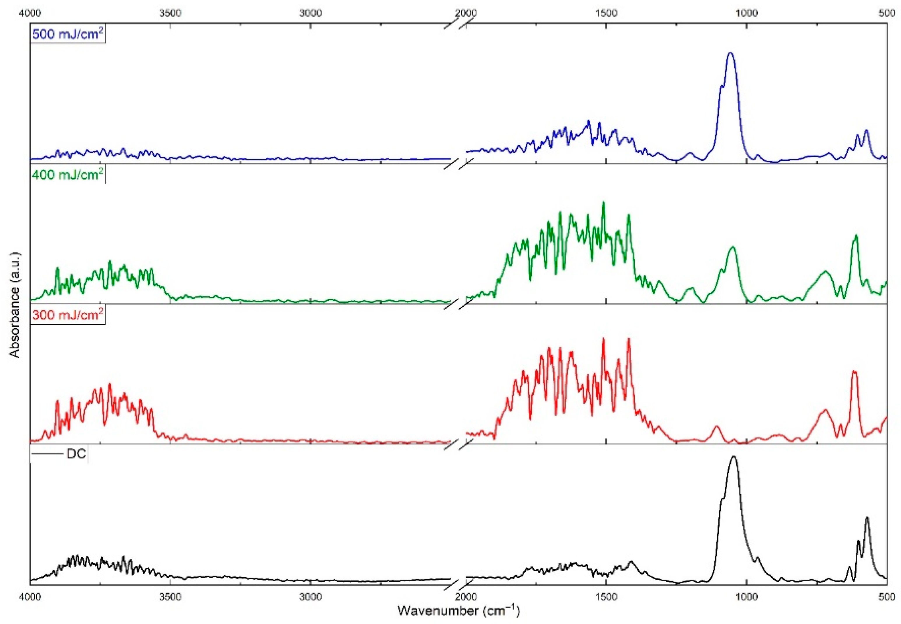
Figure 1. ATR-FTIR spectra of HAp/GO/CFZ DC and MAPLE coatings obtained at different laser fluences.
Jcs 09 00082 g001
Figure 2. Typical SEM micrographs of HAp/GO/CFZ coatings obtained by MAPLE at (a1) 300, (b1) 400, and (c1) 500 mJ/cm2 laser fluences, and the corresponding EDS spectra (a2,b2,c2).
Jcs 09 00082 g002
Figure 3. SEM micrographs with corresponding EDS maps of HAp/GO/CFZ coatings obtained by MAPLE at different laser fluences.
Jcs 09 00082 g003
Figure 4. XRD patterns for HAp/GO/CFZ coatings obtained at 500 mJ/cm2..
Jcs 09 00082 g004
Figure 5. Cell viability (MTT assay) and LDH release levels of human fetal osteoblasts after 72 h (results are calculated as means ± standard deviation of three independent experiments and shown relative to control; * p < 0.05 compared to control).
Jcs 09 00082 g005
Figure 6. Morphology of human fetal osteoblasts after 72 h of incubation by cytoskeleton staining (green: F-actin filaments labeled with FITC, and blue: nuclei labeled (with DAPI), where (a) control; (b) reference; (c) HAp/GO/CFZ coated surfaces (500 mJ/cm2 laser fluence).
Jcs 09 00082 g006
Figure 7. Graphic representation of S. aureus and E. coli biofilm development on HAp/GO/CFZ-coated surfaces (500 mJ/cm2 laser fluence) compared to uncoated control.
Jcs 09 00082 g007
Disclaimer/Publisher’s Note: The statements, opinions and data contained in all publications are solely those of the individual author(s) and contributor(s) and not of MDPI and/or the editor(s). MDPI and/or the editor(s) disclaim responsibility for any injury to people or property resulting from any ideas, methods, instructions or products referred to in the content.