Article

Assessing the Impact of Additive Manufacturing on Dental Clinical Workflows: A Process-Oriented Approach

by
Mariana Mutis Gómez
1,
Mario Guerrero Torres
2,
Sylvia María Villarreal-Archila
3 and
Jairo Núñez Rodríguez
1,*
1
Faculty of Industrial Engineering, Universidad Pontificia Bolivariana, University Campus Km 7 Vía Piedecuesta, Floridablanca 681007, Colombia
2
Faculty of Dentistry, Universidad Santo Tomas de Aquino, Bucaramanga 680001, Colombia
3
Faculty of Industrial Engineering, SOLYDO Research Group, Unidades Tecnológicas de Santander, Bucaramanga 680001, Colombia
*
Author to whom correspondence should be addressed.
J. Compos. Sci. 2025, 9(11), 579; https://doi.org/10.3390/jcs9110579
Submission received: 11 August 2025 / Revised: 7 October 2025 / Accepted: 14 October 2025 / Published: 28 October 2025
(This article belongs to the Section Composites Manufacturing and Processing)

Abstract

Additive manufacturing (AM) is rapidly transforming clinical workflows in dentistry by enabling the customized, efficient, and digitally integrated production of dental devices. However, the existing literature lacks a process-oriented perspective on its technical and operational impact. This study aims to address this gap through a dual-phase analysis using the Input–Transformation–Output (ITO) framework, providing practical insights into the operational reconfiguration enabled by AM. The first phase examined materials, image acquisition methods, design and lamination software, printing technologies, and key parameters across each stage of the AM workflow. The second phase analyzed four clinical applications (dental models, crowns and bridges, occlusal splints, and surgical guides) supported by a structured fabrication protocol and scanning electron microscopy (SEM) of 18 resin samples to assess surface quality and process-related defects. In addition, for each application, a comparative process analysis with traditional workflows was conducted using ASME diagramming. The findings indicate that AM reduces cycle times, manual intervention, and supply chain reliance while enabling production models such as Make-to-Order (MTO) and Engineer-to-Order (ETO). Its integration also fosters decentralized, in-clinic manufacturing with enhanced autonomy, flexibility, and reduced lead times. Nonetheless, this study highlights persisting challenges, including post-processing quality control, training requirements, and cost-efficiency concerns in low-volume settings. A hybrid model combining AM with conventional methods emerges as a pragmatic strategy for clinical adoption.

1. Introduction

Additive manufacturing, also known as 3D printing, is a manufacturing method that allows the manufacture of three-dimensional objects with complex geometries in a short time using 3D model data [1]. The digital workflow usually begins with the elaboration and/or acquisition of the object’s initial geometry, often using Computer-Aided Imaging (CAI) techniques, to generate a 3D model. This digital model is subsequently developed and refined using Computer-Aided Design (CAD) software. Once the design is completed, the Computer-Aided Manufacturing (CAM) stage takes over; at this point, manufacturing parameters such as the material to be used, the type of 3D printing technology and the printer’s specific settings are defined, and the CAM software processes the design (laminating it in layers) so as to generate the guidelines that will facilitate printing. Finally, the printing process is executed, followed by the necessary post-processing and inspection of the printed object to ensure its quality [2].
The evolution of computer-aided manufacturing has impacted different industries such as aerospace [3], construction [3], microfluidics [4], jewelry and biomedical fields [4] among many others; however, one of the most benefited areas has been the health sector, particularly the dental industry, as 3D printing has a series of advantages that make it attractive for this sector.
Firstly, it offers great design flexibility and rapid prototyping [3], allowing the development of complex, customized and precise geometries that would have been impossible or costly to manufacture using traditional methods. This is favorable, for example, in the production of customized prostheses and surgical devices that are tailored to the specific requirements of the patient. In addition, it has proven to be a more sustainable process compared to traditional methods, as it uses the precise amount of material needed to produce the item [1,5], significantly reducing the waste generated during the process.
However, additive manufacturing is not without its disadvantages. A notable drawback is its reduced strength [1,6], a quality that is highly valued in the dental industry. In addition, the initial costs of specialized equipment and materials are considerably high [7], which, alongside the need for additional post-production processes such as curing, sanding, or surface treatments, represents a significant barrier.
Nevertheless, computer-assisted manufacturing has redefined dentistry by enabling the production of therapeutic and auxiliary devices with high anatomical specificity. Its clinical uses include the manufacture of dental restorations, the fabrication of surgical guides, and the design of full or partial prostheses [8], among other expertise areas that continue to be explored.
The synergic integration of CAI, CAD and CAM technologies is fundamental to the materialization of these cutting-edge dental solutions. These digital systems facilitate everything, from accurate three-dimensional modeling and meticulous virtual planning to the automation of the production process. Consequently, clinical-prosthetic workflows are substantially optimized, which translates into a significant reduction in operating times [9], labor costs [10,11] and minimization of variability and human error [12] inherent in traditional dental manufacturing methods.
With regard to manufacturing processes, the dental industry works mainly with stereolithography (SLA) and digital light processing (DLP) [13], although techniques such as elective laser sintering (SLS), laminated object manufacturing (LMO), and powder bed fusion techniques (SLM, EBM, DMLS) [7,11] are also of interest.
On the other hand, different 3D printing methods currently enable the use of an ample variety of materials. Among the most common materials used within the field of dental care for both manufacturing methods (traditional and additive) are ceramics, metals, polymers, plaster, acids, cement, and resins [7,14]. Among the latter are materials such as photopolymerizable resins and UV resins. There are also composite resins, which are used in dental applications due to their notable precision and mechanical properties, such as their strength and biocompatibility [15]. Lastly, biodegradable resins have arisen as another prominent material in the field of 3D printing for dentistry [16], where it is vital to use materials that are both tolerated by the human body and efficient in the production of customized devices.
While most studies on AM in dentistry have concentrated either on a detailed analysis of clinical performance of devices or on materials science aspects [17,18,19], little attention has been given to analyzing AM as an integrated workflow and production system. This study addresses this gap by adopting a process-oriented approach that combines technical and operational perspectives within the operational context of dental manufacturing.
Building on this idea, this research seeks to answer the following question: How does additive manufacturing impact the dental industry? This question is answered by focusing on the production process and structuring the study into two complementary phases. The first phase consists of a literature review that aims to delve into and break down the additive process according to the input–transformation–output (ITO) stages, offering a process-oriented perspective still underexplored in the field. This includes the identification of materials, image collection methods, design and lamination software, types of printing technologies, equipment used, intended applications, and relevant parameters. For the second phase, we expand this approach by reviewing cases of study regarding the use of additive manufacturing in the dental industry, also framed within the input–transformation–output (ITO) structure. This was further explored with an analysis of generic and biocompatible resin materials through comparative tests with a scanning electron microscope (SEM), and a comparative study of the workflow carried out by traditional industry and additive industry, supported by the construction and analysis of ASME diagrams, in an effort to highlight the operational differences and their impact on the dental sector’s value chain.

2. Literature Review

Before proceeding, it is important to note that the literature search partially followed the PRISMA methodology. The initial search was conducted in the Scopus database, focusing on theoretical studies published between 2020 and 2025 addressing the use of additive manufacturing in the dental sector, and particularly focusing on resin-based applications. The search equation applied was “((additive AND manufacturing) OR (3D AND print)) AND (resin)”. Conference papers and articles that did not meet the predefined inclusion criteria were excluded. From this process, 752 open-access articles were initially retrieved, of which 225 were rejected for not fulfilling the acceptance criteria. Lastly, among the remaining articles, priority was given to those with at least 20 citations, resulting in a final set of 120 articles that served as the basis for the literature review. Figure 1 presents the article selection process according to the PRISMA methodology.

2.1. Materials (Input)

Within the scope of 3D printing in dentistry specifically, hydrogels such as PAM, PEG, PHEMA, PVA, metals, and ceramics are utilized, as well as thermoplastics such as ABS, PLA and PP [14]. However, resins are notably used [20] due to their ability to produce dental models and devices such as crowns, prostheses, clear aligners, splints, dentures, denture bases, surgical guides for implants, dental restorations, among others [21,22,23]. As for the production of prostheses and dental restorations, these are divided into two main groups: acrylic resins (such as PMMA and PEMA) and bis-acrylic resins [24]. They were introduced to the market in that order, and although both have performed admirably, bis-acrylics have shown “better aesthetic appearance, better mechanical properties, and less polymerization shrinkage compared to acrylic resins.” [24].
Following the initial screening, 30 articles were manually selected for the review of the most commonly used resins in dentistry, with priority given to publications from Q1 and Q2 journals. This way, based on the review, it was identified that among the most commonly used and/or researched resins in this field are acrylic resins, biocompatible resins, and resin composites (Table 1).
In clinical practice, the selection criteria for the material choice are based on the material’s properties and the intended use of the piece. This, in turn, will affect the selection of the printing method, according to the affinity with the chosen material and the desired surface finish [5,50]. However, for the vast majority of applications within the dental industry (especially for dental restorations), one requirement prevails: the materials must be biocompatible. In a few words, this term means that the material must not induce an adverse response when it comes into contact with tissues [3], not only to ensure better clinical integration, but also to minimize the risk of cytotoxic or inflammatory effects during treatment [24]. This property must be verified through certification [51,52] via tests such as the Pharmacopeia biological reactivity test (USP Class IV) or ISO 10993.
Biocompatible resins are characterized by their high impact strength, toughness, ductility, rigidity, elasticity, and resistance to tearing and temperature, as well as their ability to create pieces with internal chambers and complex or thick geometries [3]. Nevertheless, it should be noted that the mechanical properties of this type of resin vary according to its composition, altering its viscosity and thus, the impression and final finish of the device. For example, incorporating zirconium oxide enhances certain mechanical properties such as hardness and wear resistance, as well as flexural strength and impact strength when combined with fiberglass [53].
Moreover, for applications such as prostheses, one aim is to ensure that the properties of the 3D printing material are similar to those of tooth enamel. This must be determined by evaluating its biological, aesthetic, mechanical, physical and practical performance [51,52]. From an aesthetic and functional point of view, the material must then be colorless and tasteless but possess the ability to be pigmented and maintain its color and appearance after its processing. In terms of its physical and mechanical properties, the resin must be evaluated to ensure that it is durable enough to withstand masticatory forces and possible falls during manufacture.
To exemplify, according to a study by Zhang et al. [52], the compressive strength of light-curing resins must be between 146 and 448 MPa. Similarly, for acrylic resins, properties such as modulus of elasticity and impact strength must be evaluated. Dimitrova et al. [51] state that for 3D printing resins, these values must be lower than those of conventionally processed PMMA. Therefore, this last property must be at least 65 MPa according to the ISO requirements mentioned in their work, while the modulus of elasticity and impact resistance must be less than 2117.2 MPa and 14.756 kg/m2, respectively, in accordance with a study by Kawkab and Nadim [54], who identified these as the average values for a PMMA sample.
That said, although the flexural modulus does not have a standard value for resins used in dentistry, the literature review revealed that resins used for restorations and prostheses tend to have considerably high values in the range of 2 GPa. For instance, Zhang et al. [52] reported that fluid-type dental composites had flexural moduli between 2.34 and 6.32 GPa. Likewise, Sartori et al. [55] identified average values of 3.0 ± 0.2 GPa for composite resins, 1.8 ± 0.1 GPa for denture resins, and 2.7 ± 0.4 GPa for temporary crown and bridge resins.
Ultimately, it is essential to consider properties that are important for the adequate handling and processing of the material, such as viscosity and volumetric shrinkage.
The uncured resin’s viscosity should be as low as possible, especially if the resin contains fillers, as these can increase viscosity. The reason for this requirement is that high viscosity can cause clogging, irregular flow, and reduced printing accuracy, as well as cause the fillers to be distributed unevenly across the surface once they settle [53], leading to inconsistencies in the mechanical properties of the printed item.
Analogously, volumetric shrinkage should ideally be minimal, and although it has been recognized that in resins for 3D-printed dental prosthesis bases this variation is less than for conventionally processed PMMA, it should be noted that, for example, for the latter, ISO 1567: 1999; and ISO 20795-1: 2013 define the maximum value of unpolymerized monomer as 4.5% for self-curing resins and 2.2% for thermosetting resins, with average values of 8% and 0.53% volumetric and linear shrinkage, respectively [51]. This trait is particularly desirable for the making of dental molds, given that a high level of accuracy is required [56], interpreted as compliance with truthfulness and precision, so as to ensure that prostheses and other pieces designed on the models are also manufactured accurately in accordance with the requirements and dimensions of the patient’s morphology.

2.2. Design (Input)

This phase, corresponding to the input stage of the ITO process, encompasses several activities, as the generation of the file that will be processed by the CAM software to transform the product first requires a design, but not before acquiring data from the area, where the treatment will be carried out [14,57]. This highlights the importance of ensuring that CAD and CAM software is an “open system”, so as to allow data exchange between programs or devices from different ecosystems [58]. Likewise, the choice of CAD software depends on the complexity of the item being printed.
The digital process begins with the generation or acquisition of three-dimensional models. There are three alternatives to carry this out [9,14,57,59]. The first option involves creating the model from scratch using design software (CAD). The second alternative would be to obtain the models through intraoral or laboratory scanners, allowing the patient’s morphology to be captured directly and generating STL files that can be processed in CAD/CAM software. Finally, the user can choose to assemble them digitally based on tomograms (obtained mainly through CBCT), importing files in DICOM format to generate detailed three-dimensional models that can be integrated with models in STL format to achieve a complete digital workflow, which is especially useful for the design of surgical guides and planning in implantology.
These models are usually represented in file formats such as PLY or more commonly found as STL files and are then imported into computer-aided design (CAD) software where the detailed design of the part is carried out. Once the design is set in the CAD environment, slicing software is used. This software transforms the 3D model file into a sequence of two-dimensional layers, and during this process, crucial printing parameters such as layer thickness, material type, and the configuration of the required support structures are established. As a result, a file in G-code format is generated, containing the set of specific instructions for the machine. This file is finally transferred to the computer-aided manufacturing (CAM) system of the 3D printer, which interprets it to guide the physical fabrication of the object, layer by layer [14,57,60].
Based on the publications reviewed, it was found that among the most commonly used software for performing each of these tasks are: for image acquisition, software as CEREC (Bluecam and Omnicam) from Sirona, Lava COS or True Definition from 3M ESPE, iTero from Align Technology, CS 3500 from Carestream Dental LLC, Trios from 3Shape and PlanScan from Planmeca [57,58,61]; and for design, mainly DentalCAD from Exocad, but also software such as CEREC from Dentsply Sirona, Ceramill Mind from Amann Girrbach, 3Shape DentalCAD from 3Shape, Lava from 3MESPE, and MeshMixer from AutoDesk [58,62,63,64,65].
Moreover, the type of image processed by the software for image acquisition was identified. On the one hand, the most widely used is the STL format, which represents the geometry of the object using a triangle mesh and is processed by 3Shape [66], True Definition [67], iTero [68], PlanScan [69], CEREC [70], and CS 3500 from Carestream [71]. On the other hand, there is the DCM format, which is used by 3Shape software [66], and the PLY format, used by 3Shape [66], iTero [68], and Carestream’s CS 3500 (also exporting files in .udx and DICOM formats) [71], both formats being valid only within their own software ecosystems [2].

2.3. Additive Manufacturing Techniques (Transformation)

This step of the ITO process is carried out with the help of Computer-Aided Manufacturing (CAM) [7] using software and a printer model that is chosen based on its relevance to the design and type of material to be worked with. Once the design is complete, the STL file is sent for printing and processed by the 3D printer’s CAM software, the predefined parameters are adjusted, and the printing process begins with constant monitoring.
According to ISO/ASTM 52900:2021 [72], additive manufacturing processes are classified into seven categories: vat photopolymerization, material jetting, binder jetting, powder bed fusion, directed energy deposition, sheet lamination, and material extrusion. Table 2 summarizes the seven AM categories defined in ISO/ASTM 52900:2021 and their reported applications in dentistry.
Among these, vat photopolymerization has emerged as the most widely adopted for polymer-based dental applications, including crowns, occlusal splints, surgical guides, and study models [13]. Within this category, the predominant methods are stereolithography (SLA), digital light processing (DLP), and masked stereolithography (MSLA), which are consistently reported in the literature as the most frequently employed when working with resin-based materials in dentistry [14,25,60]. Owing to their widespread clinical relevance and central role in contemporary digital workflows, this study focuses primarily on these three technologies.
According to the ISO 529000s definition on VAT polymerization, these techniques all have in common the use of a liquid, thermosetting, photopolymer that is contained in a tank that has a transparent bottom to allow UV light rays to enter, and which solidifies on a platform that, once the process begins, descends to the bottom of the tank and begins to rise slowly as each layer undergoes photopolymerization [60,72].
As for DLP, this light is reflected onto the exposed layer of resin with the help of a series of micro mirrors, which follow a specific pattern and solidify the model layer by layer [14]. This technique is often used in the dental industry for the manufacture of surgical guides, models, implants, and invisible braces, due to the high precision it offers in the finishes of the printed products, its speed, and its ability to process materials widely used for dental applications, such as zirconium and alumina [14,51,78]. Among the most popular equipment for this type of printing are Sprintray Pro and MoonRay [60,62,79]; other notable printers include the NextDent 5100 from 3D Systems, Varseo XS from Bego, and CaraPrint4.0 Pro from Kulzer [51,60].
SLA differs from the previous technique due to the fact that it uses selective solidification, meaning that light is projected and directed in a precise manner using a laser that scans the surface of the resin using two mirrors, which helps to improve accuracy. However, even if it is one of the most accurate techniques, it takes longer to complete the printing process [35,60]. This technique is quite accurate, and its quality makes it ideal for creating customized products, such as models and surgical guides [78]. For this technique, one of the most favored printers is the Form from Formlabs [60,80].
Last of all, MSLA is a technique that uses a liquid crystal display (LCD) screen for resin polymerization, allowing it to perform higher-resolution printing in a shorter time compared to the two previous techniques by simultaneous exposure of the entire layer. However, it is slightly more expensive due to the cost of the LCD screen [60]. There is a wide range of printers available for this technique, but among the most used are the Phrozen Sonic series (mini, 4k 2022, Mighty 12k) and the Elegoo Mars series, although the NextDent LCD1 Anycubic Photon Mono are also common [25,60,81].
Figure 2 summarizes the main differences between tank photopolymerization techniques, highlighting their differences according to the ultraviolet light projection mechanism used to induce layer-by-layer curing of the resin.
It is worth noting that many manufacturers offer their own proprietary slicer, specifically calibrated for their equipment in order to guarantee optimal results during the additive manufacturing of dental devices [60], however other specialized programs are also used, such as Chitubox [25,81], PreForm from Formlabs [60], 3D Slicer [59], PrusaSlicer and Sli3r [81], among others.

2.4. Use of MA in Industry (Output)

Based on market trends, 3D printing has experienced rapid growth in multiple sectors such as the automotive, aerospace, jewelry, consumer products, energy, healthcare, electronics, architecture, and construction industries, among others [82,83]. This rise has been specifically attributed to the shift among large manufacturers to adopt this technology in the pursuit of mass production and customization, allowing for reduced delivery times and costs, compliance with standards (FDA, ASTM, ISO), innovation and improved manufacturing processes.
In the span of the years 2015 to 2020, according to Pearson Cases in Supply Chain Management and Analytics [82], the industry had a significant annual growth rate of 45.7%. Furthermore, the company Mordor Intelligence [83] conducted a market study based on historical data from the 3D printing market from 2019 to 2023, in which it forecasts a growth of 22.66% over the years 2024 to 2029, reflecting market dynamics such as increased adoption of additive manufacturing, demand for customization, and optimization of production costs.
Since its inception, the dental industry has sought to develop new technologies and materials. It has progressively incorporated new technologies, from early restorative tools such as the dental drill to CAD/CAM and, more recently, AM.
The early stages of 3D printing in this industry took place in the early 1980s with the manufacture of prototypes and casting models [84]. Its introduction represented a milestone in the production of dental devices, leading to the development of new materials and manufacturing methods, and it is from this moment that the concept of hybrid manufacturing emerges, an approach consisting of combining additive processes with traditional manufacturing techniques, enhancing the benefits of both methods. Consequently, 3D printing has been increasingly adopted across multiple dental specialties, including prosthodontics, oral and maxillofacial surgery, implantology, orthodontics, endodontics, and periodontics.
A comprehensive review of the literature and market trends [14,84,85] revealed that the rise of 3D printing in the dental industry has resulted from the need to improve patient safety and efficiency in procedures and appointments, optimizing care time, and the need to offer greater personalization and precision in treatments, achieving a natural appearance. Furthermore, in accordance with market research conducted by Mordor Intelligence [85], the 3D printing market in the dental industry is expected to have a compound annual growth rate (CAGR) of 19.5% for the span from 2024 to 2029. The research showed that the fastest-growing market for dental 3D printing is in the Asia-Pacific region, and that region with the largest share of the global market in 2024 is North America, with 3D Systems, Inc., Stratasys, Nexa3D, Desktop Metal, Inc., and Renishaw plc being the main companies operating in global dental 3D printing.
Consistent with the procedure adopted for the analysis of materials, a targeted selection of 32 articles was carried out for the review of the most commonly employed resins in dentistry, giving priority to publications in Q1 and Q2 journals. After reviewing the selection, it was found that the most researched and manufactured devices using 3D printing resin are prostheses, most notably denture bases, restorations (particularly crowns), orthodontic applications such as aligners, splints, brackets, occlusal splints for rehabilitation, dental models, and surgical guides (Table 3).

2.5. Post-Processing (Output)

As an integral part of the output phase, an assessment is made as to whether the piece requires additional finishing processes in order to ensure it is in optimal condition [91]. Ideally, post-processing should be minimal, but if necessary, dental pieces undergo manual finishing processes that may include sanding, polishing, washing, varnishing, and/or ultrasonic cleaning [7,14].

3. Materials and Methods

This study aims to delve into the processes involved in additive manufacturing or 3D printing with the use of resin in the dental industry, following the manufacturing steps of input–transformation–output (ITO). This methodology is particularly useful for analyzing manufacturing processes, as it allows the process to be broken down in terms of inputs (initial resources), transformation (operations that convert the inputs), and outputs (final products or results), making it easier to identify critical aspects at each stage and ensuring detailed control over each phase of the process, as well as the production of a high-quality piece.
Within the context of dental 3D printing, this method takes the form of a complete digital flow. It begins with data acquisition, device design, and material selection (input), continues with the definition of printing parameters and layer-by-layer manufacturing using additive techniques (transformation), and ends with post-processing and evaluation of the finished device (output) [5]. Thus, each stage of the ITO process involves consideration and decision-making relating to a series of variables, for example, input decisions about design software and materials, or the setup of printing parameters in the transformation phase.
In an effort to evaluate the final properties of materials transformed using additive manufacturing and to contextualize the additive manufacturing process in different dental practices, an analysis was performed on 18 resin samples that were used for the making of dental molds or impressions (11), surgical guides (3), temporary restorations (1), permanent restorations (2), and occlusal splints (1). A standard protocol was established for the analysis of all resins, which entailed: obtaining samples, identifying and implementing the method and design parameters, printing, post-processing, and microstructural characterization via SEM (Tescan, Brno, República Checa).
Considering that the study analyzes materials which were initially liquid, and by virtue of the pressure conditions to which the sample must be subjected inside the microscope, they cannot be analyzed in this initial state [92], scanning electron microscopy (SEM) was used only in the final stage of the ITO process to perform qualitative characterization and quality analysis of the surface of the 18 printed and treated materials.
For the final step, in order to identify critical activities in the operational flow and compare the ITO process of both traditional and additive manufacturing in dentistry, an analysis of the differences in each of these three stages and the impact of incorporating 3D printing into different links in the value chain was carried out. This was achieved through theoretical recognition and a case study. Specifically, the case study focused on the making of dental models, surgical guides, crowns and bridges, and occlusal splints. To this end, each part of the additive and traditional manufacturing processes was documented in an ASME process flow diagram. Figure 3 below details the steps carried out for each phase of the ITO process.

4. Results

4.1. Materials

Some of the key properties observed in the dental molding resins analyzed are their high print resolution, low volumetric shrinkage, and reduced viscosity, with the exclusion of the elastic resin. For instance, in cases where suppliers did report volumetric shrinkage values for their materials after curing, it was confirmed that most of them comply with what was previously indicated in the literature review, being less than 8%, which is considered adequate for dental applications. In addition, it was identified that Siraya Tech’s castable resin, Portux’s Model resin, and PriZma’s 3D Wide resin are specifically formulated to produce dental models, as indicated in their user guides and technical data sheets.
Despite the fact that not all suppliers provided complete information in their technical data sheets regarding the properties of the materials, Table 4 summarizes the most notable properties that allow them to be considered suitable for printing functional and accurate dental models.
Likewise, when analyzing the properties reported by suppliers in the technical data sheets (Table 5), it was noted that 5 out of the 7 evaluated resins meant for clinical use had certifications ensuring their conformity with the biocompatibility standards required for intraoral use, excluding the standard resin from Anycubic and the surgical guide resin from Antisky. Moreover, they had low viscosity values, which is favorable for the printing process. Although none of the manufacturers reported flexural modulus values in their technical data sheets, it was proven that in most samples, their flexural strength exceeded the minimum threshold of 65 MPa established in the literature as the acceptance criterion for dental resins, and it was also found that they possess flexural modulus values close to 2 GPa, consistent with what has been documented in previous studies.
Aside from their mechanical properties and biocompatibility, these resins were chosen for the manufacture of dental devices based on the fact that they have been specifically formulated for these applications, as indicated by their manufacturers. On one hand, Portux SG resin is specially designed to create surgical guides that facilitate the visualization of tissues and teeth adjacent to the area being treated, thanks to its translucent blue color; additionally, according to its supplier, it withstands deformation and discoloration after autoclave sterilization processes [106]. On the other hand, Portux Temp resin can be used to fabricate temporary restorations, including crowns, bridges, inlays, onlays, veneers, and prosthetic teeth for full dentures, ensuring a good fit, aesthetics, and compatibility with temporary cements [107,108]. Similarly, PriZma 3D Bio Crown resin can be used to create the same set of temporary restorations as stated by its supplier, but it is mainly used for permanent restorations [109]. Finally, PriZma 3D Bio Splint resin is ideal for the printing of retainers and splints due to its clear appearance and resistance [110].
Table 5. Properties of the clinical application samples collected.
ResinBiocompatibilityViscosityFlexural ModulusFlexural StrengthReferences
New stetic—Portux SGISO 10993-5, 10 and 23380 mPa.s>1.5 GPa
(ISO 10477)
>50 MPa
(ISO 10477)
[106]
New stetic—Portux TempISO 10993-5, 10 and 23 380 ± 80 mPa.s>1.8 GPa>90 MPa[107,108]
PriZma 3D—Bio Crown (tint: Bleach)In accordance with ISO 4049 standard255–500 mPa.s2.85 GPaat 5%, ≥105.5 Mpa[109,111]
PriZma 3D—Bio Crown (tint: A2)In accordance with ISO 4049 standard255–500 mPa.s2.85 GPaat 5%, ≥105.5 Mpa[109,111]
PriZma 3D—Bio SplintAnvisa Registration 80483740001190–500 mPa.s2.97 GPaat 5%, 102.8 MPa[110,112]
Anycubic—Standard Resin, clearNot applicable200–230 mPa.s1.4–1.6 GPa50–60 MPa[113]
Antisky—Dental Guide Resin, clearNot certified280–380 mPa.s2.3–2.4 GPa100–110 MPa[114]

4.2. Design

In terms of design, samples of the five generic resins were generated using Lychee software (Version 7.2.2.0, Mango 3D SAS, Paris, France), employing basic geometry for a subsequent study of the materials. The designs for the remaining pieces (Figure 4) were created using Exocad Dental CAD software (version 3.1, Exocad, Rijeka, Croatia), and the surgical guides were designed using Implant 3Shape Unite software (Version 1.7.27.6, 3Shape, Copenhagen, Denmark).

4.3. Printing

To begin with, the 3D printing technologies that best suited the properties of the resins were identified and selected. Hence, the generic resin samples were printed with the Phrozone Mini 4k (Phrozen, Hsinchu, Taiwan) and the SLA printing technique. In parallel, the remaining samples were printed on Elegoo Mars 3, Elegoo Mars 5 ultra (Elegoo, Shenzhen, China), Anycubic Photon Mono X, Anycubic Photon Mono 2, and Anycubic Photon Mono 4 Ultra 10K printers (Anycubic, Shenzhen, China) using the MSLA printing technique. Table 6 details the selection.
Following this, the printing settings or parameters were adjusted according to the specifications provided by the suppliers for the type of printer used in every case. Table 7 summarizes the specific parameters used during the printing of a selection of samples, including lifting distance and speed, exposure time, layer height, bottom layer count and exposure time corresponding to these layers.
Upon choosing the equipment, the designs were transferred to the respective slicers to add the supports and final adjustments before starting the printing process. Figure 5 shows the printed pieces, corresponding to the result of the additive manufacturing process.

4.4. Post Processing

The 5 generic resin samples were washed and cured using the Anycubic washing and curing machine (Anycubic, Shenzhen, China). The other pieces were subjected to a similar treatment, through a wash in isopropyl and/or ethyl alcohol for 5 to 10 min, followed by drying with air, and they underwent a curing process. The curing chambers used were the Elegoo Mercury plus (Elegoo, Shenzhen, China) and the Anycubic Wash and Cure 2.0 (Anycubic, Shenzhen, China), and the curing times depended on the type of part. For example, the surgical guide was subjected to 2 sessions of 15 min each, while for the temporary restorations, each session had a duration of 30 min.

4.5. Scanning Electron Microscope Analysis

SEM technology uses an electron beam that interacts with the surface of the material (which, due to the vacuum conditions inside the microscope, must be in a solid state), generating a pattern and creating with it a high-resolution image with the aid of specialized electron detectors [92]. The said pattern can then be further analyzed to identify, among many characteristics, the composition and topography of the sample.
This technology has been used in multiple studies within the field of dentistry to make an evaluation of printing parameters, such as printing direction [115], to test material-induced cytotoxicity [115], and for the analysis of microstructures and mechanical properties of materials [15,115,116]. In this context, the images that follow are not intended solely as a material characterization exercise, but rather as a complementary perspective that links surface morphology to clinical performance, by highlighting why features on the material’s surface influence the mechanical stability, aesthetics, or long-term durability of dental devices.
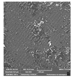
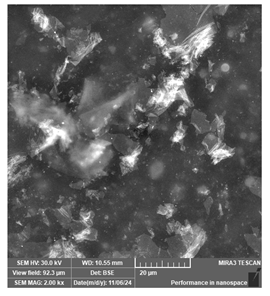
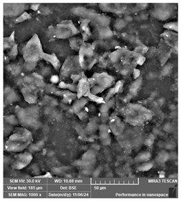
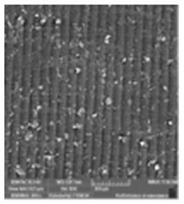
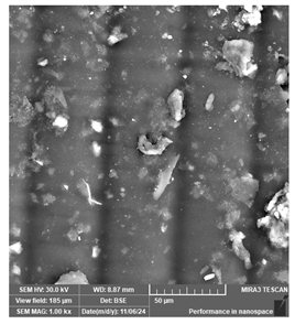
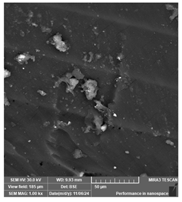
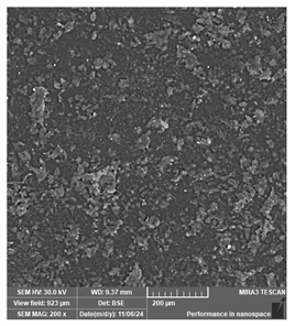
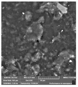
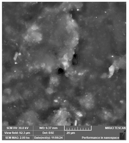
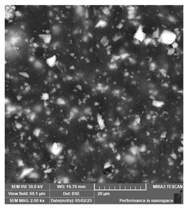
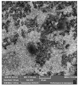
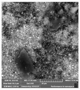
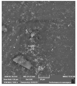
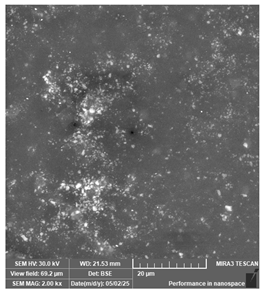
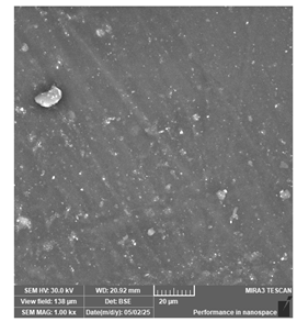
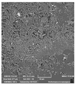
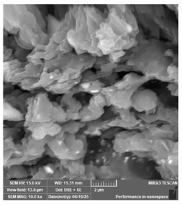
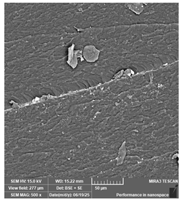
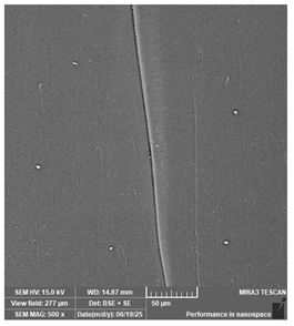
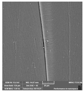
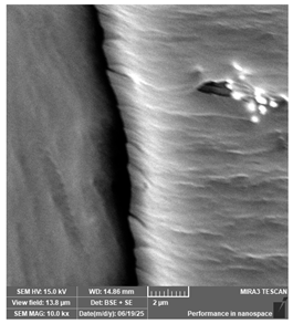
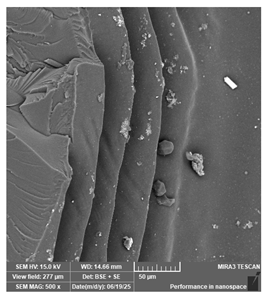
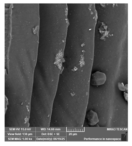
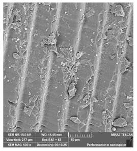
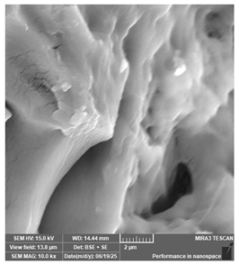
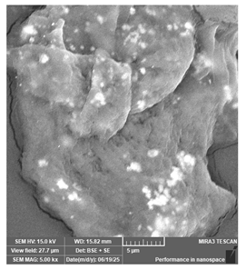
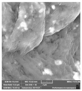
For this study, a total of 18 samples were analyzed, one for each type of resin collected. The surfaces were treated with 4 gold baths prior to analysis under the microscope to facilitate the transmission of the electrical discharge and guarantee the quality of the image. Initially, images were taken at magnifications of 200×, 1000× and 2000×, capturing the images tabulated in Table 8. To facilitate the interpretation of the scanning electron microscopy (SEM) images, explanatory notes have been incorporated as an overview of the implications of the observed morphological features. A more detailed discussion of the findings follows in the subsequent text.
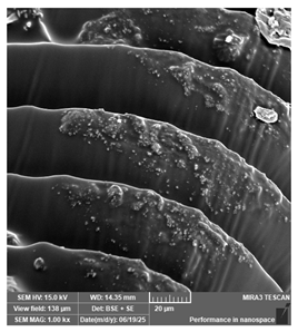
The acquired images show that the resins Sunlu Water-Wash ABS Like, Siraya Tech—Castable and PriZma 3D Bio Crown (tint: A2) have a rougher structure, while the resins Phrozen Aqua 8K, eSUN Hard-Tough resin, eSUN-eLastic, and PriZma 3D Wide show a surface pattern of parallel lines resulting from the layer-by-layer printing method.
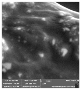
Additionally, particles of various sizes can be seen distributed throughout the samples. In samples such as the acrylic resin Prizma 3D Bio Splint, priZma 3D Wide, eSUN-eLastic and Bio Crown (tint: A2), larger particles embedded within the surface can be detected, as well as the non-uniform presence of particulate filler. Most likely, these are a sort of impurity acquired during the impression or a defect in the polymerization process. Similarly, on the samples from Phrozen Aqua 8K, eSUN Hard-Tough, and eSUN-eLastic resins, irregular particles that could be excess material or impurities are evident in detail. At 2000 magnification, the resin’s structure can be observed with greater precision, singling out some areas showing particle agglomeration and small distortions. These could be the effect of alterations in the curing process, such as incomplete curing or air bubbles trapped during the printing process, flaws that can compromise the adhesion between layers of the material.
Regarding the PriZma 3D Bio Crown shade Bleach sample, there is no evidence of an impression pattern but rather a homogeneous surface in all magnifications, featuring multiple particles of irregular sizes brightly contrasted by the BSE detector, and which could be assumed to correspond to ceramic and zirconia particles. In contrast, for the Bio Crown tint: A2 sample, though it is in essence the same material as the previous one, differing in the pigments added by the supplier, it is clear that there is not a completely uniform distribution of the constituents in the composite, evident by the trace or white mark in the center of the image.
It is worth noting that the previously mentioned characteristics, such as micro-fissures, deformations and the presence of impurities, while they have limited relevance in non-functional applications such as study models, in real clinical conditions and long-term use they could compromise the durability and stability of the devices, and with it, the effectiveness of the treatment, especially for the last two materials analyzed. It should be stated that these imperfections could be attributed to limitations or errors during fabrication, rather than to deficiencies of the material itself, thus highlighting the importance of adequately controlling the printing parameters and process conditions to ensure the correct performance of the fabricated items.
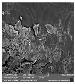
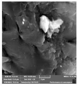
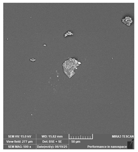
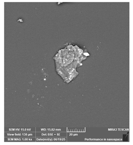
Although with the first images obtained, it was possible to identify relevant characteristics and imperfections on the samples, it was considered pertinent to increase the magnification for the unanalyzed samples in order to improve the perception of the microstructures and, in turn, obtain a more detailed characterization of the surface morphology. Therefore, magnifications of 500, 1000, 5000 and 10,000 times were used, revealing details as shown in Table 9.
Evidently, the level of detail achieved with the new magnification made it possible to identify more meticulously the qualities of the samples analyzed. To summarize some of the most outstanding findings, it was observed that the Portux SG, Anycubic water-wash +, and Anycubic Standard resins possess the clear layered printing pattern distinctive of the additive method. The surface homogeneity of the Antisky resin for surgical guides, the presence of flaws such as dirt particles, as well as the deep cavities of the Anycubic Resin Standard, also stand out; details that could be interpreted as results from an incomplete or non-uniform light-curing process. Equally noteworthy is the presence of white elements in the Portux SG resin, clearly visible at 5000 and 10,000 magnifications, which could simply be a consequence of the differences in the polymerization of the resins that make up the composite. At last, the Portux Temp resin stands out for its heterogeneous appearance and rough finish. Bearing in mind that the microscope has a backscattered electron detector (BSE) that allows distinguishing different materials using a scale of grey, it could be assumed that the structures observed could be a display of the heterogeneous distribution of the polymerization initiators and pigments in the matrix.

4.6. Comparison Between Traditional and Additive Process

Traditionally, dental restorations, including crowns, implants and dentures, are crafted with manual techniques such as lost wax and compression molding, usually assisted by a mold made from alginate and plaster. However, in recent years, there has been a surge in the use of subtractive techniques such as milling, and more recently, of additive techniques with 3D printing for the development of restorations, prostheses, splints, [14,117] among other applications.
Both subtractive and additive manufacturing rely on digital tools and CAI/CAD/CAM technologies to transform the design into a physical object; the difference lies in the fact that, in the subtractive approach, the final piece is obtained by machining or milling a block of material, while in additive manufacturing, it is built layer-by-layer through 3D printing from the digital model generated.
Taking into consideration the reviewed studies, Table 10 was constructed, summarizing the key differences in the manufacturing methods of four dental applications (models, surgical guides, restorations and occlusal splint) using traditional, subtractive and additive manufacturing techniques, facilitating our ability to visualize how the processes of obtaining the mold, design, production and finishing vary according to the approach of each manufacturing method.
Among the most distinctive disadvantages found in the literature concerning traditional methods is the high possibility of presenting deformities and distortions due to the different types of stress and temperature changes to which the materials are exposed, from the moment the impression is made to the end of the manufacturing process of the piece. Likewise, much of the success and accuracy of the final piece is attributed to the staff, who have a considerable amount of experience and skills to ensure that the models are as faithful as possible to the reality of the patients [22,79]. Furthermore, taking impressions of the oral cavity with alginate is an uncomfortable process for the patient, as it can cause irritation or even gagging due to the accumulation of material and the lengthy time it takes to make the base. In contrast, additive and subtractive manufacturing allow the use of scanners and cameras to take these images faster, more accurately and less uncomfortably and invasively for patients [14,22].
When specifically evaluating the imaging process for model generation, despite being better than traditional methods in terms of time, comfort and accuracy, the use of scanners is also not without disadvantages, as the literature has recognized that it may have limitations in capturing clear images of soft tissues as well as in the presence of saliva or blood [14].
Alternatively, the subtractive approach, just like 3D printing, presents significant advantages in terms of efficiency, speed, cost, and simplicity of the overall process. Regardless, it is still surpassed by additive manufacturing in terms of precision in the adjustment of the pieces, the lack of scalability of the process, as, in general, only one item can be printed at a time [84], but above all, the subtractive method presents shortcomings due to the amount of waste it generates. As reported by Arora, Ahmed & Maiti (2022) [79], “milling can waste up to 95% of the material, while additive techniques can generate 40% less waste, with up to 96% of it being recyclable”. Not only this, but additive manufacturing can also aim for the improvement of material efficiency at the design stage. To illustrate, a study’s findings [22] indicated that precision in mold design could be maintained by making the inside of the piece a hollow space, lowering both costs and the amount of material consumed.
Lastly, additive manufacturing offers the already mentioned advantages, as well as providing an invaluable opportunity for customization [14], reducing the number of patient visits to the clinic, along with a relatively good cost–benefit ratio, not to mention that it allows producing pieces featuring complex geometries and more aesthetically pleasing to patients, as well as making it possible to duplicate the models at any time by having the entire process systematized and digitally stored. Nevertheless, this method comes with some disadvantages, such as a limited possibility to make tests and adjustments once the item has been printed, the need for specialized post-processing procedures, and barriers resulting from the high cost of the equipment, the need for specialized training and the requirement for precise planning [14,25,65].
Built on this idea, it also seemed relevant to analyze the divergence in terms of the professional skills and expertise required by the professional based on the manufacturing method a person chooses to work with. Hence, based on the literature review, a comparative summary (Table 11) listing the qualities, knowledge, technical skills and specific abilities necessary to efficiently develop the tasks for each framework was put together, considering critical aspects of the dental device manufacturing process.
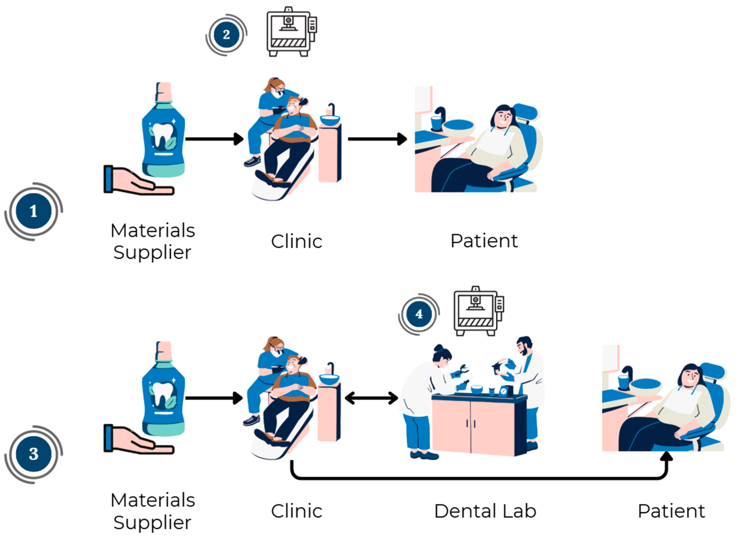
Upon the comparison supported by the literature review, we proceeded with the design of ASME diagrams in an effort to carry out a detailed comparison of the activities performed in a traditional and additive approach for the manufacturing of the dental devices previously analyzed. This task was developed upon the authors’ specialized knowledge in their respective areas, allowing a rigorous integration between the clinical perspective and the industrial engineering approach in terms of analysis and disaggregation of processes. In this sense, four different possible setups for the manufacture of the aforementioned kind of devices were identified (Figure 6), whose execution depends on the degree of digitization possible, the characteristics of the clinical case, the technical resources and experience of the professional, and the available infrastructure.
On one hand, there are cases in which the piece is completely manufactured on-site at the clinic, either by traditional methods or by additive manufacturing, which implies that the dentist is responsible for seeing through all stages of the process, from design to transformation and final adjustment of the finished device. On the other hand, there are scenarios where the transformation is performed in a decentralized manner, namely those in which the process requires the intervention of a dental laboratory, both in traditional and digital flows. For these scenarios, the dental professional at the clinic is in charge of the input and output phases, while the dental lab assumes the intermediate stages, including design and manufacturing.
Furthermore, depending on the level of customization and prior work required for design and manufacturing, 3D printed dental devices can be classified into MTO (Make to Order) or ETO (Engineer to Order) production strategies. Firstly, MTO production for dental parts refers to a fabrication made from pre-existing, standardized design files (design libraries), which are customized according to certain parameters, such as size and shape, based on intraoral scanning performed to fit the patient’s morphology. This approach leads to faster delivery cycles than those required for the manufacture of pieces under the ETO strategy. Alternatively, ETO production applies to a sort of manufacturing that requires a higher and more precise level of customization, thus requiring greater technical expertise as well as more time to develop the design.
Although most of the devices analyzed in this study are dominated by ETO model fabrication, especially in the case of surgical guides [90], given the level of detail and planning specific to each clinical case, dental restorations such as crowns can be developed in both MTO [123,124,125] and ETO [126].
The following images show the process flow for the fabrication of dental models (Figure 7), surgical guides (Figure 8), bridges and crowns (Figure 9), and occlusal splints (Figure 10) using traditional manufacturing. For every stage of the process, all the operations, transport, inspections and storage that make up this procedure are documented, enabling a thorough understanding of the manufacturing approach.
Crowns and bridges (Figure 9) are even more complex, as they require multiple laboratory-dependent stages such as glazing. In this case, the ASME diagram shows how extensive the traditional workflow is, involving multiple laboratory transfers and specialized procedures such as metal framework verification and glazing. These steps make this process the most energy-consuming among the dental devices studied and illustrate the central role of laboratories in restorative dentistry.
In relation to process productivity in terms of efficiency and effectiveness, particularly the ASME diagram for the making of occlusal splints (Figure 10) highlights the manual craftsmanship embedded in conventional production, but also its inefficiency and susceptibility to variability, as it reveals a long and labor-intensive sequence, with repeated cycles of polishing and adjustments.
One clear observation from these diagrams is the complexity and fragmentation of traditional workflows. For instance, the fabrication of dental models and restorations involves a long sequence of manual operations, many of which are related to inspection or transport. Overall, this structural complexity directly contributes to longer lead times, higher costs, and a greater probability of error or rework.
Following the analysis of the first application (Figure 7), which constitutes the foundational step for subsequent applications and the design of various other dental procedures not considered in this study, it was noted that the variability in the setup of the chain depends especially on the type of material used to make the impression of the patient’s oral cavity. For instance, when the impression is made with alginate, it is recommended to process it on-site due to the material’s rapid dimensional shrinkage, which may jeopardize the accuracy of the model if its processing is delayed; whereas if silicone is used, the model can be transferred to the dental lab without significant risk of alteration, allowing the subsequent transformation processes to be carried out outside the clinical environment. In other words, material selection introduces a decision-making component into the workflow, linking clinical choices to logistical outcomes.
Logistics also emerges as a defining characteristic of traditional workflows. Shipment to the laboratory usually takes place after disinfection of the impression, which, according to the flow charts developed, would correspond to the immediate action following step 6 in the case of occlusal splints and dental models, or step 4 in the case of surgical guides. In a similar manner, the return to the dental clinic generally occurs when the device is ready for try-in or final use. For occlusal splints, this would take place after step 19; for surgical guides, after step 11 (once the tomographic marker has been incorporated); and for dental models, after step 9 (corresponding to the setting of the model). As for restorations such as crowns and bridges, since the impression is usually made with silicone, the sending to the laboratory for the complete fabrication of the piece and the execution of the final finishing stages, which include the ceramic test and glazing, is systematically contemplated. These transfers add not only cost and time but also the risk of distortion, damage, or contamination of impressions, and, since the return of finished devices usually occurs only at advanced stages of the process, it significantly reduces clinical autonomy and lengthens patient waiting times.
In an analogous way, Figure 11, Figure 12, Figure 13 and Figure 14 display the flow chart corresponding to the production process for the studied devices using additive manufacturing, allowing the differences between the procedures to be visualized and identified in a clear and structured manner, thereby simplifying the comparison between the two approaches.
Another key improvement is that complexity is not removed with AM but rather shifted. Manual labor gives way to digital planning, design, and post-processing (washing, curing, and support removal). This transition is particularly relevant for the accurate making of surgical guides and enables a more streamlined chain with fewer opportunities for error and delay, while simultaneously raising the importance of equipment calibration and operator training as new control points.
A further advantage of additive manufacturing is the replacement of physical transport with digital file transfer. Instead of shipping impressions or models, intraoral scans can be sent electronically to a laboratory or processed in-clinic, reducing transport times, avoiding risks of damage, and enhancing data traceability. This is especially noticeable when comparing the traditional and additive manufacturing of dental restorations (Figure 13).
Delving into the initial argument, the AM ASME diagrams for dental restorations (Figure 13) and for occlusal splints (Figure 14) demonstrate some of the most significant efficiency gains thanks to the simplification of the workflow. What traditionally involved multiple laboratory exchanges, inspections and delays can now be performed in-clinic as part of an integrated digital workflow. For example, AM reduces the number of manual finishing steps for occlusal splints (Figure 14) by delivering highly accurate geometries directly from CADs. Although minor occlusal adjustments are still performed at chairside, the overall chain is shorter, more predictable, and less reliant on iterative corrections.
One of the most striking features of additive workflows is the reduction in intermediate steps. Processes such as plaster pouring, model trimming, and thermoforming are eliminated, while digital design and automated printing replace a large portion of manual tasks. In addition, Figure 11 and Figure 14 evidence that AM not only shortens production time but also reduces material waste as it bypasses tasks such as plaster preparation, setting, trimming, and mounting, reducing the process to intraoral scanning, digital design, printing, and minimal post-processing.
Despite all the benefits a complete in situ AM workflow has, a partially hybrid model remains a widely adopted alternative among practitioners. Similar to TM workflows, the 3D printing of these devices can be carried out in a different link of the supply chain: the dental laboratory. In these cases, after the imaging using intraoral scanning (usually being step 3 in the flowcharts), the digital files are transferred to the laboratory for the transformation phase, and the printed devices return to the clinic just before the occlusal adjustment (before step 15 for the splints and after step 13 for the guides) or after polymerization (step 10) for the models.
Overall, there are both strengths and limitations of hybrid models. While this approach reduces the degree of digital integration within the clinic, it still offers greater efficiency and precision compared to traditional methods and diminishes some of the vulnerabilities associated with inter-organizational logistics. It combines the rapid prototyping, customization, and accuracy of AM with the robustness and thoroughness of laboratory-based practices, reduces chairside adjustments and requires a relatively lower initial investment. However, even when AM is integrated into clinical practice, certain stages, such as final ceramic processing, continue to rely predominantly on laboratory expertise, which means that they maintain some dependency on external actors and extend lead times relative to a fully in-clinic digital workflow. Thus, hybrid models emerge as a pragmatic compromise, enabling clinicians to capitalize on the advantages of AM while mitigating challenges related to cost, training, and material performance for more complex treatments.

5. Discussion

The literature review revealed that most studies performed on additive manufacturing applied to dentistry focus on addressing clinical aspects, such as dimensional accuracy, biocompatibility of materials, mechanical comparisons between additive and subtractive techniques and validation of individual cases. However, a paucity of research that adopted an approach based on productive processes, following frameworks such as the Input–Transformation–Output (ITO) framework, was made evident. This absence is precisely a gap that this research seeks to begin to address.
In this sense, the main contribution of this work lies in its purpose in analyzing the impact additive manufacturing has on the manufacturing processes of four representative dental devices from an operational and comparative perspective when compared with traditional and subtractive methods. This approach offers a clear overview of the structure of the production workflow, capturing not only the technical steps but also the decision points, actors, possible system setups, and the logistical impact of outsourcing 3D printing. Additionally, by integrating technical analysis with workflow modeling, this study offers clinicians strategic decision-making criteria regarding the adoption of the different AM setups, while also offering manufacturers and suppliers a better understanding of where to concentrate efforts. Ultimately, the value of this research lies in positioning AM not merely as a technological alternative, but as a transformative driver of efficiency, autonomy, and innovation in dental practice.
Delving into the results, additive manufacturing proved to be a technology with high transformative potential in the dental field. Even when minor discrepancies and imperfections are still evident at the microscopic level, representing a challenge to clinical reliability, the printed pieces proved to be of high quality, with a considerable level of precision and detail, and minimal dimensional variability with respect to their original design, which is fundamental to ensure device and treatment quality and opens the opportunity to consolidate AM as a highly accurate and reproducible method. These findings are supported by previous studies such as the one conducted by Tang et al. [127], where they conclude that AM technology has reached excellent levels of accuracy in dentistry, allowing more precise, efficient and controlled procedures than before, especially, for example, in areas such as orthodontics and maxillofacial prosthetics.
It is important to emphasize that when speaking of imperfections, it is not intended to question the quality of the resins or the methods used, but to emphasize that the precision, reliability and mechanical and biological properties of the final product depend strongly on technical factors that can be controlled and optimized during the process, especially when these processes are performed in the clinic. The incorporation of SEM analysis as a quality inspection tool in this article showed that the flaws documented do not necessarily stem from the material itself, but rather respond to variations in manufacturing parameters, thus validating the importance of the standardization of parameters and ensuring adequate technical control at critical stages in the transformation of the material.
Particularly, accountability was attributed to the efficiency of the post-processing and curing stage, since this is a critical point to ensure the quality of the final product and considering that isolated surface defects were detected for some samples, such as trapped bubbles or microfractures, which could be directly related to deficiencies in that stage of the process. In accordance with this assessment, sources such as Alghauli et al. [128] point out that the final accuracy depends on variables that rely on the technical knowledge and good decision-making of the manufacturer, such as the operator’s choice of printing technology, printing orientation, layer thickness, post-curing and post-processing.
Similarly, Mandurino et al. [19] suggest that the potential to reduce defects in 3D-printed dental devices and enhance their safe and effective use lies in the development of improved material formulations, optimized polymerization techniques, and the standardization of testing methodologies. Additionally, the authors highlight that some of the challenges of these technological advances rely not only on the manufacturing of the pieces, but on variables that cannot be fully controlled, such as the complexity of the oral environment and patient habits like bruxism, hence creating an opportunity to develop more resilient materials, improve predictive modeling of device performance, and implement personalized treatment strategies that account for individual patient variability. In other words, it is confirmed that adoption of the necessary technologies is not enough to carry out 3D printing; it is also necessary to optimize the production parameters in order to minimize defects such as layer staggering, distortions or inaccuracies in the surface finish of the parts.
Furthermore, the perceived lower mechanical strength in some of the printed devices, examined qualitatively in the SEM analysis findings, is substantiated by scientific evidence such as that provided in the review carried out by Mandurino et al. [19], who acknowledge that there are some limitations to this technology. Said review highlighted that, although accuracy is high, printed restorations may be more fragile in the long term than conventionally made ones, as it was concluded that 3D printed resins for permanent restorations exhibit lower fracture and fatigue strengths than their counterparts produced by milling. As indicated by the findings of Mandurino et al. [19] this takes on particular importance if the device in question is going to be exposed to prolonged degradation processes that occur naturally in the oral environment, which implies that, for now, 3D printing is ideal for certain applications, such as temporary restorations, surgical guides, dental models and splints, while for permanent restorations it is still worth evaluating each unique case.
Building upon the same line of reasoning, the research of Katheng et al. [129] shows that the method or technology chosen plays a role in the finish and mechanical properties of the printed items, representing both a challenge, as not all methods are interchangeable, and an opportunity, since selecting the optimal technique can maximize the clinical value of AM. In their study, they favor one of the techniques we used in our research, as they proved that the SLA methodology is preferable to other digital techniques such as LCD and DLP for applications where the device is required to be strong and have high quality surfaces, demonstrating, for example, that the flexural strength of the pieces made using SLA was about 93 Mpa in contrast to 65 MPa for the parts manufactured in LCD.
Addressing the impact that 3D printing has had on the setup of clinical workflows and the supply chain in dentistry, the findings of this study allow us to affirm that the adoption of additive manufacturing has significantly transformed supply chains in the dental sector. In essence, this manufacturing method has allowed the streamlining of production, the reduction in reliance on multiple suppliers, and the flexibility of the manufacturing process, among many other advantages for an industry that, by its inherent nature, is already fully customized.
In terms of operational efficiency, it was acknowledged that the integration of 3D printing into the dental workflow substantially reduces manual intervention and the number of activities required to carry out the manufacturing process compared to the traditional analog method as well as replacing them with more agile and automated activities, for example, skipping those delay stages during setting or thermoforming by performing the printing process directly, which collectively contribute to the reduction of the lead time. This reduction in production cycles is consistent with what was reported by Mangano et al. [130], who documented that chairside additive manufacturing of a definitive crown can be completed in a single clinical session, with remarkable time efficiency, taking 90 min to produce 10 crowns, compared to 450 min with subtractive manufacturing done in the clinic and 930 min when the process is per-formed in the laboratory. Complementarily, the review by Alghauli et al. [128] shows that opting for a horizontal print orientation can halve production times and material consumption, while maintaining or even improving dimensional accuracy.
Pertaining to the premise that additive manufacturing redistributes the production process, Pillai et al. [14] support this idea by highlighting how practices that were previously considered laboratory tasks are being transferred and progressively adopted by the clinical environment thanks to technological advancements and the increasing affordability and accessibility of printing equipment. So, in this regard, the notion that additive manufacturing tends to relocate the manufacturing phase to the clinics, radically reshaping the traditional supply chain where the laboratory served as the production center, is not only a valid idea, but an ongoing phenomenon that is consolidating across contemporary dental practice.
Particularly, through the case studies, it was made evident that when the 3D printing process is outsourced through specialized laboratories, a logic more akin to a traditional one is carried on, involving processes that rely on waiting times between actors as a result of the transport of files and models or physical elements. Even so, it is still a highly appealing alternative, considering that it reduces the level of digitalization and investment required in the clinic, the greatest impact is observed when 3D printing is integrated directly into the dental clinic, enabling a decentralized flow that reduces intermediaries, response times (lead time) and logistic costs [131], enabling a just-in-time production model closer to the site of service. In this context, the clinic acting as a driving company, directly and internally managing all processes, from image acquisition to post-processing manufacturing and placement or use of the pieces, represents an opportunity to improve operational efficiency by promoting a shorter and more agile supply chain and facilitate clinical communication, reducing the need for shipments and physical storage, and enabling direct interaction with the model and facilitate collaboration with distant physicians in a way that purely digital images cannot, which also represents a decrease in capital costs and greater flexibility to the specific needs of each treatment.
The case studies developed in this research corroborate this transformation and its impact. As a case in point, it was shown that the digitization of the workflow improves traceability and communication as data travels electronically, avoiding loss or damage of models, and enabling remote collaboration in design. They also demonstrated that the choice to carry out manufacturing within the clinic makes it possible to forgo the intermediation of the dental laboratory, eliminating this link in the supply chain.
As part of this discussion, it is also necessary to reflect on the economic and educational implications arising from internalizing additive production in clinics. The challenge lies in the complexity of integration, which demands investments in equipment and staff training. Mangano et al. [130] provide comparative evidence between different workflows showing that the manufacturing of dental crowns in the clinic using 3D printing is not only faster, but also cheaper than traditional methods such as milling either in situ or in the laboratory. More precisely, they state that the unit cost showed a reduction of 71.92% per printed crown compared to the subtractive method performed in the clinic without compromising the quality of the printed crowns. Alghauli et al. [128] reinforce this and emphasize the importance of proper decision-making and the setting of print parameters, indicating that printing with a horizontal orientation is more time, material and energy efficient than doing so vertically, especially in the production of single or low-volume products.
Moreover, even though authors such as Alghauli et al. [18] claim that the additive method requires a lower initial investment compared to other methods, and Mangano et al. [131] further state that labor-based subtractive manufacturing involves a longer learning curve, the decision to adopt these technologies in a clinic should be taken after thoroughly weighing up each clinic’s circumstances; carefully factor in its production volume, available resources and operational capabilities to determine whether it is more advantageous to internalize the processes, outsource them or to abstain from adopting them at all.
At last, a crucial factor to assess is the investment in staff training, although it is recognized that this challenge tends to diminish as digital technologies continue to develop and professional training programs evolve in tandem with them. Hegedus et al. [65] address this issue by underlining the importance of early development of digital skills in professional training, and conclude that the youngest generation of dentists are already familiar with basic skills and understanding of digital dentistry, which facilitates their integration into the workflow associated with 3D printing and demonstrates the potential this technology has as a training and strategic investment for the sector.
Reflecting on this framework, it becomes evident that a key limitation lies in the fact that no single manufacturing method is currently capable of fulfilling all clinical requirements optimally. This constraint, however, simultaneously creates an opportunity to explore hybrid workflows. In line with the arguments presented by Pillai et al. [14], who advocate for the integration of multiple manufacturing approaches, the findings of this study gain further relevance by supporting the implementation of a hybrid model. Such a model would allow the exploitation of the advantages of additive manufacturing, such as speed, customization, and the ability to reproduce complex geometries without additional costs, while continuing to use traditional or subtractive techniques in those cases where the required robustness, affordability or accuracy exceeds what additive technologies can offer today.
In acknowledgment of the limitations, it should be noted that, due to its approach on engineering and process analysis, this paper does not address the evaluation of the functional performance of dental pieces in real clinical conditions, nor does it address in depth the normative or regulatory aspects required for the approval and use of medical devices. In addition, the approach is restricted to the analysis of productive processes under the input–transformation–output (ITO) framework, without delving into therapeutic, physiological or patient perception variables, which are important in the real practical setting.
On a related note, although SEM images were used for morphological analysis, the scope was restricted to qualitative assessments of defects attributed to the treatment of the materials, leaving out standardized mechanical tests or quantitative dimensional measurements that would offer a rigorous comparison of performance between technologies. Withal, the complexity of CAD and the learning curve associated with the use of digital technologies by clinical personnel were not studied in depth, and no detailed economic analysis of variables such as clinical or logistical costs and return on investment was performed to allow a quantitative comparison of the economic impact derived from the use of each productive approach.
Nonetheless, the scope of this research allows us to position it as a first approach to a new operational view, barely studied in the literature so far, shifting the analysis of the production process to real contexts and showing that additive manufacturing not only redefines finished goods, but also profoundly reconfigures the workflows and links in the supply chain, reshaping the operational dynamics of the sector. This paves the way for future research that explores hybrid manufacturing models, integrating operational efficiency metrics, cost–benefit analysis, and development of clinical staff competencies in digital technologies and flows, scopes of great value for the continuous improvement of dental services.

6. Conclusions

Resins constitute a highly versatile class of materials that have been widely utilized across industries such as construction, medicine, and electronics, among others. Their use has gained particular relevance in the field of dentistry due to their adaptability to a wide range of clinical procedures, especially when combined with advanced technologies such as additive manufacturing, which has seen a notable surge in recent years.
According to the literature, this type of material has shown remarkable effectiveness in the clinical setting, although some limitations persist in attributes such as mechanical strength and longevity. However, with continuous improvements in the composition of the materials and the correct setting up of the printing parameters, these disparities are expected to decrease.
In addition, as part of the technical analysis for this research, the use of the scanning electron microscope (SEM) proved essential to examine in detail the characteristics and properties of the light-curing resin samples. It was possible to detect imperfections, such as roughness, impurities, deformations, porosity, micro-cracks, and defects in the curing process. These findings highlight the importance of verifying both the suitability of the resin material and the appropriateness of the printing method, as well as ensuring accurate decision-making and precise calibration of process parameters to avoid compromising the quality, functionality and aesthetics of the piece. That being said, it should be noted that the implications of this type of error vary depending on the intended use of the printed device and, given that in this study a higher prevalence of imperfections was observed in the resins used for dental models, their effect on the final clinical pieces may be considered limited or negligible.
Moving from the material level to the process level, AM supported by digital workflows represents not merely an emerging trend but a new production paradigm in dentistry, positively influencing both the fabrication of dental devices and the structure of service delivery. Through a comparative analysis of traditional and additive manufacturing processes using ASME process diagrams, this study identified that the integration of additive manufacturing eliminates a key link in the traditional dental supply chain, as well as enabling the highest levels of product customization, facilitating Make-to-Order (MTO) and Engineer-to-Order (ETO) production models. This latter finding is particularly significant, as it reflects a shift toward a decentralized production model characterized by reduced reliance on external laboratories and the consolidation of a fully digital workflow within the clinic. Moreover, this digital integration was found to streamline manufacturing processes by reducing the number of manual tasks required, many of which are now executed more efficiently using CAI, CAD, and CAM technologies.
These findings are further supported by a comprehensive review of recent literature, which not only corroborated the observations made in this study but also enriched them by identifying additional operational advantages associated with integrating 3D printing into the dental production process. Notable among these are the reduction in material waste, the ability to reproduce complex geometries with high precision and detail, the decreased number of patient visits required, and the instant duplication of models, all of which contribute to optimizing the clinical experience and enhancing the efficiency of the services provided. Additionally, the technology was shown to reduce cycle times and operational costs by enabling the rapid fabrication of dental devices with minimal manual intervention, thus affording greater autonomy to dental professionals and increasing overall process efficiency.
Nevertheless, the adoption of AM is not without its challenges. These include the learning curve for clinical personnel who did not receive digital training during their professional education, as well as cost–benefit considerations that are inherently linked to the operating scale. In this context, and depending on variables such as case complexity, client base, and the degree of available digital infrastructure, the adoption of a hybrid model leveraging both traditional and additive manufacturing techniques may offer a more efficient solution.
To conclude, it is worth emphasizing that, although this study focused on clinical applications, there are other important areas of use for additive manufacturing in dentistry that remain unexplored, including academic training and research. These areas, though beyond the scope of the present study, represent a valuable opportunity for future research.

Author Contributions

Conceptualization, M.M.G. and J.N.R.; Methodology, J.N.R.; Validation, S.M.V.-A.; Formal analysis, M.G.T.; Investigation, M.M.G. and J.N.R.; Data curation, M.G.T.; Writing—original draft, M.M.G., M.G.T. and J.N.R.; Writing—review & editing, M.M.G., M.G.T. and J.N.R.; Supervision, S.M.V.-A. All authors have read and agreed to the published version of the manuscript.

Funding

This research received no external funding.

Data Availability Statement

The original contributions presented in this study are included in the article. Further inquiries can be directed to the corresponding author.

Conflicts of Interest

The authors declare no conflicts of interest.

References

  1. Lee, E.-H.; Ahn, J.-S.; Lim, Y.-J.; Kwon, H.-B.; Kim, M.-J. Effect of post-curing time on the color stability and related properties of a tooth-colored 3D-printed resin material. J. Mech. Behav. Biomed. Mater. 2022, 126, 104993. [Google Scholar] [CrossRef]
  2. Rubio, N.A. CAI: Computer-Assisted Imaging. In Digital Dental Implantology; Springer: Cham, Switzerland, 2021; pp. 3–18. [Google Scholar] [CrossRef]
  3. Wang, Q.; Liu, X.; Qiang, Z.; Hu, Z.; Cui, X.; Wei, H.; Hu, J.; Xia, Y.; Huang, S.; Zhang, J.; et al. Cellulose nanocrystal enhanced, high dielectric 3D printing composite resin for energy applications. Compos. Sci. Technol. 2022, 227, 109601. [Google Scholar] [CrossRef]
  4. Chen, R.; Cai, J.; Chin, K.C.H.; Wang, S.; Boydston, A.J.; Thevamaran, R.; Gopalan, P. Block copolymer additives for toughening 3D printable epoxy resin. Giant 2023, 17, 100204. [Google Scholar] [CrossRef]
  5. Rivera, C.; Rodriguez, J.N.; Bas, A.O. Research on the Input-Transformation-Output Process of Additive Manufacturing: Comparing PLA/Polysmooth and Resin Printed Rings. Crystals 2023, 14, 7. [Google Scholar] [CrossRef]
  6. Chhabra, M.; Kumar, M.N.; RaghavendraSwamy, K.N.; Thippeswamy, H. Flexural strength and impact strength of heat-cured acrylic and 3D printed denture base resins- A comparative in vitro study. J. Oral Biol. Craniofacial Res. 2021, 12, 1–3. [Google Scholar] [CrossRef]
  7. Carreño, A.M.; Torres, M.E.; Rojas, M.S.R.; Rodriguez, J.N. Un estudio comparativo para evaluar el proceso sustractivo y aditivo en odontología: Una revisión sistemática. Rev. Ing. USBMed 2024, 15, 70–90. [Google Scholar] [CrossRef]
  8. Suganna, M.; Kausher, H.; Ahmed, S.T.; Alharbi, H.S.; Alsubaie, B.F.; Ds, A.; Haleem, S.; Ali, A.B.M.R. Contemporary Evidence of CAD-CAM in Dentistry: A Systematic Review. Cureus 2022, 14, e31687. [Google Scholar] [CrossRef] [PubMed]
  9. Spagnuolo, G.; Sorrentino, R. The Role of Digital Devices in Dentistry: Clinical Trends and Scientific Evidences. J. Clin. Med. 2020, 9, 1692. [Google Scholar] [CrossRef]
  10. No-Cortes, J.; Ayres, A.P.; Lima, J.E.; Markarian, R.A.; Attard, N.J.; Cortes, A.R.G. Trueness, 3D Deviation, Time and Cost Comparisons Between Milled and 3D-Printed Resin Crowns. Eur. J. Prosthodont. Restor. Dent. 2022, 30, 107–112. [Google Scholar] [CrossRef]
  11. Park, J.-Y.; Kim, H.-Y.; Kim, J.-H.; Kim, J.-H.; Kim, W.-C. Comparison of prosthetic models produced by traditional and additive manufacturing methods. J. Adv. Prosthodont. 2015, 7, 294–302. [Google Scholar] [CrossRef]
  12. El Samahy, M.M.; Abdelhamid, A.M.; El Shabrawy, S.M.; Hanno, K.I. Evaluation of physicomechanical properties of milled versus 3D-printed denture base resins: A comparative in vitro study. J. Prosthet. Dent. 2023, 129, 797.e1–797.e7. [Google Scholar] [CrossRef]
  13. Schweiger, J.; Edelhoff, D.; Güth, J.-F. 3D Printing in Digital Prosthetic Dentistry: An Overview of Recent Developments in Additive Manufacturing. J. Clin. Med. 2021, 10, 2010. [Google Scholar] [CrossRef]
  14. Pillai, S.; Upadhyay, A.; Khayambashi, P.; Farooq, I.; Sabri, H.; Tarar, M.; Lee, K.T.; Harb, I.; Zhou, S.; Wang, Y.; et al. Dental 3D-Printing: Transferring Art from the Laboratories to the Clinics. Polymers 2021, 13, 157. [Google Scholar] [CrossRef]
  15. Palavicini, J.; Quin, S.L.; Zakkour, W.; Zakkour, K.; Varkiani, S.M.; Xu, X.; Lawson, N.C.; Nejat, A.H. Bond Strength of Reline Materials to 3D-Printed Provisional Crown Resins. Polymers 2023, 15, 3745. [Google Scholar] [CrossRef]
  16. Farkas, A.Z.; Galatanu, S.-V.; Nagib, R. The Influence of Printing Layer Thickness and Orientation on the Mechanical Properties of DLP 3D-Printed Dental Resin. Polymers 2023, 15, 1113. [Google Scholar] [CrossRef]
  17. Zhang, P.; He, R. 3D-printed silicon nitride ceramic implants for clinical applications: The state of the art and prospects. RSC Adv. 2025, 15, 406–419. [Google Scholar] [CrossRef]
  18. Alghauli, M.A.; Alqutaibi, A.Y. 3D-printed intracoronal restorations, occlusal and laminate veneers: Clinical relevance, properties, and behavior compared to milled restorations; a systematic review and meta-analysis. J. Esthet. Restor. Dent. 2024, 36, 1153–1170. [Google Scholar] [CrossRef]
  19. Mandurino, M.; Cortili, S.; Coccoluto, L.; Greco, K.; Cantatore, G.; Gherlone, E.F.; Vichi, A.; Paolone, G. Mechanical Properties of 3D Printed vs. Subtractively Manufactured Composite Resins for Permanent Restorations: A Systematic Review. Materials 2025, 18, 985. [Google Scholar] [CrossRef] [PubMed]
  20. Jeong, M.; Radomski, K.; Lopez, D.; Liu, J.T.; Lee, J.D.; Lee, S.J. Materials and Applications of 3D Printing Technology in Dentistry: An Overview. Dent. J. 2023, 12, 1. [Google Scholar] [CrossRef] [PubMed]
  21. Chen, Y.; Li, H.; Zhai, Z.; Nakano, T.; Ishigaki, S. Impact of internal design on the accuracy of 3-dimensionally printed casts fabricated by stereolithography and digital light processing technology. J. Prosthet. Dent. 2023, 130, 381.e1–381.e7. [Google Scholar] [CrossRef] [PubMed]
  22. Yan, Q.; Dong, H.; Su, J.; Han, J.; Song, B.; Wei, Q.; Shi, Y. A Review of 3D Printing Technology for Medical Applications. Engineering 2018, 4, 729–742. [Google Scholar] [CrossRef]
  23. Alshamrani, A.A.; Raju, R.; Ellakwa, A. Effect of Printing Layer Thickness and Postprinting Conditions on the Flexural Strength and Hardness of a 3D-Printed Resin. BioMed Res. Int. 2022, 2022. [Google Scholar] [CrossRef] [PubMed]
  24. Idrees, M.; Ibrahim, A.M.; Tekerek, E.; Kontsos, A.; Palmese, G.R.; Alvarez, N.J. The effect of resin-rich layers on mechanical properties of 3D printed woven fiber-reinforced composites. Compos. Part A Appl. Sci. Manuf. 2021, 144, 106339. [Google Scholar] [CrossRef]
  25. Kirby, S.; Pesun, I.; Nowakowski, A.; França, R. Effect of Different Post-Curing Methods on the Degree of Conversion of 3D-Printed Resin for Models in Dentistry. Polymers 2024, 16, 549. [Google Scholar] [CrossRef]
  26. AlRumaih, H.S.; Gad, M.M. The Effect of 3D Printing Layer Thickness and Post-Polymerization Time on the Flexural Strength and Hardness of Denture Base Resins. Prosthesis 2024, 6, 970–978. [Google Scholar] [CrossRef]
  27. Janssen, K.; Schnelting, G.H.; Waterink, M.; Guit, J.; Hul, J.; Ye, C.; Loos, K.; Voet, V.S. Renewable Methacrylate Resins for 3D Printing Containing Dynamic Hydroxyester Linkages for Reprocessability. Macromol. Mater. Eng. 2024, 309, 2400036. [Google Scholar] [CrossRef]
  28. Alamo, L.; Cassiano, F.B.; Bordini, E.A.F.; Stuani, V.T.; Pacheco, L.E.; Gallinari, M.d.O.; Costa, C.A.S.; Mondelli, R.F.L.; Soares, D.G. An organotypic model of oral mucosa cells for the biological assessment of 3D printed resins for interim restorations. J. Prosthet. Dent. 2024, 132, 251–259. [Google Scholar] [CrossRef]
  29. Choi, S.-S.; Lee, J.H.; Kong, H.; Park, E.-J. Biofilm removal effect of diatom complex on 3D printed denture base resin. Sci. Rep. 2024, 14, 4034. [Google Scholar] [CrossRef]
  30. Gad, M.M.; Alzaki, F.A.; Abuwarwar, F.A.; Alhammad, A.; Al Hussain, M.; Khan, S.Q.; Nassar, E.A.; Ayad, N.M. Impact of printing layer thickness on the flexural strength of nanocomposite 3D printed resins: An in vitro comparative study. Saudi Dent. J. 2024, 36, 1307–1312. [Google Scholar] [CrossRef]
  31. Poker, B.d.C.; Oliveira, V.d.C.; Macedo, A.P.; Gonçalves, M.; Ramos, A.P.; Silva-Lovato, C.H. Evaluation of surface roughness, wettability and adhesion of multispecies biofilm on 3D-printed resins for the base and teeth of complete dentures. J. Appl. Oral Sci. 2024, 32, e20230326. [Google Scholar] [CrossRef]
  32. Coelho, S.R.G.; da Silva, M.D.D.; Nunes, T.S.B.S.; Viotto, H.E.C.; Marin, D.O.M.; Pero, A.C. Effect of immersion in disinfectants on the color stability of denture base resins and artificial teeth obtained by 3D printing. J. Prosthodont. 2023, 33, 157–163. [Google Scholar] [CrossRef]
  33. Gad, M.M.; Al Hamad, H.M.; Almohsin, F.M.; Fouda, S.M.; Akhtar, S.; Khan, S.Q.; Rahoma, A.; Al-Qarni, F.D.; Baba, N.Z.; Al-Harbi, F.A. Repair strength of 3D-printed denture base resins: Effect of surface treatment and repair material type. J. Prosthodont. 2024. [Google Scholar] [CrossRef] [PubMed]
  34. Aydin, N.; Kavrama, F.U.; Yosuncigir, H.; Ucar, Y. A comparison of the shear bond strength between denture teeth and denture base resins manufactured either conventionally or with a 3D printer. J. Prosthet. Dent. 2023, 130, 742.e1–742.e6. [Google Scholar] [CrossRef] [PubMed]
  35. Al-Qarni, F.D.; Gad, M.M. Printing Accuracy and Flexural Properties of Different 3D-Printed Denture Base Resins. Materials 2022, 15, 2410. [Google Scholar] [CrossRef]
  36. Di Fiore, A.; Meneghello, R.; Brun, P.; Rosso, S.; Gattazzo, A.; Stellini, E.; Yilmaz, B. Comparison of the flexural and surface properties of milled, 3D-printed, and heat polymerized PMMA resins for denture bases: An in vitro study. J. Prosthodont. Res. 2022, 66, 502–508. [Google Scholar] [CrossRef]
  37. Zattera, A.C.A.; Morganti, F.A.; Balbinot, G.d.S.; Della Bona, A.; Collares, F.M. The influence of filler load in 3D printing resin-based composites. Dent. Mater. 2024, 40, 1041–1046. [Google Scholar] [CrossRef] [PubMed]
  38. Lassila, L.; Mangoush, E.; He, J.; Vallittu, P.K.; Garoushi, S. Effect of Post-Printing Conditions on the Mechanical and Optical Properties of 3D-Printed Dental Resin. Polymers 2024, 16, 1713. [Google Scholar] [CrossRef]
  39. Kim, J.-E.; Mangal, U.; Yu, J.-H.; Kim, G.-T.; Kim, H.; Seo, J.-Y.; Cha, J.-Y.; Lee, K.-J.; Kwon, J.-S.; Choi, S.-H. Evaluation of the effects of temperature and centrifugation time on elimination of uncured resin from 3D-printed dental aligners. Sci. Rep. 2024, 14, 15206. [Google Scholar] [CrossRef]
  40. Korkmaz, Y.N.; Buyuk, S.K.; Simsek, H.; Abay, F. Comparison of the Flexural Strength of Three Different Aged and Nonaged 3D-Printed Permanent Crown Resins. Int. J. Prosthodont. 2024, 37, S203–S207. [Google Scholar] [CrossRef]
  41. Celikel, P.; Sengul, F. Investigating the impact of post-curing cycles on surface hardness and color stability in 3D printed resin crowns. Odontology 2024, 113, 156–162. [Google Scholar] [CrossRef]
  42. Aktas, N.; Güngör, M.B. Effects of 3D-Printing Technology and Cement Type on the Fracture Resistance of Permanent Resin Crowns for Primary Teeth. Int. J. Prosthodont. 2024, 37, S195–S202. [Google Scholar] [CrossRef] [PubMed]
  43. Cardoso, B.S.; da Cruz, M.B.; Marques, J.F.; Roque, J.C.; Martins, J.P.; Malheiro, R.C.; da Mata, A.D. Cellular responses to 3D printed dental resins produced using a manufacturer recommended printer versus a third party printer. J. Adv. Prosthodont. 2024, 16, 126–138. [Google Scholar] [CrossRef] [PubMed]
  44. Baytur, S.; Turksayar, A.A.D. Effects of post-polymerization conditions on color properties, surface roughness, and flexural strength of 3D-printed permanent resin material after thermal aging. J. Prosthodont. 2024, 34, 298–307. [Google Scholar] [CrossRef]
  45. Lask, M.; Stawarczyk, B.; Reymus, M.; Meinen, J.; Mayinger, F. Impact of varnishing, coating, and polishing on the chemical and mechanical properties of a 3D printed resin and two veneering composite resins. J. Prosthet. Dent. 2024, 132, 466.e1–466.e9. [Google Scholar] [CrossRef] [PubMed]
  46. Di Fiore, A.; Stellini, E.; Alageel, O.; Alhotan, A. Comparison of mechanical and surface properties of two 3D printed composite resins for definitive restoration. J. Prosthet. Dent. 2024, 132, 839.e1–839.e7. [Google Scholar] [CrossRef]
  47. Prause, E.; Malgaj, T.; Kocjan, A.; Beuer, F.; Hey, J.; Jevnikar, P.; Schmidt, F. Mechanical properties of 3D-printed and milled composite resins for definitive restorations: An in vitro comparison of initial strength and fatigue behavior. J. Esthet. Restor. Dent. 2023, 36, 391–401. [Google Scholar] [CrossRef]
  48. Sasany, R.; Jamjoon, F.Z.; Kendirci, M.Y.; Yilmaz, B. Effect of Printing Layer Thickness on Optical Properties and Surface Roughness of 3D-Printed Resins: An In Vitro Study. Int. J. Prosthodont. 2024, 37, S165–S173. [Google Scholar] [CrossRef]
  49. Espinar, C.; Della Bona, A.; Tejada-Casado, M.; Pulgar, R.; Pérez, M.M. Optical behavior of 3D-printed dental restorative resins: Influence of thickness and printing angle. Dent. Mater. 2023, 39, 894–902. [Google Scholar] [CrossRef]
  50. Berli, C.; Thieringer, F.M.; Sharma, N.; Müller, J.A.; Dedem, P.; Fischer, J.; Rohr, N. Comparing the mechanical properties of pressed, milled, and 3D-printed resins for occlusal devices. J. Prosthet. Dent. 2020, 124, 780–786. [Google Scholar] [CrossRef]
  51. Dimitrova, M.; Corsalini, M.; Kazakova, R.; Vlahova, A.; Chuchulska, B.; Barile, G.; Capodiferro, S.; Kazakov, S. Comparison between Conventional PMMA and 3D Printed Resins for Denture Bases: A Narrative Review. J. Compos. Sci. 2022, 6, 87. [Google Scholar] [CrossRef]
  52. Zhang, X.; Zhang, Q.; Meng, X.; Ye, Y.; Feng, D.; Xue, J.; Wang, H.; Huang, H.; Wang, M.; Wang, J. Rheological and Mechanical Properties of Resin-Based Materials Applied in Dental Restorations. Polymers 2021, 13, 2975. [Google Scholar] [CrossRef]
  53. Alshamrani, A.; Alhotan, A.; Kelly, E.; Ellakwa, A. Mechanical and Biocompatibility Properties of 3D-Printed Dental Resin Reinforced with Glass Silica and Zirconia Nanoparticles: In Vitro Study. Polymers 2023, 15, 2523. [Google Scholar] [CrossRef]
  54. Al-Dwairi, Z.N.; Tahboub, K.Y.; Baba, N.Z.; Goodacre, C.J. A Comparison of the Flexural and Impact Strengths and Flexural Modulus of CAD/CAM and Conventional Heat-Cured Polymethyl Methacrylate (PMMA). J. Prosthodont. 2018, 29, 341–349. [Google Scholar] [CrossRef]
  55. Sartori, N.; Sanchez, S.A.; Oliveira, D.; Hosney, S.; Zoidis, P.; Martin, W.; Gonzaga, L.; Rocha, M.G. Flexural properties and fatigue limit of 3D-printed and milled resin-based materials. J. Prosthodont. 2024, 34, 626–634. [Google Scholar] [CrossRef] [PubMed]
  56. Wen, A.; Xiao, N.; Zhu, Y.; Gao, Z.; Qin, Q.; Shan, S.; Li, W.; Sun, Y.; Wang, Y.; Zhao, Y. Spatial Trueness Evaluation of 3D-Printed Dental Model Made of Photopolymer Resin: Use of Special Structurized Dental Model. Polymers 2024, 16, 1083. [Google Scholar] [CrossRef] [PubMed]
  57. Alghauli, M.A.; Aljohani, W.; Almutairi, S.; Aljohani, R.; Alqutaibi, A.Y. Advancements in digital data acquisition and CAD technology in Dentistry: Innovation, clinical Impact, and promising integration of artificial intelligence. Clin. eHealth 2025, 8, 32–35. [Google Scholar] [CrossRef]
  58. Alghazzawi, T.F. Advancements in CAD/CAM technology: Options for practical implementation. J. Prosthodont. Res. 2016, 60, 72–84. [Google Scholar] [CrossRef]
  59. Kim, J.; Lin, Y.; Danielak, M.; Van, M.; Lee, D.; Kim, H.; Arany, P.R. Virtual Planning and Rapid 3D Prototyping Surgical Guide for Anterior Crown Lengthening Surgery: A Clinical Case Report. J. Prosthodont. 2021, 31, 275–281. [Google Scholar] [CrossRef]
  60. Caussin, E.; Moussally, C.; Le Goff, S.; Fasham, T.; Troizier-Cheyne, M.; Tapie, L.; Dursun, E.; Attal, J.-P.; François, P. Vat Photopolymerization 3D Printing in Dentistry: A Comprehensive Review of Actual Popular Technologies. Materials 2024, 17, 950. [Google Scholar] [CrossRef]
  61. Güth, J.-F.; Runkel, C.; Beuer, F.; Stimmelmayr, M.; Edelhoff, D.; Keul, C. Accuracy of five intraoral scanners compared to indirect digitalization. Clin. Oral Investig. 2016, 21, 1445–1455. [Google Scholar] [CrossRef]
  62. Papaspyridakos, P.; AlFulaij, F.; Bokhary, A.; Sallustio, A.; Chochlidakis, K. Complete Digital Workflow for Prosthesis Prototype Fabrication with Double Digital Scanning: Accuracy of Fit Assessment. J. Prosthodont. 2022, 32, 49–53. [Google Scholar] [CrossRef]
  63. Da Son, K.; Lee, W.-S.; Lee, K.-B. Prediction of the learning curves of 2 dental CAD software programs. J. Prosthet. Dent. 2019, 121, 95–100. [Google Scholar] [CrossRef] [PubMed]
  64. Denry, I.; Kelly, J.R. State of the art of zirconia for dental applications. Dent. Mater. 2008, 24, 299–307. [Google Scholar] [CrossRef]
  65. Hegedus, T.; Kreuter, P.; Kismarczi-Antalffy, A.A.; Demeter, T.; Banyai, D.; Vegh, A.; Geczi, Z.; Hermann, P.; Payer, M.; Zsembery, A.; et al. User Experience and Sustainability of 3D Printing in Dentistry. Int. J. Environ. Res. Public Heal. 2022, 19, 1921. [Google Scholar] [CrossRef] [PubMed]
  66. Escáneres Intraorales 3Shape TRIOS—Comparar Todos los Modelos. Available online: https://www.3shape.com/es/scanners/trios (accessed on 26 May 2025).
  67. 3M True Definition Scanner. Available online: https://www.3m.com.ni/3M/es_NI/dental-la/productos/true-definition-scanner/ (accessed on 26 May 2025).
  68. iTeroEd—How to Export a File. Available online: https://www.iteroed.com/en-SG/faqs/6Ha7BtPCuv9zeJz1gIyyzB (accessed on 26 May 2025).
  69. Planmeca PlanScan Lab—MedicalVet. Available online: https://medicalvet.com.uy/product/planmeca-planscan-lab/ (accessed on 26 May 2025).
  70. Digital, Flexible y Abierto—CEREC, un Sistema Completo Para las Restauraciones, la Implantología y la Ortodoncia. Available online: https://news.dentsplysirona.com/es/business-units/cad-cam/2017/digital--flexible-y-abierto--cerec--un-sistema-completo-para-las.html (accessed on 26 May 2025).
  71. CS3500—MEDISER. Available online: https://www.medisercontrol.com/es/carestream/85-cs3500.html (accessed on 27 May 2025).
  72. ISO/ASTM 52900:2021(en); Additive Manufacturing—General Principles—Fundamentals and Vocabulary. International Standard Organization: Geneva, Switzerland, 2021. Available online: https://www.iso.org/obp/ui/en/#iso:std:iso-astm:52900:ed-2:v1:en (accessed on 18 September 2025).
  73. Miyanaji, H.; Zhang, S.; Lassell, A.; Zandinejad, A.; Yang, L. Process Development of Porcelain Ceramic Material with Binder Jetting Process for Dental Applications. JOM 2016, 68, 831–841. [Google Scholar] [CrossRef]
  74. Mostafaei, A.; Stevens, E.L.; Ference, J.J.; Schmidt, D.E.; Chmielus, M. Binder jetting of a complex-shaped metal partial denture framework. Addit. Manuf. 2018, 21, 63–68. [Google Scholar] [CrossRef]
  75. Kamio, T.; Iwata, H.; Kawai, T. Challenges and Solutions for Cost-Effective and Practical Dental Cast Fabrication With a Desktop Fused Deposition Modeling (FDM) 3D Printer. Cureus 2024, 16, e73354. [Google Scholar] [CrossRef]
  76. Gupta, A.; Alifui-Segbaya, F.; Hasanov, S.; White, A.R.; Ahmed, K.E.; Love, R.M.; Fidan, I. Material extrusion of thermoplastic acrylic for intraoral devices: Technical feasibility and evaluation. J. Mech. Behav. Biomed. Mater. 2023, 143, 105950. [Google Scholar] [CrossRef]
  77. Erdemli, B.Y.; Kalyoncuoğlu, Ü.T.; Ayyıldız, S.; Tahmasebifar, A.; Baran, E.T.; Akça, G. Plasma treatment of dental zirconia produced by nano particle jetting additive manufacturing and conventional milling. J. Aust. Ceram. Soc. 2025, 1–14. [Google Scholar] [CrossRef]
  78. Chen, Y.; Wei, J. Application of 3D Printing Technology in Dentistry: A Review. Polymers 2025, 17, 886. [Google Scholar] [CrossRef]
  79. Arora, O.; Ahmed, N.; Maiti, S. Comparison of the marginal accuracy of metal copings fabricated by 3D-printed resin and milled polymethyl methacrylate—An in vitro study. J. Adv. Pharm. Technol. Res. 2022, 13, S238–S242. [Google Scholar] [CrossRef]
  80. Ciocan, L.T.; Vasilescu, V.G.; Pantea, M.; Pițuru, S.M.; Imre, M.; Totan, A.R.; Froimovici, F.O. The Evaluation of the Trueness of Dental Mastercasts Obtained through Different 3D Printing Technologies. J. Funct. Biomater. 2024, 15, 210. [Google Scholar] [CrossRef]
  81. Ahmed, I.; Sullivan, K.; Priye, A. Multi-Resin Masked Stereolithography (MSLA) 3D Printing for Rapid and Inexpensive Prototyping of Microfluidic Chips with Integrated Functional Components. Biosensors 2022, 12, 652. [Google Scholar] [CrossRef]
  82. Parrales, K.G.M.; Lino, E.A.M.; Hernández, M.M.O. Impresión 3d como eje de desarrollo en la industria 4.0. In Serie Científica de la Universidad de las Ciencias Informáticas; Universidad de las ciencias informáticas: Havana, Cuba, 2021; Volume 14, pp. 151–160, ISSN-e 2306-2495; Available online: https://dialnet.unirioja.es/servlet/articulo?codigo=8590504&info=resumen&idioma=SPA (accessed on 26 May 2025).
  83. Mercado de Impresión 3D—Tamaño, Tendencias de la Industria, Crecimiento y Pronóstico. Available online: https://www.mordorintelligence.com/es/industry-reports/3d-printing-market (accessed on 26 May 2025).
  84. Van Noort, R. The future of dental devices is digital. Dent. Mater. 2012, 28, 3–12. [Google Scholar] [CrossRef]
  85. Mercado de Impresión Dental 3D—Tendencias, Análisis y Tamaño. Available online: https://www.mordorintelligence.com/es/industry-reports/dental-3d-printing-market (accessed on 26 May 2025).
  86. Da Silva, M.D.D.; Nunes, T.S.B.S.; Viotto, H.E.D.C.; de Souza, R.F.; Pero, A.C. Effects of brushing on denture resins for 3D printing. J. Prosthodont. Res. 2023, 68, 191–192. [Google Scholar] [CrossRef] [PubMed]
  87. Park, S.; Park, C. A combined 3D printed metal and resin digital denture: 5-year follow-up data and a creative design concept. J. Prosthet. Dent. 2024, 134, 967–970. [Google Scholar] [CrossRef] [PubMed]
  88. Da Silva, T.M.; Immich, F.; De Araujo, T.S.; Lund, R.G.; Da Silva, A.F.; Piva, E.; Rosa, W.L.D.O.D. Photosensitive resins used in additive manufacturing for oral application in dentistry: A scoping review from lab to clinic. J. Mech. Behav. Biomed. Mater. 2023, 141, 105732. [Google Scholar] [CrossRef] [PubMed]
  89. Nam, N.-E.; Hwangbo, N.-K.; Kim, J.-E. Effects of surface glazing on the mechanical and biological properties of 3D printed permanent dental resin materials. J. Prosthodont. Res. 2024, 68, 273–282. [Google Scholar] [CrossRef]
  90. Russo, L.L.; Guida, L.; Mariani, P.; Ronsivalle, V.; Gallo, C.; Cicciù, M.; Laino, L. Effect of Fabrication Technology on the Accuracy of Surgical Guides for Dental-Implant Surgery. Bioengineering 2023, 10, 875. [Google Scholar] [CrossRef]
  91. Riccio, C.; Civera, M.; Ruiz, O.G.; Pedullà, P.; Reinoso, M.R.; Tommasi, G.; Vollaro, M.; Burgio, V.; Surace, C. Effects of Curing on Photosensitive Resins in SLA Additive Manufacturing. Appl. Mech. 2021, 2, 942–955. [Google Scholar] [CrossRef]
  92. Goldstein, J.I.; Newbury, D.E.; Michael, J.R.; Ritchie, N.W.M.; Scott, J.H.J.; Joy, D.C. Scanning Electron Microscopy and X-Ray Microanalysis, 4th ed; Springer: New York, NY, USA, 2018. [Google Scholar] [CrossRef]
  93. Kuo, C.Y. Safety Data Sheet Aqua-Gray 8K. January 2024. Available online: https://cdn.shopify.com/s/files/1/0436/6965/1618/files/SDS_AquaGray_8K.pdf?v=1720766366 (accessed on 31 May 2025).
  94. Beyond 3D Ltd. Resin Aqua 8K Gray—High Resolution And Low Shrinkage Percentage. Available online: https://beyond3d.co.il/en/product/phrozen-aqua-8k-resin-gray/ (accessed on 31 May 2025).
  95. Sunlu. SUNLU Water-Wash-ABS-LikeResin Technical Data Sheet. Available online: https://cdn.shopify.com/s/files/1/0909/3450/9859/files/Water-Wash_ABS-Like_Resin.pdf?v=1735900797 (accessed on 31 May 2025).
  96. eSUN. Hard-Tough Resin Technical Data Sheet. November 2021. Available online: https://www.esun3d.com/uploads/eSUN_Hard-Tough-Resin_TDS_V4.0.pdf (accessed on 31 May 2025).
  97. Newbest Testing Service. Safety Data Sheet for Chemical Products: EResin-Elastic; Newbest Testing Service: Guangdong, China, 23 June 2022. [Google Scholar]
  98. Siraya Tech. Siraya Tech Technical Data Sheet- Castable Resin. Available online: https://drive.google.com/file/d/1WeMzB3VqyfW5yzjWkbSNQDnE6J_KbQLP/view (accessed on 8 July 2025).
  99. Makertech Labs. Resina priZma 3D Wide. Available online: https://www.makertechlabs.com.br/produto/resina-prizma-3d-wide-70424 (accessed on 31 May 2025).
  100. Jamg, H. Technical Data Sheet 2024. Shenzhen. 2024. Available online: www.jamghe.com (accessed on 8 July 2025).
  101. New Stetic S.A. Ficha Técnica Portux 3D Model Dpftpt-128. January 2024. Available online: https://www.newstetic.com/documentos/ft_portux3d_model.pdf (accessed on 10 July 2025).
  102. New Stetic S.A. Ficha de Seguridad Portux 3D Model Dpddfs-095. 2025. Available online: https://www.bing.com/ck/a?!&&p=09dfb67ec1d5f37f933d5d26e1bdd6227644d0f9473ba2d6c586f3fca3928fc7JmltdHM9MTc1OTQ0OTYwMA&ptn=3&ver=2&hsh=4&fclid=18225737-4b9d-6f03-2c3b-41454ac86e4d&u=a1aHR0cHM6Ly93d3cubmV3c3RldGljLmNvbS9kb2N1bWVudG9zL2ZzX3BvcnR1eDNkX21vZGVsLnBkZg (accessed on 22 June 2025).
  103. Anycubic. User Guide for Water-Wash Resin+. 2022. Available online: https://cdn.shopify.com/s/files/1/0245/5519/2380/files/Anycubic_Water-Wash_Resin_User_Manual_V1.0-EN_1.pdf?v=1663574590&ref=loox-pr. (accessed on 10 July 2025).
  104. Anycubic. User Guide for Standard Resin+. 2022. Available online: https://cdn.shopify.com/s/files/1/0245/5519/2380/files/Anycubic_Standard_Resin_User_Manual_V1.0-EN_1_df2a47fb-f6aa-477c-8c56-faaa2b1293ef.pdf?v=1663574587&ref=loox-pr (accessed on 10 July 2025).
  105. Anycubic. Plant-based UV Eco-Resin: Sustainable Printing with Exceptional Detail. Available online: https://store.anycubic.com/products/plant-based-uv-resin?_pos=1&_sid=514fbda10&_ss=r&variant=40701612097698&_sasdk=dMTk3OGEzZGE1YWE4MTgtMGYxMGI3MmIwZDhlMTUtMjYwMTFlNTEtOTIxNjAwLTE5NzhhM2RhNWFiOWM5 (accessed on 22 June 2025).
  106. New Stetic S.A. Ficha Técnica Portux 3D SG Dpftpt-142. October 2024. Available online: https://www.bing.com/ck/a?!&&p=0442a79c76c7de51ec6ac9f944f5a4bce8af03a4fec9472dad472f73888ce0c7JmltdHM9MTc1MjEwNTYwMA&ptn=3&ver=2&hsh=4&fclid=092de0e6-ebd3-6ecb-3fb1-ef0defd36802&u=a1aHR0cHM6Ly93d3cubmV3c3RldGljLmNvbS9kb2N1bWVudG9zL2Z0X3BvcnR1eF8zZF9zZy5wZGY&ntb=1 (accessed on 9 July 2025).
  107. New Stetic S.A. Ficha Técnica Portux 3D Temp Dpftpt-153. Available online: https://www.bing.com/ck/a?!&&p=26538badeb12d4c2be17029d26c4598e8178a1e59c744f0b5c19cd8fd425b545JmltdHM9MTc1MjEwNTYwMA&ptn=3&ver=2&hsh=4&fclid=092de0e6-ebd3-6ecb-3fb1-ef0defd36802&u=a1aHR0cHM6Ly93d3cubmV3c3RldGljLmNvbS9kb2N1bWVudG9zL2Z0X3BvcnR1eF8zZF90ZW1wLnBkZg&ntb=1 (accessed on 10 July 2025).
  108. New Stetic S.A. ICHA DE SEGURIDAD PORTUX 3D TEMP DPDDFS-115. June 2024. Available online: https://www.bing.com/ck/a?!&&p=87b9f3c46cbb53c5ebaa3c5bc030bd67dba5ef895b88bca373254aceb59c2429JmltdHM9MTc1MjEwNTYwMA&ptn=3&ver=2&hsh=4&fclid=092de0e6-ebd3-6ecb-3fb1-ef0defd36802&u=a1aHR0cHM6Ly93d3cubmV3c3RldGljLmNvbS9kb2N1bWVudG9zL2ZzX3BvcnR1eF8zZF90ZW1wLnBkZg&ntb=1 (accessed on 10 July 2025).
  109. Makertech Labs Ltda. Guia do Produto: Resina Bio Crown. Available online: https://imagens.makertechlabs.com.br/guia/bio-crown.pdf?utm_source=site&utm_medium=webpage&utm_campaign=ebook-bio-crown&utm_id=guia-produtos&_gl=1*1uowadd*_gcl_au*ODg0NzcyNDQwLjE3NTIxMTYxNjU (accessed on 9 July 2025).
  110. Makertech Labs Ltda. Guia do Produto: Resina Prizma bio-splint. Available online: https://imagens.makertechlabs.com.br/guia/bio-splint.pdf?utm_source=site&utm_medium=webpage&utm_campaign=ebook-bio-splint&utm_id=guia-produtos&_gl=1*tu9pov*_gcl_au*ODg0NzcyNDQwLjE3NTIxMTYxNjU (accessed on 10 July 2025).
  111. Krear 3D. Resina Prizma 3D Bio Crown A1 250g|Krear 3D Dental. Available online: https://www.krear3dental.com/producto/resina-prizma-3d-bio-crown-a1-250g-2/ (accessed on 1 June 2025).
  112. Makertech Labs Ltda. Ficha de Dados de Segurança—Resinas PriZma 3D Bio Splint; Makertech Labs Ltda: Tatuí, Brazil, 2019. [Google Scholar]
  113. Anycubic. Standard Resin. Available online: https://store.anycubic.com/products/colored-uv-resin?variant=34622234362018&_sasdk=dMTk3OGEzZGE1YWE4MTgtMGYxMGI3MmIwZDhlMTUtMjYwMTFlNTEtOTIxNjAwLTE5NzhhM2RhNWFiOWM5 (accessed on 22 June 2025).
  114. Antinsky. Dental Guide Resin. Available online: https://antinsky.com/products/antinsky-dental-guide-resin-for-dlp-lcd-resin-3d-printer-405nm-1kg-high-precision-and-low-shrinkage (accessed on 22 June 2025).
  115. Janjić, K.; Valentova, A.; Arellano, S.; Unterhuber, A.; Krause, A.; Oberoi, G.; Unger, E.; Tabrizi, H.A.S.; Schedle, A. The impact of print orientation and graphene nanoplatelets on biaxial flexural strength and cytotoxicity of a 3D printable resin for occlusal splints. Dent. Mater. 2024, 40, 1742–1752. [Google Scholar] [CrossRef]
  116. Bolat, Ç.; Salmaz, S. An investigation on the wear properties of the photocurable components produced by additive manufacturing for dentistry applications: Combined influences of UV exposure time, building direction, and sliding loads. Polym. Eng. Sci. 2024, 64, 5940–5958. [Google Scholar] [CrossRef]
  117. Aati, S.; Shrestha, B.; Fawzy, A. Cytotoxicity and antimicrobial efficiency of ZrO2 nanoparticles reinforced 3D printed resins. Dent. Mater. 2022, 38, 1432–1442. [Google Scholar] [CrossRef]
  118. Henao, J.; Ramos, J.S.; Valencia, C.H.; Adamms, I.; Rico, C.A.; Escandón, J.M.; Echeverri-Cárdenas, D. Elaboración de un nuevo tipo de guías quirúrgicas para implantes dentales mediante impresión 3D. Inf. Técnico 2018, 82, 78. [Google Scholar] [CrossRef]
  119. Juneja, M.; Thakur, N.; Kumar, D.; Gupta, A.; Bajwa, B.; Jindal, P. Accuracy in dental surgical guide fabrication using different 3-D printing techniques. Addit. Manuf. 2018, 22, 243–255. [Google Scholar] [CrossRef]
  120. Adrián, L.; Juan, M.; Costa, L.D.; Ignacio, M.; Ibanez, J. Protocolo T.A.C para la confección de guías quirúrgicas de precisión en Implantología. 2020. Available online: https://doi.org/10.13140/RG.2.2.35324.56963 (accessed on 28 May 2025).
  121. Nassif, M.; Haddad, C.; Habli, L.; Zoghby, A. Materials and manufacturing techniques for occlusal splints: A literature review. J. Oral Rehabil. 2023, 50, 1348–1354. [Google Scholar] [CrossRef]
  122. Grymak, A.; Waddell, J.N.; Aarts, J.M.; Ma, S.; Choi, J.J.E. Evaluation of wear behaviour of various occlusal splint materials and manufacturing processes. J. Mech. Behav. Biomed. Mater. 2022, 126, 105053. [Google Scholar] [CrossRef] [PubMed]
  123. Morón-Conejo, B.; Berrendero, S.; Bai, S.; Martínez-Rus, F.; Pradies, G. Fit comparison of interim crowns manufactured with open and proprietary 3D printing modes versus milling technology: An in vitro study. J. Esthet. Restor. Dent. 2024, 36, 1693–1703. [Google Scholar] [CrossRef]
  124. Kim, E.-K.; Park, E.Y.; Kang, S. Three-dimensional printing of temporary crowns with polylactic acid polymer using the fused deposition modeling technique: A case series. Yeungnam Univ. J. Med. 2022, 40, 302–307. [Google Scholar] [CrossRef] [PubMed]
  125. Aktaş, N.; Bani, M.; Ocak, M.; Güngör, M.B. Effects of design software program and manufacturing method on the marginal and internal adaptation of esthetic crowns for primary teeth: A microcomputed tomography evaluation. J. Prosthet. Dent. 2024, 131, 519.e1–519.e9. [Google Scholar] [CrossRef] [PubMed]
  126. Shahin, S.Y.; Nassar, E.A.; Gad, M.M. A 3D-Printed Crown Integrated with 3D-Printed Orthodontic Brackets: A Novel One-Unit Printing Technique. Materials 2025, 18, 2727. [Google Scholar] [CrossRef]
  127. Tang, Y.; Zhang, Y.; Meng, Z.; Sun, Q.; Peng, L.; Zhang, L.; Lu, W.; Liang, W.; Chen, G.; Wei, Y. Accuracy of additive manufacturing in stomatology. Front. Bioeng. Biotechnol. 2022, 10, 964651. [Google Scholar] [CrossRef] [PubMed]
  128. Alghauli, M.A.; Almuzaini, S.A.; Aljohani, R.; Alqutaibi, A.Y. Impact of 3D printing orientation on accuracy, properties, cost, and time efficiency of additively manufactured dental models: A systematic review. BMC Oral Health 2024, 24, 1550. [Google Scholar] [CrossRef] [PubMed]
  129. Katheng, A.; Prawatvatchara, W.; Chaiamornsup, P.; Sornsuwan, T.; Lekatana, H.; Palasuk, J. Comparison of mechanical properties of different 3D printing technologies. Sci. Rep. 2025, 15, 1–8. [Google Scholar] [CrossRef] [PubMed]
  130. Mangano, F.G.; Cianci, D.; Pranno, N.; Lerner, H.; Zarone, F.; Admakin, O. Trueness, precision, time-efficiency and cost analysis of chairside additive and subtractive versus lab-based workflows for manufacturing single crowns: An in vitro study. J. Dent. 2023, 141, 104792. [Google Scholar] [CrossRef]
  131. Chauhan, D.; Singh, A.P.; Chauhan, A.; Arora, R. Sustainable supply chain: An optimization and resource efficiency in additive manufacturing for automotive spare part. Sustain. Futures 2025, 9, 100563. [Google Scholar] [CrossRef]
Figure 1. Article selection diagram according to Prisma methodology.
Jcs 09 00579 g001
Figure 2. Overview of the difference between SLA, DLP, and MSLA/LCD.
Jcs 09 00579 g002
Figure 3. Approach used to analyze the ITO process of 3D printing in dentistry.
Jcs 09 00579 g003
Figure 4. Visualization of the surgical guide design (A), temporary restoration (B), occlusal splint (C), and dental model (D).
Jcs 09 00579 g004
Figure 5. Three-dimensional printed surgical guide (A), temporary restoration (B), occlusal splint (C), and dental model (D).
Jcs 09 00579 g005
Figure 6. Actors and possible setups of the dental supply chain.
Jcs 09 00579 g006
Figure 7. ASME flowchart depicting the manufacturing process of dental models using traditional manufacturing.
Jcs 09 00579 g007
Figure 8. ASME flowchart depicting the manufacturing process of surgical guides using traditional manufacturing.
Jcs 09 00579 g008
Figure 9. ASME flowchart depicting the manufacturing process of crowns and bridges using traditional manufacturing (with assistance from the dental lab).
Jcs 09 00579 g009
Figure 10. ASME flowchart depicting the manufacturing process of occlusal splints using traditional manufacturing.
Jcs 09 00579 g010
Figure 11. ASME flowchart depicting the manufacturing process of dental models using additive manufacturing.
Jcs 09 00579 g011
Figure 12. ASME flowchart depicting the manufacturing process of surgical guides using additive manufacturing.
Jcs 09 00579 g012
Figure 13. ASME flowchart depicting the manufacturing process of crowns and bridges using additive manufacturing (with assistance from the dental lab).
Jcs 09 00579 g013
Figure 14. ASME flowchart depicting the manufacturing process of occlusal splints using additive manufacturing.
Jcs 09 00579 g014
Table 1. Most commonly used and/or researched resins in dentistry.
Resin TypesArticles Where Mentioned
Acrylic[6,12,25,26,27,28,29,30,31,32,33,34,35,36]
Biocompatible[12,15,26,28,37,38,39,40,41,42,43,44]
Resin composites[15,37,40,45,46,47,48,49]
Table 2. AM process categories according to ISO/ASTM 52900:2021 and their applications in dentistry.
Process CategoriesProcessesApplications in Dentistry
Binder jettingBinder jetting (BJ)Frameworks for removable partial dentures and ceramic restorations [73,74]
Directed energy depositionLaser engineering net shape (LENS)
Electron beam additive manufacturing (EBAM)
-
Material extrusionFused deposition modeling (FDM)
Fused filament fabrication (FFF)
Scaffolds, denture bases,
implant surgical guides, orthodontic splints, impression trays, record bases and obturators for cleft palates or other maxillary defects [75,76].
Material jettingNano particle jetting (NPJ)
Drop on demand (DOD)
Dental models, temporary crowns, and experimental ceramic (zirconia) NPJ parts for fixed dental restorations [17,77].
Powder bed fusionSelective laser sintering (SLS)
Direct metal laser sintering (DMLS)
Electron beam melting (EBM)
Multi jet fusion (MJF)
Metallic prosthetic frameworks and implantology (CoCr crowns and bridges) [13]
Sheet laminationLaminate object manufacturing (LOM)-
Vat photopolymerizationStereolithography (SLA)
Direct light processing (DLP)
Continuous direct light processing (CDLP)
Direct UV printing (DUP)
Crowns, splints, surgical guides, and study models [13,17]
Table 3. Types of devices studied in the dental industry.
CategoryProduct TypeArticles Where Mentioned
ProsthodonticsDenture bases[6,12,26,29,30,33,34,35,36]
Dentures (teeth only)[32,55,83,86,87]
Complete dentures[31,88]
Dental restorationsCrowns[10,15,40,41,42,43,49,55]
Temporary restorations[48,88,89]
Permanent restorations[46,47,48,89]
OrthodonticsAligners[39]
RehabilitationOcclusal splints[88]
Other applicationsSurgical guides[88,90]
Dental models or molds[22,25,56]
Table 4. Properties of the molding resin samples collected.
ResinViscosityElastic ModulusImpact StrengthFlexural StrengthVolumetric ShrinkageReferences
Phrozen—Aqua 8K, grey280–380 mPa.s Not reported, flexural modulus: 1551 Mpa 9.6 J/m54 MpaLow
(exact value not reported)
[93,94]
Sunlu—Water-wash ABS Like400–800 mPa.s Not reported, flexural modulus: 952 Mpa120 J/m
(ISO 179)
34 Mpa
(ISO 178)
8.8%[95]
eSUN—Hard-Tough resin200–300 mPa.s at 25 °CNot reported40–110 J/m (ASTM D638)30–75 Mpa (ASTM D790)Not reported[96]
eSUN-eLastic2200–3500 mPa.s at 25°Not reportedNot reportedNot reportedNot reported[97]
Siraya Tech—Castable resin, purple300 mPa.s600 Mpa (Young’s modulus)Not reportedNot reported6%[98]
PriZma 3D—WideNot reported2080 MPaNot reported65.4 MPaNot reported[99]
Jamg He—Standard plus, grey250–400 mPa.sNot reported, flexural modulus: 1699.7 ± 10% Mpa72 ± 10% J/m57.9 ± 10% MPa0.2–0.7%[100]
Portux—Model, bone420–580 mPa.sNot reported, flexural modulus: >1900 MPaNot reported>70 MPaNot reported[101,102]
Anycubic—Water-wash+, white150–250 mPa.s at 25 °CNot reported, flexural modulus: 1500–1600 MPa21 J/m50–60 MPa3.7–4.2%[103]
Anycubic—Standard+, translucent green200 mPa.s at 25 °CNot reported, flexural modulus: 950 Mpa28 J/m32 MPa3.7–4.2%[104]
Anycubic—Plant-based resin, clear300–350 mPa.s Not reported, flexural modulus: 1400–1600 MPaNot reported42–48 MPa3.8–4.5%[105]
Table 6. Technique and technology used for the transformation of each resin.
MaterialTechnique/Technology Used
Phrozen—Aqua 8K, greySLA/Phrozen Mini 4K
Sunlu—Water-Wash ABS LikeSLA/Phrozen Mini 4K
eSUN—Hard-Tough resinSLA/Phrozen Mini 4K
eSUN-eLasticSLA/Phrozen Mini 4K
Siraya Tech—Castable resin, purpleSLA/Phrozen Mini 4K
New stetic—Portux SGMSLA/Elegoo Mars 3
New stetic—Portux TempMSLA/Anycubic Photon Mono 4 Ultra 10K
PriZma 3D—Bio Crown (tint: Bleach)MSLA/Elegoo Mars 3
PriZma 3D—Bio Crown (tint: A2)MSLA/Anycubic Photon Mono X
PriZma 3D—Bio SplintMSLA/Anycubic Photon Mono 2
PriZma—WideSLA/Uniz IBEE
Jamg He—Standard plus, greyMSLA/Anycubic Photon Mono 4 Ultra 10K
Portux—Model, boneMSLA/Anycubic Photon Mono 4 Ultra 10K
Anycubic—Water-wash+, whiteMSLA/Anycubic Photon Mono 4 Ultra 10K
Anycubic—Standard+, translucent greenMSLA/Elegoo Mars 5 ultra
Anycubic—Plant-based resin, clearMSLA/Elegoo Mars 5 ultra
Anycubic—Standard resin, clearMSLA/Elegoo Mars 5 ultra
Antisky—Dental Guide Resin, clearMSLA/Elegoo Mars 5 ultra
Table 7. Printing parameters.
MaterialLifting DistanceLifting SpeedExposure TimeLayer HeightBottom Layer CountBottom Exposure Time
Anycubic—Standard+, translucent green
Anycubic—Plant-based resin, clear
Anycubic—Standard resin, clear
Antisky—Dental guide resin, clear
--2.5 s0.05 mm232 s
Jamg He—Standard plus, grey
Portux—Model, bone
Anycubic—Water-wash+, white
3 + 5 mm120 + 240 mm/min5.5 s0.05 mm550 s
New stetic—Portux Temp2 + 2 mm65 + 180 mm/min4 s0.04 mm245 s
Table 8. Resin SEM tests.
MaterialApplication200×1000×2000×
Phrozen Aqua 8K greyDental models/impressions (generic resin)Jcs 09 00579 i001Jcs 09 00579 i002Jcs 09 00579 i003
Clear layer lines but irregular spots, suggesting potential deficiencies in detailed modeling.
Sunlu Water—Wash ABS LikeDental models/impressions (generic resin)Jcs 09 00579 i004Jcs 09 00579 i005Jcs 09 00579 i006
Rough and uneven surface, suggesting potential deficiencies in detailed modeling.
eSUN Hard—Tough resinDental models/impressions (generic resin)Jcs 09 00579 i007Jcs 09 00579 i008Jcs 09 00579 i009
Distinct parallel lines pattern. Embedded defects suggest incomplete polymerization or presence of impurities that may affect fine detail reproduction.
eSUN-eLasticDental models/impressions (generic resin)Jcs 09 00579 i010Jcs 09 00579 i011Jcs 09 00579 i012
Distinct parallel lines pattern and visible particles, suggesting incomplete polymerization or presence of impurities.
Siraya Tech—Castable resin, purpleDental models/impressions (generic resin)Jcs 09 00579 i013Jcs 09 00579 i014Jcs 09 00579 i015
Uniform but coarse surface texture. May hinder fine detail reproduction during casting.
PriZma 3D—Bio Crown (tint: Bleach)Permanent restorationsJcs 09 00579 i016Jcs 09 00579 i017Jcs 09 00579 i018
Homogeneous surface. Multiple irregular particles suggest presence of ceramic or zirconia (not impactful for clinical viability).
PriZma 3D—Bio Crown (tint: A2)Permanent restorationsJcs 09 00579 i019Jcs 09 00579 i020Jcs 09 00579 i021
Rougher surface and non-uniform particle distribution, likely linked to filler or pigment incorporation.
PriZma 3D—Bio SplintOcclusal splintsJcs 09 00579 i022Jcs 09 00579 i023Jcs 09 00579 i024
Unevenly distributed filler may comrpomise structural homogeneity.
PriZma 3D—WideDental models/impressionsJcs 09 00579 i025Jcs 09 00579 i026Jcs 09 00579 i027
The occasional large embedded particles are possibly impurities (not impactful for clinical viability).
Table 9. Resin SEM tests further magnified.
MaterialApplication500×1000×5000×10,000×
New stetic—Portux TempTemporary restorationsJcs 09 00579 i028Jcs 09 00579 i029Jcs 09 00579 i030Jcs 09 00579 i031
New stetic—Portux SGSurgical guidesJcs 09 00579 i032Jcs 09 00579 i033Jcs 09 00579 i034Jcs 09 00579 i035
Jamg He—Standard plus, greyDental models/impressionsJcs 09 00579 i036Jcs 09 00579 i037Jcs 09 00579 i038Jcs 09 00579 i039
Portux—Model, boneDental models/impressionsJcs 09 00579 i040Jcs 09 00579 i041Jcs 09 00579 i042Jcs 09 00579 i043
Anycubic—Water-wash+, whiteDental models/impressionsJcs 09 00579 i044Jcs 09 00579 i045Jcs 09 00579 i046Jcs 09 00579 i047
Anycubic—Standard+, translucent greenDental models/impressionsJcs 09 00579 i048Jcs 09 00579 i049Jcs 09 00579 i050Jcs 09 00579 i051
Anycubic—Plant-based resin, clearDental models/impressionsJcs 09 00579 i052Jcs 09 00579 i053Jcs 09 00579 i054Jcs 09 00579 i055
Anycubic—Standard, clearSurgical guidesJcs 09 00579 i056Jcs 09 00579 i057Jcs 09 00579 i058Jcs 09 00579 i059
Antisky—Dental Guide Resin, clearSurgical guidesJcs 09 00579 i060Jcs 09 00579 i061Jcs 09 00579 i062Jcs 09 00579 i063
Table 10. Comparison between traditional manufacturing (TM), subtractive manufacturing (SM), and additive manufacturing (AM) for the applications studied.
Dental
Application
Traditional ManufacturingSubtractive ManufacturingAdditive ManufacturingReferences
Dental models/impressionsCreation of plaster models using physical impressions with alginate.Not applicable.Scanning of the mouth or dental model assisted by CAI technology.
Fabrication of the piece assisted by CAM technology and 3D printing.
[14]
Surgical guidesMolding with plaster of the prosthetic pretermination.
Fabrication employing vacuum thermoforming of a thermoplastic acetate sheet on the plaster model.
Reinforcement of the piece with PMMA or light-curing resin.
Drilling and incorporation of radiopaque markers (pellets, metallic tubes, gutta-percha, barium sulfate, among others) where the implants will be placed.
Clinical validation of the guide.
Scanning of the mouth or dental model assisted by CAI technology and matching them to tomographies of the bones (obtaining and matching DICOM and STL files).
Surgery planning and surgical guide design using CAD technology.
Machining processes (milling, turning, etc.) performed on a block of material (resin).
Scanning of the mouth or dental model assisted by CAI technology and matching them to tomographies of the bones (obtaining and matching DICOM and STL files).
Surgery planning and surgical guide design using CAD technology.
Fabrication of the piece assisted by CAM technology and 3D printing.
* DLP is not suitable for this device due to the level of precision required
[14,90,118,119,120]
Restorations (crowns, bridges, etc.)Molding with plaster and alginate.
Design and fabrication using the lost wax technique.
Manual finishing
Molding with plaster and alginate and/or CAI assisted.
Device design in CAD software.
Based upon the scan, a digital model is created in STL format. It is sent to the CAD software for treatment planning and designing of the device.
Machining processes (milling, turning, etc.) performed on a block of material (resin).
Finishing includes cleaning and sanding, but less manual adjustment.
Molding with plaster and alginate and/or CAI assisted.
Device design in CAD software.
Fabrication of the piece assisted by CAM technology and 3D printing.
Finishing includes resin curing, cleaning and sanding, but less manual adjustment.
[14,25,79,84]
Occlusal splintsMolding with plaster and alginate.
Occlusal (bite) registration using wax to determine centric relation.
Design and fabrication with techniques such as lost wax, thermoforming, dusting, etc.
Manual finishing (sanding).
Molding with plaster and/or CAI assisted.
Device design in CAD software.
Machining processes (milling, turning, etc.) performed on a block of material (resin).
Finishing includes cleaning and sanding, but less manual adjustment.
Molding with plaster and/or CAI assisted.
Device design in CAD software.
Fabrication of the piece assisted by CAM technology and 3D printing.
Finishing includes resin curing, cleaning and sanding, but less manual adjustment.
[14,121,122]
Table 11. Comparison of the knowledge and skillsets required by the dental professional for traditional (TM), subtractive (SM) and additive (AM) manufacturing processes.
Traditional ManufacturingSubtractive ManufacturingAdditive Manufacturing
Manual and artistic skills for the handling of specialized materials (wax, plaster, resin) and manufacturing complex designs.Good computing skills, specialized knowledge of CAD/CAM Software.Immaculate planning, good computing skills, specialized knowledge of CAD/CAM Software, slicers and post processing equipment.
Technical knowledge on control of manufacturing variables such as quantity and type of material, temperature, setting and curing times.Technical knowledge on the control of manufacturing variables such as material properties, cutting tools and their functional limits.Technical knowledge on control of manufacturing variables such as material properties, polymerization times, print orientation, support geometry.
Accuracy, efficiency and attention to detail.Monitoring the operations performed (milling or printing, curing and finishing), knowledge and control of the digital workflow, dimensional validation of the final piece.
Ability to interpret traditional imaging and physical models.Proficiency in reading digital images such as intraoral scans, tomography (DICOM), and STL, and accuracy in image data fusion.
-Knowledge of proper calibration and preventive maintenance of equipment, and management of common technical failures
Neatness and tidiness in both the workplace and in the temporary storage of pieces (finished and semi-finished goods).Systematizing and traceability of digital files and temporary storage of finished goods.
Disclaimer/Publisher’s Note: The statements, opinions and data contained in all publications are solely those of the individual author(s) and contributor(s) and not of MDPI and/or the editor(s). MDPI and/or the editor(s) disclaim responsibility for any injury to people or property resulting from any ideas, methods, instructions or products referred to in the content.