Article

Electrospun Aligned Gelatin/Chitosan Nanofibrous Membranes for a Better Culture of Mesothelial Cells

by
Hao-Hsi Kao
1,2,†,
Darshan Tagadur Govindaraju
3,†,
Banendu Sunder Dash
3 and
Jyh-Ping Chen
3,4,5,6,*
1
Division of Nephrology, Chang Gung Memorial Hospital at Keelung, Keelung 20401, Taiwan
2
School of Medicine, College of Medicine, Chang Gung University, Kwei-San, Taoyuan 33302, Taiwan
3
Department of Chemical and Materials Engineering, Chang Gung University, Kwei-San, Taoyuan 33302, Taiwan
4
Department of Neurosurgery, Chang Gung Memorial Hospital at Linkou, Kwei-San, Taoyuan 33305, Taiwan
5
Research Center for Food and Cosmetic Safety, College of Human Ecology, Chang Gung University of Science and Technology, Taoyuan 33305, Taiwan
6
Department of Materials Engineering, Ming Chi University of Technology, Tai-Shan, New Taipei City 24301, Taiwan
*
Author to whom correspondence should be addressed.
These authors contributed equally to this work.
J. Compos. Sci. 2025, 9(1), 31; https://doi.org/10.3390/jcs9010031
Submission received: 19 November 2024 / Revised: 2 January 2025 / Accepted: 8 January 2025 / Published: 10 January 2025
(This article belongs to the Special Issue Feature Papers in Journal of Composites Science in 2024)

Abstract

:
The delivery of mesothelial cells by nanofibrous membranes (NFMs) can repair a damaged peritoneal mesothelium and enhance peritoneal healing in patients with chronic renal failure. On the other hand, the orientation of the nanofibers in NFMs may affect cell attachment, proliferation, and the phenotype of mesothelial cells in the nanostructured scaffold. We prepare composite gelatin/chitosan NFMs with aligned or random fiber orientations by electrospinning. We cross-link the nanofibers to maintain the fiber orientation during in vitro cell culture. We then study the cellular response of attached mesothelial cells to fiber orientation in the scaffold. From in vitro cell culture with rat mesothelial cells, the prepared NFMs show high biocompatibility to support cellular growth, regardless of fiber orientation. However, the alignment of electrospun nanofibers in a well-defined geometry can promote cell adhesion and proliferation rates with directional cell organization. The anisotropic arrangement of mesothelial cells in the aligned NFM also coincides with the phenotypic maintenance of the attached mesothelial cells, with biophysical cues provided by the aligned nanofibers. The aligned NFMs may find applications in tissue engineering of a damaged mesothelium layer or in other regenerative therapies where cellular alignment is critical for neo-tissue regeneration.

1. Introduction

Mesothelial cells form a monolayer of cobblestone-like cells lining the pleural, pericardial, and peritoneal cavities, and most internal organs [1]. This mesothelium monolayer can provide a slippery non-adhesive surface to facilitate the free movement of internal organs and a first-line defense against microorganisms and invading tumor cells [2]. A mesothelium layer has dynamic cellular functions and is responsible for several essential physiological functions, such as regulating fluid and solute exchange and immune surveillance [3]. However, when a peritoneal mesothelial layer is constantly exposed to peritoneal dialysis fluid during peritoneal dialysis, this may cause peritoneal damage, due to the hyperglycemic content of the peritoneal dialysis fluid [4]. This harmful effect on the peritoneal mesothelial cells can compromise the structure and function of the peritoneum and cause functional and morphologic damage to the peritoneal membrane. Human peritoneal mesothelial cells cannot last for more than 50 min when cultured with conventional peritoneal dialysis fluids [5]. Prolonged exposure to this solution also leads to peritoneal sclerosis and atrophy of mesothelial cells and hurts peritoneal ultrafiltration [6]. Regeneration of mesothelial tissue may occur through mechanisms like cell migration from the wound periphery or the adhesion and integration of free-floating mesothelial cells within serosal fluids [7]. Nonetheless, inadequate repair and cellular alterations can result in serosal adhesions and complicate the recovery process. To solve this, mesothelial cell transplantation has emerged as a promising therapeutic approach, potentially offering a solution for patients with chronic renal failure by enhancing peritoneal healing and improving dialysis efficacy [8].
Mesothelial cells can be harvested from several anatomical sources with high regenerative capacity and low immunogenic characteristics [9]. Combining these cells with a biocompatible and biodegradable scaffold can fabricate biomimetic serosal membranes to regenerate squamous-like epithelia, such as the visceral and parietal mesothelia [10]. Among their diverse roles, mesothelial cells also support tissue repair and regeneration, secrete growth factors and cytokines, and play a significant part in establishing and maintaining the extracellular matrix (ECM) [11]. These cells further modulate immune responses and reduce inflammatory reactions [12]. Therefore, mesothelial cells present distinct benefits in tissue engineering approaches for fostering tissue integration and vascularization, because of their capacity to attach to various surfaces and release bioactive chemicals [13]. Despite the progress in harnessing mesothelial cells in tissue engineering applications, several hurdles still need to be overcome. These issues include optimizing cell sources, preserving phenotypic stability in vitro, and stimulating regulated differentiation toward desirable cell lineages.
Recent progress in scaffold fabrication methods offers the possibility to make 3D scaffolds that can mimic the microenvironment of native tissue [14]. Using the regenerative capacity of mesothelial cells, these biomimetic structures can be expected to produce functional tissues with specific characteristics for transplantation [15]. To facilitate the attachment and proliferation of cells and cell differentiation into new tissues, a scaffold resembling the morphological shape and chemical makeup of a natural ECM is desirable [16,17]. Electrospinning is a preferred method to fabricate scaffolds from natural biomaterials or biocompatible synthetic polymers [18]. This process enables the formation of fibrous scaffolds using well-controlled parameters such as flow rate, spinning solution concentration, needle gauge, and distance between the collector and the needle tip. Electrospinning can produce both random and aligned fibers at micro to submicron scales, facilitating the design of scaffolds with structures that closely resemble the ECM [19].
Among the many biomaterials available to prepare scaffolds, gelatin and chitosan have been studied because of their unique biological properties and versatile applications. Gelatin is partially hydrolyzed collagen with a denatured triple helix structure, which is suitable for biomedical applications. Gelatin supports cell adhesion and proliferation, making it an ideal scaffold material in tissue engineering [20]. By blending gelatin with polycaprolactone in composite NFMs, gelatin can enhance the hydrophilicity of the composites to promote the adhesion of mesothelial cells and provide them with better morphology and improved phenotype [21]. Chitosan, a naturally derived amino polysaccharide, is known for its broad range of biological and antimicrobial properties. These features have led to the application of chitosan across various biomedical fields, such as in drug delivery systems and as tissue engineering scaffolds [22,23]. Adding chitosan in a composite NFM provides robust phenotypic maintenance of mesothelial cells and upregulates mesothelial marker gene expression and protein production [24]. Scaffolds containing gelatin and chitosan can be fabricated through favorable electrostatic interactions between anionic gelatin and cationic chitosan molecules. Moreover, incorporating gelatin in a gelatin/chitosan composite NFM can improve the hydrophilicity of chitosan, providing a surface that promotes cell attachment, spreading, and cytoskeletal organization during cell adhesion [25,26]. These scaffolds provide small pores and high porosity, which allow for efficient nutrient diffusion and cellular infiltration while preventing unwanted tissue ingrowth [27].
“Fine, order, and structure” are the three important directions of nanoscience [28]. Although many techniques have been developed for finer picotechnology, and for complex nanostructures such as core–shell [29] and Janus [30], the attention paid to the “order” of nanomaterials is limited. To this end, preparing “order” nanomaterials by electrospinning and the related process–order–performance relationship is important. Electrospun NFMs with aligned fiber structures can provide topographical cues to modulate cellular directional extension and cell differentiation [31]. They have been used successfully as scaffolds in tendon tissue engineering [32], neural tissue engineering [33], bone tissue engineering [34], and wound dressing [35]. Some reports show that mesothelial cells can have aligned morphology when cultured in vitro. When stimulated by the vascular endothelial growth factor (VEGF), mesothelial cells can align themselves specifically and closely on normal human dermal fibroblasts to form endothelial networks in vitro, with the upregulated pericyte Neural/glial antigen 2 (Ng2) marker gene [36]. When studying ovarian cancer cell invasion of a layer of mesothelial cells with aligned morphology, flaws in this well-ordered domain were associated with altered cell density, motion, and forces due to topological defects [37]. As stated before, the primary advantage of an electrospun NFM is the ability to mimic the complex 3D fibrous structure of the ECM. However, the impact of fiber alignment in NFMs on the cellular response of mesothelial cells has not been explored before.
In this study, we postulate that composite gelatin/chitosan NFMs will be suitable for mesothelial cell culture. Furthermore, the difference in cellular response to fiber alignment in the composite NFMs is also worth studying for the first time. Therefore, we use electrospinning to fabricate gelatin/chitosan composite NFMs with random or aligned nanofibers and use them as scaffolds for the in vitro culture of mesothelial cells. Due to their high solubility in aqueous environments, the as-spun membranes lack structural stability and cannot be employed as nanofibrous scaffolds. To address this, glutaraldehyde vapor, a cross-linking agent commonly utilized for its high stabilization capacity and cross-linking efficacy, was applied to enhance the structural integrity of the composite NFMs throughout the culture period [26,38]. An aligned NFM can promote cell attachment and proliferation rates and maintain the phenotype of rat peritoneal mesothelial cells.

2. Materials and Methods

2.1. Materials

Gelatin from porcine skin (type A), chitosan (degree of deacetylation = 70%, molecular weight = 60,000 to 120,000), glutaraldehyde (GTA), Dulbecco’s modified Eagle’s medium—low glucose (DMEM), fetal bovine serum (FBS), penicillin/streptomycin solution, and 3-[4,5-dimethylthiazol-2-yl]-2,5-diphenyl tetrazolium bromide (MTT) were purchased from Sigma-Aldrich (St. Louis, MO, USA). Phalloidin tetramethyl rhodamine B isothiocyanate (Phalloidin-TRITC), Hoechst 33258, and Live/Dead viability/cytotoxicity kit were purchased from Thermo Fisher Scientific (Waltham, MA, USA).

2.2. Preparation of Electrospun Nanofibrous Membranes (NFMs)

The aligned and random nanofibrous membranes (NFMs) were prepared by electrospinning with a gelatin/chitosan spinning solution. A 25% (w/v) gelatin solution and a 3% (w/v) chitosan solution were separately prepared in 80% acetic acid. The solutions were mixed at 7:3 volume ratios to prepare the spinning solution. A 10 mL syringe was filled with the spinning solution, attached with a 23G blunt metal needle, and placed in a syringe pump from Kent Scientific (Torrington, CT, USA). For aligned NFMs, the syringe pump delivers the spinning solution at 0.4 mL/h. A high-voltage power supply from Glassman High Voltage (High Bridge, NJ, USA) provides 28 kV for electrospinning. A collecting drum with a 10 cm diameter and rotating at 2500 rpm was placed horizontally from the needle tip at a 15 cm distance, to collect the nanofibers at 24 °C and 64% relative humidity. The high-voltage power supply provides an electrostatic force between the grounded collector and polymer solution ejected from the needle tip where aligned electrospun NFMs with ~0.4 mm thickness were collected by the rotating drum. For random NFMs, similar electrospinning conditions follow, but at 0.5 mL/h flow rate. The random nanofibers were collected with a static collector covered with aluminum foil and placed 15 cm from the needle tip. The temperature was at room temperature and relative humidity was controlled at ~64%. Random NFMs with ~0.4 mm thickness were removed from the collector with the aluminum foil and stored in a desiccator for use.

2.3. Cross-Linking of Electrospun Nanofibrous Membranes (NFMs)

To cross-link amino groups in gelatin and chitosan, the NFMs on the aluminum foil were treated with GTA vapor by hanging in a sealed beaker filled with 10 mL of 3% (w/v) aqueous solution of GTA for 3 h at 37 °C. The membrane was then incubated in a 0.2 M glycine solution to block unreacted aldehyde groups in GTA and reduce the cytotoxicity. After a thorough wash with distilled deionized (DDI) water, the cross-linked membrane was dried in a vacuum oven at 37 °C. The dissolution of cross-linked NFMs was studied by immersing random NFMs in a cell culture medium for 7 days at 37 °C, followed by observation with a field emission scanning electron microscope (FE-SEM) (JSM-7500F, JEOL, Tokyo, Japan).

2.4. Characterization of Electrospun Nanofibrous Membranes (NFMs)

The morphology of electrospun nanofibers was examined using a JEOL JSM-7500F FE-SEM. Five SEM images were used to estimate the fiber diameter using ImageJ software (ImageJ 1.53j java 1.8.0_112 (64-bit) version) by randomly selecting ~20 nanofibers from each SEM image. To evaluate fiber orientation, we analyzed five SEM images. For each image, we draw a horizontal reference line at the center as the baseline for orientation angle measurements. Individual fiber orientation angles were determined by measuring the deviation of each fiber axis counterclockwise to the horizontal line within 0 to 180° using the ImageJ software.
A contact angle/surface tension machine (FTA-125, First Ten Angstroms, Portsmouth, VA, USA) was used to measure the water contact angle with the sessile drop method. Images were taken after 3 s by dropping 2 μL of distilled water on the membrane at room temperature. Furthermore, to confirm fiber alignment, the water contact angle of aligned NFMs was measured from either the perpendicular or parallel direction to the fiber axis. The thermal decomposition of NFMs was determined from thermogravimetric analysis (TGA) with a TGA Q50 from TA instruments (New Castle, DE, USA). The membrane was cut into pieces and a ~10 mg sample was placed in a standard aluminum pan. The temperature increased from room temperature to 700 °C at a 10 °C/min heating rate under nitrogen. An X-ray diffraction (XRD) analysis was conducted with an X-ray diffractometer (Bruker D2 Phaser, Billerica, MA, USA) from 10 to 50o (2θ angle). An attenuated total reflectance Fourier transform infrared (ATR-FTIR) spectroscopy analysis from 500 to 4000 cm−1 was carried out with a Tensor 27 FTIR spectrophotometer (Bruker, Billerica, MA, USA). The swelling ratio of the NFMs was determined using a gravimetric method (n = 4). By drying to a constant weight at 50 °C, the dry weight of a membrane (Wd) was determined. The membrane was immersed in DDI water at different times and blotted with filter paper before measuring the wet weight (Ww). The swelling ratio is calculated as (Ww − Wd) ÷ Wd. For a biodegradation study in a collagenase solution at 37 °C, an NFM with dry weight W1 was placed in a 1.5 mL tube filled with 1 mL of collagenase solution (205 U) (n = 4). The membrane was washed three times with DDI water after incubation and dried to a constant weight (W2) for calculating the degree of degradation (%) by (W1 − W2) ÷ W1 × 100. The tensile mechanical properties were evaluated using a mechanical testing machine (Tinius Olsen H1KT, Horsham, PA, USA) (n = 4). The membrane samples (50 mm × 10 mm) were fully soaked in phosphate-buffered saline (PBS) and a wet sample was loaded uniaxially with a 100 N load cell at a 5 mm/min elongation rate to obtain a force–displacement curve. The maximum displacement, maximum loading force, and stiffness calculated from the slope within the initial linear region of the force–displacement curve were determined.

2.5. In Vitro Cell Culture

Sprague Dawley (SD) rats were sterilized at the lower abdomen and an incision was made to remove the peritoneum and isolate the mesothelial cells, following similar procedures to those reported before [39]. All experiment procedures have been approved by the Institutional Animal Care and Use Committee (IACUC) of Chang Gung University. After cutting into 2 × 2 cm pieces and thorough PBS washing, the tissue was digested in 0.2% collagenase solution and filtered to remove cell debris. A cell pellet was collected by centrifugation at 200× g for 3 min after neutralizing the enzyme activity with cell culture medium (90% DMEM and 10% FBS). A cell suspension was obtained in a cell culture medium and used for seeding on a disc-shaped NFM (12 mm diameter). Before cell seeding, an NFM was sterilized with 75% ethanol and rinsed with sterilized PBS before positioning in a 24-well culture plate. A 20 μL cell suspension containing 1 × 10⁵ cells was seeded onto the center of the membrane surface and incubated for 4 h at 37 °C for cell attachment. The cell-seeded membrane was transferred to a new well and cultured in 1 mL of cell culture medium at 37 °C. The cell culture medium was replaced twice weekly.

2.6. Cell Morphology

The cell-seeded NFMs were removed from the cell culture plate, washed with PBS, and fixed in 10% formaldehyde solution. After dehydration with ethanol from a 50% to 99.5% gradient, the fixed samples were dried in the air, coated with gold, and examined under a SEM (Hitachi S3000N, Tokyo, Japan) for cell morphology.

2.7. Cell Proliferation

The cell-seeded NFMs were removed from the culture plates and digested in a 0.2 mg/mL papain cell lysis solution prepared with 55 mM sodium citrate, 150 mM sodium chloride, and 5 mM EDTA at 60 °C for 24 h. The DNA content in the supernatant was measured using Hoechst 33258 at 360 nm/460 nm excitation/emission wavelengths after centrifugation to remove cell debris. A standard curve was constructed from calf thymus DNA to obtain the DNA content per scaffold.

2.8. Biocompatibility

As GTA used in the cross-linking reaction may induce cytotoxicity in mesothelial cells, the biocompatibility of NFMs after cross-linking was tested with extracts of the NFMs. Mesothelial cells were seeded (1 × 10⁴ cells) into each well of a 24-well plate and expanded for 3 days for testing. The cross-linked NFMs were immersed in DMEM for 24 h and the extract was used to culture mesothelial cells at 37 °C for 3 days. The culture medium was removed at predetermined times, and 0.5 mL of MTT solution (0.5 mg/mL) was added. After incubation at 37 °C for 3 h, the MTT solution was removed, and 0.5 mL of dimethyl sulfoxide was added to each well to dissolve the formazan purple crystal. The solution absorbance (optical density, OD) was determined with an ELISA plate reader at 570 nm (OD570), and the relative cell viability was determined relative to a control where cells were cultured with cell culture medium at 37 °C for 3 days.

2.9. Quantitative Real-Time Polymerase Chain Reaction (qRT-PCR)

The gene expression of calretinin, E-cadherin, the vascular endothelium growth factor (VEGF), and the intercellular adhesion molecule-1 (ICAM-1) were analyzed by quantitative real-time polymerase chain reaction (qRT-PCR). The total RNA was purified using TRIzol (Invitrogen, Carlsbad, CA, USA). The glyceraldehyde-3-phosphate dehydrogenase (GAPDH) gene was used as a housekeeping control and the primers were supplied by Tri-I Biotech Inc. (Taipei, Taiwan). For PCR reactions, the amplification was carried out in 45 cycles, 10 min at 95 °C for denaturation, 30 s at 95 °C for annealing, and 1 min at 60.9 °C (calretinin) or 67.1 °C (E-cadherin, VEGF, ICAM-1, and GAPDH) for extension. The PCR products were visualized with a SYBER Green RT-PCR kit (Bio-Red, Hercules, CA, USA). The relative gene expression was calculated by normalizing the gene expression levels with those from day 1.

2.10. Cell Viability by Live/Dead Staining

The cell viability was determined using calcein AM/propidium iodide solution in a Live/Dead staining kit. The cell-seeded NFMs were washed with PBS and stained with the calcein AM/propidium iodide solution at 37 °C for 15 min. When imaged under a TCS SP2 laser scanning confocal microscope (Leica, Wetzlar, Germany), the live cells show green fluorescence at 494/517 nm excitation/emission wavelengths, and the dead cells show red fluorescence at 528/617 nm excitation/emission wavelengths.

2.11. Cytosketelon Staining

The Phalloidin-TRITC was used to stain F-actin in the cytoskeleton after fixing the cell-seeded NFMs with 10% formaldehyde. The cells were permeabilized with 1% Triton X-100 for 5 min and F-actin was stained with Phalloidin-TRITC (50 μg/mL) for 30 min. The cell nuclei were counterstained with Hoechst 33258 (10 μg/mL) for 10 min. The stained samples were imaged with a TCS SP8 laser scanning confocal microscope (Leica, Wetzlar, Germany) at 540 nm/570 nm excitation/emission wavelengths for F-actin and 340 nm/488 nm excitation/emission wavelengths for cell nuclei.

2.12. Immunofluorescence (IF) Staining

The protein expression of E-cadherin and calretinin was assessed through immunofluorescence (IF) staining after fixing the sample in 10% formaldehyde for 60 min. The fixed samples were washed with 1% Tween 20 in PBS (PBST) and blocked with a HyBlock blocking buffer. A rabbit anti-mouse primary antibody against E-cadherin or calretinin was used to bind the protein at 4 °C overnight. After PBST rinsing, FITC-conjugated AffiniPure goat anti-rabbit IgG secondary antibody was used to bind with the primary antibody for 2 h at 37 °C. The sample was washed again with PBST, followed by staining cell nuclei with Hoechst 33258. A Leica TCS SP8 laser scanning confocal microscope (Wetzlar, Germany) was used for imaging. The fluorescein green fluorescence was observed at an excitation/emission wavelength of 490 nm/525 nm and the Hoechst 33258 blue fluorescence was observed at an excitation/emission wavelength of 350 nm/461 nm. To semi-quantitatively determine E-cadherin and calretinin protein expression, each IF staining image was analyzed four times using the ImageJ software (ImageJ 1.53j java 1.8.0_112 (64-bit) version). The percentage of green fluorescence signal area in the total image area was divided by the number of cell nuclei in each image.

2.13. Statistical Analysis

All data reported are presented as mean ± standard deviation (SD). A one-way analysis of variance (ANOVA) was used for statistical analysis and p < 0.05 was considered significant.

3. Results and Discussion

3.1. Characterization of Electrospun Nanofibrous Membranes (NFMs)

From SEM analysis of as-prepared NFMs, the average fiber diameters are 163 ± 45 nm and 247 ± 82 nm for aligned and random NFMs, respectively, and show no significant difference (Figure 1A). From the histograms below the SEM images, the random NFMs show a wide distribution of fiber orientation angles, indicating an isotropic arrangement of nanofibers. In contrast, aligned NFMs showed a narrow distribution of fiber orientation angles, confirming unidirectional alignment. The reduced diameter for aligned compared to random nanofibers may be related to the increased surface charge density of the ejected jet, which intensifies the elongation forces exerted along the jet’s path. This enhanced charge density increases jet bending instability, resulting in fibers with smaller diameters, as reported in previous studies [26,40]. The SEM analysis reveals smooth fiber morphology devoid of beads, and confirms the microporous nature of both electrospun membranes. This porosity could facilitate the diffusion of nutrients and metabolic by-products to support the attachment and proliferation of mesothelial cells. However, small pore size may also limit cell infiltration, necessitating control over membrane thickness (~0.4 μm) during electrospinning for optimal functionality.
To improve scaffold stability during cell culture, NFMs were cross-linked by glutaraldehyde (GTA) vapors. As illustrated in Figure 1B, nanofibers preserve their structure after cross-linking. The average fiber diameters are 268 ± 70 nm and 284 ± 82 nm for aligned and random NFMs, respectively. The alignment of the fibers was also preserved by comparing the histograms of fiber orientation angle distribution. The presence of moisture during the GTA vapor cross-linking process appeared to influence fiber morphology. Although the mean value of diameter changed, cross-linking did not significantly alter the fiber morphology, size, or pore interconnectivity, suggesting preservation of the macroporous structure. The cross-linked NFMs were tested for stability by immersion in a cell culture medium for up to 7 days at 37 °C. As shown in Figure 1C, the nanofibrous architecture remained stable, with insignificant changes in fiber diameters and fiber alignment. This stability in the fiber structure post incubation confirms the effectiveness of GTA as a cross-linking agent to cross-link chitosan and gelatin in the nanofibers, endowing the NFMs with enhanced structural integrity and resistance to dissolution. The preserved nanoscale fiber diameter and interconnectivity of pores post cross-linking highlight the potential of GTA-treated NFMs as durable and stable scaffolds. The SEM characterization underscores that the GTA-cross-linked NFMs can maintain their robust morphology in cell culture medium, providing a foundation for investigation into their suitability for mesothelial cell culture. All further characterization and cell culture were thus carried out with the cross-linked NFMs.
To evaluate the surface wettability of NFMs, which are critical for facilitating cell adhesion and proliferation, static water contact angles were measured to quantify the differences in hydrophilicity between different membranes. Cross-linking with GTA reduces the hydrophilicity of NFMs by forming covalent bonds between its aldehyde groups and the amino groups in gelatin and chitosan. The availability of the hydrophilic NH₂ groups on the membrane surface will thus decrease. Furthermore, the inherent surface roughness and porosity of the NFMs may contribute to increased water contact angles, where air pockets trapped within the surface roughness enhance the apparent hydrophobicity. Therefore, the water contact angles of NFMs may reflect the combined influence of GTA cross-linking, altered surface chemistry, and the structural features of the membrane, to result in an elevated value compared with a dense membrane film made of gelatin and chitosan. [41]. The random NFM exhibited a water contact angle of 64.3° ± 1.5°, indicative of moderate hydrophilicity (Figure 2A). In contrast, the aligned NFM demonstrated significantly varied contact angles, depending on measurement orientation relative to the fiber axis. The contact angle is 49.3° ± 0.4° when measured perpendicularly to the fiber axis, and it increased to 59.7° ± 0.5° when measured parallel to the fiber axis (Figure 2A). This anisotropic wettability arises from the directional spread of water along the fiber axis, which reduces the perpendicular water contact angle and enhances apparent hydrophilicity [42]. Such alignment-dependent wettability characteristics, absent in random fibers, underline the impact of fiber orientation on scaffold surface properties, which may influence the cellular response of attached mesothelial cells.
As the cross-linked NFMs resist dissolution for up to 7 days, the water absorption and biodegradation studies were carried out within this period. The swelling ratio of the cross-linked membrane exhibits rapid water uptake (Figure 2B). At an initial time point of up to 1 h, the membrane shows a rapid increase in swelling ratio to ~4.3, which gradually stabilizes and reaches ~5.8 within 120 h. The fast-swelling kinetics is consistent with the hydrophilicity shown from water contact angle measurements. The high equilibrium swelling ratio or water absorption ability of NFMs is largely due to the abundance of hydrophilic amino acids in the gelatin molecules [43]. Collagenase can cleave the peptide bonds formed between glycine (Gly) and a neutral amino acid (X) in the Pro-X-Gly-Pro repeated amino acid sequence in gelatin. Chitosan, however, is a polysaccharide composed of glucosamine and N-acetylglucosamine units, and it lacks the peptide bonds that collagenase can recognize and break down. The degradation of NFMs was thus studied in a collagenase solution (205 U/mL). As shown in Figure 2C, collagenase can destabilize the overall structure and influence membrane integrity by degrading gelatin in the NFMs, which results in membrane dissolution and weight loss. However, as the percentage of gelatin in the NFM was 94% (w/w) based on the composition of the spinning solution, the degree of degradation could only reach ~80%. The gelatin/chitosan composite NFM is thus suitable for applications in tissue engineering where controlled degradation is crucial.
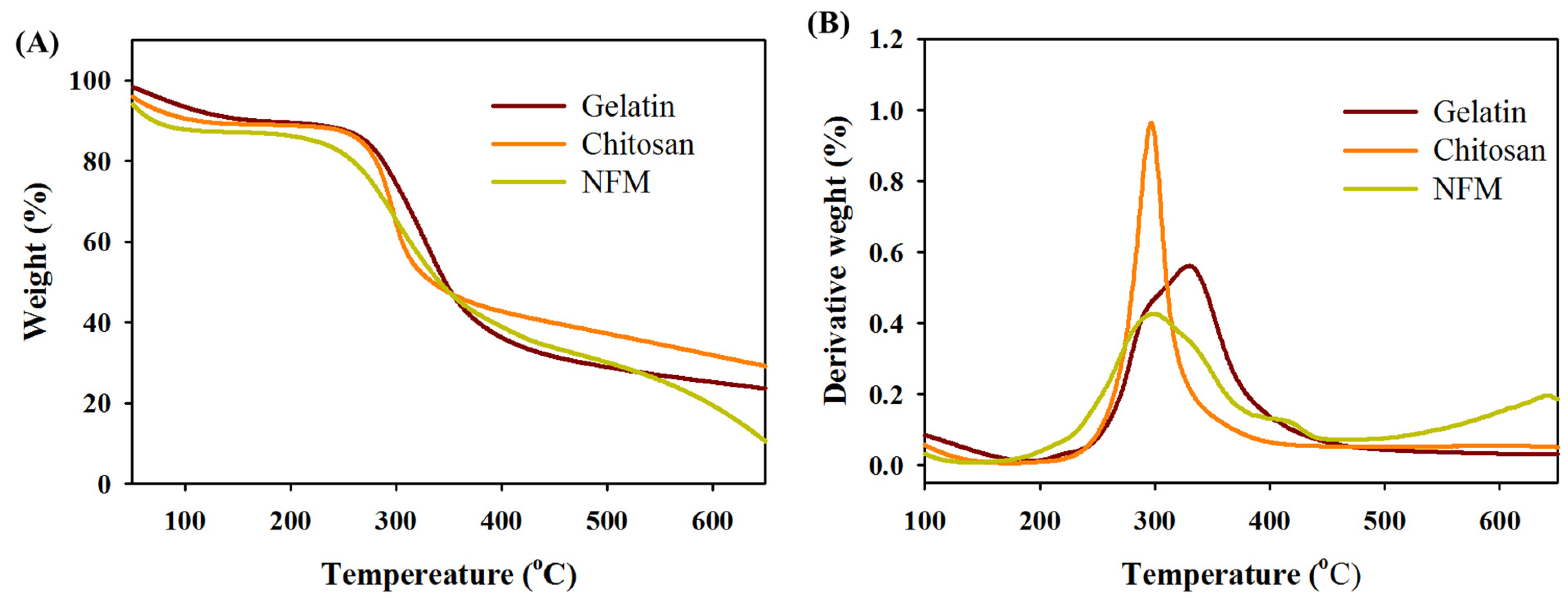
The thermal properties of gelatin, chitosan, and the NFMs were analyzed through thermogravimetric analysis (TGA) (Figure 3A) and differential thermal analysis (DTA) (Figure 3B). Gelatin exhibits an initial weight loss between 50 °C and 120 °C, attributed to moisture evaporation. A secondary degradation phase occurs between 250 °C and 420 °C and shows a peak decomposition temperature at 330 °C, marking the thermal breakdown of its protein structure. At the end of this stage, ~24% residual weight representing carbonaceous ash formation was found. Chitosan shows some weight loss before 100 °C due to moisture release. A major decomposition stage is noted between 216 °C and 370 °C, with a peak decomposition temperature at 296 °C, attributed to the breakdown of its carbohydrate polymer structure, and it leaves ~29% residual ash. The composite NFM displays distinct stages of degradation, reflective of the combined properties of its components. Initial weight loss below 120 °C corresponds to moisture evaporation, indicating its high hydrophilicity, and is consistent with the individual water affinity characteristics of gelatin and chitosan. The first major degradation phase occurs between 150 °C and 220 °C, primarily due to the breakdown of peptide bonds in gelatin, initiating weight loss at lower temperatures compared to chitosan, which exhibits greater thermal stability due to its polysaccharide backbones. The principal degradation phase, spanning from 220 °C to 440 °C, is dominated by chitosan’s decomposition, involving cleavage of C–N and C–O bonds in glucosamine units, resulting in considerable mass reduction. Gelatin continues to degrade within this range, contributing to further mass loss. The NFM demonstrates enhanced thermal stability due to intermolecular hydrogen bonding between gelatin and chitosan, which delays degradation onset. Cross-linking between amine groups of chitosan and gelatin further stabilizes the composite, making it well suited for biomedical applications requiring controlled degradation. Beyond 400 °C, a small amount of carbonaceous residue remains, mainly from chitosan’s ash, which is resistant to further decomposition. This thermal profile, with clearly defined stages for moisture loss, initial, and main degradation, highlights the composite’s robust degradation behavior, affirming its suitability as a tissue engineering scaffold where gradual, predictable breakdown is crucial for promoting cell infiltration and scaffold integration over time.
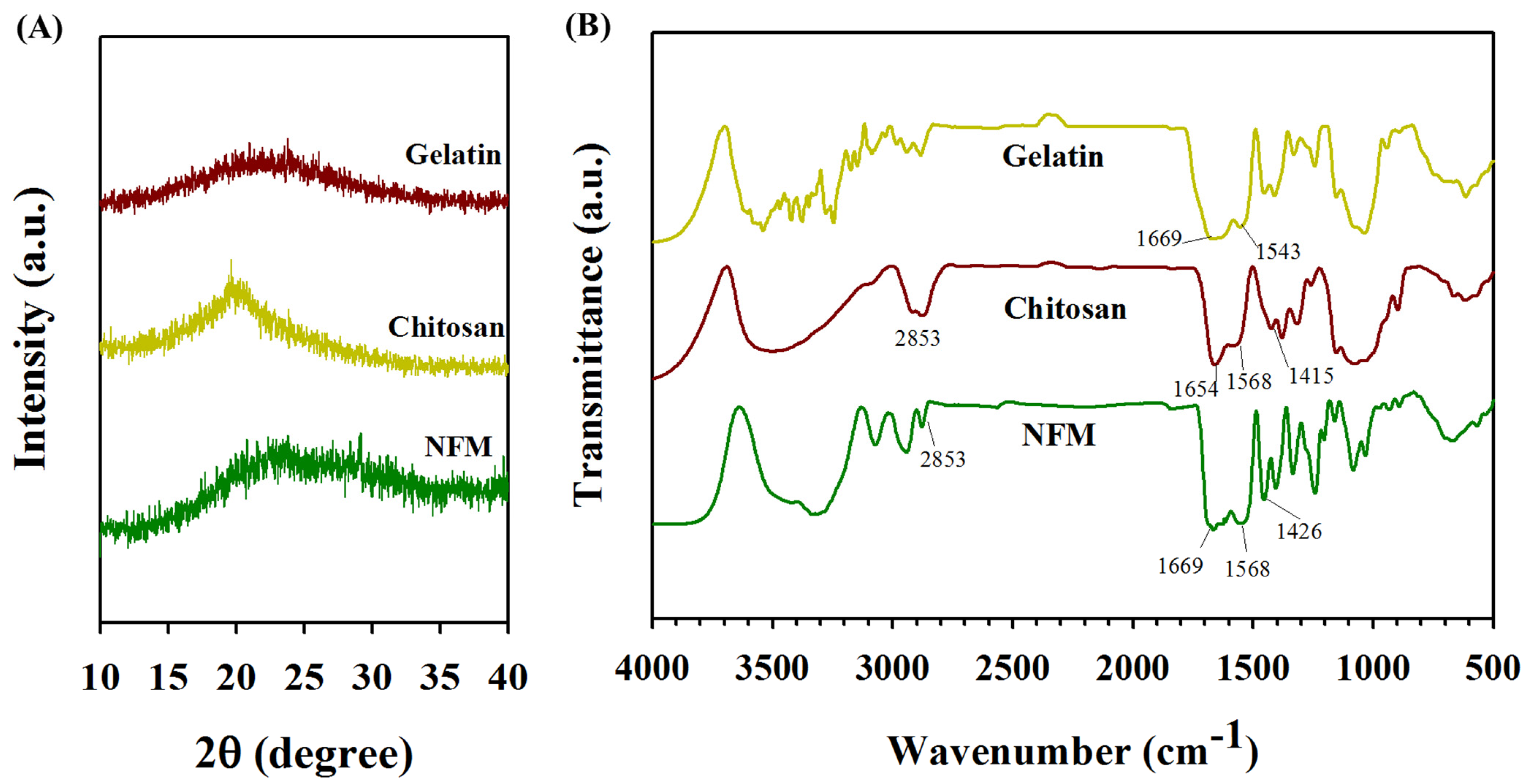
The X-ray diffraction (XRD) analysis of NFMs reveals a predominantly amorphous structure, with broad, low-intensity peaks between 2θ = 10° to 30°, attributed to the amorphous nature of gelatin and the semi-crystalline characteristics of chitosan (Figure 4A). The reduced crystallinity and broadened peaks suggest intermolecular hydrogen bonding and cross-linking between polymer chains, which disrupts chitosan’s crystalline regions [44]. This amorphous profile may enhance the membrane’s biocompatibility by offering an irregular surface conducive to cell attachment and proliferation. The Fourier transform infrared (FTIR) spectral analysis of the NFM revealed all characteristic peaks belonging to gelatin and chitosan and affirmed the presence of both components in the composite structure (Figure 4B). Gelatin exhibited peaks at 1669 cm−1, 1543 cm−1, and 3300 cm−1, which correspond to C–O stretching vibrations, coupled N–H in-plane bending with C–N stretching, and N–H stretching vibrations, respectively. Chitosan shows characteristic peaks at 1078 cm−1 for C–O bond, 1415 cm−1 for amide and ether bonds, 1568 and 1654 cm−1 for N–H stretch, 2853 cm−1 for C–H stretch, and 3331 cm−1 for O–H stretch [45]. For cross-linked NFM, all absorption peaks of gelatin and chitosan could be identified. However, we cannot identify an absorption peak at 1640 cm−1 arising from the new imide bonds formed after cross-linking, which occurs due to interference from the strong absorption of the amide I peak present in both gelatin and chitosan. As a protein-derived polymer, gelatin exhibits extensive hydrogen bonding between peptide chains and with water molecules, contributing to the broad and overlapping bands commonly associated with amide I and amide II vibrations. Consequently, the 1699 cm−1 band may be attributed to intermolecular associations, while the 1640 cm−1 band corresponds primarily to imide bonds [46]. The presence of these distinct peaks in the NFM confirms the homogeneous blending of gelatin and chitosan in the spinning solution for preparing composite NFMs.
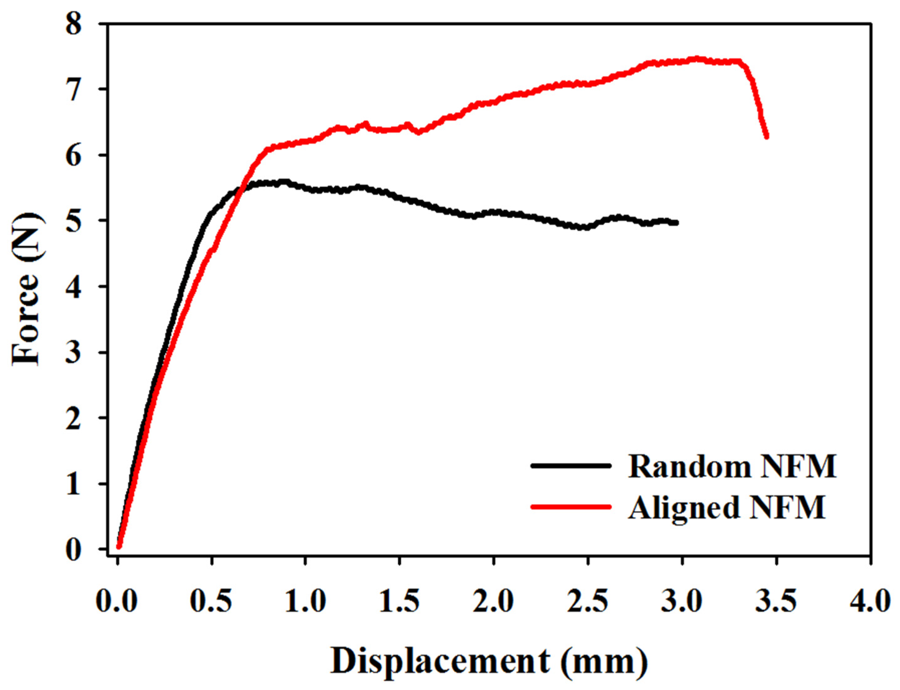
As the mechanical properties of an NFM should include its ability to resist the stress experienced during application, uniaxial tensile testing was conducted with a mechanical testing machine to determine an NFM’s stiffness, ultimate strength, and elongation during a break. The tested NFM sample was wet after being fully soaked in PBS for 7 days to simulate the cell culture condition. Typical force–displacement curves are shown in Figure 5 for random and aligned NFMs. The stiffness, maximum load, and maximum displacement can be obtained from the curves and the average values of four samples are compared in Table 1. The aligned NFM shows higher mean values of maximum load and maximum displacement, but no significance was found when compared with the random NFM. However, aligned NFMs display significantly higher stiffness when tested alongside the fiber axis, due to their parallel configuration, which distributes applied stress effectively and resists deformation. This directional strength makes aligned fibers ideal for applications requiring mechanical reinforcement in specific orientations, such as ligament or tendon scaffolds [23,47]. Conversely, random NFMs exhibit lower stiffness but offer isotropic mechanical properties, supporting uniform performance in all directions. This random orientation mimics the irregular structure of natural ECM, enhancing multidirectional cell attachment and proliferation, thus favoring applications in soft tissue engineering where balanced mechanical integrity is necessary. Overall, the different mechanical behaviors revealed from aligned and random fiber configurations highlight the adaptability of NFMs, which can allow for tailored properties through electrospinning to meet specific biomechanical needs.

3.2. In Vitro Cell Culture

The peritoneum mesothelial cells isolated from SD rats grow smoothly on TCPS during in vitro cell culture and with elongated, flattened, and squamous-like morphology (Figure 6A). The cell viability from MTT assays indicates a fast cell proliferation rate up to 5 days, and the cell number reaches saturation thereafter, due to contact inhibition (Figure 6B).
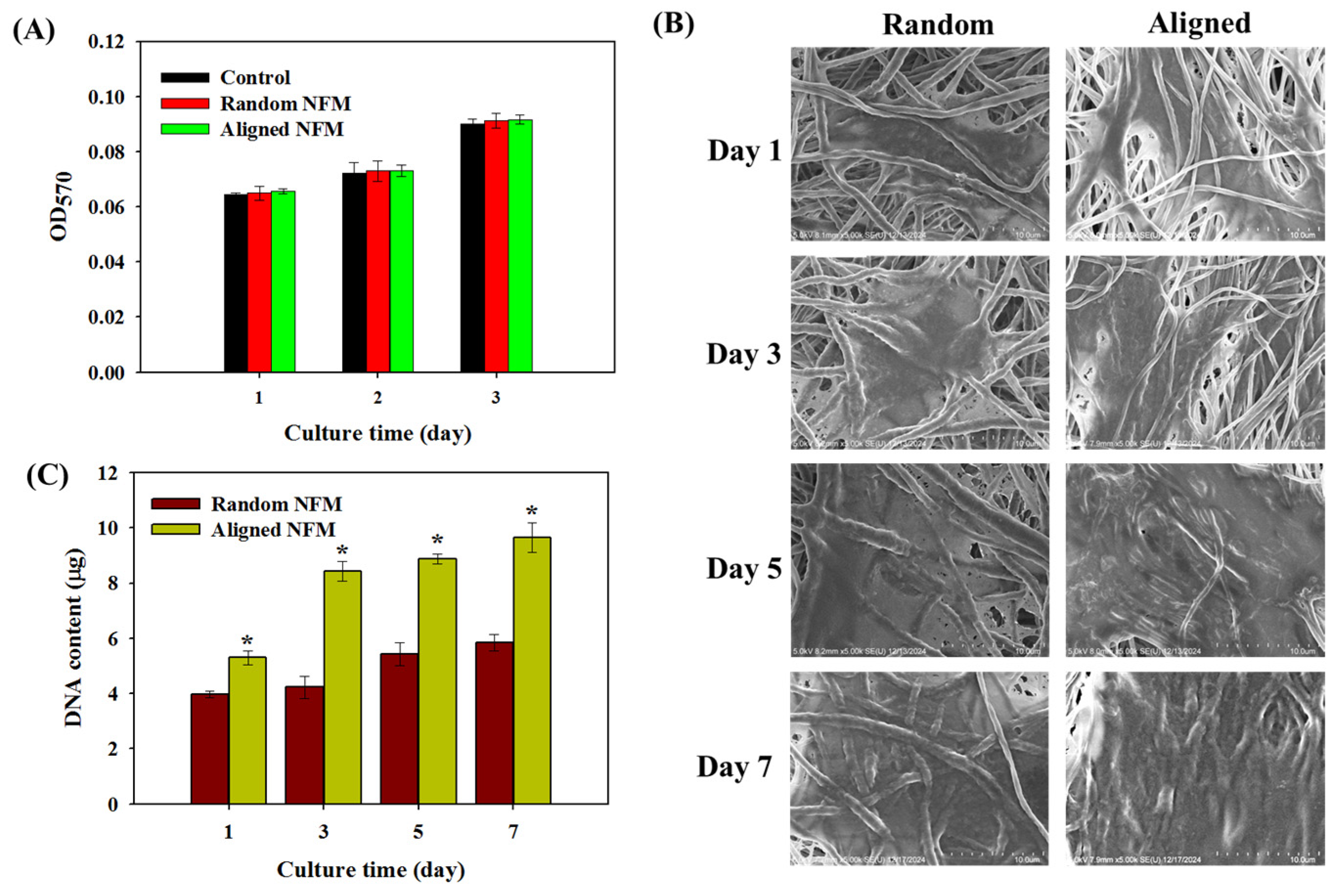
The biocompatibility of random and aligned NFMs with mesothelial cells was assessed by culturing mesothelial cells with extracts of each NFM and evaluating the metabolic activity of cells via MTT assays. As depicted in Figure 7A, there is no significant difference in cell viability between mesothelial cells cultured in cell culture medium (control) and in the extract of aligned and random NFMs, validating both scaffolds’ biocompatibility. For mesothelial cells cultured in the NFMs, the SEM analysis at different culture times demonstrates an excellent cellular response toward mesothelial cells for cell adhesion, spreading, and proliferation (Figure 7B). On day 1 post seeding, cells adhered to the membrane surface and displayed a spindle-shaped morphology, initiating spread across the scaffold. By day 3, a pronounced alignment of cells along the axis of aligned fibers became evident, with cell density substantially higher on aligned NFMs than on random NFMs. This cell organization persisted and intensified on days 5 and 7, resulting in a continuous cellular layer particularly prominently on aligned NFMs, where cells became more flattened and cohesive. This is consistent with a previous finding, where cross-linked gelatin/chitosan nanofibers provided endothelial cells with direction cell attachment and migration along aligned fibers [26]. The SEM images further revealed ECM deposition surrounding the adhered cells, which may be attributed to the positive charge of chitosan, which not only enhances cell adhesion but also binds with the negatively charged ECM components. Chitosan in the scaffold can also serve to improve cell attachment by leveraging this electrostatic interaction. The nanofibrous architecture of NFMs offers a large surface-to-volume ratio and provides an ideal milieu for the attachment of mesothelial cells. It also facilitates cell proliferation and migration, allowing efficient nutrient and waste exchange within the cellular microenvironment.
To evaluate cell proliferation, the cell numbers in the NFMs were compared by determining the total DNA contents in each scaffold at different culture times (Figure 7C). A noticeable increase in DNA content was observed with time in both scaffolds, endorsing their suitability for the growth of mesothelial cells. Gelatin contains a repetitive Arg-Gly-Asp (RGD) tripeptide to serve as a key cell-recognition motif. This RGD sequence binds to integrin, a receptor family, on cell surfaces specific to ECM proteins, which mediates cell adhesion and promotes cell attachment. When RGD binds to integrin, it not only strengthens cell attachment but also acts as a signal transducer, triggering an intracellular signaling cascade that upregulates gene expression associated with cell growth and differentiation. The interaction of RGD with integrin thus underscores the use of gelatin in the composite NFMs to enhance cell attachment and activate signaling pathways that support cellular growth and differentiation [48]. Even though the DNA content increased in both scaffolds, it is worth noting that aligned NFMs may offer a more favorable environment for mesothelial cell attachment and growth. Using the DNA content on day 1 as an indication of cell attachment, this value is significantly higher for the aligned NFM than its random counterpart. Comparing the cell proliferation rate, the DNA content increase rate from day 1 to day 7 is significantly higher for aligned NFMs (1.8 folds) than random NFMs (1.4 folds). This higher cell proliferation rate may be attributed to aligned topography. It is expected that aligned nanofibers provide a larger surface area along the fiber axis for cells to grow than cells grown on random nanofibers. Furthermore, aligned architecture may encourage the growth of mesothelial cells through the contact guidance effect.
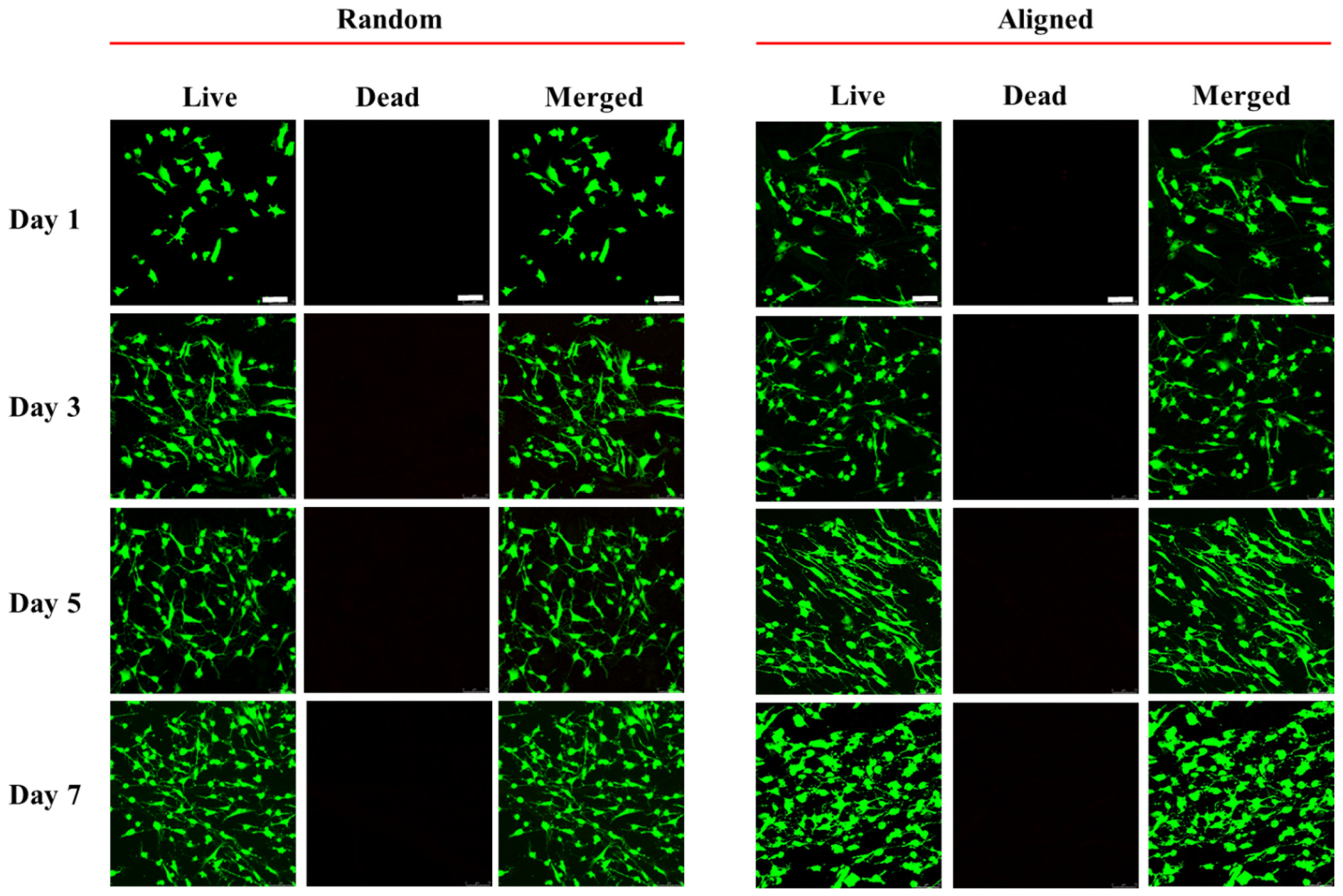
The viability of mesothelial cells in NFMs was confirmed from Live/Dead staining with laser scanning confocal microscopy. As shown in Figure 8, stacked confocal images indicate that cells are highly viable from day 1 to day 7, with negligible dead cells to support the excellent biocompatibility of NFMs. However, the aligned NFM reveals more live cells showing directional cell morphology, highlighting that aligned fiber orientation can guide cell attachment and proliferation more effectively. The structured topography of aligned NFMs offers a supportive environment that promotes the proliferation of viable mesothelial cells, an essential feature for tissue engineering applications [49]. Overall, these findings demonstrate that although both NFMs provide a beneficial environment for mesothelial cells, the aligned NFM may provide a better milieu for mesothelial cell attachment and proliferation with oriented morphology.
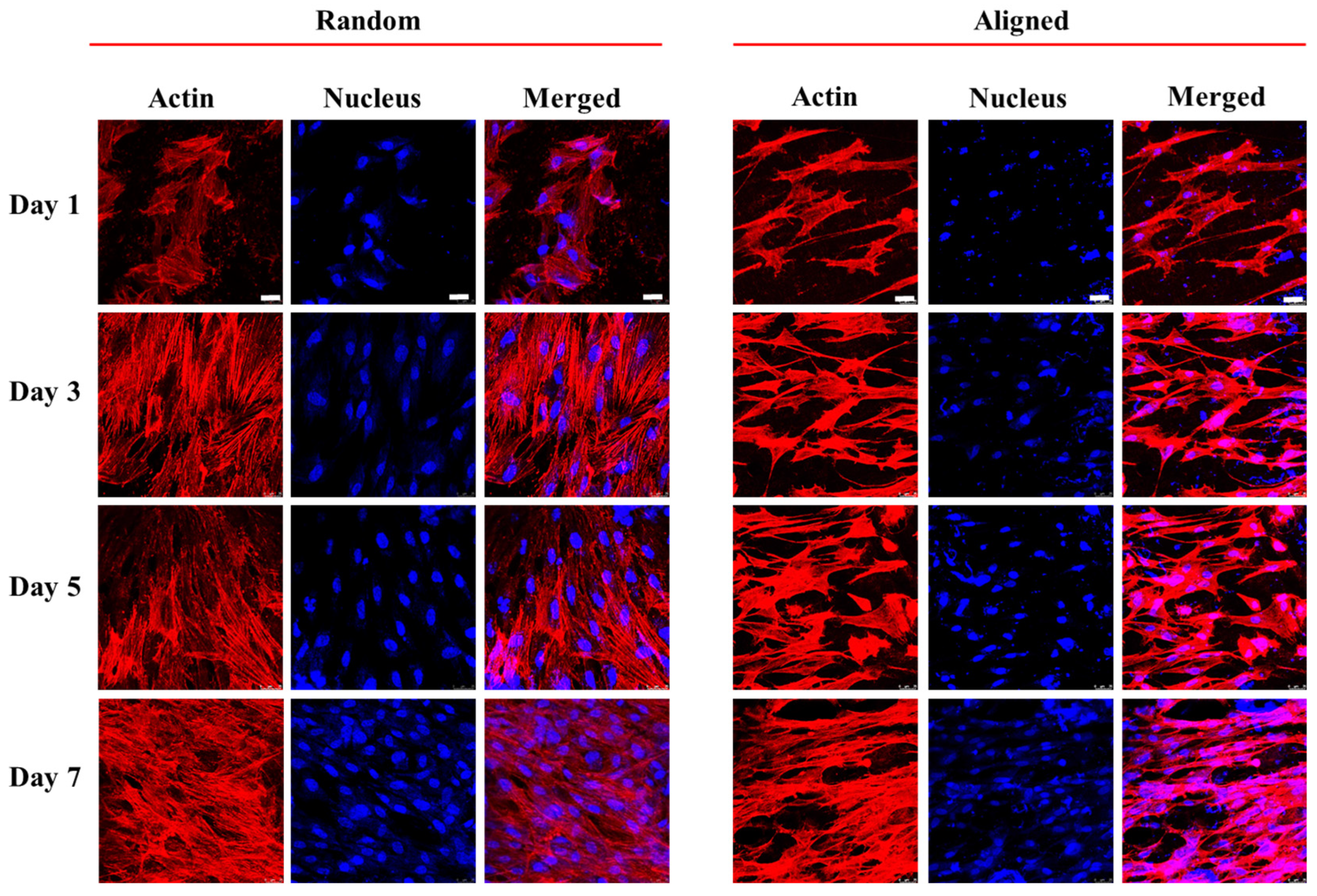
To reveal cytoskeleton expression using confocal microscopy, mesothelial cells grown in aligned and random NFMs were stained with Phalloidin-TRITC for F-actin and Hoechst 33258 for nuclei. As shown in Figure 9, spindle-shaped cell morphology with direction distribution of filamentous F-actin (red) and elongated nucleus (blue) was shown in aligned NFMs. In comparison, cells in the random NFM exhibit a polygonal shape with round nuclei. Mesothelial cells in both NFMs showed increasing actin expression from day 1 to day 7. However, the actin expression is more concentrated, and the actin cytoskeleton arrangement is less isotropic in the aligned NFM than in the random NFM.
The impact of fiber alignment on the gene expression of mesothelial cells, including E-cadherin, calretinin, the intercellular adhesion molecule-1 (ICAM-1), and vascular endothelial growth factor (VEGF), were studied using qRT-PCR. As shown in Figure 10, mesothelial cells in the aligned NFM displayed significantly higher expression levels of these mesothelial markers than the random NFM. This upregulation is likely due to the aligned fiber direction cue toward mesothelial cells, which can not only promote cytoskeletal alignment and cell elongation, but also enhance gene expression linked to cell adhesion, differentiation, and phenotype maintenance. Mesothelial cells express calretinin, a calcium-binding protein involved in calcium signaling. Although the precise role of calretinin remains scarcely elucidated, there is a hypothesis that it influences the cell cycle [50]. However, several immunohistochemistry investigations have shown that calretinin serves as a valuable indicator for mesothelial cells [51]. Mesoderm is the source of mesothelial cells, which show distinctive features by expressing many cell adhesion molecules, such as ICAM-1 and E-cadherin [52]. E-cadherin is a calcium-dependent transmembrane epithelial adhesion protein that is uniquely expressed in epithelial lineage cells and facilitates intercellular adhesion [53]. As epithelial cells adopt a mesenchymal migratory and invasive characteristic with downregulated E-cadherin expression, this protein may play a role in the epithelial-to-mesenchymal (EMT) transition [54]. The intercellular adhesion molecule-1 (ICAM-1) or CD54 is constitutively expressed in mesothelial cells. Moreover, it has been shown that ICAM-1 has promising potential as a distinguishing factor between mesothelial cells and fibroblasts [55]. Additionally, the introduction of soluble ICAM-1 into cells leads to a significant decrease in neutrophil transmigration [56]. The proangiogenic factor vascular endothelial growth factor (VEGF) is involved in endothelial cell proliferation and vascular permeability [57]. It is a main growth factor for mesothelial cells to stimulate their growth. The local production of the VEGF by transitional mesothelial cells plays an important role during the peritoneal angiogenesis process, and the EMT of mesothelial cells is the mechanism responsible for the upregulation of the VEGF [58]. Therefore, transitional expression of the VEGF by mesothelial cells may suggest the maintenance of mesothelial cell characteristics. Overall, gene expression analysis suggests that the oriented topography of aligned NFMs can direct gene expression profiles that mimic in vivo mesothelial tissue. These findings support the potential application of aligned NFMs in regenerative medicine, where scaffold-induced gene expression and phenotype retention are critical for tissue engineering outcomes.
To investigate the expression of mesothelial marker proteins, immunofluorescence (IF) staining of calretinin and E-cadherin was studied using confocal microscopy (Figure 11). On day 3, both random and aligned NFMs showed a random distribution of these cell-associated proteins. On day 7, a more organized alignment of calretinin and E-cadherin appeared, likely influenced by directional cell growth. From day 3 to day 7, a marked increase in the synthesis of these proteins was observed, showing uniform distribution in both membranes; however, the aligned NFM appears to support a higher protein expression level. A semi-quantitative analysis of the green fluorescence-stained areas in the IF images was performed to evaluate marker protein synthesis ability (Table 2). The calretinin and E-cadherin levels continuously increased with time for both NFMs; however, the aligned NFM consistently demonstrated a higher synthesis rate for these mesothelial marker proteins. This trend suggests that the structural orientation provided by the aligned nanofiber arrangement facilitates an environment more beneficial to the maintenance of mesothelial cell phenotypes. Such outcomes underscore the use of aligned NFMs to maintain mesothelial phenotypic traits for tissue engineering applications, and its role in providing a favorable milieu for mesothelial tissue growth.

4. Conclusions

In this study, we demonstrate that an aligned gelatin/chitosan composite NFM offers a favorable cellular response toward mesothelial cells than its random counterpart NFM. NFMs can maintain their structural integrity after cross-linking with GTA vapor, which is essential for long-term vitro cell culture. Their nanofibrous architecture can provide a high surface area and effective nutrient transfer to support cellular infiltration and ECM formation for tissue engineering applications. The combination of gelatin and chitosan in the NFMs provided an ideal milieu for the adhesion and proliferation of mesothelial cells with high viability, and fiber alignment was found to be a suitable cue to promote cell attachment and proliferation rate. The aligned nanofibers can induce cell orientation along the fiber axis and promote gene expression and protein synthesis of key mesothelial markers, due to biophysical cues provided by the aligned nanofibers. Overall, the aligned NFM exhibits tailored structural and biological properties for application in the regenerative therapy of a damaged mesothelium layer. It also can be employed in applications where cellular alignment is critical for various regenerative therapies.

Author Contributions

Conceptualization, H.-H.K., D.T.G., and J.-P.C.; methodology, H.-H.K., D.T.G., and J.-P.C.; formal analysis, H.-H.K. and D.T.G.; resources, J.-P.C.; data curation, D.T.G. and B.S.D.; investigation, D.T.G. and B.S.D.; writing—original draft preparation, D.T.G.; writing—review and editing, J.-P.C.; supervision, J.-P.C.; funding acquisition, H.-H.K. All authors have read and agreed to the published version of the manuscript.

Funding

We acknowledge the financial support of Chang Gung Memorial Hospital (CMRPG2N0031) and the National Science and Technology Council, Taiwan (NSTC-112-2314-B-182A-059).

Institutional Review Board Statement

This study was conducted according to the guidelines of the Declaration of Helsinki and was approved by the Institutional Review Board of Chang Gung University (IACUC approval no. CGU111-237).

Data Availability Statement

The data presented in this study are available on request from the corresponding author.

Acknowledgments

We acknowledge the technical support provided by the Microscope Core Laboratory, Chang Gung Memorial Hospital, Linkou, and the Microscopy Center, Chang Gung University.

Conflicts of Interest

The authors declare no conflicts of interest. The funders had no role in study design, collection, analyses, or interpretation of the data, manuscript writing, or decision to publish the results.

References

  1. Zou, J.; Li, L.; Yang, Z.; Chen, X. Phototherapy meets immunotherapy: A win–win strategy to fight against cancer. Nanophotonics 2021, 10, 3229–3245. [Google Scholar] [CrossRef]
  2. Mutsaers, S.E.; Prêle, C.M.; Lansley, S.M.; Herrick, S.E. The origin of regenerating mesothelium: A historical perspective. Int. J. Artif. Organs 2007, 30, 484–494. [Google Scholar] [CrossRef]
  3. Mutsaers, S.E. Mesothelial cells: Their structure, function and role in serosal repair. Respirology 2002, 7, 171–191. [Google Scholar] [CrossRef]
  4. Roumeliotis, S.; Dounousi, E.; Salmas, M.; Eleftheriadis, T.; Liakopoulos, V. Unfavorable Effects of Peritoneal Dialysis Solutions on the Peritoneal Membrane: The Role of Oxidative Stress. Biomolecules 2020, 10, 768. [Google Scholar] [CrossRef]
  5. Brunkhorst, R.; Mahiout, A. Pyruvate neutralizes peritoneal dialysate cytotoxicity: Maintained integrity and proliferation of cultured human mesothelial cells. Kidney Int. 1995, 48, 177–181. [Google Scholar] [CrossRef]
  6. Uiterwijk, H.; Franssen, C.F.M.; Kuipers, J.; Westerhuis, R.; Nauta, F.L. Glucose Exposure in Peritoneal Dialysis Is a Significant Factor Predicting Peritonitis. Am. J. Nephrol. 2020, 51, 237–243. [Google Scholar] [CrossRef]
  7. Mutsaers, S.E.; Birnie, K.; Lansley, S.; Herrick, S.E.; Lim, C.B.; Prêle, C.M. Mesothelial cells in tissue repair and fibrosis. Front. Pharmacol. 2015, 6, 113. [Google Scholar] [CrossRef]
  8. Mutsaers, S.E.; Prele, C.M.; Pengelly, S.; Herrick, S.E. Mesothelial cells and peritoneal homeostasis. Fertil. Steril. 2016, 106, 1018–1024. [Google Scholar] [CrossRef]
  9. Mutsaers, S.E.; Wilkosz, S. Structure and function of mesothelial cells. Cancer Treat. Res. 2007, 134, 1–19. [Google Scholar] [CrossRef] [PubMed]
  10. Lachaud, C.C.; Rodriguez-Campins, B.; Hmadcha, A.; Soria, B. Use of Mesothelial Cells and Biological Matrices for Tissue Engineering of Simple Epithelium Surrogates. Front. Bioeng. Biotechnol. 2015, 3, 117. [Google Scholar] [CrossRef] [PubMed]
  11. Hiriart, E.; Deepe, R.; Wessels, A. Mesothelium and Malignant Mesothelioma. J. Dev. Biol. 2019, 7, 7. [Google Scholar] [CrossRef] [PubMed]
  12. Herrick, S.E.; Mutsaers, S.E. Mesothelial progenitor cells and their potential in tissue engineering. Int. J. Biochem. Cell Biol. 2004, 36, 621–642. [Google Scholar] [CrossRef] [PubMed]
  13. Osawa, H.; Nishimura, J.; Hiraki, M.; Takahashi, H.; Haraguchi, N.; Hata, T.; Ikenaga, M.; Murata, K.; Yamamoto, H.; Mizushima, T.; et al. Regeneration of peritoneal mesothelial cells after placement of hyaluronate carboxymethyl-cellulose (Seprafilm®). Surg. Today 2017, 47, 130–136. [Google Scholar] [CrossRef] [PubMed]
  14. Nikolova, M.P.; Chavali, M.S. Recent advances in biomaterials for 3D scaffolds: A review. Bioact. Mater. 2019, 4, 271–292. [Google Scholar] [CrossRef] [PubMed]
  15. Sharma, C.; Dinda, A.K.; Mishra, N.C. Fabrication and characterization of natural origin chitosan-gelatin-alginate composite scaffold by foaming method without using surfactant. J. Appl. Polym. Sci. 2013, 127, 3228–3241. [Google Scholar] [CrossRef]
  16. Krithica, N.; Natarajan, V.; Madhan, B.; Sehgal, P.K.; Mandal, A.B. Type I Collagen Immobilized Poly(caprolactone) Nanofibers: Characterization of Surface Modification and Growth of Fibroblasts. Adv. Eng. Mater. 2012, 14, B149–B154. [Google Scholar] [CrossRef]
  17. Mitragotri, S.; Anderson, D.G.; Chen, X.; Chow, E.K.; Ho, D.; Kabanov, A.V.; Karp, J.M.; Kataoka, K.; Mirkin, C.A.; Petrosko, S.H.; et al. Accelerating the Translation of Nanomaterials in Biomedicine. ACS Nano 2015, 9, 6644–6654. [Google Scholar] [CrossRef] [PubMed]
  18. Ye, K.; Kuang, H.; You, Z.; Morsi, Y.; Mo, X. Electrospun Nanofibers for Tissue Engineering with Drug Loading and Release. Pharmaceutics 2019, 11, 182. [Google Scholar] [CrossRef] [PubMed]
  19. Li, D.; Chen, W.; Sun, B.; Li, H.; Wu, T.; Ke, Q.; Huang, C.; Ei-Hamshary, H.; Al-Deyab, S.S.; Mo, X. A comparison of nanoscale and multiscale PCL/gelatin scaffolds prepared by disc-electrospinning. Colloids Surf. B Biointerfaces 2016, 146, 632–641. [Google Scholar] [CrossRef]
  20. Echave, M.C.; Saenz del Burgo, L.; Pedraz, J.L.; Orive, G. Gelatin as Biomaterial for Tissue Engineering. Curr. Pharm. Des. 2017, 23, 3567–3584. [Google Scholar] [CrossRef]
  21. Govindaraju, D.T.; Kao, H.-H.; Chien, Y.-M.; Chen, J.-P. Composite Polycaprolactone/Gelatin Nanofiber Membrane Scaffolds for Mesothelial Cell Culture and Delivery in Mesothelium Repair. Int. J. Mol. Sci. 2024, 25, 9803. [Google Scholar] [CrossRef] [PubMed]
  22. Chang, J.-J.; Lee, Y.-H.; Wu, M.-H.; Yang, M.-C.; Chien, C.-T. Electrospun anti-adhesion barrier made of chitosan alginate for reducing peritoneal adhesions. Carbohydr. Polym. 2012, 88, 1304–1312. [Google Scholar] [CrossRef]
  23. Govindaraju, D.T.; Chen, C.H.; Kuo, C.Y.; Shalumon, K.T.; Chien, Y.M.; Kao, H.H.; Chen, J.P. Development of high resilience spiral wound suture-embedded gelatin/PCL/heparin nanofiber membrane scaffolds for tendon tissue engineering. Int. J. Biol. Macromol. 2022, 221, 314–333. [Google Scholar] [CrossRef]
  24. Kao, H.-H.; Kuo, C.-Y.; Govindaraju, D.T.; Chen, K.-S.; Chen, J.-P. Polycaprolactone/Chitosan Composite Nanofiber Membrane as a Preferred Scaffold for the Culture of Mesothelial Cells and the Repair of Damaged Mesothelium. Int. J. Mol. Sci. 2022, 23, 9517. [Google Scholar] [CrossRef]
  25. Jalaja, K.; Naskar, D.; Kundu, S.C.; James, N.R. Potential of electrospun core-shell structured gelatin-chitosan nanofibers for biomedical applications. Carbohydr. Polym. 2016, 136, 1098–1107. [Google Scholar] [CrossRef]
  26. Qian, Y.-F.; Zhang, K.-H.; Chen, F.; Ke, Q.-F.; Mo, X.-M. Cross-linking of gelatin and chitosan complex nanofibers for tissue-engineering scaffolds. J. Biomater. Sci. Polym. Ed. 2011, 22, 1099–1113. [Google Scholar] [CrossRef] [PubMed]
  27. Casper, C.L.; Yamaguchi, N.; Kiick, K.L.; Rabolt, J.F. Functionalizing Electrospun Fibers with Biologically Relevant Macromolecules. Biomacromolecules 2005, 6, 1998–2007. [Google Scholar] [CrossRef]
  28. Li, W.; Zhang, X.; Chen, Y.; Yang, G.; Huang, J.; Xiong, L. Acupoint catgut from electrospun tri-layer core–shell fibers loaded with adenosine for a long-term analgesia effect. Eur. Polym. J. 2024, 220, 113447. [Google Scholar] [CrossRef]
  29. Chen, X.; Liu, Y.; Liu, P. Electrospun Core–Sheath Nanofibers with a Cellulose Acetate Coating for the Synergistic Release of Zinc Ion and Drugs. Mol. Pharm. 2024, 21, 173–182. [Google Scholar] [CrossRef] [PubMed]
  30. Wang, M.; Huang, C.; Chen, Y.; Ji, Y.; Yu, D.-G.; Bligh, S.W.A. Medicated tri-layer fibers based on cellulose acetate and polyvinylpyrrolidone for enhanced antibacterial and wound healing properties. Carbohydr. Polym. 2025, 348, 122856. [Google Scholar] [CrossRef] [PubMed]
  31. Wang, S.; Hu, F.; Li, J.; Zhang, S.; Shen, M.; Huang, M.; Shi, X. Design of electrospun nanofibrous mats for osteogenic differentiation of mesenchymal stem cells. Nanomedicine 2018, 14, 2505–2520. [Google Scholar] [CrossRef] [PubMed]
  32. Chen, C.H.; Li, D.L.; Chuang, A.D.; Dash, B.S.; Chen, J.P. Tension Stimulation of Tenocytes in Aligned Hyaluronic Acid/Platelet-Rich Plasma-Polycaprolactone Core-Sheath Nanofiber Membrane Scaffold for Tendon Tissue Engineering. Int. J. Mol. Sci. 2021, 22, 11215. [Google Scholar] [CrossRef]
  33. Xie, J.; Liu, W.; MacEwan, M.R.; Bridgman, P.C.; Xia, Y. Neurite Outgrowth on Electrospun Nanofibers with Uniaxial Alignment: The Effects of Fiber Density, Surface Coating, and Supporting Substrate. ACS Nano 2014, 8, 1878–1885. [Google Scholar] [CrossRef] [PubMed]
  34. Xie, J.; Shen, H.; Yuan, G.; Lin, K.; Su, J. The effects of alignment and diameter of electrospun fibers on the cellular behaviors and osteogenesis of BMSCs. Mater. Sci. Eng. C 2021, 120, 111787. [Google Scholar] [CrossRef] [PubMed]
  35. Rezvani Ghomi, E.; Khosravi, F.; Neisiany, R.E.; Shakiba, M.; Zare, M.; Lakshminarayanan, R.; Chellappan, V.; Abdouss, M.; Ramakrishna, S. Advances in electrospinning of aligned nanofiber scaffolds used for wound dressings. Curr. Opin. Biomed. Eng. 2022, 22, 100393. [Google Scholar] [CrossRef]
  36. Koukorava, C.; Ward, K.; Ahmed, K.; Almaghrabi, S.; Dauleh, S.; Pereira, S.M.; Taylor, A.; Haddrick, M.; Cross, M.J.; Wilm, B. Mesothelial Cells Exhibit Characteristics of Perivascular Cells in an In Vitro Angiogenesis Assay. Cells 2023, 12, 2436. [Google Scholar] [CrossRef] [PubMed]
  37. Zhang, J.; Yang, N.; Kreeger, P.K.; Notbohm, J. Topological defects in the mesothelium suppress ovarian cancer cell clearance. APL Bioeng. 2021, 5, 036103. [Google Scholar] [CrossRef]
  38. Chen, J.-P.; Chang, G.-Y.; Chen, J.-K. Electrospun collagen/chitosan nanofibrous membrane as wound dressing. Colloids Surf. A Physicochem. Eng. Asp. 2008, 313–314, 183–188. [Google Scholar] [CrossRef]
  39. Kao, H.H.; Kuo, C.Y.; Chen, K.S.; Chen, J.P. Preparation of Gelatin and Gelatin/Hyaluronic Acid Cryogel Scaffolds for the 3D Culture of Mesothelial Cells and Mesothelium Tissue Regeneration. Int. J. Mol. Sci. 2019, 20, 4527. [Google Scholar] [CrossRef]
  40. Jafari, J.; Emami, S.H.; Samadikuchaksaraei, A.; Bahar, M.A.; Gorjipour, F. Electrospun chitosan-gelatin nanofiberous scaffold: Fabrication and in vitro evaluation. Biomed. Mater. Eng. 2011, 21, 99–112. [Google Scholar] [CrossRef]
  41. Zhan, J.; Morsi, Y.; Ei-Hamshary, H.; Al-Deyab, S.S.; Mo, X. In vitro evaluation of electrospun gelatin–glutaraldehyde nanofibers. Front. Mater. Sci. 2016, 10, 90–100. [Google Scholar] [CrossRef]
  42. Peng, N.B.; He, J.H. Insight into the Wetting Property of a Nanofiber Membrane by the Geometrical Potential. Recent Pat. Nanotechnol. 2020, 14, 64–70. [Google Scholar] [CrossRef] [PubMed]
  43. Campiglio, C.E.; Contessi Negrini, N.; Fare, S.; Draghi, L. Cross-Linking Strategies for Electrospun Gelatin Scaffolds. Materials 2019, 12, 2476. [Google Scholar] [CrossRef]
  44. Beppu, M.M.; Vieira, R.S.; Aimoli, C.G.; Santana, C.C. Crosslinking of chitosan membranes using glutaraldehyde: Effect on ion permeability and water absorption. J. Membr. Sci. 2007, 301, 126–130. [Google Scholar] [CrossRef]
  45. Bharadwaj, T.; Chrungoo, S.; Verma, D. Self-assembled chitosan/gelatin nanofibrous aggregates incorporated thermosensitive nanocomposite bioink for bone tissue engineering. Carbohydr. Polym. 2024, 324, 121544. [Google Scholar] [CrossRef]
  46. Dizdarević, A.; Marić, M.; Shahzadi, I.; Ari Efiana, N.; Matuszczak, B.; Bernkop-Schnürch, A. Imine bond formation as a tool for incorporation of amikacin in self-emulsifying drug delivery systems (SEDDS). Eur. J. Pharm. Biopharm. 2021, 162, 82–91. [Google Scholar] [CrossRef] [PubMed]
  47. Govindaraju, D.T.; Chen, C.H.; Shalumon, K.T.; Kao, H.H.; Chen, J.P. Bioactive Nanostructured Scaffold-Based Approach for Tendon and Ligament Tissue Engineering. Nanomaterials 2023, 13, 1847. [Google Scholar] [CrossRef] [PubMed]
  48. Abdul Kafi, M.; El-Said, W.A.; Kim, T.H.; Choi, J.W. Cell adhesion, spreading, and proliferation on surface functionalized with RGD nanopillar arrays. Biomaterials 2012, 33, 731–739. [Google Scholar] [CrossRef] [PubMed]
  49. Gui, X.; Hu, J.; Han, Y. Random and aligned electrospun gelatin nanofiber mats for human mesenchymal stem cells. Mater. Res. Innov. 2018, 23, 208–215. [Google Scholar] [CrossRef]
  50. Doglioni, C.; Dei Tos, A.P.; Laurino, L.; Iuzzolino, P.; Chiarelli, C.; Celio, M.R.; Viale, G. Calretinin: A novel immunocytochemical marker for mesothelioma. Am. J. Surg. Pathol. 1996, 20, 1037–1046. [Google Scholar] [CrossRef] [PubMed]
  51. Gotzos, V.; Vogt, P.; Celio, M.R. The calcium binding protein calretinin is a selective marker for malignant pleural mesotheliomas of the epithelial type. Pathol. Res. Pract. 1996, 192, 137–147. [Google Scholar] [CrossRef] [PubMed]
  52. Nagel, H.; Hemmerlein, B.; Ruschenburg, I.; Hüppe, K.; Droese, M. The value of anti-calretinin antibody in the differential diagnosis of normal and reactive mesothelia versus metastatic tumors in effusion cytology. Pathol. Res. Pract. 1998, 194, 759–764. [Google Scholar] [CrossRef] [PubMed]
  53. Perl, A.K.; Wilgenbus, P.; Dahl, U.; Semb, H.; Christofori, G. A causal role for E-cadherin in the transition from adenoma to carcinoma. Nature 1998, 392, 190–193. [Google Scholar] [CrossRef] [PubMed]
  54. Takeichi, M. Morphogenetic roles of classic cadherins. Curr. Opin. Cell Biol. 1995, 7, 619–627. [Google Scholar] [CrossRef] [PubMed]
  55. Suassuna, J.H.; Das Neves, F.C.; Hartley, R.B.; Ogg, C.S.; Cameron, J.S. Immunohistochemical studies of the peritoneal membrane and infiltrating cells in normal subjects and in patients on CAPD. Kidney Int. 1994, 46, 443–454. [Google Scholar] [CrossRef]
  56. Yáñez-Mó, M.; Lara-Pezzi, E.; Selgas, R.; Ramírez-Huesca, M.; Domínguez-Jiménez, C.; Jiménez-Heffernan, J.A.; Aguilera, A.; Sánchez-Tomero, J.A.; Bajo, M.A.; Álvarez, V.; et al. Peritoneal Dialysis and Epithelial-to-Mesenchymal Transition of Mesothelial Cells. N. Engl. J. Med. 2003, 348, 403–413. [Google Scholar] [CrossRef] [PubMed]
  57. Ferrara, N.; Gerber, H.-P.; LeCouter, J. The biology of VEGF and its receptors. Nat. Med. 2003, 9, 669–676. [Google Scholar] [CrossRef] [PubMed]
  58. Aguilera, A.; Yáñez-Mo, M.; Selgas, R.; Sánchez-Madrid, F.; López-Cabrera, M. Epithelial to mesenchymal transition as a triggering factor of peritoneal membrane fibrosis and angiogenesis in peritoneal dialysis patients. Curr. Opin. Investig. Drugs 2005, 6, 262–268. [Google Scholar]
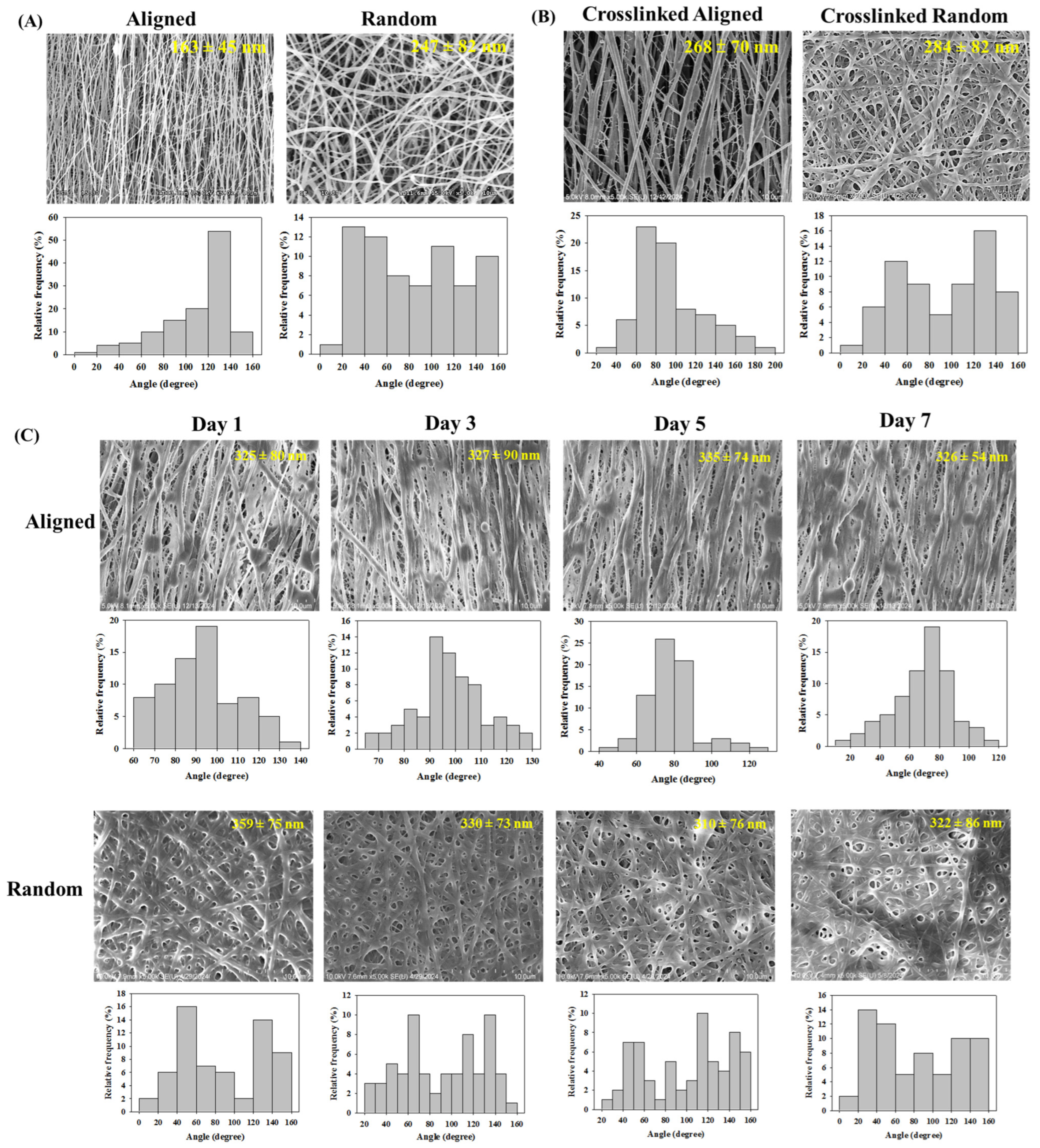
Figure 1. The scanning electron microscope (SEM) micrographs of aligned and random nanofibrous membranes (NFMs) before (A) and after cross-linking (B) (bar = 10 μm) with the average fiber size (mean ± SD) shown in each SEM image. (C) The SEM micrographs of cross-linked aligned NFMs after being immersed in cell culture medium for up to 7 days (bar = 10 μm) with the average fiber size (mean ± SD) shown in each SEM image. The histogram is the fiber orientation angle distribution.
Jcs 09 00031 g001
Figure 2. The water contact angles (A), swelling ratio (B), and degradation in a collagenase solution (C) of aligned and random nanofibrous membranes (NFMs).
Jcs 09 00031 g002
Figure 3. The thermogravimetric analysis (TGA) (A) and the differential thermal gravimetric (DTG) analysis (B) of gelatin, chitosan, and nanofibrous membranes (NFMs).
Jcs 09 00031 g003
Figure 4. The X-ray diffraction (XRD) (A) and the Fourier transform infrared (FTIR) spectroscopy (B) analysis of gelatin, chitosan, and nanofibrous membranes (NFMs).
Jcs 09 00031 g004
Figure 5. Typical tensile force–displacement curves of random and aligned nanofibrous membranes (NFMs) in wet conditions.
Jcs 09 00031 g005
Figure 6. The growth mesothelial cells on tissue culture polystyrene (TCPS) by observing with an inverted optical microscope (bar = 100 μm) (A) and determining the cell viability from MTT assays with solution absorbance at 570 nm (OD570) (B).
Jcs 09 00031 g006
Figure 7. (A) The biocompatibility of random and aligned nanofibrous membranes (NFMs). Mesothelial cells were cultured in a cell culture medium (control) and the extract of an NFM. The cell viability was determined from metabolic activity from solution absorbance at 570 nm (OD570). (B) The scanning electron microscopy (SEM) analysis of mesothelial cells in random and aligned NFMs (bar = 10 μm). (C) The proliferation of mesothelial cells in random and aligned NFMs by determining the cellular DNA contents. * p < 0.05 compared with random NFMs on each day.
Jcs 09 00031 g007
Figure 8. The confocal microscopy images after Live/Dead staining of mesothelial cells in random and aligned nanofibrous membranes (NFMs). Bar = 75 µm.
Jcs 09 00031 g008
Figure 9. The confocal microscopy images after actin cytoskeleton and nucleus staining of mesothelial cells in random and aligned nanofibrous membranes (NFMs). Bar = 25 µm.
Jcs 09 00031 g009
Figure 10. The quantitative real-time polymerase chain reaction (qRT-PCR) for quantifying the gene expression of mesothelial cells in random and aligned nanofibrous membranes (NFMs). The relative gene expression levels (normalized to day 1) of calretinin, E-cadherin, vascular endothelial factor (VEGF), and intercellular adhesion molecule-1 (ICAM-1) were compared. * p < 0.05 compared with random NFM on each day.
Jcs 09 00031 g010
Figure 11. The immunofluorescence staining of calretinin (A) and E-cadherin (B) by labeling with fluorescein-conjugated antibodies to show green fluorescence after culturing mesothelial cells in random and aligned nanofibrous membranes (NFMs). The nucleus was stained with Hoechst 33258 to show blue fluorescence. Bar = 25 μm.
Jcs 09 00031 g011
Table 1. The tensile mechanical properties of random and aligned nanofibrous membranes (NFMs).
MembranesMaximum Load (N)Maximum Displacement (mm)Stiffness (N/mm)
Random NFM6.24 ± 0.603.54 ± 0.559.88 ± 0.53
Aligned NFM6.68 ± 0.694.03 ±0.5412.28 ± 0.38 *
* p < 0.05 compared with random NFM.
Table 2. A semi-quantitative analysis of the area percentage of the green fluorescence areas, normalized by the number of nuclei in immunofluorescence images of calretinin and E-cadherin by culture mesothelial cells in random and aligned nanofibrous membranes (NFMs).
Culture Time (Days)Calretinin (%)E-Cadherin (%)
RandomAlignedRandomAligned
30.25 ± 0.020.35 ± 0.01 *0.23 ± 0.020.29 ± 0.01 *
70.43 ± 0.010.50 ± 0.02 *0.32 ± 0.020.43 ± 0.01 *
* p < 0.05 compared with random NFM on each day.
Disclaimer/Publisher’s Note: The statements, opinions and data contained in all publications are solely those of the individual author(s) and contributor(s) and not of MDPI and/or the editor(s). MDPI and/or the editor(s) disclaim responsibility for any injury to people or property resulting from any ideas, methods, instructions or products referred to in the content.