Communication

Ruby Nanoparticles for Greenhouse Farming: Synthesis, Features and Application

by
Mark O. Paskhin
1,
Kuder O. Aiyyzhy
1,
Roman V. Pobedonostsev
1,
Dina V. Kazantseva
1,
Ignat I. Rakov
1,
Ekaterina V. Barmina
1,
Denis V. Yanykin
1,2 and
Sergey V. Gudkov
1,3,*
1
Prokhorov General Physics Institute, Russian Academy of Sciences, Vavilov Str. 38, 119991 Moscow, Russia
2
Institute of Basic Biological Problems, Federal Research Center “Pushchino Scientific Center for Biological Research” FRC PSCBR, Russian Academy of Sciences, 2 Institutskaya Str., 142290 Pushchino, Russia
3
Department of Biophysics, Lobachevsky State University, 23 Gagarin Avenue, 603950 Nizhny Novgorod, Russia
*
Author to whom correspondence should be addressed.
J. Compos. Sci. 2024, 8(1), 7; https://doi.org/10.3390/jcs8010007
Submission received: 21 November 2023 / Revised: 20 December 2023 / Accepted: 22 December 2023 / Published: 25 December 2023
(This article belongs to the Section Nanocomposites)

Abstract

:
In this work, we investigated the effect of photoconversion covers based on ruby (chromium-doped alumina (Al2O3:Cr3+)) particles (PCC-R) on the growth and development of lettuce (Lactuca sativa) plants. Ruby particles (from 100 nm to 2 μm) were obtained by the sequential application of spall laser ablation and further laser fragmentation. The content of chromium ions relative to aluminum ions in the nanoparticles was 3.3 × 10⁻³. The covers with different densities of applied ruby particles (2 × 10⁷ m−2 (PCC-R7), 2 × 10⁸ m−2 (PCC-R8), 2 × 10⁹ m−2 (PCC-R9)) were studied in the present work. The PCC-Rs had two wide bands of luminescence excitation. The first one was from 350 nm to 450 nm with a maximum at 405 nm, and the second one was from 500 nm to 600 nm with a peak at 550 nm. Synthesized covers emitted in the range of 650 nm to 750 nm, with a peak at 695 nm. It has been shown that PCC-R8, in contrast to PCC-R7 and PCC-R9, provided an increase in yield by 40% and was characterized by increased water use efficiency during dark respiration and assimilation of carbon dioxide in plants. It is assumed that the observed positive effect of PCC-R8 photoconversion covers is associated with the activation of regulatory mechanisms due to a qualitative change in the light spectrum.

1. Introduction

Throughout human history, the scarcity of food resources has always motivated society to develop new technologies that make it easier to obtain food and increase its amount. The introduction of each new technology has temporarily solved the problem of a shortage of food resources and stimulated population growth, which in turn has led to new food shortages. Currently, human society is approaching another food crisis. By 2050, it is predicted that the global population will reach nine billion [1,2,3,4]. Therefore, the development of new technologies for agriculture is one of the most important tasks of modern society. Intensifying photosynthesis in crop plants is a promising approach for accelerating plant growth and development [5,6] and increasing crop yield. One of the main factors limiting photosynthesis is light: its quantitative and qualitative characteristics. It is known that plants absorb visible light in the region of photosythetically active radiation (400–700 nm), with red (600–700 nm) and blue (400–450 nm) light photons being used most effectively [7,8,9]. In conditions of low lighting, red photons are very strong stimulators of photosynthesis [10,11,12] because they provide the highest photosynthetic quantum yield [12]. In addition to blue and red light, ultraviolet light (UV), which is in the high energy wavelength range (200–400 nm), has a great influence on plant growth and development. Near ultraviolet (UV-A, 320–400 nm) is one of the photoinhibitory factors [13], which nevertheless determines plant development. It stimulates the accumulation of secondary metabolites: phenols, flavonoids, anthocyanins and ascorbic acid [14]. Photons of other spectral ranges (green, yellow, orange and far-red light) have no less effect on plants [15,16,17,18,19] and the ratio of different spectral ranges has a significant regulatory effect on plants [20,21,22,23,24]. Detailed information on the effect of light quality on plant growth is presented in a recent review [25].
Currently, special photoconversion covers are being actively developed that absorb light of certain wavelengths and convert it into light of another wavelength. Typically, PCCs absorb light beyond photosynthetically active radiation (PAR, 400–700 nm) [26,27,28,29,30,31,32], as well as green photons [33,34,35,36], due to the relatively low plant requirement for them. These covers re-emit in the PAR region, increasing its intensity as well as changing the ratio of spectral bands, triggering the plants’ regulatory mechanisms. The advantages of PCC are its relative ease of maintenance and the absence of energy costs. At the same time, some photoconversion covers are capable of self-generating electricity for the needs of greenhouses [37,38,39]. This provides a great advantage over artificial lighting systems, which consume large amounts of electricity on farms. However, despite the wide variety of phosphors and matrices used, photoconversion covers are a relatively “crude” technology, as they have a number of significant disadvantages. On the one hand, most phosphors with a high light conversion yield (for example, organic dyes) have a very short service life, and relatively stable phosphors (for example, nanoparticles of metal compounds) have a low quantum yield. On the other hand, photoconversion materials are sensitive to environmental influences due to the impossibility of selecting an ideal matrix. For example, water vapor near heated and/or excited phosphors forms reactive oxygen species that irreversibly damage luminescence centers. Moreover, most PCCs are still expensive to manufacture.
A good alternative to the phosphors known from the literature are ruby nanoparticles (Al2O3:Cr3+), since they absorb well in a wide range of wavelengths from 350 nm to 450 nm with a maximum at 405 nm and from 500 nm to 600 nm with a maximum at 550 nm, and fluoresce in the range from 660 nm to 720 nm with a maximum at 695 nm [40,41]. Ruby nanoparticles do not change their luminescent properties when heated and can easily be incorporated into polymer matrices.
Currently, there are various methods for producing luminophores, such as chemical vapor deposition, electrospraying, ultrasonication and ball milling [42,43,44,45]. Nevertheless, laser ablation of solids in liquids is a well-established technique that allows the generation of a large variety of nanoparticles without discharging any surface-active substances in comparison to conventional manufacturing routes [46,47,48]. The parameters of the generated nanoparticles (size distribution, chemical composition, etc.) depend on a number of experimental parameters, such as laser wavelength, laser pulse duration, laser fluence on the target, laser peak intensity, and the surrounding atmosphere. During laser ablation in a liquid, it is quite difficult to control the size distribution of nanoparticles. They have a wide dispersion, which is not always suitable for practical applications. One way to solve this problem is to subsequently irradiate colloidal solutions in the absence of a target (the laser fragmentation process). In this case, laser radiation interacts with particles inside the laser beam waist during the laser pulse. As a role, fragmentation proceeds through the melting of the particles and their interaction with the surrounding vapors of the liquid. As a rule, this leads to a shift of the maximum of the distribution into an area of smaller sizes and its narrowing. This process has been thoroughly studied both experimentally and theoretically [46,49,50,51,52].
In our previous work [40], ruby grains were synthesized by laser heating a mixture of aluminum and chromium oxides in air. In this work, for the first time, we created a series of PCCs based on ruby nanoparticles with a nominal composition of Al2O3:Cr3+ obtained by laser ablation and tested their effect on the growth and development of lettuce (Lactuca sativa) grown in laboratory conditions.

2. Materials and Methods

2.1. Nanoparticle Characterization

For nanoparticle preparation, an industrial corundum ceramic already containing Cr3+ ions (VK94-1, also called 22XC, Thermocomponents, Mytishchi, Moscow, Russia) was used.
The hydrodynamic diameter of the obtained ruby particles was measured by the dynamic light scattering (DLS) method using a Zetasizer ULTRA Red Label (Malvern Panalytical Ltd., Malvern, UK) operating at a laser wavelength of 632.8 nm.
The morphology of the synthesized particles was analyzed using a JEOL JSM 5910-LV scanning electron microscope (SEM) (Jeol Ltd., Tokyo, Japan). The chemical composition of VK94-1 before and after laser exposure was determined by Energy-dispersive X-ray spectroscopy (EDX) with a spectral energy resolution of 133 eV (Oxford Instruments plc, Abingdon, Oxfordshire, UK). In all cases, the initial and fragmented particles were deposited on a silicone substrate for SEM and EDX analysis. Element content was calculated using SEM microscope software Aztec ENERGY (v. 6.1). A histogram of the size distribution of synthesized ruby nanoparticles was obtained by analyzing SEM images in the ImageJ program (National Institutes of Health, Bethesda, MD, USA) using circle approximation based on 300 measurements.
Absorption spectra were measured by spectrophotometer Cintra 4040 (GBS Scientific Equipment Pty Ltd., Melbourne, VIC, Australia) in 10-mm quartz cuvettes at room temperature (22 °C).

2.2. Preparation of Luminescent Polymer Composite

Acetone colloidal mixtures containing 0.2 mg × L−1, 2 mg × L−1 and 20 mg × L−1 ruby particles were used to prepare PCC-R7, PCC-R8 and PCC-R9, respectively. The colloid was mixed with fluoroplast-32L in a ratio of 1/100 (Fluoroplast-32L (St. Petersburg Paint and Varnish Plant, KRASKI SPB LLC, Saint Petersburg, Russia)). The obtained mixture was applied to clean, grease-free glass using a spray gun with nozzle No. 4 under a pressure of 2.5–3 atm at a distance of 15–25 cm. Thus, covers containing photoconversion ruby particles formed on the surface of the glass. The application density was 2 × 10⁷ particles per one m2, 2 × 10⁸ particles per one m2 and 2 × 10⁹ particles per one m2. Luminescence 3D spectra of PCC-Rs were obtained using a Jasco FP-8300 spectrofluorometer (JASCO Applied Sciences, Victoria, BC, Canada) with a photoluminescence excitation range of 200–900 nm.

2.3. Planting and Growing Conditions

The experiment was performed using lettuce plants (Lactuca sativa). 5 seeds were planted in a 0.3-L pot of soil. The soil was pre-washed with 5 volumes of water. After planting the seeds, the pots were immediately placed under PCC, without (control) and with (experimental) phosphor. Plants were grown under a light regime of 16 h/8 h (day/night) and a temperature of 22–23 °C during the day and 16–17 °C at night. Lighting was provided by a combination of LED (JH-5WBVG14G24-Y6C, Ledguhon, Guangzhou Juhong Optoelectronics Co., Ltd., Guangzhou, China), incandescent lamps (40 W, LLC “Electrolighting”, Tver, Russia) and UV fluorescent lamps (Litarc Lighting & Electronic Ltd., Shenzhen, China). The intensity of photosynthetically active radiation (PAR, 400 nm ˃ λ ˃ 700 nm) was ≈120 µmol photons s−1 m−2 and 5 µmol photons s−1 m−2, respectively. Illumination intensity was measured using a PG200N spectrometer (UPRtek, Zhunan, Miaoli, Taiwan). After seed germination, the plants were watered with a fertilizer solution containing 0.05 g/L KNO3; 0.17 g/L Mg(NO3)2·6H2O; 1.06 g/L Ca(NO3)2·4H2O; 0.38 g/L K2SO4; 0.135 g/L KH2PO4; 0.49 g/L MgSO4·7H2O. A week after germination, one plant was left in each pot, and the concentration of nutrients in the solution was doubled.

2.4. Measuring Chlorophyll Content in Leaves

In the experiment, chlorophyll content in lettuce leaves was measured both on intact leaves using a CL-01 chlorophyll meter (Hansatech, Norfolk, UK) and in an ethanol extract of the leaves. The second procedure was performed as follows. Leaf samples (0.3 g) were ground in an atmosphere of liquid nitrogen and placed in ethanol (95% v/v). After 10 min of dark incubation, the extract was filtered and centrifuged for 5 min at 15,000 rpm. The concentration of chlorophyll in the solution was calculated from absorbance at 664 nm and 648 nm using the formula:
C = 5.24   A 664 + 22.24   A 648
where A(664)—absorption at λ = 664 nm, A(648)—absorption at λ = 648 nm [53].
During the main experiment, the leaf chlorophyll content was measured using CL-01 only. To convert the data obtained using CL-01 into generally accepted units of measurement “mg Chl × (g fresh weight)−1”, a calibration curve was constructed, and a linear relationship was calculated (y = 0.32x + 0.08, R2 = 0.64341), as previously described [54].

2.5. Measurement of Morphological Parameters of Plants

The number of leaves was determined manually. The leaf area was determined using GreenImage software [55].

2.6. Measuring the Kinetics of Photoinduced Changes in Chlorophyll a Fluorescence and the Intensity of Carbon Dioxide Assimilation and Transpiration

To measure chlorophyll a fluorescence and gas exchange rates in the plant leaves, a DUAL-PAM-100 gas analyzer integrated with the GFS-3000 (Waltz, Eichenring, Effeltrich, Germany) was used. The measurements were carried out according to the method described in a recent work [29].

2.7. Statistical Analysis

To determine statistically significant differences between plant groups, a one-way analysis of variance (ANOVA) was performed, followed by post hoc comparisons using the Student’s t test for independent means. The difference was considered statistically significant at p ≤ 0.05.

3. Results

3.1. Synthesis and Analysis of the Synthesized Ruby Particles

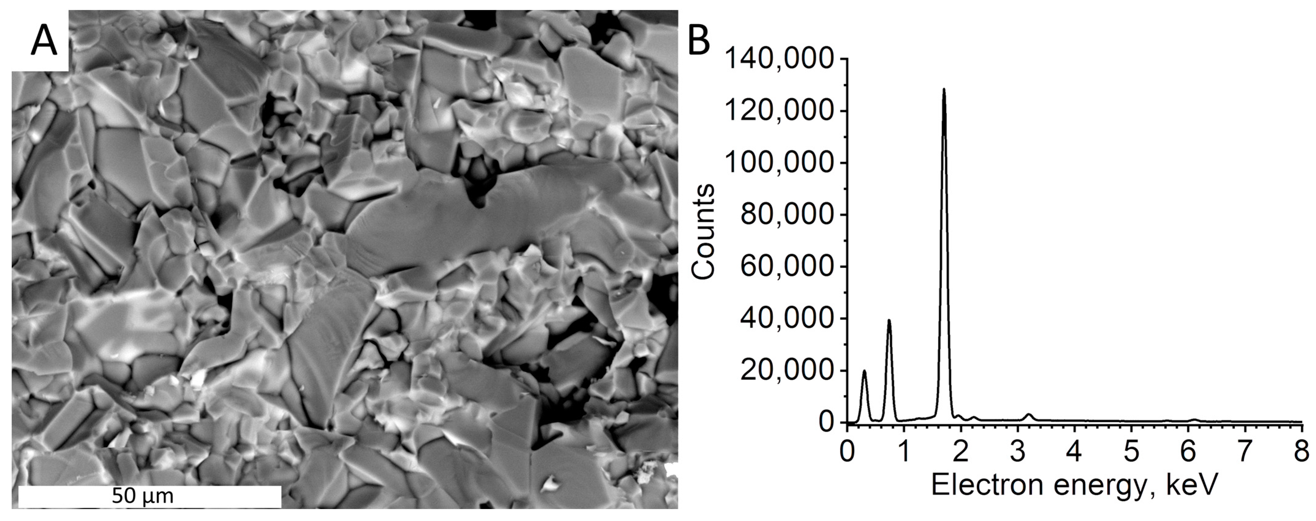
Figure 1A shows a general view of industrial corundum ceramics already containing Cr3+ ions, from which nanoparticles for PCC were prepared. Luminescence (λ = 695 nm) induced by a laser spot (λ = 405 nm) is shown (Figure 1A). A block diagram of the experimental setup for laser ablation of corundum ceramics is presented in Figure 1B. SEM images show that the morphology of VK94-1 is not uniform and consists of grains of various sizes, up to several tens of micrometers (Figure 2). VK94-1 contained chromium ions at a chromium to aluminum ratio of 3.3 × 10⁻³ and some chemical impurities: Si and Mn (Table 1). Manganese and silicon content were caused by the initial concentration of industrial corundum ceramics.
Nanoparticles were obtained in two stages. The primary ruby particles were generated by laser ablation of VK94-1 in isopropanol. It was chosen as a working fluid due to its low reactivity with aluminum oxide. To increase the particle production rate, a flow cell was applied as the basis of the experimental setup. A typical block diagram of the experimental installation is presented in Figure 1B. The radiation source was a solid-state Nd:YAG laser operating at a wavelength of 1064 nm, a pulse duration of 10 ns, a pulse repetition rate of 10 kHz, and a pulse energy of 1 mJ (SOL Instruments Ltd., Augsburg, Germany). Laser radiation onto the target surface was focused by an F-Theta objective with an F = 90 mm (Wavelength Opto-Electronic (S) Pte Ltd., Singapore). A focused laser beam with a diameter of 30 μm was moved along a circular path along the inner region of the target at a speed of 1000 mm/s by a galvano-optical mirror system. The final laser exposure area was about 1 cm2 and the fluence on the target surface was 140 J/cm2. The laser ablation time was 10 min.
Laser ablation of VK94-1 samples resulted in the production of primary particles. Analysis of SEM images of the primary ruby particles Figure 3A and Figure S1) showed that their morphology has an irregular shape with sizes ranging from 1 µm to 10 μm (Figure 3B). The formation of primary microparticles occurs as a result of mechanical destruction of the surface caused by laser ablation of corundum ceramics in isopropanol.
Elemental analysis (EDX) of the primary particles of ruby resulted in the fact that the chromium to aluminum ratio did not change (3.3 × 10⁻³) (Table 1). An increase in silicon content is caused by the use of a silicon substrate during analysis.
It should be noted that laser ablation and fragmentation of particles are generally accompanied by plasma formation, which leads to partial decomposition of isopropanol under its optical breakdown, as was determined in our previous work [56,57,58,59,60,61]. Further replacement of the carbon components in the clean environment (acetone) was carried out by centrifuging the colloid of corundum nanoparticles.
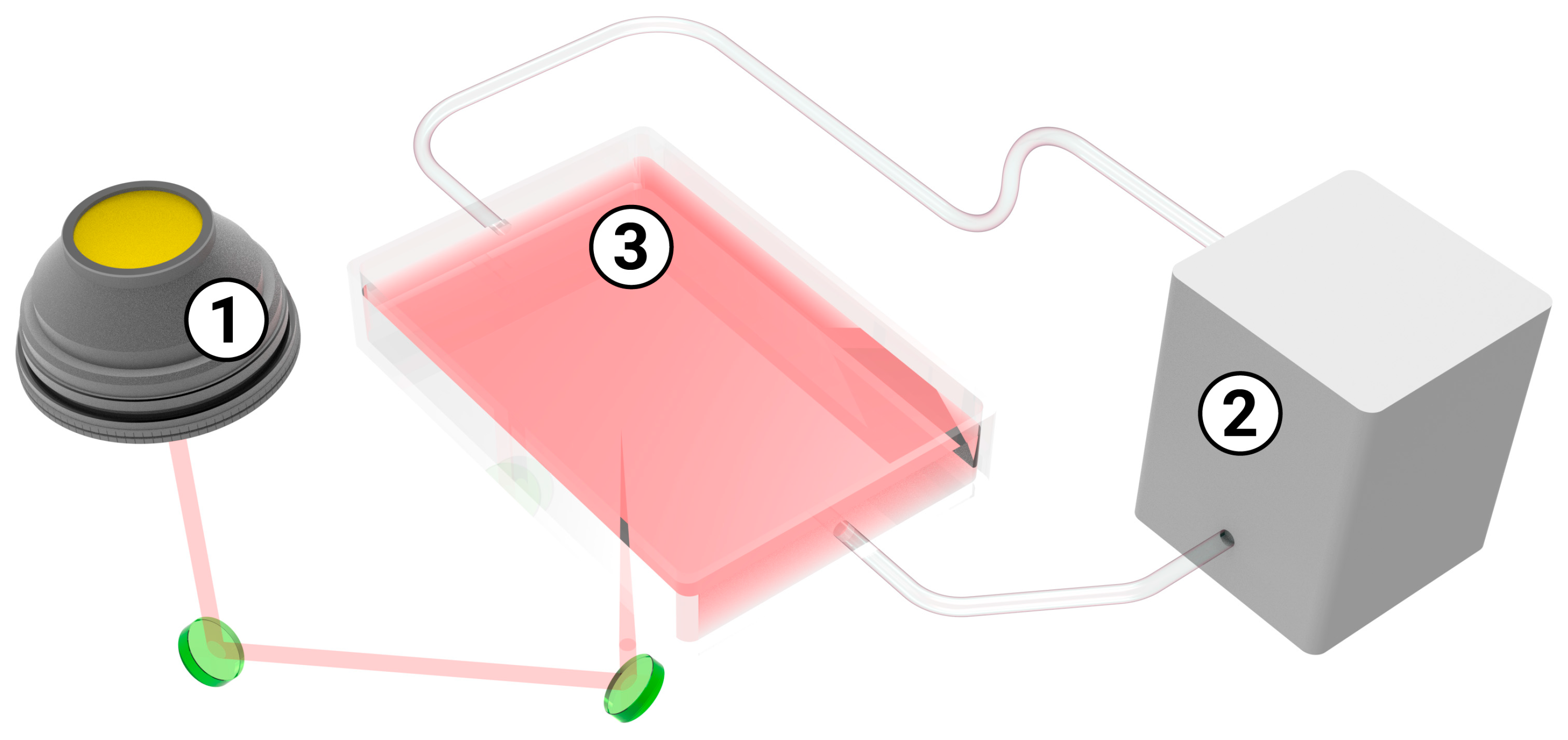
To reduce the particle size, the colloid of the primary ruby particles was exposed to laser fragmentation for 60 min. In this case, a suspension of generated ruby microparticles was irradiated by a laser beam entering through the glass from the bottom side of the cuvette in a flow experimental scheme (Figure 4). The laser fragmentation process used the same radiation source used for laser ablation.
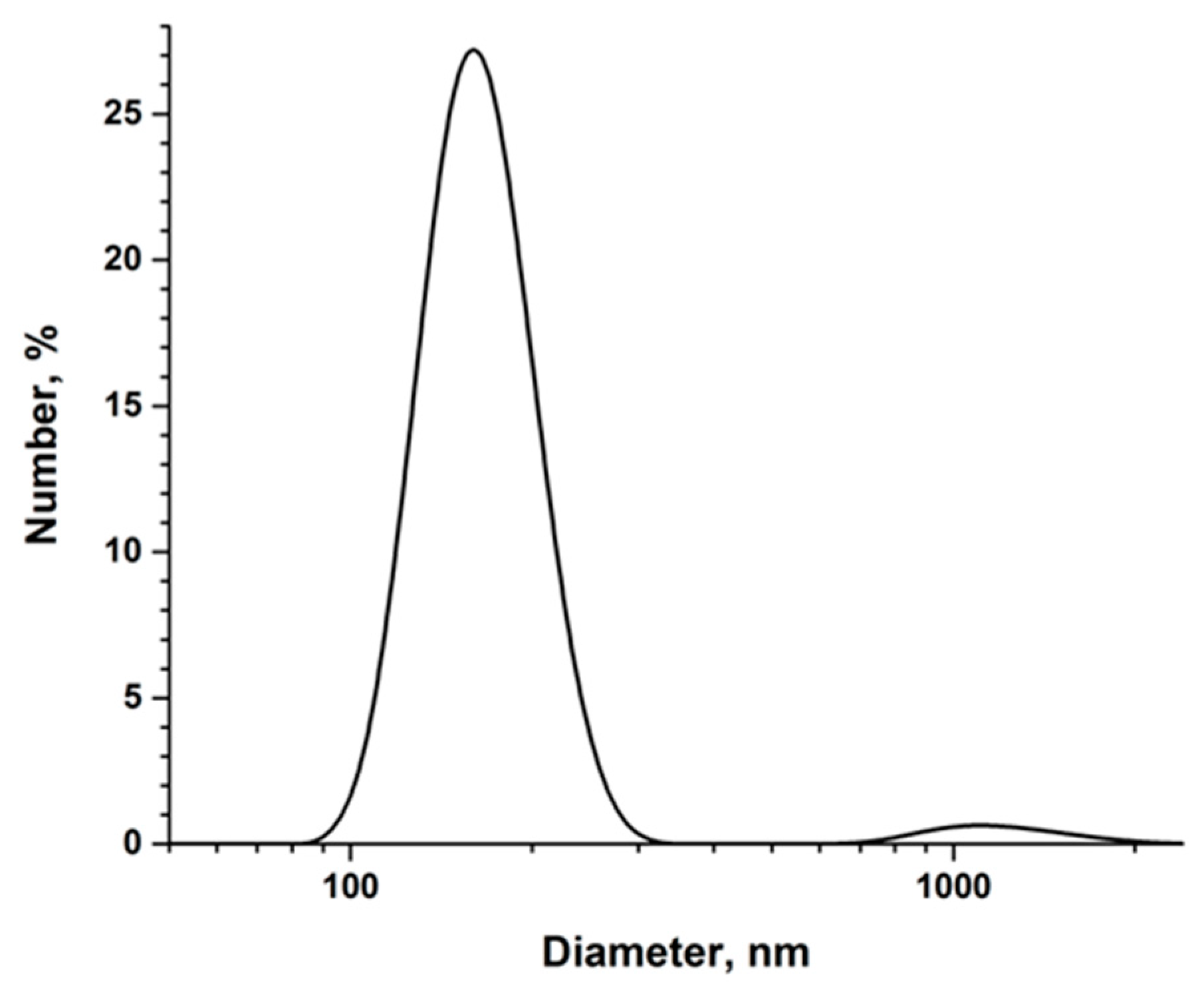
Analysis of SEM images of the secondary particles showed that the laser fragmentation led to the formation of spherical micro- and nanoparticles (Figure 5A and Figure S2). This means that the laser fragmentation occurred through the melting and redistribution of the melt under the surrounding environmental vapors. EDX analysis of the secondary particles did not reveal changes in particle content (Table 1). The size distribution histogram (Figure 5B) of the synthesized ruby nanoparticles (based on the analysis of SEM images) showed that the main part of the particles had a size of about 200 nm. These data are consistent with the DLS measurements, which determined the hydrodynamic diameter of the nanoparticles (Figure 6). It was shown that the laser fragmentation of primary particles led to the formation of two pools of particles. The first one had an average size from 100 nm to 300 nm, and the second one was from 600 nm to up to 2 μm (Figure 6).
The difference luminescence maps of PCC-R8 are shown in Figure 7. The luminescence of covers containing secondary ruby particles ranged from 650 nm to 750 nm, with a peak at 695 nm. The PCC-R8 had two wide bands of excitation of luminescence: from 350 nm to 450 nm with a maximum at 405 nm and from 500 nm to 600 nm with a peak at 550 nm. The UV-absorption spectra of both primary and secondary ruby particles had a maximum at 200–240 nm (Figure S3), which corresponds to earlier data [62,63].

3.2. The Effect of PCCs on Plant Growth and Development

Further work was aimed at studying the effect of the developed covers on the growth and development of lettuce Lactuca sativa grown under artificial lighting simulating sunlight.
Figure 8 shows that PCC-R8 covers had the greatest impact on plant growth performance. In plants grown under this cover, the number of leaves and their total area were greater than in control plants by 10% and 40%, respectively. Plants grown under PCC-R9 had a slightly smaller number of leaves but without a statistically significant decrease in their total area. PCC-R7 covers did not affect these parameters. All PCC-Rs increased the chlorophyll content in plant leaves (25–35%). Thus, it was revealed that the density of phosphors applied to the covers that is optimal for the growth of lettuce plants is 2 × 10⁸ m−2.
The purpose of further experiments was to identify the reasons for the positive effect of PCC-R8 on the growth of lettuce plants. For these purposes, measurements of the photosynthetic activity of the leaves of the tested groups of plants were carried out. Table 2 presents the measurement results of the gas exchange parameters in leaves. Among the measured parameters were intensity of CO2 assimilation (A), intensity of transpiration (E), and water use efficiency parameters (instantaneous water use efficiency (WUEleaf), intrinsic water use efficiency (WUEi)) (Table 2). The intensity of CO2 assimilation and transpiration processes in the dark did not differ in plants of different groups (except for PCC-R8 plants) and amounted to 0.31–0.36 µmol CO2 m−2 s−1 and 0.30–0.36 mmol H2O m−2 s−1, respectively. The intensity of CO2 assimilation in PCC-R8 plants was statistically significantly higher than in control plants (0.31 µmol CO2 m−2 s−1 versus 0.39 µmol CO2 m−2 s−1), which may indicate more intense respiration in such plants. The intensity of transpiration in PCC-R8 plants was 0.09 mmol H2O m−2 s−1, which was significantly lower than in the other plants. Turning on the light activated the processes of CO2 assimilation and transpiration. If the intensity of CO2 assimilation under the light in the studied groups of plants did not have statistically significant differences, the transpiration rate in the leaves of PCC-R8 plants was activated less efficiently than in other groups (up to 0.46 mmol H2O m−2 s−1 and 0.65–0.68 mmol H2O m−2 s−1, respectively). However, the differences in carbon dioxide assimilation and transpiration by leaves were not as remarkable as the differences in the efficiency of plant water use. The process of transpiration is necessary for the plant to maintain both light-dependent and light-independent processes. It was found that water use efficiency (WUE), calculated from dark respiration, was significantly higher in PCC-R8 plants, while this parameter in PCC-R7 and PCC-R9 remained approximately at the level noted for control plants (Table 2). Figure 9 shows that turning on light not only activates the processes of assimilation and transpiration in plant leaves but also changes the balance of water use by plants. In the first minutes, in all groups of plants, the efficiency of water use, calculated both in the ratio of CO2 assimilation/transpiration (instantaneous water use efficiency, WUEleaf) and in the ratio of CO2 assimilation/stomal conductance (intrinsic water use efficiency, WUEi), showed a sharp increase. After reaching maximum values, WUE gradually decreased. The main reason for the differences between WUE kinetics was the different activation rates of CO2 assimilation and transpiration. It was found that in PCC-R8 plants, the maximum WUE values were up to three times higher than those in control and PCC-R7/PCC-R9 plants. After 20 min of illumination, the WUE values in PCC-R8 plants reached those measured in other plant groups, while in the case of the WUEi parameter, the differences between plant groups disappeared.
Table 3 presents data on measuring the photochemical activity of plants by recording photoinduced changes in chlorophyll fluorescence. At the same time, no statistically significant differences were found between the plants in the control and experimental groups. It is assumed that the observed positive effect of PCC-R8 photoconversion covers is not associated with an increase in the intensity of photosynthetically active radiation directly but with the activation of regulatory mechanisms due to a qualitative change in the light spectrum.
Thus, we obtained nano- and microparticles of ruby, which could be effectively used in photoconversion covers for greenhouses due to their size, shape and luminescent properties.

4. Discussion

Nanomaterials can be produced using several approaches: mechanical milling, electrospinning, lithography, sputtering, the arc discharge method, laser ablation/fragmentation, chemical vapor deposition, solvothermal and hydrothermal methods, the sol-gel method, soft and hard templating methods and reverse micelle methods [64,65]. Laser ablation in liquid or ball milling methods is usually used to synthesize ruby particles [41,66,67,68]. The advantage of laser-generated materials is their purity because there is no need for any acids or polymer stabilizers [46], as well as the absence of impurities formed during ball milling.
In our present work, we used an experimental set-up consisting of a nanosecond laser, scanator systems, and a flow cell. The use of the flow cell allows us to both increase the rate of particle production and reduce the costs of nanoparticles generated in industrial applications. Compared to the equipment used in previously used femtosecond laser ablation [41,67], nanosecond laser ablation does not require special optics or expensive maintenance. At the same time, the productivity of femtosecond laser ablation was significantly lower. Laser irradiation of corundum ceramic with a nanosecond impulse resulted in the formation of fragments with versatile shapes (Figure 3A).
Such morphology is caused by the detaching of the initial protrusions (grains) of ceramics (Figure 3) due to cracking from the target under spall ablation. It has been suggested that under nanosecond laser impulses, thermal shockwaves are formed and reflected from the cold to heated stages of the material [69]. Such processes were observed earlier under laser ablation of selenium targets in liquid and boron targets in isopropanol [56,70]. Mechanisms for the formation of ruby particles by laser ablation of corundum ceramic are different in comparison to laser synthesis of ruby grains [40]. During the laser synthesis of ruby grains by laser heating, a mixture of aluminum and chromium oxide powders, spherical particles (0.5–1 mm) are formed [40]. In this case, the chromium content in the particles is easily controlled and, if necessary, adjusted. However, laser synthesis is a relatively slow process.
The formation of nano-sized particles and regular spherical shapes was achieved by laser fragmentation (Figure 6A). Laser fragmentation of irregularly shaped particles occurs through the melting and redistribution of the melt under the influence of environmental vapors [46]. Despite the two-stage process, the technique we used allows us to process a large amount of material relatively quickly and obtain nanoparticles of a spherical shape. This is a definite advantage of the proposed approach. The great advantage of ruby (as a material) and the nanoparticles obtained from it is its ability to retain luminescent properties both during laser processing and after incorporation into the polymer material used in the production of PCC-R compared to the spectra from Figure 7 and published earlier [40].
It was revealed that photoconversion covers containing ruby particles can change the qualitative composition of the light passing through them (Table 4). The ratio of intensity of blue to red light (calculated by the number of photons) decreases under PCC-R8 (from 0.74 to 0.69) and is practically no different between the control and PCC-R7 and PCC-R9. On the contrary, the ratio of red to blue light increases. In this case, the effect of PCC-R7 was insignificant, probably due to the insufficient density of phosphors in the cover. The maximum R:B ratio was found under PCC-R8 (1.34), while PCC-R9 took an intermediate position (1.31), probably due to the fact that the phosphors shaded each other. It is known that phosphors not only convert light from one wavelength to another but also absorb light. At the same time, the quantum efficiency of luminescence is far from unity. In addition, at high application densities, phosphors in PCC-Rs will not only shade plants but also have the effect of concentration quenching of luminescence [71], reducing the effectiveness of PCC-Rs and causing undesirable effects. In this regard, it is very important to determine the optimal density of phosphors applied to PCC-Rs.
The presented data indicate the ability of PCC-Rs to increase the proportion of red light by converting blue photons. At the same time, we obtained an idea of the optimal density of nanoparticles when qualitative changes in the spectrum are already sufficient to optimize plant growth, but the particle density that shades the plants has not been reached.
On the one hand, even a slight increase in the proportion of red light stimulated plant growth (Figure 8) [72,73,74], as observed in our experiments (Figure 8). Moreover, the effect of additional red light is most pronounced under conditions of low-light insolation [7]. On the other hand, changing the R:B ratio leads to changes in the intensity and efficiency of transpiration since blue light regulates the opening of stomata [75,76]. In our work, a significant decrease in transpiration intensity was shown in PCC-R8 plants, both in the dark and in the light. An increase in the intensity of the transpiration rate was accompanied by an increase in CO2 assimilation in the dark, with a constant intensity of assimilation. Similar data were obtained earlier: illumination of tomato leaves with light of different spectral compositions led to the same rates of carbon export with different stomatal conductance and transpiration rates, strongly dependent on the light spectrum, which controls the opening of stomata [77,78]. Adding blue light to red increases the intensity of transpiration, and adding red light to blue decreases its intensity [79]. The efficiency of water use under red light is higher than under blue light [80]. In our experiments, the increase in the R:B ratio by PCC-R8 increased the water use efficiency of plant leaves in both light and dark conditions (Table 4). In addition, an increase in the proportion of red light (compared to far-red light) can activate the plant phytochrome system, whose regulatory function is well known [81,82,83,84,85]. Our experiments showed an increased release of carbon dioxide from the leaves of PCC-R8 plants, which may have caused more active respiration. It is known that plants not only store assimilated carbon but also use it to grow biomass and maintain the functions of existing tissues, which is accompanied by active respiration in all living tissues [86,87,88]. During the process of respiration, solar energy, accumulated by plants in the light in the form of energy from chemical bonds of organic molecules, is directed to the production of high-energy molecules such as ATP, NADH and NADPH. On the one hand, an increase in respiration intensity may indicate an increase in growth processes associated with the accumulation of biomass; on the other hand, increased respiration activity is a sign of stress in plants. Previously, in a series of works by different scientific groups, it was shown that relatively small changes in the lighting spectrum of plants caused by photoconversion covers have a significant effect on both the productivity of plants and the intensity of physiological processes in them [26,27,28,29,30,33,35,36,55,89,90,91,92,93,94,95,96,97,98,99,100,101,102,103,104,105], which correlates with our data.
It is assumed that the observed positive effect of PCC-R8 photoconversion covers is not directly associated with an increase in the intensity of photosynthetically active radiation but with the acidization of regulatory mechanisms due to a qualitative change in the light spectrum.

Supplementary Materials

The following supporting information can be downloaded at: https://www.mdpi.com/article/10.3390/jcs8010007/s1, Figure S1: SEM image of primary ruby microparticles in isopropanol; Figure S2: SEM image of secondary ruby nanoparticles in isopropanol; Figure S3: UV-absorption spectra of primary ruby microparticles (black curve) and secondary ruby nanoparticles (red curve) in isopropanol

Author Contributions

Conceptualization, E.V.B. and S.V.G.; methodology, E.V.B., S.V.G. and D.V.Y.; investigation, E.V.B., K.O.A., I.I.R., D.V.Y., M.O.P., R.V.P. and D.V.K.; writing—original draft preparation, E.V.B., K.O.A., M.O.P. and D.V.Y.; writing—review and editing, D.V.Y. and S.V.G.; visualization, M.O.P., K.O.A. and I.I.R.; funding acquisition, S.V.G. All authors have read and agreed to the published version of the manuscript.

Funding

This work was supported by a grant from the Ministry of Science and Higher Education of the Russian Federation (075-15-2022-315) for the organization and development of the world-class research center “Photonics”.

Data Availability Statement

The data presented in this study are available on request from the corresponding author.

Conflicts of Interest

The authors declare no conflict of interest.

References

  1. Godfray, H.C.J.; Beddington, J.R.; Crute, I.R.; Haddad, L.; Lawrence, D.; Muir, J.F.; Pretty, J.; Robinson, S.; Thomas, S.M.; Toulmin, C. Food Security: The Challenge of Feeding 9 Billion People. Science 2010, 327, 812–818. [Google Scholar] [CrossRef] [PubMed]
  2. Lambin, E.F.; Meyfroidt, P. Global Land Use Change, Economic Globalization, and the Looming Land Scarcity. Proc. Natl. Acad. Sci. USA 2011, 108, 3465–3472. [Google Scholar] [CrossRef] [PubMed]
  3. Lowry, G.V.; Avellan, A.; Gilbertson, L.M. Opportunities and Challenges for Nanotechnology in the Agri-Tech Revolution. Nat. Nanotechnol. 2019, 14, 517–522. [Google Scholar] [CrossRef] [PubMed]
  4. Shang, Y.; Kamrul Hasan, M.; Ahammed, G.J.; Li, M.; Yin, H.; Zhou, J. Applications of Nanotechnology in Plant Growth and Crop Protection: A Review. Molecules 2019, 24, 2558. [Google Scholar] [CrossRef]
  5. Ort, D.R.; Merchant, S.S.; Alric, J.; Barkan, A.; Blankenship, R.E.; Bock, R.; Croce, R.; Hanson, M.R.; Hibberd, J.M.; Long, S.P.; et al. Redesigning Photosynthesis to Sustainably Meet Global Food and Bioenergy Demand. Proc. Natl. Acad. Sci. USA 2015, 112, 8529–8536. [Google Scholar] [CrossRef] [PubMed]
  6. Batista-Silva, W.; da Fonseca-Pereira, P.; Martins, A.O.; Zsögön, A.; Nunes-Nesi, A.; Araújo, W.L. Engineering Improved Photosynthesis in the Era of Synthetic Biology. Plant Commun. 2020, 1, 100032. [Google Scholar] [CrossRef] [PubMed]
  7. McCree, K.J. The Action Spectrum, Absorptance and Quantum Yield of Photosynthesis in Crop Plants. Agric. Meteorol. 1971, 9, 191–216. [Google Scholar] [CrossRef]
  8. Engelmann, T.W. Untersuchungen Über Die Quantitativen Beziehungen Zwischen Absorption Des Lichtes Und Assimilation in Pflanzenzellen. Bot. Zeit. 1884, 44, 43–52. [Google Scholar]
  9. Timiriazev, K.A.S.A. The Life of the Plant; Longmans, Green & Co.: London, UK; New York, NY, USA, 1912. [Google Scholar]
  10. Folta, K.M.; Childers, K.S. Light as a Growth Regulator: Controlling Plant Biology with Narrow-Bandwidth Solid-State Lighting Systems. HortScience 2008, 43, 1957–1964. [Google Scholar] [CrossRef]
  11. Landi, M.; Zivcak, M.; Sytar, O.; Brestic, M.; Allakhverdiev, S.I. Plasticity of Photosynthetic Processes and the Accumulation of Secondary Metabolites in Plants in Response to Monochromatic Light Environments: A Review. Biochim. Biophys. Acta BBA Bioenerg. 2020, 1861, 148131. [Google Scholar] [CrossRef]
  12. Rahman, M.M.; Field, D.L.; Ahmed, S.M.; Hasan, M.T.; Basher, M.K.; Alameh, K. LED Illumination for High-Quality High-Yield Crop Growth in Protected Cropping Environments. Plants 2021, 10, 2470. [Google Scholar] [CrossRef] [PubMed]
  13. Tyystjärvi, E. Photoinhibition of Photosystem II. Int. Rev. Cell Mol. Biol. 2013, 300, 243–303. [Google Scholar] [CrossRef] [PubMed]
  14. Chen, Y.; Li, T.; Yang, Q.; Zhang, Y.; Zou, J.; Bian, Z.; Wen, X. UVA Radiation Is Beneficial for Yield and Quality of Indoor Cultivated Lettuce. Front. Plant Sci. 2019, 10, 492746. [Google Scholar] [CrossRef] [PubMed]
  15. Smith, H.L.; Mcausland, L.; Murchie, E.H. Don’t Ignore the Green Light: Exploring Diverse Roles in Plant Processes. J. Exp. Bot. 2017, 68, 2099–2110. [Google Scholar] [CrossRef] [PubMed]
  16. Dougher, T.A.O.; Bugbee, B. Evidence for Yellow Light Suppression of Lettuce Growth. Photochem. Photobiol. 2001, 73, 208–212. [Google Scholar] [CrossRef] [PubMed]
  17. Hogewoning, S.W.; Wientjes, E.; Douwstra, P.; Trouwborst, G.; van Ieperen, W.; Croce, R.; Harbinson, J. Photosynthetic Quantum Yield Dynamics: From Photosystems to Leaves. Plant Cell 2012, 24, 1921–1935. [Google Scholar] [CrossRef] [PubMed]
  18. Kono, M.; Kawaguchi, H.; Mizusawa, N.; Yamori, W.; Suzuki, Y.; Terashima, I. Far-Red Light Accelerates Photosynthesis in the Low-Light Phases of Fluctuating Light. Plant Cell Physiol. 2020, 61, 192–202. [Google Scholar] [CrossRef] [PubMed]
  19. Brazaitytė, A.; Duchovskis, P.; Urbonavičiūtė, A.; Samuolienė, G.; Jankauskienė, J.; Kasiulevičiūtė-Bonakėrė, A.; Blizkinas, Z.; Novickovas, A.; Breive, K.; Žukauskas, A. The effect of light-emitting diodes lighting on cucumber transplants and after-effect on yield. Zemdirb. Agric. 2009, 96, 102–118. [Google Scholar]
  20. Huché-Thélier, L.; Crespel, L.; Gourrierec, J.L.; Morel, P.; Sakr, S.; Leduc, N. Light Signaling and Plant Responses to Blue and UV Radiations—Perspectives for Applications in Horticulture. Environ. Exp. Bot. 2016, 121, 22–38. [Google Scholar] [CrossRef]
  21. Christie, J.M.; Blackwood, L.; Petersen, J.; Sullivan, S. Plant Flavoprotein Photoreceptors. Plant Cell Physiol. 2015, 56, 401–413. [Google Scholar] [CrossRef]
  22. Galvão, V.C.; Fankhauser, C. Sensing the Light Environment in Plants: Photoreceptors and Early Signaling Steps. Curr. Opin. Neurobiol. 2015, 34, 46–53. [Google Scholar] [CrossRef] [PubMed]
  23. Kong, S.G.; Okajima, K. Diverse Photoreceptors and Light Responses in Plants. J. Plant Res. 2016, 129, 111–114. [Google Scholar] [CrossRef] [PubMed]
  24. Yadav, A.; Singh, D.; Lingwan, M.; Yadukrishnan, P.; Masakapalli, S.K.; Datta, S. Light Signaling and UV-B-Mediated Plant Growth Regulation. J. Integr. Plant Biol. 2020, 62, 1270–1292. [Google Scholar] [CrossRef] [PubMed]
  25. Kochetova, G.V.; Avercheva, O.V.; Bassarskaya, E.M.; Zhigalova, T.V. Light Quality as a Driver of Photosynthetic Apparatus Development. Biophys. Rev. 2022, 14, 779–803. [Google Scholar] [CrossRef]
  26. Burmistrov, D.E.; Yanykin, D.V.; Simakin, A.V.; Paskhin, M.O.; Ivanyuk, V.V.; Kuznetsov, S.V.; Ermakova, J.A.; Alexandrov, A.A.; Gudkov, S.V. Cultivation of Solanum Lycopersicum under Glass Coated with Nanosized Upconversion Luminophore. Appl. Sci. 2021, 11, 10726. [Google Scholar] [CrossRef]
  27. Yanykin, D.V.; Burmistrov, D.E.; Simakin, A.V.; Ermakova, J.A.; Gudkov, S.V. Effect of Up-Converting Luminescent Nanoparticles with Increased Quantum Yield Incorporated into the Fluoropolymer Matrix on Solanum Lycopersicum Growth. Agronomy 2022, 12, 108. [Google Scholar] [CrossRef]
  28. Yanykin, D.V.; Paskhin, M.O.; Simakin, A.V.; Burmistrov, D.E.; Pobedonostsev, R.V.; Vyatchinov, A.A.; Vedunova, M.V.; Kuznetsov, S.V.; Ermakova, J.A.; Alexandrov, A.A.; et al. Plant Photochemistry under Glass Coated with Upconversion Luminescent Film. Appl. Sci. 2022, 12, 7480. [Google Scholar] [CrossRef]
  29. Paskhin, M.O.; Yanykin, D.V.; Popov, A.V.; Pobedonostsev, R.V.; Kazantseva, D.V.; Dorokhov, A.S.; Izmailov, A.Y.; Vyatchinov, A.A.; Orlovskaya, E.O.; Shaidulin, A.T.; et al. Two Types of Europium-Based Photoconversion Covers for Greenhouse Farming with Different Effects on Plants. Horticulturae 2023, 9, 846. [Google Scholar] [CrossRef]
  30. Zhang, Z.; Zhao, Z.; Lu, Y.; Wang, D.; Wang, C.; Li, J. One-Step Synthesis of Eu3+-Modified Cellulose Acetate Film and Light Conversion Mechanism. Polymers 2020, 13, 113. [Google Scholar] [CrossRef]
  31. Wang, D.; Yu, Y.; Ai, X.; Pan, H.; Zhang, H.; Dong, L. Polylactide/Poly(Butylene Adipate-Co-Terephthalate)/Rare Earth Complexes as Biodegradable Light Conversion Agricultural Films. Polym. Adv. Technol. 2019, 30, 203–211. [Google Scholar] [CrossRef]
  32. Yu, Y.; Xu, P.; Jia, S.; Pan, H.; Zhang, H.; Wang, D.; Dong, L. Exploring Polylactide/Poly(Butylene Adipate-Co-Terephthalate)/Rare Earth Complexes Biodegradable Light Conversion Agricultural Films. Int. J. Biol. Macromol. 2019, 127, 210–221. [Google Scholar] [CrossRef] [PubMed]
  33. Nishimura, Y.; Wada, E.; Fukumoto, Y.; Aruga, H.; Shimoi, Y. The Effect of Spectrum Conversion Covering Film on Cucumber in Soilless Culture. Acta Hortic 2012, 956, 481–487. [Google Scholar] [CrossRef]
  34. Novoplansky, A.; Sachs, T.; Cohen, D.; Bar, R.; Bodenheimer, J.; Reisfeld, R. Increasing Plant Productivity by Changing the Solar Spectrum. Sol. Energy Mater. 1990, 21, 17–23. [Google Scholar] [CrossRef]
  35. Sánchez-Lanuza, M.B.; Menéndez-Velázquez, A.; Peñas-Sanjuan, A.; Navas-Martos, F.J.; Lillo-Bravo, I.; Delgado-Sánchez, J.M. Advanced Photonic Thin Films for Solar Irradiation Tuneability Oriented to Greenhouse Applications. Materials 2021, 14, 2357. [Google Scholar] [CrossRef] [PubMed]
  36. Ke-li, Z.; Liang-jie, Y.; Mei-yun, X.; You-zu, Y.; Ju-tang, S. The Application of Lights-Conversed Polyethylene Film for Agriculture. Wuhan Univ. J. Nat. Sci. 2002, 7, 365–367. [Google Scholar] [CrossRef]
  37. Hassanien, R.; Hassanien, E.; Li, M. Influences of Greenhouse-Integrated Semi-Transparent Photovoltaics on Microclimate and Lettuce Growth. Int. J. Agric. Biol. Eng. 2017, 10, 11–22. [Google Scholar] [CrossRef]
  38. Hassanien, R.; Hassanien, E.; Li, M.; Yin, F. The Integration of Semi-Transparent Photovoltaics on Greenhouse Roof for Energy and Plant Production. Renew Energy 2018, 121, 377–388. [Google Scholar] [CrossRef]
  39. Aira, J.R.; Gallardo-Saavedra, S.; Eugenio-Gozalbo, M.; Alonso-Gómez, V.; Muñoz-García, M.Á.; Hernández-Callejo, L. Analysis of the Viability of a Photovoltaic Greenhouse with Semi-Transparent Amorphous Silicon (a-Si) Glass. Agronomy 2021, 11, 1097. [Google Scholar] [CrossRef]
  40. Aiyyzhy, K.O.; Barmina, E.V.; Shafeev, G.A. Laser Synthesis of Ruby for Photo-Conversion of Solar Spectrum. Laser Phys. Lett. 2022, 20, 046001. [Google Scholar] [CrossRef]
  41. Aizuddin, W.; Razali, W.; Kasim, A.; Senawi, A.; Hashim, A.; Yahya, N.; Rafaie, H.A. Fabrication and Characterization of Ruby Nanoparticles. Malays. J. Anal. Sci. 2018, 22, 458–464. [Google Scholar] [CrossRef]
  42. Zhang, Y.; Zhang, L.; Zhou, C. Review of Chemical Vapor Deposition of Graphene and Related Applications. Acc. Chem. Res. 2013, 46, 2329–2339. [Google Scholar] [CrossRef] [PubMed]
  43. Jaworek, A.; Sobczyk, A.T. Electrospraying Route to Nanotechnology: An Overview. J. Electrostat. 2008, 66, 197–219. [Google Scholar] [CrossRef]
  44. Afzal, A.; Nawfal, I.; Mahbubul, I.M.; Kumbar, S.S. An Overview on the Effect of Ultrasonication Duration on Different Properties of Nanofluids. J. Therm. Anal. Calorim. 2019, 135, 393–418. [Google Scholar] [CrossRef]
  45. Giri, P.K.; Bhattacharyya, S.; Singh, D.K.; Kesavamoorthy, R.; Panigrahi, B.K.; Nair, K.G.M. Correlation between Microstructure and Optical Properties of ZnO Nanoparticles Synthesized by Ball Milling. J. Appl. Phys. 2007, 102, 93515. [Google Scholar] [CrossRef]
  46. Zhang, D.; Gökce, B.; Barcikowski, S. Laser Synthesis and Processing of Colloids: Fundamentals and Applications. Chem. Rev. 2017, 117, 3990–4103. [Google Scholar] [CrossRef] [PubMed]
  47. Kazakevich, P.V.; Simakin, A.V.; Voronov, V.V.; Shafeev, G.A. Laser Induced Synthesis of Nanoparticles in Liquids. Appl. Surf. Sci. 2006, 252, 4373–4380. [Google Scholar] [CrossRef]
  48. Sajti, C.L.; Sattari, R.; Chichkov, B.; Barcikowski, S. Ablation Efficiency of α-Al2O3 in Liquid Phase and Ambient Air by Nanosecond Laser Irradiation. Appl. Phys. A Mater. Sci. Process 2010, 100, 203–206. [Google Scholar] [CrossRef]
  49. Delfour, L.; Itina, T.E. Mechanisms of Ultrashort Laser-Induced Fragmentation of Metal Nanoparticles in Liquids: Numerical Insights. J. Phys. Chem. C 2015, 119, 13893–13900. [Google Scholar] [CrossRef]
  50. Ziefuß, A.R.; Reichenberger, S.; Rehbock, C.; Chakraborty, I.; Gharib, M.; Parak, W.J.; Barcikowski, S. Laser Fragmentation of Colloidal Gold Nanoparticles with High-Intensity Nanosecond Pulses Is Driven by a Single-Step Fragmentation Mechanism with a Defined Educt Particle-Size Threshold. J. Phys. Chem. C 2018, 122, 22125–22136. [Google Scholar] [CrossRef]
  51. Amendola, V.; Meneghetti, M. Controlled Size Manipulation of Free Gold Nanoparticles by Laser Irradiation and Their Facile Bioconjugation. J. Mater. Chem. 2007, 17, 4705–4710. [Google Scholar] [CrossRef]
  52. Strasser, M.; Setoura, K.; Langbein, U.; Hashimoto, S. Computational Modeling of Pulsed Laser-Induced Heating and Evaporation of Gold Nanoparticles. J. Phys. Chem. C 2014, 118, 25748–25755. [Google Scholar] [CrossRef]
  53. Lichtenthaler, H.K. [34] Chlorophylls and Carotenoids: Pigments of Photosynthetic Biomembranes. Methods Enzymol. 1987, 148, 350–382. [Google Scholar] [CrossRef]
  54. Kalaji, H.M.; Dąbrowski, P.; Cetner, M.D.; Samborska, I.A.; Łukasik, I.; Brestic, M.; Zivcak, M.; Tomasz, H.; Mojski, J.; Kociel, H.; et al. A Comparison between Different Chlorophyll Content Meters under Nutrient Deficiency Conditions. J. Plant Nutr. 2017, 40, 1024–1034. [Google Scholar] [CrossRef]
  55. Simakin, A.V.; Ivanyuk, V.V.; Dorokhov, A.S.; Gudkov, S.V. Photoconversion Fluoropolymer Films for the Cultivation of Agricultural Plants Under Conditions of Insufficient Insolation. Appl. Sci. 2020, 10, 8025. [Google Scholar] [CrossRef]
  56. Aiyyzhy, K.O.; Barmina, E.V.; Voronov, V.V.; Shafeev, G.A.; Novikov, G.G.; Uvarov, O.V. Laser Ablation and Fragmentation of Boron in Liquids. Opt. Laser Technol. 2022, 155, 108393. [Google Scholar] [CrossRef]
  57. Fongarland, P.; Vilcocq, L.; Djakovitch, L. Catalytic Liquefaction of Kraft Lignin with Solvothermal Approach. Catalysts 2021, 11, 875. [Google Scholar] [CrossRef]
  58. Chang, K.L.; Lin, Y.C.; Qiu, M.Z.; Tu, C.W.; Chang, C.P.; Wu, J.L.; Lin, Y.C.; Chang, C.K. Gas-Phase Isopropanol Degradation by Nonthermal Plasma Combined with Mn-Cu/-Al2O3. Environ. Sci. Pollut. Res. Int. 2021, 28, 40693–40702. [Google Scholar] [CrossRef]
  59. Jo, J.O.; Mok, Y.S. Oxidation of Isopropyl Alcohol in Air by a Catalytic Plasma Reactor System. Appl. Chem. Eng. 2014, 25, 531–537. [Google Scholar] [CrossRef]
  60. Czylkowski, D.; Hrycak, B.; Miotk, R.; Jasiński, M.; Mizeraczyk, J.; Dors, M. Microwave Plasma for Hydrogen Production from Liquids. Nukleonika 2016, 61, 185–190. [Google Scholar] [CrossRef]
  61. Mashhadani, Z.T.A. A Comparison of the Conversion of Isopropyl Alcohol by Non-Thermal Plasma and Thermally-Driven Catalysis Using In-Situ FTIR Spectroscopy. Ph.D. Thesis, Newcastle University, Newcastle upon Tyne, UK, 2018. [Google Scholar]
  62. Loh, E. Ultraviolet absorption and excitation spectrum of ruby and sapphire. J. Chem. Phys 1965, 44, 1940–1945. [Google Scholar] [CrossRef]
  63. Kusuma, H.H.; Astuti, B.; Ibrahim, Z. Absorption and emission properties of ruby (Cr:Al2O3) single crystal. J. Phys. Conf. Ser. 2019, 1170, 012054. [Google Scholar] [CrossRef]
  64. Baig, N.; Kammakakam, I.; Falath, W.; Kammakakam, I. Nanomaterials: A Review of Synthesis Methods, Properties, Recent Progress, and Challenges. Mater. Adv. 2021, 2, 1821–1871. [Google Scholar] [CrossRef]
  65. Nam, N.H.; Luong, N.H. Nanoparticles: Synthesis and Applications. Mater. Biomed. Eng. Inorg. Micro-Nanostructures 2019, 7, 211–240. [Google Scholar] [CrossRef]
  66. Yang, X.; Maleki, A.; Lipey, N.A.; Zheng, X.; Santiago, M.; Connor, M.; Sreenivasan, V.K.A.; Dawes, J.M.; Lu, Y.; Zvyagin, A.V. Lifetime-Engineered Ruby Nanoparticles (Tau-Rubies) for Multiplexed Imaging of μ-Opioid Receptors. ACS Sensors 2021, 6, 1375–1383. [Google Scholar] [CrossRef] [PubMed]
  67. Edmonds, A.M.; Sobhan, M.A.; Sreenivasan, V.K.A.; Grebenik, E.A.; Rabeau, J.R.; Goldys, E.M.; Zvyagin, A.V. Nano-Ruby: A Promising Fluorescent Probe for Background-Free Cellular Imaging. Part. Part. Syst. Charact. 2013, 30, 506–513. [Google Scholar] [CrossRef]
  68. Cortes-Vega, F.D.; Yang, W.; Zarate-Medina, J.; Brankovic, S.R.; Calderon, H.A.; Robles Hernandez, F.C. Mechanochemical Synthesis of α-Al2O3-Cr3+ (Ruby) and χ-Al2O3. J. Am. Ceram. Soc. 2019, 102, 976–980. [Google Scholar] [CrossRef]
  69. Inogamov, N.A.; Zhakhovsky, V.V.; Faenov, A.Y.; Khokhlov, V.A.; Shepelev, V.V.; Skobelev, I.Y.; Kato, Y.; Tanaka, M.; Pikuz, T.A.; Kishimoto, M.; et al. Spallative Ablation of Dielectrics by X-Ray Laser. Appl. Phys. A Mater. Sci. Process 2010, 101, 87–96. [Google Scholar] [CrossRef]
  70. Ayyyzhy, K.O.; Voronov, V.V.; Gudkov, S.V.; Rakov, I.I.; Simakin, A.V.; Shafeev, G.A. Laser Fabrication and Fragmentation of Selenium Nanoparticles in Aqueous Media. Phys. Wave Phenom. 2019, 27, 113–118. [Google Scholar] [CrossRef]
  71. Bojarski, C.; Domsta, J. Tyeoriya Vliyaniya Kontsyentratsii Na Lyuminyestsyentsiyu Tvyerdykh Rastvorov. Acta Phys. Acad. Sci. Hung. 1971, 30, 145–166. [Google Scholar] [CrossRef]
  72. Oh, H.E.; Yoon, A.; Park, Y.G. Red Light Enhances the Antioxidant Properties and Growth of Rubus Hongnoensis. Plants 2021, 10, 2589. [Google Scholar] [CrossRef]
  73. Rehman, M.; Fahad, S.; Saleem, M.; Hafeez, M.; Rahman, M.U.; Liu, F.; Deng, G. Red Light Optimized Physiological Traits and Enhanced the Growth of Ramie (Boehmeria nivea L.). Photosynthetica 2020, 58, 922–931. [Google Scholar] [CrossRef]
  74. Zhang, S.; Ma, J.; Zou, H.; Zhang, L.; Li, S.; Wang, Y. The Combination of Blue and Red LED Light Improves Growth and Phenolic Acid Contents in Salvia Miltiorrhiza Bunge. Ind. Crop. Prod. 2020, 158, 112959. [Google Scholar] [CrossRef]
  75. Petroutsos, D.; Tokutsu, R.; Maruyama, S.; Flori, S.; Greiner, A.; Magneschi, L.; Cusant, L.; Kottke, T.; Mittag, M.; Hegemann, P.; et al. A Blue-Light Photoreceptor Mediates the Feedback Regulation of Photosynthesis. Nature 2016, 537, 563–566. [Google Scholar] [CrossRef] [PubMed]
  76. Lim, S.; Kim, J. Light Quality Affects Water Use of Sweet Basil by Changing Its Stomatal Development. Agronomy 2021, 11, 303. [Google Scholar] [CrossRef]
  77. Kinoshita, T.; Doi, M.; Suetsugu, N.; Kagawa, T.; Wada, M.; Shimazaki, K.I. Phot1 and Phot2 Mediate Blue Light Regulation of Stomatal Opening. Nature 2001, 414, 656–660. [Google Scholar] [CrossRef]
  78. Lanoue, J.; Leonardos, E.D.; Ma, X.; Grodzinski, B. The Effect of Spectral Quality on Daily Patterns of Gas Exchange, Biomass Gain, and Water-Use-Efficiency in Tomatoes and Lisianthus: An Assessment of Whole Plant Measurements. Front. Plant Sci. 2017, 8, 266308. [Google Scholar] [CrossRef]
  79. Cheng, X.; Wang, R.; Liu, X.; Zhou, L.; Dong, M.; Rehman, M.; Fahad, S.; Liu, L.; Deng, G. Effects of Light Spectra on Morphology, Gaseous Exchange, and Antioxidant Capacity of Industrial Hemp. Front. Plant Sci. 2022, 13, 937436. [Google Scholar] [CrossRef]
  80. Lanoue, J.; Leonardos, E.D.; Grodzinski, B. Effects of Light Quality and Intensity on Diurnal Patterns and Rates of Photo-Assimilate Translocation and Transpiration in Tomato Leaves. Front. Plant Sci. 2018, 9, 370722. [Google Scholar] [CrossRef]
  81. Kreslavski, V.D.; Los, D.A.; Schmitt, F.J.; Zharmukhamedov, S.K.; Kuznetsov, V.V.; Allakhverdiev, S.I. The Impact of the Phytochromes on Photosynthetic Processes. Biochim. Biophys. Acta BBA Bioenerg. 2018, 1859, 400–408. [Google Scholar] [CrossRef]
  82. Cao, K.; Yu, J.; Xu, D.; Ai, K.; Bao, E.; Zou, Z. Exposure to Lower Red to Far-Red Light Ratios Improve Tomato Tolerance to Salt Stress. BMC Plant Biol. 2018, 18, 92. [Google Scholar] [CrossRef]
  83. Sharrock, R.A. The Phytochrome Red/Far-Red Photoreceptor Superfamily. Genome Biol. 2008, 9, 230. [Google Scholar] [CrossRef] [PubMed]
  84. Mathews, S. Phytochrome-Mediated Development in Land Plants: Red Light Sensing Evolves to Meet the Challenges of Changing Light Environments. Mol. Ecol. 2006, 15, 3483–3503. [Google Scholar] [CrossRef] [PubMed]
  85. Casson, S.A.; Hetherington, A.M. Environmental Regulation of Stomatal Development. Curr. Opin. Plant Biol. 2010, 13, 90–95. [Google Scholar] [CrossRef] [PubMed]
  86. Mccree, K.J. An Equation for the Rate of Respiration of White Clover Grown under Controlled Conditions. In Prediction and Measurement of Photosynthetic Productivity, Proceedings of the IBP/PP Technical Meeting, Trebon, Czech Republic, 14–21 September 1969; PUDOC: Wageningen, The Netherlands, 1970. [Google Scholar]
  87. Amthor, J.S. The Role of Maintenance Respiration in Plant Growth. Plant Cell Environ. 1984, 7, 561–569. [Google Scholar] [CrossRef]
  88. Lötscher, M.; Klumpp, K.; Schnyder, H. Growth and Maintenance Respiration for Individual Plants in Hierarchically Structured Canopies of Medicago Sativa and Helianthus Annuus: The Contribution of Current and Old Assimilates. New Phytol. 2004, 164, 305–316. [Google Scholar] [CrossRef] [PubMed]
  89. Parrish, C.H.; Hebert, D.; Jackson, A.; Ramasamy, K.; McDaniel, H.; Giacomelli, G.A.; Bergren, M.R. Optimizing Spectral Quality with Quantum Dots to Enhance Crop Yield in Controlled Environments. Commun. Biol. 2021, 4, 124. [Google Scholar] [CrossRef] [PubMed]
  90. Gudkov, S.V.; Simakin, A.V.; Bunkin, N.F.; Shafeev, G.A.; Astashev, M.E.; Glinushkin, A.P.; Grinberg, M.A.; Vodeneev, V.A. Development and Application of Photoconversion Fluoropolymer Films for Greenhouses Located at High or Polar Latitudes. J. Photochem. Photobiol. B 2020, 213, 112056. [Google Scholar] [CrossRef] [PubMed]
  91. Ivanyuk, V.V.; Shkirin, A.V.; Belosludtsev, K.N.; Dubinin, M.V.; Kozlov, V.A.; Bunkin, N.F.; Dorokhov, A.S.; Gudkov, S.V. Influence of Fluoropolymer Film Modified with Nanoscale Photoluminophor on Growth and Development of Plants. Front. Phys. 2020, 8, 616040. [Google Scholar] [CrossRef]
  92. Wu, W.; Zhang, Z.; Dong, R.; Xie, G.; Zhou, J.; Wu, K.; Zhang, H.; Cai, Q.; Lei, B. Characterization and Properties of a Sr2Si5N8:Eu2+-Based Light-Conversion Agricultural Film. J. Rare Earths 2020, 38, 539–545. [Google Scholar] [CrossRef]
  93. Yoon, H.I.; Kim, J.H.; Park, K.S.; Namgoong, J.W.; Hwang, T.G.; Kim, J.P.; Son, J.E. Quantitative Methods for Evaluating the Conversion Performance of Spectrum Conversion Films and Testing Plant Responses under Simulated Solar Conditions. Hortic Environ. Biotechnol. 2020, 61, 999–1009. [Google Scholar] [CrossRef]
  94. Yoon, H.I.; Kang, J.H.; Kang, W.H.; Son, J.E. Subtle Changes in Solar Radiation under a Green-to-Red Conversion Film Affect the Photosynthetic Performance and Chlorophyll Fluorescence of Sweet Pepper. Photosynthetica 2020, 58, 1107–1115. [Google Scholar] [CrossRef]
  95. Schettini, E.; de Salvador, F.R.; Scarascia-Mugnozza, G.; Vox, G. Radiometric Properties of Photoselective and Photoluminescent Greenhouse Plastic Films and Their Effects on Peach and Cherry Tree Growth. J. Hortic Sci. Biotechnol. 2011, 86, 79–83. [Google Scholar] [CrossRef]
  96. Hamada, K.; Shimasaki, K.; Ogata, T.; Nishimura, Y.; Nakamura, K.; Oyama-Egawa, H.; Yoshida, K. Effects of Spectral Composition Conversion Film and Plant Growth Regulators on Proliferation of Cymbidium Protocorm Like Body (PLB) Cultured In Vitro. Environ. Control. Biol. 2010, 48, 127–132. [Google Scholar] [CrossRef]
  97. Liu, X.Y.; Chang, T.T.; Guo, S.R.; Xu, Z.G.; Li, J. Effect of Different Light Quality of LED on Growth and Photosynthetic Character in Cherry Tomato Seedling. Acta Hortic 2011, 907, 325–330. [Google Scholar] [CrossRef]
  98. Edser, C. Light Manipulating Additives Extend Opportunities for Agricultural Plastic Films. Plast. Addit. Compd. 2002, 4, 20–24. [Google Scholar] [CrossRef]
  99. González, A.; Rodríguez, R.; Bañón, S.; Franco, J.A.; Fernández, J.A.; Salmerón, A.; Espí, E. Strawberry and Cucumber Cultivation under Fluorescent Photoselective Plastic Films Cover. Acta Hortic 2003, 614, 407–413. [Google Scholar] [CrossRef]
  100. De Salvador, F.R.; Mugnozza, G.S.; Vox, G.; Schettini, E.; Mastrorilli, M.; Bou Jaoudé, M. Innovative Photoselective and Photoluminescent Plastic Films for Protected Cultivation. Acta Hortic 2008, 801, 115–121. [Google Scholar] [CrossRef]
  101. Hidaka, K.; Yoshida, K.; Shimasaki, K.; Murakami, K.; Yasutake, D.; Kitano, M. Spectrum Conversion Film for Regulation of Plant Growth. J. Fac. Agric. Kyushu Univ. 2008, 53, 549–552. [Google Scholar] [CrossRef]
  102. In Yoon, H.; Hyeun Kang, J.; Kim, D.; Eek Son, J. Seedling Quality and Photosynthetic Characteristic of Vegetables Grown Under a Spectrum Conversion Film. J. Bio-Environ. Control. 2021, 30, 110–117. [Google Scholar] [CrossRef]
  103. Burmistrov, D.E.; Yanykin, D.V.; Paskhin, M.O.; Nagaev, E.V.; Efimov, A.D.; Kaziev, A.V.; Ageychenkov, D.G.; Gudkov, S.V. Additive Production of a Material Based on an Acrylic Polymer with a Nanoscale Layer of Zno Nanorods Deposited Using a Direct Current Magnetron Discharge: Morphology, Photoconversion Properties, and Biosafety. Materials 2021, 14, 6586. [Google Scholar] [CrossRef]
  104. Park, Y.; Runkle, E.S. Spectral-Conversion Film Potential for Greenhouses: Utility of Green-to-Red Photons Conversion and Far-Red Filtration for Plant Growth. PLoS ONE 2023, 18, e0281996. [Google Scholar] [CrossRef] [PubMed]
  105. Paskhin, M.O.; Pobedonostsev, R.V.; Kazantseva, D.V.; Simakin, A.V.; Gorudko, I.V.; Yanykin, D.V.; Gudkov, S.V. The Influence of Composite Luminescent Materials Based on Graphene Oxide on the Growth and Development of Solanum Lycopersicum in Greenhouses. J. Compos. Sci. 2023, 7, 474. [Google Scholar] [CrossRef]
Figure 1. (A) General view of corundum ceramics (diameter 5 cm), already containing Cr3+ ions (1) and its luminescence induced by laser irradiation (λ = 405 nm) (2). (B) Block diagram of the experimental setup for laser ablation of corundum ceramics in isopropanol. (1) Source of radiation Nd:YAG laser with a wavelength of 1064 nm, pulse duration of 10 ns, pulse repetition rate of 10 kHz and pulse energy of 1 mJ; (2) peristaltic pump; (3) working liquid (isopropanol); (4) corundum ceramic target.
Jcs 08 00007 g001
Figure 2. SEM image (A) and EDX spectrum (B) of the VK94-1 target.
Jcs 08 00007 g002
Figure 3. SEM image (A), size distribution (B) and EDX spectrum (C) of primary ruby microparticles in isopropanol.
Jcs 08 00007 g003
Figure 4. Block diagram of the experimental setup for laser fragmentation of ruby particles in isopropanol in a flow cell. (1) Source of radiation Nd:YAG laser with a wavelength of 1064 nm, pulse duration of 10 ns, pulse repetition rate of 10 kHz and pulse energy of 1 mJ; (2) colloid of ruby particles, (3) peristaltic pump.
Jcs 08 00007 g004
Figure 5. SEM image of ruby nanoparticles (A), size distribution (B) and EDX spectrum (C) of ruby nanoparticles after laser fragmentation.
Jcs 08 00007 g005
Figure 6. Distribution of secondary ruby particles over the hydrodynamic diameter.
Jcs 08 00007 g006
Figure 7. Difference 3D luminescence spectrum (“spectrum of PCC-R8” minus “spectrum of common cover”) of ruby particles.
Jcs 08 00007 g007
Figure 8. The effect of PCC-Rs on the leaf number (A), area (B) and chlorophyll content (C) of L. sativa plants (thirty-fourth day of the experiment). Plants were grown under control glasses (1), PCC-R7 (2), PCC-R8 (3), PCC-R9 (4). The data are the means of at least 10 measurements, with the standard error of the mean. The letters a, b and c denote statistically significant differences between groups of plants at p ≤ 0.05.
Jcs 08 00007 g008
Figure 9. Kinetics of light-induced (λ = 625 nm, 140 µmol photons m−2 s−1) changes in water use efficiency in the leaves of L. sativa, calculated on the basis of transpiration intensity (A) and stomatal conductance (B). Measurements were performed using control plants (black curve), PCC-R7-plants (red curve), PCC-R8-plants (blue curve) and PCC-R9-plants (green curve). Temperature, relative air humidity and CO2 content in the measuring cell were set to 25 °C, 65% and 200 ppm, respectively. The measurements were repeated at least three times.
Jcs 08 00007 g009
Table 1. Elemental analysis of VK94-1, primary and secondary particles obtained with EDX. The data are the means of 3 (VK94-1 and secondary particles) or 4 (primary ruby particles) measurements, with the standard deviation of the mean.
SampleProportion of the Atoms, %Chromium to Aluminum Ratio
OAlSiCrMn
VK94-160.1 ± 0.139.4 ± 0.40.26 ± 0.010.13 ± 0.010.20 ± 0.063.3 × 10−3
Primary ruby particles61.1 ± 0.133.2 ± 0.85.43 ± 0.640.11 ± 0.010.15 ± 0.013.3 × 10−3
Secondary ruby particles61.1 ± 0.233.1 ± 0.35.52 ± 0.480.13 ± 0.010.21 ± 0.023.3 × 10−3
Table 2. Effect of PCC-Rs on the parameters of gas exchange in leaves of L. sativa. A is the intensity of CO2 assimilation, E is the intensity of transpiration, WUEleaf is instantaneous water use efficiency, WUEi is intrinsic water use efficiency. The measurements were repeated at least three times. The data are the means with the standard error of the mean.
PCC-R VersionControlPCC-R7PCC-R8PCC-R9
Parameter
A, µmol CO2 m−2 s−10 min−0.31 ± 0.02 a−0.34 ± 0.03 a−0.39 ± 0.01 b−0.36 ± 0.01 a
20 min0.71 ± 0.07 a0.92 ± 0.23 a0.81 ± 0.03 a0.85 ± 0.13 a
E, mmol H2O m−2 s−10 min0.30 ± 0.04 a0.36 ± 0.05 a0.09 ± 0.01 b0.36 ± 0.10 a
20 min0.65 ± 0.10 a0.68 ± 0.09 a0.46 ± 0.06 b0.65 ± 0.15 a
WUEleaf, µmol CO2 mmol H2O−10 min−0.97 ± 0.02 a−0.93 ± 0.03 a−3.94 ± 0.30 b−1.04 ± 0.03 a
8 min1.06 ± 0.22 a1.72 ± 0.14 a2.80 ± 0.17 b1.42 ± 0.09 a
20 min1.22 ± 0.18 a1.36 ± 0.19 a1.82 ± 0.20 b1.34 ± 0.21 a
WUEi, nmol CO2 mmol H2O−10 min−9 ± 2 a−5 ± 1 a−21 ± 4 b−6 ± 2 a
8 min7 ± 1 a9 ± 1 a21 ± 1 b8 ± 1 a
20 min9 ± 1 a8 ± 2 a10 ± 1 a9 ± 1 a
Letters indicate statistically significant difference between different samples (p ≤ 0.05).
Table 3. Effect of PCC-Rs on chlorophyll a fluorescence in leaves of L. sativa measured after 20 min of illumination. Fv/Fm is the maximum quantum yield of photosystem II; Y(II) is the effective quantum yield of photochemistry of photosystem II; ETR(II) is the rate of linear electron transfer in photosystem II; qN is non-photochemical quenching chlorophyll a fluorescence; qP is photochemical quenching chlorophyll a fluorescence; ETR (I) is the rate of linear electron transfer in photosystem I; Y(I) is the effective quantum yield of photochemistry of photosystem I. The measurements were repeated at least three times. The data are the means with the standard error of the mean.
ControlPCC-R7PCC-R8PCC-R9
Fv/Fm0.80 ± 0.002 a0.80 ± 0.02 a0.79 ± 0.02 a0.80 ± 0.01 a
Y(II)0.37 ± 0.02 a0.39 ± 0.06 a0.35 ± 0.01 a0.37 ± 0.01 a
ETR(II), µmol electrons (PSII s)−123.9 ± 1.4 a25.5 ± 3.9 a22.6 ± 0.6 a24.1 ± 0.7 a
qN0.57 ± 0.02 a0.56 ± 0.03 a0.58 ± 0.01 a0.58 ± 0.02 a
qP0.55 ± 0.03 a0.58 ± 0.07 a0.53 ± 0.01 a0.56 ± 0.01 a
Y(I)0.80 ± 0.05 a0.78 ± 0.03 a0.81 ± 0.03 a0.81 ± 0.01 a
ETR(I), µmol electrons (PSI s)−151.4 ± 3.3 a50.5 ± 2.2 a52.5 ± 2.0 a52.1 ± 0.6 a
Letters indicate statistically significant difference between different samples (p ≤ 0.05).
Table 4. Changes in light range ratios induced by PCC-Rs. B—blue light range (400–500 nm); G—green light range (500–600 nm); R–red light range (600–700 nm). The data are the means with the standard error of the mean.
ControlPCC-R7PCC-R8PCC-R9
B:G ratio 0.74 ± 0.003 a0.73 ± 0.02 a0.69 ± 0.001 b0.71 ± 0.002 a
R:B ratio 1.24 ± 0.01 a1.27 ± 0.01 a1.34 ± 0.004 b1.31 ± 0.01 c
Letters indicate statistically significant difference between different samples (p ≤ 0.05).
Disclaimer/Publisher’s Note: The statements, opinions and data contained in all publications are solely those of the individual author(s) and contributor(s) and not of MDPI and/or the editor(s). MDPI and/or the editor(s) disclaim responsibility for any injury to people or property resulting from any ideas, methods, instructions or products referred to in the content.