Article

Optimal Acyl Chain Length for Imparting Rigidity and Water Resistance to Cellulose–Hydroxyapatite Composites


Department of Applied Chemistry, Faculty of Science and Engineering, Doshisha University, 1-3, Tatara-miyakotani, Kyotanabe, Kyoto 610-0394, Japan
*
Author to whom correspondence should be addressed.
J. Compos. Sci. 2026, 10(9), 472; https://doi.org/10.3390/jcs10090472
Submission received: 4 August 2026 / Revised: 26 August 2026 / Accepted: 31 August 2026 / Published: 2 September 2026
(This article belongs to the Special Issue The Properties and Applications of Advanced Functional Biocomposites)

Abstract

An acylation reaction was performed on the crystalline surface of cellulose in a composite consisting of microfibrillated cellulose (MFC) and hydroxyapatite (HAP) with an inorganic weight fraction of 68%. The composite was acylated using acetic anhydride, propanoic anhydride, and butanoic anhydride in pyridine in the presence of potassium carbonate at 120 °C for 1 h. The formation of ester linkages was confirmed by infrared spectroscopy, and X-ray diffraction analysis showed that the crystalline structure of cellulose was retained after acylation. From the intensity of the carbonyl stretching vibration in the infrared spectra, the degree of substitution of the acetylated sample was estimated to be approximately 0.2. The acylated MFC–HAP composites were uniaxially hot-pressed at 120 °C and 300 MPa, and the resulting molded specimens were subjected to three-point bending tests. A yield point appeared at a bending strain of 1.1–1.4%, followed by plastic deformation and final fracture, indicating that they exhibited ductile fracture. The elastic moduli were 5.9 GPa (acetyl), 7.6 GPa (propanoyl), 7.4 GPa (butanoyl), 3.6 GPa (hexanoyl), and 7.1 GPa (before acylation), indicating that acyl groups with medium chain lengths did not reduce the rigidity of the composites. When the molded specimens were immersed in water at room temperature for 24 h, the water absorption ratios were 29% (acetyl), 24% (propanoyl), 17% (butanoyl), and 18% (hexanoyl), demonstrating that water resistance improved with increasing acyl chain length. In summary, propanoylation and butanoylation improved the water resistance of the composites without compromising their rigidity in the dry state.

1. Introduction

Bioceramics such as bone and teeth possess characteristic properties including light weight, high stiffness, high toughness, and low environmental impact [1,2,3,4,5,6,7,8,9]. The mechanical properties of bone span a wide range, from 2 to 35 GPa for elastic modulus, from 30 to 320 MPa for bending strength [10,11,12,13], and from 200 to 6200 J/m2 for work of fracture [14]. However, no method has yet been established to synthesize materials with performance comparable to bioceramics on a large scale. Bone is a composite consisting of approximately 70 wt% carbonate apatite, 20 wt% collagen, and 10 wt% water [15,16,17,18]. Collagen is a protein, and its use in large quantities as a structural material is costly. If cellulose, a biomass-derived material, could be used in place of collagen, it would represent a step toward the development of practically applicable materials [19,20,21,22].
Composites of microfibrillated cellulose (MFC) and hydroxyapatite (HAP), prepared via the crystallization of hydroxyapatite in an aqueous dispersion of microfibrillated cellulose, exhibited elastic moduli of 7.0–8.7 GPa, bending strengths of 60–98 MPa and work of fracture values of 227–560 J/m2. The composites contained 42–62 wt% of HAP. Within this range of inorganic weight fraction, an increase in the inorganic content resulted in a higher elastic modulus and a lower work of fracture. It demonstrates that stiffness and toughness are mutually incompatible, such that improving one property necessarily entails a compromise in the other. Another notable characteristic of the composites of MFC and HAP is that they do not undergo brittle fracture. The fracture surfaces of molded composites of hydroxyapatite with starch [23], carboxymethyl cellulose [24], and poly(vinyl alcohol) [25] observed in bending tests were smooth, indicating brittle fracture behavior. Composites with TEMPO-oxidized cellulose nanofibers likewise fractured in a brittle manner when the nanofibers were well dispersed, whereas composites prepared from partially aggregated nanofibers did not exhibit brittle fracture [26]. These results indicate that when the organic phase possesses a fibrous structure at the micrometer to nanometer scale, mechanical energy can be efficiently absorbed, thereby suppressing brittle fracture.
One of the issues of the composites of MFC-HAP is that the composites adsorbed a significant amount of water when immersed in water, and the compacts lost rigidity and cannot retain their shape. Water absorption in the composites is considered to occur through several pathways, including absorption into the voids between hydroxyapatite particles and cellulose particles, absorption by the cellulose phase, and adsorption at the interfaces between cellulose and hydroxyapatite. Masuhara and coworkers reported that the CNC–HAP composite becomes hydrophobic upon the addition of chitosan [27]. Extensive studies have been conducted on the hydrophobization of cellulose [28,29], and hydrophobizing cellulose within a composite may improve the water resistance of the composite material. It was demonstrated that acylation of the cellulose nanofibers–HAP [30] and starch–HAP [31] composites with vinyl carboxylates [32] imparted hydrophobicity and improved their water resistance. In the study, we found that choice of the acyl groups affected water resistance and rigidity of the composites: upon the introduction of aliphatic acyl groups, increasing the acyl chain length improves water resistance, while reducing the rigidity of the composites [30,31]. In this paper, we attempted to clarify the effects of the chain length of linear aliphatic acyl groups on the mechanical properties and water resistance of the composites. We also reported that benzoylation of MFC-HAP with vinyl benzoate afforded water-resistant and rigid composites, which exhibited ductile fracture [33]. The reaction temperature above 110 °C was necessary to sufficiently benzoylate the composite. Because of the elevated reaction temperature, the application of this reaction to short-chain vinyl carboxylates with low boiling points presents significant challenges, particularly in large-scale operations. Therefore, the exploration of alternative acylating agents is highly desirable. Acylation of cellulose with carboxylic anhydrides is a well-studied reaction [34,35,36,37,38,39,40,41]. The boiling points of acetic anhydride and propanoic anhydride are 140 °C and 168 °C, respectively, much higher than vinyl acetate (72 °C) and vinyl propanoate (95 °C), allowing reactions to be conducted at higher temperatures. In this paper we performed acylation of microfibrillated cellulose–hydroxyapatite composites with acid anhydrides with short-chain acyl groups. We investigated the effects of acyl chain lengths on the stiffness and water resistance of the resulting composites.

2. Experiment

2.1. Materials and Instrumentation

Microfibrillated cellulose, 10 wt% in water, Celish KY100G, was provided by Daicel Corporation, Tokyo, Japan. Calcium chloride dihydrate, disodium hydrogen phosphate, sodium hydroxide, acetic anhydride, methanol, pyridine, and acetone were purchased from FUJIFILM Wako Pure Chemical Corporation, Osaka, Japan. Propanoic anhydride, butanoic anhydride, and vinyl hexanoate were purchased from Tokyo Chemical Industry, Co., Ltd., Tokyo, Japan.
Thermogravimetric and differential thermal analysis (TG-DTA) was performed with a Shimadzu DTA-60 thermal analysis instrument (Kyoto, Japan). The sample was heated in an alumina pan from 30 °C to 100 °C at a rate of 20 °C/min, kept at 100 °C for 10 min, heated to 1000 °C at a rate of 20 °C/min, and kept at 1000 °C for 10 min. Powder X-ray diffraction (XRD) of the composite powder was recorded on a Rigaku SmartLab diffractometer (Tokyo, Japan) with Cu-Kα radiation. Fourier transform infrared (FT-IR) spectra were recorded as KBr pellets on a JASCO FT/IR-4600 spectrophotometer (Tokyo, Japan). Scanning electron microscopy (SEM) observations and energy dispersive X-ray spectroscopy (EDS) were carried out with a Hitachi TM3030Plus microscope (Tokyo, Japan). Three-point bending test was carried out using a bending testing machine MZ-603, Maruto Instrument Co., Ltd., Tokyo, Japan.

2.2. Preparation of MFC-HAP with a Feed Weight Ratio of 30:70 (MFC:HAP)

An aqueous dispersion of 10 wt% MFC 23.3 g was placed in a 500 mL Erlenmeyer flask, and 0.2 M Na2HPO4 (162 mL) and 1 M NaOH (43.57 mL) was added. The dispersion was stirred at 50 °C for 15 min and at 70 °C for 15 min. Then 0.2 M CaCl2 270.4 mL was added dropwise at 70 °C, and the mixture was stirred for 1 h at 70 °C. The reaction mixture was cooled to room temperature and the white powder was collected by suction filtration. The powder was washed with acetone (200 mL), water-acetone (1:1, v/v, 1 L) and acetone (1 L). The composite powder was dried in vacuo at 50 °C for 2 h, and pulverized in a mortar.

2.3. Acetylation of the MFC-HAP Composite

The MFC-HAP composite powder (1.5 g), K2CO3 (13.8 g), acetic anhydride (2.36 mL), and pyridine (50 mL) were placed in a round bottom flask and heated at 120 °C for 1 h. After the mixture was cooled to room temperature, the powder was collected by suction filtration, and washed with acetone (400 mL). The powder was washed with water until the filtrate was at a neutral pH. The powder was dried in vacuo at 50 °C for 2 h, and pulverized in a mortar.

2.4. Propanoylation of the MFC-HAP Composite

The MFC-HAP composite powder (1.5 g), K2CO3 (13.8 g), propanoic anhydride (3.22 mL), and pyridine (50 mL) were placed in a round bottom flask and heated at 120 °C for 1 h. After the mixture was cooled to room temperature, the powder was collected by suction filtration, and washed with acetone (400 mL). The powder was washed with water until the filtrate was at a neutral pH. The powder was dried in vacuo at 50 °C for 2 h, and pulverized in a mortar.

2.5. Butanoylation of the MFC-HAP Composite

The MFC-HAP composite powder (1.5 g), K2CO3 (13.8 g), butanoic anhydride (4.07 mL), and pyridine (50 mL) were placed in a round bottom flask and heated at 120 °C for 1 h. After the mixture was cooled to room temperature, the powder was collected by suction filtration, and washed with acetone (400 mL). The powder was washed with water until the filtrate was at a neutral pH. The powder was dried in vacuo at 50 °C for 2 h, and pulverized in a mortar.

2.6. Hexanoylation of the MFC-HAP Composite with Vinyl Hexanoate

The MFC-HAP composite powder (1.5 g), K2CO3 (0.5 g), vinyl hexanoate (1.98 mL), and DMF (50 mL) were placed in a round bottom flask and heated at 100 °C for 4 h. After the mixture was cooled to room temperature, the powder was collected by suction filtration, and washed with methanol (1 L). The powder was dried in vacuo at 80 °C for 2 h, and pulverized in a mortar.

2.7. Inorganic Contents of the Composite

To determine the inorganic contents (IC) of the composites, the composite powder was subjected to TG-DTA analysis. The sample was heated in an alumina pan in air from room temperature to 100 °C at a rate of 20 °C/min, kept at 100 °C for 10 min, heated to 1000 °C at a rate of 20 °C/min, and kept at 1000 °C for 10 min. The sample was cooled to 100 °C at a rate of 100 °C/min. The weight% of inorganic components was calculated as a ratio of the final weight to that at 100 °C in the first heating.

2.8. Densification of the Acylated MFC-HAP Composite Powder and Three-Point Bending Test

The acylated MFC-HAP composite powder was placed in a tungsten carbide mold of 4 mm × 13 mm, and uniaxially pressed at 300 MPa at 120 °C for 5 min to obtain the molded composites of 4 mm × 13 mm × 1.5–1.8 mm. The bulk density of the compacts (dbulk) was determined from the ratio of the sample weight to the volume of the rectangular parallelepiped. A three-point bending test was conducted immediately after the compression molding process. The load is applied to the sample with a cross-head speed of 0.5 mm/min. Stress P was calculated by
P = 3 F L 2 w t 2
where F is the load (N), L is the length between the supporting points (8 mm), w is the breadth (4 mm) and t is the thickness (1.5–1.8 mm) of the specimen. Strain ε was calculated according to
ε = 6 t s L 2
where s (m) is the displacement of the crosshead. Bending elastic modulus, Eb, was calculated from the slope of the stress–strain line according to
E b = Δ P Δ ε
The work of fracture [42] of the composite (Wf) was measured by determining the area under the load (F)-displacement (s) curve and dividing by the area of the fracture surface using the following equation.
W f = 1 w t 0 F d s = L 9 0 P d ε
The work required to reach the yield point (Wf) was determined using the following equation.
W f = 1 w t 0 s b F d s = L 9 0 ε b P d ε
where sb and εb represent the displacement and strain at the yield point, respectively.
The mechanical properties were generally presented as the mean values and standard errors of the mean obtained from three to four measurements. Although the L/t ratio was smaller than the value of 15 recommended for a standard three-point bending test, bending tests of specimens with a molded thickness of 0.5 mm could not be performed successfully using our apparatus, owing to a relatively large strain at the yield point.

2.9. Porosity of the Molded Composite

The molded composite was assumed to consist of pores with a volume fraction of ϕ and composite particles with a volume fraction of 1 − ϕ. The porosity, ϕ, was determined as follows. Let dbulk denote the density of the molded body and dmax the maximum density of a hypothetical fully dense body without pores. For a molded body with a unit volume, its mass is given by (1 − ϕ) dmax. Accordingly, dbulk can be expressed in terms of dmax and the porosity ϕ as
d b u l k = 1 d m a x
By rearranging this equation, the porosity can be expressed as
= 1 d b u l k d m a x
The density of the hypothetical pore-free composite, dmax, was determined based on the following assumptions. Let χ be the volume fraction of HAP in the composite particle and (1 − χ) the volume fraction of cellulose. Let dHAP denote the density of HAP (3.16 g/cm3) and dcellulose the density of cellulose (1.63 g/cm3). In a composite with a unit volume, the mass of HAP is χdHAP, and the mass of cellulose is (1 − χ) dcellulose. The total mass of the composite is therefore given by χ dHAP + (1 − χ) dcellulose, and hence the maximum theoretical density without pores is expressed as
d m a x = χ d H A P + 1 χ d c e l l u l o s e
On the other hand, the inorganic weight fraction of the composite, IC, is given by
I C = χ d H A P χ d H A P + 1 χ d c e l l u l o s e
By eliminating χ from these two equations, the maximum density can be expressed as
d m a x = d H A P   d c e l l u l o s e d H A P 1 I C + d c e l l u l o s e I C
The value of IC was obtained from thermogravimetric analysis of the acylated samples, and the maximum density was calculated using Equation (10).

2.10. Water Absorption Ratio

The potential water adsorption sites in the composite are considered to be: (1) the pore regions of the composite, (2) the cellulose phase, (3) the HAP phase, and (4) the interfacial regions between cellulose and HAP. The water absorption ratio, assuming that water adsorption occurs exclusively within the pores of the composite, was estimated as follows. We assumed that water penetrates only into the pores, that no water is absorbed by the composite particles themselves, and that the porosity does not change upon water immersion. Under these assumptions, the water absorption ratio, Δw/w (pore), can be determined as follows. Assuming that the volume of the molded body is unity, the mass of the dry specimen is equal to dbulk. When the specimen is immersed in water and the pores are completely filled with water, the pore volume is equal to the porosity ϕ, and the mass of absorbed water is given by ϕdwater, where dwater (1.0 g/cm3) is the density of water. Since the water absorption ratio is defined as the mass of absorbed water divided by the mass of the dry specimen,
w w ( p o r e ) = d w a t e r d b u l k
By substituting the expression for porosity Equation (7), the water absorption ratio can also be expressed as
w w ( p o r e ) = 1 d b u l k 1 d m a x d w a t e r

3. Results and Discussion

3.1. Preparation of MFC-HAP Composite and Acylation

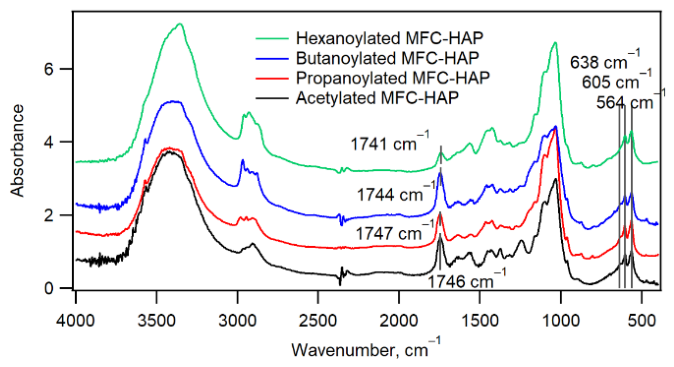
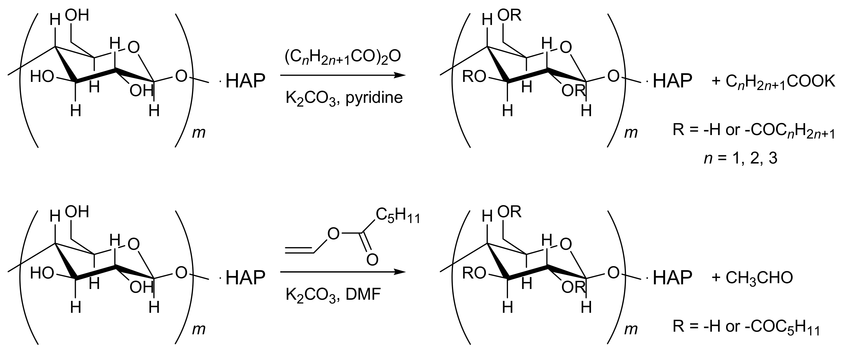
The composites of MFC and HAP were prepared by the crystallization of hydroxyapatite in an alkaline aqueous dispersion of MFC at 70 °C [43]. The inorganic content of the composite determined by thermogravimetric analysis was 68 wt%. The MFC-HAP composite powder was acylated using acetic anhydride, propanoic anhydride, and butanoic anhydride at 120 °C for 1 h in pyridine in the presence of K2CO3 (Scheme 1). Hexanoylation was performed with vinyl hexanoate at 100 °C for 4 h in DMF. The inorganic contents of the acylated composites were 58 (acetylated), 62 (propanoylated), 64 (butanoylated), and 54 wt% (hexanoylated), showing that the fraction of the organic phase increased owing to acylation. As shown in Figure 1, IR spectra of the acylated composites showed the ester carbonyl stretching at 1746 (acetylated), 1747 (propanoylated), 1744 (butanoylated), and 1741 cm−1 (hexanoylated). The acetylated composite showed the characteristic ester C-O stretching at 1246 cm−1 and the methyl in-plane bending at 1374 cm−1. They also exhibited characteristic signals of HAP, the O-P-O bending vibrations at 604 cm−1 and 564 cm−1 and the OH libration mode at 638 cm−1 (shoulder). The degree of substitution of acetyl groups was estimated to be 0.2 from the ratio of absorbance at 1747 cm−1 to that of 3400 cm−1 according to Hurtubise [44]. Based on the absorbance of the carbonyl stretching vibration, the degrees of substitution of the propanoylated, butanoylated, and hexanoylated composites were estimated to be 0.24, 0.37, and 0.04, respectively (see supporting information).
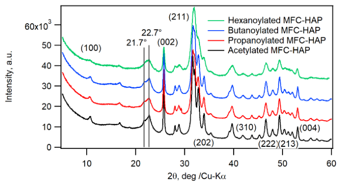
X-ray diffraction patterns of the acylated MFC-HAP composites are shown in Figure 2. The diffraction peak at 22.7° 2θ (200), which was also observed in the starting MFC, is attributed to the cellulose I crystal structure. The peak at 21.7° 2θ [(200), (1–10)] was also observed as a minor peak for acetylated, propanoylated and butanoylated composites, indicating that some crystalline region was transformed into the cellulose III phase upon acylation [45]. The diffraction peaks of HAP were also observed to be sharp, and the diffraction pattern was almost unchanged compared with that before acylation (Figure S1), indicating that acylation using carboxylic anhydrides caused little to no decomposition of the HAP crystals or reduction in their crystallinity.

3.2. Densification of the Composites and Three-Point Bending Test

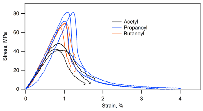
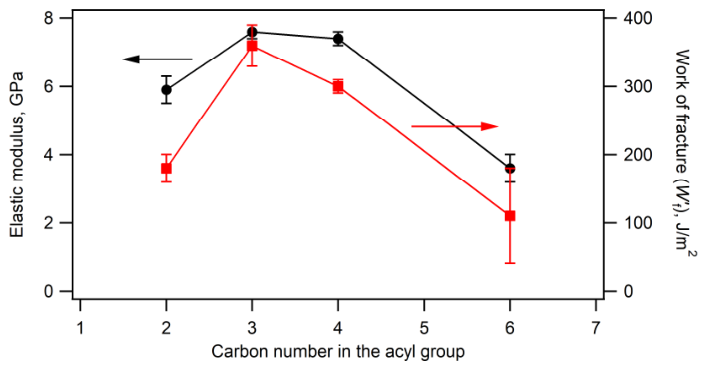
The composite powder was uniaxially pressed at 120 °C at 300 MPa to prepare the compacts for a three-point bending test. Representative stress–strain curves are shown in Figure 3. All composites showed a yield point at the strain of ca. 1%, and after the yield point plastic deformation followed, and the stress gradually decreased to zero. These stress–strain curves indicate that the composites are not fragile but ductile. The mechanical properties are summarized in Table 1. In Figure 4 shows the relationship between elastic modulus and work of fracture versus the number of carbon atoms in the acyl groups. The elastic moduli of the acetylated and hexanoylated composites were smaller than that of the unacylated MFC-HAP composite (7.1 GPa), whereas the elastic moduli of the propanoylated and butanoylated composites were larger. In general, acylation is expected to reduce the elastic modulus because the inorganic weight fraction decreases upon modification; however, we demonstrated that the introduction of acyl groups with intermediate chain lengths does not necessarily lead to a reduction in elastic modulus. Because both the cellulose and the hydroxyapatite constituting this composite are intrinsically rigid, it is possible that flexible acyl groups with an appropriate chain length fill the interfacial region between the rigid organic and inorganic phases, thereby enhancing the overall stiffness of the composite. Work of fracture of the butanoylated and propanoylated composites were larger than acetylated and hexanoylated composites, showing that propanoylation and butanoylation improved toughness of the composite.
Because the inorganic contents of the composites ranged from 54 to 68%, the effects of the inorganic contents on the mechanical properties should be taken into consideration to discuss the effects of acyl chain length. The elastic modulus, bending strength, and work to fracture of the MFC–HAP composites with inorganic contents of 44% and 62% were 7.9 and 8.4 GPa, 98 and 66 MPa, and 560 and 250 J/m2, respectively [43]. While the elastic modulus increased only slightly with increasing inorganic weight fraction, the flexural strength and work of fracture decreased markedly as the inorganic weight fraction increased. The decrease in bending strength and work of fracture upon acylation may be partly attributable to the concomitant decrease in inorganic weight fraction. In contrast, the increase in elastic modulus observed upon propanoylation and butanoylation, despite the decrease in inorganic weight fraction, is contrary to what would be expected from the decrease in inorganic content. Interestingly, despite exhibiting the highest porosity, the propanoylated composite possesses the highest elastic modulus.
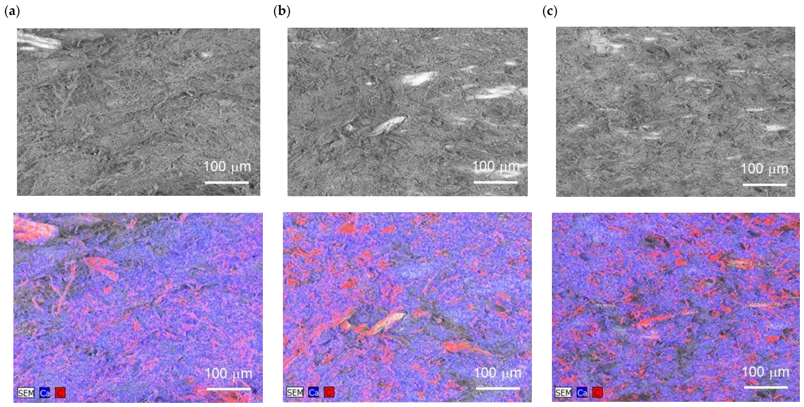
Figure 5 shows SEM images of the fracture surfaces after bending tests. All samples exhibit a fibrous structure on the fracture surfaces, indicating that the cracks do not propagate linearly. The fracture surface exhibited features similar to those of bovine femoral bone [46]. In addition, the results of elemental analysis of the fracture surfaces by EDS are summarized in Table 2. The propanoylated composite exhibits the highest C/Ca ratio, indicating that fracture preferentially occurs in the cellulose fiber regions. As shown in Figure S3, for the propanoylated and butanoylated samples, the carbon content at the fracture surface is higher than that calculated from the stoichiometric composition, whereas the phosphorus and calcium contents are lower than the corresponding calculated values. These results indicate that, in these composites, the cellulose phase is preferentially exposed at the fracture surface.

3.3. Water Absorption Test

The compressed composites were immersed in water at room temperature for 24 h. The compact of the unacylated composite collapsed when immersed in water, while the compacts of the acylated composites retained their shape. Water absorption ratios, ∆w/w, bulk densities, and porosities, calculated according to Equation (7), are listed in Table 3. The water absorption ratios decreased as the chain length of the acyl groups increased. Assuming that water is adsorbed only in the pore, we calculated water absorption ratios, ∆w/w (pore), according to Equation (11). As shown in Table 3, for the acetylated sample the water absorption ratio, ∆w/w, was larger than ∆w/w (pore). This indicates that water adsorption occurs not only in the pores but also in the cellulose phase or at the cellulose–hydroxyapatite interface, or that the porosity changes upon immersion in water [47]. In contrast, for the acylated samples other than the acetyl derivative, ∆w/w is equal to or smaller than ∆w/w (pore), suggesting that water adsorption is limited to the pores.
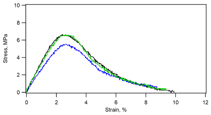
Table 4 lists the mechanical properties of the compacts after immersion in water. The elastic moduli of the compacts were reduced to 0.2–1.4 GPa, and stiffness was significantly affected. Figure 6 shows the stress–strain curves of the compacts of the propanoylated composite after immersion in water. The elastic modulus and flexural strength decreased after immersion in water. Water acted as a plasticizer, and the flexural strain reached 10%. The rigidity and water resistance of the composites were found to depend strongly on the carbon chain length of the acyl groups.

4. Conclusions

The acylation of the cellulose crystal surfaces in MFC–HAP composites containing 68 wt% HAP was carried out, and the dependence of stiffness and water resistance of the molded composites on the chain length of the aliphatic acyl groups was investigated. All of the acylated composites did not exhibit brittle fracture. In addition, the elastic moduli of the propanoylated and butanoylated composites were comparable to, or slightly higher than, those before acylation. The water absorption rates of the propanoyl and butanoyl derivatives were significantly lower than that of the unacylated sample. After immersion in water at room temperature for 24 h, the elastic modulus of the butanoylated composite decreased from 7.4 GPa to 0.7 GPa, indicating that challenges remain in terms of mechanical performance after immersion in water.

Supplementary Materials

The following supporting information can be downloaded at: https://www.mdpi.com/article/10.3390/jcs10090472/s1, Figure S1: XRD pattern of MFC-HAP composite with 68 wt% HAP; Figure S2: TG curves of acylated and unacylated MFC-HAP composites; Figure S3: Comparison of the atomic compositions determined by EDS and those calculated from the chemical formula, [C6H7O2(OH)3−x(OCO(CH2)nCH3][Ca10(PO4)6(OH)2]y where x is the degree of substitution of the acyl group, n is the number of methylene groups in the acyl groups and y is determined based on the inorganic contents of the composites. The value of x was assumed to be 0.2 for all composites. Table S1: Evaluation of the degree of substitution of the acylated composites. The Supplementary Information (SI): TG of acylated and unacylated MFC-HAP composites; XRD pattern of unacylated MFC-HAP composite; atomic compositions of the fracture surface of the composites; the degree of acylation determined from the IR spectra.

Author Contributions

Conceptualization, T.M.; Methodology, Y.M., E.K., A.T. and T.M.; Validation, E.K. and A.T.; Formal analysis, E.K. and A.T.; Investigation, A.M., Y.M., E.K. and A.T.; Data curation, A.M., Y.M., E.K. and A.T.; Writing—original draft, A.M. and Y.M.; Writing—review & editing, T.M.; Visualization, T.M.; Supervision, E.K., A.T. and T.M.; Project administration, T.M.; Funding acquisition, T.M. All authors have read and agreed to the published version of the manuscript.

Funding

This research received no external funding.

Data Availability Statement

The raw data supporting the conclusions of this article will be made available by the authors on request.

Acknowledgments

We thank Hiroyuki Matsumura, Tomohiro Hashizume, and Yohei Minami, Daicel Corporation, for supplying MFC samples and valuable discussion.

Conflicts of Interest

The authors declare no conflicts of interest.

References

  1. Suchanek, W.; Yoshimura, M. Processing and properties of hydroxyapatite-based biomaterials for use as hard tissue replacement implants. J. Mater. Res. 1998, 13, 94–117. [Google Scholar] [CrossRef] [Scilit]
  2. Yan, J.; Mecholsky, J.J., Jr.; Clifton, K.B. How tough is bone? Application of elastic-plastic fracture mechanics to bone. Bone 2007, 40, 479–484. [Google Scholar] [CrossRef] [Scilit] [PubMed]
  3. Dorozhkin, S. Calcium Orthophosphates in Nature, Biology and Medicine. Materials 2009, 2, 399–498. [Google Scholar] [CrossRef] [Scilit]
  4. Currey, J.D. The structure and mechanics of bone. J. Mater. Sci. 2012, 47, 41–54. [Google Scholar] [CrossRef] [Scilit]
  5. Stock, S.R. The Mineral-Collagen Interface in Bone. Calcif. Tissue Int. 2015, 97, 262–280. [Google Scholar] [CrossRef] [Scilit] [PubMed]
  6. Wagermaier, W.; Klaushofer, K.; Fratzl, P. Fragility of Bone Material Controlled by Internal Interfaces. Calcif. Tissue Int. 2015, 97, 201–212. [Google Scholar] [CrossRef] [Scilit] [PubMed]
  7. Nair, A.K.; Gautieri, A.; Chang, S.; Buehler, M.J. Molecular mechanics of mineralized collagen fibrils in bone. Nat. Commun. 2013, 4, 1724. [Google Scholar] [CrossRef] [Scilit] [PubMed]
  8. Almora-Barrios, N.; De Leeuw, N.H. A Density Functional Theory Study of the Interaction of Collagen Peptides with Hydroxyapatite Surfaces. Langmuir 2010, 26, 14535. [Google Scholar] [CrossRef] [Scilit] [PubMed]
  9. Wang, Y.; Naleway, S.E.; Wang, B. Biological and bioinspired materials: Structure leading to functional and mechanical performance. Bioact. Mater. 2020, 5, 745–757. [Google Scholar] [CrossRef] [Scilit] [PubMed]
  10. Wang, R.; Gupta, H.S. Deformation and Fracture Mechanisms of Bone and Nacre. Annu. Rev. Mater. Res. 2011, 41, 41–73. [Google Scholar] [CrossRef] [Scilit]
  11. Rho, J.; Kuhn-Spearing, L.; Zioupos, P. Mechanical properties and the hierarchical structure of bone. Med. Eng. Phys. 1998, 20, 92–102. [Google Scholar] [CrossRef] [Scilit] [PubMed]
  12. Athanasiou, K.A.; Zhu, C.F.; Lanctot, D.R.; Agrawal, C.M.; Wang, X. Fundamentals of Biomechanics in Tissue Engineering of Bone. Tissue Eng. 2000, 6, 361–381. [Google Scholar] [CrossRef] [Scilit] [PubMed]
  13. Currey, J.D. What determines the bending strength of compact bone? J. Exp. Biol. 1999, 202, 2495–2503. [Google Scholar] [CrossRef] [Scilit] [PubMed]
  14. Wang, X.; Puram, S. The Toughness of Cortical Bone and Its Relationship with Age. Ann. Biomed. Eng. 2004, 32, 123–135. [Google Scholar] [CrossRef] [Scilit] [PubMed]
  15. Dorozhkin, S.V.; Epple, M. Biological and Medical Significance of Calcium Phosphates. Angew. Chem. Int. Ed. 2002, 41, 3130–3146. [Google Scholar] [CrossRef] [Scilit] [PubMed]
  16. Olszta, M.J.; Cheng, X.; Jee, S.S.; Kumar, R.; Kim, Y.; Kaufman, M.J.; Douglas, E.P.; Gower, L.B. Bone structure and formation: A new perspective. Mater. Sci. Eng. R Rep. 2007, R58, 77–116. [Google Scholar] [CrossRef] [Scilit]
  17. Currey, J.D.; Zioupos, P.; Peter, D.; Casinos, A. Mechanical properties of nacre and highly mineralized bone. Proc. R. Soc. Lond. B 2001, 268, 107–111. [Google Scholar] [CrossRef] [Scilit] [PubMed]
  18. Palmer, L.C.; Newcomb, C.J.; Kaltz, S.R.; Spoerke, E.D.; Stupp, S.I. Biomimetic Systems for Hydroxyapatite Mineralization Inspired By Bone and Enamel. Chem. Rev. 2008, 108, 4754–4783. [Google Scholar] [CrossRef] [Scilit] [PubMed]
  19. Berrahou, S.; Latifi, S.; Saoiabi, S.; Abidi, N.; Saoiabi, S.; Azzaoui, K.; Hanbali, G.; Jodeh, S.; Hammouti, B.; Sabbahi, R. Hydroxyapatite–cellulose composites: Properties, fabrication methods, and applications. J. Mater. Sci. Mater. Med. 2026, 37, 27. [Google Scholar] [CrossRef] [Scilit] [PubMed]
  20. Furko, M.; Balázsi, K.; Balázsi, C. Calcium Phosphate Loaded Biopolymer Composites—A Comprehensive Review on the Most Recent Progress and Promising Trends. Coatings 2023, 13, 360. [Google Scholar] [CrossRef] [Scilit]
  21. Shi, R.; Lang, J.; Wang, T.; Zhou, N.; Ma, M. Fabrication, Properties, and Biomedical Applications of Calcium-Containing Cellulose-Based Composites. Front. Bioeng. Biotechnol. 2022, 10, 937266. [Google Scholar] [CrossRef] [Scilit] [PubMed]
  22. Arkharova, N.A.; Severin, A.V.; Khripunov, A.K.; Krasheninnikov, S.V.; Tkachenko, A.A.; Orekhov, A.S.; Davydova, G.A.; Rakova, E.V.; Klechkovskaya, V.V. Composite Films Based on Bacterial Cellulose and Nanocrystals of Hydroxyapatite: Morphology, Structure, and Properties. Polym. Sci. Ser. A 2019, 61, 650–658. [Google Scholar] [CrossRef] [Scilit]
  23. Okuda, Y.; Hirota, K.; Mizutani, T.; Aoyama, Y. Co-precipitation of tapioca starch and hydroxyapatite. Effects of phosphorylation of starch on mechanical properties of the composites. Results Mater. 2019, 3, 100035. [Google Scholar] [CrossRef] [Scilit]
  24. Okuda, Y.; Shigemasa, R.; Hirota, K.; Mizutani, T. In Situ Crystallization of Hydroxyapatite on Carboxymethyl Cellulose as a Biomimetic Approach to Biomass-Derived Composite Materials. ACS Omega 2022, 7, 12127–12137. [Google Scholar] [CrossRef] [Scilit] [PubMed]
  25. Kusakabe, A.; Hirota, K.; Mizutani, T. Crystallisation of hydroxyapatite in phosphorylated poly(vinyl alcohol) as a synthetic route to tough mechanical hybrid materials. Mater. Sci. Eng. C 2017, 70, 487–493. [Google Scholar] [CrossRef] [Scilit] [PubMed]
  26. Okuda, K.; Mizutani, T.; Hirota, K.; Hayashi, T.; Zinno, K. Nonbrittle Nanocomposite Materials Prepared by Coprecipitation of TEMPO-Oxidized Cellulose Nanofibers and Hydroxyapatite. ACS Sustain. Chem. Eng. 2021, 9, 158–167. [Google Scholar] [CrossRef] [Scilit]
  27. Matsuo, Y.; Sato, R.; Tabata, K.; Makino, T.; Saito, T.; Sato, K.; Arita, T.; Masuhara, A. Hybrid composite cellulose nanocrystal, hydroxyapatite, and chitosan material with controlled hydrophilic/hydrophobic properties as a remineralizable dental material. Cellulose 2024, 31, 2267–2279. [Google Scholar] [CrossRef] [Scilit]
  28. Raza, M.; Abu-Jdayil, B. Cellulose nanocrystals from lignocellulosic feedstock: A review of production technology and surface chemistry modification. Cellulose 2022, 29, 685–722. [Google Scholar] [CrossRef] [Scilit]
  29. Rodríguez-Fabià, S.; Torstensen, J.; Johansson, L.; Syverud, K. Hydrophobization of lignocellulosic materials part II: Chemical modification. Cellulose 2022, 29, 8957–8995. [Google Scholar] [CrossRef] [Scilit]
  30. Okuda, Y.; Kido, E.; Hirota, K.; Mizutani, T. Water-resistant tough composites of cellulose nanofibers and hydroxyapatite. ACS Appl. Polym. Mater. 2023, 5, 8082–8088. [Google Scholar] [CrossRef] [Scilit]
  31. Okuda, Y.; Aoyama, Y.; Hirota, K.; Mizutani, T.; Okuda, K. Effects of Hydration on Mechanical Properties of Acylated Hydroxyapatite-Starch Composites. ACS Appl. Polym. Mater. 2022, 4, 1666–1674. [Google Scholar] [CrossRef] [Scilit]
  32. Çetin, N.S.; Tingaut, P.; Özmen, N.; Henry, N.; Harper, D.; Dadmun, M.; Sèbe, G. Acetylation of Cellulose Nanowhiskers with Vinyl Acetate under Moderate Conditions. Macromol. Biosci. 2009, 9, 997–1003. [Google Scholar] [CrossRef] [Scilit] [PubMed]
  33. Taniguchi, N.; Mitsushima, Y.; Okuda, Y.; Takagi, A.; Kido, E.; Hirota, K.; Mizutani, T. Benzoylation of microfibrillated cellulose–hydroxyapatite composites for green and water-resistant mechanical materials. RSC Appl. Polym. 2026, 4, 298. [Google Scholar] [CrossRef] [Scilit]
  34. Heinze, T.; Liebert, T. Unconventional methods in cellulose functionalization. Prog. Polym. Sci. 2001, 26, 1689–1762. [Google Scholar] [CrossRef] [Scilit]
  35. Kim, D.; Nishiyama, Y.; Kuga, S. Surface acetylation of bacterial cellulose. Cellulose 2002, 9, 361–367. [Google Scholar] [CrossRef] [Scilit]
  36. Tingaut, P.; Zimmermann, T.; Lopez-Suevos, F. Synthesis and Characterization of Bionanocomposites with Tunable Properties from Poly(lactic acid) and Acetylated Microfibrillated Cellulose. Biomacromolecules 2009, 11, 454. [Google Scholar] [CrossRef] [Scilit] [PubMed]
  37. Lin, N.; Huang, J.; Chang, P.R.; Feng, J.; Yu, J. Surface acetylation of cellulose nanocrystal and its reinforcing function in poly(lactic acid). Carbohydr. Polym. 2011, 83, 1834–1842. [Google Scholar] [CrossRef] [Scilit]
  38. Eyley, S.; Thielemans, W. Surface modification of cellulose nanocrystals. Nanoscale 2014, 6, 7764–7779. [Google Scholar] [CrossRef] [Scilit] [PubMed]
  39. Wang, Y.; Wang, X.; Xie, Y.; Zhang, K. Functional nanomaterials through esterification of cellulose: A review of chemistry and application. Cellulose 2018, 25, 3703–3731. [Google Scholar] [CrossRef] [Scilit]
  40. Oberlintner, A.; Likozar, B.; Novak, U. Hydrophobic functionalization reactions of structured cellulose nanomaterials: Mechanisms, kinetics and in silico multi-scale models. Carbohydr. Polym. 2021, 259, 117742. [Google Scholar] [CrossRef] [Scilit] [PubMed]
  41. Li, W.; Li, W.; Xiong, Z.; Zhang, P. Controllable acetylation of cellulose nanocrystal by uniform design and response surface methodology. Carbohydr. Polym. 2024, 333, 121990. [Google Scholar] [CrossRef] [Scilit] [PubMed]
  42. Bai, H.; Walsh, F.; Gludovatz, B.; Delattre, B.; Huang, C.; Chen, Y.; Tomsia, A.P.; Ritchie, R.O. Bioinspired Hydroxyapatite/Poly(methyl methacrylate) Composite with a Nacre-Mimetic Architecture by a Bidirectional Freezing Method. Adv. Mater. 2016, 28, 50–56. [Google Scholar] [CrossRef] [Scilit] [PubMed]
  43. Okuda, Y.; Sasaki, R.; Kido, E.; Hirota, K.; Mizutani, T. One-Pot Hybridization of Microfibrillated Cellulose and Hydroxyapatite as a Versatile Route to Eco-Friendly Mechanical Materials. ACS Omega 2024, 9, 44457–44464. [Google Scholar] [CrossRef] [Scilit] [PubMed]
  44. Sassi, J.; Chanzy, H. Ultrastructural aspects of the acetylation of cellulose. Cellulose 1995, 5, 111–127. [Google Scholar] [CrossRef] [Scilit]
  45. French, A.D. Idealized powder diffraction patterns for cellulose polymorphs. Cellulose 2013, 21, 885–896. [Google Scholar] [CrossRef] [Scilit]
  46. Ziv, V.; Wagner, H.D.; Weiner, S. Microstructure-Microhardness Relations in Parallel-Fibered and Lamellar Bone. Bone 1996, 18, 417–428. [Google Scholar] [CrossRef] [Scilit] [PubMed]
  47. Nishiyama, Y. Thermodynamics of the swelling work of wood and non-ionic polysaccharides: A revisit. Carbohydr. Polym. 2023, 320, 121227. [Google Scholar] [CrossRef] [Scilit] [PubMed]
Figure 1. IR spectra of acylated MFC-HAP composites. The absorbance was normalized so that the intensity of the O–P–O bending vibration of hydroxyapatite at 564 cm−1 was set to unity.
Jcs 10 00472 g001
Figure 2. Powder X-ray diffraction patterns of acylated MFC-HAP composites. The major peaks of HAP are indexed.
Jcs 10 00472 g002
Figure 3. Stress–strain curves of three-point bending test of acetylated MFC-HAP, propanoylated MFC-HAP, and butanoylated MFC-HAP. To confirm reproducibility, the bending test was conducted three times.
Jcs 10 00472 g003
Figure 4. Plot of elastic modulus (red line) and work of fracture (black line) against the number of carbon atoms in the acyl groups.
Jcs 10 00472 g004
Figure 5. SEM images of the fracture surface and elemental mapping with energy-dispersive X-ray spectroscopy at an acceleration voltage of 15 kV (calcium in blue and carbon in red). (a) Acetyl, (b) propanoyl and (c) butanoyl composites.
Jcs 10 00472 g005
Figure 6. Stress–strain curves of three-point bending test of propanoylated MFC-HAP after immersion in water at room temperature for 24 h. The results of three point bending tests are shown superimposed.
Jcs 10 00472 g006
Scheme 1. Acylation of MFC-HAP composites with carboxylic anhydrides and vinyl hexanoate.
Jcs 10 00472 sch001
Table 1. Mechanical properties of acylated MFC-HAP composites a.
Elastic Modulus, GPaBending Strength, MPaStrain at Yield Point, %Work of Fracture (Wf), J m−2Work of Fracture (Wf), J m−2
Acetylated5.9 ± 0.441 ± 41.11 ± 0.02370 ± 30180 ± 20
Propanoylated7.6 ± 0.278 ± 31.4 ± 0.1580 ± 30360 ± 30
Butanoylated7.4 ± 0.268 ± 21.27 ± 0.03400 ± 20300 ± 10
Hexanoylated3.6 ± 0.429 ± 20.8 ± 0.1--- b110 ± 70
MFC-HAP7.1 ± 0.389 ± 51.54 ± 0.08--- b650 ± 70
a Averages and standard errors of mean are calculated based on at least three independent bending tests. b Not determined.
Table 2. The compositions (in at.%) of the fracture surface measured by EDS.
SamplesCOPCaC/Ca
Acetylated30.053.75.610.72.8
Propanoylated34.550.55.59.53.6
Butanoylated34.249.45.411.03.1
Table 3. Water absorption ratios, ∆w/w, water absorption ratios, ∆w/w (pore), porosity, and bulk densities of acylated MFC-HAP compacts after immersion in water at room temperature for 24 h a.
Samplesw/w, %w/w (Pore), %Bulk Density, g/cm−3Porosity, %
Acetylated29 ± 222 ± 21.66 ± 0.0427 ± 2
Propanoylated24 ± 129.2 ± 0.41.58 ± 0.0132.3 ± 0.3
Butanoylated17 ± 122.3 ± 0.41.73 ± 0.0122.3 ± 0.3
Hexanoylated18 ± 118.0 ± 2
a Averages and standard errors of mean are calculated based on at least three independent bending tests.
Table 4. Mechanical properties of acylated MFC-HAP compacts after immersion in water at room temperature for 24 h a.
SamplesElastic Modulus, GPaBending Strength, MPaStrain at Yield Point, %Work of Fracture (Wf), J m−2Work of Fracture (W′f), J m−2
Propanoylated0.23 ± 0.036.3 ± 0.43.1 ± 0.1240 ± 1092 ± 9
Butanoylated0.7 ± 0.115.3 ± 12.6 ± 0.2250 ± 20190 ± 20
Hexanoylated1.4 ± 0.212.8 ± 0.81.1 ± 0.2--- b70 ± 20
a Averages and standard errors of mean are calculated based on at least three independent bending tests. b Not determined.
Disclaimer/Publisher’s Note: The statements, opinions and data contained in all publications are solely those of the individual author(s) and contributor(s) and not of MDPI and/or the editor(s). MDPI and/or the editor(s) disclaim responsibility for any injury to people or property resulting from any ideas, methods, instructions or products referred to in the content.