1. Introduction
Bone grafts have been utilized for many years in maxillofacial surgery to induce tissue regeneration and repair. The bone grafts must be used in combination with a membrane to ensure the mechanical stability of the particulate grafts, especially when small biomaterial granules are used. With this procedure, the success rate increases. Some membranes are made from polymers such as polysulfone, polyvinylidene fluoride, and polypropylene. The disadvantage of this kind of membrane is that it needs a second surgery to remove. Collagen-based membranes outperform non-absorbable membranes in situations where bone formation is necessary. The collagen membranes act as a barrier against aggressive agents and assist bone reconstruction. Research in this area is essential for developing new materials that improve the regenerative processes [
1].
Collagen is the most abundant structural protein present in the extracellular matrix and makes an important contribution to wound healing due to its ability to support cell adhesion, migration, and proliferation. Its fibrillar architecture enhances fibroblast function, while its high water-binding capacity helps maintain a moist microenvironment that promotes the formation of the granulation tissue, angiogenesis, and re-epithelialization. Notwithstanding these advantages, native collagen has limitations, such as low mechanical strength and rapid degradation under the inflammatory process, which may compromise its performance in deep or highly exuding wounds [
2,
3].
Hydroxyapatite (HA) is a calcium phosphate compound that closely resembles the mineral phase of bone; it is commonly used to reinforce collagen-based scaffolds. Its chemical bioactivity provides strong interactions with host tissues through the release of ions (Ca
2+ and PO
43−), supporting fibroblast proliferation, angiogenesis, and local pH buffering. Additionally, HA provides mechanical stiffness and compressive strength, which are important in load-bearing environments or wounds involving bone defects. However, its effectiveness in biological and load-bearing characteristics depends on its concentration and the context, for example, as excessive mineral content may reduce scaffold flexibility and impair cell migration in soft-tissue applications [
2,
4].
The collagen and hydroxyapatite combination results in a hybrid scaffold that resembles the hierarchical structure of the native tissues. These composites conjugate the biological properties of collagen—such as elasticity, hydration, and cell signaling—with the mechanical strength and osteoconductive capacity of HA. The resulting composite exhibits enhanced cellular interactions, controlled biodegradation, improved mechanical stability, and a favorable microenvironment for tissue healing. These synergistic properties make collagen–HA composites particularly suitable for complex wounds, including chronic ulcers and bone-exposed defects [
3,
5].
The combination of collagen and micrometer-sized hydroxyapatite (HA) particles has shown advantages over either material alone. When implanted into cranial defects in rats, the matrix, synthesized by cross-linking collagen fibers with modified HA, demonstrated good biocompatibility. The HA results showed greater osteoconductive potential than the matrices composed of either cross-linked collagen or cross-linked HA alone [
2,
6]. A tri-component hydroxyapatite–collagen–HA composite has offered new possibilities for application as a bone implant material [
7].
Literature results from in vitro experiments have demonstrated that the incorporation of HA into collagen gels enhances the strength of the collagen gels, thereby inhibiting fibroblast contraction on the collagen matrices [
8]. This indicates that the rheological behavior and thermal stability of the scaffold determine its resistance to contraction. Although several investigations on collagen–HA scaffold materials have been conducted to characterize their biochemical and biological properties [
5,
6,
9,
10], the effect of temperature on their conformational and mechanical properties remains unclear.
The ability to promote osteoconductivity and vascularization is the most significant advantage of these composites. HA–collagen scaffolds induce the recruitment and activity of mesenchymal stem cells, osteoblasts, and osteoclasts, facilitating new bone formation. The HA–collagen composite enhances angiogenesis, which is essential for nutrient delivery and long-term tissue viability. Studies have demonstrated that these composites improve cell viability and regenerative outcomes compared to single-component materials, and their performance can be further enhanced through the incorporation of bioactive molecules or ions [
3,
5].
Another critical parameter influencing the biological and mechanical performance of HA–collagen composites is porosity. Highly porous structures promote cell infiltration, vascular network formation, and nutrient diffusion, accelerating tissue regeneration. Although increased porosity may reduce mechanical strength, it enhances elasticity and allows the scaffold to conform to irregular defect sites, which is advantageous during surgical procedures. In addition, the presence of collagen improves the handling properties of the biomaterial, including ease of shaping, adhesion to the defect site, and support for clot formation, further contributing to the clinical applicability of these bioinspired materials [
2,
4].
Several complementary tests are necessary to assess the performance of new biomaterials. Among the tests, mechanical testing, wettability measurement, cell culture, and immunohistochemical testing are necessary to establish material biocompatibility [
11]. Ultrastructure analysis using SEM and TEM provides extremely reliable results. Several characteristics of collagen can be identified, such as the shape and arrangement of its fibers. Based on electron microscope images, it is possible to estimate the performance of the biomaterial, the presence of organic structures, and the presence of foreign bodies on the surface [
12,
13,
14].
The combination of nanohydroxyapatite and collagen has been widely employed for bone regeneration [
15,
16], whether incorporated into a single composite material [
17,
18] or used separately [
19]. Extensive evidence in the literature demonstrates that their association provides significant benefits to the bone regeneration process and, in many cases, is essential to achieve satisfactory outcomes [
20,
21].
Histology is an excellent tool for analyzing the structures of biological materials. It can be used to assess the quality and composition of the matrix. Collagen, as a biological material, can be analyzed by histology to investigate the regeneration and formation of soft and hard tissues [
22].
The aim of this study was to show that the association of a novel collagen membrane with nanohydroxyapatite will promote bone and soft tissue regeneration comparable to or superior to that achieved with commercially available products.
2. Materials and Methods
Two bone grafts (xenogeneic and synthetic) used in this study serve as scaffolds to guide the formation of new bone. After bone regeneration, dental implants can be inserted. The disadvantage of xenografts compared to synthetic materials is the possibility of disease transmission.
In the present work, in vitro and in vivo testing were conducted. The performance of two particulate bone grafts (Bio-Oss®, Wolhusen, Switzerland and Blue-Bone®, Curitiba, Brazil) and two membranes (Mucograft®, Wolhusen, Switzerland and Green Membrane Perio®, Curitiba, Brazil) for maxillofacial surgery application was analyzed and compared. The membranes were used to maintain the mechanical stability of particulate grafts. The membranes were characterized by SEM, TEM, roughness, and wettability analyses.
Bio-Oss® is a deproteinized bovine bone xenogeneic biomaterial used in regenerative dentistry and manufactured by Geistlich Pharma AG (Wolhusen, Switzerland). Mucograft® is a resorbable collagen membrane available from Geistlich. This biomaterial is made from sterilized bovine bone by removing the organic part, leaving the mineral structure (hydroxyapatite).
The Blue-Bone® graft is a synthetic composite biomaterial developed by the authors and is a mixture of nanometric particles of hydroxyapatite (80%) and β-TCP (20%). The Green Membrane Perio® is a biomaterial developed from non-crosslinked type I collagen fibers obtained from bovine Achilles tendons. Due to the need for impurity-free samples, the company Regener Biomaterials’ (Curitiba, Brazil) laboratory infrastructure was used for manufacturing the tested biomaterials.
2.1. Roughness Measurement
The membrane surface roughness was analyzed using the Zygo NewView® 7100 optical interferometric roughness meter (Zygo Corporation, Middlefield, CT, USA). The interferometric optical profilometer is a key instrument for measuring surface roughness and topography. The operation of the optical profilometer essentially involves projecting white light onto the sample surface. The light reflected by the surface interferes with the reference light, generating an interference pattern. This pattern is captured by an optical system and processed by software, which reconstructs the three-dimensional geometry near the surface. The equipment performs this measurement non-destructively and without physical contact with the sample, allowing for precise analysis of roughness, waviness, and shape with high vertical resolution. In the present study, we measured the surface roughness parameters Ra, RMS, Rku, PV, Rpk, and R3z. Details of the definition of these parameters are described in the literature.
2.2. Wettability
Surface wettability was determined by measuring contact angles using a goniometer (First Ten Angstroms FTA-100®, First Ten Angstroms Co., Portsmouth, VA, USA).
Wettability was determined using contact angle measurements. For the measurement, a drop of 0.9% sodium chloride was placed on the membrane surface, and an image of the drop was captured. The static contact angle was defined by fitting the Young–Laplace to the drop. The contact angles were determined by averaging the values obtained from five different drops on three samples of each group.
2.3. Tensile Test
The tensile testing was performed using a Universal Testing Machine (EMIC DL10000, Emic, Paraná, Brazil) in accordance with ASTM D638-14 (Standard Test Method for Tensile Properties of Plastics).
Sheet specimens were cut with a length of 30 mm, a width of 12.4 mm, and a thickness of 3.1 mm. Before conducting the mechanical tests, the sample surfaces were inspected to ensure they were free of visible flaws, scratches, and imperfections. Tensile tests were performed at room temperature with a crosshead speed of 10 mm/min. A high-definition force transducer with a capacity of 50 N was used. Extreme care was taken to ensure that the sample was inserted and clamped so that its long axis coincided with the direction of pull through the center line of the grip assembly. The maximum load (in grams) at the extreme loading point was measured, and the tensile strength (maximum stress, in MPa) was calculated.
2.4. Measurement of Thermodynamic Properties Using DSC
The thermal stability of the mixture of nano-hydroxyapatite, β-Tricalcium phosphate, and type I collagen was determined by differential scanning calorimetry (DSC). The thermal behavior of the samples was analyzed by differential scanning calorimetry (DSC) using a Shimadzu DSC60 DSC calorimeter (Shimadzu, Kyoto, Japan). Two samples are used for each DSC testing. The first sample was used to determine the thermal properties, and the second sample, of known thermal properties, was used as a reference. All energy variations in the mixture of nano-hydroxyapatite, β-Tricalcium phosphate, and type I collagen during heating were calculated relative to the reference sample.
Samples with 5.0 ± 0.5 mg were placed in aluminum pans. The pans were closed and weighed. Scans were performed between 25 °C and 500 °C at a rate of 10 °C/min. The tests were conducted without atmospheric control. The reference sample was an empty aluminum pan. The mixture and reference samples were kept at the same temperatures, and the difference in energy required to raise their temperatures was determined. The reference sample has a defined thermal capacity throughout the scanned temperature range. The test determined the variation in energy with the temperature of the phase transformations and transitions.
2.5. Surgery
For the in vivo test, a rat animal model was employed. The animal protocols used in this work were evaluated and approved by the Animal Ethics Committee (CEUA) of the Institute of Biology, Rio de Janeiro State University (Protocol No. 001/2019). They are in accordance with the laboratory animal care guidelines of this university. Forty adult male Wistar rats weighing 250 g and aged 8 weeks were used, provided by the Biology Institute of the State University of Rio de Janeiro. The animals were kept in cages with ad libitum access to food and water. The light/dark cycle (lights on at 7:00 a.m. and off at 7:00 p.m.) and temperature (22 °C) were maintained at constant levels throughout the experiment.
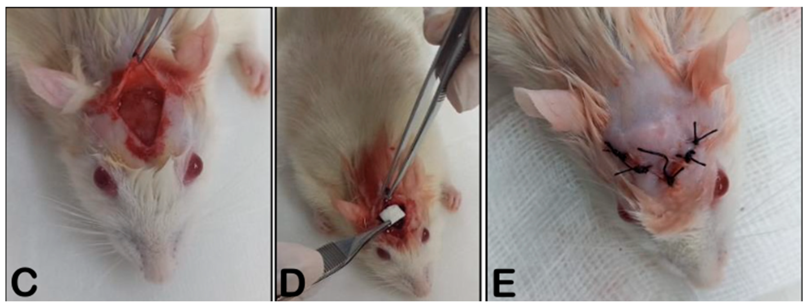
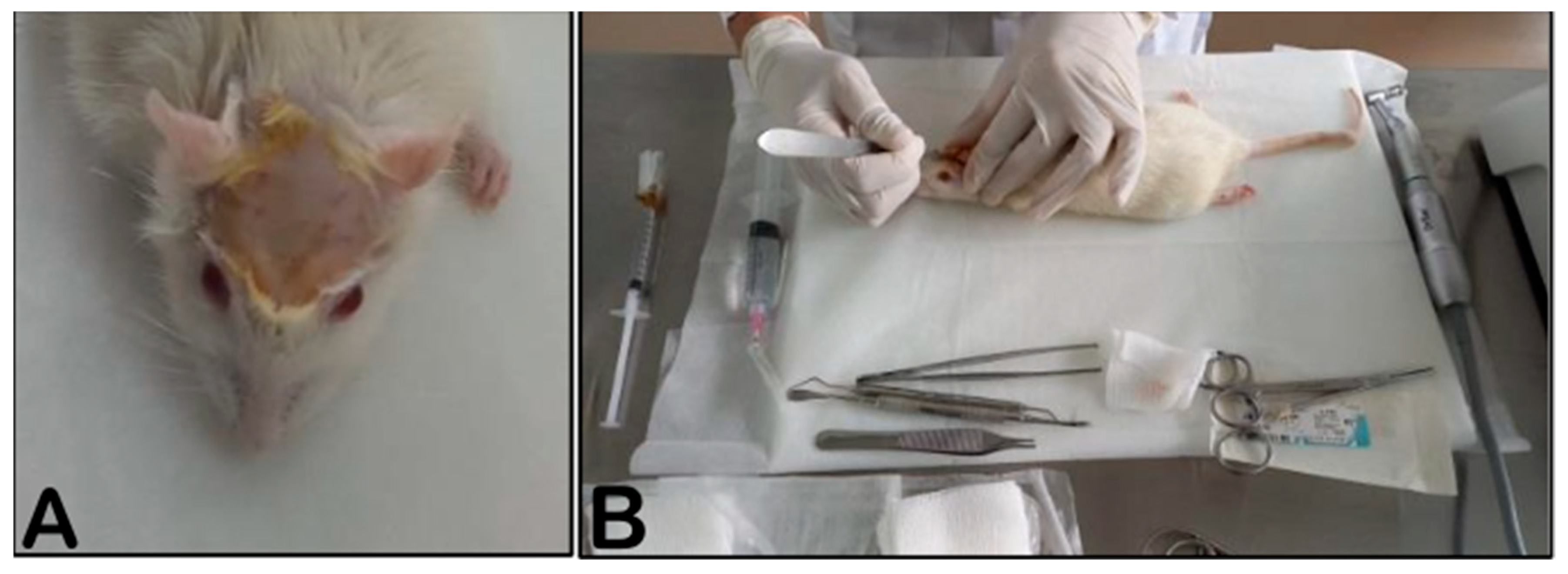
The rats were anesthetized with Ketamine and Xylazine intraperitoneally. Trichotomy was initiated (A), then a triangular incision was made (BC), and critical bone defects of 8 mm in diameter were created in the temporoparietal region using a trephine and the membrane. The critical defects were filled with bone substitute particles Bio-Oss® and Blue-Bone®.
2.6. Animal Groups
Forty male adult Wistar rats (200–220 g body weight) were used. The animals were provided by the Institute of Biology, Universidade Estadual do Rio de Janeiro.
The rats were divided into five groups (
n = 8 per group). In the sham group, defects were created without a bone graft. The defect was filled with an animal blood clot. In four groups, the defects were filled with the same bone graft volume, either with or without a membrane. A membrane, with a diameter of 9 mm, was used to maintain the mechanical stability of the graft particles at the surgical site.
Table 1 shows the five groups that were used.
Figure 1 and
Figure 2 show details of the surgery procedure.
2.7. Euthanasia
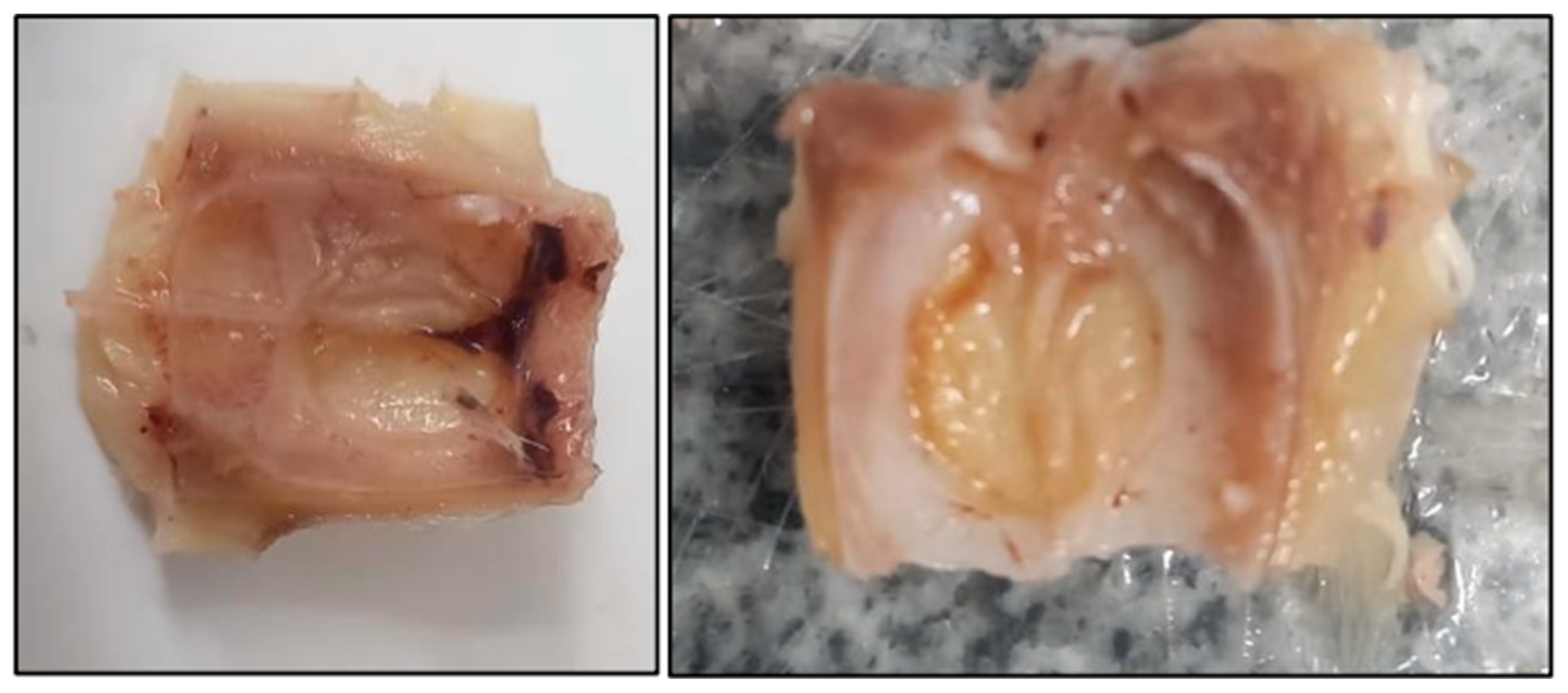
Eight weeks after surgery, the animals were sacrificed with a ketamine hydrochloride/xylazine solution (1/1, 0.3 mg/kg, ip) under anesthesia. The animals were decapitated, and the heads were used in the histological procedures (
Figure 3).
After euthanasia, tests were performed to determine the percentage of collagen in the subepithelial region of the cavities. Immunohistochemical tests were performed to quantify the degree of tissue regeneration.
2.8. Morphological Analysis Protocol
The animal heads were cut and decalcified in EDTA (7.0%) in phosphate-buffered saline (PBS) (0.1 M, pH 7.4) for 40 days. Specimens were washed in distilled water, dehydrated in alcohol (70%, 95%, and 100%), cleared in dimethylbenzene, and embedded in Paraplast® (Sigma, Aldrich, St. Louis, MO, USA) at 65 °C. Serial 7 µm sections were cut with a microtome (Leica, Nussloch, Germany) and collected on silanized slides.
2.9. Goldner’s Trichrome Staining Protocol
The slices were deparaffinized, rehydrated with alcohol (100%, 95%, and 70%), rinsed in distilled water, and immersed in Weigert’s iron hematoxylin® (Sigma-Aldrich, St. Louis, MO, USA) for 10 min. After rinsing in distilled water, the slides were immersed in Biebrich’s fuchsin solution (Sigma-Aldrich) for 15 min, then rinsed again in distilled water and differentiated in phosphomolybdovanate acid solution for 10 min. Then, the slides were immersed in an aniline blue solution for 5 min, rinsed with distilled water, dehydrated, and covered with coverslips.
2.10. Histomorphometry Analysis
Histomorphometry analysis was performed on a Goldner trichrome-stained slide. Three randomized Goldner trichrome, PAS-stained, and immunolabeled slides were photographed using an optical microscope (Carl Zeiss-JVC TK-1270 color video camera (Victor Company of Japan), Oberkochen, Germany) at 400× magnification. Images were quantified using GraphPad Prism Version 8.0 (GraphPad, San Diego, CA, USA).
2.11. Statistical Analysis
The data were analyzed using one-way ANOVA followed by a Wilcoxon Matched-Pairs test (p < 0.05). All statistical analyses were conducted with specific software (GraphPad Prism Version 8.0 and BioEstat 5.0).
2.12. Immunohistochemical Analysis
The preparation and analysis of the samples started with deparaffinizing the sections in 3 xylol baths (5 min for each bath); hydrating the sections (100%, 90%, and 70% ethanol, distilled water—5 min each bath). The sections were incubated in 3% hydrogen peroxide diluted in distilled water for 15 min, protected from light to inhibit endogenous peroxidase, then washed in 3 baths of TBS (Tris-Buffered Saline) pH 7.4 buffer (5 min each bath). The antigen retrieval was achieved in Tris/EDTA (ethylenediaminetetraacetic acid) buffer pH 9.0 at 95 °C for 20 min; cooled and washed in 3 baths of TBS buffer pH 7.4 (5 min each bath); and blocked non-specific sites with 3% TBS/BSA for 20 min.
The final steps were to incubate with the primary anti-Collagen I antibody diluted in TBS/BSA at 1% (1:200), overnight, in a refrigerator (4 °C), then placed in a humid chamber; wash in 3 baths of TBS buffer pH 7.4 (5 min each bath); incubated with biotinylated secondary antibody (VECTASTAIN® Universal Quick HRP Kit, Newark, CA, USA) for 30 min at room temperature; washed in 3 baths of TBS buffer pH 7.4 (5 min each bath); incubated with streptavidin VECTASTAIN® Universal Quick HRP Kit) for 15 min at room temperature; washed in PBS buffer pH 7.2; develop with DAB (diaminobenzidine); stained with hematoxylin; dehydrate (distilled water, 70%, 90% and 100% ethanol—5 min each bath); and clarified in 3 xylol baths (5 min each bath) and finally assembled with Entellan, Merck KGaA, Darmstadt, Germany.
2.13. Ultrastructural Analyses Using TEM and SEM
Thin membrane sections were analyzed using a JEOL JEM-1011 transmission electron microscope (JEOL, Ltd., Akishima, Tokyo, Japan), operating at 60 kV. Digital micrographs were captured using an ORIUS CCD digital camera (Gatan, Inc., Pleasanton, CA, USA) at magnifications of 8000×, 10,000×, and 25,000×.
The sample preparation for TEM analysis started with fixation in 2.5 wt% glutaraldehyde diluted in 0.1 M cacodylate buffer solution (overnight); washed in 3 baths in cacodylate buffer solution (0.1 M) for 15 min each bath; dehydrated in 30 vol% acetone bath (15 min), 50 vol% acetone, 70 vol% acetone (15 min), 90 vol% acetone (15 min), 100 vol% acetone (15 min), and 100 vol% acetone (15 min); infiltrated in acetone + epon mixture (2:1) for 2 h; acetone + epon (1:1) for 2 h; acetone + epon mixture (1:2) for 2 h, infiltration in pure epon (overnight); incubated in epon and allowed to polymerize between 48 and 72 h at 60 °C; cut into plates with a thickness of 1 μm and staining with toluidine blue and cut with an ultramicrotome to obtain 70 nm slides, which were collected on 300 mesh copper grids. Contrasting the slides was achieved with uranyl acetate (for 20–30 min).
3. Results and Discussion
Collagen membranes are used in oral and maxillofacial surgery and medicine to improve tissue healing and bone remodeling. Type 1 collagen membranes increase wound closure rates to 90% within 72 h [
23]. These data are interesting because the groups that had collagen membranes presented a more efficient healing process than those without membranes.
The extracellular matrix has numerous components. Therefore, some tests are essential to characterize these elements. A study employed SEM and TEM to determine the pattern of collagen fibers, thereby elucidating how fibroblasts respond to collagen degradation or deposition [
24].
3.1. Roughness
The collagen membrane has homogeneous morphology and high roughness parameters. Irregularities on the surface influence the shape of the adhered proteins, cells and the structure of the extracellular matrix. The surface morphology and chemical composition alter the time required for membrane reabsorption. These changes affect the tissue regeneration process [
8,
12,
25,
26].
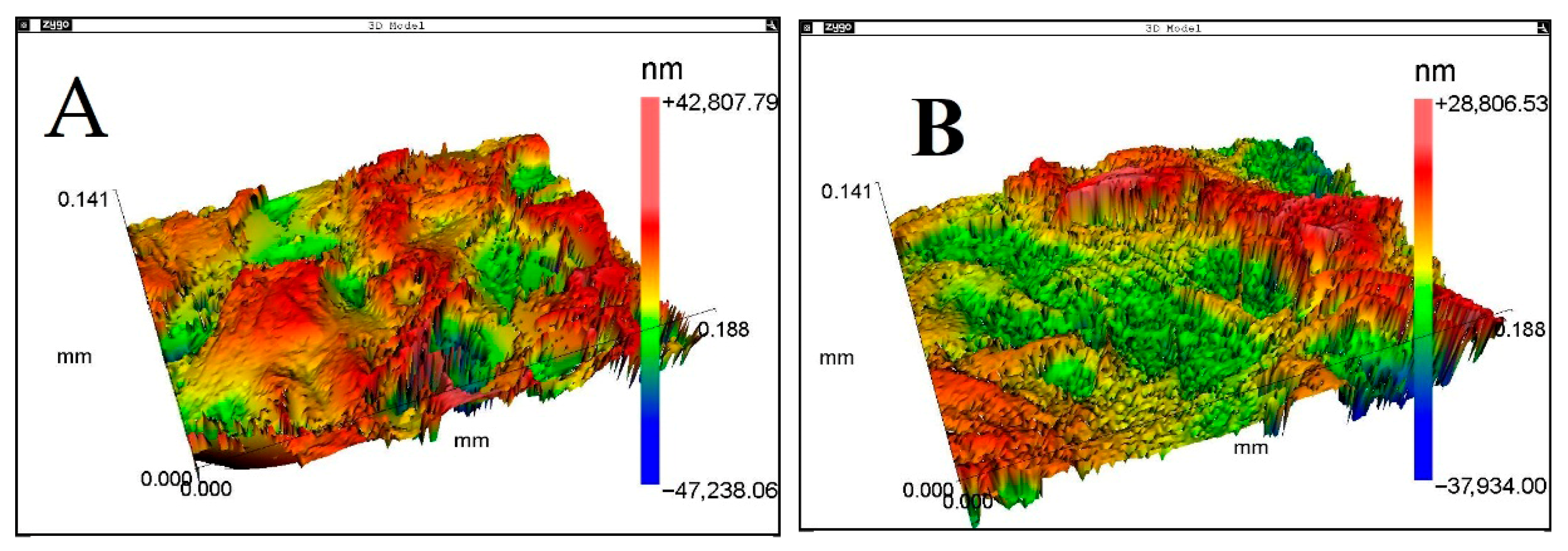
Table 2 shows the measured membrane surface roughness parameters.
Figure 4 shows the representative surface morphologies of the membranes obtained by the interferometric roughness meter. The membrane surface roughness Ra parameter is higher than that of the dental implant surface. Normally, the dental implant surface parameter Ra is from 1.0 to 1.4 µm.
The membrane surface roughness influences cell adhesion, degradation, and reabsorption. A surface with controlled roughness provides an energetically and mechanically favorable environment for inducing protein and cell adhesion. The greater the number of contact points, the more sites for cell receptors (proteins), and the greater the adsorption and physical anchoring of proteins. This behavior strengthens and densifies attachments, enabling better bone formation. Very rough or superhydrophobic surfaces hinder the adhesion of proteins (fibronectin and sialoprotein) and interactions with bone-forming cells (osteoblasts), while moderate roughness is usually ideal for growth and proliferation.
To our knowledge, no work in the literature has analyzed the influence of surface roughness or determined the ideal membrane value at the micro- and nanoscales. The magnitude of the roughness influences the type of cell that adheres to the surface. Based on the analysis of dental implant surfaces, surfaces with low or extreme roughness inhibit the adhesion of proteins and cells. The ideal membrane surface roughness is not defined. The idea is that the roughness of the membrane surface allows protein adhesion, provides physical adhesion, and mechanical anchorage for cells, which are crucial for the stability and formation of mineralized bone. Clinical experience shows that the membrane has a rough side that promotes bone growth and a smooth side that prevents soft-tissue invasion. More studies are needed to determine the influence and the appropriate membrane.
3.2. Wettability
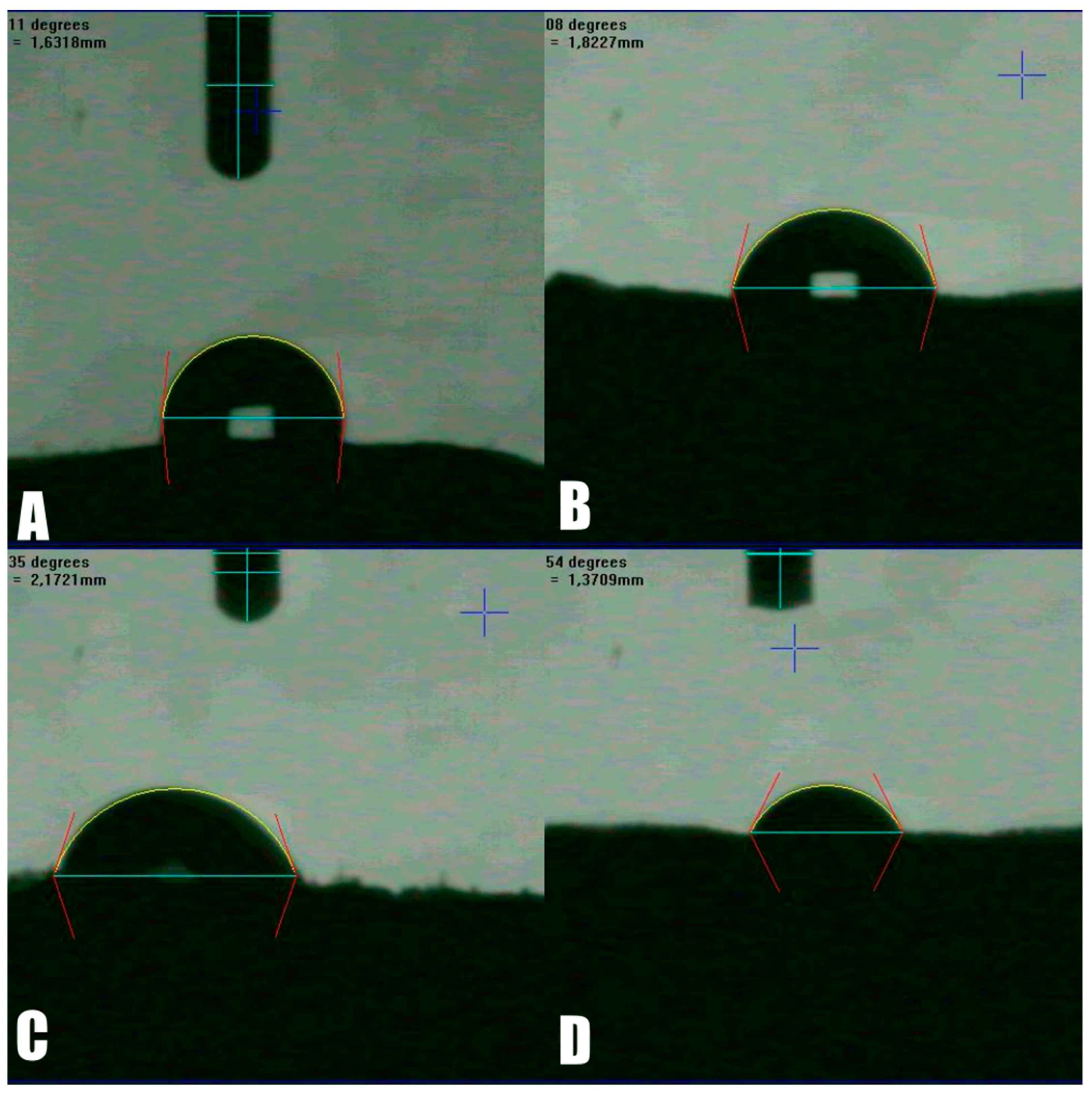
The wettability test showed that both membranes are highly hydrophilic, with a droplet absorption time of 10 s. The initial contact angle of the droplet with the surface was 105.47 degrees (
Figure 5A); however, after five seconds, the surface had absorbed the entire droplet (
Figure 5D).
Surface roughness significantly influences the wettability of biomaterials, which is directly related to the surface energy of the material. In general, higher surface energy corresponds to increased wettability.
Wettability plays a critical role in mediating interactions with bone-forming cells (osteoblasts) as well as soft tissue cells (fibroblasts). Hydrophilic and hydrophobic surfaces can selectively attract or repel specific proteins. By modulating wettability, protein adsorption may be either enhanced or inhibited, thereby influencing the strength of cell adhesion depending on the protein type involved.
In general, hydrophilic surfaces tend to promote greater protein adsorption. However, in physiological conditions, albumin is the most abundant serum protein, and its preferential adsorption should be minimized, as it can inhibit the adhesion of other proteins and subsequently impair cell attachment.
For biomaterials, moderate wettability—typically corresponding to a contact angle between 40° and 70°—is considered optimal for biocompatibility, since it provides a balance between protein adsorption and repulsion.
Currently, there is a lack of studies in the literature evaluating the performance of membranes with varying wettability. Ideally, such membranes should promote the formation of a suitable protein layer, allow permeability for the diffusion of biological fluids into the graft, and support strong adhesion of bone-forming cells.
3.3. Tensile Testing
The results of the tensile tests to evaluate the quality of the membranes are important because, during surgery, when suturing the membrane, the tensile force of the suture can tear the material. The tensile strength of the Green® membrane was 5.2 MPa. The Mucograft® membrane resisted 4.8 MPa. The difference in tensile strength between the two membranes can be attributed to chemical composition. The tensile test results indicate that both membranes resist the tensile force of the positioning suture.
A previous study [
27] evaluated and compared the morphology and mechanical properties of different membranes for application in guided bone regeneration. The study evaluated silkworm-cocoon-derived silk membrane (SM), collagen membrane (CM), and polytetrafluoroethylene membrane (PTFEM). The tensile strengths for SM, CM, and PTFEM were 27.8 ± 4.4, 3.4 ± 0.2, and 4.3 ± 0.1 MPa, respectively. The percentages of elongation were 39.3 ± 7.1, 9.6 ± 0.1, and 301.6 ± 7.7, respectively. The reason for the difference in tensile strength between the previous study and the present work is the use of a dry membrane in the present work. In contrast, in the previous study, the samples were immersed in distilled water for 24 h before the mechanical test. The second difference was that the study used a synthetic membrane with a different material.
3.4. Differential Scanning Calorimetry
In DSC tests to determine energy variations with temperature, it is possible to determine the denaturation temperature, the water loss, and other phenomena.
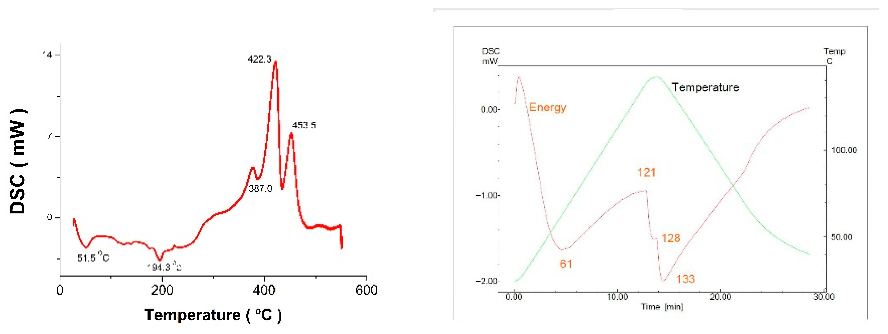
Figure 6 shows the DSC thermal curve results.
The collagen denaturation temperature influences its hydration, which usually varies from 58 to 75 °C. During denaturation, the triple helix structure of collagen changes, forming a random coil. The denaturation of hydrated collagen can also be associated with the phase change from solid to liquid. In the case of hydrogel, heating above 65 °C breaks the inter- and intramolecular crosslinks.
The term “biopolymer hydration” is based on the thermodynamic characteristics of water evaporation. However, the polymer has adsorbed water and chemically bonded to it. The bonds include hydrogen bonds, ionic and covalent bonds, and hydrophilic and hydrophobic interactions. To break the polymer bonds with water, heating at different temperatures is necessary. These temperatures can be determined by measuring the enthalpy of water evaporation from the sample using differential scanning calorimetry. This method estimates the water bonding state in biopolymers and the levels of structural organization. For example, the binding of water to collagen fibers is measured [
28]. As the DSC test temperature increases, the deflection of the Blue-Bone
® and Green Membrane Perio
® curves (
Figure 6) shows that the energy absorbed by collagen increases. The Blue-Bone
® and Green Membrane Perio
® main transition structures begin at temperatures (T1) of 51.5 °C and 61.1 °C, respectively.
The Green Membrane Perio® main transition temperature quantifies the collagen gelatinization process in a hydrated environment. At this temperature, the internal crosslinks are broken. At the transition temperature, changes in the shapes inside the collagen fibrils were observed. At 121.4 °C, the denaturation temperature (T2) is reached, and partial shrinkage of the fibril length occurs. In the case of natural collagen, it is observed that the fibrils undergo thermal denaturation, which involves the rupture of hydrogen bonds between the chains of molecules, resulting in the formation of an amorphous material. In the DSC test, it is possible to determine the temperature at the onset of denaturation (Tonset), the temperature at the end of denaturation (Tendset), and the temperature at which the denaturation peak occurs, characterized by the maximum endothermic reaction (Tp). At the Tp temperature, the collagen structure unfolds, changing its helix-spiral shape. As the test temperature increases, a second transition occurs (T3). The second transition is associated with a change in the shape of collagen molecules. The structure changes from a triple helix to a random spiral.
The conformational transformation is characterized by the viscous movement of fibrils upon heating. There are no significant variations in the transition range from TH to RC among the different types of bone. Results of DSC bone tests show characteristic peaks related to the denaturation of albumin and hemoglobin proteins, as well as the fusion of fats. As the temperature increases, water loss occurs during volumetric contraction.
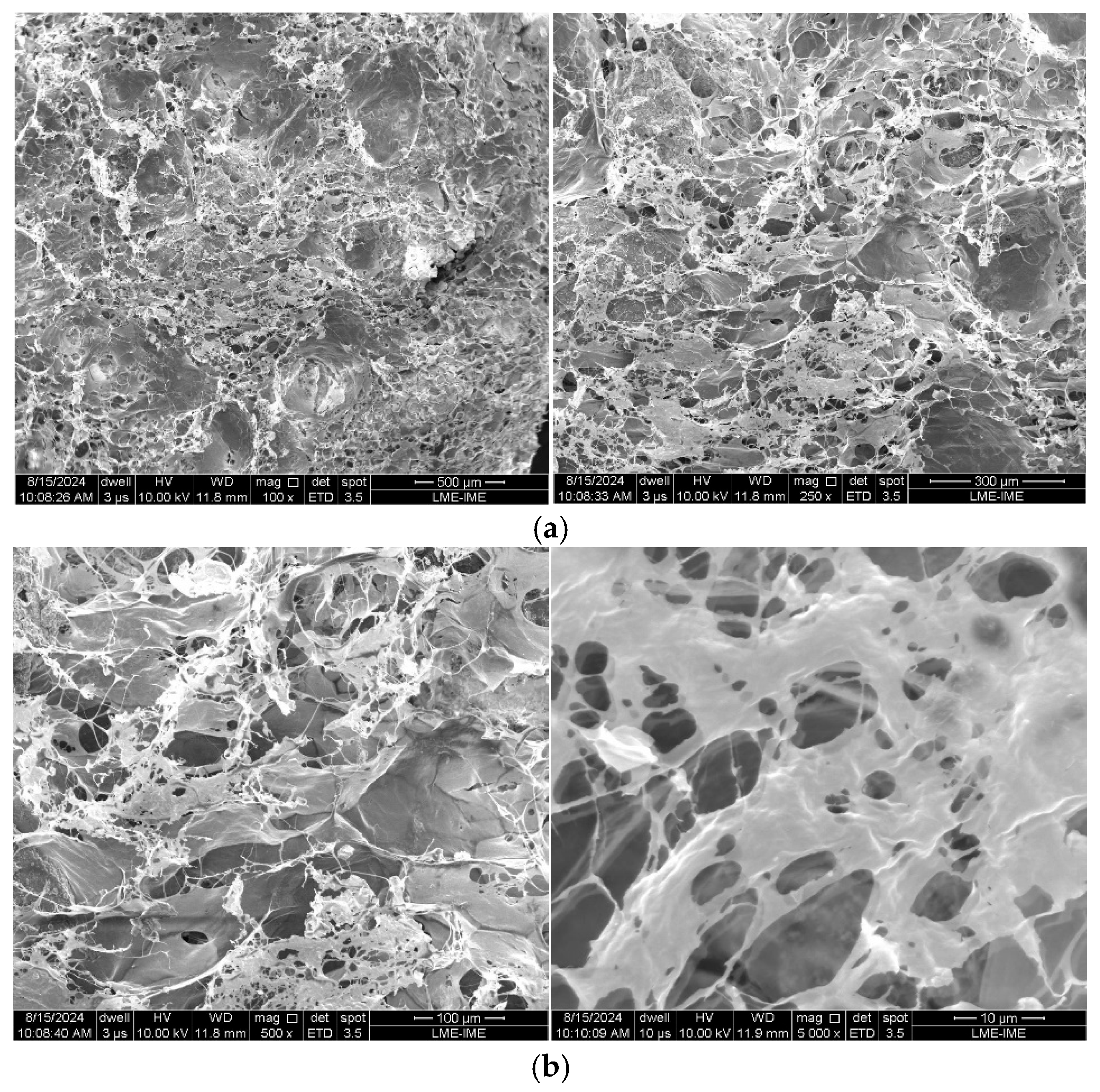
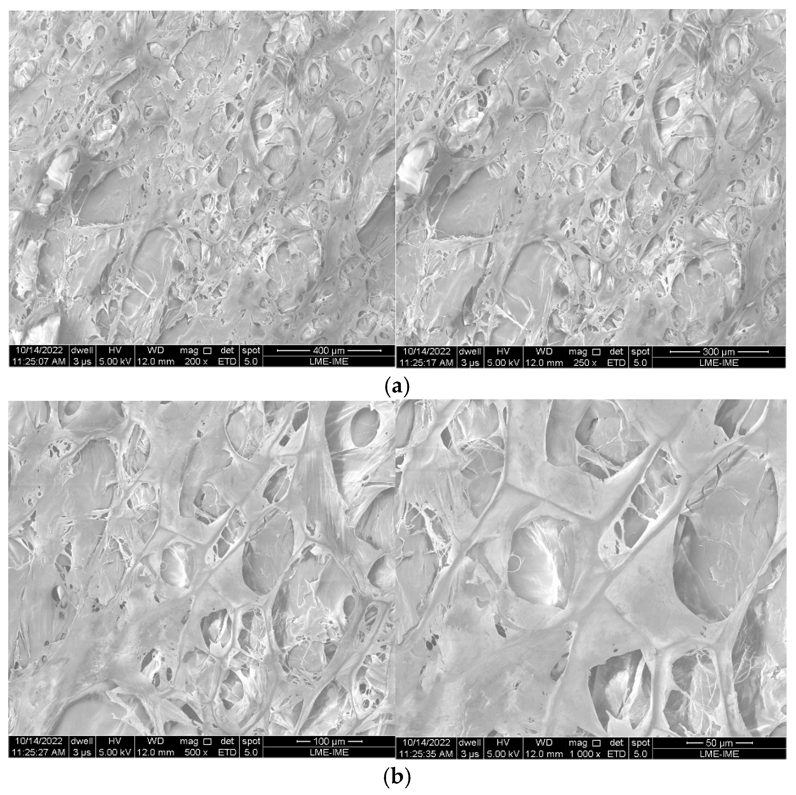
3.5. Scanning Electron Microscopy
3.6. Transmission Electron Microscopy (TEM)
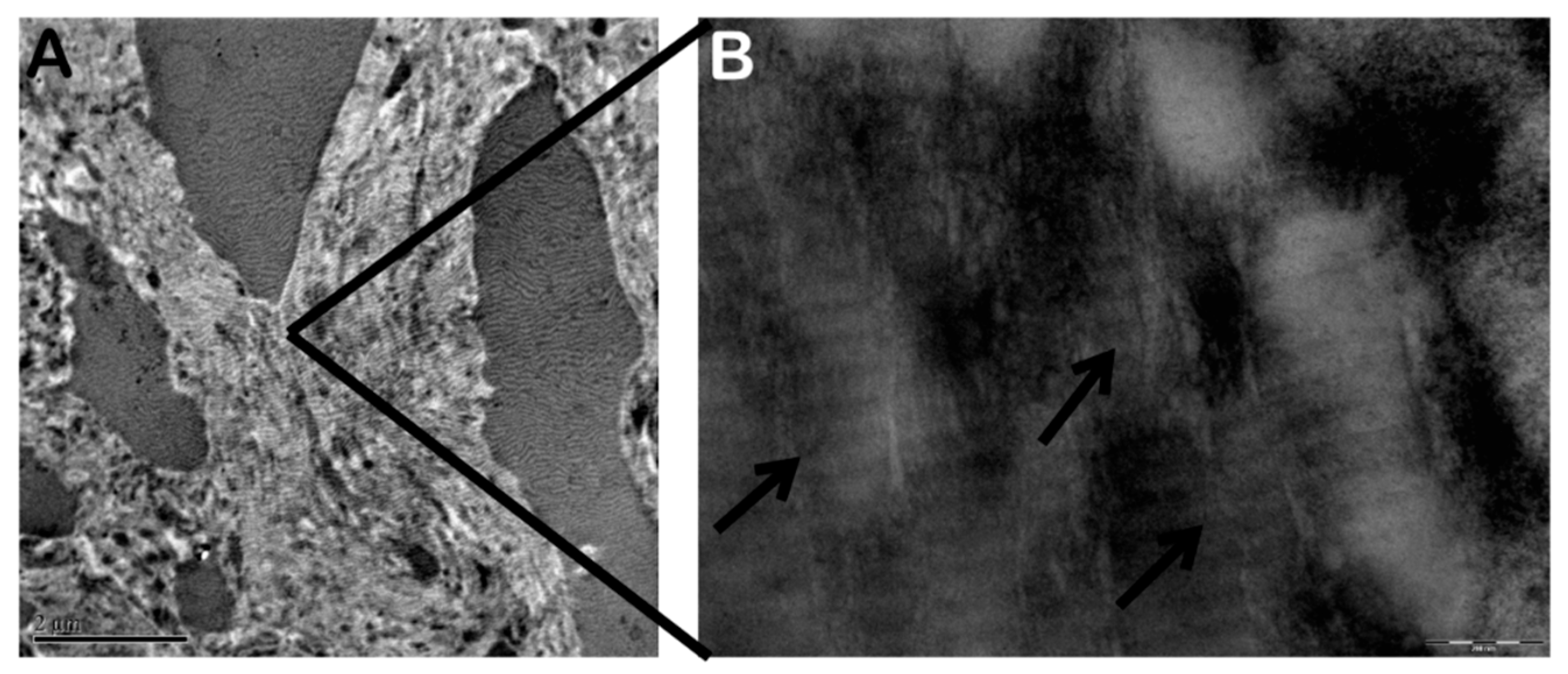
Figure 9 shows transmission electron microscopy of a type 1 collagen membrane. It is possible to observe well-defined collagen fiber bundles (indicated by the arrow).
3.7. Immunohistochemical Analysis
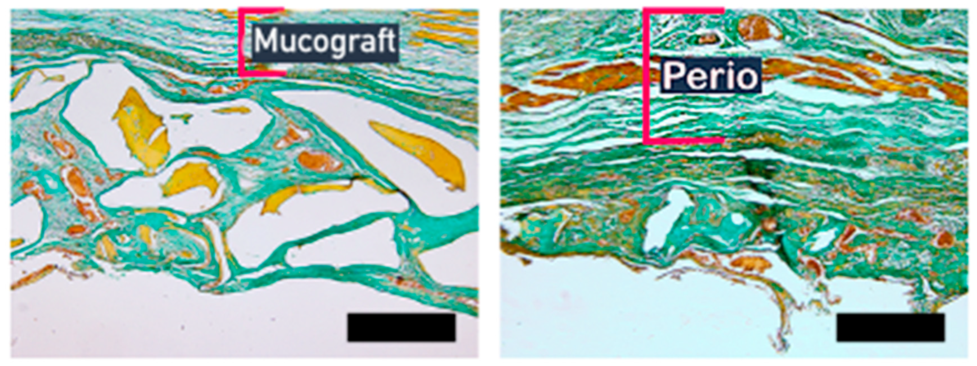
For histomorphometric analysis, the slides were stained with Goldner’s trichrome. The objective was to evaluate and quantify the formation of bone and other tissues involved in regeneration. With the staining and the different shades of color, it is possible to differentiate between mineralized bone, non-mineralized osteoid, and other cell types.
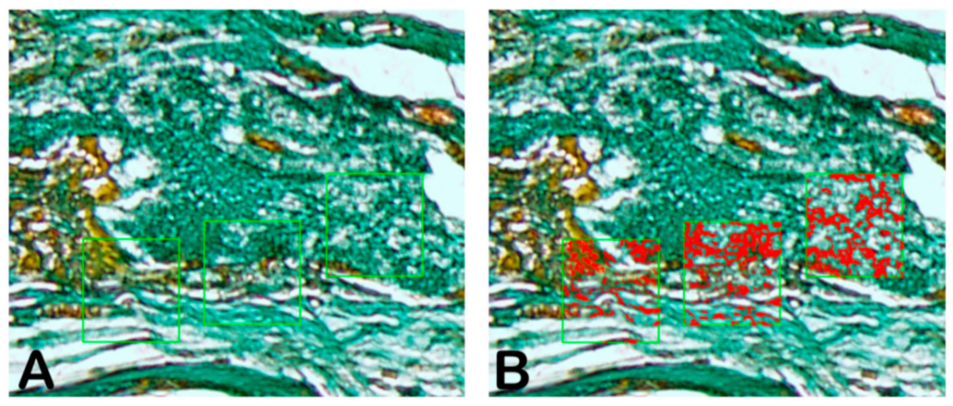
Figure 10 shows a representative green-stained area in Goldner’s trichrome. In
Figure 11, it is possible to observe newly formed collagen type 1 in the representative scheme performed by GraphPad Prism Version 8.0 (GraphPad, San Diego, CA, USA).
Figure 10 shows the areas chosen for measurement (A) and the percentage of green coloration representing newly formed collagen type 1 (B).
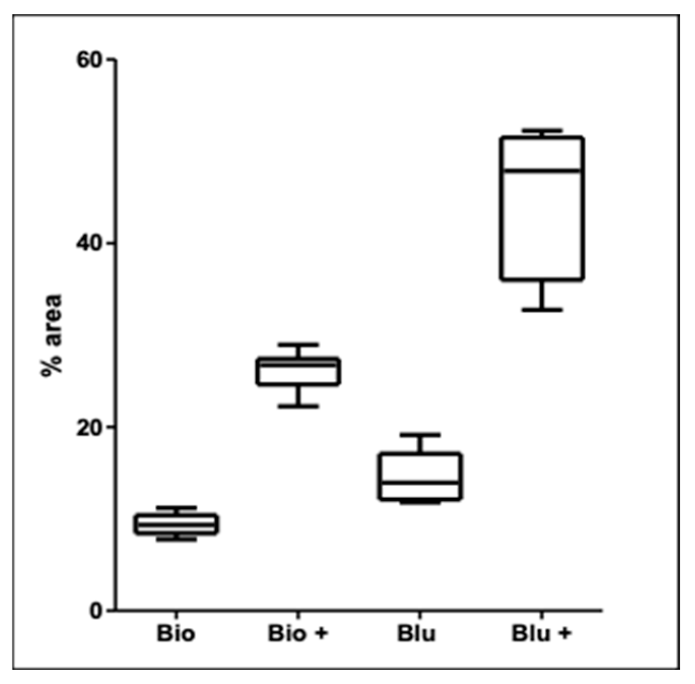
Figure 12 shows the histomorphometric analysis results using Goldner’s trichrome staining. The Blue-Bone
® bone graft particles stabilized with Green Membrane Perio
® had the highest collagen type 1 content.
Table 3 presents the statistical analysis of the differences among the groups. All groups were statistically different, including those that used membranes to improve type 1 collagen formation.
Statistical analysis indicated that using a membrane to stabilize graft particles in the surgical cavity is important for tissue regeneration. Groups in which the grafts were stabilized with a membrane showed better results. Statistical analysis indicated a significant difference in performance between the stabilized BioOss® bone graft group and the group without the Mucograft® membrane (p = 0.0313). The group in which the cavities were filled with Blue-Bone® graft and the particles stabilized with the Green Membrane Perio® showed a higher percentage of type 1 collagen than the BioOss® groups with and without the use of the Mucograft® membrane.
The statistical analysis showed differences among the groups. All groups were significantly relevant, including the group that used membranes to improve the type 1 collagen, (** = Statistical significance).
3.8. Immunohistochemical Analysis
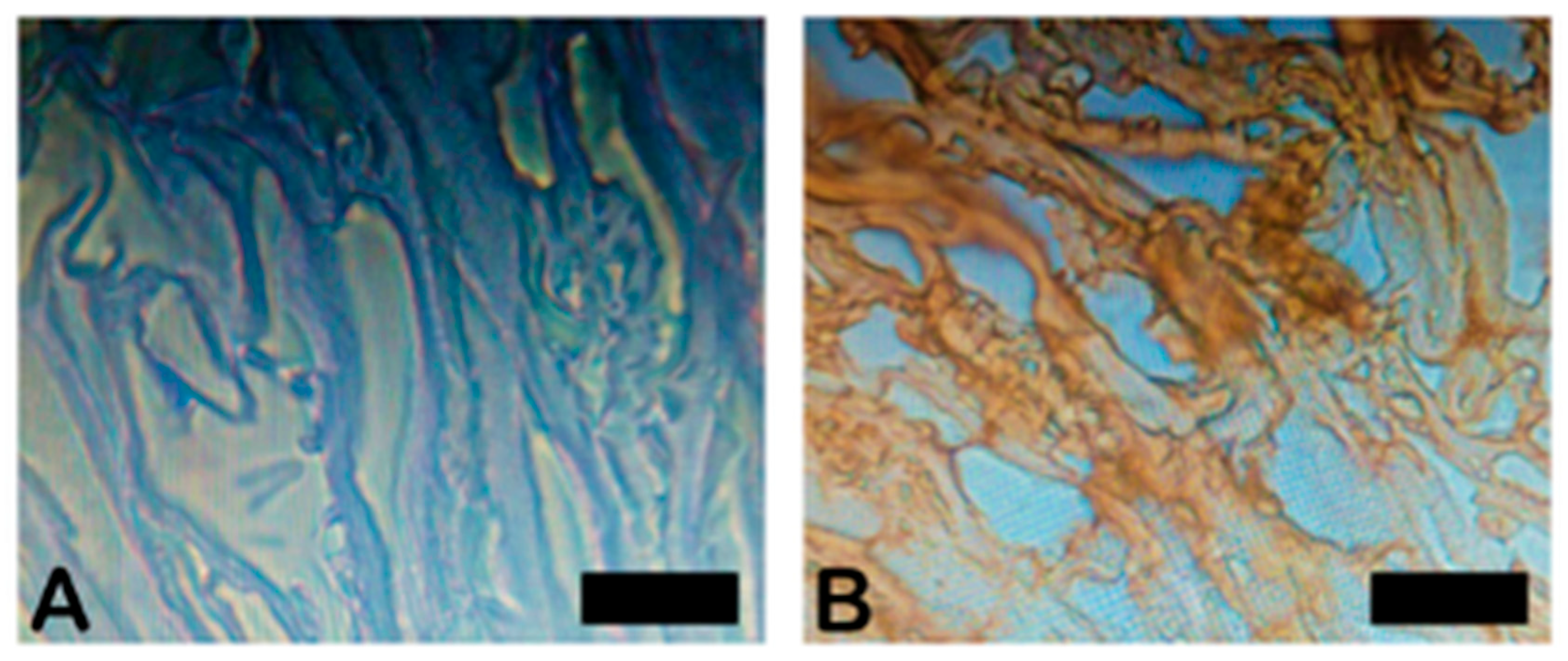
Figure 13 illustrates the morphology of type 1 collagen as determined by immunohistochemical analysis. The negative control test showed no stains, certifying the efficiency of the manufacturing process.
The present work showed that the more similar the collagen fiber pattern is to the extracellular matrix, the greater the stability of the collagen fiber [
29]. As shown by the results, a characteristic fibrillar pattern of collagen was observed.
Collagen plays a structural role within the organism. Collagens are classified into 28 distinct types, designated as types I through XXVIII. Type I collagen is by far the most abundant and structurally significant of all collagens. It is found in skin, tendons, ligaments, blood vessels, organs, and bone. Collagen can be either fibrillar (fiber-forming) or non-fibrillar [
30]. Collagen type I provides tensile strength and is the principal structural element of skin and bone. In the present work, a collagen type I membrane was used.
Membrane roughness is crucial for tissue regeneration, as it prevents the rapid growth of soft tissues in bone defects. A study evaluating macrophage activity on the surfaces of different collagen membranes found that varying topographies did not alter macrophage morphology or the release of pro- and anti-inflammatory cytokines. This result showed that the effect of surface roughness on macrophage behavior could depend on other factors [
31]. The results of the present work show a difference in roughness between the Mucograft
® and Green Membrane Perio
®. With both membranes, tissue formation of satisfactory quality occurred, starting with membranes that have characteristics that stimulate protein production, as corroborated by other researchers [
32,
33].
Histology is one of the primary methods for determining the percentage of collagen in a sample. Several studies have shown that among the stain procedures, Goldner’s trichrome and Masson’s trichrome are the two most prominent stains for assessing collagen [
34,
35]. In a study analyzing the subcutaneous implantation of a collagen membrane in 25 rats at various times, it was observed through Masson–Goldner staining of the material interaction, incorporation, and degradation until the final stage of regeneration [
36]. And we can consider the evaluation in a single time step a limitation of this study. However, the Masson–Goldner trichrome proved to be very efficient for statistical verification among the groups analyzed.




 ,
,