Review

Horizontal Nystagmus as a Coupled-Integrator Network Phenotype: A Clinical–Conceptual Framework Linking Gaze Holding, Velocity Storage, and Nodulus–Uvula Supervision


by
Leonardo Manzari
Msa Ent Academy Center, 03043 Cassino, Italy
J. Otorhinolaryngol. Hear. Balanc. Med. 2026, 7(1), 22; https://doi.org/10.3390/ohbm7010022
Submission received: 22 April 2026 / Revised: 16 June 2026 / Accepted: 17 June 2026 / Published: 22 June 2026
(This article belongs to the Section Otology and Neurotology)

Abstract

Horizontal nystagmus is still commonly interpreted at the bedside through a pragmatic peripheral-versus-central dichotomy. Although this heuristic is often clinically useful, it may be misleading because distributed brainstem–cerebellar disorders can generate peripheral-appearing phenotypes. This paper presents a narrative clinical–conceptual review proposing that a substantial subset of horizontal nystagmus patterns may be understood more coherently as expressions of dysfunction within a coupled vestibulo-ocular integrative network rather than as direct signatures of a single lesion site. Within this framework, two core dynamical domains are separated conceptually: a vestibular nuclei (VN)-centered velocity-storage process and an NPH-centered gaze-holding integrator. These processes are proposed to operate under cerebellar regulatory influence, with the nodulus–uvula (NU) acting as a plausible regulator of storage gain, temporal persistence, adaptation stability, and oscillatory behavior. Clinically, the velocity-storage domain is expressed through low-frequency vestibulo-ocular reflex behavior and optokinetic after-nystagmus-related dynamics, whereas the gaze-holding domain is expressed through eccentric gaze stability, gaze-evoked nystagmus, and post-saccadic drift. This framework carries a clinically relevant implication: horizontal nystagmus phenotypes may be interpreted more effectively by asking which functional process is predominantly abnormal—gaze holding, storage-related vestibular persistence, or cerebellar regulatory stability—rather than by relying solely on a binary peripheral–central label. On this basis, we outline a clinician-facing workflow linking gaze dependence, periodicity, direction reversals, head-shaking behavior, and Alexander-law mismatch to operational bedside criteria and candidate quantitative readouts. The proposed model is intended as a clinical–conceptual framework rather than a deterministic localization tool. Its main value lies in organizing discordant vestibular findings, strengthening the mechanistic interpretation of bedside and instrumented observations, and identifying testable directions for future validation studies in acute dizziness and ocular motor disorders.

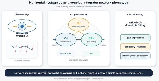
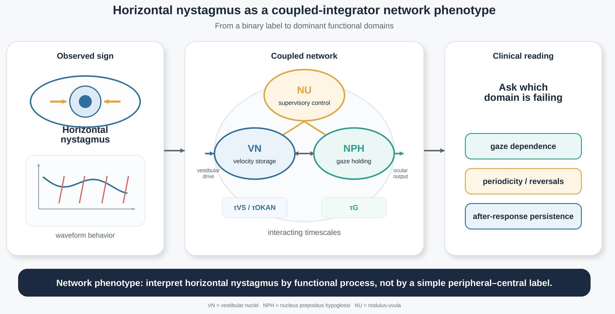
Graphical Abstract

1. Introduction

Horizontal nystagmus is one of the most common abnormal eye movement signs encountered in patients presenting with acute vertigo and dizziness. At the bedside, its interpretation often remains anchored to a practical but simplified rule: peripheral unless proven otherwise. Although this heuristic is clinically useful in many situations, it may become unreliable when distributed brainstem–cerebellar disorders generate phenotypes that resemble unilateral peripheral vestibulopathy [1,2].
One reason for this persistent diagnostic ambiguity may be that horizontal nystagmus is often interpreted primarily as a directional sign and only secondarily as the output of an integrated control system. In reality, vestibulo-ocular behavior depends on multiple interacting neural processes operating across different timescales. Among the most relevant are the mechanisms responsible for gaze holding and those that shape, prolong, and stabilize vestibular responses over time through velocity storage [3,4,5].
This paper proposes that a substantial subset of horizontal nystagmus phenotypes may be better understood as manifestations of dysfunction within a coupled-integrator network composed of at least three interacting functional domains: a vestibular nuclei-based velocity-storage process, an NPH-centered gaze-holding process, and a cerebellar regulatory domain in which the nodulus–uvula plays a key stabilizing role [6,7]. Within this framework, peripheral-appearing and central-appearing patterns are not mutually exclusive categories, but observable expressions of different dominant failures within a distributed network. The aim is to complement, not replace, established peripheral-versus-central reasoning and bedside algorithms [6,7].
The present work is not intended as a formal review of all literature on horizontal nystagmus. Rather, it is a narrative clinical-conceptual synthesis that translates selected anatomical, physiological, and lesion-based observations into a clinically testable framework [1,8]. Its purpose is not to introduce new signs, but to reorganize familiar signs within a systems-level model that distinguishes dominant dysfunction of gaze holding, velocity storage, or supervisory stability.
Horizontal nystagmus remains a frequent bedside finding in acute dizziness, yet clinicians still lack a compact framework linking known circuitry to observable waveform behavior and to practical next-step interpretation [1,3]. By shifting emphasis from a binary peripheral–central categorization to dominant dynamical domains, the proposed framework may help clinicians and investigators interpret mixed phenotypes more coherently, explain selected discordant vestibular findings, and identify which measurements are most informative when instrumented assessment is available. In this sense, its intended contribution is both conceptual and translational.

Literature Selection and Scope of the Review

This article is a narrative clinical–conceptual review rather than a systematic review. The literature was selected to support the development of a mechanistic framework linking horizontal nystagmus to vestibular nuclei-related velocity storage, NPH-mediated gaze holding, and cerebellar regulatory influence. Priority was given to anatomical, physiological, experimental, lesion-based, and clinically relevant studies addressing vestibular nuclei circuitry, gaze-holding mechanisms, velocity storage, optokinetic after-nystagmus, periodic alternating nystagmus, Alexander-law behavior, and bedside evaluation of acute vestibular syndromes [1,2,3,4,5,6,7]. The aim was not to provide exhaustive coverage of all horizontal nystagmus disorders, but to integrate key evidence into a clinically testable interpretative model. No statistical analysis was performed because no original patient dataset or quantitative meta-analysis was generated in this review.

2. Biological Basis

2.1. Vestibular Nuclei and Velocity Storage

A central premise of the proposed framework is that the vestibular nuclei provide a biologically plausible substrate for storage-related vestibulo-ocular dynamics. Rather than acting as a simple relay of canal signals, vestibular nuclei circuitry is thought to contribute to the prolongation of vestibular responses beyond the immediate mechanical behavior of the semicircular canals, thereby supporting velocity-storage behavior [4,9,10]. In clinical terms, this domain is relevant to low-frequency vestibulo-ocular reflex behavior and after-responses such as optokinetic after-nystagmus (OKAN), which together reflect the temporal persistence or “memory” of vestibular drive [4,9,10].
This interpretation is strengthened by the fact that vestibular nuclei computations are not purely canal-based, but reflect multimodal convergence, including canal–otolith interactions and context-dependent processing [9,11,12]. Such convergence supports the view that storage-related vestibular behavior is an emergent property of distributed vestibular circuitry rather than the output of a single anatomical node. Within the present model, the vestibular nuclei are therefore treated as the principal expression site of storage-related dynamics and as a major determinant of sustained vestibulo-ocular behavior.

2.2. NPH and Gaze Holding

The second major domain is related predominantly to gaze holding. This function is anchored mainly in an NPH-centered network that transforms eye-velocity commands into sustained eye-position signals required for eccentric gaze stability [3,5,13]. In physiological and clinical terms, dysfunction within this domain is expected to manifest as gaze-evoked nystagmus, post-saccadic drift, and related signs of impaired gaze stabilization [3,5,13].
Within the present framework, gaze holding is not considered in isolation from vestibular behavior. Rather, it is treated as a functionally coupled partner of storage-related processing. A bedside pattern that appears at first glance to reflect simple unilateral vestibular imbalance may therefore instead arise from dominant dysfunction within a gaze-holding process embedded in a broader vestibulo-ocular network [3,5,14]. This interpretation is consistent with the close anatomical and functional relationship between the NPH and neighboring vestibular nuclei circuitry, which provides a plausible substrate through which vestibular drive and gaze stability interact [3,5,6,13,14].

2.3. Nodulus–Uvula and Supervisory Control

Within the proposed framework, the nodulus–uvula (NU) is assigned a supervisory role. It is proposed to influence storage gain, temporal persistence, adaptation stability, and the conditions under which oscillatory behaviors emerge [8,15]. This perspective provides a biologically plausible basis for interpreting periodic alternating nystagmus (PAN), direction reversals, and other unstable phenotypes as expressions of dysregulated supervisory control rather than as fixed directional imbalances alone [8,15].
Importantly, this does not imply that the nodulus–uvula is the sole determinant of such behaviors, nor that every phenotype can be localized rigidly to a triad composed of the vestibular nuclei, NPH, and NU. Rather, the nodulus–uvula is treated here as a particularly plausible stabilizing controller within a broader distributed cerebellar–brainstem network. Its role in the present model is therefore primarily regulatory: it is invoked to explain how storage-related behavior may be tuned, stabilized, or destabilized over time, especially in oscillatory or periodic phenotypes [8,15].

2.4. Regime Dependence Across Vestibular Paradigms

A further implication of this biological framework is that different stimulus regimes probe different dynamical windows of the same coupled system. High-acceleration impulse paradigms mainly interrogate rapid direct vestibular pathways, whereas low-frequency rotations and optokinetic paradigms are more likely to reveal sustained, convergence-dependent, and storage-related behavior [10,16,17,18]. Apparent discordance across testing conditions may therefore reflect regime dependence within a coupled vestibulo-ocular system rather than inconsistency of the clinical data.
This point is clinically important because it helps explain why horizontal nystagmus phenotypes may appear contradictory when assessed with different methods. From the perspective of the present model, dissociation between rapid and sustained vestibular measures is not necessarily paradoxical. Instead, it may indicate that different domains of the same system are being sampled under different dynamical conditions [9,10,11,12,17,18].
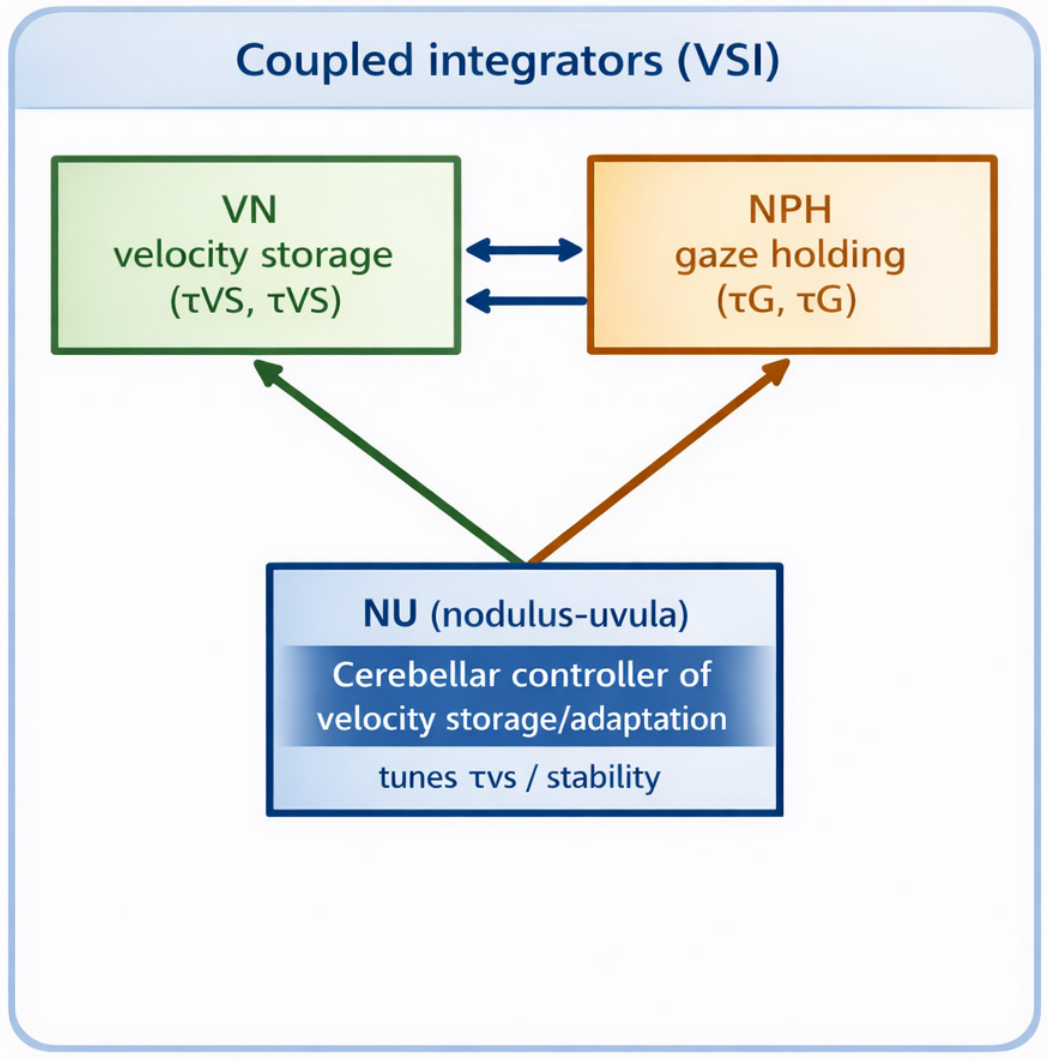
The proposed biological architecture is summarized schematically in Figure 1, which illustrates the interaction between vestibular nuclei-related velocity storage, NPH-mediated gaze holding, and nodulus–uvula supervisory control.

3. Proposed Framework

3.1. Horizontal Nystagmus as a Coupled-Integrator Network Phenotype

The present framework proposes that horizontal nystagmus may often appear “peripheral-like” despite brainstem–cerebellar involvement because it reflects the output of a coupled vestibulo-ocular control system rather than the direct signature of a single anatomical lesion [1,2]. Within this view, peripheral-appearing and central-appearing patterns are not mutually exclusive categories, but observable expressions of dominant dysfunction within interacting dynamical domains.
The model emphasizes at least three functionally relevant domains: a vestibular nuclei-related storage domain, an NPH-centered gaze-holding domain, and a cerebellar regulatory domain in which the nodulus–uvula plays a stabilizing role [3,5,6,7,8]. These domains are conceptually separable, but physiologically coupled. Their interaction is central to the interpretation proposed here, because vestibular signals reaching ocular motor output are shaped not only by peripheral drive, but also by storage-related persistence, gaze-holding stability, and higher-order regulatory influence.
In this sense, horizontal nystagmus is interpreted not simply as a directional sign, but as the observable output of a distributed control system operating across different timescales. This perspective may help explain why some phenotypes resemble unilateral peripheral vestibulopathy at the bedside even when the underlying dysfunction lies within central vestibulo-cerebellar circuitry [1,2,14].

3.2. Interacting Dynamical Domains

Within the proposed model, the storage-related and gaze-holding domains may be represented in simplified terms as coupled leaky integrators. One is dominated by storage-like behavior and influences the temporal persistence of vestibular responses; the other is dominated by gaze-holding behavior and supports the transformation of eye-velocity commands into stable eye-position signals [3,5,9,10,11,12,13]. Although analytically separable, these domains are expected to interact continuously in practice.
This interaction has direct clinical implications. The vestibular signal reaching gaze-holding circuitry is shaped by storage dynamics, while dysfunction within the gaze-holding domain may in turn distort the apparent vestibular phenotype observed clinically [3,5,14]. For this reason, a bedside pattern that initially appears to reflect simple unilateral vestibular imbalance may instead arise from dominant dysfunction within a distributed integrative network.
The model therefore encourages interpretation according to dominant dynamical behavior rather than strict lesion labels. Gaze-holding abnormalities may be inferred from waveform behavior during eccentric gaze, whereas storage-related abnormalities may be approached through low-frequency persistence, after-responses, and dissociation between rapid and sustained vestibular paradigms [1,3,10,16,17,18].

3.3. Cerebellar Regulatory Instability and Oscillatory Behavior

A distinctive feature of the present framework is the inclusion of a cerebellar regulatory domain, centered conceptually on the nodulus–uvula, to account for modulation of storage gain, adaptation stability, and oscillatory control [7,11]. Within this view, periodic alternating nystagmus (PAN), direction reversals, and related unstable phenotypes may reflect dysregulated cerebellar–brainstem tuning rather than fixed directional imbalance alone [7,11].
This interpretation does not imply that all oscillatory or periodic phenotypes can be reduced to a single cerebellar mechanism. Rather, it treats the nodulus–uvula as a biologically plausible regulatory node within a broader distributed cerebellar–brainstem system. Its inclusion in the framework is intended to explain how instability may emerge in a network already shaped by coupled storage-related and gaze-holding processes [8,15].
By incorporating supervisory instability into the model, the framework expands the interpretation of horizontal nystagmus beyond static asymmetry. Periodicity and reversals are thus approached as network behaviors that may reveal altered regulation of persistence and adaptation over time [1,8,15].

3.4. From Circuitry to Clinical Interpretation

A key implication of the proposed framework is that bedside and instrumented findings may be organized according to dominant dynamical domains rather than strict anatomical categories. In this formulation, the relevant clinical question is not only whether a pattern appears peripheral or central, but which functional domain appears to be predominantly disturbed: gaze holding, storage-related vestibular persistence, or supervisory stability [1,3,16].
This shift in emphasis may help explain selected discordant findings across bedside examination and instrumented testing. Apparent inconsistencies may become physiologically informative when interpreted as the consequence of different dominant failures within the same coupled system. The model is therefore intended not as a deterministic localization scheme, but as a clinical–conceptual framework linking circuitry, waveform behavior, and candidate next-step quantitative readouts.
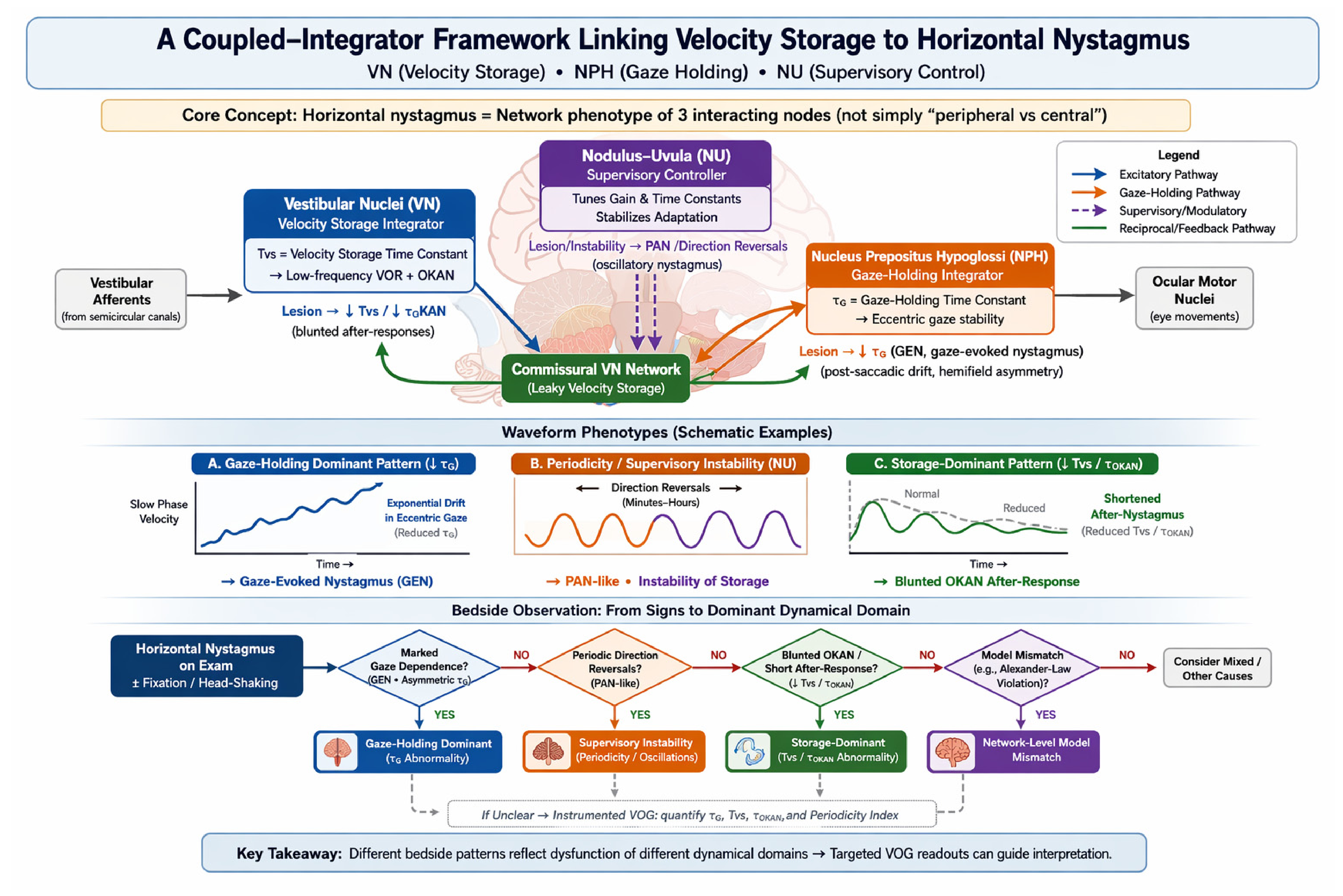
The proposed framework is summarized in Figure 2, which integrates the coupled-integrator architecture with representative waveform phenotypes and bedside-oriented interpretation.

4. Clinical Interpretation and Testable Predictions

4.1. Bedside Interpretation According to Dominant Dynamical Domains

A practical implication of the proposed framework is that horizontal nystagmus may be approached clinically by identifying the functional process that best explains the observed waveform behavior: gaze holding, storage-related vestibular persistence, or cerebellar regulatory stability [1,3,16]. This approach does not replace the established peripheral–central distinction; rather, it provides a second interpretative layer when bedside findings are mixed, evolving, or difficult to reconcile with a simple unilateral peripheral model [19,20].
Operationally, the examiner should first document whether the nystagmus is present in primary position, whether it is modified by visual fixation, whether it changes with eccentric gaze, and whether slow-phase velocity shows exponential drift, periodic reversal, or disproportionate persistence. Eccentric gaze-evoked nystagmus with post-saccadic centripetal drift suggests predominant gaze-holding dysfunction [3,5,6,13]. By contrast, reduced or abnormal low-frequency persistence and after-responses suggest storage-related dysfunction [10,17,18]. PAN-like periodicity or direction reversal suggests instability of cerebellar–brainstem regulation rather than a static vestibular asymmetry [1,8,15]. Mismatch with a simple unilateral peripheral model, including selected violations of Alexander’s law, should be treated as a warning sign that higher-order integration or feedback mechanisms may be involved [2,16].
These observations are not intended as rigid one-to-one localizing signs. They are proposed as operational bedside clues that help decide which aspect of nystagmus should be quantified next. Table 1 summarizes the main observable features, their most plausible functional interpretation, candidate bedside signs, quantitative readouts, and interpretative cautions.

4.2. Waveform Phenotypes and Clinical Pattern Recognition

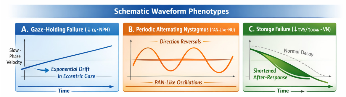
From the perspective of the proposed model, waveform behavior may provide a useful bridge between circuitry and clinical interpretation. Schematic phenotypes associated with dominant dynamical domains are illustrated in Figure 3. Gaze-holding failure is represented by eccentric gaze instability with exponential drift, consistent with a reduced gaze-holding time constant and a leaky-integrator phenotype [3,5,13]. Supervisory instability is represented by PAN-like oscillatory behavior with direction reversals, suggesting altered regulation of persistence and adaptation over time [8,15]. Storage-related deficit is represented by shortened or blunted after-responses relative to expected decay, consistent with reduced storage-related persistence [10,17,18].
These patterns are not intended to serve as fixed diagnostic templates. Instead, they illustrate the logic of matching observable waveform behavior to dominant dynamical domains. Their value lies in helping clinicians move from a purely descriptive label toward a more structured physiological interpretation when bedside signs are ambiguous or apparently contradictory.
The major schematic waveform phenotypes associated with the framework are shown in Figure 3.

4.3. Instrumented Vestibular–Ocular Assessment

Instrumented assessment provides a second and more structured way of examining the proposed framework. Routine video-oculography may be used to characterize waveform class, quantify gaze dependence, document asymmetry across gaze positions, and distinguish exponential from more linear slow-phase behavior. Within the present model, these measures are especially relevant for identifying dominant disturbance of the gaze-holding domain [3,5,13].
More specialized vestibular assessment may then be used to explore storage-related behavior. Low-frequency vestibular persistence, blunted or shortened after-responses, and dissociation between rapid impulse-based responses and more sustained vestibular paradigms would be particularly relevant findings. In this context, optokinetic after-nystagmus and related low-frequency measures are not assumed to be specific to a single anatomical level, but may still provide useful evidence for altered storage-related dynamics when interpreted in context [10,17,18,21].
Instrumented observation may also help characterize supervisory instability more precisely. Cycle duration, reversal pattern, stability across visual conditions, and the relationship between oscillatory behavior and fixation state may help determine whether a phenotype is more compatible with disturbance of supervisory regulation than with a static directional imbalance [8,15].
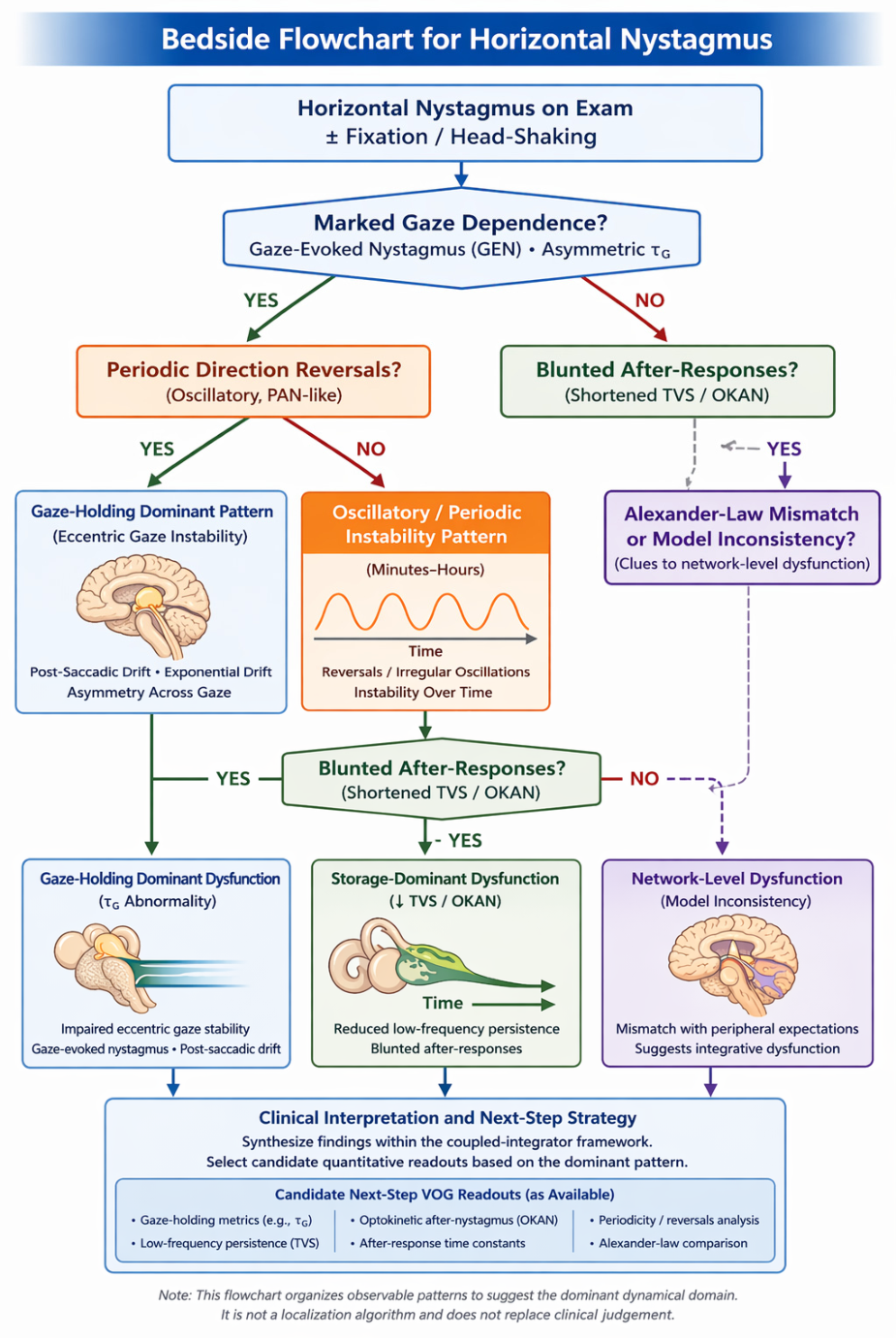
A bedside-oriented clinical pathway derived from the proposed framework is summarized in Figure 4, which links observable horizontal nystagmus patterns to dominant dynamical domains and to candidate next-step quantitative assessments.

4.4. Complementary Clinical Vestibular Tests, Including VEMPs

The proposed framework should be applied together with established bedside and laboratory vestibular tests. These tests do not map one-to-one onto the three functional domains, but they provide complementary information about sensory input, brainstem ocular motor output, and the temporal behavior of the vestibulo-ocular system. Bedside examination and video-oculography are most useful for documenting fixation effects, gaze dependence, slow-phase velocity waveform, and post-saccadic drift. The video head-impulse test mainly probes rapid high-acceleration canal-driven pathways, whereas caloric testing, low-frequency rotation, and optokinetic after-nystagmus are more suited to sustained or storage-related behavior [6,7,10,17,18,22,23].
Vestibular evoked myogenic potentials (VEMPs) deserve specific mention because they provide information that is complementary to horizontal nystagmus analysis. Cervical VEMPs and ocular VEMPs are not direct measures of horizontal gaze holding or velocity storage. Instead, they assess otolith-dependent vestibular pathways and help identify saccular, utricular, and vestibular nerve input asymmetries that may influence canal–otolith convergence at the vestibular nuclei [24,25,26]. In patients with mixed or discordant vestibular–ocular findings, VEMPs may therefore help determine whether a peripheral otolith or maculo-vestibular input imbalance is contributing to a broader network-level phenotype. They should be interpreted as contextual evidence about afferent input to the system rather than as a standalone test of the coupled-integrator model.
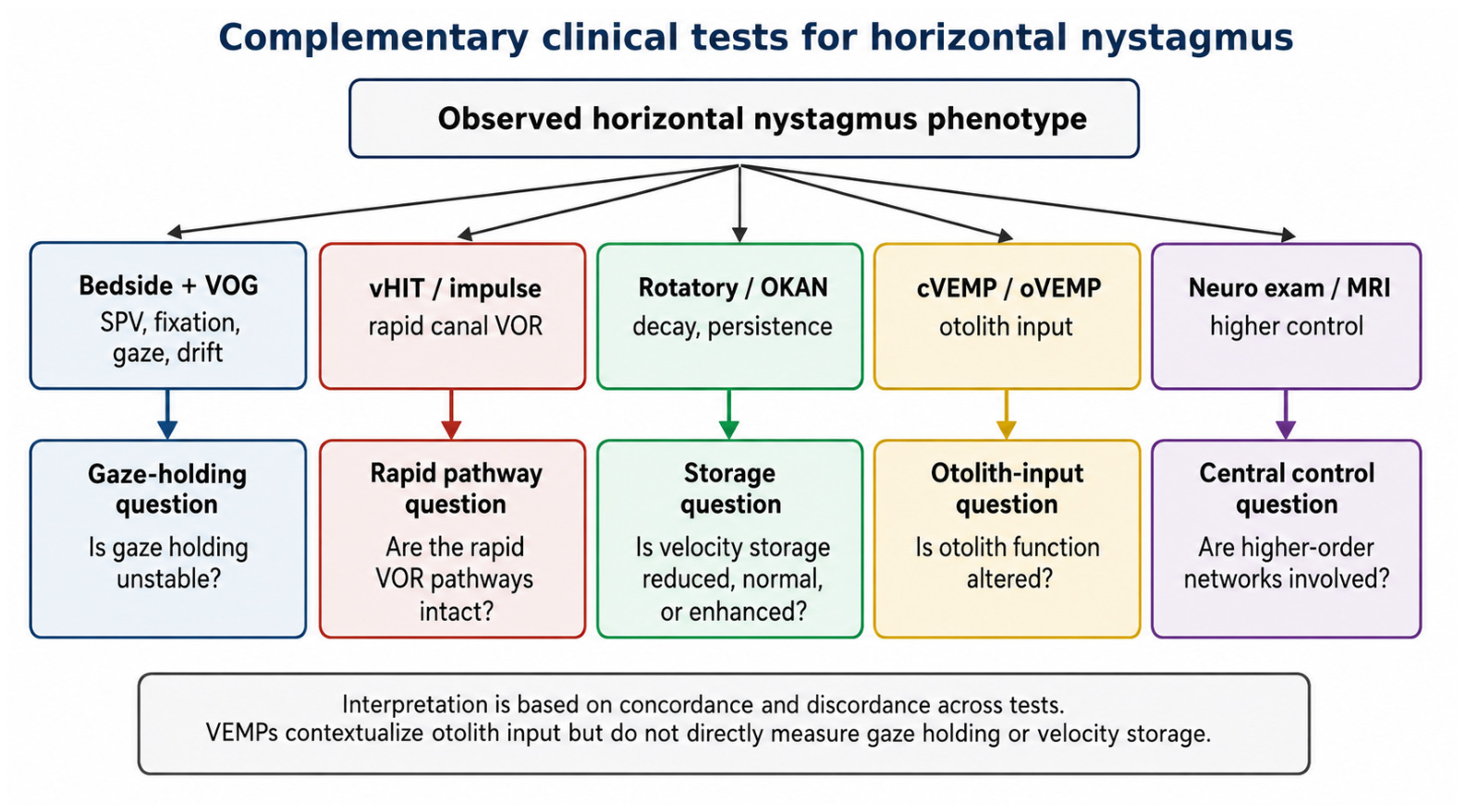
A practical test map is shown in Figure 5, summarizing how bedside examination, video-oculography, impulse testing, rotatory/optokinetic paradigms, and VEMPs can be used together to identify which functional question is most relevant in a given patient.

4.5. Testable Predictions Generated by the Framework

Several predictions generated by this framework are amenable to direct clinical and experimental evaluation. First, some patients with peripheral-appearing spontaneous horizontal nystagmus should show dominant abnormalities of gaze holding, including marked asymmetry across gaze positions and post-saccadic drift, supporting the view that gaze-holding dysfunction may masquerade as primary vestibular imbalance [6,13].
Second, some patients should show dissociation between rapid and sustained vestibular measures. In particular, relatively preserved high-frequency head-impulse performance may coexist with abnormal low-frequency persistence or impaired after-responses, suggesting that storage-related behavior can be selectively disturbed even when direct rapid vestibular pathways remain relatively intact [10,17,18,21].
Third, PAN-like behavior, direction reversals, and related oscillatory phenotypes should show patterns more consistent with instability of supervisory regulation than with static vestibular bias alone. If so, these findings would support the view that some unstable horizontal nystagmus phenotypes arise from dysregulated control of persistence and adaptation rather than from fixed peripheral asymmetry [8,15].
Fourth, mismatch with Alexander’s law should correlate more closely with integrative or feedback-domain abnormalities than with simple peripheral asymmetry measures. Within this framework, such mismatch is expected to function primarily as a clue to model insufficiency rather than as a definitive localizing sign [2,16].
Finally, the observed phenotype should vary in part according to stimulus regime. High-acceleration impulse paradigms are expected to emphasize fast direct vestibular pathways, whereas low-frequency rotational and optokinetic paradigms are expected to reveal more sustained, convergence-dependent, and storage-related behavior. Discordance across these testing conditions would therefore support the broader claim that horizontal nystagmus reflects different dynamical windows of a coupled vestibulo-ocular system rather than a single uniform mechanism [9,10,11,12,21].

4.6. Criteria That Would Support the Framework

The proposed framework is not intended to be validated by one decisive experiment, but by examining whether bedside and instrumented observations consistently cluster around the dominant dynamical domains it predicts. Support for the framework would come from reproducible dissociations between gaze-holding and storage-related behavior, from identifiable supervisory-instability phenotypes, and from systematic regime dependence across complementary methods of vestibular–ocular assessment.
Conversely, the framework would be weakened if horizontal nystagmus phenotypes failed to show any meaningful clustering around these proposed domains, if gaze-holding and storage-related abnormalities consistently overlapped without interpretable structure, or if regime dependence proved clinically uninformative across complementary testing conditions.

5. Implications, Limitations, and Future Directions

5.1. Conceptual Implications

The proposed framework suggests that horizontal nystagmus may be interpreted more usefully as a network phenotype shaped by coupled vestibulo-ocular integrators than as a simple binary sign of peripheral versus central disease. Its main conceptual contribution is the separation of dominant dynamical domains, particularly gaze-holding behavior and storage-related vestibular persistence, within a broader distributed system subject to cerebellar supervisory regulation [3,8,19]. Within this view, horizontal nystagmus is not reduced to a directional label, but is approached as the observable output of interacting processes operating across different timescales.
A major implication of this perspective is that apparently contradictory findings may no longer need to be regarded as inconsistencies. A patient may appear “peripheral” by one criterion and “central” by another not because the examination is unreliable, but because different tests are sampling different dynamical windows of the same coupled system [1,3,10]. From this standpoint, discordant findings may become physiologically informative rather than merely confusing.

5.2. Clinical Implications

If correct, this framework may reduce diagnostic anchoring in acute vertigo and ocular motor disorders by encouraging clinicians to ask a different question. Instead of asking only whether a pattern is peripheral or central, the examiner may ask which dynamical domain appears to be predominantly disturbed: gaze holding, storage-related vestibular persistence, or supervisory stability [1,2,16]. This shift in emphasis may be especially helpful when bedside signs are mixed, evolving, or difficult to reconcile.
The model is not intended to replace established emergency algorithms or neuro-otological heuristics. Rather, it is meant to complement them by offering a mechanistic interpretation of phenotypes that do not fit comfortably within standard rules [1,3]. Its usefulness may therefore be greatest in borderline cases, in apparently inconsistent vestibular datasets, and in settings where bedside observation can be combined with video-oculography or more specialized vestibular testing.
Another practical strength of the framework is its scalability. At the simplest level, it may function as a conceptual bedside aid. In more instrumented settings, it may guide waveform interpretation and the prioritization of candidate quantitative readouts. In specialized laboratories, it may provide a framework for more formal interrogation of low-frequency vestibular persistence, after-responses, and periodicity-related behavior [3,10,17,18].

5.3. Limitations of the Proposed Framework

Several limitations should be emphasized. First, gaze holding and velocity storage are emergent properties of distributed networks, and the decomposition proposed here into vestibular nuclei-related storage, NPH-centered gaze holding, and nodulus–uvula supervisory control is an operational abstraction rather than a claim of strict anatomical exclusivity [3,4,5,6,7,8]. The framework should therefore be read as a clinically useful simplification, not as a definitive lesion-localization scheme.
Second, not all candidate correlates of the framework are equally accessible in routine practice. Some observations may be appreciated clinically at the bedside, whereas others require video-oculography or specialized vestibular laboratory paradigms. In particular, storage-related after-responses and optokinetic measures are unlikely to be available uniformly across healthcare systems [17,18]. The framework is therefore intended to be scalable and context-dependent rather than universally executable in identical form.
Third, the model does not yet provide validated thresholds, normative ranges, or universally accepted extraction rules for all proposed quantitative correlates. For this reason, the present paper should not be read as a diagnostic algorithm. Rather, it should be viewed as a structured clinical–conceptual framework that identifies candidate variables for future study [3,17,18].

5.4. Beyond the Nodulus–Uvula

Although the nodulus–uvula is emphasized in this framework as a plausible regulator of storage and adaptation, it is unlikely to act alone. Other cerebellar and brainstem regions, including broader vermian, floccular, parafloccular, fastigial, and distributed ocular motor networks, almost certainly contribute to the final phenotype [1,3,8,15]. Their influence may become especially relevant in mixed cerebellar syndromes, in combined ocular motor–postural disorders, and in cases where bedside findings cannot be explained sufficiently by the simplified core architecture proposed here.
The framework also acknowledges higher-order ocular motor and vestibular influences. Cortical, thalamic, basal ganglia, brainstem reticular, vestibulo-cerebellar, visual–vestibular, pursuit, fixation-suppression, and spatial-orientation networks may influence the expression of horizontal nystagmus. These structures are not explicitly represented in the simplified core model because the purpose of the framework is clinical usability rather than exhaustive circuit mapping. Future expanded versions should incorporate these influences, particularly in degenerative, vascular, multisystem, and complex cerebellar disorders [19,20,23].
This incompleteness is intentional. The present framework is designed as a clinically useful core model rather than a comprehensive account of every cerebellar and brainstem contribution to horizontal nystagmus. Its value will depend on whether future experimental, lesion-based, and clinical studies confirm that this simplified architecture captures a meaningful part of real-world vestibulo-ocular behavior [19,20].

5.5. Priorities for Future Validation

Future work should focus on reproducibility, inter-observer agreement, and prospective clinical validation. Quantitative assessment of nystagmus will be essential for clinical translation. Particularly important variables include slow-phase velocity in primary and eccentric gaze, gaze-dependent drift slope, post-saccadic drift metrics, fixation-suppression effects, head-shaking response, VEMP amplitude and latency/asymmetry patterns, low-frequency vestibular persistence, OKAN time constants, rotatory-chair decay measures, periodicity measures, and reversal dynamics. Future studies should determine whether these variables can be extracted reliably across platforms and operators, whether storage-related and otolith-input phenotypes can be measured consistently in real clinical settings, and whether the proposed functional domains correlate meaningfully with lesion-defined syndromes, longitudinal compensation, and mixed vestibular–ocular motor phenotypes [6,14,17,18,19,24,25,26].
More broadly, the framework will be most useful if it helps generate a structured research agenda linking circuitry, waveform analysis, and clinically relevant phenotypes. Its success will depend not only on theoretical plausibility, but also on whether it improves interpretation of real patients across bedside, video-oculographic, and laboratory-based settings. Establishing normative ranges, clinically meaningful thresholds, and minimal bedside decision rules will be necessary before the framework can be applied as a practical clinical tool.

6. Conclusions

Horizontal nystagmus is commonly interpreted through a peripheral-versus-central dichotomy, but this distinction may be insufficient for a substantial subset of clinically relevant phenotypes. The present paper proposes that selected horizontal nystagmus patterns may be understood more coherently as the output of a coupled vestibulo-ocular network involving gaze holding, storage-related vestibular persistence, and cerebellar regulatory influence. Within this framework, peripheral-appearing and central-appearing patterns are not mutually exclusive categories, but observable expressions of dominant dysfunction within interacting functional processes.
By shifting emphasis from lesion labels alone to observable network behavior, the proposed framework offers a mechanistic interpretation of apparently discordant bedside and instrumented findings. Its value lies not in defining a rigid localization scheme, but in providing a biologically plausible, clinically scalable, and testable way of organizing horizontal nystagmus phenotypes across different observational settings.
If future clinical and instrumented studies support this view, the framework may help reduce diagnostic anchoring, clarify mixed vestibular–ocular phenotypes, and provide a more explicit bridge between vestibular physiology and ocular motor interpretation.

Funding

This research received no external funding.

Institutional Review Board Statement

Not applicable.

Informed Consent Statement

Not applicable.

Data Availability Statement

No new data were created or analyzed in this study. Data sharing is not applicable to this article.

Acknowledgments

The author gratefully acknowledges the anonymous reviewers for their constructive comments and valuable suggestions, which contributed to improving the clarity and balance of the manuscript. During the preparation of this work, the author used ChatGPT (OpenAI, San Francisco, CA, USA; GPT-5.5 Thinking) for drafting support, organization of the review, and English-language editing. All scientific judgement and evaluation were performed independently by the author. After using this tool, the author reviewed and edited the content as needed, and takes full responsibility for the content of the published article.

Conflicts of Interest

The author declares no conflicts of interest.

Abbreviations

The following abbreviations are used in this manuscript:
GENgaze-evoked nystagmus;
NPHnucleus prepositus hypoglossi;
NUnodulus–uvula;
OKANoptokinetic after-nystagmus;
PANperiodic alternating nystagmus;
VNvestibular nuclei;
VOGvideo-oculography;
VORvestibulo-ocular reflex;
τGgaze-holding time constant;
τOKANoptokinetic after-nystagmus time constant;
τVSvelocity-storage time constant.

References

  1. Leigh, R.J.; Zee, D.S. Diagnosis of nystagmus and saccadic intrusions. In The Neurology of Eye Movements, 5th ed.; Oxford University Press: New York, NY, USA, 2015; pp. 657–768. [Google Scholar] [CrossRef] [Scilit]
  2. Choi, J.H.; Oh, E.H.; Kim, H.S.; Park, J.Y.; Lee, S.M.; Choi, S.Y.; Kim, H.J.; Choi, J.Y.; Kim, J.S.; Otero-Millan, J.; et al. Spontaneous nystagmus violating Alexander’s law: Neural substrates and mechanisms. Cerebellum 2025, 24, 34. [Google Scholar] [CrossRef] [Scilit] [PubMed]
  3. Leigh, R.J.; Zee, D.S. Gaze holding and the neural integrator. In The Neurology of Eye Movements, 5th ed.; Oxford University Press: New York, NY, USA, 2015; pp. 360–385. [Google Scholar] [CrossRef] [Scilit]
  4. Highstein, S.M.; Holstein, G.R. The anatomy of the vestibular nuclei. Prog. Brain Res. 2006, 151, 157–203. [Google Scholar] [CrossRef] [Scilit] [PubMed]
  5. McCrea, R.A.; Horn, A.K.E. Nucleus prepositus. Prog. Brain Res. 2006, 151, 205–230. [Google Scholar] [CrossRef] [Scilit] [PubMed]
  6. Eggers, S.D.Z.; Bisdorff, A.; von Brevern, M.; Zee, D.S.; Kim, J.S.; Perez-Fernandez, N.; Welgampola, M.S.; Della Santina, C.C.; Newman-Toker, D.E. Classification of vestibular signs and examination techniques: Nystagmus and nystagmus-like movements. J. Vestib. Res. 2019, 29, 57–87. [Google Scholar] [CrossRef] [Scilit] [PubMed]
  7. Strupp, M.; Bisdorff, A.; Furman, J.; Hornibrook, J.; Jahn, K.; Maire, R.; Newman-Toker, D.; Magnusson, M. Acute unilateral vestibulopathy/vestibular neuritis: Diagnostic criteria. J. Vestib. Res. 2022, 32, 389–406. [Google Scholar] [CrossRef] [Scilit] [PubMed]
  8. Waespe, W.; Cohen, B.; Raphan, T. Dynamic modification of the vestibulo-ocular reflex by the nodulus and uvula. Science 1985, 228, 199–202. [Google Scholar] [CrossRef] [Scilit] [PubMed]
  9. Curthoys, I.S.; Markham, C.H. Convergence of labyrinthine influences on units in the vestibular nuclei of the cat. I. Natural stimulation. Brain Res. 1971, 35, 469–490. [Google Scholar] [CrossRef] [Scilit] [PubMed]
  10. Fetter, M.; Zee, D.S. Recovery from unilateral labyrinthectomy in rhesus monkey. J. Neurophysiol. 1988, 59, 370–393. [Google Scholar] [CrossRef] [Scilit] [PubMed]
  11. Dickman, J.D.; Angelaki, D.E. Vestibular convergence patterns in vestibular nuclei neurons of alert primates. J. Neurophysiol. 2002, 88, 3518–3533. [Google Scholar] [CrossRef] [Scilit] [PubMed]
  12. Zhou, W.; Tang, B.F.; Newlands, S.D.; King, W.M. Responses of monkey vestibular-only neurons to translation and angular rotation. J. Neurophysiol. 2006, 96, 2915–2930. [Google Scholar] [CrossRef] [Scilit] [PubMed]
  13. Dale, A.; Cullen, K.E. The nucleus prepositus predominantly outputs eye movement-related information during passive and active self-motion. J. Neurophysiol. 2013, 109, 1900–1911. [Google Scholar] [CrossRef] [Scilit] [PubMed]
  14. Kim, S.H.; Zee, D.S.; du Lac, S.; Kim, H.J.; Kim, J.S. Nucleus prepositus hypoglossi lesions produce a unique ocular motor syndrome. Neurology 2016, 87, 2026–2033. [Google Scholar] [CrossRef] [Scilit] [PubMed]
  15. Halmagyi, G.M.; Rudge, P.; Gresty, M.A.; Leigh, R.J.; Zee, D.S. Treatment of periodic alternating nystagmus. Ann. Neurol. 1980, 8, 609–611. [Google Scholar] [CrossRef] [Scilit] [PubMed]
  16. Robinson, D.A.; Zee, D.S.; Hain, T.C.; Holmes, A.; Rosenberg, L.F. Alexander’s law: Its behavior and origin in the human vestibulo-ocular reflex. Ann. Neurol. 1984, 16, 714–722. [Google Scholar] [CrossRef] [Scilit] [PubMed]
  17. Ireland, D.J.; Jell, R.M. Optokinetic after-nystagmus in man after loss or reduction of labyrinthine function: A preliminary report. J. Otolaryngol. 1982, 11, 86–90. [Google Scholar] [PubMed]
  18. Hain, T.C.; Mattox, D.; Herdman, S.J.; Zee, D.S.; Holliday, M.; Byskosh, A.T. Localizing value of optokinetic afternystagmus. Ann. Otol. Rhinol. Laryngol. 1994, 103, 806–811. [Google Scholar] [CrossRef] [Scilit] [PubMed]
  19. Shaikh, A.G.; Zee, D.S. Why brains behave differently: Insights from eye movement disorders. Exp. Eye Res. 2025, 261, 110643. [Google Scholar] [CrossRef] [Scilit] [PubMed]
  20. Dieterich, M.; Glasauer, S.; Brandt, T. Why acute unilateral vestibular midbrain lesions rarely manifest with rotational vertigo: A clinical and modeling approach to head direction cell function. J. Neurol. 2018, 265, 1184–1198. [Google Scholar] [CrossRef] [Scilit] [PubMed]
  21. Manzari, L. Multiscale integration of acceleration and jerk sensing in the vestibular system. Audiol. Res. 2026, 16, 21. [Google Scholar] [CrossRef] [Scilit] [PubMed]
  22. Tarnutzer, A.A.; Edlow, J.A. Bedside testing in acute vestibular syndrome: Evaluating HINTS Plus and beyond—A critical review. Audiol. Res. 2023, 13, 670–685. [Google Scholar] [CrossRef] [Scilit] [PubMed]
  23. Cullen, K.E. Internal models of self-motion: Neural computations by the vestibular cerebellum. Trends Neurosci. 2023, 47, 85–86. [Google Scholar] [CrossRef] [Scilit]
  24. Curthoys, I.S.; Vulovic, V.; Burgess, A.M.; Manzari, L.; Sokolic, L.; Pogson, J.; Robins, M.; Mezey, L.E.; Goonetilleke, S.; Cornell, E.D.; et al. Neural basis of new clinical vestibular tests: Otolithic neural responses to sound and vibration. Clin. Exp. Pharmacol. Physiol. 2014, 41, 371–380. [Google Scholar] [CrossRef] [Scilit] [PubMed]
  25. Curthoys, I.S.; Grant, J.W.; Burgess, A.M.; Pastras, C.J.; Brown, D.J.; Manzari, L. Otolithic receptor mechanisms for vestibular-evoked myogenic potentials: A review. Front. Neurol. 2018, 9, 366. [Google Scholar] [CrossRef] [Scilit] [PubMed]
  26. Rosengren, S.M.; Colebatch, J.G.; Young, A.S.; Govender, S.; Welgampola, M.S. Vestibular evoked myogenic potentials in practice: Methods, pitfalls and clinical applications. Clin. Neurophysiol. Pract. 2019, 4, 47–68. [Google Scholar] [CrossRef] [Scilit] [PubMed]
Figure 1. Minimal conceptual architecture of the coupled-integrator model. Simplified schematic of the proposed vestibulo-ocular network framework. The vestibular nuclei (VN) are represented as the principal expression site of velocity-storage behavior, characterized by storage-related time constants such as τVS and τOKAN. The nucleus prepositus hypoglossi (NPH) is represented as the principal contributor to the gaze-holding integrator, characterized by the gaze-holding time constant τG. Bidirectional coupling between VN and NPH indicates reciprocal brainstem interactions linking vestibular drive to gaze stability. The nodulus–uvula (NU) is depicted as a cerebellar supervisory module that modulates storage gain, temporal persistence, and adaptation stability through closed-loop interactions with brainstem circuitry. Abbreviations: VN, vestibular nuclei; NPH, nucleus prepositus hypoglossi; NU, nodulus–uvula; τVS, velocity-storage time constant; τOKAN, optokinetic after-nystagmus time constant; τG, gaze-holding time constant.
Ohbm 07 00022 g001
Figure 2. Integrated coupled-integrator framework linking velocity storage to horizontal nystagmus. Clinician-facing schematic integrating three interacting domains that shape horizontal nystagmus as a network phenotype rather than a simple peripheral–central label. The vestibular nuclei (VN), including commissural vestibular circuitry, are represented as the storage-related domain and influence storage gain and time constants relevant to low-frequency vestibulo-ocular reflex behavior and optokinetic after-responses. The nucleus prepositus hypoglossi (NPH) is represented as the gaze-holding domain, with gaze stability depending on the gaze-holding time constant τG; dysfunction within this domain is expected to produce gaze-evoked nystagmus, post-saccadic drift, and gaze asymmetry. The nodulus–uvula (NU) is represented as a supervisory regulatory domain influencing storage gain, adaptation, and oscillatory stability; instability within this domain may manifest as periodic alternating nystagmus (PAN) or direction reversals. The middle panel illustrates schematic slow-phase phenotypes corresponding to dominant disturbance of gaze holding, supervisory stability, or storage-related behavior. The lower panel presents a conceptual bedside workflow linking gaze dependence, periodicity, after-response abnormalities, and Alexander-law mismatch to dominant dynamical domains and candidate next-step quantitative readouts. Abbreviations: VN, vestibular nuclei; NPH, nucleus prepositus hypoglossi; NU, nodulus–uvula; VOR, vestibulo-ocular reflex; PAN, periodic alternating nystagmus; OKAN, optokinetic after-nystagmus; τG, gaze-holding time constant; τVS, velocity-storage time constant; τOKAN, OKAN time constant.
Ohbm 07 00022 g002
Figure 3. Schematic waveform phenotypes associated with dominant dynamical domains. Three conceptual slow-phase velocity patterns illustrate separable dynamical signatures within the proposed coupled-integrator framework. (A) Gaze-holding failure: exponential drift during eccentric gaze, consistent with a reduced gaze-holding time constant (τG) and gaze-evoked nystagmus. (B) Supervisory instability: PAN-like oscillations with direction reversals, consistent with unstable regulation of velocity storage and adaptation and describable by periodicity-related measures such as cycle duration and reversal slope. (C) Velocity-storage deficit: shortened after-response relative to normal decay (dashed trace), consistent with reduced storage-related time constants (τVS and τOKAN) and blunted after-nystagmus. The traces are schematic and are intended to illustrate the logic of matching waveform behavior to dominant dynamical domains rather than to define diagnostic thresholds. Abbreviations: NPH, nucleus prepositus hypoglossi; NU, nodulus–uvula; VN, vestibular nuclei; PAN, periodic alternating nystagmus; τG, gaze-holding time constant; τVS, velocity-storage time constant; τOKAN, optokinetic after-nystagmus time constant.
Ohbm 07 00022 g003
Figure 4. Bedside flowchart for framework-guided interpretation of horizontal nystagmus. Conceptual clinical pathway beginning with horizontal nystagmus observed in the primary position, with or without fixation. Early branches assess gaze dependence, gaze-evoked nystagmus, exponential drift, periodicity, and direction reversals. Additional branches incorporate storage-related after-response abnormalities and mismatch with a simple unilateral peripheral model, including Alexander-law inconsistency. The flowchart is intended to organize observable phenotypes according to dominant network behavior and to suggest candidate next-step quantitative assessments when instrumented testing is available. It is not intended as a rigid lesion-localization algorithm. Abbreviations: VN, vestibular nuclei; NPH, nucleus prepositus hypoglossi; NU, nodulus–uvula; PAN, periodic alternating nystagmus; VOG, video-oculography; τG, gaze-holding time constant; τVS, velocity-storage time constant; τOKAN, optokinetic after-nystagmus time constant.
Ohbm 07 00022 g004
Figure 5. Complementary vestibular and ocular motor tests for applying the framework. The diagram summarizes how bedside examination, video-oculography (VOG), vHIT, rotatory/optokinetic measures, vestibular evoked myogenic potentials (VEMPs), and neurologic assessment may be used to interrogate different clinical questions within the proposed framework. VEMPs do not directly measure horizontal nystagmus, gaze holding, or velocity storage; rather, cVEMPs and oVEMPs provide information about otolith-dependent vestibular pathways and help contextualize peripheral otolith input to the vestibular nuclei. Abbreviations: cVEMP, cervical vestibular evoked myogenic potential; oVEMP, ocular vestibular evoked myogenic potential; OKAN, optokinetic after-nystagmus; SPV, slow-phase velocity; VEMPs, vestibular evoked myogenic potentials; vHIT, video head-impulse test; VOG, video-oculography.
Ohbm 07 00022 g005
Table 1. Operational bedside and laboratory correlates of the proposed framework.
Observable FeatureSuggested Dominant ProcessAccessible Bedside SignCandidate Quantitative ReadoutInterpretative Caution
Gaze-evoked nystagmus with exponential centripetal driftGaze-holding dysfunctionEccentric gaze instability; post-saccadic driftSlow-phase velocity slope; gaze-position dependence; post-saccadic drift metrics on VOGNot specific for an isolated NPH lesion; may reflect broader ocular motor dysfunction
Reduced or shortened after-response/poor OKANStorage-related vestibular persistenceReduced persistence after optokinetic or rotatory stimulation when availableOKAN time constant; low-frequency VOR time constant; rotatory-chair decay measuresUsually requires instrumented laboratory assessment; not a single-level localizing sign
PAN-like periodicity or direction reversalCerebellar–brainstem regulatory instabilityReversal of nystagmus direction over time; oscillatory behaviorCycle duration; reversal slope; fixation dependence; slow-phase velocity time seriesMay involve nodulus–uvula, floccular, vermian, fastigial, or broader brainstem–cerebellar networks
Mismatch with a simple unilateral peripheral modelHigher-order integration or feedback-domain dysfunctionAlexander-law inconsistency; unexpected gaze dependence; discordant fixation effectsComparison of slow-phase velocity across gaze positions and fixation conditionsShould trigger broader central and network-level interpretation, not a standalone diagnosis
Dissociation between rapid and sustained vestibular paradigmsRegime dependence of the coupled systemRelatively preserved impulse behavior with abnormal sustained behaviorvHIT gain vs. low-frequency rotation, OKAN, or after-response metricsDifferent tests probe different dynamical windows; apparent discordance may be physiologically informative
Disclaimer/Publisher’s Note: The statements, opinions and data contained in all publications are solely those of the individual author(s) and contributor(s) and not of MDPI and/or the editor(s). MDPI and/or the editor(s) disclaim responsibility for any injury to people or property resulting from any ideas, methods, instructions or products referred to in the content.