Proceeding Paper

MEMS Biomimetic Superficial and Canal Neuromasts for Flow Sensing †

by
Ajay Giri Prakash Kottapalli
1,2,*,
Debarun Sengupta
1,
Elgar Kanhere
2,
Meghali Bora
2,
Jianmin Miao
3 and
Michael Triantafyllou
4
1
Advanced Production Engineering (APE) Department, Engineering and Technology Institute Groningen (ENTEG), University of Groningen, 9747 AG Groningen, The Netherlands
2
Center for Environmental Sensing and Modeling (CENSAM) IRG Singapore-MIT Alliance for Research and Technology (SMART), Singapore 138602, Singapore
3
School of Mechanical and Aerospace Engineering, Nanyang Technological University, Singapore 639798, Singapore
4
MIT Sea Grant College Program, Massachusetts Institute of Technology (MIT), Cambridge, MA 02139, USA
*
Author to whom correspondence should be addressed.
Presented at the Eurosensors 2018 Conference, Graz, Austria, 9–12 September 2018.
Proceedings 2018, 2(13), 1044; https://doi.org/10.3390/proceedings2131044
Published: 12 November 2018
(This article belongs to the Proceedings of EUROSENSORS 2018)

Abstract

:
Neuromast sensors found in fishes detect flows around their body and are known to generate hydrodynamic awareness. This work reports the development of superficial neuromast (SN) and canal neuromast (CN) inspired MEMS flow sensors. The MEMS artificial neuromast sensors mimic the key features of the SN and the CN such as the shape and the mechanical properties of the cupula. In case of both SN and CN MEMS sensors, hydrogels with varying initiator concentrations were used to replicate the Young’s modulus of the biological cupula. The MEMS SN and CN sensors were experimentally characterized in steady-state and oscillatory flows respectively.

1. Introduction

The cupula is a naturally occurring polymer-like material that extends from the skin of the fish into the surrounding flows and interacts with the flows through a drag force based sensing mechanism [1]. Most flow sensors developed in the past through inspiration from a biological neuromast mimicked the cupula as a solid cylindrical structure that extends into the flow [2,3].
This work reports the design and fabrication of MEMS steady-state and oscillatory flow sensors that are inspired by the superficial neuromast (SN) and canal neuromast (CN) sensors in fishes respectively. The MEMS sensors that are developed in this work attempt to mimic the structural, material and functional properties of both these neuromast sensors. The SN inspired MEMS sensors feature liquid crystal polymer membranes with embedded strain gauges and measure the steadystate flow velocities. The CN inspired MEMS sensors feature Pb(Zr0.52Ti0.48)O3 PZT membranes as sensing elements and measure oscillatory flows. Methacrylic anhydride modified Hyaulronic acid hydrogel is used to form the artificial cupula for both the SN and CN inspired MEMS sensors. However, the Young’s modulus of the HA-MA hydrogel cupula was matched with the biological cupula (10 Pa in case of SN and 10 kPa in case of CN) by regulating the initiator concentration.

2. Experimental

2.1. Biomimetic MEMS SN Sensors

In case of the MEMS SN sensor, the principal sensing element is a patterned serpentine gold strain gauge on a liquid crystal polymer(LCP) layer aligned and bonded to a silicon bottom substrate (with a cavity formed by deep reactive ion etching (DRIE)) by means of intermediate SU-8 adhesion layer. The sensor fabrication methodology is explained in detail in [4]. A polycarbonate hair cell structure formed by setereolithography, is mounted at the center of the patterned LCP membrane (Figure 1a). Electrospinning is carried out on the assembly to form the nanofibril scaffold (Figure 1c). In order to mimic the soft cupular material of 10 Pa, 2% HA-MA with 0.1% I2959 initiator is used followed by a ten minute exposure to UV at 365 nm. Nanoindentation and rheology techniques were used to determine the Young’s modulus and hardness of the artificial cupula material. The synthesized hydrogel is drop casted (prolate spheroid shaped) on the electrospun nanofibril scaffold finally to mimic the cupula found in SNs (Figure 1d,e).

2.2. Biomimetic MEMS CN Sensors

In case of the CN-inspired MEMS sensor, the sensing is done by a Pb(Zr0.52Ti0.48)O3 PZT membrane with a high aspect ratio rigid cylindrical polycarbonate pillar (Figure 2a) [5]. In order to achieve a stiffer cupula of 10 kPa (determined by Rheology and Nanoindentation techniques), 8% HA-MA with a 1% I2959 initiator concentration was prepared and exposed to UV for 10 min. Like in case of the previous SN-inspired MEMS sensor, for this device the high aspect ratio artificial hair cell pillar drop-casted (in a hemispherical shape) with the harder hydrogel acts as the main structural element which interacts with the flow (Figure 2b,c).

3. Results and Discussions

One of the key differences between SNs and CNs is in the shape of their cupula. The cupula in SN has a prolate spheroid shape whereas the one found in CN has a hemispherical shape. This work exploits this particular structural difference between the two neuromast sensors to demonstrate two biomimetic sensors capable of detecting steady state flows and oscillatory flows.

3.1. Steady-State Flow Sensing Using the Biomimetic MEMS SN Sensors

The SN sensor is subjected to a wind-tunnel test to demonstrate its capability of detecting steady state flows. The sensor is placed in the test section of the wind-tunnel and tested over a flow velocity range of 0–9 m/s. The Figure 3a shows the schematic representation of the experimental set up. The variable resistance output from the sensor is connected to an external Wheatstone bridge circuit configuration and the voltage output from the bridge circuit is recorded using a National Instruments data acquisition setup with LabView software. The experiment is repeated over the aforementioned velocity range for five times and the results are plotted (Figure 3).

3.2. Oscillatory Flow Sensing Using the Biomimetic MEMS CN Sensors

The natural CNs are known to detect oscillatory flows pattern generated around swimming fishes. The fabricated biomimetic MEMS CN with the cupula was subjected to an oscillatory flow using a dipole stimulus. A vibrating sphere or dipole (16 mm in diameter) connected to a permanent magnet mini-shaker (model 4810, Brüel & Kjær, Minden, NV, USA) by means of a 16 cm long rod (4 mm in diameter) is used for generation of oscillatory flow. Figure 4a shows the schematic representation of the experimental set up. When the dipole is vibrated at a particular frequency, the water in its vicinity starts vibrating at the same frequency which leads to induction of drag force on the cupula causing it to vibrate at the same frequency. The sensor is subjected to three different dipole stimulus frequencies (5 Hz, 10 Hz, and 35 Hz) and the output for each case is recorded individually (Figure 4b).

4. Conclusions

In this work, the design, fabrication, and testing of MEMS steady-state and oscillatory flow sensors that are inspired by the SN and CN sensors in fishes are reported. The MEMS sensors that are developed in this work attempt to mimic the structural, material and functional properties of both these neuromast sensors. The SN inspired MEMS sensors featuring liquid crystal polymer membranes with embedded strain gauges are demonstrated to measure the steady-state flow velocities (over a range of 0–9 m/s) successfully. Similarly, the CN inspired MEMS sensors featuring Pb(Zr0.52Ti0.48)O3 PZT membranes as sensing elements are demonstrated to measure oscillatory flows for three different dipole stimulus frequencies. This work successfully demonstrates the application of biomimetic MEMS SN and CN sensors for flow sensing applications.

Acknowledgments

This research is supported by the National Research Foundation Singapore under its Campus for Research Excellence and Technological Enterprise programme.

Conflicts of Interest

The authors declare no conflict of interest.

References

  1. Peleshanko, S.; Julian, M.D.; Ornatska, M.; McConney, M.E.; LeMieux, M.C.; Chen, N.; Tucker, C.; Yang, Y.; Liu, C.; Humphrey, J.A.C.; et al. Hydrogel-encapsulated microfabricated haircells mimicking fish cupula neuromast. Adv. Mater. 2007, 19, 2903–2909. [Google Scholar] [CrossRef]
  2. Prakash Kottapalli, A.G.; Asadnia, M.; Miao, J.; Triantafyllou, M. Touch at a distance sensing: Lateralline inspired MEMS flow sensors. Bioinspir. Biomim. 2014, 9, 046011. [Google Scholar] [CrossRef] [PubMed]
  3. Chen, N.; Tucker, C.; Engel, J.M.; Yang, Y.; Pandya, S.; Liu, C. Design and characterization of artificial haircell sensor for flow sensing with ultrahigh velocity and angular sensitivity. J. Microelectromech. Syst. 2007, 16, 999–1014. [Google Scholar] [CrossRef]
  4. Kottapalli, A.G.P.; Bora, M.; Asadnia, M.; Miao, J.; Venkatraman, S.S.; Triantafyllou, M. Nanofibril scaffold assisted MEMS artificial hydrogel neuromasts for enhanced sensitivity flow sensing. Sci. Rep. 2016, 6, 19336. [Google Scholar] [CrossRef] [PubMed]
  5. Bora, M.; Kottapalli, A.G.P.; Miao, J.M.; Triantafyllou, M.S. Fish-inspired self-powered microelectromechanical flow sensor with biomimetic hydrogel cupula. APL Mater. 2017, 5, 104902. [Google Scholar] [CrossRef]
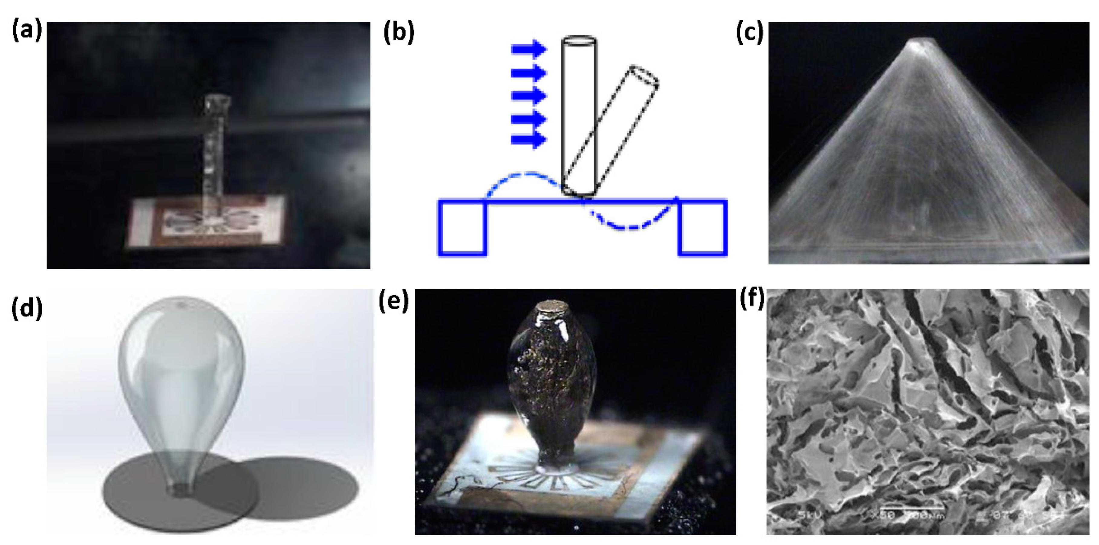
Figure 1. Superficial neuromast inspired steady-state flow sensor. (a) Photograph of LCP membrane with polycarbonate structure. (b) Schematic representation of the sensing mechanism. (c) Photograph of the electrospun nanofibril scaffold (d) Schematic representation of the SN cupula. (e) Photograph of the sensor with HA-MA hydrogel cupula. (f) SEM image of the cross-section of hydrogel.
Proceedings 02 01044 g001
Figure 2. Canal neuromast inspired oscillatory flow sensor (a) photograph of PZT membrane with hair-like copper structure and (b) HA-MA hydrogel of Young’s modulus 10 kPa. (hydrogel is coloured with rhodamine dye) (c) SEM image of the cross-section of HA-MA hydrogel cupula.
Proceedings 02 01044 g002
Figure 3. (a) Schematic representation of the experimental setup for testing the steady-state response of biomimetic MEMS SN sensor. (b) Plot showing the responses of the biomimetic MEMS SN sensor when subjected to a steady state flow over a velocity range of 0–8 m/s [4].
Proceedings 02 01044 g003
Figure 4. (a) Schematic of the experimental setup for testing the oscillatory response of MEMS CN sensor. (b) Response of the CN sensor to three different dipole stimulus frequencies.
Proceedings 02 01044 g004
Publisher’s Note: MDPI stays neutral with regard to jurisdictional claims in published maps and institutional affiliations.