Proceeding Paper

Effects of Vine Shoot Extract on Riboflavin-Induced DNA Damage in HepG2 Cells †

Department of Chemistry, Division of Food Chemistry and Toxicology, Technische Universität Kaiserslautern, Erwin-Schrödinger-Str. 52, 67663 Kaiserslautern, Germany
*
Author to whom correspondence should be addressed.
Presented at Natural Products and the Hallmarks of Chronic Diseases—COST Action 16112, Luxemburg 25–27 March 2019.
Proceedings 2019, 11(1), 19; https://doi.org/10.3390/proceedings2019011019
Published: 17 April 2019
(This article belongs to the Proceedings of CA16112 - Luxemburg 2019)

Abstract

:
Winery by-products, including pomace, stem, vine leaves and shoots, arise in high amounts during the wine-making process. Due to high contents of secondary plant metabolites, such as polyphenols, their usage in the food, cosmetics and pharmaceutical industry is possible. In this study, we investigated effects of an extract prepared from vine shoots (Vitis vinifera L. cv. Riesling) on riboflavin-induced DNA damage in human hepatocellular carcinoma cells HepG2 by comet assay. Significant decreases of DNA damages were detected after 2 h and 24 h extract incubation at concentration ranges of 1 to 30 µg/mL (2 h) and 1 to 10 µg/mL (24 h), respectively. These effects may be attributable to polyphenolic compounds, which has to be further investigated.

1. Introduction

During the wine-making process winery by-products, such as pomace, stem, vine leaves and shoots, arise. Vine shoots for example occur in huge amounts at pruning in springtime and are traditionally used as natural fertilizer [1]. Due to secondary plant metabolites, such as polyphenols, the usage of winery by-products as sources of bioactive compounds offers an opportunity to obtain value-added products for the food, cosmetics, and pharmaceutical industry [2]. It is known that polyphenols protect cells against oxidative stress due to their antioxidative and radical scavenging abilities and are associated with numerous beneficial health effects. A broad spectrum of biological activities is described, including antioxidant, anticancer and anti-inflammatory activities, amongst others [3]. Vine shoots are rich sources of phenolic acids, flavonoids and especially stilbenoids, such as resveratrol [4]. The aim of the present study was to investigate the effects of an extract from vine shoots of Vitis vinifera L. cv. Riesling on riboflavin-induced DNA damage by the comet assay. Additional treatment of DNA with formamidopyrimidin glycosylase (FPG) enhanced the sensitivity towards reactive oxygen species (ROS) induced DNA lesions and thus enabled the detection of oxidative DNA damage.

2. Materials and Methods

2.1. Chemicals, Cells and Media

Vine shoots of Vitis vinifera L. cv. Riesling were provided from a vineyard (conventional production) in Rhineland-Palatinate (Grünstadt-Sausenheim). They were cut-off before pruning of the vines in springtime 2016 and stored at room temperature till extraction. Chemicals and solvents were all of analytical grade or compliant with standards required for cell culture experiments. Formamidopyrimidine glycosylase (FPG) was provided by Prof. A. Collins (University of Oslo, Norway). HepG2 cells were obtained from DSMZ (Braunschweig, Germany), riboflavin, catalase, dimethyl sulfoxide (DMSO) 99.6%, Amberlite® XAD16N, ethanol absolute (≥99.8%), trypan blue solution and ethidium bromide were purchased from Sigma Aldrich (Munich, Germany), hydrochloric acid from CHEMSOLUTE®, Th. Geyer GmbH & Co. KG (Renningen, Germany), low and normal melting agarose from Bio-Rad (Munich, Germany), Trypsin from Serva (Heidelberg, Germany). Cell culture media (RPMI 1640) and supplements (fetal calf serum (FCS), penicillin/streptomycin) were purchased from Invitrogen (Karlsruhe, Germany). Cell culture materials (petri dishes, flasks, etc.) were from Greiner Bio-One (Essen, Germany).

2.2. Vine shoot Extract Preparation

Lyophilized and grinded vine shoots were extracted twice at the ratio of 1 g of solid per 25 mL of extraction solvent (methanol/water/1 N HCl: 80/19/1 (v/v/v)) at room temperature under constant stirring (1st: 60 min, 2nd: 30 min). After sterile filtration (0.2 µm) the extraction solvent was removed under reduced pressure at 40 °C and transferred into the water phase. To remove sugars, salts and organic acids solid-phase extraction was carried out using Amberlite® XAD16N resin. After conditioning with ethanol (90%) and equilibration with double distilled water, the column was loaded with the aqueous extract and washed with 5 fold bed volumes of double distilled water. Retained substances were eluted with 5 fold bed volumes of ethanol (90%). The eluate was evaporated under reduced pressure at 40 °C, transferred into water phase and then lyophilized. The powdery extract was homogenized and stored at −20 °C in the dark until used.

2.3. Cell Culture and Incubation Procedure

HepG2 cells were cultivated in 175 cm2 flasks in RPMI 1640 medium with addition of 10% FCS, 100 U/mL penicillin and 100 µg/mL streptomycin (humidified atmosphere with 5% CO2 at 37 °C). The cells were seeded in 6 cm Petri dishes, cultivated for 24 h, washed with PBS and in the first step incubated with vine shoot extract in incubation medium containing 5% FCS when performing 24 h incubation and FCS-free medium when performing 2 h incubation. Vine shoot extract was dissolved in DMSO, with a final DMSO-concentration of 0.5% in media (2 h: 1 to 30 µg/mL; 24 h: 1 to 50 µg/mL). Secondly, cells were kept on ice, washed twice with PBS and treated with the photosensitizer riboflavin (2 h preincubation: 100 µM; 24 h preincubation 250 µM; in PBS) in combination with irradiation with a halogen lamp (distance to the bottom of the Petri dishes: 33 cm) for 10 min. Afterwards the cells were washed three times with PBS and isolated by trypsination (0.5 mg/mL trypsin). To inhibit the formation of extracellular hydrogen peroxide (H2O2) by pro-oxidative interaction of phenolic compounds with cell media constituents [5], incubations were performed in presence of catalase (100 U/mL).

2.4. DNA Damage Measurement (Comet Assay)
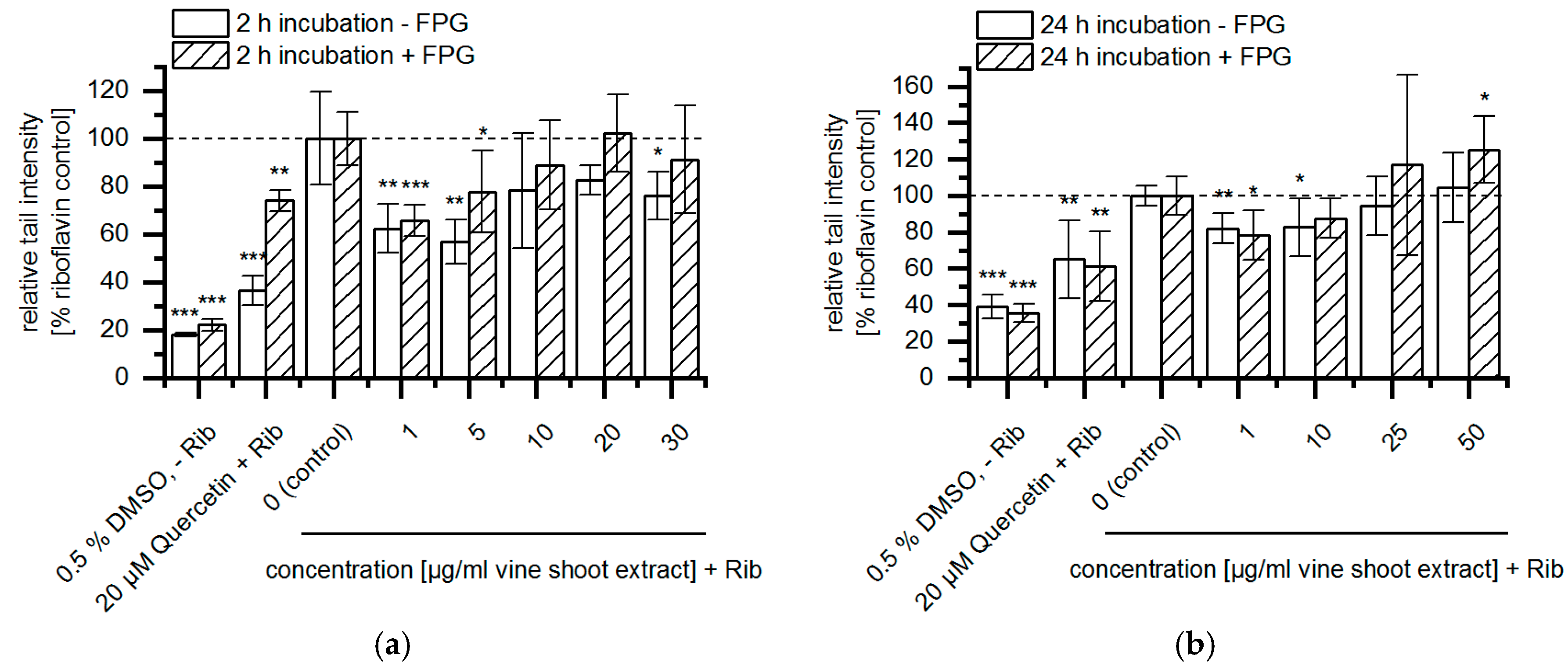
Alkaline single cell gel electrophoresis was performed according to Collins et al. and Bakuradze et al. [6,7], with slight modifications. First aliquots of suspended cells were examined for cell viability by trypan blue exclusion. Absolute viability was determined (in %; viable cells in percent of total cells). To exclude DNA damage resulting from cell death only cell suspensions with viability >80% were tested in comet assay. Briefly, 4 × 10⁴ cells per preparation were centrifuged (10 min, 2000 rpm, 4 °C), the pellets were resuspended in 65 µl low melting agarose (0.5%) and applied on microscope slides pre-coated with normal melting agarose (1.5%) (2 gels per slide, 2 slides each treatment, with and without FPG, respectively). The cells were lysed over night at 4 °C. Afterwards the slides were washed in enzyme buffer three times. Either FPG in enzyme buffer or enzyme buffer (without FPG) were applied on the slides (50 µL per gel) and incubated for 30 min at 37 °C. After DNA unwinding (pH 13.5, 20 min, 4 °C) and horizontal gel electrophoresis (20 min, 25 V, 300 mA), the slides were neutralized by washing with neutralization buffer three times, fixed with ethanol (95%), dried and stored at room temperature in the dark. Analysis was performed after labeling of DNA with ethidium bromide using fluorescence microscope (Zeiss, Oberkochen, Germany). Overall 2 × 50 cells per slide were analyzed. DNA damage was calculated as tail intensity (TI%, intensity of DNA in the comet tail, as percent of total DNA) [6,8,9]. Results were expressed as percent of riboflavin treated control. They are presented as mean ± SD of 3 to 5 independent experiments. Differences were determined by independent one-sided t-test by Origin®, version 2019 (OriginLab Corporation, Northampton, MA, USA).

3. Results

Comet assay was used to study the effects of vine shoot extract on riboflavin-induced DNA damage in HepG2 cells (Figure 1). An additional treatment with FPG led to an enhanced sensitivity towards ROS induced DNA lesions allowing a distinction between riboflavin-induced DNA damage and oxidative DNA damage. Treatment of HepG2 cells only with DMSO (0.5%) led to basal levels of DNA damage of 18 to 39% TI. After 2 h vine shoot extract preincubation significant decreased riboflavin-induced DNA damage was detected within the concentration range from 1 to 30 µg/mL, whereby lower vine shoot extract concentrations showed more distinct effects. Oxidative DNA damages were significantly reduced from 1 to 5 µg/mL vine shoot extract. For instance, 2 h preincubation with 1 µg/mL vine shoot extract showed a reduction of oxidative DNA damage down to about 66% compared to the riboflavin control.
Only preincubations with vine shoot extract concentrations of 1–10 µg/mL led to significant reduced riboflavin-induced DNA damage after 24 h. Oxidative DNA damage was decreased after incubation with 1 µg/mL vine shoot extract, whereas preincubation with 50 µg/mL vine shoot extract led to significant increase of oxidative DNA damage.

4. Discussion and Conclusion

In previous studies, polyphenol rich extracts have been shown to protect against oxidative DNA damage. After preincubations of Caco-2 cells with apple juice and bilberry extract and subsequent induction of oxidative DNA damage by menadione, “U-shaped” forms of concentration curves were observed [10,11]. DNA-protective effects for example were described after 24 h incubation with 50 µg/mL bilberry extract and 5 µg/mL bilberry extract with FPG-treatment, respectively. In contrast, prooxidative effects were detected at high concentrations of bilberry extract (250–500 µg/mL) [10]. Furthermore, several polyphenolic compounds were described to be protective against oxidative DNA damage. Relatively low concentrations of grape seed polyphenols, namely catechin and gallic acid, have led to decreased H2O2-induced DNA damage in mice spleen cells [12]. In addition, resveratrol protected against arsenic-trioxide-induced oxidative damage at simultaneous exposure to 20 µM As2O3 and 5 µM resveratrol in human bronchial epithelial (HBE) cells [13].
Taken together, vine shoot extract showed protective effects against riboflavin-induced DNA damage, which was significantly reduced after 2 and 24 h preincubation with vine shoot extract at concentrations of 1–30 µg/mL and 1–10 µg/mL, respectively. These effects may be attributable to contained polyphenolic substances, which has to be further investigated.

Author Contributions

Supervision, E.R.; Conceptualization, E.R.; T.B. and C.F.; Investigation, C.F.; Writing—original draft preparation, C.F.; Writing—review and editing, E.R.; T.B.

Funding

This research was funded by Stiftung Rheinland-Pfalz für Innovation, project no. 1164.

Acknowledgments

We thank the winemaker, Gerhard Siebert (Sausenheim, Germany) for providing vine shoot samples. This work is based upon work from COST Action NutRedOx-CA16112 supported by COST (European Cooperation in Science and Technology).

Conflicts of Interest

The authors declare no conflict of interest.

References

  1. Luque-Rodríguez, J.M.; Pérez-Juan, P.; Luque de Castro, M.D. Extraction of polyphenols from vine shoots of Vitis vinifera by superheated ethanol-water mixtures. J. Agric. Food Chem. 2006, 54, 8775–8781. [Google Scholar] [CrossRef] [PubMed]
  2. Martins, S.; Mussatto, S.I.; Martínez-Avila, G.; Montañez-Saenz, J.; Aguilar, C.N.; Teixeira, J.A. Bioactive phenolic compounds: Production and extraction by solid-state fermentation. A review. Biotechnol. Adv. 2011, 29, 365–373. [Google Scholar] [CrossRef] [PubMed]
  3. Xia, E.Q.; Deng, G.F.; Guo, Y.J.; Li, H.B. Biological activities of polyphenols from grapes. Int. J. Mol. Sci. 2010, 11, 622–646. [Google Scholar] [CrossRef] [PubMed]
  4. Cebrián, C.; Sánchez-Gómez, R.; Salinas, M.R.; Alonso, G.L.; Zalacain, A. Effect of post-pruning vine-shoots storage on the evolution of high-value compounds. Ind. Crop. Prod. 2017, 109, 730–736. [Google Scholar] [CrossRef]
  5. Lapidot, T.; Walker, M.D.; Kanner, J. Can apple antioxidants inhibit tumor cell proliferation? generation of H2O2 during interaction of phenolic compounds with cell culture media. J. Agric. Food Chem. 2002, 50, 3156–3160. [Google Scholar] [CrossRef] [PubMed]
  6. Collins, A.R.; Dusinská, M.; Gedik, C.M.; Stĕtina, R. Oxidative damage to DNA: Do we have a reliable biomarker? Environ. Health Perspect. 1996, 104, 465–469. [Google Scholar] [CrossRef] [PubMed]
  7. Bakuradze, T.; Lang, R.; Hofmann, T.; Stiebitz, H.; Bytof, G.; Lantz, I.; Baum, M.; Eisenbrand, G.; Janzowski, C. Antioxidant effectiveness of coffee extracts and selected constituents in cell-free systems and human colon cell lines. Mol. Nutr. Food Res. 2010, 12, 1734–1743. [Google Scholar] [CrossRef] [PubMed]
  8. Collins, A.R.; Oscoz, A.A.; Brunborg, G.; Gaivão, I.; Giovannelli, L.; Kruszewski, M.; Smith, C.C.; Stetina, R. The comet assay: Topical issues. Mutagenesis 2008, 23, 143–151. [Google Scholar] [CrossRef] [PubMed]
  9. Tice, R.R.; Agurell, E.; Anderson, D.; Burlinson, B.; Hartmann, A.; Kobayashi, H.; Miyamae, Y.; Rojas, E.; Ryu, J.C.; Sasaki, Y.F. Single cell gel/comet assay: Guidelines for in vitro and in vivo genetic toxicology testing. Environ. Mol. Mutagenesis 2000, 35, 206–221. [Google Scholar] [CrossRef]
  10. Schantz, M.; Mohn, C.; Baum, M.; Richling, E. Antioxidaitve effiency of an anthocyanin rich bilberry extract in the human colon tumor cell lines Caco-2 and HT-29. J. Berry Res. 2010, 1, 25–33. [Google Scholar] [CrossRef]
  11. Schaefer, S.; Baum, M.; Eisenbrand, G.; Dietrich, H.; Will, F.; Janzowski, C. Polyphenolic apple juice extracts and their major constituents reduce oxidative damage in human colon cell lines. Mol. Nutr. Food Res. 2006, 50, 24–33. [Google Scholar] [CrossRef] [PubMed]
  12. Fan, P.; Lou, H. Effects of polyphenols from grape seeds on oxidative damage to cellular DNA. Mol. Cell. Biochem. 2004, 1, 67–74. [Google Scholar] [CrossRef] [PubMed]
  13. Chen, C.; Jiang, X.; Lai, Y.; Liu, Y.; Zhang, Z. Resveratrol protects against arsenic trioxide-induced oxidative damage through maintenance of glutathione homeostasis and inhibition of apoptotic progression. Environ. Mol. Mutagenesis 2015, 3, 333–346. [Google Scholar] [CrossRef] [PubMed]
Figure 1. Modulation of riboflavin-induced DNA damage in HepG2 cells after (a) 2 h and (b) 24 h vine shoot extract preincubation (2 h: 1 to 30 µg/mL vine shoot extract, 100 µM riboflavin; 24 h: 1 to 50 µg/mL vine shoot extract, 250 µM riboflavin); quercetin (20 µM) was used as positive control. Results were calculated as percent of riboflavin treated control; 2 h: n = 3 – 5, 24 h: n = 4–5 (mean ±SD). Differences were determined by independent one-sided t-test related to riboflavin treated control: * p < 0.05, ** p < 0.01, *** p < 0.001.
Proceedings 11 00019 g001

