Abstract
The development of miniaturized potentiostats capable of measuring in a wide range of conditions and with full characteristics (e.g., wide bandwidth and capacitive/inductive contribution to sensor’s impedance) is still an unresolved challenge in bioelectronics. We present a simple analogue design coupled to a digital filter based on a lock-in amplifier as an alternative to complex architectures reported hitherto. A low-cost, miniaturized and fully integrated acquisition electronic system was developed, tested for a fully integrated three-lead electrochemical biosensor and benchmarked against a commercial potentiostat. The portable potentiostat was coupled to an array of miniaturized gold working electrodes to perform complex impedance analyses for tumor necrosis factor α (TNF-α) cytokine detection. This wearable potentiostat is very promising for the development of low-cost point-of-care (POC) with low power consumption.
1. Introduction
Heart failure (HF) is a very fast growing cardiovascular disorder (CVD) with around 1 million new patients being diagnosed every year [1]. Bioelectronic devices such as left ventricle assisted devices (LVADs) cause many secondary effects in patients, triggering increased inflammatory cytokine levels, e.g., Interleukin 1 (IL-1), IL-10 and tumor necrosis factor α (TNF-α). Traditional techniques for the detection of CVD cytokines, such as enzyme-linked immunosorbent assays (ELISA), although accurate and sensitive, still require specialized personnel and high volumes of sample for analysis [2,3]. In this light, several electrochemical biosensors have been developed for the monitoring of inflammatory cytokines [4,5]. Nevertheless, such biosensors cannot be implemented in point of care (POC) environments unless potentiostatic systems are miniaturized as well.
In this sense, only few attempts of potentiostat miniaturization can be found in the literature [6,7,8,9], and some important concerns always arise. Some authors focused on adapting already existing portable electronics to perform cyclic voltammetry. This is the case of Cruz et al. [6], who integrated a LMP91000EVM commercial chip and a BeagleBone (Texas Instruments, Dallas, TX, USA) development board. As for portable impedance measurements, an interesting work is presented by Zhang et al. [7], who, according to the same philosophy, integrated a commercial miniaturized impedance analyzer AD5933 (Analog Devices, Norwood, MA, USA) with an Arduino UNO board and a Bluetooth module for BSA immunosensing applications. Similarly, Li et al. [8] developed a detector combined with a 3D-printed USB-compatible chip to sense aflatoxins in rice. They benchmarked their device against a Zahner commercial station, achieving good limit of detection.
After exhaustive revision of the literature, we reached the conclusion that there are three main issues regarding the performance of portable home-made potentiostats that have not been properly solved yet: (a) the expansion of the frequency range to low values (<10 Hz); (b) the determination of the capacitive/inductive contribution to the sensor complex impedance (magnitude and phase) and (c) the extension of the miniaturized electronic module to perform parallel measurements.
In the present work we intend to individually tackle the two former issues. We focused on well-described electronic circuit topologies to develop a potentiostat integrable into a three-lead electrochemical configuration. We also designed some additional conditioning electronics and implemented a digital lock-in filter for signal post-processing in order to extract the appropriate features for antigen quantification, which is of crucial importance in some clinical and environmental approaches.
2. Materials and Methods
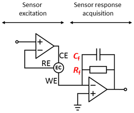
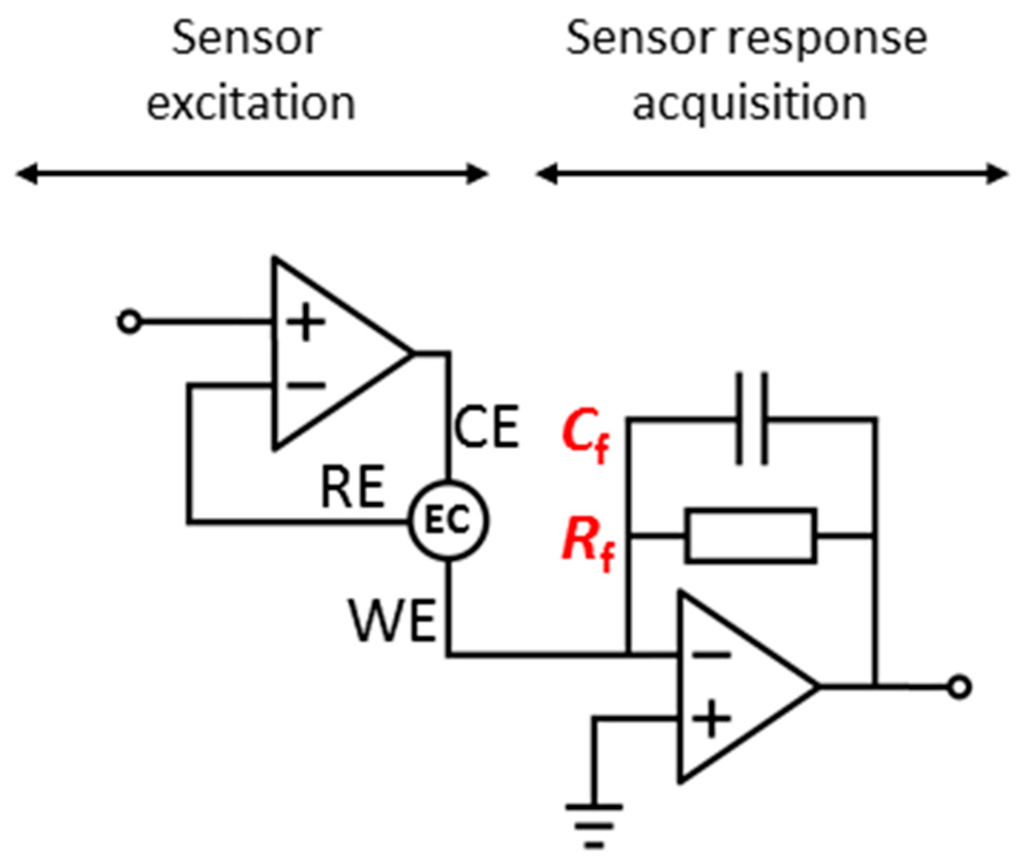
A final integrated printed circuit board (PCB) holding the potentiostat architecture in Figure 1 was developed. This well-known potentiostatic configuration consists of an operational amplifier with one input and the output leads connected to the electrochemical cell through the reference and counter electrodes, respectively, and the excitation signal connected to the positive input lead. The sensor intensity response was converted to a measurable voltage quantity with a transimpedance amplifier at the working electrode terminal. All the analogue circuitry was integrated in a PCB together with two microcontrollers for sensor excitation, response acquisition and computer interfacing for data processing. A graphical user interface (GUI) was developed in MATLAB (The Mathworks, Inc., Natick, MA, USA) to allow the user choose the parameters of the excitation potential, i.e., DC magnitude and sign and small AC signal amplitude and frequency.

Figure 1.
Electronic schematic of the two core parts of the potentiostat: the sensor excitation stage including the three-lead approach and an electrochemical cell (EC), and the inverter sensor response acquisition stage, with the feedback capacitor and resistor, to transform the sensor response current to a measurable voltage.
The device was tested with a removable and fully integrated chip including a silver/silver chloride reference microelectrode, a platinum auxiliary microelectrode and eight gold working microelectrodes. First, the bare gold WEs were diazotized and activated with EDC/HNS, and they were then bio-functionalized with anti-TNF-α to detect the corresponding TNF-α cytokines. Details of the biosensor fabrication, characterization and bio-functionalization were previously reported using the same microelectrodes array [4]. The WEs were first measured with a commercial VMP3 potentiostat within the frequency range of 200 kHz to 100 mHz, and impedance analyses were then modelled using Randomized + Simplex algorithm to extract electronic parameters. These were useful to calibrate the portable device in a full frequency range. Finally, bare gold WEs were tested with the portable potentiostat and the magnitude and phase of the impedance in the frequency region of 1–10 Hz was extracted by a digital lock-in amplifier implementation. This frequency range was found to be the most significant for the detection process.
3. Results
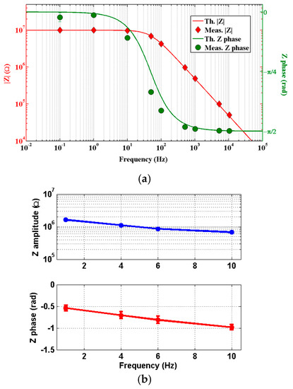
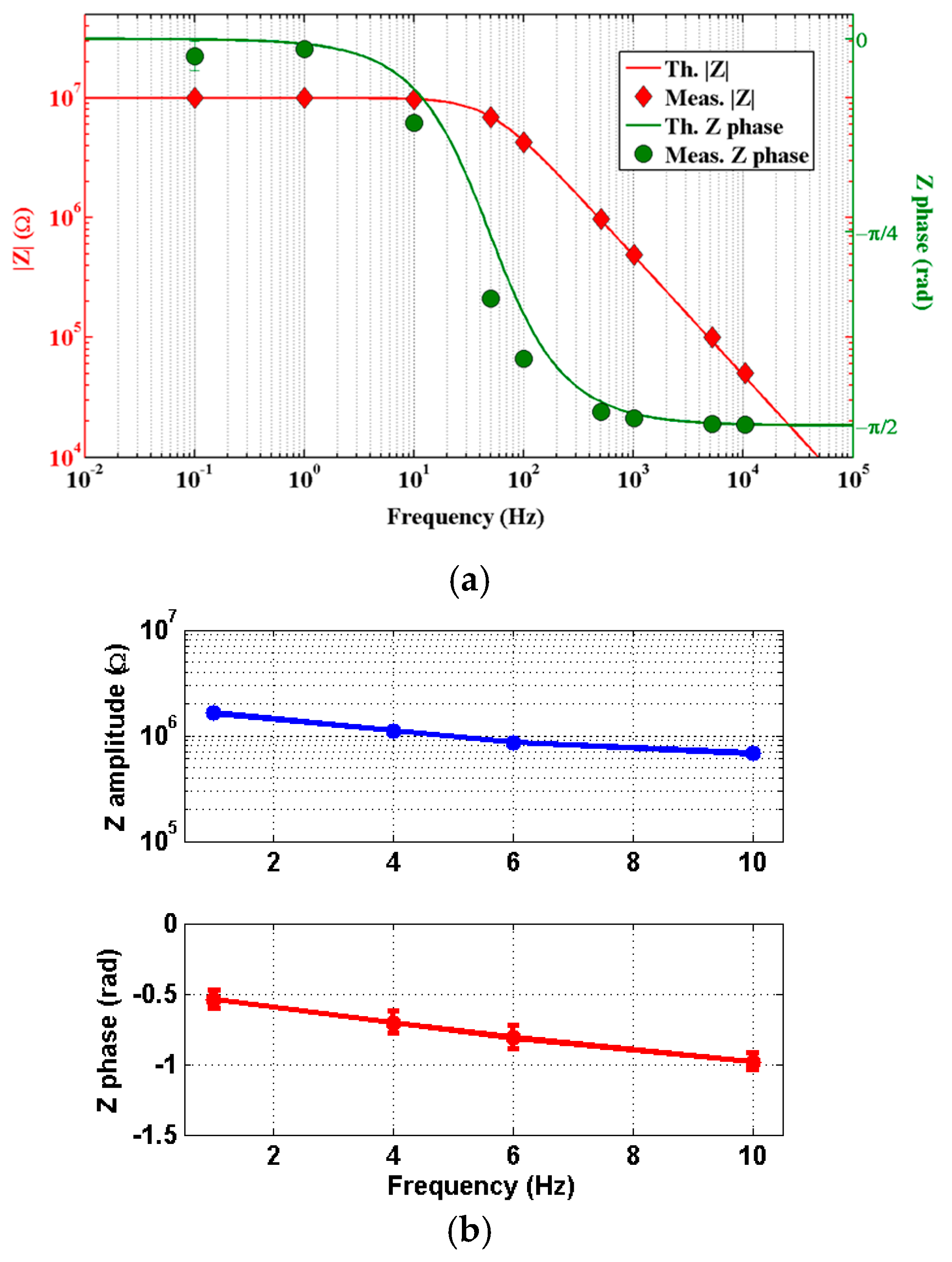
First, the portable potentiostat was tested with an equivalent circuit made with SMD resistors and capacitors, emulating the behavior of a real biosensor. In the present case, a parallel circuit of 10 MΩ and 330 pF was used for this purpose. The corresponding Bode diagram for both impedance amplitude and phase is shown in Figure 2a. The behavior was tested in a wide frequency range; this is, from 100 mHz to 10 kHz, to show the capabilities of the developed device. The theoretical behavior of this system is also shown as red and green lines for amplitude and phase, respectively.

Figure 2.
(a) Full range calibration of the portable electronic device for a parallel configuration of resistor and capacitor (experimental data) and fitting of the theoretical model; (b) Bare gold WE impedance amplitude and phase measured with the portable home-made electronic device in the frequency region comprised between 1 Hz and 10 Hz.
Afterwards, the device was tested on a real biosensor array. The WE was cleaned and derivatized with a diazonium salt. After NHS/EDC activation of the diazonium’s COOH- termination followed by 1 h incubation with anti-TNF-α, the electrodes were ready for detection of TNF-α.
A single concentration of TNF-α was tested as a proof of concept. Impedance measurements with a commercial potentiostat revealed that the most adequate frequency range for detection in the present case, this is where impedance magnitudes differ more between concentrations of TNF-α, was 1–10 Hz. Figure 2b shows both Bode plots for impedance amplitude and phase of the real biosensor, in the aforementioned frequency range.
4. Conclusions
In summary, our approach succeeds in fixing some of which we consider important issues, such as the performance of measurements at low frequencies and the determination of the sensor’s contribution to its complex. The results suggest that the developed device could be useful and easily embeddable in point of care (POC) environments and biomedical applications.
Future work will consider the detection of TNF-α at several concentrations and simultaneously in several WEs, as well as the determination of the limit of detection (LOD) and limit of quantification (LOQ).
Author Contributions
R.P., F.P. and M.L. designed, tested and validated the electronic circuitry and the control software. A.B., J.B. and A.E. designed and manufactured the miniaturized biosensor. R.P., A.B. and A.E. performed the integration of biosensor and potentiostat and conducted the experiments. All authors gave final approval for publication.
Acknowledgments
The research leading to these results has received funding from the European Union’s 7FP (SEA-on-a-CHIP, Grant Agreement No. 614168) and the European Union’s Horizon 2020 (HEARTEN, Grant Agreement No. 643694). R.P. acknowledges an FPU grant from the Spanish Ministerio de Educación, Cultura y Deporte.
Conflicts of Interest
The authors declare no conflict of interest. The funding sponsors had no role in the design of the study; in the collection, analyses, or interpretation of data; in the writing of the manuscript, and in the decision to publish the results.
References
- Jessup, M.; Brozena, S. Medical progress. Heart failure. N. Engl. J. Med. 2003, 348, 2007–2018. [Google Scholar] [CrossRef] [PubMed]
- Caruso, R.; Trunfio, S.; Milazzo, F.; Campolo, J.; De Maria, R.; Colombo, T.; Parolini, M.; Cannata, A.; Russo, C.; Paino, R.; et al. Early expression of pro-and anti-inflammatory cytokines in left ventricular assist device recipients with multiple organ failure syndrome. ASAIO J. 2010, 56, 313–318. [Google Scholar] [CrossRef] [PubMed]
- Navarri, R.; Lunghetti, S.; Cameli, M.; Mondillo, S.; Favilli, R.; Scarpini, F.; Puccetti, L. Neurohumoral improvement and torsional dynamics in patients with heart failure after treatment with levosimendan. IJC Heart Vasc. 2015, 7, 153–157. [Google Scholar] [CrossRef] [PubMed][Green Version]
- Baraket, A.; Lee, M.; Zine, N.; Sigaud, M.; Bausells, J.; Errachid, A. A fully integrated electrochemical biosensor platform fabrication process for cytokines detection. Biosens. Bioelectron. 2017, 93, 170–175. [Google Scholar] [CrossRef] [PubMed]
- Lee, M.; Zine, N.; Baraket, A.; Zabala, M.; Campabadal, F.; Caruso, R.; Trivella, M.G.; Jaffrezic-Renault, N.; Errachid, A. A novel biosensor based on hafnium oxide: Application for early stage detection of human interleukin-10. Sens. Actuator 2012, 175, 201–207. [Google Scholar] [CrossRef]
- Cruz, A.F.D.; Norena, N.; Kaushik, A.; Bhansali, S. A low-cost miniaturized potentiostat for point-of-care diagnosis. Biosens. Bioelectron. 2014, 62, 249–254. [Google Scholar] [CrossRef] [PubMed]
- Zhang, D.; Lu, Y.; Zhang, Q.; Liu, L.; Li, S.; Yao, Y.; Jiang, J.; Liu, G.L.; Liu, Q. Protein detecting with smartphone-controlled electrochemical impedance spectroscopy for point-of-care applications. Sens. Actuator 2016, 222, 994–1002. [Google Scholar] [CrossRef]
- Li, Z.; Ye, Z.; Fu, Y.; Xiong, Y.; Li, Y. A portable electrochemical immunosensor for rapid detection of trace aflatoxin B 1 in rice. Anal. Methods 2016, 8, 548–553. [Google Scholar] [CrossRef]
- Yu, X.; Esanu, M.; MacKay, S.; Chen, J.; Sawan, M.; Wishart, D.; Hiebrt, W. An impedance detection circuit for applications in a portable biosensor system. In Proceedings of the 2016 IEEE International Symposium on Circuits and Systems (ISCAS), Montreal, QC, Canada, 22–25 May 2016; pp. 1518–1521. [Google Scholar] [CrossRef]
Publisher’s Note: MDPI stays neutral with regard to jurisdictional claims in published maps and institutional affiliations. |
© 2017 by the authors. Licensee MDPI, Basel, Switzerland. This article is an open access article distributed under the terms and conditions of the Creative Commons Attribution (CC BY) license (https://creativecommons.org/licenses/by/4.0/).


