Article

Surface Properties and Adherence of Bradyrhizobium diazoefficiens to Glycine max Roots Are Altered When Grown in Soil Extracted Nutrients

by
Armaan Kaur Sandhu
1,
Senthil Subramanian
1,2 and
Volker S. Brözel
1,3,*
1
Department of Biology and Microbiology, South Dakota State University, Brookings, SD 57006, USA
2
Department of Agronomy, Horticulture and Plant Science, South Dakota State University, Brookings, SD 57006, USA
3
Department of Biochemistry, Genetics and Microbiology, University of Pretoria, Pretoria 0004, South Africa
*
Author to whom correspondence should be addressed.
Nitrogen 2021, 2(4), 461-473; https://doi.org/10.3390/nitrogen2040031
Submission received: 14 October 2021 / Revised: 4 November 2021 / Accepted: 11 November 2021 / Published: 15 November 2021

Abstract

:
Soybean roots are colonized and nodulated by multiple strains of compatible nitrogen-fixing rhizobia primarily belonging to the Genus Bradyrhizobium. Motility towards the root and attachment to root hairs are key determinants of competitive colonization and subsequent nodulation. Bacterial surface properties and motility are known to vary with chemical composition of the culture medium, and root adhesion and nodulation occur in a soil environment rather than laboratory medium. We asked whether the nodulation-promoting factors motility, surface hydrophobicity and surface adhesion of Bradyrhizobium are affected by growth in a soil nutrient environment. B. diazoefficiens USDA 110, 126, 3384, and B. elkanii USDA 26 were grown in mineral salt medium with peptone, yeast extract and arabinose (PSY), and in a soil extracted soluble organic matter (SESOM) medium. Surface hydrophobicity was determined by partitioning into hydrocarbon, motility by transition through soft agar, and surface-exposed saccharides by lectin profiling, followed by biofilm formation and soybean root adhesion capacity of populations. SESOM-grown populations were generally less motile and more hydrophobic. They bound fewer lectins than PSY-grown populations, indicating a simpler surface saccharide profile. SESOM populations of USDA 110 did not form detectable biofilm, but showed increased binding to soy roots. Our results indicate that growth in a soil environment impacts surface properties, motility, and subsequent soy root adhesion propensity. Hence, evaluation of Bradyrhizobium for nodulation efficiency should be performed using soil from the specific field where the soybeans are to be planted, rather than laboratory culture media.

1. Introduction

Rhizobium bacteria contribute combined nitrogen to many leguminous plants while occurring inside specialized root structures called nodules [1]. The productivity of soybeans (Glycine max) is enhanced significantly through nodule occupancy by prolific fixers such as B. diazoefficiens USDA 110 [2]. Early during plant growth, root hairs signal soil-borne rhizobia by releasing isoflavonoids [3]. Rhizobia respond to isoflavonoids such as genistein by synthesizing Nod Factors, which in turn initiate curling of soy root hairs, followed by infection thread and nodule formation [4,5]. The nitrogen contributing efficacy of rhizobia varies widely, necessitating screening and evaluation of candidate bacterial strains for application in fields. Successful colonization of the infection thread and nodules require temporal adhesion and colonization of rhizobia at the root hair surface. Adhesion to the root surface is dependent on physical proximity, facilitated by bacterial motility in the soil. Adherence and colonization by proximal bacteria depend on the physiochemical compatibility of both the root and rhizobial surfaces.
Surface properties of bacterial cells have a considerable influence on attachment to the root surface, so compatible surface properties contribute to increased adhesion. More hydrophobic mutants of Bradyrhizobium japonicum have been associated with more competitive nodule formation [6]. Bacterial lipopolysaccharides (LPS) have direct relation to hydrophobicity and more hydrophobic LPS is positively correlated to surface hydrophobicity [7,8]. Bradyrhizobium producing altered or limited extracellular polysaccharides (EPS) were defective in biofilm formation and binding to soy lectin and resulted in pseudo nodules [9,10]. Along with surface properties, there is evidence that motility can provide the cell with some significant advantages to adhere to the root surface [11,12]. On the other hand, not much is known about soy root surface properties. Soy roots produce surface-exposed lectins which sequester cognate sugar moieties at the surface of bacteria [13,14], especially in acidic soil. Lectins are proteinaceous, specific carbohydrate-binding proteins. They interact with carbohydrates in a highly specific but non-covalent manner [15]. In more alkaline soil, root hairs produce the protein rhicadhesin which promotes the bacterial attachment [16]. The surface properties of rhizobia therefore play an important role in the initial adhesion to soy root surface.
Bacteria respond to specific chemical and physical cues by condition-specific gene expression and altered phenotype. This is also the case with Bradyrhizobium, as phenotypic variations have been reported for populations cultured in different sugar sources. While B. diazoefficiens swims by sub-polar flagella, L-arabinose induces the production of lateral flagella, which lead to swarming on moist surfaces [17]. The EPS composition of B. japonicum varies by the available sugar sources [9]. Bradyrhizobium are generally cultured using a mineral salt medium with peptone, yeast extract and either arabinose or mannitol as a carbon source. We asked whether Bradyrhizobium adapts its adhesion-specific phenotype when growing in soil. Arguing that the primary drivers of phenotypic change would be water-diffusible substances able to enter the cell, we cultured Bradyrhizobium in soybean field aqueous soil extract and characterized adhesion-pertaining phenotypes.

2. Materials and Methods

2.1. Bacterial Strains and Culture Media

B. diazoefficiens USDA 110, 126, 3384 and B. elkanii USDA 26 were obtained from the NRRL Culture Collection of the Agricultural Research Service, United State Department of Agriculture. Cultures were grown in liquid or on solid PSY and SESOM, a filter-sterilized liquid soil extract [18]. PSY was prepared as mentioned by Mesa et al. [19], containing per liter: KH2PO4, 300 mg; Na2HPO4, 300 mg; CaCl2·2H2O, 5 mg; MgSO4·7H2O, 100 mg; peptone, 3 g; yeast extract, 1 g; H3BO3, 10 mg; ZnSO4·7H2O, 1 mg; CuSO4·5H2O, 0.5 mg; Na2MoO4·2H2O, 0.1 mg; MnCl2·4H2O, 0.1 mg; FeCl3, 0.19 mg; thiamine-HCl, 1 mg; biotin, 1 mg; Na-panthothenate, 1 mg; L-arabinose.
Soil for SESOM was obtained from a field under soybean cultivation directly after harvest, was dried at 55 °C and stored at 4 °C. SESOM was prepared as described previously [18] by adding 200 g of dry soil to 1 l of prewarmed (60 °C) sterile MOPS buffer (10 mM, pH 7.0) in a 2 L Erlenmeyer and shaken for 4 h at 150 rpm. To remove soil particles, the suspension was filtered sequentially through filter paper and cellulose acetate filters of pore sizes 8, 4, 1.2, 0.8, 0.4 and 0.2 µM. The clear extract was filtered to sterility through a 0.2 µM bottle top filter. Each extract was checked for sterility by plating 20 µL on R2A agar and incubating at 30 °C for 72 h. The sterile SESOM was supplemented with 0.1 g/L Bacto peptone and 0.1 g/L L(+)-Arabinose.

2.2. Motility

The effect of culture medium on swimming motility was determined by inoculating the center of low percentage agar plates (0.35%). Precultures were prepared by inoculating grown in PSY and SESOM and incubating at 30 °C for 24 h while shaking at 250 rpm. Cultures were diluted to A600 of 0.100 and 20 µL was drop inoculated at the center of PSY or SESOM low agar plates. Plates were incubated at 30 °C for 10 d when the colony radius was measured.

2.3. Microbial Adhesion to Hydrocarbons

Microbial adhesion to hydrocarbons (MATH) was assessed to analyze surface hydrophobicity of cultures. Exponential phase cultures (50 mL) prepared as outlined above were harvested by centrifugation for 10 min at 10,000× g and resuspended in 15 mL sterile Phosphate Urea Magnesium Sulfate Buffer (PUM) containing 22.2 g/L K2HPO4·3H2O, 7.26 g/L KH2PO4, 1.8g/L Urea and 0.02 g/L MgSO4·7H2O with pH adjusted to 7.1 [20,21]. Following a second round of harvesting, cells were suspended in 15 mL PUM by vortexing. To three acid washed glass tubes, we added 4 mL cell suspension, and supplemented with 1 mL n-hexadecane, retaining the final 3 mL suspension as blank. Cell suspended in PUM were exposed to hexadecane by vortexing for 1 min and allowing the mixture to separate at room temperature for 1 h. After separation, 1 mL of the aqueous phase was withdrawn very carefully using acid washed, sterile glass pipettes, and transferred to a quartz cuvette. The absorbance was measured at 600 nm, using hexadecane-supplemented PUM without bacteria added as blank, termed Absorbance Math (AM). The absorbance of untreated cell suspensions was measured at 600 nm, using PUM buffer as blank, and termed Absorbance original (AO). The fraction of cells that partitioned to the hydrocarbon phase was calculated as:
Fp = 1 − (AM/AO)

2.4. Lectin Binding Assay

Surface-exposed sugar moieties were characterized using a collection of 24 lectins (Vector Laboratories, Burlingame, CA 94010, USA) (Table 1). Nine of these were fluorescein-conjugated, and the remaining fifteen were biotinylated. Exponential phase 24 h old cultures were prepared as described above, harvested, and washed with HSB buffer (2.383 g/L HEPES, 8.766 g/L NaCl, 0.011 g/L CaCl2 and 0.8 g/L sodium azide) (Vector Laboratories), and resuspended to absorbance of 0.100 at 600 nm. Lectins were diluted to 20 µg/ mL in HSB, and 100 µL added to 50 µL of cell suspension. After mixing by brief vortexing, lectins were allowed to bind for 10 min at room temperature, followed by vortexing and a further 10 min rest period. The cells were harvested by centrifugation at 16,200× g for 7 min. Unbound lectin was removed by three wash cycles using HSB buffer. For fluorescein-conjugated lectins, the resulting pellet was resuspended in 20 µL of HSB and the entire sample transferred to a clean microscopic slide. The drop of cell suspension was covered with a glass coverslip, pressed slightly to reduce cell movement, and the sides sealed using clear nail varnish to prevent evaporation. For biotinylated lectins, lectin-exposed washed cells were supplemented with 50 µL of 20 µg/mL streptavidin-FITC and left to bind for 30 min. Unbound streptavidin-FITC was removed by washing cells twice. The pellet was resuspended in 20 µL HSB and transferred to a microscope slide.
Samples were viewed by fluorescence microscopy using an Olympus BX53 Upright Compound Microscope with 466/40 nm excitation and a 525/50 nm emission filter, captured using an Olympus DP70 digital camera. Sample images were captured using 100, 200, 400, 600, 800, 1000 and 1200 ms exposure time. Binding intensity was scored by the shortest exposure time yielding visible fluorescence, with 100 ms scoring level 4, and <1200 ms scoring level 0. The proportion of cells displaying fluorescence was determined by Image J by preparing the binary image under Process. The resulting binary image was further processed for analysis by using the following programs sequentially > Mask > Watershed > Fill holes. For the analyses, the analyze particles program was selected under the Analyze icon.

2.5. Biofilm Formation

The effect of culture medium on biofilm formation was determined by the widely used polystyrene crystal violet assay [22,23], with some modifications. Exponential phase cultures were prepared as described above, harvested by centrifugation, suspended in sterile water to remove residual medium, harvested, and resuspended in fresh PSY or SESOM to absorbance of 0.100 at 600 nm. Nunclon Delta Surface Polystyrene 96-well plates (Thermo Scientific, Waltham, MA, USA; catalog 167008) were loaded with 150 µL of cells in 7 replicates. The sterile culture media were loaded into additional wells to serve as blank. After 24 h incubation at 30 °C, the planktonic cells were removed by pipetting. Each inoculated well of the plate was washed twice by pipetting using sterile water to facilitate removal of all planktonic cells. Biofilms were stained by adding 200 µL of 0.1% aqueous crystal violet solution, shaking at room temperature for 20 min. The crystal violet solution was removed from the wells by pipette, followed by two cycles of careful washing with water by pipetting. The plate was left open for 30 min to dry and 200 µL of 95% ethanol was added to dissolve the crystal violet. The plates were incubated for 10 min at room temperature with shaking and absorbance determined at 570 nm.

2.6. Root Adherence

To determine bacterial adherence to young roots, exponential phase cultures were exposed to germinating soy seeds. Seeds were sterilized as described previously [24]. Briefly, seeds were sterilized by washing in 30% of concentrated bleach for 5 min while shaking gently, followed by three consecutive washes with sterilized deionized water under shaking. Seeds were suspended in 70% ethanol while being shaken gently for 20 min, followed by seven consecutive washes with sterilize deionized water under shaking. Following a final 20 min soak in sterile deionized water, seven seeds were placed onto each R2A agar plate (Difco) and left in the dark at 30 °C for 9 d. R2A comprised of yeast extract 0.5 g/L, proteose peptone No. 3 0.5 g/L, casamino acids 0.5 g/L, dextrose 0.5 g/L, soluble starch 0.5 g/L, sodium pyruvate 0.3 g/L, dipotassium phosphate 0.3 g/L, magnesium sulfate 0.05 g/L and agar 15 g/L. The plates were inspected every 2 d, and plates with seedlings showing microbial outgrowth on the agar were discarded. By day 9, roots varied from 8 to 12 cm in length.
The exponential phase cultures were prepared as described above and diluted in fresh medium to absorbance of 0.100 at 600 nm. Aliquots were taken, and the culturable count was determined using the droplet plate count technique (Lindsay and VonHoly, 1999). Briefly, samples were serially diluted and 20 µL volumes spotted onto R2A plates. After incubation at 30 °C for 4 d, colonies were counted. Five seedlings were added to each 250 mL flask containing 50 mL of exponential phase cultures (50 mL) prepared in either PSY or SESOM as outlined above, left for 60 min while shaking gently. Seeds were removed aseptically using sterile tweezers, washed twice with sterilize deionized water and transferred to 50 mL conical tubes containing 50 mL PBS with 0.02% Tween 20 [24]. Tubes were immersed in a sonicator bath (Fisher FS20) and exposed to ultrasound for 30 s, and then transferred to ice water for 30 s to prevent undue heating. This sonication and cooling cycle was repeated 4 times. One mL of treated sample was drawn to perform serial dilutions and triplicate 20 µL volumes were plated on R2A plates. After incubation at 30 °C for 4 d, colonies were counted.

3. Results

3.1. Growth Medium Can Significantly Influence Cell Surface Hydrophobicity and Motility of Bradyrhizobium Strains

Surface properties of bacterial cells affect interaction with surfaces. We determined surface hydrophobicity of populations cultured in PSY and SESOM using the MATH partitioning assay. Hydrophobicity of the four strains varied, when grown in PSY medium with arabinose. USDA 110 was most hydrophilic while USDA 26 was the most hydrophobic (with USDA126 and USDA3384 displaying intermediate hydrophobicity), indicating strain-specific surface phenotype (Figure 1). When grown in the soil extract medium, SESOM, two of the strains underwent a significant increase in surface hydrophobicity. For USDA 3384, the fraction partitioned to the hydrocarbon phase increased by 700% and for USDA 110 by 300%. For USDA 110, some absorbance values of aqueous phase after partitioning were negative, suggesting minor emulsification due to hydrocarbon droplets into the aqueous phase. However, no significant change was observed for USDA 26 and 126 (Figure 1). These results indicate that surface hydrophobicity of Bradyrhizobium has some inherent strain-specific differences, and it is affected by the culture medium, with increased hydrophobicity predicted for cells growing in soil.
To determine whether growth in a soil environment affects motility, strains were inoculated into classical motility low agar plates (0.35%) in either PSY or SESOM. The strains varied in degree of motility in either medium, with USDA 110 and 126 the most motile, USDA 3384 less motile and 26 the least motile (Figure 2). USDA 26, 110 and 3384 showed a significant decrease in motility when grown in SESOM, while 126 remained unchanged (Figure 2). Strains 110 and 3384 showed the largest change. Collectively, USDA 110 and USDA 3384 became more hydrophobic and less motile when cultured in SESOM.

3.2. Lectin Binding Profiles of Bradyrhizobium Strains Change When Cultured in SESOM

The presence of surface-exposed sugars was characterized semi-quantitatively using 24 lectins. Lectin binding intensity was scored by visible fluorescence after different exposure times, yielding a lectin fingerprint for each strain (Figure 3). USDA 110 bound to the highest number of lectins, followed by strains 26, 126 and then 3384. Each strain bound not one but multiple lectins, but also displayed binding to a unique combination of lectins, indicating different combinations of surface-exposed sugars. The fluorescence intensity varied among lectins and strains, indicating differences in the quantity of bound lectin. Importantly, SESOM-grown populations all bound fewer lectins than those grown in PSY. This indicates a decrease in the diversity of surface-exposed sugars and therefore shifts in extracellularly expressed polysaccharides. SESOM-grown USDA 110 bound only four of the 13 lectins bound ex PSY. Intriguingly, no new lectin binding was observed ex SESOM, except for USDA 126 which bound sWGA and STL. Collectively, this indicates a decrease in the variety of surface-exposed sugars in Bradyrhizobium when growing in a soil environment.
Only a small proportion of cells displayed fluorescence, so cells appeared to either bind or not bind a specific lectin (Figure 4). To ascertain whether this was due to an insufficient quantity of lectin in the binding reaction, cultures were exposed to increased lectin concentrations, up to 300 μg/mL. This did not increase the proportion of lectin binding cells (data not shown), but instead indicated the phenotypic variability among exponential phase cells in a liquid culture. As the slow-growing Bradyrhizobium are more prone to contamination during culture, experimentation included regular sub-culturing on agar to ensure cultures were not contaminated. SESOM-cultured populations generally displayed lower proportions of lectin binding cells than ex PSY, with some exceptions such as USDA 110 binding to WGA and USDA 126 binding to RCA I. These results indicate a differentiation in surface properties among cells in Bradyrhizobium populations cultured under homogenous conditions (liquid shake culture).

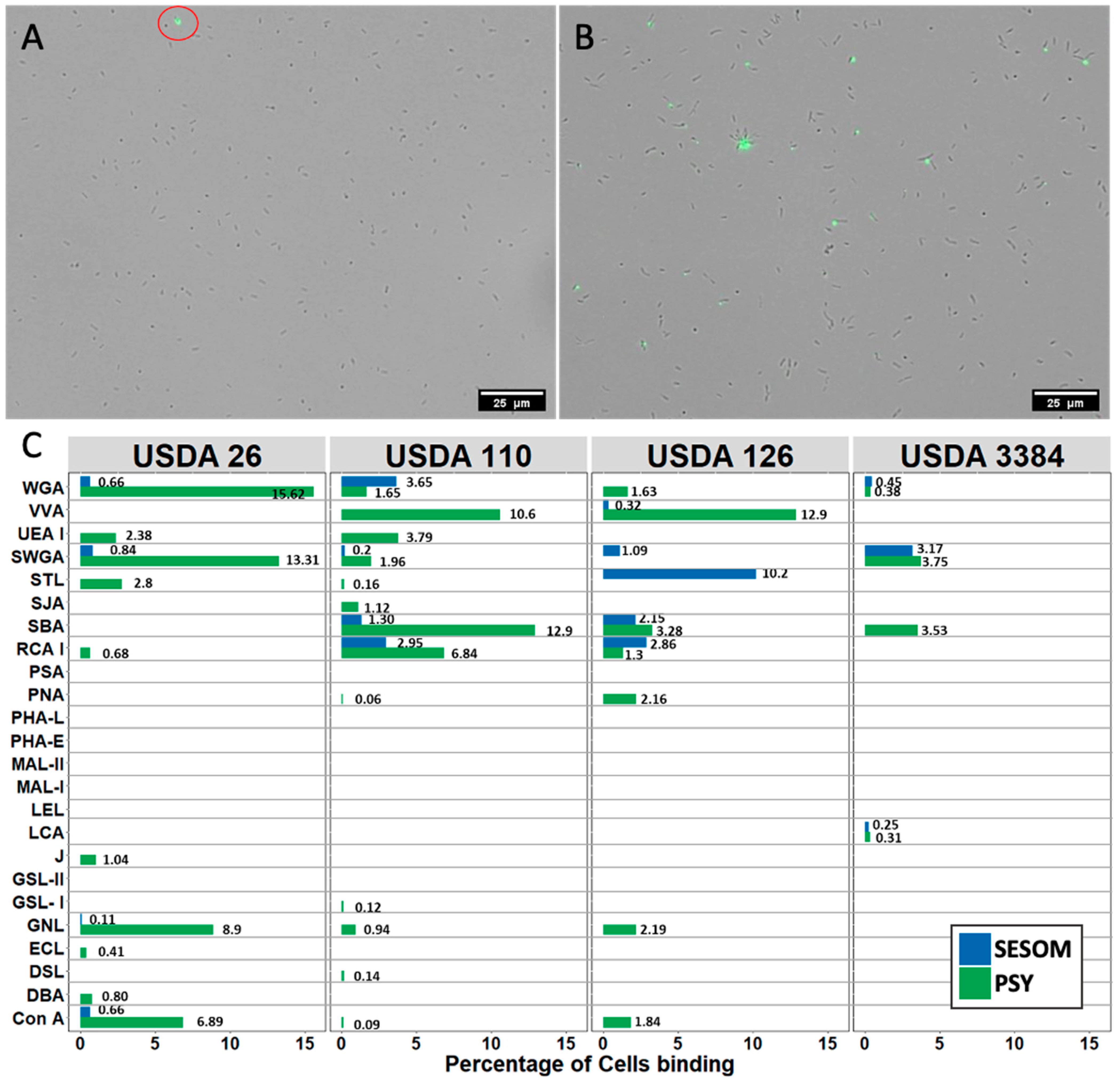
3.3. SESOM-Grown Populations Form No Biofilm but Display Increased Adherence to Soy Roots

Bradyrhizobium growing in SESOM displayed altered attachment-associated traits, so we quantified in vitro biofilm formation using the standard microtitre plate biofilm assay. PSY-grown USDA 110 and 26 formed biofilms on polystyrene within 24 h (Figure 5A), while USDA 126 and 3384 did not, even when incubated for 96 h (data not shown). Intriguingly, no strain formed biofilm in SESOM, with cells remaining in the planktonic phase. These results indicate that Bradyrhizobium growing in soil would not attach and form biofilms on more hydrophilic surfaces such as the Nunclon Delta Surface Polystyrene used in these experiments.
To gain insight into association with young soy roots, Bradyrhizobium was grown in PSY or SESOM, suspensions of exponentially growing cells were exposed to 9-day-old gnotobiotic seedlings for 60 min, and the adhering cells were counted. Adherence of all four PSY-cultured populations was scant (Figure 5B). In contrast, SESOM-grown USDA 110 displayed significantly more root adhesion. The adhesion data obtained for SESOM-grown cultures were more varied than for ex PSY. While root adhesion of strains 26, 126 and 3384 was not found statistically significant, it was higher than in PSY in multiple cases (Figure 5B). This greater variability in observed phenotype among individual experiments may be due to either variability among young roots, or due to variation in Bradyrhizobium population phenotype, as is also observed in lectin binding experiments.

4. Discussion

Our data reveal that Bradyrhizobium adopts adhesion-related surface properties when grown in the liquid soil extract SESOM. We observed the effect of soil nutrient environment on the surface hydrophobicity, motility, lectin binding spectrum and adherence capacity of the cells to soy roots. SESOM-grown populations of strains 110 and 3384 were more hydrophobic in nature, whereas there was no significant difference with strains 26 and 126. An effect of growth media on the surface hydrophobicity of bacterial cells has been reported for other bacterial species [25,26,27]. The hydrophobic properties of both the root surface and the bacteria involved in symbiosis contributes to one of the strongest forces in the initial root association. Bacterial cell surface hydrophobicity has been correlated directly to root attachment and competitive nodule forming ability [6,28]. Structural changes brought in LPS increase the cell surface hydrophobicity, which likely is significantly involved in the endocytotic infection process [7]. It is likely that the soil nutrients make 110 and 3384 more hydrophobic, and hence increase their chance of attachment by supporting them in their competitiveness to other bacteria. This was observed in the root adherence assay with strain 110 but not 3384.
The motility of rhizobia in response to chemical signaling plays a critical role in the initial contact, attachment and colonization of the root hair [29]. Mutants of Rhizobium meliloti LS-30 with defective motility took 5–20 times more time to adsorb to the root cell, and had delayed nodule formation, making them less competitive than other bacteria [11]. In this study, SESOM-grown populations were observed to be less motile. This could be because B. japonicum grown in L-arabinose as sugar source expresses lateral flagella along with subpolar flagella, making them move faster than those grown in other sugars [17]. Our lectin data indicated reduced surface sugar diversity when growing in SESOM. Alterations of the surface sugar moieties and their effect on symbiotic capability have been observed in Bradyrhizobium [9,30,31,32]. Surface sugar diversity has been reported to vary in composition when grown on different carbon sources [33,34]. These data suggest that the carbon sources in SESOM lead Bradyrhizobium to produce a different combination of extracellular polysaccharides and other sugar-containing polymers. Specifically, SESOM-grown populations appear to produce a smaller number of such extracellular polymers.
The contrast between biofilm formation on polystyrene and adherence to soy roots was unexpected, especially in the case of strain 110. While SESOM-grown populations failed to form biofilm on polystyrene, they were more prone to adhere to the soy roots. Arabinose as carbon source yielded opposite outcome. Medium characteristics such as osmolarity, nutrients, and factors derived from the biotic environment, may also affect attachment and/or biofilm formation. Little is known about the distribution of surface properties on young soybean roots, but the SESOM-grown, more hydrophobic strain 110 attached to the roots. In contrast it did not form detectable biofilm on the hydrophilic-modified Nunclon Delta Surface plates used. As confirmation, the hydrophilic ex PSY populations attached to the hydrophilic polystyrene, but not the soy roots. Biofilm formation of Rhizobium is affected by growth medium and incubation conditions [35]. Soybean agglutinin (SBA) lectin is thought to contribute to root-surface adherence [36]. While strains 110 and 126 displayed SBA binding under both culture condition (Figure 3), root adherence was significantly different. This suggest that root adherence may be driven largely by other factors.
The binding of specific lectins to only some cells in the population was surprising. Selective binding was not due to a shortage of lectin concentration as confirmed by experiments with increased concentration. The purity of the culture had also been verified regularly, so selective lectin binding pointed to differentiation in surface-exposed sugar polymers. This apparent differentiation of surface properties of cells in liquid shake cultures was unexpected. Our methodology included the removal of residual sugars from the culture medium by washing cells, and a wash step to remove unbound lectins after binding. These wash steps could have contributed to a loss of EPS into the liquid phase, leading to less or no bound lectin retained on cells. A similar observation of differential polysaccharide positioning has also been reported in Agrobacterium tumefaciens and Rhizobium leguminosarum [37,38]. While phenotypic population differentiation has been studied extensively in Bacillus subtilis [39,40], it is better known from environments allowing niche-specific physicochemical conditions such as in biofilms. The ecophysiological role of phenotypic differentiation of Bradyrhizobia presents itself as a new field of study.
From the results, it is clear that growth in a soil nutrient environment does affect the surface properties and adherence capacity of Bradyrhizobium. This is true especially for USDA 110, which is an agriculturally important rhizobial bacteria for soybean nitrogen fixation, where SESOM increased hydrophobicity and root adherence capability significantly [41]. The observed differences in the surface and phenotypic properties of the soil nutrient grown population leads to the conclusion that Bradyrhizobium adjust with the chemical cues that are present around them. The results from this study emphasize the need to further explore the effect of (secreted) root exudates on the surface and phenotypic behavior of Bradyrhizobium cells and resulting root adherence.

Author Contributions

Conceptualization, S.S. and V.S.B.; methodology, A.K.S. and V.S.B.; formal analysis, A.K.S.; investigation, A.K.S.; resources, S.S.; data curation, A.K.S.; writing—original draft preparation, A.K.S.; writing—review and editing, A.K.S., S.S. and V.S.B.; visualization, A.K.S.; supervision, V.S.B.; project administration, S.S; funding acquisition, S.S. All authors have read and agreed to the published version of the manuscript.

Funding

This material is based upon work supported by the National Science Foundation/EPSCoR RII Track-1: Building on The 2020 Vision: Expanding Research, Education and Innovation in South Dakota, Award OIA-1849206 and by the South Dakota Board of Regents.

Institutional Review Board Statement

Not applicable.

Informed Consent Statement

Not applicable.

Acknowledgments

We thank the NRRL Culture Collection of the Agricultural Research Service, United State Department of Agriculture for providing the bacterial cultures used in this study. We acknowledge use of the SDSU-FGCF supported in part by NSF/EPSCoR Grant No. 0091948 and by the State of South Dakota. We thank K M Taufiqur Rahman for his help with image analysis.

Conflicts of Interest

The authors declare no conflict of interest. The funders had no role in the design of the study; in the collection, analyses, or interpretation of data; in the writing of the manuscript, or in the decision to publish the results.

Disclaimer

Any opinions: findings, and conclusions or recommendations expressed in this material are those of the author(s) and do not necessarily reflect the views of the National Science Foundation.

References

  1. Peix, A.; Ramirez-Bahena, M.H.; Velazquez, E.; Bedmar, E.J. Bacterial Associations with Legumes. Crit. Rev. Plant Sci. 2015, 34, 17–42. [Google Scholar] [CrossRef]
  2. Kaneko, T.; Nakamura, Y.; Sato, S.; Minamisawa, K.; Uchiumi, T.; Sasamoto, S.; Watanabe, A.; Idesawa, K.; Iriguchi, M.; Kawashima, K.; et al. Complete genomic sequence of nitrogen-fixing symbiotic bacterium Bradyrhizobium japonicum USDA110. DNA Res. 2002, 9, 189–197. [Google Scholar] [CrossRef]
  3. Wang, Q.; Liu, J.E.; Zhu, H.Y. Genetic and Molecular Mechanisms Underlying Symbiotic Specificity in Legume-Rhizobium Interactions. Front. Plant Sci. 2018, 9, 313. [Google Scholar] [CrossRef]
  4. Liu, C.W.; Murray, J.D. The Role of Flavonoids in Nodulation Host-Range Specificity: An Update. Plants 2016, 5, 33. [Google Scholar] [CrossRef] [PubMed] [Green Version]
  5. Broghammer, A.; Krusell, L.; Blaise, M.; Sauer, J.; Sullivan, J.T.; Maolanon, N.; Vinther, M.; Lorentzen, A.; Madsen, E.B.; Jensen, K.J.; et al. Legume receptors perceive the rhizobial lipochitin oligosaccharide signal molecules by direct binding. Proc. Natl. Acad. Sci. USA 2012, 109, 13859–13864. [Google Scholar] [CrossRef] [PubMed] [Green Version]
  6. Ozawa, T.; Ogata, H.; Doi, R.; Komai, Y. Isolation of transposon Tn5-induced hydrophobic mutants of a Bradyrhizohium japonicum strain with improved competitive nodulation abilities. Soil Sci. Plant Nutr. 1992, 38, 545–552. [Google Scholar] [CrossRef]
  7. Kannenberg, E.L.; Carlson, R.W. Lipid A and O-chain modifications cause Rhizobium lipopolysaccharides to become hydrophobic during bacteroid development. Mol. Microbiol. 2001, 39, 379–391. [Google Scholar] [CrossRef] [Green Version]
  8. Park, K.M.; So, J.S. Altered cell surface hydrophobicity of lipopolysaccharide-deficient mutant of Bradyrhizobium japonicum. J. Microbiol. Meth. 2000, 41, 219–226. [Google Scholar] [CrossRef]
  9. Karr, D.B.; Liang, R.T.; Reuhs, B.L.; Emerich, D.W. Altered exopolysaccharides of Bradyrhizobium japonicum mutants correlate with impaired soybean lectin binding, but not with effective nodule formation. Planta 2000, 211, 218–226. [Google Scholar] [CrossRef]
  10. Quelas, J.I.; Mongiardini, E.J.; Casabuono, A.; Lopez-Garcia, S.L.; Althabegoiti, M.J.; Covelli, J.M.; Perez-Gimenez, J.; Couto, A.; Lodeiro, A.R. Lack of Galactose or Galacturonic Acid in Bradyrhizobium japonicum USDA 110 Exopolysaccharide Leads to Different Symbiotic Responses in Soybean. Mol. Plant Microbe Interact. 2010, 23, 1592–1604. [Google Scholar] [CrossRef] [Green Version]
  11. Caetanoanolles, G.; Wall, L.G.; Demicheli, A.T.; Macchi, E.M.; Bauer, W.D.; Favelukes, G. Role of Motility and Chemotaxis in Efficiency of Nodulation by Rhizobium meliloti. Plant Physiol. 1988, 86, 1228–1235. [Google Scholar] [CrossRef] [Green Version]
  12. Mitchell, J.G.; Kogure, K. Bacterial motility: Links to the environment and a driving force for microbial physics. FEMS Microbiol. Ecol. 2006, 55, 3–16. [Google Scholar] [CrossRef] [Green Version]
  13. Loderio, A.R.; Lopez-Garcia, S.L.; Vazquez, T.E.E.; Favelukes, G. Stimulation of adhesiveness, infectivity, and competitiveness for nodulation of Bradyrhizobium japonicum by its pretreatment with soybean seed lectin. FEMS Microbiol. Lett. 2000, 191, 157–158. [Google Scholar]
  14. De Hoff, P.L.; Brill, L.M.; Hirsch, A.M. Plant lectins: The ties that bind in root symbiosis and plant defense. Mol. Genet. Genom. 2009, 282, 1–15. [Google Scholar] [CrossRef] [PubMed] [Green Version]
  15. Ambrosi, M.; Cameron, N.R.; Davis, B.G. Lectins: Tools for the molecular understanding of the glycocode. Org. Biomol. Chem. 2005, 3, 1593–1608. [Google Scholar] [CrossRef] [PubMed]
  16. Smit, G.; Tubbing, D.M.J.; Kijne, J.W.; Lugtenberg, B.J.J. Role of Ca2+ in the Activity of Rhicadhesin from Rhizobium-Leguminosarum Biovar Viciae, Which Mediates the 1st Step in Attachment of Rhizobiaceae Cells to Plant-Root Hair Tips. Arch. Microbiol. 1991, 155, 278–283. [Google Scholar] [CrossRef]
  17. Covelli, J.M.; Althabegoiti, M.J.; Lopez, M.F.; Lodeiro, A.R. Swarming motility in Bradyrhizobium japonicum. Res. Microbiol. 2013, 164, 136–144. [Google Scholar] [CrossRef]
  18. Vilain, S.; Luo, Y.; Hildreth, M.B.; Brozel, V.S. Analysis of the life cycle of the soil saprophyte Bacillus cereus in liquid soil extract and in soil. Appl. Environ. Microbiol. 2006, 72, 4970–4977. [Google Scholar] [CrossRef] [Green Version]
  19. Mesa, S.; Hauser, F.; Friberg, M.; Malaguti, E.; Fischer, H.M.; Hennecke, H. Comprehensive assessment of the Regulons controlled by the FixLJ-FixK(2)-FixK(1) cascade in Bradyrhizobium japonicum. J. Bacteriol. 2008, 190, 6568–6579. [Google Scholar]
  20. Rosenberg, M. Microbial adhesion to hydrocarbons: Twenty-five years of doing MATH. FEMS Microbiol. Lett. 2006, 262, 129–134. [Google Scholar] [CrossRef]
  21. Zoueki, C.W.; Tufenkji, N.; Ghoshal, S. A modified microbial adhesion to hydrocarbons assay to account for the presence of hydrocarbon droplets. J. Colloid Interface Sci. 2010, 344, 492–496. [Google Scholar] [CrossRef] [Green Version]
  22. O’Toole, G.A. Microtiter Dish Biofilm Formation Assay. J. Vis. Exp. JoVE 2011, 47, 2473. [Google Scholar] [CrossRef]
  23. O’Toole, G.A.; Kolter, R. Initiation of biofilm formation in Pseudomonas fluorescens WCS365 proceeds via multiple, convergent signalling pathways: A genetic analysis. Mol. Microbiol. 1998, 28, 449–461. [Google Scholar] [CrossRef] [PubMed]
  24. White, L.J.; Brozel, V.S.; Subramanian, S. Isolation of Rhizosphere Bacterial Communities from Soil. Bio-protocol 2015, 5, e1569. [Google Scholar] [CrossRef]
  25. Ngwai, Y.B.; Sabiya, G. Cultivation in different growth media affects the expression of the cell surface hydrophobicity of bacteria. Cameroon J. Exp. Biol. 2007, 3, 26–29. [Google Scholar]
  26. Nwanyanwu, C.E.; Abu, G. Influence of growth media on hydrophobicity of phenol-utilizing bacteria found in petroleum refinery effluent. Int. Res. J. Biol. Sci. 2013, 2, 6–11. [Google Scholar]
  27. Deptula, A.; Mikucka, A.; Gospodarek, E. Effect of growth conditions on cell surface hydrophobicity of multiresistant Pseudomonas aeruginosa strains. Med. Dosw. I Mikrobiol. 2004, 56, 359–364. [Google Scholar]
  28. Ozawa, T.; Tokuda, S.; Komai, Y. Correlation between Competitive Nodulation Ability and Cell Surface Hydrophobicity of Bradyrhizobium japonicum. Bull. Jpn. Soc. Microb. Ecol. 1991, 6, 19–23. [Google Scholar] [CrossRef] [Green Version]
  29. Tambalo, D.D.; Yost, C.K.; Hynes, M.F. Motility and chemotaxis in the rhizobia. Biol. Nitrogen Fixat. 2015, 1, 337–348. [Google Scholar]
  30. Noh, J.G.; Jeon, H.E.; So, J.S.; Chang, W.S. Effects of the Bradyrhizobium japonicum waaL (rfaL) Gene on Hydrophobicity, Motility, Stress Tolerance, and Symbiotic Relationship with Soybeans. Int. J. Mol. Sci. 2015, 16, 16778–16791. [Google Scholar] [CrossRef] [Green Version]
  31. Lee, H.I.; In, Y.H.; Jeong, S.Y.; Jeon, J.M.; Noh, J.; So, J.S.; Chang, W.S. Inactivation of the lpcC gene alters surface-related properties and symbiotic capability of Bradyrhizobium japonicum. Lett. Appl. Microbiol. 2014, 59, 9–16. [Google Scholar] [CrossRef]
  32. Chang, W.S.; Park, K.M.; Koh, S.C.; So, J.S. Characterization of the Bradyrhizobium japonicum galE gene: Its impact on lipopolysaccharide profile and nodulation of soybean. FEMS Microbiol. Lett. 2008, 280, 242–249. [Google Scholar] [CrossRef] [Green Version]
  33. Staudt, A.K.; Wolfe, L.G.; Shrout, J.D. Variations in exopolysaccharide production by Rhizobium tropici. Arch. Microbiol. 2012, 194, 197–206. [Google Scholar] [CrossRef] [PubMed]
  34. Janczarek, M. Environmental Signals and Regulatory Pathways That Influence Exopolysaccharide Production in Rhizobia. Int. J. Mol. Sci. 2011, 12, 7898–7933. [Google Scholar] [CrossRef] [PubMed] [Green Version]
  35. Rinaudi, L.; Fujishige, N.A.; Hirsch, A.M.; Banchio, E.; Zorreguieta, A.; Giordano, W. Effects of nutritional and environmental conditions on Sinorhizobium meliloti biofilm formation. Res. Microbiol. 2006, 157, 867–875. [Google Scholar] [CrossRef] [PubMed]
  36. Pérez-Giménez, J.; Mongiardini, E.J.; Althabegoiti, M.J.; Covelli, J.; Quelas, J.I.; López-García, S.L.; Lodeiro, A.R. Soybean Lectin Enhances Biofilm Formation by Bradyrhizobium japonicum in the Absence of Plants. Int. J. Microbiol. 2009, 2009, 8. [Google Scholar] [CrossRef] [Green Version]
  37. Xu, J.; Kim, J.; Koestler, B.J.; Choi, J.H.; Waters, C.M.; Fuqua, C. Genetic analysis of Agrobacterium tumefaciens unipolar polysaccharide production reveals complex integrated control of the motile-to-sessile switch. Mol. Microbiol. 2013, 89, 929–948. [Google Scholar] [CrossRef] [Green Version]
  38. Laus, M.C.; Logman, T.J.; Lamers, G.E.; Van Brussel, A.A.N.; Carlson, R.W.; Kijne, J.W. A novel polar surface polysaccharide from Rhizobium leguminosarum binds host plant lectin. Mol. Microbiol. 2006, 59, 1704–1713. [Google Scholar] [CrossRef]
  39. Cao, H.J.; Kuipers, O.P. Influence of global gene regulatory networks on single cell heterogeneity of green fluorescent protein production in Bacillus subtilis. Microb. Cell Factories 2018, 17, 134. [Google Scholar] [CrossRef]
  40. Gallegos-Monterrosa, R.; Christensen, M.N.; Barchewitz, T.; Koppenhofer, S.; Priyadarshini, B.; Balint, B.; Maroti, G.; Kempen, P.J.; Dragos, A.; Kovacs, A.T. Impact of Rap-Phr system abundance on adaptation of Bacillus subtilis. Commun. Biol. 2021, 4, 468. [Google Scholar] [CrossRef]
  41. Zhang, L.; Liu, J.Y.; Gu, H.; Du, Y.F.; Zuo, J.F.; Zhang, Z.B.; Zhang, M.L.; Li, P.; Dunwell, J.M.; Cao, Y.R.; et al. Bradyrhizobium diazoefficiens USDA 110-Glycine max Interactome Provides Candidate Proteins Associated with Symbiosis. J. Proteome Res. 2018, 17, 3061–3074. [Google Scholar] [CrossRef] [PubMed] [Green Version]
Figure 1. Surface hydrophobicity of Bradyrhizobium strains cultured in either PSY-Arabinose or SESOM to mid exponential phase and partitioned into hexadecane as an apolar solvent. Data were analyzed by Student t-test, and p values indicate statistical significance (ns not significant, ** p < 0.01, *** p< 0.001).
Nitrogen 02 00031 g001
Figure 2. Motility radius of Bradyrhizobium strains spot-inoculated into either PSY-Arabinose or SESOM solidified with 0.35% agar and incubated for 10 d. Data were analyzed by Student t-test, and p values indicate statistical significance (ns not significant, ** p < 0.01, **** p < 0.0001).
Nitrogen 02 00031 g002
Figure 3. Lectin binding profiles of Bradyrhizobium strains cultured in either PSY-Arabinose or SESOM to mid-exponential phase. Binding intensity was scored by the shortest exposure time yielding visible fluorescence, with 100 ms scoring level 4, and <1200 ms scoring level 0.
Nitrogen 02 00031 g003
Figure 4. Lectin binds only to a subset of cells in the population. Binding of sWGA to B diazoefficiens USDA 26 grown in SESOM (A) and PSY (B), and the percentage of cells binding to the 25 lectins after culturing in PSY and SESOM (C).
Nitrogen 02 00031 g004
Figure 5. Biofilm (A) and soy root adherence (B) of Bradyrhizobium strains. Exponential phase Bradyrhizobium cultured in either PSY-Arabinose or SESOM were inoculated into 96-well polystyrene plates and biofilm formed after 24 h quantified by crystal violet staining (A), or exposed to 9 d old roots for 60 min (B). Data were analyzed by Student t-test, and p values indicate statistical significance (ns not significant, **** p < 0.0001).
Nitrogen 02 00031 g005
Table 1. List of lectins used.
Lectin NameLectin AbbreviationSugar Specificity *Conjugated with
Concanavalin ACon ABranched and terminal mannose [High-Man, Man α-1,6(Mana-1,3)]FITC
Dolichos biflorus agglutinin DBAGlcNAc β-1,4 GlcNAc oligomers and LacNAc (Gal β 1,4 GlcNAc)FITC
Peanut agglutininPNATerminal Gal (β-OR) FITC
Ricinus communis agglutinin I RCA IGalFITC
Soybean agglutininSBAα- or βLinked terminal GaINAc, GalNAc α-1,3 GalFITC
Ulex europaeus agglutinin IUEA Iα-FucoseFITC
Wheat germ agglutininWGAβ-GlcNAc, sialic acid, GalNAcFITC
Datura Stramonium LectinDSLGlcNAc β-1,4 GlcNAc oligomers and LacNAc (Gal β 1,4 GlcNAc)FITC
Galanthus Nivalis LectinGNLTerminal α-1, 3 mannoseFITC
Lens culinaris agglutininLCAComplex (man/GlcNAc core with α-1,6 Fuc)Biotin
Pisum sativum agglutininPSAMan, (Fuc a-1,6 GlcNAc, α-D-Glc, α-D-Man)Biotin
Griffonia simplicifolia lectin I GSL-Iα-Galactose, also binds some GalNAcBiotin
Sophora japonica agglutininSJAβGalNAcBiotin
Vicia villosa agglutininVVAGaINAcBiotin
Lycopersicon esculentum (tomato) lectinLELβ-1,4 GlcNAc oligomersBiotin
Solanum tuberosum (potato) lectinSTLGlcNAc oligomers, LacNAcBiotin
Griffonia simplicifolia lectin IIGSL-IITerminal GlcNAcBiotin
Succinylated Wheat germ agglutininsWGAGalcNAcBiotin
Erythrina cristagalli lectinECLGal β-1,4 GalNAcBiotin
Artocarpus integrifolia (Jacalin)JGal β-1,3 GalNAcBiotin
Phaseolus vulgaris erythroagglutininPHA-EComplex-type N-glycans with outer Gal and bisecting GlcNAcBiotin
Phaseolus vulgaris leucoagglutininPHA-Lβ-1,6 Brandched tri mannosyl core N-linked glycansBiotin
Maackia amurensis lectin IMAL-IGalactosyl (β-1,4) N-acetylglucos amine, (α-2,3) sialic acidBiotin
Maackia amurensis lectin IIMAL-IIα-2,3 sialic acid-LacNAc structureBiotin
* Vector Laboratories.
Publisher’s Note: MDPI stays neutral with regard to jurisdictional claims in published maps and institutional affiliations.

