Fractal Parameters as Independent Biomarkers in the Early Diagnosis of Pediatric Onset Inflammatory Bowel Disease
Abstract
1. Introduction
2. Materials and Methods
2.1. Patients
2.2. Intestinal Tissue Preparation and Staining
2.3. Medical Image Preprocessing
2.4. Niblack Thresholding
2.5. Fractal Analysis
2.6. Statistical Analysis
3. Results
3.1. Demographic and Clinical Characteristics
3.2. Histological Activity
3.3. Fractal Dimension
3.4. Lacunarity
3.5. Intersegmental Comparison
4. Discussion
5. Conclusions
Author Contributions
Funding
Institutional Review Board Statement
Informed Consent Statement
Data Availability Statement
Acknowledgments
Conflicts of Interest
References
- Husain, A.; Nanda, M.N.; Chowdary, M.S.; Sajid, M. Fractals: An eclectic survey, part II. Fractal Fract. 2022, 6, 379. [Google Scholar] [CrossRef]
- de Mattos, A.C.; Florindo, J.B.; Adam, R.L.; Lorand-Metze, I.; Metze, K. The fractal dimension suggests two chromatin configurations in small cell neuroendocrine lung cancer and is an independent unfavorable prognostic factor for overall survival. Microsc. Microanal. 2022, 28, 522–526. [Google Scholar] [CrossRef]
- Karri, S.; Aviel-Ronen, S.; Firer, M.A. Fractal and textural imaging identify new subgroups of patients with colorectal cancer based on biophysical properties of the cancer cells. Pathol. Res. Pract. 2022, 238, 154040. [Google Scholar] [CrossRef]
- Dinčić, M.; Todorović, J.; Nešović-Ostojić, J.; Kovačević, S.; Dunđerović, D.; Lopičić, S.; Spasić, S.; Radojević-Škodrić, S.; Stanisavljević, D.; Ilić, A.Ž. The fractal and GLCM textural parameters of chromatin may be potential biomarkers of papillary thyroid carcinoma in Hashimoto’s thyroiditis specimens. Microsc. Microanal. 2020, 26, 717–730. [Google Scholar] [CrossRef]
- Dinčić, M.; Popović, T.B.; Kojadinović, M.; Trbovich, A.M.; Ilić, A.Ž. Morphological, fractal, and textural features for the blood cell classification: The case of acute myeloid leukemia. Eur. Biophys. J. 2021, 50, 1111–1127. [Google Scholar] [CrossRef] [PubMed]
- Einstein, A.J.; Wu, H.-S.; Gil, J. Self-affinity and lacunarity of chromatin texture in benign and malignant breast epithelial cell nuclei. Phys. Rev. Lett. 1998, 80, 397–400. [Google Scholar] [CrossRef]
- Ioelovich, M. Fractal dimensions of cell wall in growing cotton fibers. Fractal Fract. 2020, 4, 6. [Google Scholar] [CrossRef]
- Arsac, L.M.; Weissland, T. Multifractality in the movement system when adapting to arm cranking in wheelchair athletes, able-bodied athletes, and untrained people. Fractal Fract. 2022, 6, 176. [Google Scholar] [CrossRef]
- Smith, T.G., Jr.; Lange, G.D.; Marks, W.B. Fractal methods and results in cellular morphology—Dimensions, lacunarity and multifractals. J. Neurosci. Methods 1996, 69, 123–136. [Google Scholar] [CrossRef] [PubMed]
- Tél, T. Fractals, multifractals, and thermodynamics: An introductory review. Z. Naturforschung A 1988, 43, 1154–1174. [Google Scholar] [CrossRef]
- Charisis, V.S.; Hadjileontiadis, L.J.; Liatsos, C.N.; Mavrogiannis, C.C.; Sergiadis, G.D. Capsule endoscopy image analysis using texture information from various colour models. Comput. Methods Programs Biomed. 2012, 107, 61–74. [Google Scholar] [CrossRef]
- Oprić, D.; Stankovich, A.D.; Nenadović, A.; Kovačević, S.; Obradović, D.D.; De Luka, S.; Nešović-Ostojić, J.; Milašin, J.; Ilić, A.Ž.; Trbovich, A.M. Fractal analysis tools for early assessment of liver inflammation induced by chronic consumption of linseed, palm and sunflower oils. Biomed. Sign. Process. Control 2020, 61, 101959. [Google Scholar] [CrossRef]
- Stojić, D.; Radošević, D.; Rajković, N.; Marić, D.L.; Milošević, N.T. Classification by morphology of multipolar neurons of the human principal olivary nucleus. Neurosci. Res. 2021, 170, 66–75. [Google Scholar] [CrossRef]
- Lyu, X.; Jajal, P.; Tahir, M.Z.; Zhang, S. Fractal dimension of retinal vasculature as an image quality metric for automated fundus image analysis systems. Sci. Rep. 2022, 12, 11868. [Google Scholar] [CrossRef]
- Freeborn, T.J.; Fu, B. Fatigue-induced Cole electrical impedance model changes of biceps tissue bioimpedance. Fractal Fract. 2018, 2, 27. [Google Scholar] [CrossRef]
- Gladun, K.V. Higuchi fractal dimension as a method for assessing response to sound stimuli in patients with diffuse axonal brain injury. Sovrem. Tehnologii Med. 2020, 12, 63. [Google Scholar] [CrossRef]
- Naik, G.R.; Arjunan, S.; Kumar, D. Applications of ICA and fractal dimension in sEMG signal processing for subtle movement analysis: A review. Australas. Phys. Eng. Sci. Med. 2011, 34, 179–193. [Google Scholar] [CrossRef] [PubMed]
- Stylianou, O.; Kaposzta, Z.; Czoch, A.; Stefanovski, L.; Yabluchanskiy, A.; Racz, F.S.; Ritter, P.; Eke, A.; Mukli, P. Scale-free functional brain networks exhibit increased connectivity, are more integrated and less segregated in patients with Parkinson’s disease following dopaminergic treatment. Fractal Fract. 2022, 6, 737. [Google Scholar] [CrossRef]
- Goh, V.; Sanghera, B.; Wellsted, D.M.; Sundin, J.; Halligan, S. Assessment of the spatial pattern of colorectal tumour perfusion estimated at perfusion CT using two-dimensional fractal analysis. Eur. Radiol. 2009, 19, 1358–1365. [Google Scholar] [CrossRef]
- Streba, L.; Forţofoiu, M.C.; Popa, C.; Ciobanu, D.; Gruia, C.L.; Mogoantă, S.; Streba, C.T. A pilot study on the role of fractal analysis in the microscopic evaluation of colorectal cancers. Rom. J. Morphol. Embryol. 2015, 56, 191–196. [Google Scholar] [PubMed]
- Watanabe, H.; Hayano, K.; Ohira, G.; Imanishi, S.; Hanaoka, T.; Hirata, A.; Kano, M.; Matsubara, H. Quantification of structural heterogeneity using fractal analysis of contrast-enhanced CT image to predict survival in gastric cancer patients. Dig Dis Sci. 2021, 66, 2069–2074. [Google Scholar] [CrossRef] [PubMed]
- Dzik-Jurasz, A.; Walker-Samuel, S.; Leach, M.O.; Brown, G.; Padhani, A.; George, M.; Collins, D.J. Fractal parameters derived from analysis of DCE-MRI data correlates with response to therapy in rectal carcinoma. In Proceedings of the International Society for Magnetic Resonance in Medicine 11, ISMRM 12th Scientific Meeting, Kyoto, Japan, 15–21 May 2004; p. 2503. [Google Scholar]
- Tochigi, T.; Kamran, S.C.; Parakh, A.; Noda, Y.; Ganeshan, B.; Blaszkowsky, L.S.; Ryan, D.P.; Allen, J.N.; Berger, D.L.; Wo, J.Y.; et al. Response prediction of neoadjuvant chemoradiation therapy in locally advanced rectal cancer using CT-based fractal dimension analysis. Eur. Radiol. 2022, 32, 2426–2436. [Google Scholar] [CrossRef]
- Jain, S.; Seal, A.; Ojha, A.; Krejcar, O.; Bureš, J.; Tachecí, I.; Yazidi, A. Detection of abnormality in wireless capsule endoscopy images using fractal features. Comput. Biol. Med. 2020, 127, 104094. [Google Scholar] [CrossRef] [PubMed]
- Gryglewski, A.; Henry, B.M.; Mrozek, M.; Żelawski, M.; Piech, K.; Tomaszewski, K.A. Sensitivity and specificity of fractal analysis to distinguish between healthy and pathologic rectal mucosa microvasculature seen during colonoscopy. Surg. Laparosc. Endosc. Percutan. Tech. 2016, 26, 358–363. [Google Scholar] [CrossRef] [PubMed]
- Paramasivam, A.; Kamalanand, K.; Emmanuel, C.; Mahadevan, B.; Sundravadivelu, K.; Raman, J.; Jawahar, P.M. Influence of electrode surface area on the fractal dimensions of electrogastrograms and fractal analysis of normal and abnormal digestion process. In Proceedings of the 2018 International Conference on Recent Trends in Electrical, Control and Communication (RTECC 2018), Malaysia, Malaysia, 20–22 March 2018; pp. 245–250. [Google Scholar] [CrossRef]
- Yan, R.; Guo, X. Nonlinear fractal dynamics of human colonic pressure activity based upon the box-counting method. Comput. Methods Biomech. Biomed. Eng. 2013, 16, 660–668. [Google Scholar] [CrossRef]
- Dimoulas, C.; Kalliris, G.; Papanikolaou, G.; Kalampakas, A. Long-term signal detection, segmentation and summarization using wavelets and fractal dimension: A bioacoustics application in gastrointestinal-motility monitoring. Comput. Biol. Med. 2007, 37, 438–462. [Google Scholar] [CrossRef]
- Weber, M.C.; Schmidt, K.; Buck, A.; Kasajima, A.; Becker, S.; Wilhelm, D.; Friess, H.; Neumann, P.A. P067 Fractal analysis of extracellular matrix as a new histological method for observer-independent quantification of intestinal fibrosis in Crohn’s disease. J. Crohn’s Colitis 2023, 17 (Suppl. 1), i234. [Google Scholar] [CrossRef]
- Hadjileontiadis, L.J.; Rekanos, I.T. Detection of explosive lung and bowel sounds by means of fractal dimension. IEEE Signal Process. Lett. 2003, 10, 311–314. [Google Scholar] [CrossRef]
- Almassalha, L.; Tiwari, A.; Ruhoff, P.; Stypula-Cyrus, Y.; Cherkezyan, L.; Matsuda, H.; Dela Cruz, M.A.; Chandler, J.E.; White, C.; Maneval, C.; et al. The global relationship between chromatin physical topology, fractal structure, and gene expression. Sci. Rep. 2017, 7, 41061. [Google Scholar] [CrossRef]
- Bancaud, A.; Lavelle, C.; Huet, S.; Ellenberg, J. A fractal model for nuclear organization: Current evidence and biological implications. Nucl. Acids Res. 2012, 40, 8783–8792. [Google Scholar] [CrossRef]
- Metze, K.; Adam, R.; Florindo, J.B. The fractal dimension of chromatin—A potential molecular marker for carcinogenesis, tumor progression and prognosis. Expert Rev. Mol. Diagn. 2019, 19, 299–312. [Google Scholar] [CrossRef] [PubMed]
- Vesković, M.; Labudović-Borović, M.; Zaletel, I.; Rakočević, J.; Mladenović, D.; Jorgačević, B.; Vučević, D.; Radosavljević, T. The effects of betaine on the nuclear fractal dimension, chromatin texture, and proliferative activity in hepatocytes in mouse model of nonalcoholic fatty liver disease. Microsc. Microanal. 2018, 24, 132–138. [Google Scholar] [CrossRef]
- Ray, G.; Longworth, M.S. Epigenetics, DNA organization, and inflammatory bowel disease. Inflamm. Bowel Dis. 2019, 25, 235–247. [Google Scholar] [CrossRef]
- Fedor, I.; Zold, E.; Barta, Z. Temporal relationship of extraintestinal manifestations in inflammatory bowel disease. J. Clin. Med. 2021, 10, 5984. [Google Scholar] [CrossRef] [PubMed]
- Gajendran, M.; Loganathan, P.; Jimenez, G.; Catinella, A.P.; Ng, N.; Umapathy, C.; Ziade, N.; Hashash, J.G. A comprehensive review and update on ulcerative colitis. Dis. Mon. 2019, 65, 100851. [Google Scholar] [CrossRef] [PubMed]
- Sathiyasekaran, M.; Shivbalan, S. Crohn’s disease. Indian J. Pediatr. 2006, 73, 723–729. [Google Scholar] [CrossRef]
- Fuller, M.K. Pediatric inflammatory bowel disease: Special considerations. Surg. Clin. N. Am. 2019, 99, 1177–1183. [Google Scholar] [CrossRef]
- De Roche, T.C.; Xiao, S.Y.; Liu, X. Histological evaluation in ulcerative colitis. Gastroenterol. Rep. 2014, 2, 178–192. [Google Scholar] [CrossRef]
- Torres, J.; Bonovas, S.; Doherty, G.; Kucharzik, T.; Gisbert, J.P.; Raine, T.; Adamina, M.; Armuzzi, A.; Bachmann, O.; Bager, P.; et al. ECCO Working Group. ECCO Guidelines on Therapeutics in Crohn’s Disease: Medical Treatment. J. Crohn’s Colitis 2020, 14, 4–22. [Google Scholar] [CrossRef]
- Szymanska, E.; Wierzbicka, A.; Dadalski, M.; Kierkus, J. Fecal zonulin as a noninvasive biomarker of intestinal permeability in pediatric patients with inflammatory bowel diseases—Correlation with disease activity and fecal calprotectin. J. Clin. Med. 2021, 10, 3905. [Google Scholar] [CrossRef]
- Ordog, T.; Syed, S.; Hayashi, Y.; Asuzu, D.T. Epigenetics and chromatin dynamics: A review and a paradigm for functional disorders. Neurogastroenterol. Motil. 2012, 24, 1054–1068. [Google Scholar] [CrossRef]
- Ansari, I.; Raddatz, G.; Gutekunst, J.; Ridnik, M.; Cohen, D.; Abu-Remaileh, M.; Tuganbaev, T.; Shapiro, H.; Pikarsky, E.; Elinav, E.; et al. The microbiota programs DNA methylation to control intestinal homeostasis and inflammation. Nat. Microbiol. 2020, 5, 610–619. [Google Scholar] [CrossRef] [PubMed]
- Ruifrok, A.C.; Johnston, D.A. Quantification of histochemical staining by color deconvolution. Anal. Quant. Cytol. Histol. 2001, 23, 291–299. [Google Scholar]
- Landini, G.; Martinelli, G.; Piccinini, F. Colour deconvolution: Stain unmixing in histological imaging. Bioinformatics 2021, 37, 1485–1487. [Google Scholar] [CrossRef] [PubMed]
- Singh, T.R.; Roy, S.; Singh, O.I.; Sinam, T.; Singh, K.M. A new local adaptive thresholding technique in binarization. IJCSI Int. J. Comp. Sci. 2011, 8, 271–277. [Google Scholar] [CrossRef]
- Karperien, A.L.; Jelinek, H.F. Box-counting fractal analysis: A primer for the clinician. In The Fractal Geometry of the Brain; Di Ieva, A., Ed.; Springer: New York, NY, USA, 2016; pp. 91–108. [Google Scholar] [CrossRef]
- Glickman, J.N.; Bousvaros, A.; Farraye, F.A.; Zholudev, A.; Friedman, S.; Wang, H.H.; Leichtner, A.M.; Odze, R.D. Pediatric patients with untreated ulcerative colitis may present initially with unusual morphologic findings. Am. J. Surg. Pathol. 2004, 28, 190–197. [Google Scholar] [CrossRef]
- Bancaud, A.; Huet, S.; Daigle, N.; Mozziconacci, J.; Beaudouin, J.; Ellenberg, J. Molecular crowding affects diffusion and binding of nuclear proteins in heterochromatin and reveals the fractal organization of chromatin. EMBO J. 2009, 28, 3785–3798. [Google Scholar] [CrossRef] [PubMed]
- Mobley, A.S. Chapter 4—Induced pluripotent stem cells. In Neural Stem Cells and Adult Neurogenesi; Mobley, A.S., Ed.; Academic Press: London, UK, 2019; ISBN 9780128110140. [Google Scholar] [CrossRef]
- Chessum, N.; Jones, K.; Pasqua, E.; Tucker, M. Recent advances in cancer therapeutics. Prog. Med. Chem. 2015, 54, 1–63. [Google Scholar] [CrossRef]
- De Bruyn, J.; Wichers, C.; Radstake, T.; Broen, J.; D’Haens, G. Histone deacetylases in inflammatory mucosa distinguish Crohn’s disease from ulcerative colitis. J. Crohn’s Colitis 2015, 9 (Suppl. 1), S87–S88. [Google Scholar] [CrossRef]
- Blanchard, F.; Chipoy, C. Histone deacetylase inhibitors: New drugs for the treatment of inflammatory diseases? Drug. Discov. Today 2005, 10, 197–204. [Google Scholar] [CrossRef] [PubMed]


| Variable | Control | UC | CD | Overall p |
|---|---|---|---|---|
| Gender | ||||
| Male | 60.0% | 60.0% | 78.6% | 0.498 |
| Female | 40.0% | 40.0% | 21.4% | |
| Age | 11.3 ± 5.1 | 14.4 ± 3.3 | 12.1 ± 4.8 | 0.271 |
| PUCAI | 32.5 ± 19.0 | |||
| PCDAI | 17.5 ± 6.3 |
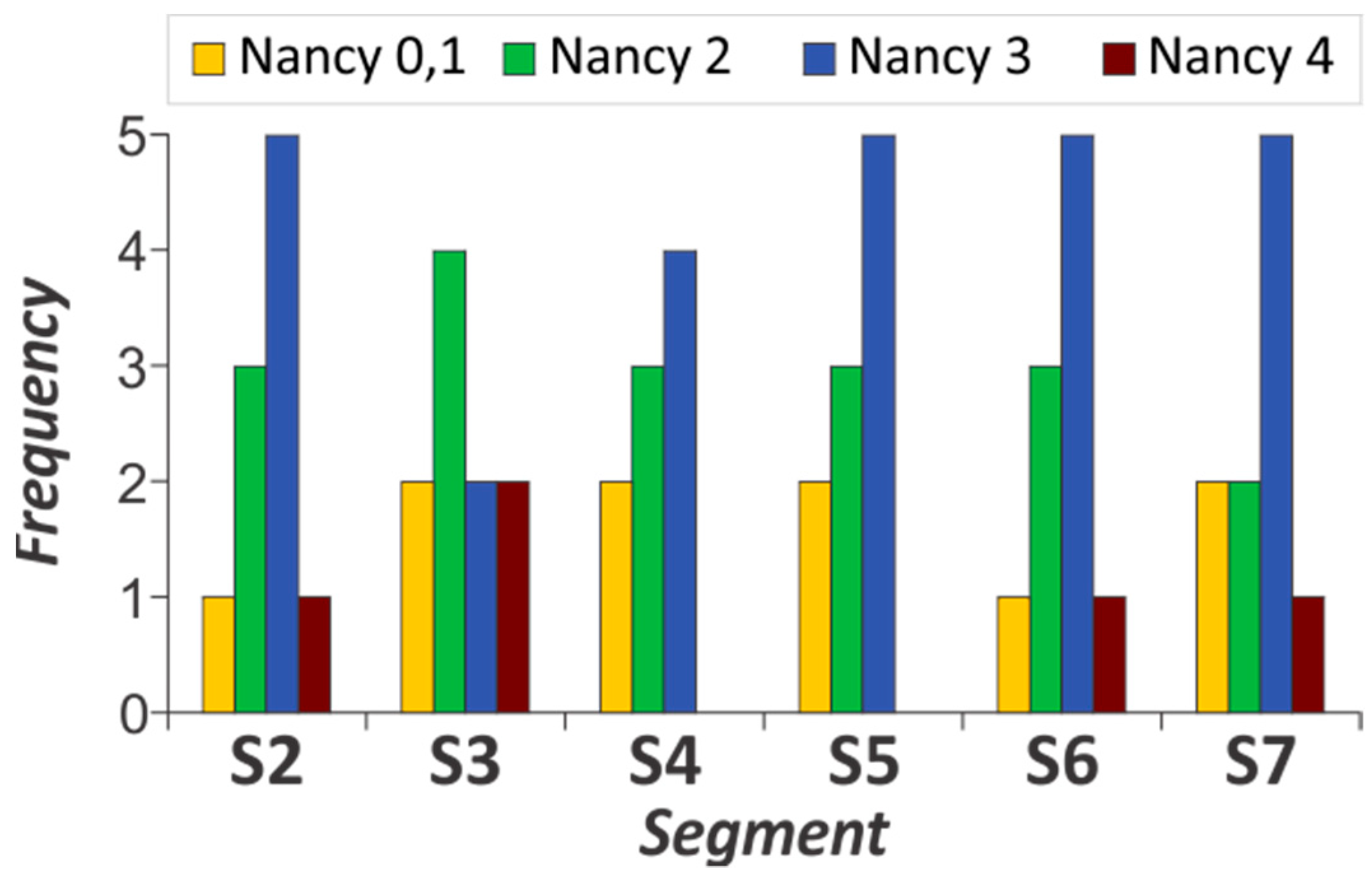
| Segment | Nancy 0, 1 | Nancy 2 | Nancy 3 | Nancy 4 |
|---|---|---|---|---|
| S2 | 1 (10.0%) | 3 (30.0%) | 5 (50.0%) | 1 (10.0%) |
| S3 | 2 (20.0%) | 4 (40.0%) | 2 (20.0%) | 2 (20.0%) |
| S4 | 2 (22.2%) | 3 (33.3%) | 4 (44.4%) | |
| S5 | 2 (20.0%) | 3 (30.0%) | 5 (50.0%) | |
| S6 | 1 (10.0%) | 3 (30.0%) | 5 (50.0%) | 1 (10.0%) |
| S7 | 2 (20.0%) | 2 (20.0%) | 5 (50.0%) | 1 (10.0%) |
| Segment | GHAS 1,2,3,4 | GHAS 5,6,7 | GHAS 8,9,10 | GHAS 11–16 |
|---|---|---|---|---|
| S1 | 3 (21.43%) | 6 (42.86%) | 4 (28.57%) | 1 (7.14%) |
| S2 | 6 (42.86%) | 5 (35.71%) | 1 (7.14%) | 2 (14.29%) |
| S3 | 6 (42.86%) | 4 (28.57%) | 2 (14.29%) | 2 (14.29%) |
| S4 | 4 (28.57%) | 4 (28.57%) | 5 (35.71%) | 1 (7.14%) |
| S5 | 7 (50.00%) | 2 (14.29%) | 2 (14.29%) | 3 (21.43%) |
| S6 | 5 (35.71%) | 4 (28.57%) | 3 (21.43%) | 2 (14.29%) |
| S7 | 7 (50.00%) | 3 (21.43%) | 4 (28.57%) |
| Intestinal Segment | Fractal Dimension (FD) | Statistical Signif. (p Values) | ||
|---|---|---|---|---|
| Crohn’s Disease (CD) | Ulcerative Colitis (UC) | Control Group | ||
| S1 | 1.728(1.586–1.831) | 1.722 (1.593–1.802) | 1.728 (1.531–1.819) | CD–UC (0.042) |
| CD–Control (1.000) | ||||
| UC–Control (0.107) | ||||
| S2 | 1.732 (1.523–1.842) | 1.755 (1.559–1.834) | 1.741 (1.599–1.818) | CD–UC (<0.0001) |
| CD–Control (0.097) | ||||
| UC–Control (0.035) | ||||
| S3 | 1.732 (1.529–1.829) | 1.752 (1.135–1.840) | 1.736 (1.547–1.835) | CD–UC (<0.0001) |
| CD–Control (0.814) | ||||
| UC–Control (0.004) | ||||
| S4 | 1.736 (1.566–1.831) | 1.738 (1.048–1.843) | 1.728 (1.538–1.812) | CD–UC (1.000) |
| CD–Control (0.106) | ||||
| UC–Control (0.023) | ||||
| S5 | 1.734 (1.593–1.816) | 1.747 (1.118–1.948) | 1.734 (1.555–1.810) | CD–UC (0.004) |
| CD–Control (1.000) | ||||
| UC–Control (0.001) | ||||
| S6 | 1.722 (1.521–1.940) | 1.749 (1.601–1.947) | 1.727 (1.585–1.826) | CD–UC (<0.0001) |
| CD–Control (1.000) | ||||
| UC–Control (<0.0001) | ||||
| S7 | 1.719 (1.524–1.944) | 1.733 (1.511–1.824) | 1.730 (1.536–1.818) | CD–UC (0.010) |
| CD–Control (0.014) | ||||
| UC–Control (1.000) | ||||
| S8 | 1.731 (1.617–1.788) | 1.749 (1.605–1.834) | CD–Control (0.005) | |
| Intestinal Segment | Lacunarity (Lac) | Statistical Signif. (p Values) | ||
|---|---|---|---|---|
| Crohn’s Disease (CD) | Ulcerative Colitis (UC) | Control Group | ||
| S1 | 0.279 (0.172–0.430) | 0.285 (0.209–0.455) | 0.279 (0.197–0.517) | All groups (0.074) |
| S2 | 0.282 (0.163–0.470) | 0.253 (0.167–0.422) | 0.266 (0.178–0.407) | CD–UC (<0.0001) |
| CD–Control (0.087) | ||||
| UC–Control (0.002) | ||||
| S3 | 0.279 (0.174–0.460) | 0.260 (0.175–0.404) | 0.274 (0.192–0.481) | CD–UC (<0.0001) |
| CD–Control (1.000) | ||||
| UC–Control (<0.0001) | ||||
| S4 | 0.271 (0.184–0.442) | 0.269 (0.181–0.554) | 0.280 (0.198–0.482) | CD–UC (0.350) |
| CD–Control (0.039) | ||||
| UC–Control (<0.0001) | ||||
| S5 | 0.273 (0.273–0.430) | 0.258 (0.037–0.365) | 0.275 (0.191–0.475) | CD–UC (<0.0001) |
| CD–Control (1.000) | ||||
| UC–Control (<0.0001) | ||||
| S6 | 0.287 (0.042–0.510) | 0.257 (0.038–0.378) | 0.280 (0.190–0.467) | CD–UC (<0.0001) |
| CD–Control (1.000) | ||||
| UC–Control (<0.0001) | ||||
| S7 | 0.289 (0.040–0.480) | 0.272 (0.183–0.450) | 0.276 (0.182–0.504) | CD–UC (0.001) |
| CD–Control (0.015) | ||||
| UC–Control (0.909) | ||||
| S8 | 0.276 (0.221–0.396) | 0.263 (0.199–0.418) | CD–Control (0.126) | |
| Intestinal Segment | Fractal Dimension (FD) | ||
|---|---|---|---|
| Crohn’s Disease (CD) | Ulcerative Colitis (UC) | Control Group | |
| S1 | 1.728 (1.586–1.832) | 1.723 (1.593–1.802) | 1.728 (1.531–1.819) |
| S2 | 1.732 (1.524–1.843) | 1.755 (1.559–1.834) | 1.741 (1.600–1.819) |
| S3 | 1.732 (1.529–1.829) | 1.752 (1.135–1.840) | 1.736 (1.547–1.835) |
| S4 | 1.736 (1.567–1.831) | 1.738 (1.048–1.843) | 1.728 (1.538–1.812) |
| S5 | 1.734 (1.593–1.816) | 1.747 (1.118–1.948) | 1.734 (1.555–1.811) |
| S6 | 1.722 (1.521–1.940) | 1.749 (1.601–1.948) | 1.727 (1.585–1.826) |
| S7 | 1.719 (1.524–1.944) | 1.733 (1.511–1.824) | 1.729 (1.536–1.818) |
| Stat.sign. (p values) | S7-S1, S7-S3, S7-S2, S7-S5, S7-S4 (p < 0.025) | S1-S4, S1-S6, S1-S5, S1-S3, S1-S2 (p < 0.002) S7-S6, S7-S5, S7-S3, S7-S2 (p < 0.013) | S1-S2, S4-S2, S7-S2 (p < 0.090) |
| Intestinal Segment | Lacunarity (Lac) | ||
|---|---|---|---|
| Crohn’s Disease (CD) | Ulcerative Colitis (UC) | Control Group | |
| S1 | 0.279 (0.173–0.430) | 0.285 (0.209–0.455) | 0.279 (0.197–0.517) |
| S2 | 0.279 (0.163–0.470) | 0.253 (0.167–0.422) | 0.266 (0.178–0.407) |
| S3 | 0.279 (0.174–0.460) | 0.259 (0.175–0.404) | 0.274 (0.192–0.482) |
| S4 | 0.271 (0.184–0.442) | 0.269 (0.181–0.555) | 0.280 (0.198–0.482) |
| S5 | 0.273 (0.184–0.430) | 0.258 (0.037–0.365) | 0.275 (0.191–0.475) |
| S6 | 0.287 (.0420–0.510) | 0.257 (0.038–0.378) | 0.280 (0.190–0.467) |
| S7 | 0.289 (0.040–0.480) | 0.272 (0.183–0.450) | 0.276 (0.182–0.504) |
| Stat.sign. (p values) | S2-S7, S3-S7, S4-S7, S5-S7 (p < 0.022) | S2-S1, S3-S1, S4-S1, S6-S1 (p < 0.0001), S2-S7, S3-S7, S6-S7 (p < 0.011) S2-S4 (p < 0.025) | S2-S1, S2-S4, S2-S6 (p < 0.040) |
Disclaimer/Publisher’s Note: The statements, opinions and data contained in all publications are solely those of the individual author(s) and contributor(s) and not of MDPI and/or the editor(s). MDPI and/or the editor(s) disclaim responsibility for any injury to people or property resulting from any ideas, methods, instructions or products referred to in the content. |
© 2023 by the authors. Licensee MDPI, Basel, Switzerland. This article is an open access article distributed under the terms and conditions of the Creative Commons Attribution (CC BY) license (https://creativecommons.org/licenses/by/4.0/).
Share and Cite
Makević, V.; Milovanovich, I.D.; Popovac, N.; Janković, R.; Trajković, J.; Vuković, A.; Milosević, B.; Jevtić, J.; de Luka, S.R.; Ilić, A.Ž. Fractal Parameters as Independent Biomarkers in the Early Diagnosis of Pediatric Onset Inflammatory Bowel Disease. Fractal Fract. 2023, 7, 619. https://doi.org/10.3390/fractalfract7080619
Makević V, Milovanovich ID, Popovac N, Janković R, Trajković J, Vuković A, Milosević B, Jevtić J, de Luka SR, Ilić AŽ. Fractal Parameters as Independent Biomarkers in the Early Diagnosis of Pediatric Onset Inflammatory Bowel Disease. Fractal and Fractional. 2023; 7(8):619. https://doi.org/10.3390/fractalfract7080619
Chicago/Turabian StyleMakević, Vedrana, Ivan D. Milovanovich, Nevena Popovac, Radmila Janković, Jelena Trajković, Andrija Vuković, Bojana Milosević, Jovan Jevtić, Silvio R. de Luka, and Andjelija Ž. Ilić. 2023. "Fractal Parameters as Independent Biomarkers in the Early Diagnosis of Pediatric Onset Inflammatory Bowel Disease" Fractal and Fractional 7, no. 8: 619. https://doi.org/10.3390/fractalfract7080619
APA StyleMakević, V., Milovanovich, I. D., Popovac, N., Janković, R., Trajković, J., Vuković, A., Milosević, B., Jevtić, J., de Luka, S. R., & Ilić, A. Ž. (2023). Fractal Parameters as Independent Biomarkers in the Early Diagnosis of Pediatric Onset Inflammatory Bowel Disease. Fractal and Fractional, 7(8), 619. https://doi.org/10.3390/fractalfract7080619


