

Subject-Specific BCI Frameworks for Motor Imagery Classification Based on BSS-Free and BSS-Equipped Pipelines


Abstract
1. Introduction
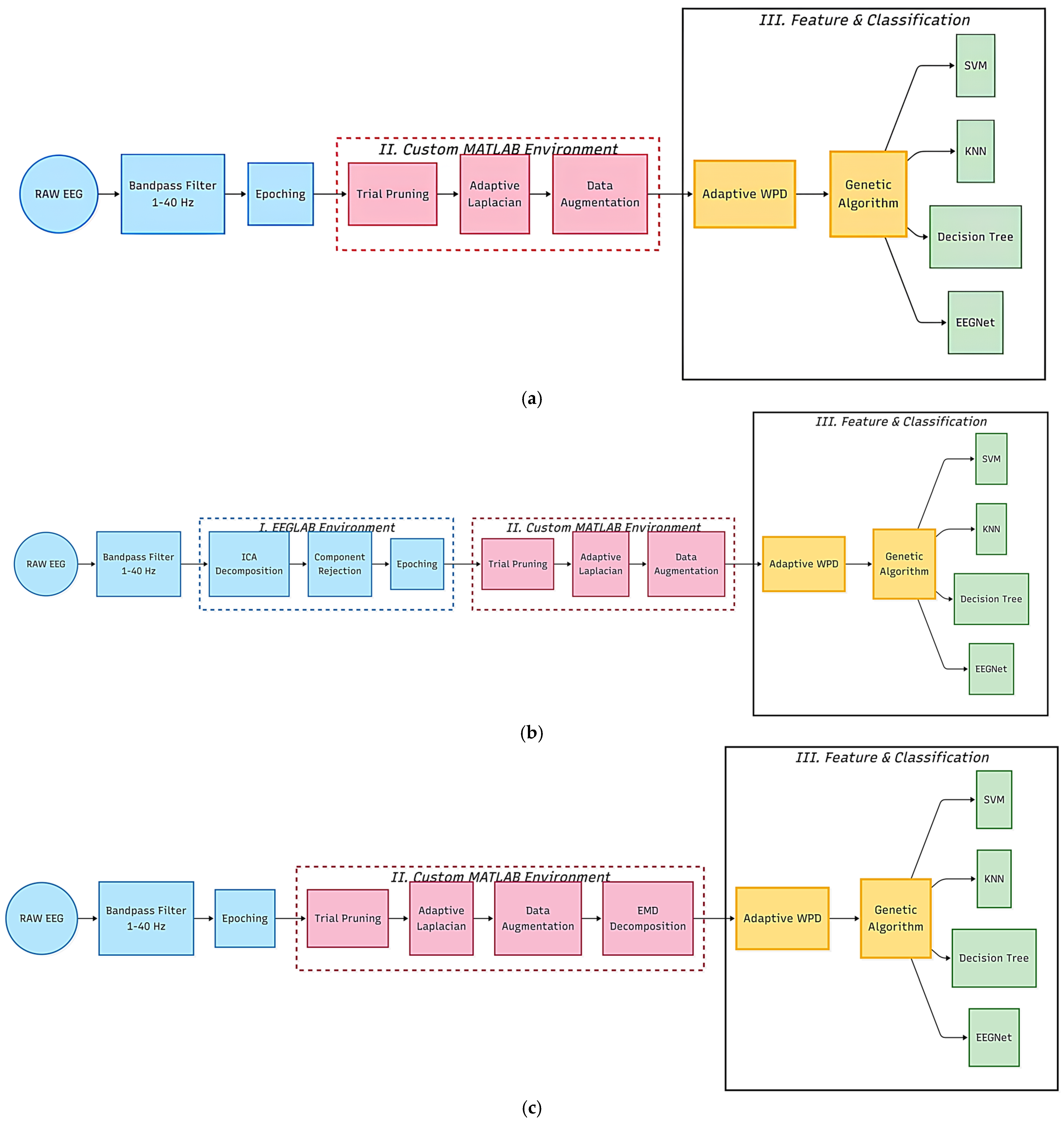
- Low-Latency BCI Framework: Formulated a BSS-free pipeline that removes the need for computationally expensive decomposition (ICA/EMD) without sacrificing accuracy, specifically optimized for real-time applications.
- Integrated Artifact Suppression: Developed a dual-layer strategy combining Adaptive Laplacian Spatial Filtering with statistical trial pruning to eliminate volume conduction and high-amplitude noise while maintaining a minimal computational footprint.
- Adaptive Manifold Optimization: Resolved subject-specific variance and BCI illiteracy by implementing an adaptive WPD framework that dynamically aligns signal frequency sub-bands, further optimized by a GA for high-precision feature selection.
- Robust Data Augmentation: Implemented a stochastic Gaussian noise injection strategy that introduces non-repeating signal variations across epochs, mitigating data scarcity and enhancing model generalizability against temporal non-stationarity.
- Cross-Architecture Stability Benchmarking: Validated the framework’s robustness against temporal non-stationarity by benchmarking it across diverse models (SVM, KNN, Decision Tree, EEGNet) for evaluating BCI signal processing reliability.
2. Related Work
2.1. Engineering Trade-Offs in MI-BCI Design
2.2. Overview of Data Augmentation
2.3. Multi-Resolution Analysis: DWT vs. WPD
2.4. Overview of Feature Selection
3. Material and Methods
3.1. Data Acquisition
3.1.1. BCI Competition IV Dataset 2A
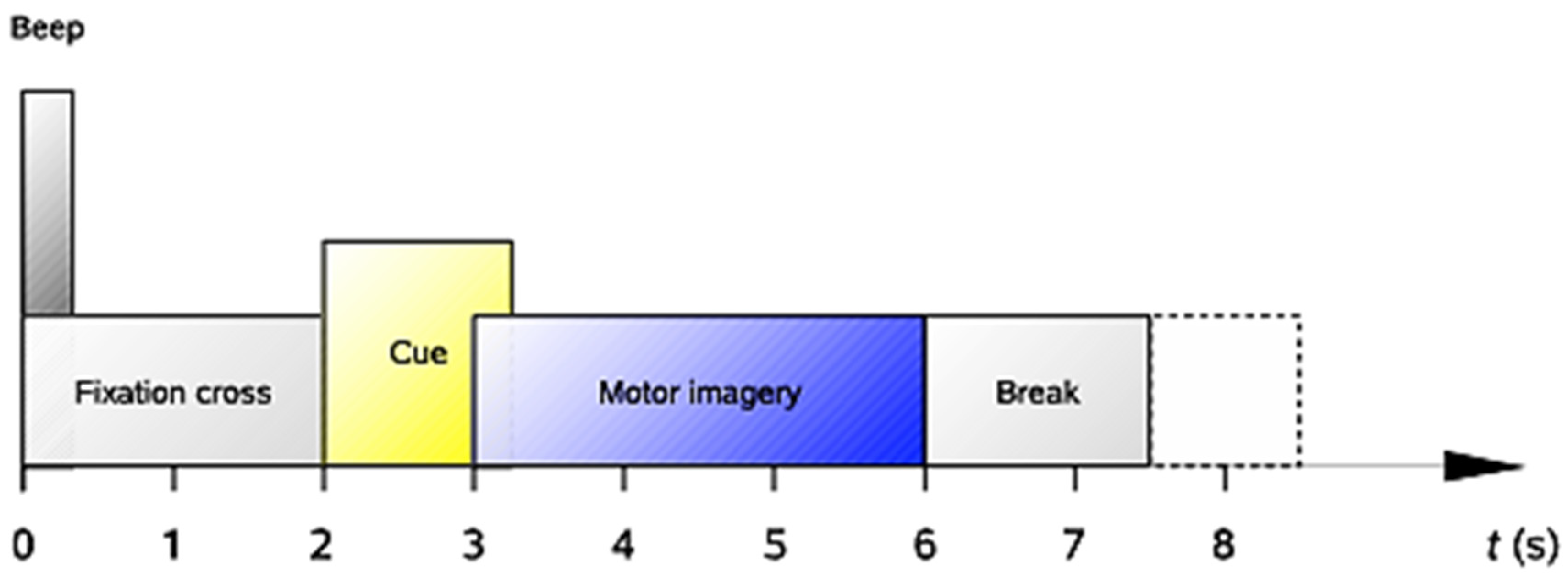
- t = 0 s: A fixation cross appeared on a black screen accompanied by an acoustic tone to signal the trial’s onset.
- t = 2 s: A directional arrow (left, right, down, or up) appeared for 1.25 s to cue the specific motor imagery task.
- t = 2 s to 6 s: Subjects performed the cued task without feedback until the fixation cross was removed at t = 6 s.
- Post-trial: A brief rest period with a black screen occurred before the next trial sequence began.
3.1.2. PhysioNet EEG Motor Imagery Dataset
- Event Synchronization: Since the meaning of the markers T1 and T2 depends on the specific run index, a manual re-mapping was implemented in EEGLAB. In runs involving the hands (Runs 4, 8, 12), markers were mapped to T1 (Left Hand) and T2 (Right Hand). In runs involving bilateral coordination (Runs 6, 10, 14), these were mapped to T3 (Both Hands) and T4 (Both Feet), resulting in a consistent 4-class framework.
- Multi-Run Concatenation: All task-relevant runs for each subject were merged into a single continuous EEG structure. This was performed prior to artifact rejection to provide the ICA algorithm with a statistically robust signal length, ensuring more accurate decomposition of non-neural sources.
3.2. Signal Processing
3.2.1. Digital Filtering
3.2.2. Epoching
3.2.3. Trial Pruning
3.3. Blind Source Separation (BSS) Comparison
3.3.1. Independent Component Analysis
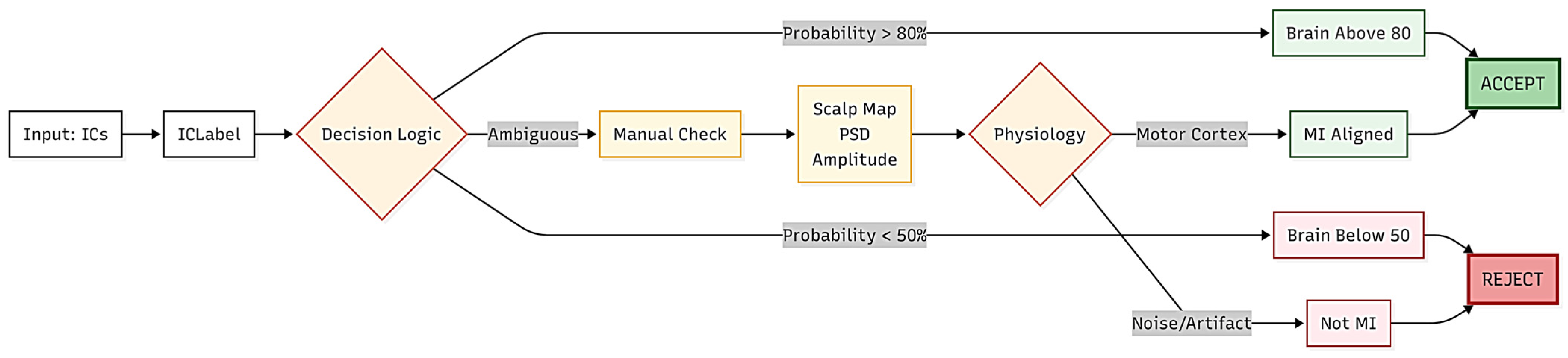
- Automated Filtering: Components were automatically accepted if the Brain class probability exceeded 80% and combined noise (Muscle, Eye, Heart) remained below 20%. In contrast, components were rejected if Brain probability fell below 50% or combined noise exceeded 20%.
- Structured Manual Protocol: Ambiguous components (where probabilities fell between 50 and 80%) were subjected to an objective assessment based on three criteria:
- Spatial Distribution: Components were rejected if the scalp map displayed localized, non-cortical activity such as ocular or temporal artifacts rather than the bilateral distribution expected during MI.
- Spectral Profiling: Components were rejected if the Power Spectral Density (PSD) exhibited non-physiological characteristics, such as high-frequency wide-band noise from muscle tension, or excessive low-frequency power, from blink artifacts.
- Temporal Stability: Components were cross-referenced with time-series amplitude to identify transient, non-stationary artifacts.
3.3.2. Fast Empirical Mode Decomposition
- Decomposition Model and IMF Constraints
- To focus on the most significant neural signatures, such as Mu and Beta rhythms, the algorithm is constrained to isolate a maximum of three IMFs. The original signal x(t) is modelled as defined in Equation (2):Restricting the output to three IMFs isolates relevant neural oscillations from background baseline drifts. This ensures a stable feature space and minimizes frequency leakage, resulting in a more robust and consistent decomposition [37].
- 2.
- Boundary Constraints and PCHIP Interpolation
- Throughout the execution, the system identifies local maxima and minima to build the upper and lower signal boundaries. By including the initial and final signal samples as boundary constraints, the PCHIP interpolation maintains its mathematical integrity and avoids edge divergence as shown in Equation (3):Choosing PCHIP over traditional cubic splines ensures the interpolation is shape preserving and prevents overshoot.
- 3.
- Mean Envelope Extraction
- As a result, the mean of these envelopes is removed from the signal to extract a distinct neural component, with the leftover residue serving as the input for subsequent extraction phases. The local mean m(t) is calculated as Equation (5):
3.4. Spatial Enhancement and Optimization
3.4.1. Adaptive Laplacian Spatial Filter
- Spatial Correlation Weighting
- While traditional fixed filters use rigid, pre-set coefficients based solely on where the electrodes are placed, the adaptive approach constantly recalibrates its weights based on the statistical relationship between the target electrode and its neighbors. For a target electrode , the raw spatial weights for each neighbour are determined by the Pearson Correlation coefficient. A threshold of 0.3 is applied to ensure only spatially redundant noise is considered:This threshold is designed to mitigate the smearing effect while ensuring that subtle, task-specific MI patterns remain preserved. By filtering only those electrode pairings with a correlation above 0.3, redundant spatial noise is effectively suppressed without losing the fine-grained information essential for high-accuracy MI classification. This flexibility is important for managing the session-to-session variability and low SNR typical of motor imagery tasks, as it allows the pipeline to adjust to the specific brain activity of each subject.
- 2.
- Weight Normalization
- The spatial weights are normalized to unity ( as shown in Equation (7), ensuring the resulting noise estimate is proportional to the target channel’s local field potential. This normalization prevents artificial amplitude distortion and guarantees that the conservatism factor remains scaled consistently, regardless of subject-specific or session-to-session signal variance.
- 3.
- Weighted Spatial Noise Estimation
- The smeared noise L(t) is constructed by calculating the weighted sum, as shown in Equation (8), of the neighboring channels identified in Table 1:

| Target Electrodes | BCI Competition IV 2A Neighbors | PhysioNet Neighbors |
|---|---|---|
| C3 | 2, 7, 9, 14 | 2, 16, 8, 10 |
| Cz | 4, 9, 11, 16 | 4, 18, 10, 12 |
| C4 | 6, 11, 13, 18 | 6, 20, 12, 14 |
- 4.
- Signal Sharpening and Preservation
- The final spatially enhanced signal is obtained by subtracting the noise estimate, scaled by a conservatism factor (α = 0.25), from the original target channel defined by Equation (9). This factor acts as a protective mechanism, preventing the over-subtraction of the signal, which could otherwise result in the unintended loss of task-relevant neural activity:
3.4.2. Data Augmentation
- Noise Level: To ensure that the added noise remains proportional to the biological signal amplitude, the noise magnitude () was dynamically scaled according to Equation (10). A baseline standard deviation () was calculated across the original epochs. The noise level coefficient of 2% was selected to ensure that the perturbation strength remains subtle, providing the necessary stochastic variety for model generalization without overriding the signal’s core physiological characteristics. By scaling the noise magnitude proportionally to the baseline standard deviation, the methodology ensures the injected noise remains within the physiological noise floor. This approach maintains the structural integrity of task-relevant oscillatory components, such as Mu and Beta rhythms, while preventing the classifier from overfitting to the training data:
- Augmentation Factor: An augmentation factor of 3 was employed, tripling the original number of epochs.
3.5. Feature Extraction–Adaptive Wavelet Packet Decomposition
3.6. Feature Selection
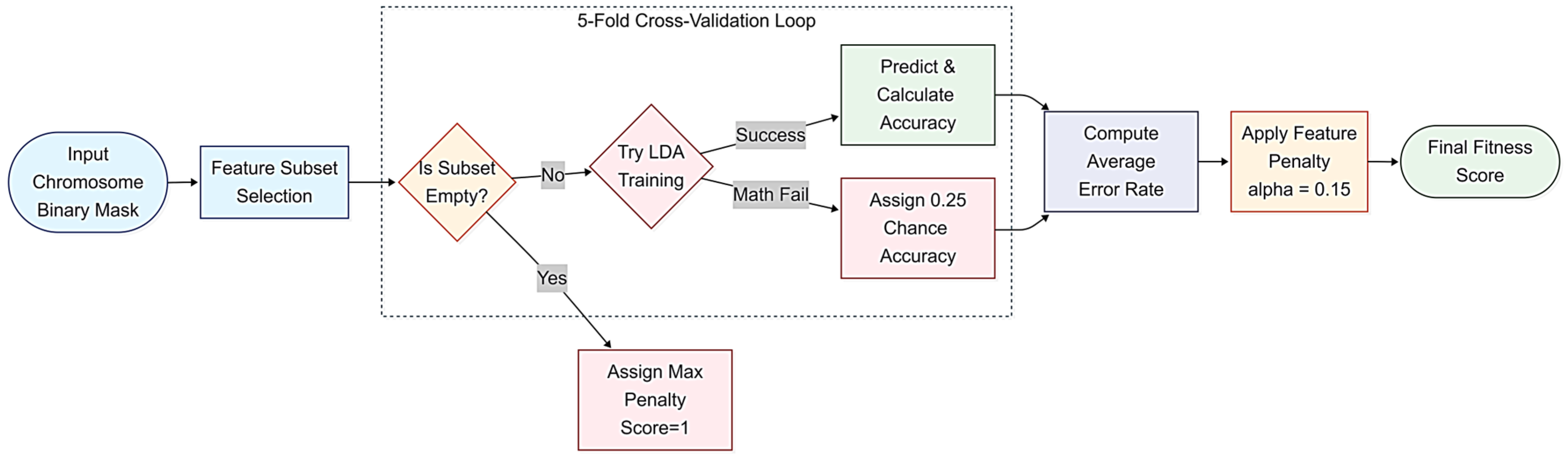
- Accuracy: Determined via 5-fold cross-validation using an LDA classifier to avoid feature bias with the final classifiers. This internal cross-validation ensures that the feature selection process remains robust against overfitting and data leakage within the training set.
- (Penalty Factor): Set to 0.15 to penalize redundant features and favor low-dimensional subsets that reduce computational overhead for real-time BCI applications. This penalty factor serves as a dimensionality constraint, preventing the GA from chasing marginal gains in training accuracy that often result from overfitting. Instead, the algorithm is incentivized to prioritize the selection of robust, task-relevant feature subsets. By utilizing this subject-specific evolutionary search, the pipeline effectively isolates the synergistic interactions between the log-transformed power, relative power, and spatial ratios. This ensures that the final model is trained exclusively on features that exhibit the highest SNR for each participant, significantly improving the stability of the BCI MI predictions.

3.7. Classification
3.7.1. KNN
3.7.2. SVM
3.7.3. Regression Tree
3.7.4. Modified EEGNet
3.8. Performance Metrics
3.9. Experimental Setup
- Global Stochastic Augmentation: A global, label-preserving data augmentation stage was implemented. As detailed in Section 3.4.2, stochastic Gaussian noise injection was applied to the entire dataset to increase feature space density. This process is entirely unsupervised and label-blind, relying on signal-specific characteristics to generate synthetic variants without introducing label-dependent bias.
- Trial-Level Partitioning: The dataset was partitioned into a 70% training set and a 30% testing set at the trial level before feature extraction.
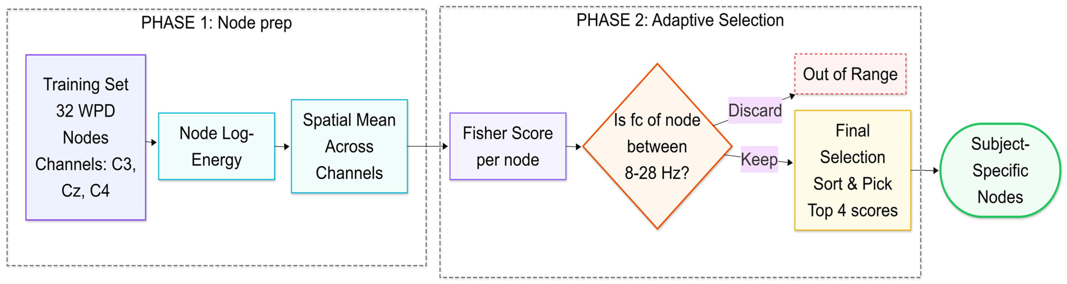
- Supervised Optimization (Training-Exclusive): This stage constitutes the supervised learning component of the pipeline. To isolate motor imagery (MI) activity, the analysis was restricted to the C3, C4, and Cz channels. Within this channel space, all operations sensitive to class discriminability, specifically the identification of optimal WPD nodes and the GA-optimized feature selection, were performed exclusively on the training partition.
- Fixed Transformation (Testing): The normalization parameters and optimized feature masks derived during the training phase were applied as fixed, independent transforms to the testing trials.
4. Results
4.1. BCI Competition IV Dataset 2A Results
4.1.1. Dimensionality Efficiency
4.1.2. Within-Subject Multi-Model Analysis
4.2. PhysioNet EEG MI Dataset Results
4.2.1. Dimensionality Efficiency
4.2.2. Within-Subject Multi-Model Analysis
4.3. Computational Efficiency
4.4. Comparative Performance Analysis
5. Discussion
6. Conclusions
Author Contributions
Funding
Institutional Review Board Statement
Informed Consent Statement
Data Availability Statement
Acknowledgments
Conflicts of Interest
Abbreviations
| BCI | Brain–Computer Interface |
| BSS | Blind Source Separation |
| DWT | Discrete Wavelet Transform |
| EEG | Electroencephalography |
| EMD | Empirical Mode Decomposition |
| EMG | Electromyography |
| EOG | Electrooculogram |
| ERD | Event-Related Desynchronization |
| ERS | Event-Related Synchronization |
| GA | Genetic Algorithm |
| ICA | Independent Component Analysis |
| IMF | Intrinsic Mode Functions |
| KNN | k-Nearest Neighbors |
| LDA | Linear Discriminant Analysis |
| MI | Motor Imagery |
| NCA | Neighbor Component Analysis |
| OvR | One vs Rest |
| PPF | Piecewise Polynomial Function |
| STFT | Short-Time Fourier Transform |
| SVM | Support Vector Machine |
| WPD | Wavelet Packet Decomposition |
Appendix A
| Node (l = 5) | Centre Frequency of Node | Frequency Range | Physiological Band |
|---|---|---|---|
| 0 | 1.95 Hz | 0–3.91 Hz | Delta |
| 1 | 5.86 Hz | 3.91–7.82 Hz | Theta |
| 2 | 9.77 Hz | 7.82–11.73 Hz | Alpha |
| 3 | 13.68 Hz | 11.71–15.64 Hz | Low Beta |
| 4 | 17.59 Hz | 15.64–19.55 Hz | Beta |
| 5 | 21.50 Hz | 19.55–23.46 Hz | Beta |
| 6 | 25.41 Hz | 23.46–27.37 Hz | Beta |
| 7 | 29.32 Hz | 27.37–31.28 Hz | High Beta/Gamma |
| Node (l = 5) | Centre Frequency of Node | Frequency Range | Physiological Band |
|---|---|---|---|
| 0 | 1.25 Hz | 0–2.5 Hz | Delta |
| 1 | 3.75 Hz | 2.5–5 Hz | Theta |
| 2 | 6.25 Hz | 5–7.5 Hz | Alpha |
| 3 | 8.75 Hz | 7.5–10 Hz | Low Beta |
| 4 | 11.25 Hz | 10–12.5 Hz | Beta |
| 5 | 13.75 Hz | 12.5–15 Hz | Beta |
| 6 | 16.25 Hz | 15–17.5 Hz | Beta |
| 7 | 18.75 Hz | 17.5–20 Hz | Beta |
| 8 | 21.25 Hz | 20–22.5 Hz | Beta |
| 9 | 23.75 Hz | 22.5–25 Hz | Beta |
| 10 | 26.25 Hz | 25–27.5 Hz | High Beta |
| Optimization Hyperparameter | Configuration Setting | Rationale |
|---|---|---|
| Solver Algorithm | Stochastic Gradient Descent with Momentum (‘sgdm’) | Provides smooth parameter trajectories to escape local minima in uncurated feature spaces. |
| Maximum Epoch Limit | 100 | Limits total execution length, terminating training before the network over-fits to non-neural residual components. |
| Mini-Batch Size | 32 | Optimized profile scaled to handle expanded data pools resulting from noise injection. |
| Initial Learning Rate | 0.001 | Conservative baseline parameter chosen to prevent catastrophic early gradient divergence. |
| Learning Rate Schedule | Piecewise decay (‘piecewise’) | Dynamically tightens solver stepping precision as network optimization nears a convergence plateau. |
| Drop Period and Drop Factor | Decreases by 0.5 every 30 epochs | Drops learning rate by half at structural milestones to safely resolve fine-grained hyperplane choices. |
| L2 Regularization (Weight Decay) | 0.02 | Balances weight penalty constraints to regularize loss boundaries against data scarcity. |
| Data Shuffling Paradigm | Shuffled every epoch (‘every-epoch’) | Breaks localized sequencing dependencies across trials to protect against cross-trial validation leakage. |
| Validation Frequency | Evaluated every 10 iterations | Provides granular, periodic diagnostic checks on training progress and generalization trends. |
| Validation Patience | Infinite patience (inf) | Forces full completion of the 100 epochs to establish a steady baseline for structural analysis without early-termination bias. |
References
- Mdluli, B.; Khumalo, P.; Maswanganyi, R.C. Signal Preprocessing, Decomposition and Feature Extraction Methods in EEG-Based BCIs. Appl. Sci. 2025, 15, 12075. [Google Scholar] [CrossRef] [Scilit]
- Ramsoonder, N.; Maswanganyi, R.C.; Khumalo, P. Comparative Performance Analysis of Machine Learning Computational Pipelines and Deep Learning Architectures in EEG Motor Imagery BCIs. Mathematics 2026, 14, 1520. [Google Scholar] [CrossRef] [Scilit]
- Maswanganyi, R.C.; Tu, C.; Owolawi, P.A.; Du, S. Statistical Evaluation of Factors Influencing Inter-Session and Inter-Subject Variability in EEG-Based Brain Computer Interface. IEEE Access 2022, 10, 96821–96839. [Google Scholar] [CrossRef] [Scilit]
- Molla, M.K.I.; Al Shiam, A.; Islam, M.R.; Tanaka, T. Discriminative Feature Selection-Based Motor Imagery Classification Using EEG Signal. IEEE Access 2020, 8, 98255–98265. [Google Scholar] [CrossRef] [Scilit]
- Singh, A.; Hussain, A.A.; Lal, S.; Guesgen, H.W. A Comprehensive Review on Critical Issues and Possible Solutions of Motor Imagery Based Electroencephalography Brain–Computer Interface. Sensors 2021, 21, 2173. [Google Scholar] [CrossRef] [Scilit] [PubMed]
- Khan, R.A.; Rashid, N.; Shahzaib, M.; Malik, U.F.; Arif, A.; Iqbal, J.; Saleem, M.; Khan, U.S.; Tiwana, M. A Novel Framework for Classification of Two-Class Motor Imagery EEG Signals Using Logistic Regression Classification Algorithm. PLoS ONE 2023, 18, e0276133. [Google Scholar] [CrossRef] [Scilit] [PubMed]
- Jiang, X.; Bian, G.B.; Tian, Z. Removal of Artifacts from EEG Signals: A Review. Sensors 2019, 19, 987. [Google Scholar] [CrossRef] [Scilit] [PubMed]
- Mannan, M.M.N.; Kamran, M.A.; Jeong, M.Y. Identification and Removal of Physiological Artifacts from Electroencephalogram Signals: A Review. IEEE Access 2018, 6, 30630–30652. [Google Scholar] [CrossRef] [Scilit]
- Dhole, P.V.; Chaudhary, D.G.; Dhangar, V.D.; Shejul, S.D.; Datwase, S.S.; Gawali, B.W. A Review of EEG Artifact Removal Techniques for Brain–Computer Interface. SN Comput. Sci. 2025, 6, 1026. [Google Scholar] [CrossRef] [Scilit]
- Wu, C.; Yao, B.; Zhang, X.; Li, T.; Wang, J.; Pu, J. The Application of Entropy in Motor Imagery Paradigms of Brain–Computer Interfaces. Brain Sci. 2025, 15, 168. [Google Scholar] [CrossRef] [Scilit] [PubMed]
- Titkanlou, M.K.; Pham, D.T.; Mouček, R. Classification of EEG Signal Using Deep Learning Architectures Based Motor-Imagery for an Upper-Limb Rehabilitation Exoskeleton. SN Comput. Sci. 2025, 6, 193. [Google Scholar] [CrossRef] [Scilit]
- Sung, D.J.; Kim, K.T.; Jeong, J.H.; Kim, L.; Lee, S.J.; Kim, H.; Kim, S.J. Improving Inter-Session Performance via Relevant Session-Transfer for Multi-Session Motor Imagery Classification. Heliyon 2024, 10, e37343. [Google Scholar] [CrossRef] [Scilit] [PubMed]
- Amer, N.S.; Belhaouari, B. EEG Signal Processing for Medical Diagnosis, Healthcare, and Monitoring: A Comprehensive Review. IEEE Access 2023, 11, 139457–139465. [Google Scholar] [CrossRef] [Scilit]
- Shyu, K.K.; Huang, S.C.; Tung, K.J.; Lee, L.H.; Lee, P.L.; Chen, Y.H. Common Spatial Pattern and Riemannian Manifold-Based Real-Time Multiclass Motor Imagery EEG Classification. IEEE Access 2023, 11, 139457–139465. [Google Scholar] [CrossRef] [Scilit]
- Delorme, A. EEG Is Better Left Alone. Sci. Rep. 2023, 13, 2372. [Google Scholar] [CrossRef] [Scilit] [PubMed]
- Siviero, I.; Menegaz, G.; Storti, S.F. Functional Connectivity and Feature Fusion Enhance Multiclass Motor-Imagery Brain–Computer Interface Performance. Sensors 2023, 23, 7520. [Google Scholar] [CrossRef] [Scilit] [PubMed]
- Velasco, I.; Sipols, A.; De Blas, C.S.; Pastor, L.; Bayona, S. Motor Imagery EEG Signal Classification with a Multivariate Time Series Approach. Biomed. Eng. Online 2023, 22, 29. [Google Scholar] [CrossRef] [Scilit] [PubMed]
- Yan, Y.; Zhou, H.; Huang, L.; Cheng, X.; Kuang, S. A Novel Two-Stage Refine Filtering Method for EEG-Based Motor Imagery Classification. Front. Neurosci. 2021, 15, 657540. [Google Scholar] [CrossRef] [Scilit] [PubMed]
- Zhang, Z.; Duan, F.; Sole-Casals, J.; Dinares-Ferran, J.; Cichocki, A.; Yang, Z.; Sun, Z. A Novel Deep Learning Approach with Data Augmentation to Classify Motor Imagery Signals. IEEE Access 2019, 7, 15945–15954. [Google Scholar] [CrossRef] [Scilit]
- Sharma, N.; Sharma, M.; Singhal, A.; Vyas, R.; Malik, H.; Hossaini, M.A.; Afthanorhan, A. An Efficient Approach for Recognition of Motor Imagery EEG Signals Using the Fourier Decomposition Method. IEEE Access 2023, 11, 122782–122791. [Google Scholar] [CrossRef] [Scilit]
- Li, M.A.; Zhu, W.; Liu, H.N.; Yang, J.F. Adaptive Feature Extraction of Motor Imagery EEG with Optimal Wavelet Packets and SE-Isomap. Appl. Sci. 2017, 7, 390. [Google Scholar] [CrossRef] [Scilit]
- George, O.; Smith, R.; Madiraju, P.; Yahyasoltani, N.; Ahamed, S.I. Data Augmentation Strategies for EEG-Based Motor Imagery Decoding. Heliyon 2022, 8, e10240. [Google Scholar] [CrossRef] [Scilit] [PubMed]
- Petrov, M. Optimizing Motor Imagery BCIs: A Motor Imagery EEG Classification Protocol Using Evolutionary Optimization and Low Sample Training-Sets. Master’s Thesis, New College of Florida, Sarasota, FL, USA, 2024. Available online: https://digitalcommons.ncf.edu/theses_etds/6586 (accessed on 15 May 2026).
- Dhiman, R. Wavelet Packet Decomposition of Electroencephalogram Signals for Brain Computer Interface. Available online: https://aeteps2023.wordpress.com/wp-content/uploads/2023/06/aeteps2023_id_278.pdf (accessed on 15 May 2026).
- Geng, X.; Wang, L.; Yu, P.; Hu, W.; Liang, Q.; Zhang, X.; Chen, C.; Zhang, X. A Method of EEG Signal Feature Extraction Based on Hybrid DWT and EMD. Alex. Eng. J. 2025, 113, 195–204. [Google Scholar] [CrossRef] [Scilit]
- Kabir, M.H.; Akhtar, N.I.; Tasnim, N.; Miah, A.S.M.; Lee, H.S.; Jang, S.W.; Shin, J. Exploring Feature Selection and Classification Techniques to Improve the Performance of an Electroencephalography-Based Motor Imagery Brain–Computer Interface System. Sensors 2024, 24, 4989. [Google Scholar] [CrossRef] [Scilit] [PubMed]
- BCI Competition IV. Available online: https://www.bbci.de/competition/iv/ (accessed on 10 July 2026).
- Schalk, G.; McFarland, D.J.; Hinterberger, T.; Birbaumer, N.; Wolpaw, J.R. EEG Motor Movement/Imagery Dataset. PhysioNet, 2009. Available online: https://physionet.org/content/eegmmidb/1.0.0/ (accessed on 25 April 2026).
- Savadi, S. A Comprehensive Review of Feature Extraction and Classification Techniques in EEG Signal Analysis. Int. J. Comput. Eng. Res. 2025, 15, 51–62. Available online: http://www.ijceronline.com (accessed on 10 May 2026).
- Bahhah, M.A.; Attar, E.T. Enhancing Epilepsy Seizure Detection Through Advanced EEG Preprocessing Techniques and Peak-to-Peak Amplitude Fluctuation Analysis. Diagnostics 2024, 14, 2525. [Google Scholar] [CrossRef] [Scilit] [PubMed]
- Shieh, C.P.; Yang, S.H.; Liu, Y.S.; Kuo, Y.T.; Lo, Y.C.; Kuo, C.H.; Chen, Y.Y. Simultaneously Spatiospectral Pattern Learning and Contaminated Trial Pruning for Electroencephalography-Based Brain Computer Interface. Symmetry 2020, 12, 1387. [Google Scholar] [CrossRef] [Scilit]
- Visu, P.; Smitha, P.S.; Velayutham, M.; Ahmad, M.W. Enhanced EEG Classification Using Adaptive DWT and Heuristic-ICA Algorithm. Automatika 2023, 64, 827–836. [Google Scholar] [CrossRef] [Scilit]
- Blum, S.; Jacobsen, N.S.J.; Bleichner, M.G.; Debener, S. A Riemannian Modification of Artifact Subspace Reconstruction for EEG Artifact Handling. Front. Hum. Neurosci. 2019, 13, 141. [Google Scholar] [CrossRef] [Scilit] [PubMed]
- Van Jaarsveldt, C.; Peters, G.W.; Ames, M.; Chantler, M. Tutorial on Empirical Mode Decomposition: Basis Decomposition and Frequency Adaptive Graduation in Non-Stationary Time Series. IEEE Access 2023, 11, 94442–94478. [Google Scholar] [CrossRef] [Scilit]
- Rabbath, C.A.; Corriveau, D. A Comparison of Piecewise Cubic Hermite Interpolating Polynomials, Cubic Splines and Piecewise Linear Functions for the Approximation of Projectile Aerodynamics. Def. Technol. 2019, 15, 741–757. [Google Scholar] [CrossRef] [Scilit]
- Wang, F.; Liu, H.; Zhao, L.; Su, L.; Zhou, J.; Gong, A.; Fu, Y. Improved Brain–Computer Interface Signal Recognition Algorithm Based on Few-Channel Motor Imagery. Front. Hum. Neurosci. 2022, 16, 880304. [Google Scholar] [CrossRef] [Scilit] [PubMed]
- Ji, N.; Ma, L.; Dong, H.; Zhang, X. EEG Signals Feature Extraction Based on DWT and EMD Combined with Approximate Entropy. Brain Sci. 2019, 9, 201. [Google Scholar] [CrossRef] [Scilit] [PubMed]
- Beniczky, S.; Schomer, D.L. Electroencephalography: Basic Biophysical and Technological Aspects Important for Clinical Applications. Epileptic Disord. 2020, 22, 697–715. [Google Scholar] [CrossRef] [Scilit] [PubMed]
- Bradshaw, L.A.; Wikswo, J.P. Spatial Filter Approach for Evaluation of the Surface Laplacian of the Electroencephalogram and Magnetoencephalogram. Ann. Biomed. Eng. 2001, 29, 202–213. [Google Scholar] [CrossRef] [Scilit] [PubMed]
- Gu, Q.; Li, Z.; Han, J. Generalized Fisher Score for Feature Selection. In Proceedings of the Twenty-Seventh Conference on Uncertainty in Artificial Intelligence, Barcelona, Spain, 14–17 July 2011; AUAI Press: Arlington, VA, USA, 2011. [Google Scholar]
- Degirmenci, M.; Yuce, Y.K.; Perc, M.; Isler, Y. EEG Channel and Feature Investigation in Binary and Multiple Motor Imagery Task Predictions. Front. Hum. Neurosci. 2024, 18, 1525139. [Google Scholar] [CrossRef] [Scilit] [PubMed]
- Mateen, N.; Naeem, M.; Khan, M.J.; Yousaf, T.; Ali, A.; Altabey, W.A.; Noori, M.; Kouritem, S.A. Subject Based Feature Selection for Hybrid Brain Computer Interface Using Genetic Algorithm and Support Vector Machine. Results Eng. 2025, 27, 105649. [Google Scholar] [CrossRef] [Scilit]
- Dhiman, R.; Anshul. Brain Computer Interface Using Genetic Algorithm with Modified Genome and Phenotype Structures. J. Inf. Technol. Manag. 2023, 15, 47–68. [Google Scholar] [CrossRef]
- Konjkav, A.; Jahangiri, F.; Sajedin, A. Classification Algorithms of EEG Signals Based on Motor Imagery. Int. J. Res. Technol. Electr. Ind. 2025, 4, 517–525. [Google Scholar] [CrossRef]
- Yassine, F.A.; Abdelkader, G. Enhancing EEG-Based Brain–Computer Interface Systems through Efficient Machine Learning Classification Techniques. Indones. J. Electr. Eng. Comput. Sci. 2024, 34, 2045–2054. [Google Scholar] [CrossRef] [Scilit]
- Chen, M.; Lee, C.; Chun, Y. A Machine Learning Approach Using Spatially Explicit K-Nearest Neighbors for House Price Predictions. ISPRS Int. J. Geo-Inf. 2026, 15, 46. [Google Scholar] [CrossRef] [Scilit]
- Syriopoulos, P.K.; Kalampalikis, N.G.; Kotsiantis, S.B.; Vrahatis, M.N. KNN Classification: A Review. Ann. Math. Artif. Intell. 2025, 93, 43–75. [Google Scholar] [CrossRef] [Scilit]
- Lu, Y.; Wang, W.; Lian, B.; He, C. Feature Extraction and Classification of Motor Imagery EEG Signals in Motor Imagery for Sustainable Brain–Computer Interfaces. Sustainability 2024, 16, 6627. [Google Scholar] [CrossRef] [Scilit]
- Selim, S.; Tantawi, M.M.; Shedeed, H.A.; Badr, A. A CSP\AM-BA-SVM Approach for Motor Imagery BCI System. IEEE Access 2018, 6, 49192–49208. [Google Scholar] [CrossRef] [Scilit]
- Ferracuti, F.; Iarlori, S.; Mansour, Z.; Monteriù, A.; Porcaro, C. Comparing between Different Sets of Preprocessing, Classifiers, and Channels Selection Techniques to Optimise Motor Imagery Pattern Classification System from EEG Pattern Recognition. Brain Sci. 2022, 12, 57. [Google Scholar] [CrossRef] [Scilit] [PubMed]
- Deng, X.; Zhang, B.; Yu, N.; Liu, K.; Sun, K. Advanced TSGL-EEGNet for Motor Imagery EEG-Based Brain–Computer Interfaces. IEEE Access 2021, 9, 25118–25130. [Google Scholar] [CrossRef] [Scilit]
- Mdluli, B.; Khumalo, P.; Maswanganyi, R.C. Subject-Specific Comparative Performance Analysis of Deep Learning Architectures for Motor Imagery Classification. Mathematics 2026, 14, 1527. [Google Scholar] [CrossRef] [Scilit]
- Xie, Y.; Oniga, S. Classification of Motor Imagery EEG Signals Based on Data Augmentation and Convolutional Neural Networks. Sensors 2023, 23, 1932. [Google Scholar] [CrossRef] [Scilit] [PubMed]
- Geng, X.; Li, D.; Chen, H.; Yu, P.; Yan, H.; Yue, M. An Improved Feature Extraction Algorithms of EEG Signals Based on Motor Imagery Brain–Computer Interface. Alex. Eng. J. 2022, 61, 4807–4820. [Google Scholar] [CrossRef] [Scilit]
- Antony, M.J.; Sankaralingam, B.P.; Mahendran, R.K.; Gardezi, A.A.; Shafiq, M.; Choi, J.G.; Hamam, H. Classification of EEG Using Adaptive SVM Classifier with CSP and Online Recursive Independent Component Analysis. Sensors 2022, 22, 7596. [Google Scholar] [CrossRef] [Scilit] [PubMed]


| Layer Index | Name | Layer Type/Block | Filter/Kernel Size | Stride/Padding | Output Shape(H × W × C) | Operational Description and Purpose |
|---|---|---|---|---|---|---|
| 1 | ‘Input’ | Image Input Layer | - | - | 1 × numF × 1 | Receives the 4D reshaped vector of pre-selected GA features; applies zero-center normalization to stabilize early-stage gradients. |
| 2 | ‘Conv_Features’ | 2D Convolution | 1 × 3 | Stride: 1 × 1 Padding: ‘same’ | 1 × numF × 32 | Scans across adjacent pre-selected feature columns to extract local pattern correlations. |
| 3 | ‘BN1’ | Batch Normalization | - | - | 1 × numF × 32 | Normalizes intermediate activations; mitigates internal covariate shift across training mini-batches. |
| 4 | ‘ReLU1’ | Rectified Linear Unit | - | - | 1 x numF x 32 | Implements non-linear thresholding to preserve discriminative MI traits. |
| 5 | ‘Conv_Global’ | 2D Convolution | 1 × 5 | Stride: 1 × 1 Padding: ‘same’ | 1 × numF × 64 | Broadens the horizontal receptive field to detect global time-frequency combinations among the features. |
| 6 | ‘BN2’ | Batch Normalization | - | - | 1 × numF × 64 | Stabilizes downstream feature scaling prior to dimensional downsampling. |
| 7 | ‘ReLU2’ | Rectified Linear Unit | - | - | 1 × numF × 64 | Secondary activation mapping to enforce sparse, highly defined feature representation. |
| 8 | ‘AvgPool’ | Average Pooling | 1 × 2 | Stride: 1 × 2 Padding: 0 | 1 [numF/2] × 64 | Downsamples the feature width via linear smoothing to suppress residual session-to-session and subject noise. |
| 9 | ‘Dropout’ | Dropout Regularization | - | - | 1 × [numF/2] × 64 | Randomly deactivates 40% (p = 0.4) of nodes per iteration to combat over-fitting across small sample sets. |
| 10 | ‘FC_Output’ | Fully Connected (Dense) | - | - | 1 × 1 × numC | Maps the high-dimensional optimized feature map directly down to the targeted multi-class space. |
| 11 | ‘Softmax’ | Softmax Activation | - | - | 1 × 1 × numC | Computes a normalized probability distribution across the independent motor imagery categories. |
| 12 | ‘Output_Layer’ | Cross-Entropy Loss | - | - | - | Computes categorical cross-entropy error metrics during model backpropagation. |
| A01 | A02 | A03 | A04 | A05 | A06 | A07 | A08 | A09 | |
|---|---|---|---|---|---|---|---|---|---|
| Optimal Nodes | 2 3 5 6 | 6 4 5 3 | 2 3 6 4 | 5 6 3 2 | 3 6 5 4 | 2 6 3 5 | 5 6 3 4 | 3 2 6 4 | 3 2 5 6 |
| Number of Features | 12 | 10 | 12 | 18 | 12 | 7 | 14 | 11 | 15 |
| A01 | A02 | A03 | A04 | A05 | A06 | A07 | A08 | A09 | |
|---|---|---|---|---|---|---|---|---|---|
| Optimal Nodes | 2 3 5 6 | 6 4 5 3 | 2 3 6 5 | 5 6 4 3 | 2 5 3 6 | 6 2 5 4 | 6 5 4 2 | 3 5 2 4 | 3 2 5 6 |
| Number of Features | 10 | 16 | 12 | 12 | 11 | 6 | 11 | 12 | 9 |
| A01 | A02 | A03 | A04 | A05 | A06 | A07 | A08 | A09 | |
|---|---|---|---|---|---|---|---|---|---|
| Optimal Nodes | 2 3 5 6 | 4 6 3 2 | 2 3 6 4 | 5 6 3 2 | 3 6 4 2 | 2 6 3 5 | 5 6 3 2 | 3 2 6 4 | 3 2 5 6 |
| Number of Features | 12 | 4 | 10 | 9 | 13 | 14 | 12 | 12 | 10 |
| KNN | SVM | Decision Tree | Modified EEGNet | |
|---|---|---|---|---|
| No-BSS vs. EMD | 0.054 | 0.088 | 0.409 | 0.033 |
| No-BSS vs. ICA | 0.811 | 0.082 | 0.637 | 0.643 |
| A01 | A02 | A03 | A04 | A05 | A06 | A07 | A08 | A09 | Average | |
|---|---|---|---|---|---|---|---|---|---|---|
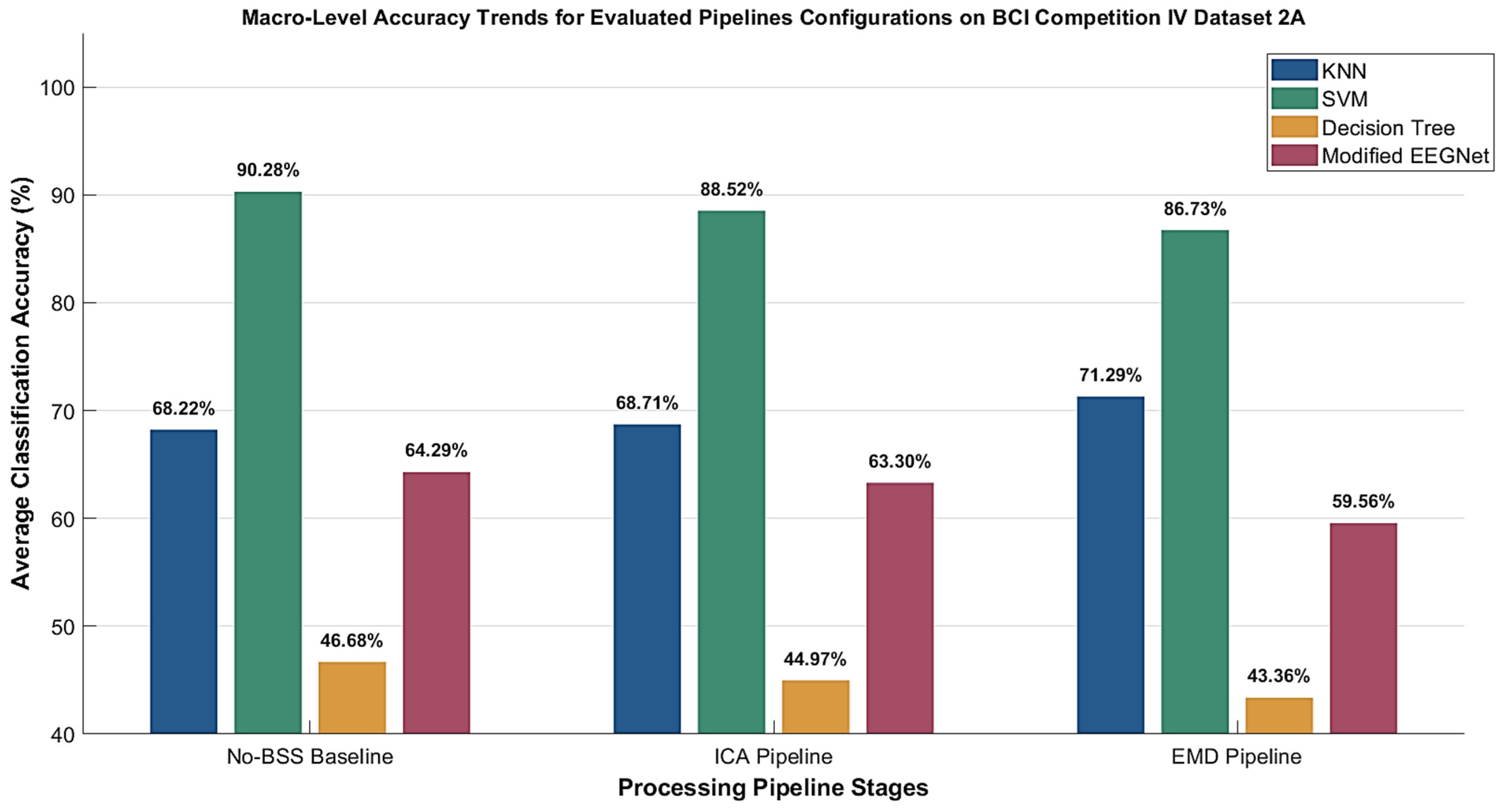
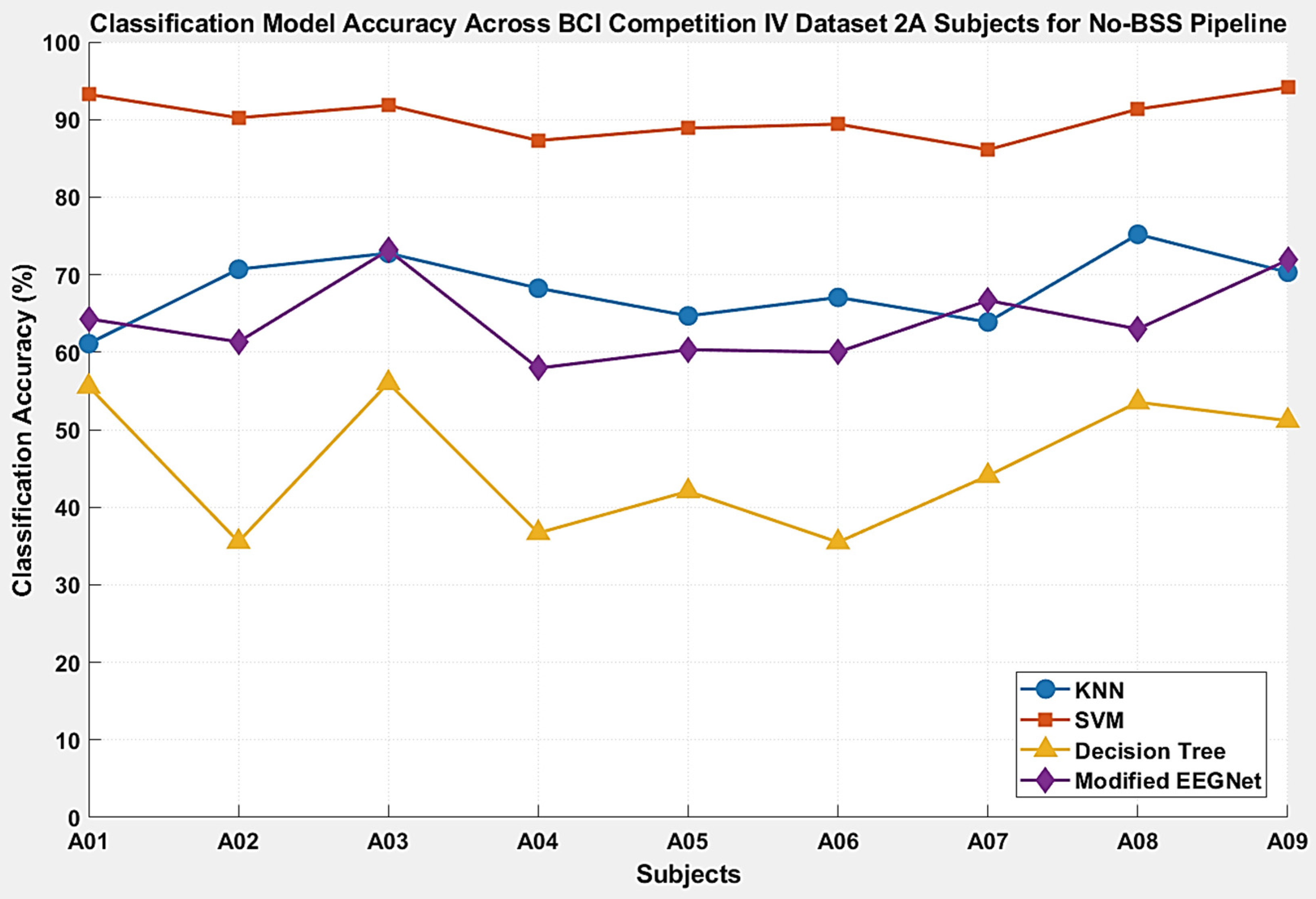
| KNN | 61.11% | 70.70% | 72.76% | 68.25% | 64.68% | 67.06% | 63.89% | 75.20% | 70.31% | 68.22% ± 4.26% |
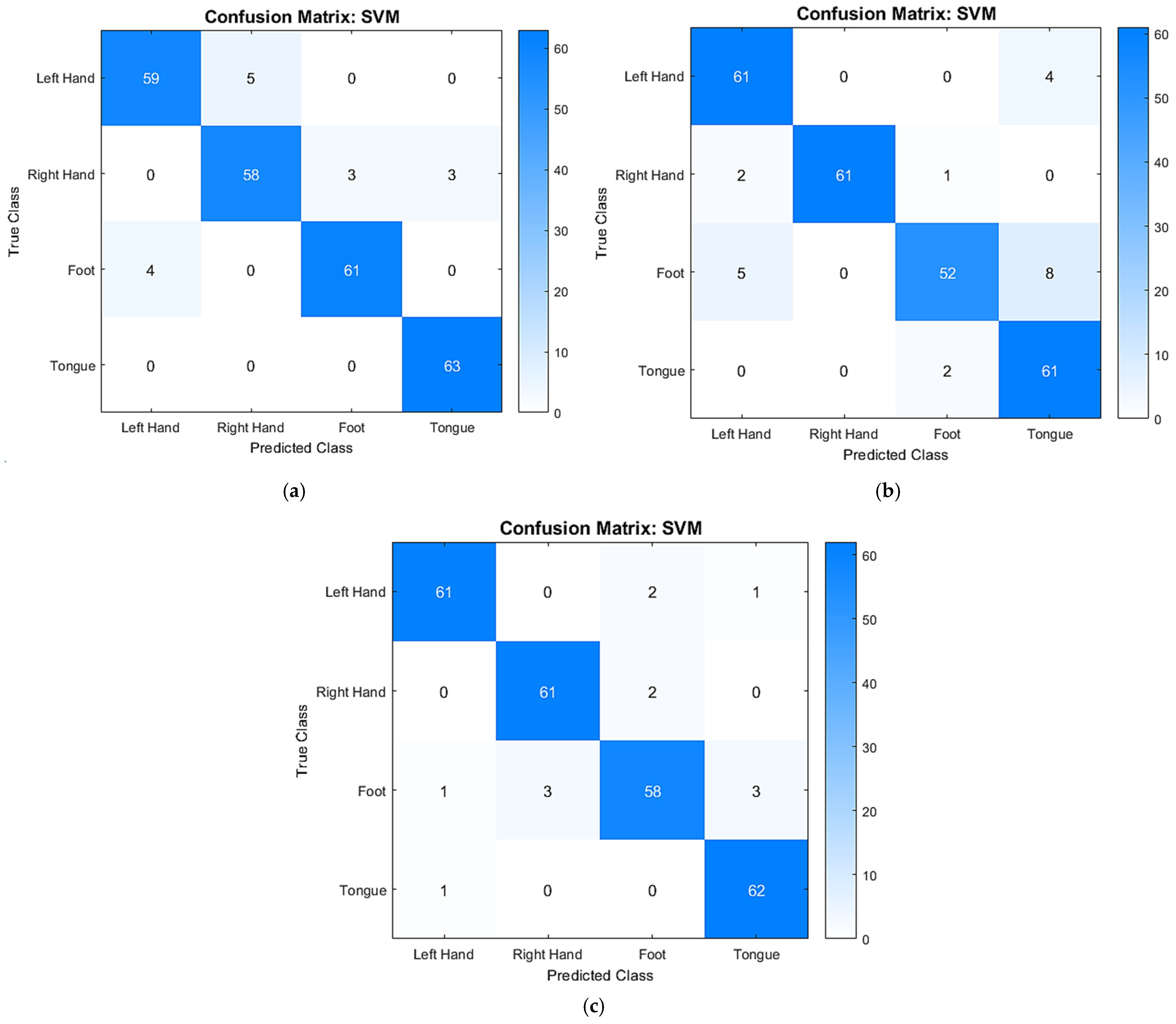
| SVM | 93.25% | 90.23% | 91.83% | 87.30% | 88.89% | 89.41% | 86.11% | 91.34% | 94.14% | 90.28% ± 2.50% |
| Decision Tree | 55.56% | 35.55% | 56.03% | 36.68% | 42.06% | 35.49% | 44.05% | 53.54% | 51.17% | 46.68% ± 7.36% |
| Modified EEGNet | 64.29% | 61.33% | 73.15% | 57.94% | 60.32% | 60.00% | 66.67% | 62.99% | 71.88% | 64.29% ± 5.02% |
| A01 | A02 | A03 | A04 | A05 | A06 | A07 | A08 | A09 | Average | |
|---|---|---|---|---|---|---|---|---|---|---|
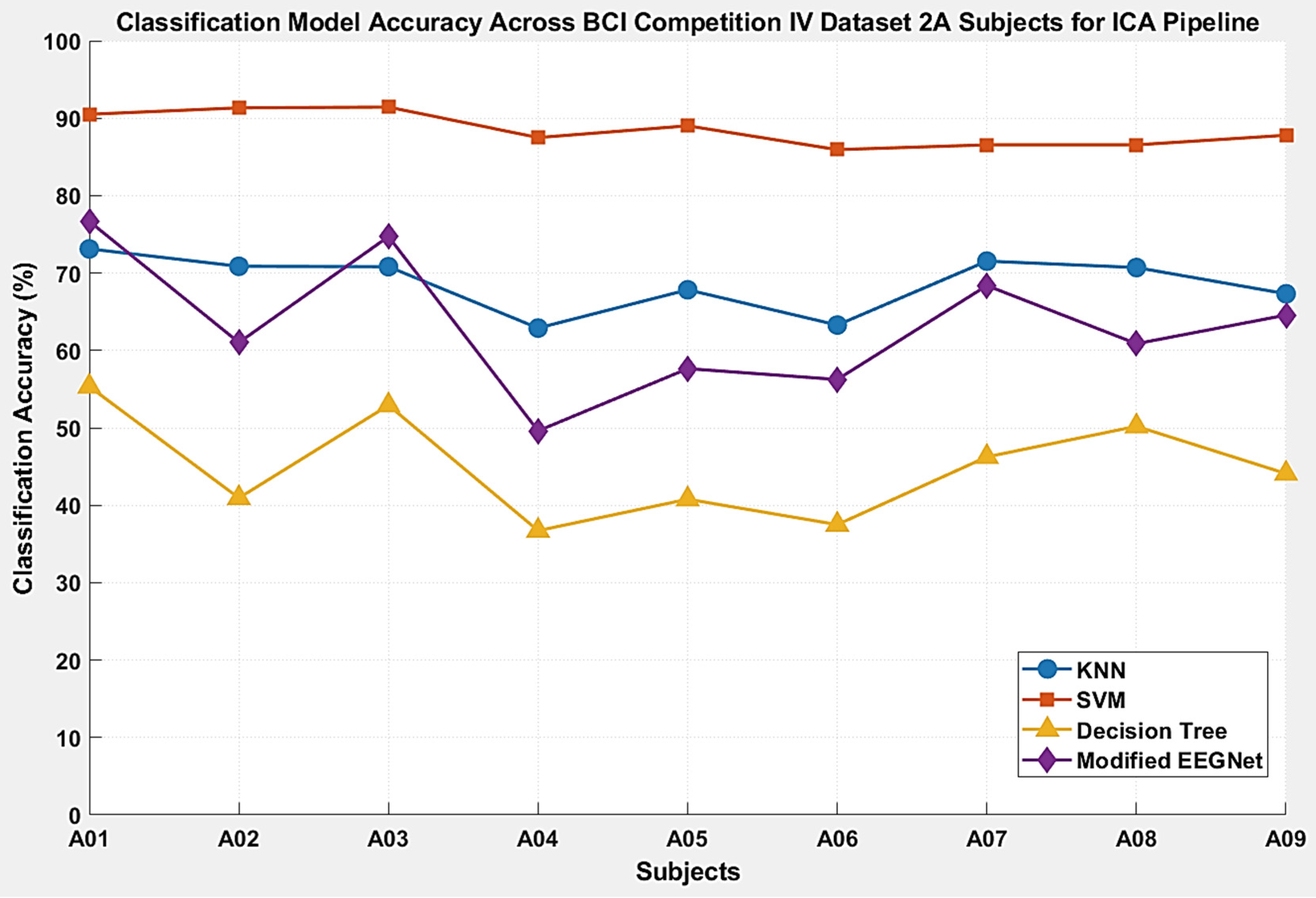
| KNN | 73.12% | 70.87% | 70.82% | 62.89% | 67.84% | 63.28% | 71.54% | 70.71% | 67.32% | 68.71% ± 3.45% |
| SVM | 90.51% | 91.34% | 91.44% | 87.50% | 89.02% | 85.94% | 86.56% | 86.56% | 87.80% | 88.52% ± 2.02% |
| Decision Tree | 55.34% | 40.94% | 52.92% | 36.72% | 40.78% | 37.50% | 46.25% | 50.20% | 44.09% | 44.97% ± 6.31% |
| Modified EEGNet | 76.68% | 61.02% | 74.71% | 49.61% | 57.65% | 56.25% | 68.38% | 60.87% | 64.57% | 63.30% ± 8.28% |
| A01 | A02 | A03 | A04 | A05 | A06 | A07 | A08 | A09 | Average | |
|---|---|---|---|---|---|---|---|---|---|---|
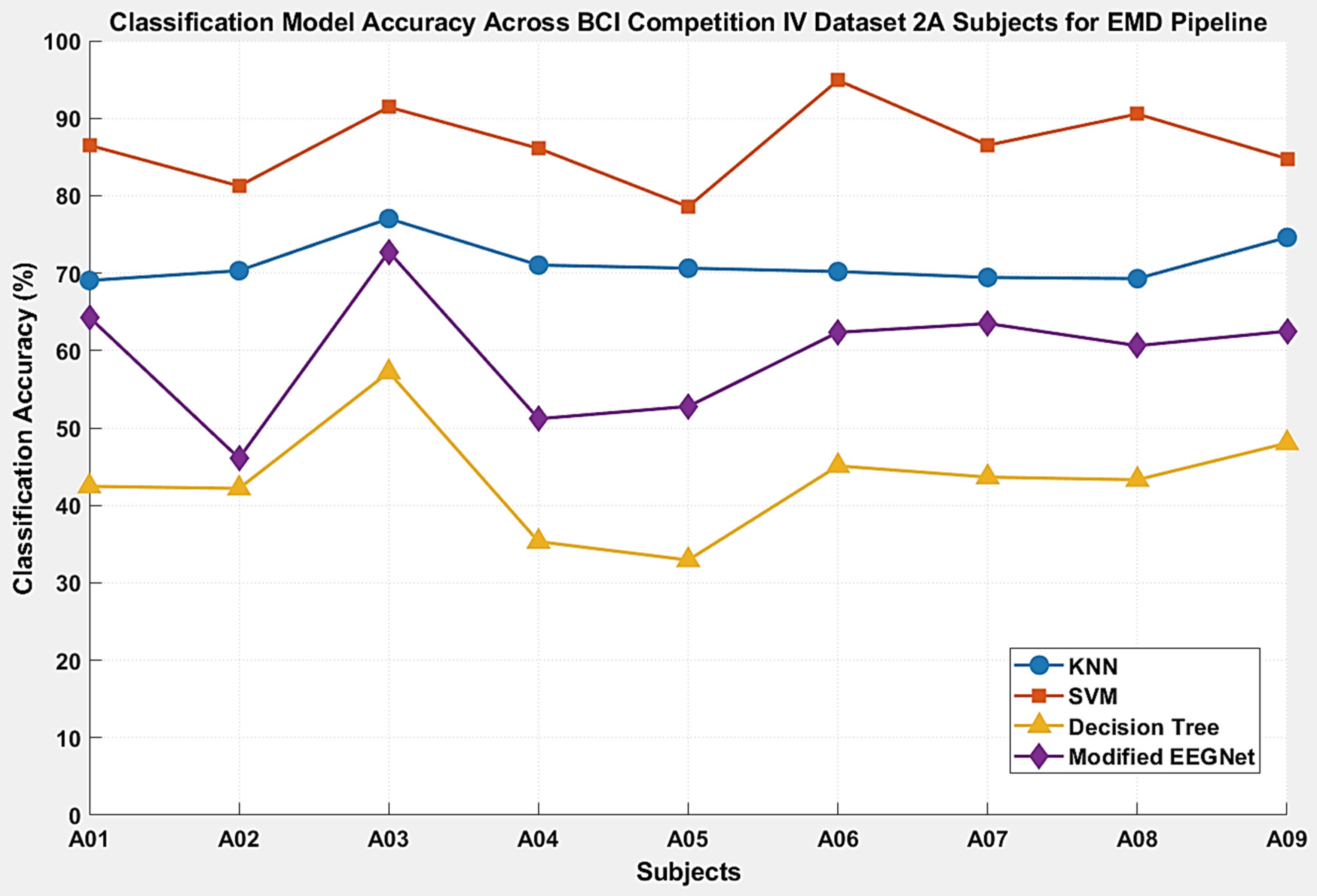
| KNN | 69.05% | 70.31% | 77.04% | 71.03% | 70.63% | 70.20% | 69.44% | 69.29% | 74.61% | 71.29% ± 3.42% |
| SVM | 86.51% | 81.25% | 91.44% | 86.11% | 78.57% | 94.90% | 86.51% | 90.55% | 84.77% | 86.73% ± 3.42% |
| Decision Tree | 42.46% | 42.19% | 57.20% | 35.32% | 32.94% | 45.10% | 43.65% | 43.31% | 48.05% | 43.36% ± 6.60% |
| Modified EEGNet | 64.29% | 46.09% | 72.76% | 51.19% | 52.78% | 62.35% | 63.49% | 60.63% | 62.50% | 59.56% ± 7.65% |
| Performance Metrics | No BSS | ICA | EMD |
|---|---|---|---|
| Kappa | 0.5761 ± 0.0569 | 0.5823 ± 0.0459 | 0.6171 ± 0.0342 |
| F1-Score | 0.6819 ± 0.0435 | 0.6868 ± 0.0345 | 0.7110 ± 0.0262 |
| Recall | 0.6820 ± 0.0423 | 0.6870 ± 0.0344 | 0.7128 ± 0.0259 |
| Precision | 0.6885 ± 0.0430 | 0.6870 ± 0.0357 | 0.7174 ± 0.0267 |
| AUC | 0.8989 ± 0.0201 | 0.8997 ± 0.0158 | 0.9052 ± 0.138 |
| Performance Metrics | No BSS | ICA | EMD |
|---|---|---|---|
| Kappa | 0.8704 ± 0.0333 | 0.8469 ± 0.0269 | 0.8231 ± 0.0636 |
| F1-Score | 0.9023 ± 0.0251 | 0.8849 ± 0.0202 | 0.8672 ± 0.0474 |
| Recall | 0.9028 ± 0.0250 | 0.8852 ± 0.0203 | 0.8673 ± 0.0477 |
| Precision | 0.9059 ± 0.0242 | 0.8885 ± 0.0197 | 0.8712 ± 0.0465 |
| AUC | 0.9733 ± 0.0086 | 0.9712 ± 0.0075 | 0.9596 ± 0.0230 |
| Performance Metrics | No BSS | ICA | EMD |
|---|---|---|---|
| Kappa | 0.2935 ± 0.0929 | 0.2665 ± 0.0842 | 0.2443 ± 0.0886 |
| F1-Score | 0.4515 ± 0.0794 | 0.4365 ± 0.0699 | 0.4195 ± 0.0753 |
| Recall | 0.4697 ± 0.0696 | 0.4502 ± 0.0635 | 0.4331 ± 0.0666 |
| Precision | 0.5312 ± 0.0696 | 0.4803 ± 0.0695 | 0.4744 ± 0.0602 |
| AUC | 0.7140 ± 0.0576 | 0.7232 ± 0.0561 | 0.6837 ± 0.0606 |
| Performance Metrics | No BSS | ICA | EMD |
|---|---|---|---|
| Kappa | 0.5177 ± 0.0735 | 0.5107 ± 0.1103 | 0.4608 ± 0.0765 |
| F1-Score | 0.6418 ± 0.0515 | 0.6307 ± 0.0836 | 0.5946 ± 0.0761 |
| Recall | 0.6416 ± 0.0483 | 0.6334 ± 0.0827 | 0.5954 ± 0.0767 |
| Precision | 0.6457 ± 0.0505 | 0.6355 ± 0.0840 | 0.5969 ± 0.0763 |
| AUC | 0.8532 ± 0.0366 | 0.8533 ± 0.0496 | 0.8279 ± 0.0526 |
| S001 | S002 | S003 | S004 | S005 | S006 | S007 | S008 | S009 | |
|---|---|---|---|---|---|---|---|---|---|
| Optimal Nodes | 6 8 7 5 | 5 6 8 7 | 9 8 10 5 | 4 5 10 7 | 4 7 3 5 | 4 6 5 3 | 3 6 9 8 | 6 5 8 7 | 9 5 3 10 |
| Number of Features | 14 | 13 | 10 | 21 | 22 | 15 | 17 | 19 | 15 |
| S001 | S002 | S003 | S004 | S005 | S006 | S007 | S008 | S009 | |
|---|---|---|---|---|---|---|---|---|---|
| Optimal Nodes | 7 5 4 10 | 5 6 8 7 | 9 8 10 3 | 6 4 10 7 | 4 5 8 7 | 7 5 6 8 | 3 7 10 6 | 6 4 10 7 | 3 7 6 5 |
| Number of Features | 15 | 18 | 16 | 12 | 12 | 24 | 17 | 13 | 14 |
| S001 | S002 | S003 | S004 | S005 | S006 | S007 | S008 | S009 | |
|---|---|---|---|---|---|---|---|---|---|
| Optimal Nodes | 6 8 9 7 | 8 5 10 9 | 9 8 10 3 | 5 4 10 6 | 4 7 3 8 | 6 4 5 3 | 3 7 6 8 | 6 5 4 10 | 5 7 3 10 |
| Number of Features | 10 | 15 | 12 | 19 | 18 | 21 | 11 | 15 | 19 |
| KNN | SVM | Decision Tree | Modified EEGNet | |
|---|---|---|---|---|
| No-BSS vs. EMD | <0.001 | 0.017 | 0.580 | 0.657 |
| No-BSS vs. ICA | 0.288 | 0.084 | 0.653 | 0.061 |
| S001 | S002 | S003 | S004 | S005 | S006 | S007 | S008 | S009 | Average | |
|---|---|---|---|---|---|---|---|---|---|---|
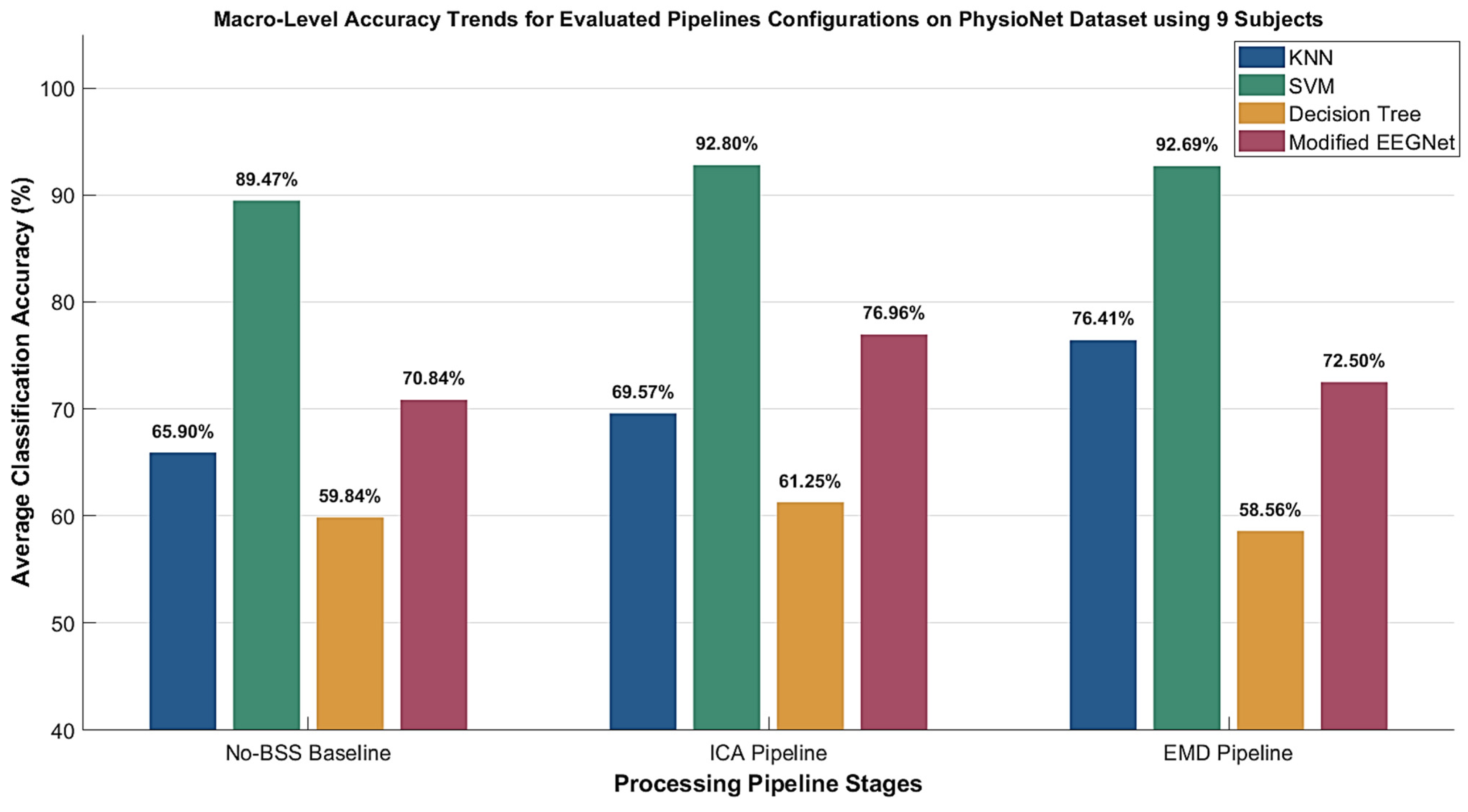
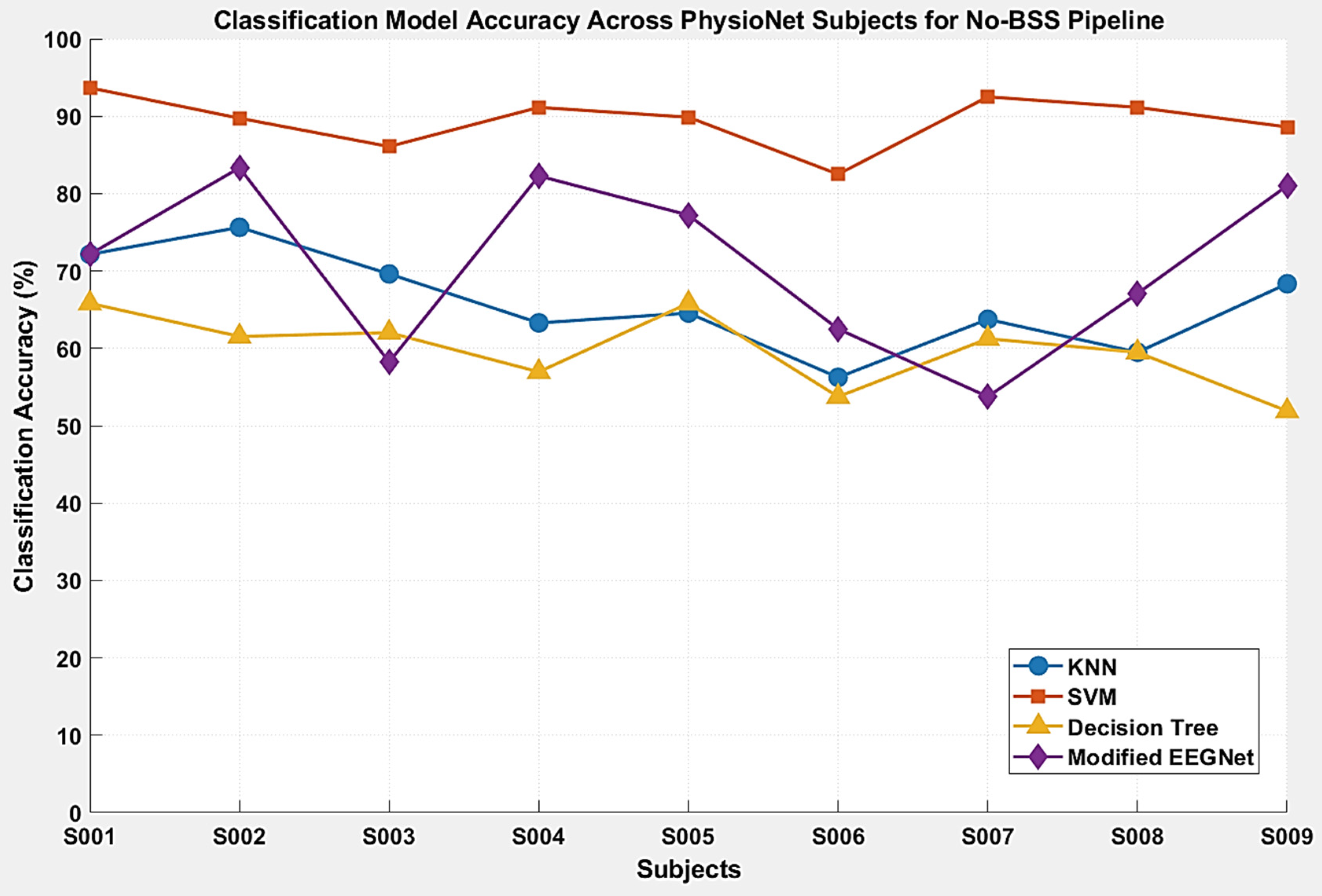
| KNN | 72.15% | 75.64% | 69.62% | 63.29% | 64.56% | 56.25% | 63.75% | 59.49% | 68.35% | 65.90% ± 5.79% |
| SVM | 93.67% | 89.74% | 86.08% | 91.14% | 89.87% | 82.50% | 92.50% | 91.14% | 88.61% | 89.47% ± 3.22% |
| Decision Tree | 65.82% | 61.54% | 62.03% | 56.96% | 65.82% | 53.75% | 61.25% | 59.49% | 51.90% | 59.84% ± 4.59% |
| Modified EEGNet | 72.15% | 83.33% | 58.23% | 82.28% | 77.22% | 62.50% | 53.75% | 67.09% | 81.01% | 70.84% ± 10.34% |
| S001 | S002 | S003 | S004 | S005 | S006 | S007 | S008 | S009 | Average | |
|---|---|---|---|---|---|---|---|---|---|---|
| KNN | 70.89% | 63.64% | 75.32% | 80.00% | 68.35% | 65.82% | 66.25% | 66.67% | 69.23% | 69.57% ± 4.88% |
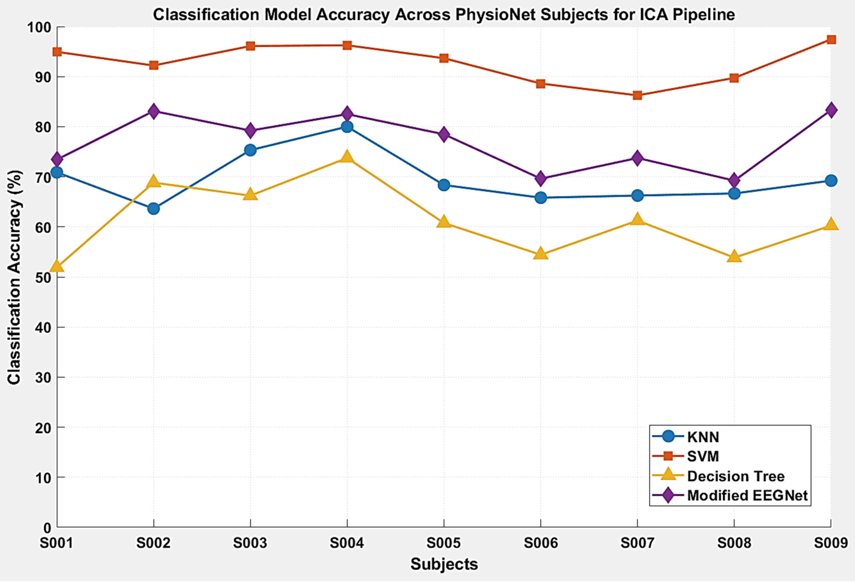
| SVM | 94.94% | 92.21% | 96.10% | 96.25% | 93.67% | 88.61% | 86.25% | 89.74% | 97.44% | 92.80% ± 3.65% |
| Decision Tree | 51.90% | 68.83% | 66.23% | 73.75% | 60.76% | 54.43% | 61.25% | 53.85% | 60.26% | 61.25% ± 6.90% |
| Modified EEGNet | 73.42% | 83.12% | 79.22% | 82.50% | 78.48% | 69.62% | 73.75% | 69.23% | 83.33% | 76.96% ± 5.30% |
| S001 | S002 | S003 | S004 | S005 | S006 | S007 | S008 | S009 | Average | |
|---|---|---|---|---|---|---|---|---|---|---|
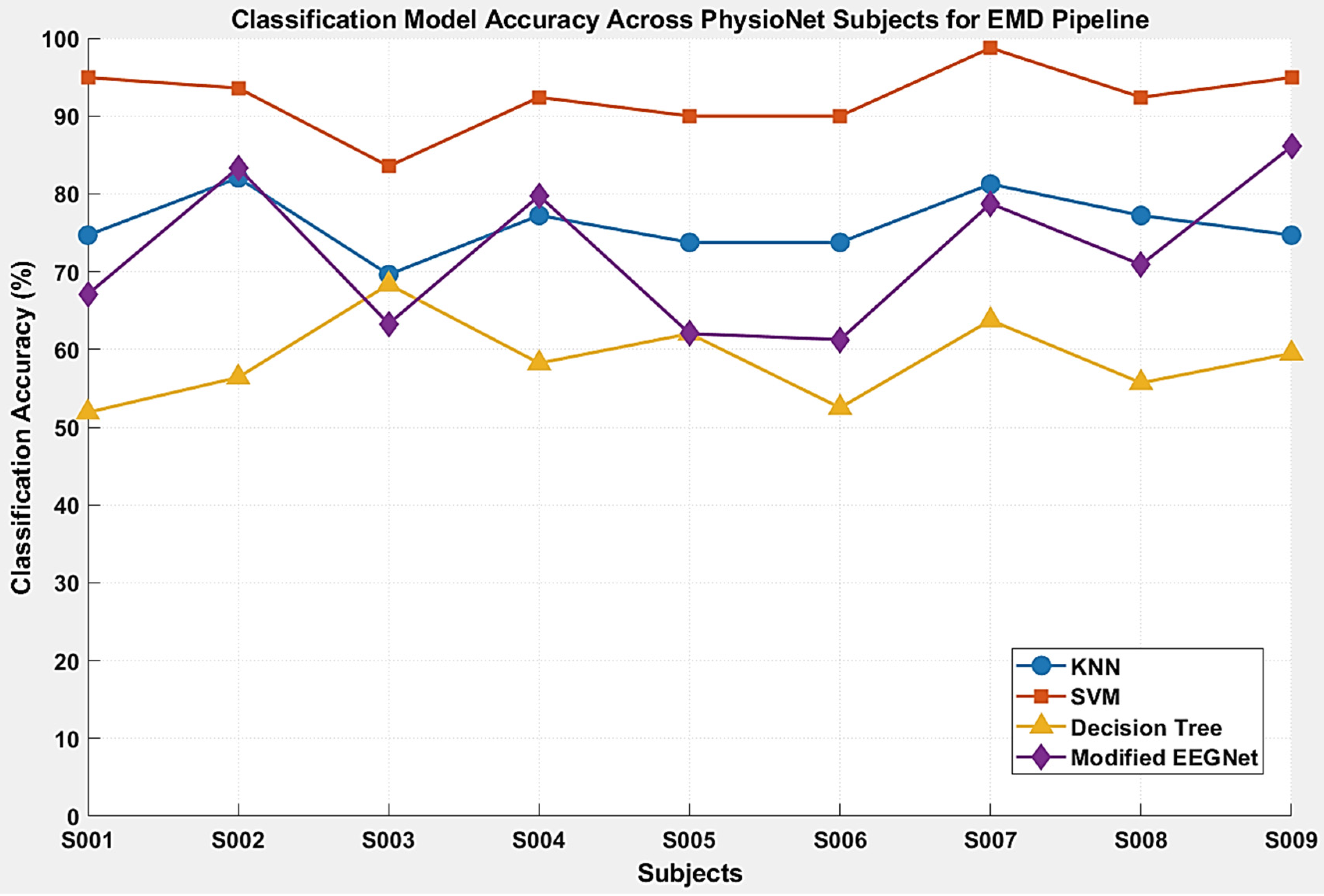
| KNN | 74.68% | 82.05% | 69.62% | 77.22% | 77.22% | 73.75% | 81.25% | 77.22% | 74.68% | 76.41% ± 3.60% |
| SVM | 94.94% | 93.59% | 83.54% | 92.41% | 93.67% | 90.00% | 98.75% | 92.41% | 94.94% | 92.69% ± 3.94% |
| Decision Tree | 51.90% | 56.41% | 68.35% | 58.23% | 60.76% | 52.50% | 63.75% | 55.70% | 59.49% | 58.56% ± 4.97% |
| Modified EEGNet | 67.09% | 83.33% | 63.29% | 79.75% | 62.03% | 61.25% | 78.75% | 70.89% | 86.08% | 72.50% ± 9.11% |
| Performance Metrics | No BSS | ICA | EMD |
|---|---|---|---|
| Kappa | 0.5448 ± 0.0579 | 0.5939 ± 0.0653 | 0.7186 ± 0.0721 |
| F1-Score | 0.6594 ± 0.0556 | 0.6938 ± 0.0495 | 0.7633 ± 0.0368 |
| Recall | 0.6584 ± 0.0573 | 0.6949 ± 0.0499 | 0.7634 ± 0.0344 |
| Precision | 0.6770 ± 0.0483 | 0.7055 ± 0.0470 | 0.7808 ± 0.0372 |
| AUC | 0.8923 ± 0.0326 | 0.8965 ± 0.0252 | 0.9313 ± 0.0163 |
| Performance Metrics | No BSS | ICA | EMD |
|---|---|---|---|
| Kappa | 0.8560 ± 0.0429 | 0.9097 ± 0.0404 | 0.9025 ± 0.0525 |
| F1-Score | 0.8944 ± 0.0309 | 0.9267 ± 0.0368 | 0.9258 ± 0.0396 |
| Recall | 0.8940 ± 0.0312 | 0.9284 ± 0.0359 | 0.9311 ± 0.0378 |
| Precision | 0.9051 ± 0.0271 | 0.9331 ± 0.0366 | 0.9258 ± 0.0396 |
| AUC | 0.9686 ± 0.0174 | 0.9815 ± 0.0174 | 0.9782 ± 0.0197 |
| Performance Metrics | No BSS | ICA | EMD |
|---|---|---|---|
| Kappa | 0.4642 ± 0.0607 | 0.4829 ± 0.0919 | 0.4479 ± 0.0656 |
| F1-Score | 0.5955 ± 0.0409 | 0.6043 ± 0.0712 | 0.5820 ± 0.0468 |
| Recall | 0.5976 ± 0.0428 | 0.6109 ± 0.0686 | 0.5856 ± 0.0490 |
| Precision | 0.6248 ± 0.0487 | 0.6468 ± 0.0459 | 0.6184 ± 0.0404 |
| AUC | 0.8218 ± 0.0270 | 0.8251 ± 0.0515 | 0.8058 ± 0.0357 |
| Performance Metrics | No BSS | ICA | EMD |
|---|---|---|---|
| Kappa | 0.6109 ± 0.1380 | 0.6927 ± 0.0709 | 0.6330 ± 0.1216 |
| F1-Score | 0.7074 ± 0.1030 | 0.7696 ± 0.0509 | 0.7218 ± 0.0951 |
| Recall | 0.7076 ± 0.1014 | 0.7704 ± 0.0527 | 0.7248 ± 0.0915 |
| Precision | 0.7173 ± 0.1014 | 0.7848 ± 0.0536 | 0.7378 ± 0.0931 |
| AUC | 0.8997 ± 0.0459 | 0.9154 ± 0.0320 | 0.9011 ± 0.0454 |
| Dataset Pool | No BSS | ICA | EMD |
|---|---|---|---|
| BCI Competition IV Dataset 2A | |||
| Training Time (s) | 0.1927 ± 0.0775 | 0.3626 ± 0.2083 | 0.2250 ± 0.1187 |
| Testing Time (s) | 0.0695 ± 0.0147 | 0.0842 ± 0.0368 | 0.0697 ± 0.0398 |
| PhysioNet | |||
| Training Time (s) | 0.2477 ± 0.1852 | 0.1800 ± 0.0599 | 0.4136 ± 0.2129 |
| Testing Time (s) | 0.0792 ± 0.0444 | 0.0766 ± 0.0252 | 0.1482 ± 0.0842 |
| Dataset Pool | No BSS | ICA | EMD |
|---|---|---|---|
| BCI Competition IV Dataset 2A | |||
| Training Time (s) | 0.5562 ± 0.1012 | 0.7455 ± 0.2575 | 0.6389 ± 0.1739 |
| Testing Time (s) | 0.0841 ± 0.0212 | 0.1104 ± 0.0370 | 0.0889 ± 0.0209 |
| PhysioNet | |||
| Training Time (s) | 0.6224 ± 0.2291 | 0.6373 ± 0.1414 | 0.9279 ± 0.3123 |
| Testing Time (s) | 0.0896 ± 0.0335 | 0.0906 ± 0.0414 | 0.1422 ± 0.0585 |
| Dataset Pool | No BSS | ICA | EMD |
|---|---|---|---|
| BCI Competition IV Dataset 2A | |||
| Training Time (s) | 0.1599 ± 0.0255 | 0.2327 ± 0.1242 | 0.1926 ± 0.0588 |
| Testing Time (s) | 0.0154 ± 0.0051 | 0.0231 ± 0.0188 | 0.0139 ± 0.0040 |
| PhysioNet | |||
| Training Time (s) | 0.1720 ± 0.0629 | 0.1845 ± 0.0433 | 0.2623 ± 0.0860 |
| Testing Time (s) | 0.0152 ± 0.0051 | 0.0197 ± 0.0053 | 0.0227 ± 0.0075 |
| Dataset Pool | No BSS | ICA | EMD |
|---|---|---|---|
| BCI Competition IV Dataset 2A | |||
| Training Time (s) | 99.9989 ± 24.7504 | 137.1620 ± 36.2134 | 147.3385 ± 104.5006 |
| Testing Time (s) | 0.1287 ± 0.0345 | 0.1430 ± 0.0391 | 0.1465 ± 0.0272 |
| PhysioNet | |||
| Training Time (s) | 44.3482 ± 19.9092 | 35.6024 ± 7.3621 | 56.3234 ± 21.8473 |
| Testing Time (s) | 0.1154 ± 0.0713 | 0.0991 ± 0.0260 | 0.1283 ± 0.0433 |
| Methodology | Dataset | Subject Count | Average Accuracy |
|---|---|---|---|
| ICA-WT-CSP [54] | PhysioNet | None Specified | 81.75% |
| DWT-EMD-Approximate Entropy [37] | BCI Competition IV Dataset 2A | 9 | 85.71% |
| ICA-Wavelet-CSP [55] | BCI Competition IV Dataset 2A | 9 | 82.00% |
| ORICA-CSP [55] | BCI Competition IV Dataset 2A | 9 | 89.00% |
| Proposed Pipeline (No-BSS) | PhysioNet | 9 | 89.47% |
| Proposed Pipeline (No-BSS) | BCI Competition IV Dataset 2A | 9 | 90.28% |
Disclaimer/Publisher’s Note: The statements, opinions and data contained in all publications are solely those of the individual author(s) and contributor(s) and not of MDPI and/or the editor(s). MDPI and/or the editor(s) disclaim responsibility for any injury to people or property resulting from any ideas, methods, instructions or products referred to in the content. |
© 2026 by the authors. Licensee MDPI, Basel, Switzerland. This article is an open access article distributed under the terms and conditions of the Creative Commons Attribution (CC BY) license.
Share and Cite
Ramsoonder, N.; Maswanganyi, R.C.; Khumalo, P. Subject-Specific BCI Frameworks for Motor Imagery Classification Based on BSS-Free and BSS-Equipped Pipelines. Big Data Cogn. Comput. 2026, 10, 280. https://doi.org/10.3390/bdcc10080280
Ramsoonder N, Maswanganyi RC, Khumalo P. Subject-Specific BCI Frameworks for Motor Imagery Classification Based on BSS-Free and BSS-Equipped Pipelines. Big Data and Cognitive Computing. 2026; 10(8):280. https://doi.org/10.3390/bdcc10080280
Chicago/Turabian StyleRamsoonder, Nerita, Rito Clifford Maswanganyi, and Philani Khumalo. 2026. "Subject-Specific BCI Frameworks for Motor Imagery Classification Based on BSS-Free and BSS-Equipped Pipelines" Big Data and Cognitive Computing 10, no. 8: 280. https://doi.org/10.3390/bdcc10080280
APA StyleRamsoonder, N., Maswanganyi, R. C., & Khumalo, P. (2026). Subject-Specific BCI Frameworks for Motor Imagery Classification Based on BSS-Free and BSS-Equipped Pipelines. Big Data and Cognitive Computing, 10(8), 280. https://doi.org/10.3390/bdcc10080280