


Assessment of Causes and Potential Prevention and Therapy for Autoimmune Diseases Through Evolutionary Medicine


Abstract
1. Introduction
1.1. Evolutionary Medicine ([1], s. #1.1)
1.2. Ecological Niche ([1], s. #1.2.2)
1.3. Mismatch ([1], s. #1.2.4)
1.4. Holobiont ([1], s. #1.2.5)
1.5. The Autoimmune Diseases
- -
- The primary cause, or causes, of the disease can be genetic only in a small fraction of cases, as the frequency of a harmful gene cannot significantly increase within one or few generations;
- -
- To a large extent, the disease results from an alteration of the ecological niche to which the species is adapted. The rapidity of an alteration, on the evolutionary timescale, does not allow natural selection to spread appropriate genetic adaptations, and therefore dysfunctions or disease conditions result;
2. Epidemiological Data on Autoimmune Diseases
2.1. Autoimmune Diseases in the USA
- -
- Mild cases are not always diagnosed or reported;
- -
- Multiple autoimmune diseases can coexist in an individual (according to the Autoimmune Association, up to 25% of individuals with autoimmune diseases have more than one such disease [30]);
- -
- Many autoimmune diseases are rare and poorly understood. They are sometimes confused with more common and better-known diseases;
- -
- For some of the diseases listed in Table 1, the nature of the disease, i.e., whether autoimmune or another type, is not confirmed with certainty.

| Part A | ||
|---|---|---|
| With min. Frequency >200 | Level of Evidence | US Cases Min–Max |
| Psoriasis | T-cells | 8665–12,332 |
| Autoimmune urticaria | Autoantibodies | 2666 |
| Celiac disease | Autoantibodies | 2500–3333 |
| Graves’ disease (Autoimmune thyrotoxicosis) | Autoantibodies | 2096 |
| Crohn’s disease | T-cells | 1998–2566 |
| Autoimmune thyroiditis (Hashimoto’s thyroiditis) | Autoantibodies | 1733–1933 |
| Ulcerative colitis | Immune-mediated | 1634–2100 |
| Rheumatoid arthritis | Autoantibodies | 1100–2633 |
| Type 1 diabetes mellitus (T1D) | Autoantibodies | 867–1133 |
| Rheumatoid vasculitis | Autoantibodies | 863–1107 |
| Multiple sclerosis | Autoantibodies | 799–804 |
| Polymyalgia rheumatica | Immune-mediated | 738–850 |
| Alopecia areata | Autoantibodies | 699–930 |
| Idiopathic pulmonary fibrosis | Immune-mediated | 674–1648 |
| Sjögren’s disease | Autoantibodies | 500–1433 |
| Pediatric autoimmune neuropsychiatric disorder associated with streptococcus | Autoantibodies suspected | 362 |
| Uveitis | T-cells | 344 |
| Vitiligo | Autoantibodies | 334–667 |
| Reactive arthritis | Immune-mediated | 260–354 |
| Cutaneous lupus erythematosus | Autoantibodies | 233–273 |
| Systemic lupus erythematosus (Lupus) | Autoantibodies | 218–270 |
| With min. frequency <200 and >10 | ||
| Psoriatic arthritis | Immune-mediated | 200–833 |
| Temporal arteritis (Takayasu’s arteritis) | T-cells | 182–288 |
| Episcleritis | Autoantibodies | 175 |
| Lichen sclerosus | Immune-mediated | 167 |
| Antiphospholipid syndrome | Autoantibodies | 133–167 |
| Acute febrile mucocutaneous limph node syndrome (Kawasaki’s disease) | Autoantibodies | 116 |
| Primary biliary cholangitis | Autoantibodies | 98 |
| Bullous pemphigoid | Autoantibodies | 86 |
| Dermatomyositis | Autoantibodies | 71 |
| SLE glomerulonephritis syndrome | Autoantibodies | 70–72 |
| Evans syndrome | Autoantibodies | 58–86 |
| Autoimmune hepatitis | Autoantibodies | 56 |
| Urticarial vasculitis | Autoantibodies | 53–720 |
| Systemic sclerosis (Scleroderma) | Autoantibodies | 50–100 |
| Myasthenia gravis | Autoantibodies | 50–67 |
| Autoimmune disorder of inner ear (Ménière’s disease) | Immune-mediated | 50 |
| Cataplexy and narcolepsy | Autoantibodies | 47 |
| Autoimmune encephalitis | Autoantibodies | 46 |
| Myositis | Autoantibodies | 43–52 |
| Warm autoimmune hemolytic anemia | Autoantibodies | 37–43 |
| Felty syndrome | Autoantibodies | 33 |
| Juvenile rheumatoid arthritis | Autoantibodies | 29–37 |
| Lupus vasculitis | Autoantibodies | 26–27 |
| Dermatitis herpetiformis | Autoantibodies | 25–37 |
| Autoimmune hemolytic anemia | Autoantibodies | 20–70 |
| Behcet’s syndrome | Autoantibodies | 17 |
| Polymyositis | Autoantibodies | 17–73 |
| Pemphigus | Autoantibodies | 17–18 |
| Rheumatic fever | Autoantibodies | 17 |
| Primary sclerosing cholangitis | T-cells | 16–24 |
| Antineutrophil cytoplasmic antibody positive vasculitis | Autoantibodies | 15–61 |
| Autoimmune pancreatitis | Immune-mediated | 15 |
| Autoimmune thrombocytopenic purpura | Autoantibodies | 15–32 |
| Necrotizing vasculitis | Immune-mediated | 15 |
| Addison’s disease | Autoantibodies | 14–74 |
| IgA nephropathy | Immune-mediated | 10–15 |
| TOTAL | 31,372–43,739 | |
| With min. frequency < 10 | ||
| Acute disseminated encephalomyelitis (Autoantibodies); Adult onset Still’s disease (Autoantibodies); Angiofollicular lymph node hyperplasia (Autoantibodies); Antisynthetase syndrome (Autoantibodies); Autoimmune angioedema (Autoantibodies suspected); Autoimmune neutropenia (Autoantibodies); Chronic inflammatory demyelinating polyradiculoneuropathy (T-cells); Cold agglutinin disease (Autoantibodies); Cryptogenic organizing pneumonia (Autoantibodies); Guillain-Barré syndrome (Autoantibodies); Immunoglobulin A vasculitis (Autoantibodies); Immunoglobulin G4 related disease (Autoantibodies); Intermediate uveitis (T-cells); Mixed collagen vascular disease (Autoantibodies); Myelin oligodendrocyte glycoprotein antibody-associated disease (Autoantibodies); Neuromyelitis optica (Autoantibodies); Paraneoplastic cerebellar degeneration (Autoantibodies); Relapsing polychondritis (Autoantibodies); Steroid-responsive encephalopathy associated with autoimmune thyroiditis (Hashimoto’s encephalopathy) (Autoantibodies); * Transverse myelitis (Autoantibodies); | ||
| With min. frequency < 1 | ||
| Acquired epidermolysis bullosa (Autoantibodies); Acquired hemophilia (Autoantibodies); Anti-glomerular basement membrane disease (Autoantibodies); Aplastic anemia (T-cells); Autoimmune encephalitis caused by N-methyl D-aspartate receptor antibody (Autoantibodies); Autoimmune Gastritis (Autoantibodies); Autoimmune lymphoproliferative syndrome (T-cells); Autoimmune oophoritis (Immune-mediated); Autoimmune orchitis (Autoantibodies); Autoimmune progesterone dermatitis (Autoantibodies suspected); Autoimmune retinopathy (Autoantibodies suspected); Autoimmune vasculitis (Autoantibodies); Balo concentric sclerosis (Autoantibodies); Benign mucous membrane pemphigoid (Autoantibodies); Cogan syndrome (Autoantibodies); Eaton-Lambert syndrome (Autoantibodies); Enthesitis (Autoantibodies); Essential mixed cryoglobulinemia (Immune-mediated); Herpes gestationis (Autoantibodies); Hypersensitivity angiitis (Autoantibodies); Infertility associated with antisperm antibodies (Autoantibodies); Linear IgA dermatosis (Autoantibodies); Microscopic polyangiitis (Autoantibodies); Myocarditis due to autoimmune disease (Autoantibodies); Neuromyotonia (Autoantibodies); Ophthalmoplegia due to graves’ disease (Autoantibodies); Opsoclonus-myoclonus syndrome (Immune-mediated); Polyglandular autoimmune syndrome type 1 (Autoantibodies); Polyglandular autoimmune syndrome type 2 (Autoantibodies); Polyglandular autoimmune syndrome type 3 (Autoantibodies); Polyglandular autoimmune syndrome type 4 (Autoantibodies); Pure red cell aplasia (Autoantibodies); Rasmussen syndrome (Autoantibodies); Retinocochleocerebral vasculopathy (Autoantibodies suspected); Stiff-person syndrome (Autoantibodies); Sympathetic uveitis (Autoantibodies); Systemic sclerosis with limited cutaneous involvement (CREST syndrome) (Autoantibodies suspected); Vogt-Koyanagi-Harada disease (Autoantibodies); | ||
| Part B | ||
| With min. frequency > 200 | US cases Min–Max | |
| Chronic post-COVID 19 syndrome | 16,718–16,961 | |
| * Restless legs | 13,332–96,657 | |
| * Raynaud’s phenomenon | 6933–29,030 | |
| Fibromyalgia | 4733–6899 | |
| Epilepsy | 4175–5480 | |
| Hidradenitis suppurativa | 1767–2800 | |
| Lichen planus | 933–1089 | |
| Ankylosing spondylitis | 867 | |
| Endometriosis | 788–825 | |
| Chronic fatigue syndrome | 767–14,432 | |
| Microscopic colitis | 343 | |
| Sarcoidosis | 197–201 | |
| With min. frequency < 200 and >10 | ||
| Primary idiopathic dilated cardiomyopathy | 122 | |
| Acute lichenoid pityriasis | 102 | |
| Postmyocardial infarction syndrome | 89 | |
| Systemic mast cell disease | 86–97 | |
| Complex regional pain syndrome | 69 | |
| * Undifferentiated connective tissue disease | 47–467 | |
| Chronic interstitial cystitis | 35–75 | |
| Eosinophilic esophagitis | 33–190 | |
| Cutaneous mastocytosis | 33 | |
| Pyoderma gangrenosum | 19–20 | |
| Scleritis | 17 | |
| TOTAL | 52,205–176,865 | |
| With min. frequency < 10 | ||
| Erythema nodosum; Fasciitis with eosinophilia syndrome; Inclusion body myositis; Morphea (localized scleroderma); Subacute bacterial endocarditis | ||
| With min. frequency < 1 | ||
| Chronic Lyme disease; Chronic multifocal osteomyelitis; Ligneous conjunctivitis; Lipomatosis dolorosa; Mooren’s ulcer; Moyamoya disease; Neuralgic amyotrophy; Palindromic rheumatism; Paroxysmal nocturnal hemoglobinuria; POEMS syndrome; Polyarteritis nodosa; Progressive hemifacial atrophy; Rheumatic chorea; Schnitzler syndrome; Tolosa-Hunt syndrome | ||
2.2. Rarity in Populations Following Ancestral Lifestyles or in Nations in the Early Stages of Modernization
2.3. Increase in Frequency with Modernization of Lifestyles and Within a Few Decades
2.4. Current Significant Frequency in Nations Other than the USA
2.5. Difference in Frequency in Parts of Similar or Identical Populations with Different Lifestyles
2.6. Sex-Related Difference in Autoimmune Disease Frequency
3. Autoimmune Diseases as a Consequence of Alterations in the Ecological Niche
3.1. Autoimmune Diseases as Caused by Alterations of the Ecological Niche Not Regarding the Holobiont
3.2. Autoimmune Diseases as Caused by Alterations of the Ecological Niche Regarding the Holobiont
- (A)
- In the 1970s, when studying the frequency of autoimmune diseases as a cause of hospitalization in Nigeria, diseases such as Addison’s disease, Hashimoto’s thyroiditis, thyrotoxicosis myxedema, ulcerative colitis, systemic collagen diseases, rheumatoid arthritis, pernicious anemia, and myasthenia gravis were found to be rare. However, a high percentage of rheumatoid factor positivity was observed in the served populations without apparent disease conditions. It was also observed that in many parts of tropical Africa there was a high prevalence of parasitic infestations correlated with immunological changes and that in the same areas autoimmune diseases were rare. From this it could be deduced, as a plausible hypothesis, that the interaction between host and parasite somehow curbed the abnormal immunological mechanisms involved in the pathogenesis of autoimmune diseases [40,81].
- (B)
- Before the 1940s, a large portion of the US population, both among adults and children, carried helminths. This condition was even more common among the poor in major cities and in rural areas of the South [82] and, subsequently, in disadvantaged populations such as those of Indian reservations [83]. By 1990, both in the US and Europe, the presence of helminths had dramatically declined, while it was still found in immigrants from developing countries [84].
- (C)
- As discussed in [85], populations and groups with a higher prevalence of helminth infections show a low risk for inflammatory bowel disease (IBD). Modern hygiene practices lead to a lack of exposure to helminths and this appears to be a major contributing factor to the onset of IBD. Furthermore, there is an inverse correlation between Crohn’s disease (CD) and helminth colonization. In the US and Europe, this disease is more common in urban versus rural populations, and in northern versus southern regions. A similar correlation is observed when comparing developed versus less developed countries. For all these correlations regarding CD, the opposite is observed for the presence of helminths. Until modern times, the presence of intestinal helminths in both children and adults was the norm. Helminths and the human immune system have coevolved in close proximity since time immemorial. Helminths modulates the host’s immune system to prevent excessive reactions that would block their persistence in the host [74,77].
- (D)
- Subjects with helminthic presence show reduced immunological responses to unrelated concomitant antigenic exposures. This suggests that helminth eradication is a risk factor for autoimmune diseases [82,86,87]. The interaction between helminths and their hosts is the result of a long co-evolution that has led to complex and multifaceted characteristics that allow for benign coexistence. Helminths activate Th2 responses, which help limit worm numbers in the host, and stimulate the production of potent immunomodulatory molecules such as IL-10 and TGF-β, and the proliferation of regulatory T cells [88].
- (E)
- It has been emphasized that the relationships between host and parasitic worms have persisted for many millions of years. In this coevolution of host and parasites, the worms attempt to slow down and circumvent the host’s defenses while the host tries to avoid the excessive presence of parasites. This ancestral balance has certainly profoundly shaped the human immune system, and it is likely that the presence of parasitic worms is necessary to preserve our immunological health [89].
- (F)
- There are significant epidemiological data and numerous animal studies supporting the hypothesis that helminths immunomodulation lowers for the host the risk of autoimmune diseases and allergies. For example, the presence of helminths is correlated with high serum levels of IL-10, which can be protective against atopy [90]. Stimulation of the production of substances such as IL-4, IL-10, TGF-β, and the multiplication of regulatory T cells, induced by the presence of helminths, can be a protective factor against the development of IBD and other immunological diseases [85].
- (G)
- In rats, infection with Schistosoma mansoni significantly attenuates colitis induced by 2,4,6-Trinitrobenzene sulphonic acid [91].
- (H)
- In a study on rhesus macaques (Macaca mulatta) about a form of alopecia that shows chronic inflammatory infiltrates of the dermis and is analogous to human disorders belonging to the category of autoimmune diseases, animals born and raised in captivity were compared with animals born in the wild that, unlike those in captivity, were infected with lung mites. This second group of animals, in comparison with the former, showed reduced inflammation and cellular infiltrates of the dermis and a significant lower incidence of alopecia [92].
- (I)
- Domesticated animals (dogs, cats, horses) and animals kept in captivity in zoos (baboons, chimpanzees, rhesus monkeys, gorillas, cotton-top tamarins) are routinely subjected to pharmacological interventions to eliminate worms. This is done with the intent of improving their health, but they suffer from allergic and autoimmune diseases that appear similar to those of humans (many examples and an extensive discussion are reported in [93], pp. 299–302).
- (J)
- Epidemiological data and animal studies indicate that helminth eradication is certainly contributing to the increased incidence of autoimmune and immune-mediated diseases among populations with improved sanitation. These diseases include Crohn’s disease, ulcerative colitis, multiple sclerosis, T1D, rheumatoid arthritis, food allergy, and asthma [94]. For example, it has been highlighted that (i) the increasing incidence of T1D cannot be exclusively explained by genetic factors; and (ii) the ability of helminth infection to influence the onset and progression of T1D is important for possible therapeutic applications [95].
4. Treatment of Autoimmune Diseases
4.1. Measures That Address the Symptoms and Pathogenic Mechanisms of Autoimmune Diseases
- -
- The first is their high cost as: “they are some of the most expensive treatments, resulting in a degree of hesitancy to introduce new monoclonal antibody agents.” [121]. As an example of costs, in 2016, the additional costs of one year of treatment for juvenile idiopathic arthritis with two monoclonal antibodies (adalimumab or tociluzimab) or two biologic agents (etanercept or abatacept) compared to methotrexate therapy have been estimated at £38,127, £38,656, £32,526, and £39,536, respectively [122]. If these figures are multiplied by the number of subjects treated, or even more so by the number of subjects who should be treated for autoimmune diseases, the possible amount reached are hardly sustainable for rich nations and completely unaffordable for economically poor or low-income nations.
- -
- The second objection is that monoclonal antibodies or other biologic agents are a therapeutic tool, with limited effects over time. They do not permanently eliminate the disease and, moreover, have no preventative value. Or rather, if they did have preventative value, their widespread use, even for asymptomatic individuals, is not feasible, both because of the resulting astronomical cost and the potential side effects in asymptomatic individuals. It should also be emphasized that while diseases such as cancer result in either recovery or death, both of which lead to the end of therapy, autoimmune diseases in general are not resolved with monoclonal antibodies and can persist for a lifetime.
- (i)
- Not all autoimmune diseases can be effectively treated with monoclonal antibodies or other biological agents. For example, for three liver and biliary tract diseases (autoimmune hepatitis, primary sclerosing cholangitis, and primary biliary cholangitis), which are certainly autoimmune diseases, there are no specific monoclonal antibodies or biologic agents available that can offer effective specific treatment [123].
- (ii)
- their efficacy is partial and not always sufficient. In the treatment of moderately to severely active ulcerative colitis, the use of one of several monoclonal antibodies or other biological agents showed significant efficacy compared to placebo, but the efficacy was partial and no treatment was always effective [124].
- (iii)
- there are side effects that limit their use. For example, in the treatment of inflammatory bowel disease, the use of tumor necrosis factor α (TNFα) antagonists (some monoclonal antibodies, biological agents, and small-molecule inhibitors/modulators) increases the risk of serious infections because the treatment reduces the body’s defenses against infections [125], a risk confirmed in the treatment of ulcerative colitis in the aforementioned study [124]. As another example, in the comparison between the use of adalumimab and ten other monoclonal antibodies or other agents with biological effects in the treatment of psoriasis, adalimumab, the reference treatment, showed a frequency for all adverse events of 614/1000 patient-years, while the other antibodies and drugs had an incidence that was sometimes lower and sometimes higher, but never negligible, even for the incidence of malignant neoplasms [126].
4.2. Measures Aimed at Preventing the Onset and Worsening of Autoimmune Diseases
- -
- it is necessary to develop drugs that are currently unavailable;
- -
- it is necessary to determine to what extent they reproduce the beneficial immunotolerance effects naturally induced by macroparasites;
- -
- it may be difficult to faithfully mimic the immunomodulatory effects of the natural infection by macroparasites;
- -
- the tolerability and possible side effects of such drugs must also be assessed;
- -
- lastly, and not least, the costs associated with their development and large-scale use must also be assessed.
- (1)
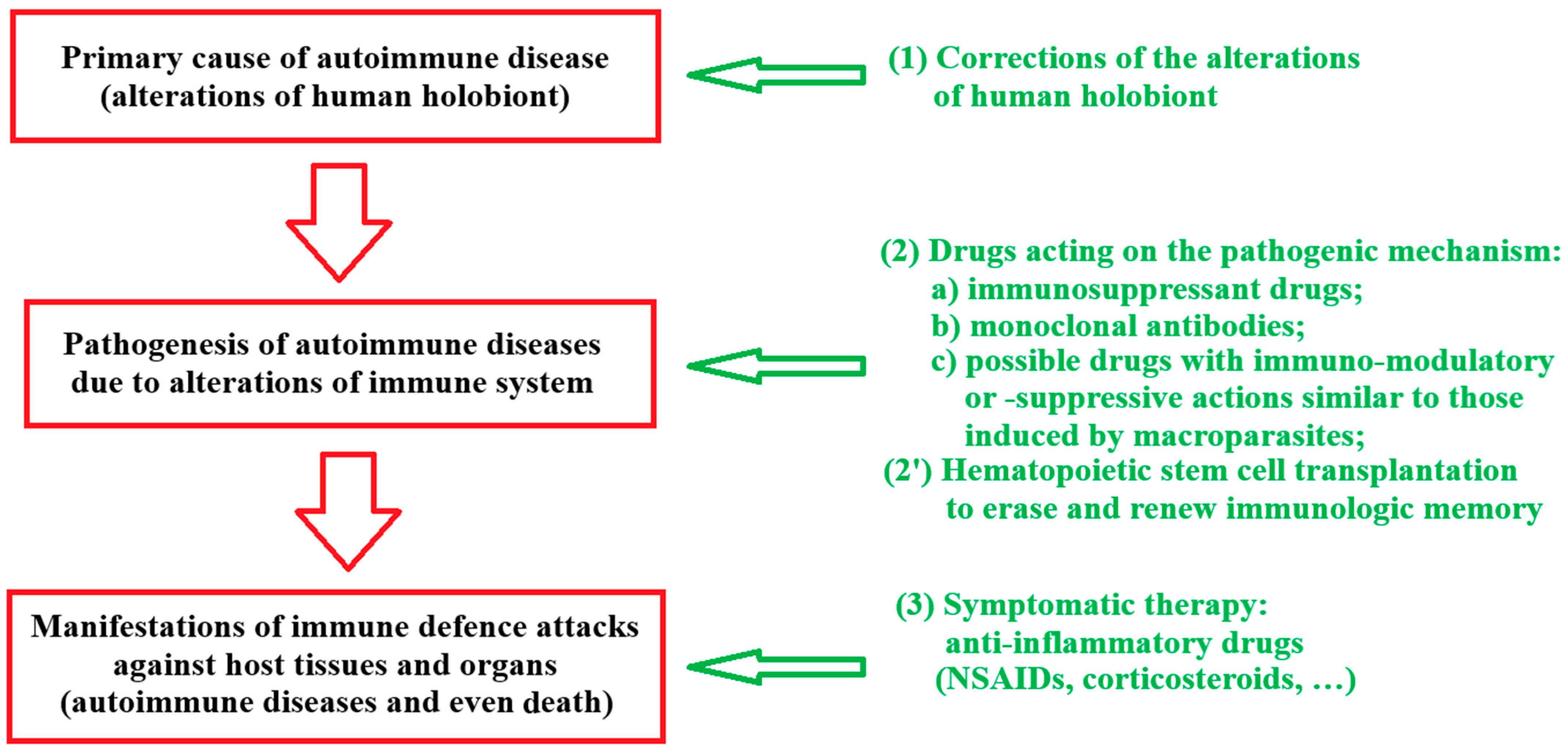
- An autoimmune disease is not a disease without a cause. Evidence suggests that autoimmune diseases have, in most cases, as primary cause the eradication of macroparasites, which is part of a severe alteration of the human holobiont;
- (2)
- Therefore, it is rational to prevent the alteration of the holobiont by avoiding the eradication of the macroparasites or by a new infection with macroparasites;
- (3)
- If the eradication of the macroparasites has already taken place, the alteration of the holobiont must be corrected as best and soon as possible by reinserting the helminths;
- (4)
- This should be achieved by reintroducing macroparasites to which our species has long adapted (e.g., Enterobius vermicularis, pinworm; Ascaris lumbricoides, roundworm). It should not be attempted using parasites for which adaptation is nonexistent or incomplete (e.g., parasites adapted to other species—as Thichuris suis, the pig whipworm, and Hymenolepis diminuta, the rat tapeworm cysticerci—or parasites common in tropical environments for which populations living for millennia in other habitats have little or no evolutionary experience—as Necator americanus). Reinfection with macroparasites does not exclude the possibility that, if necessary, e.g., because they are poorly tolerated or cause problems, they can be limited in their proliferation or eradicated through the use of dewormers;
- (5)
- Reintegration of macroparasites should occur as early as possible in life when the immune system is developing. Contacts between host and parasites occur under evolutionarily “natural” conditions at an early age, and it is by no means guaranteed that contact at later (not “normal”) ages will lead to a complete correction of the possible abnormal development of the immune system.
5. Conclusions
Author Contributions
Funding
Data Availability Statement
Conflicts of Interest
References
- Libertini, G.; Corbi, G.; Shubernetskaya, O.; Komici, K.; Ferrara, N. The Essential Revolution of Evolutionary Medicine; Springer: Berlin/Heidelberg, Germany, 2024. [Google Scholar]
- Nesse, R.M. The importance of Evolution for Medicine. In Evolutionary Medicine and Health: New Perspectives; Trevathan, W.R., Smith, E.O., McKenna, J.J., Eds.; Oxford University Press: New York, NY, USA, 2008; pp. 416–431. [Google Scholar]
- Dobzhansky, T. Nothing in biology makes sense except in the light of evolution. Am. Biol. Teach. 1973, 35, 125–129. [Google Scholar] [CrossRef] [Scilit]
- Varki, A. Nothing in medicine makes sense, except in the light of evolution. J. Mol. Med. 2012, 90, 481–494. [Google Scholar] [CrossRef] [Scilit] [PubMed]
- Libertini, G. Ragionamenti Evoluzionistici; Book in Italian; Società Editrice Napoletana: Napoli, Italy, 1983; English Edition: Evolutionary Arguments on Aging, Disease, and Other Topics; Azinet Press: Crownsville, MD, USA, 2011. [Google Scholar]
- Eaton, S.B.; Konner, M.; Shostak, M. Stone agers in the fast lane: Chronic degenerative diseases in evolutionary perspective. Am. J. Med. 1988, 84, 739–749. [Google Scholar] [CrossRef] [Scilit] [PubMed]
- Sánchez-Cañizares, C.; Jorrín, B.; Poole, P.S.; Tkacz, A. Understanding the holobiont: The interdependence of plants and their microbiome. Curr. Opin. Microbiol. 2017, 38, 188–196. [Google Scholar] [CrossRef] [Scilit] [PubMed]
- Fiuza, B.S.D.; Fonseca, H.F.; Meirelles, P.M.; Marques, C.R.; da Silva, T.M.; Figueiredo, C.A. Understanding Asthma and Allergies by the Lens of Biodiversity and Epigenetic Changes. Front. Immunol. 2021, 12, 623737. [Google Scholar] [CrossRef] [Scilit] [PubMed]
- Wildschutte, J.H.; Williams, Z.H.; Meagan Montesion, M.; Subramian, R.P.; Kidd, J.M.; Coffin, J.M. Discovery of unfixed endogenous retrovirus insertions in diverse human populations. Proc. Natl. Acad. Sci. USA 2016, 113, E2326–E2334. [Google Scholar] [CrossRef] [Scilit] [PubMed]
- Pride, D.T.; Salzman, J.; Haynes, M.; Rohwer, F.; Davis-Long, C.; White, R.A. Evidence of a robust resident bacteriophage population revealed through analysis of the human salivary virome. ISME J. 2012, 6, 915–926. [Google Scholar] [CrossRef] [Scilit] [PubMed]
- Abeles, S.R.; Pride, D.T. Molecular bases and role of viruses in the human microbiome. J. Mol. Biol. 2014, 426, 3892–3906. [Google Scholar] [CrossRef] [Scilit] [PubMed]
- Ghose, C.; Ly, M.; Schwanemann, L.K.; Shin, J.H.; Atab, K.; Barr, J.J.; Little, M.; Schooley, R.T.; Chopyk, J.; Pride, D.T. The Virome of Cerebrospinal Fluid: Viruses Where We Once Thought There Were None. Front. Microbiol. 2019, 10, 2061. [Google Scholar] [CrossRef] [Scilit] [PubMed]
- Moustafa, A.; Xie, C.; Kirkness, E.; Biggs, W.; Wong, E.; Turpaz, Y.; Bloom, K.; Delwart, E.; Nelson, K.E.; Venter, J.C.; et al. The blood DNA virome in 8000 humans. PLoS Pathog. 2017, 13, e1006292. [Google Scholar] [CrossRef] [Scilit] [PubMed]
- Liang, G.; Bushman, F.D. The human virome: Assembly, composition and host interactions. Nat. Rev. Microbiol. 2021, 19, 514–527. [Google Scholar] [CrossRef] [Scilit] [PubMed]
- Pannaraj, P.S.; Ly, M.; Cerini, C.; Saavedra, M.; Aldrovandi, G.M.; Saboory, A.A.; Johnson, K.M.; Pride, D.T. Shared and distinct features of human milk and infant stool viromes. Front. Microbiol. 2018, 9, 1162. [Google Scholar] [CrossRef] [Scilit] [PubMed]
- Grice, E.A.; Segre, J.A. The human microbiome: Our second genome. Annu. Rev. Genom. Hum. Genet. 2012, 13, 151–170. [Google Scholar] [CrossRef] [Scilit] [PubMed]
- Hou, K.; Wu, Z.X.; Chen, X.Y.; Wang, J.Q.; Zhang, D.; Xiao, C.; Zhu, D.; Koya, J.B.; Wei, L.; Li, J.; et al. Microbiota in health and diseases. Signal. Transduct. Target Ther. 2022, 7, 135. [Google Scholar] [CrossRef] [Scilit] [PubMed]
- LeBlanc, J.G.; Milani, C.; de Giori, G.S.; Sesma, F.; van Sinderen, D.; Ventura, M. Bacteria as vitamin suppliers to their host: A gut microbiota perspective. Curr. Opin. Biotechnol. 2013, 24, 160–168. [Google Scholar] [CrossRef] [Scilit] [PubMed]
- Zhan, Q.; Wang, R.; Thakur, K.; Feng, J.Y.; Zhu, Y.Y.; Zhang, J.G.; Wei, Z.J. Unveiling of dietary and gut-microbiota derived B vitamins: Metabolism patterns and their synergistic functions in gut-brain homeostasis. Crit. Rev. Food Sci. Nutr. 2022, 22, 4046–4058. [Google Scholar] [CrossRef] [Scilit] [PubMed]
- Wu, S.; Grimm, R.; German, J.B.; Lebrilla, C.B. Annotation and structural analysis of sialylated human milk oligosaccharides. J. Proteome Res. 2011, 10, 856–868. [Google Scholar] [CrossRef] [Scilit] [PubMed]
- Urashima, T.; Hirabayashi, J.; Sato, S.; Kobata, A. Human Milk Oligosaccharides as Essential Tools for Basic and Application Studies on Galectins. Trends Glycosci. Glycotechnol. 2018, 30, SJ11–SJ24. [Google Scholar] [CrossRef] [Scilit]
- Wu, S.; Tao, N.; German, J.B.; Grimm, R.; Lebrilla, C.B. Development of an annotated library of neutral human milk oligosaccharides. J. Proteome Res. 2010, 9, 4138–4151. [Google Scholar] [CrossRef] [Scilit] [PubMed]
- Pisetsky, D.S. Pathogenesis of autoimmune disease. Nat. Rev. Nephrol. 2023, 19, 508–524. [Google Scholar] [CrossRef] [Scilit] [PubMed]
- Mu, S.; Wang, W.; Liu, Q.; Ke, N.; Li, H.; Sun, F.; Zhang, J.; Zhu, Z. Autoimmune disease: A view of epigenetics and therapeutic targeting. Front. Immunol. 2024, 15, 1482728. [Google Scholar] [CrossRef] [Scilit] [PubMed]
- Goldman, M.D.; Cooney, K.A. (Eds.) Goldman-Cecil Medicine, 27th ed.; Elsevier Science Health Science: Philadelphia, PA, USA.
- Miller, F.W. The increasing prevalence of autoimmunity and autoimmune diseases: An urgent call to action for improved understanding, diagnosis, treatment, and prevention. Curr. Opin. Immunol. 2023, 80, 102266. [Google Scholar] [CrossRef] [Scilit] [PubMed]
- Tallis, R.; Fillit, H.; Brocklehurst, J.C. (Eds.) Brocklehurst’s Textbook of Geriatric Medicine and Gerontology, 5th ed.; Churchill Livingston: London, UK, 1998. [Google Scholar]
- Michel, J.P.; Beattie, B.L.; Martin, F.C.; Walston, J. Oxford Textbook of Geriatric Medicine, 3rd ed.; Oxford University Press: New York, NY, USA, 2018. [Google Scholar]
- Autoimmune Registry 2026. Available online: https://www.autoimmuneregistry.org/autoimmune-diseases (accessed on 10 January 2026).
- Autoimmune Association. About Autoimmunity. Available online: https://autoimmune.org/resource-center/about-autoimmunity/ (accessed on 10 January 2026).
- Autoimmune Registry 2023. Available online: https://www.autoimmuneregistry.org/autoimmune-diseases (accessed on 7 April 2023).
- Eaton, S.B.; Shostak, M.; Konner, M. The Paleolithic Prescription: A Program of Diet and Exercise and a Design for Living; Harper and Row: New York, NY, USA, 1988. [Google Scholar]
- Early, J.; Peters, J. The Population Dynamics of the Mucajai Yanomamo; Academic Press: New York, NY, USA, 1990. [Google Scholar]
- Hill, K.; Hurtado, A.M. Ache Life History; Aldine De Gruyter: New York, NY, USA, 1996. [Google Scholar]
- Foster, R.M.; Harries, J.R. Multiple sclerosis in the African. Br. Med. J. 1970, 3, 628. [Google Scholar] [CrossRef] [Scilit] [PubMed]
- Gelfand, M. The Sick African. A Clinical Study, 3rd ed.; Juta: Cape Town, South Africa, 1957. [Google Scholar]
- Trowell, H.C. Non-Infective Disease in Africa; The Peculiarities of Medical Non-Infective Diseases in the Indigenous Inhabitants of Africa South of the Sahara; Edward Arnold: London, UK, 1960. [Google Scholar]
- Haddock, D.R.W. Neurological disorders in Tanzania. J. Trop. Med. Hyg. 1965, 68, 161–166. [Google Scholar] [PubMed]
- Dean, G. Annual incidence, prevalence, and mortality of multiple sclerosis in white South-African-born and in white immigrants to South Africa. Br. Med. J. 1967, 2, 724–730. [Google Scholar] [CrossRef] [Scilit] [PubMed]
- Greenwood, B.M. Autoimmune disease and parasitic infections in Nigerians. Lancet 1968, 292, 380–382. [Google Scholar] [CrossRef] [Scilit] [PubMed]
- Tsega, E.; Choremi, H.; Bottazzo, G.F.; Doniach, D. Prevalence of autoimmune diseases and autoantibodies in Ethiopia. Trop. Geogr. Med. 1980, 32, 231–236. [Google Scholar] [PubMed]
- Clark, I.A.; Al-Yaman, F.M.; Cowden, W.B.; Rockett, K.A. Does malarial tolerance, through nitric oxide, explain the low incidence of autoimmune disease in tropical Africa? Lancet 1996, 348, 1492–1494. [Google Scholar] [CrossRef] [Scilit] [PubMed]
- Adebajo, A.O. Low frequency of autoimmune disease in tropical Africa. Lancet 1997, 349, 361–362. [Google Scholar] [CrossRef] [Scilit] [PubMed]
- Lerner, A.; Jeremias, P.; Matthias, T. The World Incidence and Prevalence of Autoimmune Diseases is Increasing. Int. J. Celiac Dis. 2015, 3, 151–155. [Google Scholar] [CrossRef] [Scilit]
- Onkamo, P.; Väänänen, S.; Karvonen, M.; Tuomilehto, J. Worldwide increase in incidence of Type I diabetes–The analysis of the data on published incidence trends. Diabetologia 1999, 42, 1395–1403, Erratum in Diabetologia 2000, 43, 685. [Google Scholar] [CrossRef] [Scilit] [PubMed]
- Harjutsalo, V.; Sund, R.; Knip, M.; Groop, P.H. Incidence of type 1 diabetes in Finland. JAMA 2013, 310, 427–428. [Google Scholar] [CrossRef] [Scilit] [PubMed]
- West, J.; Fleming, K.M.; Tata, L.J.; Card, T.R.; Crooks, C.J. Incidence and prevalence of celiac disease and dermatitis herpetiformis in the UK over two decades: Population-based study. Am. J. Gastroenterol. 2014, 109, 757–768. [Google Scholar] [CrossRef] [Scilit] [PubMed]
- Kerr, D.; Nakazawa, D.J. Foreword. In The Autoimmune Epidemic; Touchstone: New York, NY, USA, 2008. [Google Scholar]
- Bach, J.F. The effect of infections on susceptibility to autoimmune and allergic diseases. N. Engl. J. Med. 2002, 347, 911–920. [Google Scholar] [CrossRef] [Scilit] [PubMed]
- Tuomilehto, J.; Karvonen, M.; Pitkäniemi, J.; Virtala, E.; Kohtamäki, K.; Toivanen, L.; Tuomilehto-Wolf, E. Record-high incidence of Type I (insulin-dependent) diabetes mellitus in Finnish children. The Finnish Childhood Type I Diabetes Registry Group. Diabetologia 1999, 42, 655–660. [Google Scholar] [CrossRef] [Scilit] [PubMed]
- Farrokhyar, F.; Swarbrick, E.T.; Irvine, E.J. A critical review of epidemiological studies in inflammatory bowel disease. Scand. J. Gastroenterol. 2001, 36, 2–15, Erratum in Scand. J. Gastroenterol. 2001, 36, 560. [Google Scholar] [CrossRef] [Scilit]
- Pugliatti, M.; Sotgiu, S.; Solinas, G.; Castiglia, P.; Pirastru, M.I.; Murgia, B.; Mannu, L.; Sanna, G.; Rosati, G. Multiple sclerosis epidemiology in Sardinia: Evidence for a true increasing risk. Acta Neurol. Scand. 2001, 103, 20–26. [Google Scholar] [CrossRef] [Scilit] [PubMed]
- Latvijas Oficiala Statistika—Incidence and Prevalence of Diabetes Mellitus by Type of Diabetes and Regions 2010–2024. Available online: https://data.stat.gov.lv/pxweb/en/OSP_PUB/START__VES__VS__VSS/VSS200/table/tableViewLayout1/ (accessed on 16 June 2026).
- Jacobson, D.L.; Gange, S.J.; Rose, N.R.; Graham, N.M. Epidemiology and estimated population burden of selected autoimmune diseases in the United States. Clin. Immunol. Immunopathol. 1997, 84, 223–243. [Google Scholar] [CrossRef] [Scilit] [PubMed]
- Eaton, W.W.; Rose, N.R.; Kalaydjian, A.; Pedersen, M.G.; Mortensen, P.B. Epidemiology of autoimmune diseases in Denmark. J. Autoimmun. 2007, 29, 1–9. [Google Scholar] [CrossRef] [Scilit] [PubMed]
- Wang, L.; Wang, F.S.; Gershwin, M.E. Human autoimmune diseases: A comprehensive update. J. Intern. Med. 2015, 278, 369–395. [Google Scholar] [CrossRef] [Scilit] [PubMed]
- Cooper, G.S.; Bynum, M.L.; Somers, E.C. Recent insights in the epidemiology of autoimmune diseases: Improved prevalence estimates and understanding of clustering of diseases. J. Autoimmun. 2009, 33, 197–207. [Google Scholar] [CrossRef] [Scilit] [PubMed]
- Schweizer, J.J.; von Blomberg, B.M.; Bueno-de Mesquita, H.B.; Mearin, M.L. Coeliac disease in The Netherlands. Scand. J. Gastroenterol. 2004, 39, 359–364. [Google Scholar] [CrossRef] [Scilit] [PubMed]
- Kondrashova, A.; Reunanen, A.; Romanov, A.; Karvonen, A.; Viskari, H.; Vesikari, T.; Ilonen, J.; Knip, M.; Hyöty, H. A six-fold gradient in the incidence of type 1 diabetes at the eastern border of Finland. Ann. Med. 2005, 37, 67–72. [Google Scholar] [CrossRef] [Scilit] [PubMed]
- Kondrashova, A.; Mustalahti, K.; Kaukinen, K.; Viskari, H.; Volodicheva, V.; Haapala, A.M.; Ilonen, J.; Knip, M.; Mäki, M.; Hyoty, H.; et al. Lower economic status and inferior hygienic environment may protect against celiac disease. Ann. Med. 2008, 40, 223–231. [Google Scholar] [CrossRef] [Scilit] [PubMed]
- Forsyth, K.S.; Jiwrajka, N.; Lovell, C.D.; Toothacre, N.E.; Anguera, M.C. The conneXion between sex and immune responses. Nat. Rev. Immunol. 2024, 24, 487–502. [Google Scholar] [CrossRef] [Scilit] [PubMed]
- Ramos-Casals, M.; Brito-Zerón, P.; Kostov, B.; Sisó-Almirall, A.; Bosch, X.; Buss, D.; Trilla, A.; Stone, J.H.; Khamashta, M.A.; Shoenfeld, Y. Google-driven search for big data in autoimmune geoepidemiology: Analysis of 394,827 patients with systemic autoimmune diseases. Autoimmun. Rev. 2015, 14, 670–679. [Google Scholar] [CrossRef] [Scilit] [PubMed]
- Xing, E.; Billi, A.C.; Gudjonsson, J.E. Sex Bias and Autoimmune Diseases. J. Invest. Dermatol. 2022, 142, 857–866. [Google Scholar] [CrossRef] [Scilit] [PubMed]
- Pasche, B.; Kalaydjiev, S.; Franz, T.J.; Kremmer, E.; Gailus-Durner, V.; Fuchs, H.; de Angelis, M.H.; Lengeling, A.; Busch, D.H. Sex-dependent susceptibility to Listeria monocytogenes infection is mediated by differential interleukin-10 production. Infect. Immun. 2005, 73, 5952–5960. [Google Scholar] [CrossRef] [Scilit] [PubMed]
- Moulton, V.R. Sex Hormones in Acquired Immunity and Autoimmune Disease. Front Immunol. 2018, 9, 2279. [Google Scholar] [CrossRef] [Scilit] [PubMed]
- Kelvin, E.A.; Carpio, A.; Bagiella, E.; Leslie, D.; Leon, P.; Andrews, H.; Hauser, W.A. Ecuadorian Neurocysticercosis Group. The association of host age and gender with inflammation around neurocysticercosis cysts. Ann. Trop. Med. Parasitol. 2009, 103, 487–499. [Google Scholar] [CrossRef] [Scilit] [PubMed]
- Tchuenté, L.T.; Behnke, J.M.; Gilbert, F.S.; Southgate, V.R.; Vercruysse, J. Polyparasitism with Schistosoma haematobium and soil-transmitted helminth infections among school children in Loum, Cameroon. Trop. Med. Int. Health 2003, 8, 975–986, Erratum in Trop. Med. Int. Health 2004, 9, 314. [Google Scholar] [CrossRef] [Scilit] [PubMed]
- Wright, J.E.; Werkman, M.; Dunn, J.C.; Anderson, R.M. Current epidemiological evidence for predisposition to high or low intensity human helminth infection: A systematic review. Parasit. Vectors 2018, 2018, 11. [Google Scholar] [CrossRef] [Scilit] [PubMed]
- Borba, V.V.; Zandman-Goddard, G.; Shoenfeld, Y. Exacerbations of autoimmune diseases during pregnancy and postpartum. Best Pract. Res. Clin. Endocrinol. Metab. 2019, 33, 101321. [Google Scholar] [CrossRef] [Scilit] [PubMed]
- Christen, U.; Hintermann, E. Pathogens and autoimmune hepatitis. Clin. Exp. Immunol. 2019, 195, 35–51. [Google Scholar] [CrossRef] [Scilit] [PubMed]
- Maisha, J.A.; El-Gabalawy, H.S.; O’Neil, L.J. Modifiable risk factors linked to the development of rheumatoid arthritis: Evidence, immunological mechanisms and prevention. Front. Immunol. 2023, 14, 1221125, Erratum in Front. Immunol. 2024, 15, 1370893. https://doi.org/10.3389/fimmu.2024.1370893. [Google Scholar] [CrossRef] [Scilit] [PubMed]
- Tiwari, V.; Brent, L.H. Psoriatic Arthritis. In StatPearls; StatPearls Publishing: Treasure Island, FL, USA, 2025. [Google Scholar] [PubMed]
- Strachan, D.P. Hay fever, hygiene, and household size. BMJ 1989, 299, 1259–1260. [Google Scholar] [CrossRef] [Scilit] [PubMed]
- Okada, H.; Kuhn, C.; Feillet, H.; Bach, J.-F. The “hygiene hypothesis” for autoimmune and allergic diseases: An update. Clin. Exp. Immunol. 2010, 160, 1–9. [Google Scholar] [CrossRef] [Scilit] [PubMed]
- Zandman-Goddard, G.; Shoenfeld, Y. Parasitic infection and autoimmunity. Lupus 2009, 18, 1144–1148. [Google Scholar] [CrossRef] [Scilit] [PubMed]
- Ben-Ami Shor, D.; Harel, M.; Eliakim, R.; Shoenfeld, Y. The hygiene theory harnessing helminths and their ova to treat autoimmunity. Clin. Rev. Allergy Immunol. 2013, 45, 211–216. [Google Scholar] [CrossRef] [Scilit] [PubMed]
- Versini, M.; Jeandel, P.Y.; Bashi, T.; Bizzaro, G.; Blank, M.; Shoenfeld, Y. Unraveling the Hygiene Hypothesis of helminthes and autoimmunity: Origins, pathophysiology, and clinical applications. BMC Med. 2015, 13, 81. [Google Scholar] [CrossRef] [Scilit] [PubMed]
- Colafrancesco, S.; Perricone, C.; Priori, R.; Valesini, G.; Shoenfeld, Y. Sjogren’s syndrome: Another facet of the autoimmune/inflammatory syndrome induced by adjuvants (ASIA). J. Autoimmun. 2014, 51, 10–16. [Google Scholar] [CrossRef] [Scilit] [PubMed]
- Kivity, S.; Arango, M.T.; Ehrenfeld, M.; Tehori, O.; Shoenfeld, Y.; Anaya, J.-M.; Agmon-Levin, N. Infection and autoimmunity in Sjogren’s syndrome: A clinical study and comprehensive review. J. Autoimmun. 2014, 51, 17–22. [Google Scholar] [CrossRef] [Scilit] [PubMed]
- Peng, J.; Narasimhan, S.; Marchesi, J.R.; Benson, A.; Wong, F.S.; Wen, L. Long term effect of gut microbiota transfer on diabetes development. J. Autoimmun. 2014, 53, 85–94. [Google Scholar] [CrossRef] [Scilit] [PubMed]
- Greenwood, B.M.; Herrick, E.M.; Voller, A. Can parasitic infection suppress autoimmune disease? Proc. R. Soc. Med. 1970, 63, 19–20. [Google Scholar] [CrossRef] [Scilit] [PubMed]
- Elliott, D.E.; Urban, J.F., Jr.; Argo, C.K.; Weinstock, J.V. Does the failure to acquire helminthic parasites predispose to Crohn’s disease? FASEB J. 2000, 14, 1848–1855. [Google Scholar] [CrossRef] [Scilit] [PubMed]
- Healy, G.R.; Gleason, N.N.; Bokat, R.; Pond, H.; Roper, M. Prevalence of ascariasis and amebiasis in Cherokee Indian school children. Public Health Rep. 1969, 84, 907–914. [Google Scholar] [CrossRef] [Scilit] [PubMed]
- Salas, S.D.; Heifetz, R.; Barrett-Connor, E. Intestinal parasites in Central American immigrants in the United States. Arch. Inter. Med. 1990, 150, 1514–1516. [Google Scholar] [CrossRef] [PubMed]
- Weinstock, J.V.; Summers, R.; Elliott, D.E. Helminths and harmony. Gut 2004, 53, 7–9. [Google Scholar] [CrossRef] [Scilit] [PubMed]
- Sabin, E.A.; Araujo, M.I.; Carvalho, E.M.; Pearce, E.J. Impairment of tetanus toxoid-specific Th1-like immune responses in humans infected with Schistosoma mansoni. J. Infect. Dis. 1996, 173, 269–272. [Google Scholar] [CrossRef] [Scilit] [PubMed]
- Borkow, G.; Leng, Q.; Weisman, Z.; Stein, M.; Galai, N.; Kalinkovich, A.; Bentwich, Z. Chronic immune activation associated with intestinal helminth infections results in impaired signal transduction and anergy. J. Clin. Invest. 2000, 106, 1053–1060. [Google Scholar] [CrossRef] [Scilit] [PubMed]
- Doetze, A.; Satoguina, J.; Burchard, G.; Rau, T.; Löliger, C.; Fleischer, B.; Hoerauf, A. Antigen-specific cellular hyporesponsiveness in a chronic human helminth infection is mediated by T(h)3/T(r)1-type cytokines IL-10 and transforming growth factor-beta but not by a T(h)1 to T(h)2 shift. Int. Immunol. 2000, 12, 623–630. [Google Scholar] [CrossRef] [Scilit] [PubMed]
- Zaccone, P.; Fehervari, Z.; Phillips, J.M.; Dunne, D.W.; Cooke, A. Parasitic worms and inflammatory diseases. Parasite Immunol. 2006, 28, 515–523. [Google Scholar] [CrossRef] [Scilit] [PubMed]
- van den Biggelaar, A.H.; van Ree, R.; Rodrigues, L.C.; Lell, B.; Deelder, A.M.; Kremsner, P.G.; Yazdanbakhsh, M. Decreased atopy in children infected with Schistosoma haematobium: A role for parasite-induced interleukin-10. Lancet 2000, 356, 1723–1727. [Google Scholar] [CrossRef] [Scilit] [PubMed]
- Moreels, T.G.; Nieuwendijk, R.J.; De Man, J.G.; De Winter, B.Y.; Herman, A.G.; Van Marck, E.A.; Pelckmans, P.A. Concurrent infection with Schistosoma mansoni attenuates inflammation induced changes in colonic morphology, cytokine levels, and smooth muscle contractility of trinitrobenzene sulphonic acid induced colitis in rats. Gut 2004, 53, 99–107. [Google Scholar] [CrossRef] [Scilit] [PubMed]
- Kramer, J.; Fahey, M.; Santos, R.; Carville, A.; Wachtman, L.; Mansfield, K. Alopecia in Rhesus macaques correlates with immunophenotypic alterations in dermal inflammatory infiltrates consistent with hypersensitivity etiology. J. Med. Primatol. 2010, 39, 112–122. [Google Scholar] [CrossRef] [Scilit] [PubMed]
- Velasquez-Manoff, M. An Epidemic of Absence: A New Way of Understanding Allergies and Autoimmune Diseases; Simon and Schuster: New York, NY, USA, 2012. [Google Scholar]
- Weinstock, J.V.; Elliott, D.E. Helminth infections decrease host susceptibility to immune-mediated diseases. J. Immunol. 2014, 193, 3239–3247. [Google Scholar] [CrossRef] [Scilit] [PubMed]
- Zaccone, P.; Hall, S.W. Helminth infection and type 1 diabetes. Rev. Diabet. Stud. 2012, 9, 272–286. [Google Scholar] [CrossRef] [Scilit] [PubMed]
- Gershwin, M.E.; Tsokos, G.C.; Diamond, B. The Rose and Macky Textbook of Autoimmune Diseases, 7th ed.; Elsevier: Amsterdam, The Netherlands, 2024. [Google Scholar]
- Lu, R.M.; Hwang, Y.C.; Liu, I.J.; Lee, C.C.; Tsai, H.Z.; Li, H.J.; Wu, H.C. Development of therapeutic antibodies for the treatment of diseases. J. Biomed. Sci. 2020, 27, 1. [Google Scholar] [CrossRef] [Scilit] [PubMed]
- Katsavos, S.; Coles, A. Alemtuzumab as Treatment for Multiple Sclerosis. Cold Spring Harb. Perspect. Med. 2018, 8, a032029. [Google Scholar] [CrossRef] [Scilit] [PubMed]
- Hill, A.; Hill, Q.A. Autoimmune hemolytic anemia. Hematol. Am. Soc. Hematol. Educ. Program. 2018, 2018, 382–389. [Google Scholar] [CrossRef] [Scilit] [PubMed]
- Fasano, S.; Isenberg, D.A. Present and novel biologic drugs in primary Sjögren’s syndrome. Clin. Exp. Rheumatol. 2019, 37, 167–174. [Google Scholar] [PubMed]
- Kesharwani, D.; Paliwal, R.; Satapathy, T.; Das Paul, S. Rheumatoid Arthritis: An Updated Overview of Latest Therapy and Drug Delivery. J. Pharmacopunct. 2019, 22, 210–224. [Google Scholar] [CrossRef] [Scilit] [PubMed]
- Lucchini, E.; Zaja, F.; Bussel, J. Rituximab in the treatment of immune thrombocytopenia: What is the role of this agent in 2019? Haematologica 2019, 104, 1124–1135. [Google Scholar] [CrossRef] [Scilit] [PubMed]
- Krajnc, N.; Bsteh, G.; Berger, T.; Mares, J.; Hartung, H.P. Monoclonal Antibodies in the Treatment of Relapsing Multiple Sclerosis: An Overview with Emphasis on Pregnancy, Vaccination, and Risk Management. Neurotherapeutics 2022, 19, 753–773. [Google Scholar] [CrossRef] [Scilit] [PubMed]
- Slentz, D.H.; Nelson, C.C.; Smith, T.J. Teprotumumab: A novel therapeutic monoclonal antibody for thyroid-associated ophthalmopathy. Expert Opin. Investig. Drugs 2020, 29, 645–649. [Google Scholar] [CrossRef] [Scilit] [PubMed]
- Burek-Michalska, A.; Turno-Kręcicka, A. Adalimumab in the treatment of non-infectious uveitis. Adv. Clin. Exp. Med. 2020, 29, 1231–1236. [Google Scholar] [CrossRef] [Scilit] [PubMed]
- Alabbad, S.; AlGaeed, M.; Sikorski, P.; Kaminski, H.J. Monoclonal Antibody-Based Therapies for Myasthenia Gravis. BioDrugs 2020, 34, 557–566. [Google Scholar] [CrossRef] [Scilit] [PubMed]
- Song, Z.; Zhang, J.; Meng, J.; Jiang, G.; Yan, Z.; Yang, Y.; Chen, Z.; You, W.; Wang, Z.; Chen, G. Different Monoclonal Antibodies in Myasthenia Gravis: A Bayesian Network Meta-Analysis. Front. Pharmacol. 2022, 12, 790834. [Google Scholar] [CrossRef] [Scilit] [PubMed]
- Ke, Q.; Kroger, C.J.; Clark, M.; Tisch, R.M. Evolving Antibody Therapies for the Treatment of Type 1 Diabetes. Front. Immunol. 2021, 11, 624568. [Google Scholar] [CrossRef] [Scilit] [PubMed]
- Ebrahimiadib, N.; Berijani, S.; Ghahari, M.; Pahlaviani, F.G. Ankylosing Spondylitis. J. Ophthalmic Vis. Res. 2021, 16, 462–469. [Google Scholar] [CrossRef] [Scilit] [PubMed]
- Yang, J.; Liu, X. Immunotherapy for Refractory Autoimmune Encephalitis. Front. Immunol. 2021, 12, 790962. [Google Scholar] [CrossRef] [Scilit] [PubMed]
- Berentsen, S.; Fattizzo, B.; Barcellini, W. The choice of new treatments in autoimmune hemolytic anemia: How to pick from the basket? Front. Immunol. 2023, 14, 1180509. [Google Scholar] [CrossRef] [Scilit] [PubMed]
- Deng, W.P. Recent advances in the treatment of systemic lupus erythematosus with belimumab in children. Chin. J. Contemp. Pediatr. 2021, 23, 1069–1074. [Google Scholar] [CrossRef] [Scilit] [PubMed]
- Joy, A.; Muralidharan, A.; Alfaraj, M.; Shantharam, D.; Cherukuri, A.S.S.; Muthukumar, A. The Role of Belimumab in Systemic Lupus Erythematosis: A Systematic Review. Cureus 2022, 14, e25887. [Google Scholar] [CrossRef] [Scilit] [PubMed]
- Nie, T.; Blair, H.A. Inebilizumab: A Review in Neuromyelitis Optica Spectrum Disorder. CNS Drugs 2022, 36, 1133–1141. [Google Scholar] [CrossRef] [Scilit] [PubMed]
- Briani, C.; Visentin, A. Therapeutic Monoclonal Antibody Therapies in Chronic Autoimmune Demyelinating Neuropathies. Neurotherapeutics 2022, 19, 874–884. [Google Scholar] [CrossRef] [Scilit] [PubMed]
- Didona, D.; Paolino, G.; Di Zenzo, G.; Didona, B.; Pampena, R.; Di Nicola, M.R.; Mercuri, S.R. Pemphigus Vulgaris: Present and Future Therapeutic Strategies. Dermatol. Pract. Concept. 2022, 12, e2022037. [Google Scholar] [CrossRef] [Scilit] [PubMed]
- Yun, Z.; Duan, L.; Liu, X.; Cai, Q.; Li, C. An update on the biologics for the treatment of antiphospholipid syndrome. Front. Immunol. 2023, 14, 1145145. [Google Scholar] [CrossRef] [Scilit] [PubMed]
- Ly, S.; Nedosekin, D.; Wong, H.K. Review of an Anti-CD20 Monoclonal Antibody for the Treatment of Autoimmune Diseases of the Skin. Am. J. Clin. Dermatol. 2023, 24, 247–273. [Google Scholar] [CrossRef] [Scilit] [PubMed]
- Olbrich, H.; Sadik, C.D.; Ludwig, R.J.; Thaçi, D.; Boch, K. Dupilumab in Inflammatory Skin Diseases: A Systematic Review. Biomolecules 2023, 13, 634. [Google Scholar] [CrossRef] [Scilit] [PubMed]
- Autoimmune Association. The Economic Effects of Autoimmune Disease Research. Available online: https://autoimmune.org/advocacy/economic-effects-of-autoimmune-disease-research/ (accessed on 10 January 2026).
- Lim, K.K.; Ng, K.; Balachandran, S.; Russell, M.D.; Boalch, A.; Sinclair, A.; Coker, B.; Vinnakota, K.; Mansoor, R.; Douiri, A.; et al. Measuring the impact of monoclonal antibody therapies. Front. Med. 2023, 10, 1256712. [Google Scholar] [CrossRef] [Scilit] [PubMed]
- Shepherd, J.; Cooper, K.; Harris, P.; Picot, J.; Rose, M. The clinical effectiveness and cost-effectiveness of abatacept, adalimumab, etanercept and tocilizumab for treating juvenile idiopathic arthritis: A systematic review and economic evaluation. Health Technol. Assess. 2016, 20, 1–222. [Google Scholar] [CrossRef] [Scilit] [PubMed]
- Kolev, M.; Kremer, A.E.; Sokollik, C.; Gershwin, M.E.; Terziroli Beretta-Piccoli, B. Unmet needs in autoimmune liver diseases. Curr. Opin. Immunol. 2025, 95, 102565. [Google Scholar] [CrossRef] [Scilit] [PubMed]
- Jairath, V.; Raine, T.; Leahy, T.P.; Potluri, R.; Wosik, K.; Gruben, D.; Cappelleri, J.C.; Hur, P.; Bartolome, L. Efficacy and safety of advanced therapies for moderately to severely active ulcerative colitis in induction and maintenance: Systematic literature review and Bayesian network meta-analysis. J. Comp. Eff. Res. 2025, 14, e240225. [Google Scholar] [CrossRef] [Scilit] [PubMed]
- Solitano, V.; Facciorusso, A.; Jess, T.; Ma, C.; Hassan, C.; Repici, A.; Jairath, V.; Armuzzi, A.; Singh, S. Comparative Risk of Serious Infections with Biologic Agents and Oral Small Molecules in Inflammatory Bowel Diseases: A Systematic Review and Meta-Analysis. Clin. Gastroenterol. Hepatol. 2023, 21, 907–921.e2. [Google Scholar] [CrossRef] [Scilit] [PubMed]
- Olivares-Guerrero, M.; Daudén, E.; Riera-Monroig, J.; González-Quesada, A.; Sahuquillo-Torralba, A.; Rivera-Díaz, R.; Carrascosa, J.M.; Belinchón, I.; Gómez-García, F.J.; Herrera-Acosta, E.; et al. Risk of adverse events of psoriasis treatment with biologic agents and new small molecules-BIOBADADERM Registry. J. Eur. Acad. Dermatol. Venereol. 2025, 39, 1643–1655. [Google Scholar] [CrossRef] [Scilit] [PubMed]
- Blank, M.; Shoenfeld, Y. Helminth derivative tuftsin-phopshorylcholine to treat autoimmunity. Autoimmun. Rev. 2024, 23, 103601. [Google Scholar] [CrossRef] [Scilit] [PubMed]
- Novikova, N.S.; Diatlova, A.S.; Derevtsova, K.Z.; Korneva, E.A.; Viktorovna, T.V.; Ostrinki, Y.; Abraham, L.; Quinn, S.; Segal, Y.; Churilov, L.P.; et al. Tuftsin-phosphorylcholine attenuate experimental autoimmune encephalomyelitis. J. Neuroimmunol. 2019, 337, 577070, Erratum in J Neuroimmunol. 2020, 339, 577137. https://doi.org/10.1016/j.jneuroim.2019.577137. [Google Scholar] [CrossRef] [Scilit] [PubMed]
- Croci, S.; Bonacini, M.; Muratore, F.; Caruso, A.; Fontana, A.; Boiardi, L.; Soriano, A.; Cavazza, A.; Cimino, L.; Belloni, L.; et al. The therapeutic potential of tuftsin-phosphorylcholine in giant cell arteritis. J. Autoimmun. 2019, 98, 113–121. [Google Scholar] [CrossRef] [Scilit] [PubMed]
- Shemer, A.; Kivity, S.; Shovman, O.; Bashi, T.; Perry, O.; Watad, A.; Shor, D.B.; Volkov, A.; Barshack, I.; Bragazzi, N.L.; et al. Tuftsin-phosphorylcholine (TPC) equally effective to methylprednisolone in ameliorating lupus nephritis in a mice model. Clin. Exp. Immunol. 2018, 193, 160–166. [Google Scholar] [CrossRef] [Scilit] [PubMed]
- Ni, Y.; Xiong, R.; Zhu, Y.; Luan, N.; Yu, C.; Yang, K.; Wang, H.; Xu, X.; Yang, Y.; Sun, S.; et al. A target-based discovery from a parasitic helminth as a novel therapeutic approach for autoimmune diseases. eBioMedicine 2023, 95, 104751. [Google Scholar] [CrossRef] [Scilit] [PubMed]
- Hosseini, A.; Gharibi, T.; Marofi, F.; Javadian, M.; Babaloo, Z.; Baradaran, B. Janus kinase inhibitors: A therapeutic strategy for cancer and autoimmune diseases. J. Cell. Physiol. 2020, 235, 5903–5924. [Google Scholar] [CrossRef] [Scilit] [PubMed]
- Blache, U.; Tretbar, S.; Koehl, U.; Mougiakakos, D.; Fricke, S. CAR T cells for treating autoimmune diseases. RMD Open 2023, 9, e002907. [Google Scholar] [CrossRef] [Scilit] [PubMed]
- Schett, G.; Mackensen, A.; Mougiakakos, D. CAR T-cell therapy in autoimmune diseases. Lancet 2023, 402, 2034–2044. [Google Scholar] [CrossRef] [Scilit] [PubMed]
- Alexander, T.; Greco, R. Hematopoietic stem cell transplantation and cellular therapies for autoimmune diseases: Overview and future considerations from the Autoimmune Diseases Working Party (ADWP) of the European Society for Blood and Marrow Transplantation (EBMT). Bone Marrow Transplant. 2022, 57, 1055–1062. [Google Scholar] [CrossRef] [Scilit] [PubMed]
- Alexander, T.; Sharrack, B.; Rovira, M.; Saccardi, R.; Farge, D.; Snowden, J.A.; Greco, R. Autoimmune Disease. In The EBMT Handbook: Hematopoietic Cell Transplantation and Cellular Therapies, 8th ed.; Sureda, A., Corbacioglu, S., Greco, R., Kröger, N., Carreraset, E., Eds.; Springer: Cham, Switzerland, 2024; Chapter 92. [Google Scholar] [CrossRef] [Scilit] [PubMed]
- Conti, V.; Corbi, G.; Costantino, M.; De Bellis, E.; Manzo, V.; Sellitto, C.; Stefanelli, B.; Colucci, F.; Filippelli, A. Biomarkers to Personalize the Treatment of Rheumatoid Arthritis: Focus on Autoantibodies and Pharmacogenetics. Biomolecules 2020, 10, 1672. [Google Scholar] [CrossRef] [Scilit] [PubMed]
- Fugger, L.; Jensen, L.T.; Rossjohn, J. Challenges, Progress, and Prospects of Developing Therapies to Treat Autoimmune Diseases. Cell 2020, 181, 63–80. [Google Scholar] [CrossRef] [Scilit] [PubMed]
- Song, Y.; Li, J.; Wu, Y. Evolving understanding of autoimmune mechanisms and new therapeutic strategies of autoimmune disorders. Signal Transduct. Target Ther. 2024, 9, 263, Erratum in Signal Transduct. Target Ther. 2025, 10, 426. https://doi.org/10.1038/s41392-025-02525-z. [Google Scholar] [CrossRef] [Scilit] [PubMed]
- Correale, J. Helminth/Parasite Treatment of Multiple Sclerosis. Curr. Treat. Options Neurol. 2014, 16, 296. [Google Scholar] [CrossRef] [Scilit] [PubMed]
- Sipahi, A.M.; Baptista, D.M. Helminths as an alternative therapy for intestinal diseases. World J. Gastroenterol. 2017, 23, 6009–6015. [Google Scholar] [CrossRef] [Scilit] [PubMed]
- Lothstein, K.E.; Gause, W.C. Mining Helminths for Novel Therapeutics. Trends Mol. Med. 2021, 27, 345–364. [Google Scholar] [CrossRef] [Scilit] [PubMed]
- Arai, T.; Lopes, F. Potential of human helminth therapy for resolution of inflammatory bowel disease: The future ahead. Exp. Parasitol. 2022, 232, 108189, Erratum in Exp. Parasitol. 2021, 234, 108218. [Google Scholar] [CrossRef] [Scilit] [PubMed]
- Shi, W.; Xu, N.; Wang, X.; Vallée, I.; Liu, M.; Liu, X. Helminth Therapy for Immune-Mediated Inflammatory Diseases: Current and Future Perspectives. J. Inflamm. Res. 2022, 15, 475–491. [Google Scholar] [CrossRef] [Scilit] [PubMed]
- Shields, V.E.; Cooper, J. Use of helminth therapy for management of ulcerative colitis and Crohn’s disease: A systematic review. Parasitology 2022, 149, 145–154. [Google Scholar] [CrossRef] [Scilit] [PubMed]
- Acharya, S.; Da’dara, A.A.; Skelly, P.J. Schistosome immunomodulators. PLoS Pathog. 2021, 17, e1010064. [Google Scholar] [CrossRef] [Scilit] [PubMed]
| multiple sclerosis | [98] |
| autoimmune hemolytic anemia | [99] |
| primary Sjögren’s syndrome | [100] |
| rheumatoid arthritis | [101] |
| immune thrombocytopenia | [102,103] |
| thyroid-associated ophthalmopathy | [104] |
| uveitis | [105] |
| myasthenia gravis | [106,107] |
| T1D | [108] |
| ankylosing spondylitis | [109] |
| autoimmune encephalitis | [110,111] |
| systemic lupus erythematosus | [112,113] |
| neuromyelitis optica spectrum disorder | [114] |
| chronic autoimmune demyelinating neuropathies | [115] |
| pemphigus vulgaris | [116] |
| antiphospholipid syndrome | [117] |
| autoimmune diseases of the skin (“pemphigus vulgaris, cutaneous lupus erythematous, dermatomyositis, systemic sclerosis, thyroid dermopathy, autoimmune pemphigoid diseases, and cutaneous vasculitis diseases.”) | [118] |
| inflammatory skin diseases (“bullous autoimmune diseases, eczema, prurigo, alopecia areata, chronic spontaneous urticaria, Netherton syndrome and a variety of other chronic inflammatory skin diseases.”) | [119] |
Disclaimer/Publisher’s Note: The statements, opinions and data contained in all publications are solely those of the individual author(s) and contributor(s) and not of MDPI and/or the editor(s). MDPI and/or the editor(s) disclaim responsibility for any injury to people or property resulting from any ideas, methods, instructions or products referred to in the content. |
© 2026 by the authors. Published by MDPI on behalf of the Italian Society of Gerontology and Geriatrics (SIGG). Licensee MDPI, Basel, Switzerland. This article is an open access article distributed under the terms and conditions of the Creative Commons Attribution (CC BY) license.
Share and Cite
Libertini, G.; Corbi, G.; Conti, V.; Ferrara, N. Assessment of Causes and Potential Prevention and Therapy for Autoimmune Diseases Through Evolutionary Medicine. J. Gerontol. Geriatr. 2026, 74, 19. https://doi.org/10.3390/jgg74030019
Libertini G, Corbi G, Conti V, Ferrara N. Assessment of Causes and Potential Prevention and Therapy for Autoimmune Diseases Through Evolutionary Medicine. Journal of Gerontology and Geriatrics. 2026; 74(3):19. https://doi.org/10.3390/jgg74030019
Chicago/Turabian StyleLibertini, Giacinto, Graziamaria Corbi, Valeria Conti, and Nicola Ferrara. 2026. "Assessment of Causes and Potential Prevention and Therapy for Autoimmune Diseases Through Evolutionary Medicine" Journal of Gerontology and Geriatrics 74, no. 3: 19. https://doi.org/10.3390/jgg74030019
APA StyleLibertini, G., Corbi, G., Conti, V., & Ferrara, N. (2026). Assessment of Causes and Potential Prevention and Therapy for Autoimmune Diseases Through Evolutionary Medicine. Journal of Gerontology and Geriatrics, 74(3), 19. https://doi.org/10.3390/jgg74030019

