Review

Mycetoma: The Spectrum of Clinical Presentation

by
Ahmed Hassan Fahal
1,*,
Suliman Hussein Suliman
2 and
Roderick Hay
3
1
The Mycetoma Research Centre, University of Khartoum, Khartoum 11111, Sudan
2
Department of Surgery, Faculty of Medicine, University of Khartoum, Khartoum 11111, Sudan
3
The International Foundation for Dermatology, London W1P 5HQ, UK
*
Author to whom correspondence should be addressed.
Trop. Med. Infect. Dis. 2018, 3(3), 97; https://doi.org/10.3390/tropicalmed3030097
Submission received: 4 August 2018 / Revised: 22 August 2018 / Accepted: 30 August 2018 / Published: 4 September 2018

Abstract

:
Mycetoma is a chronic infection, newly designated by the World Health Organization (WHO) as a neglected tropical disease, which is endemic in tropical and subtropical regions. It follows implantation of infectious organisms, either fungi (eumycetomas) or filamentous bacteria (actinomycetomas) into subcutaneous tissue, from where infection spreads to involve skin, bone and subcutaneous sites, leading to both health related and socioeconomic problems. In common with other NTDs, mycetoma is most often seen in rural areas amongst the poorest of people who have less access to health care. The organisms form small microcolonies that are discharged onto the skin surface via sinus tracts, or that can burrow into other adjacent tissues including bone. This paper describes the clinical features of mycetoma, as early recognition is a key to early diagnosis and the institution of appropriate treatment including surgery. Because these lesions are mostly painless and the majority of infected individuals present late and with advanced disease, simplifying early recognition is an important public health goal.

1. Introduction

Mycetoma is a unique and troubling neglected tropical disease, endemic in many tropical and subtropical areas. In addition, it is reported from different parts of the world [1,2]. It is a chronic granulomatous subcutaneous inflammatory disease that has a profound and negative impact on numerous medical, health-related and socioeconomic aspects of the lives of patients and communities in the endemic regions [3,4]. The causative organisms of mycetoma are of bacterial or fungal origin and hence these are classified as actinomycetomas and eumycetomas respectively [5,6]. Traumatic implantation of these causative organisms, which are soil and environmental inhabitants, into the subcutaneous tissue via minor trauma or injury is believed to be the route of entry in the development of mycetoma [7,8]. During the evolution of the infection, the causative organisms grow into modified localised microcolonies known as grains that are surrounded by inflammatory cells and give rise to inflammatory sinus tracts. Mycetoma is basically a localised disease; the inflammatory process usually involves the local subcutaneous tissue but it then spreads along the different tissue planes to invade the skin, deep tissues and structures and eventually the bones [9,10]. If not treated appropriately, it can lead to enormous tissue damage, disfigurement and disability that hinder the patients’ normal daily activities [11,12]. Mycetoma patients are unique. They are of low socioeconomic status and health educational level, reside in remote, poor rural areas with meagre medical and health resources. Furthermore, their lesions are usually painless and hence the majority of them present late and with advanced pathology [13,14].

2. Age, Gender and Occupation in Mycetoma

No age is exempted from infection but the majority of patients are young adults in the age group of 15–30 years [15,16]. In most of the reported series, children account for 30% of the cases [17]. This has massive implications for the patients, families and communities in the endemic areas as these are the working and wage-earning members of each society and many of these drop out of education due to illness, which increases the pressure on family and community income and resources in households that are already impoverished [18,19].
Male predominance is a constant finding in mycetoma with a gender ratio of 3.7:1 [20,21]. This is commonly attributed to the greater risk of exposure to microorganisms in the soil during outdoor work-related activities. However, in mycetoma-endemic areas, females are also involved in outdoor work activities and thus other genetic or immunological factors cannot be ruled out [22,23].
Mycetoma is seen in most communities in farmers, field labourers and in herdsmen who come in contact with the land, although in endemic areas people from other occupations are affected [24,25].
The patients’ photographs were all obtained following due informed consent procedure.

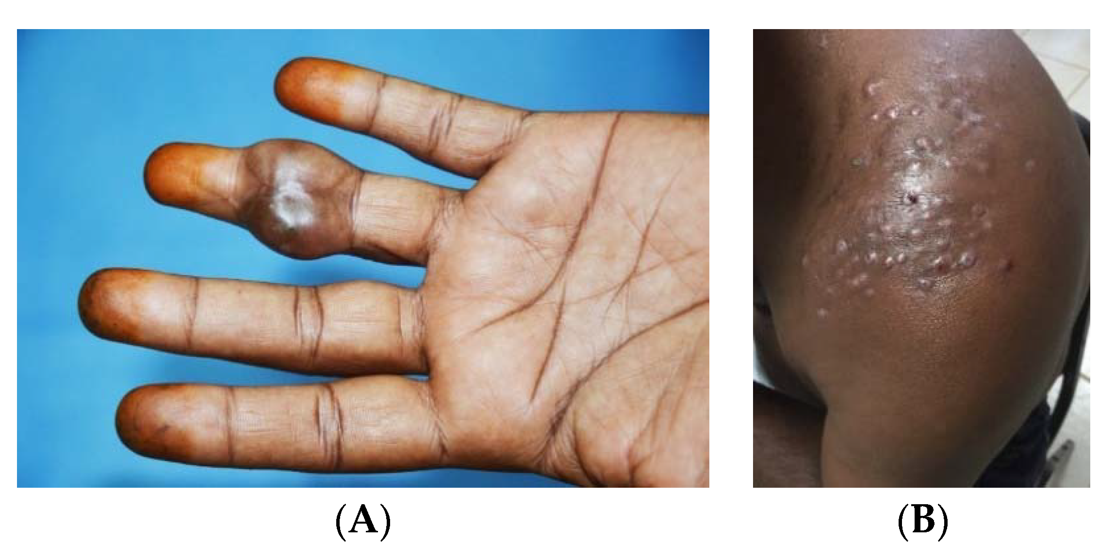
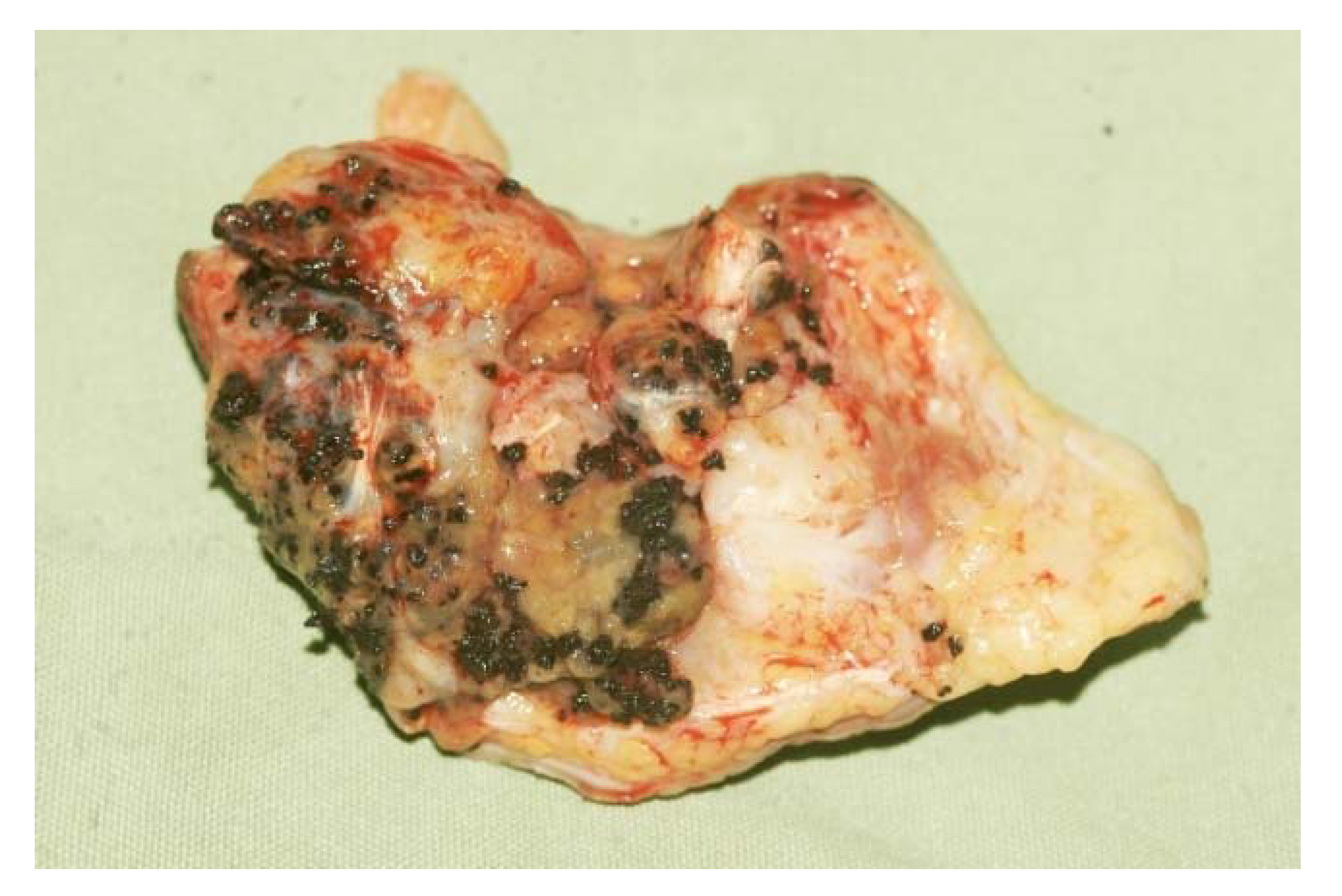
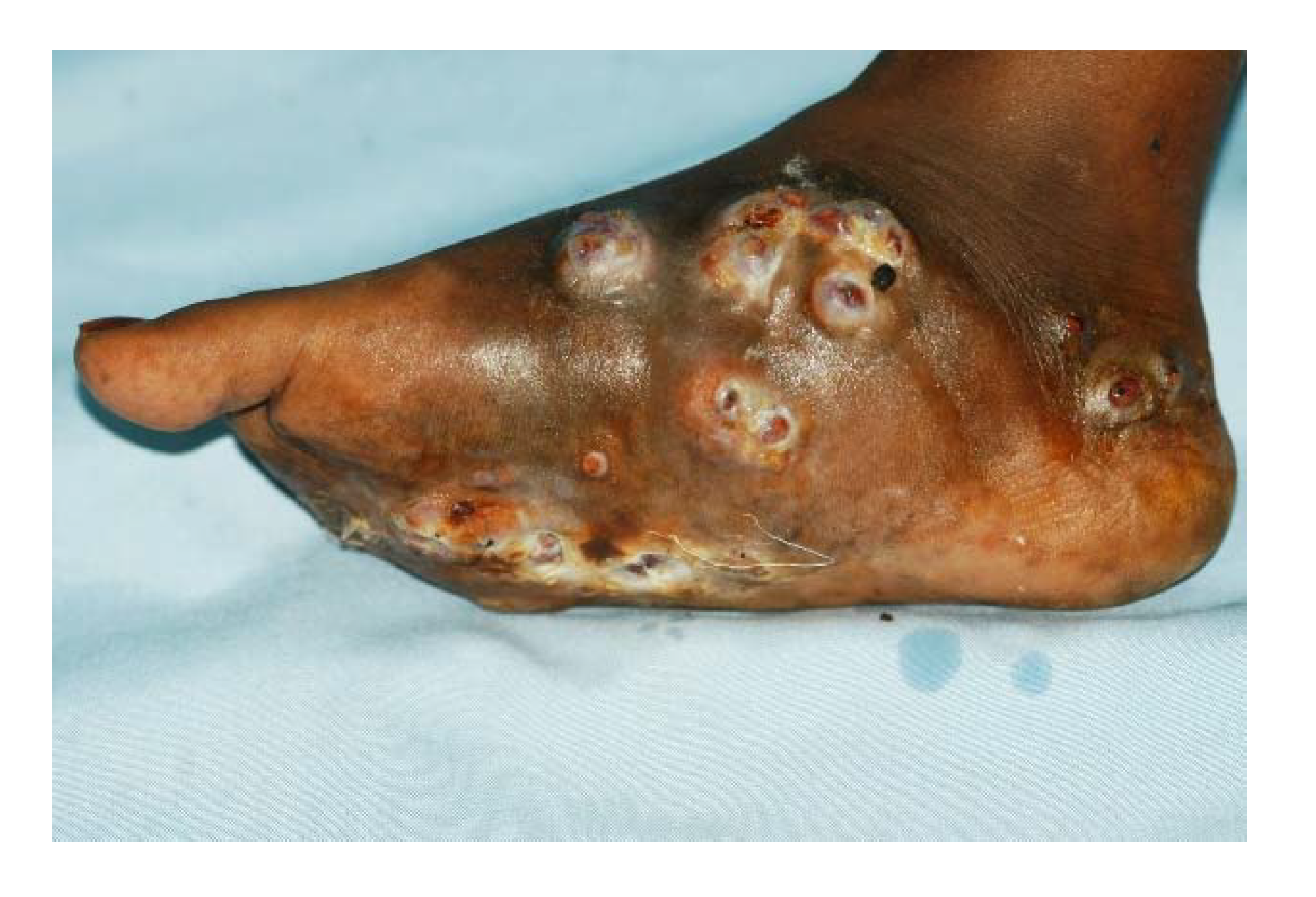
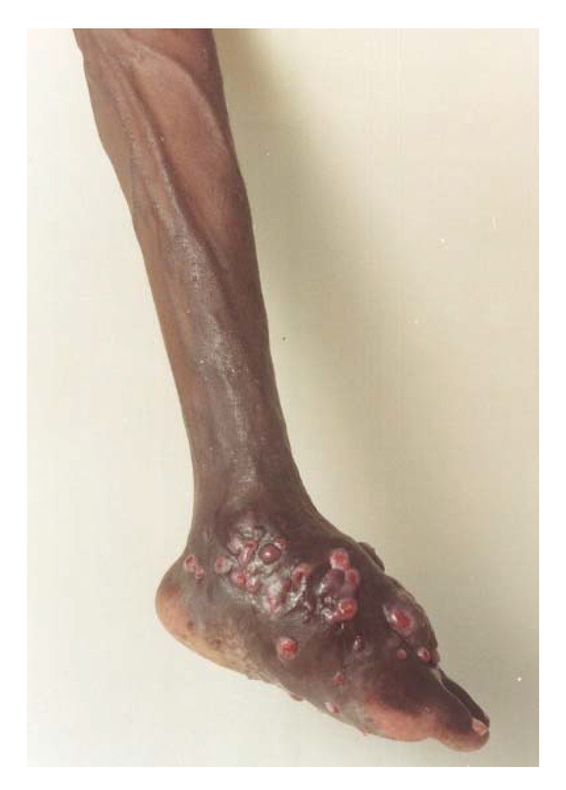
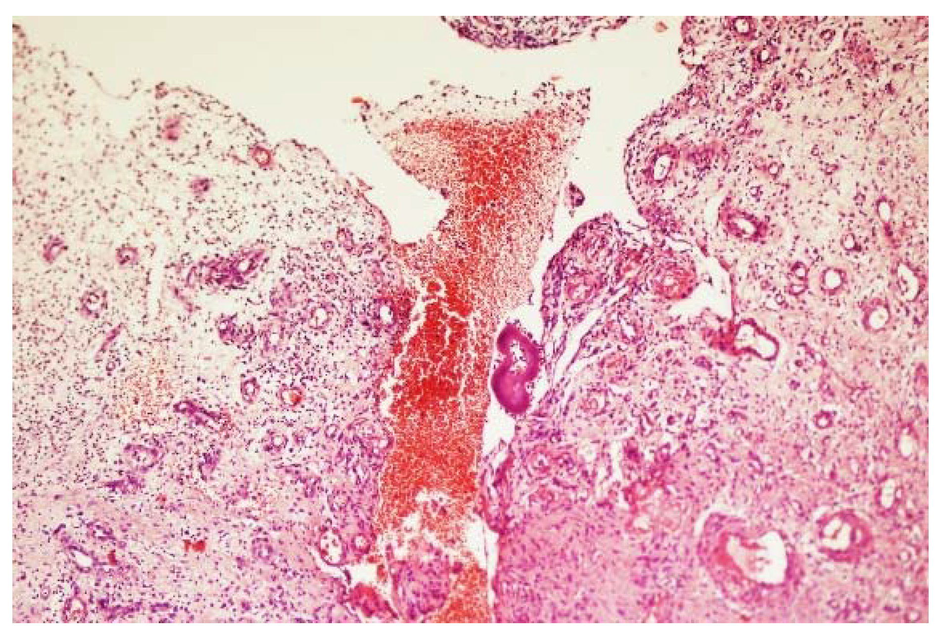
3. The Clinical Presentation

More than 70 microorganisms are incriminated in causing mycetoma. Nevertheless, the clinical presentation of mycetoma is almost indistinguishable regardless of the causal microorganism [26]. The triad of painless subcutaneous swelling, multiple draining sinuses and discharge that contains grains is characteristics of mycetoma. The swelling is usually firm and rounded but it may be soft, lobulated and, rarely, cystic and it is often mobile (Figure 1) [27]. Multiple secondary nodules then evolve, the nodules may suppurate and drain through multiple sinus tracts, and these sinuses may close transiently after discharge during the active phase of the disease. Prior to discharge, pustules may be visible. Fresh adjacent sinuses may open while some of the old ones may heal completely. The sinuses are connected with each other, with deep abscesses and with the skin surface (Figure 2) [28]. The discharge is usually serous, serosanguinous or purulent. During the active phase of the disease, the sinuses discharge grains, consisting of the microorganisms encapsulated in a cement-like material, melanin and other substances. The grains are believed to protect these microorganisms against host defence mechanisms, antimicrobials and antifungals [29]. The grain colour depends on the causative organism. The grains can be black, yellow, white or red and they are of variable size and consistency. In most mycetomas, they are visible to the naked eye and may be noticed by patients; the exception is actinomycetoma, due to the Nocardia species, in which the grains are very small and only visible under the microscope. The black grains are most commonly due to M. mycetomatis and related species, the red ones are due to A. pelletierii, the yellow to Streptomyces somaliensis and the white grains can be due to A. madurae [30]. However, black fungi may also produce grains of pale colour. Pus, exudate, the dressing gauze and biopsy material should be examined for the presence of the grains as they may give a clue to the causative organisms although the identity needs to be confirmed as visual inspection alone is not sufficiently accurate (Figure 3).
The onset and progress rate is more rapid with actinomycetoma than with eumycetoma. In eumycetoma, the lesion grows slowly with clearly defined margins and remains encapsulated for a long period, whereas, in actinomycetoma, the lesion is more inflammatory, more destructive and invades the bone at an earlier period; this is more evident in A. pelletierii actinomycetoma [31].
As the mycetoma granuloma increases in size, the skin over it becomes attached and stretched. The skin may become smooth, shiny and areas of hypo or hyperpigmentation may develop (Figure 4) [32]. In some patients, there may be areas of local hyperhidrosis confined to the mycetoma lesion itself and the skin around it. This is commonly seen with massive lesions in patients with active disease and the cause is unclear but increased local temperature due to brisk arterial circulation may a cause (Figure 5 and Figure 6). In Nocardia actinomycetomas the chest wall or back is often affected and here the edges of the lesions are unclear and hard to define.
In long-standing massive mycetoma lesions, dilated tortuous veins at, and proximal to, the mycetoma site are noted. They are frequently confused with varicose veins. They develop as a secondary phenomenon to accommodate the brisk venous return due to the increased arterial blood flow caused by the chronic inflammatory process at the mycetoma site (Figure 7) [33].
Unusually and for unknown reasons, the tendons and the nerves are spared until very late in the disease process; this may explain the rarity of neurological and trophic changes even in patients with long-standing mycetomas. The absence of trophic changes may be explained by the more than adequate blood supply in the mycetoma-infected area [34].
In the majority of patients, the regional lymph nodes are small and shotty. An enlarged regional lymph node is not uncommon and this may be due to secondary bacterial infection, the genuine lymphatic spread of mycetoma or it may be due to immune complex deposition as part of a local immune response to mycetoma infection [35].
In general, the infection remains localised and constitutional disturbances are rare but when they do occur they are generally due to secondary bacterial infection of the open sinus tracts and generalised immunosuppression. Cachexia and anaemia may be seen in late mycetoma. This is often due to malnutrition, sepsis and mental depression [36]. Co-morbidities in mycetoma are rare and the commonest are diabetes mellitus and hypertension, as in the general population. HIV and malignant transformation are not reported in mycetoma [30].
Mycetoma is usually painless in nature; it has been suggested that the mycetoma produces substances that have an anaesthetic action. At a late stage of the disease, the pain may also become negligible due to nerve damage by the tense fibrous tissue reaction, endarteritis obliterans or poor vascularisation of the nerves [37]. Pain at the mycetoma site is reported in 20% of patients and it is commonly produced by the secondary bacterial infection or transiently when a new closed sinus is about to open onto the skin surface.
Mycetoma can produce many distortions, deformities and disabilities and that is due to structural impairment that includes bone destruction or periostitis, loss of function and disuse atrophy of the affected limb. The current treatment options are limited and suboptimal; many patients undergo repeated massive surgical excisions leading to more tissue destruction and fibrosis and disability (Figure 8) [38].

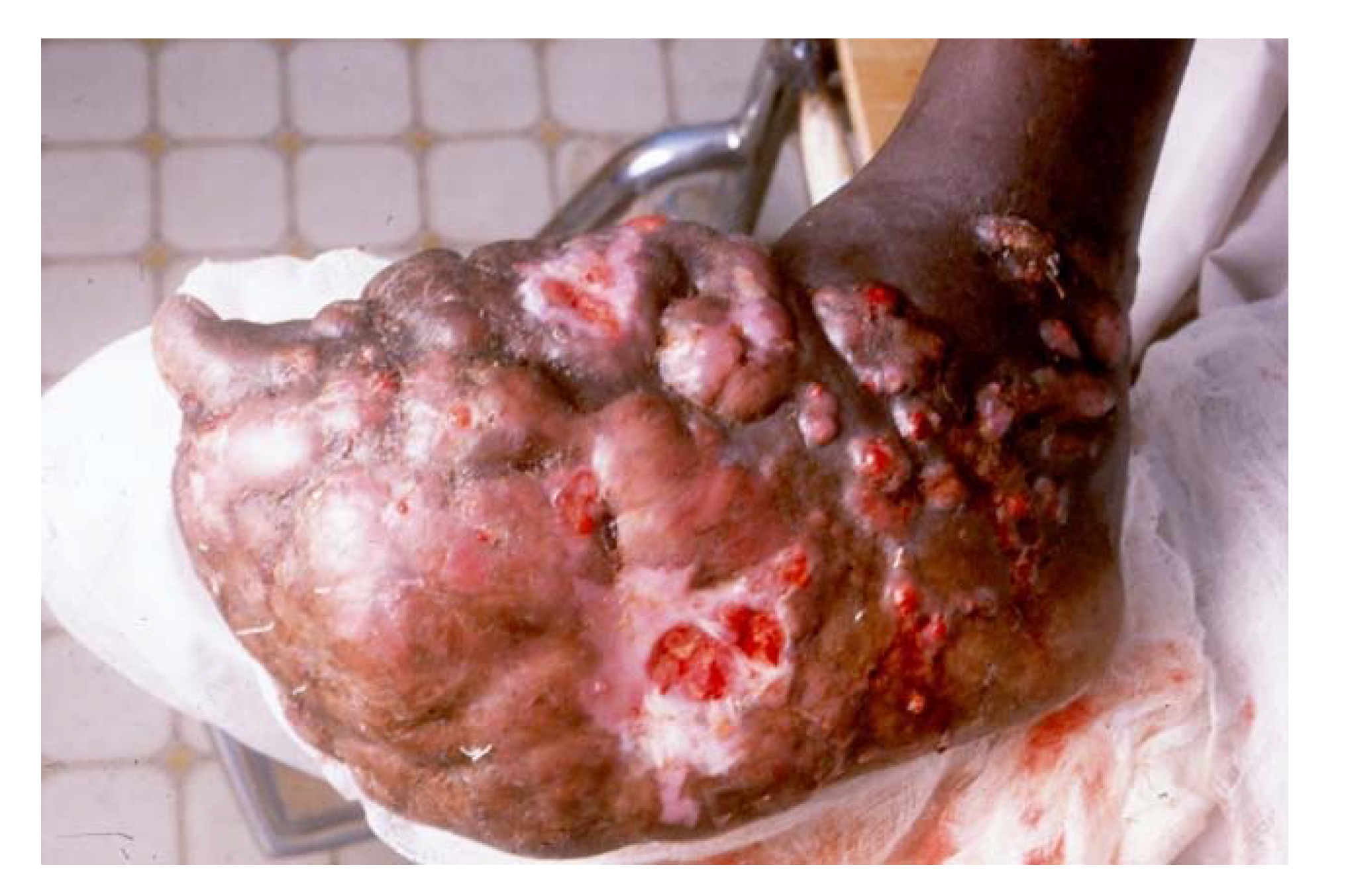
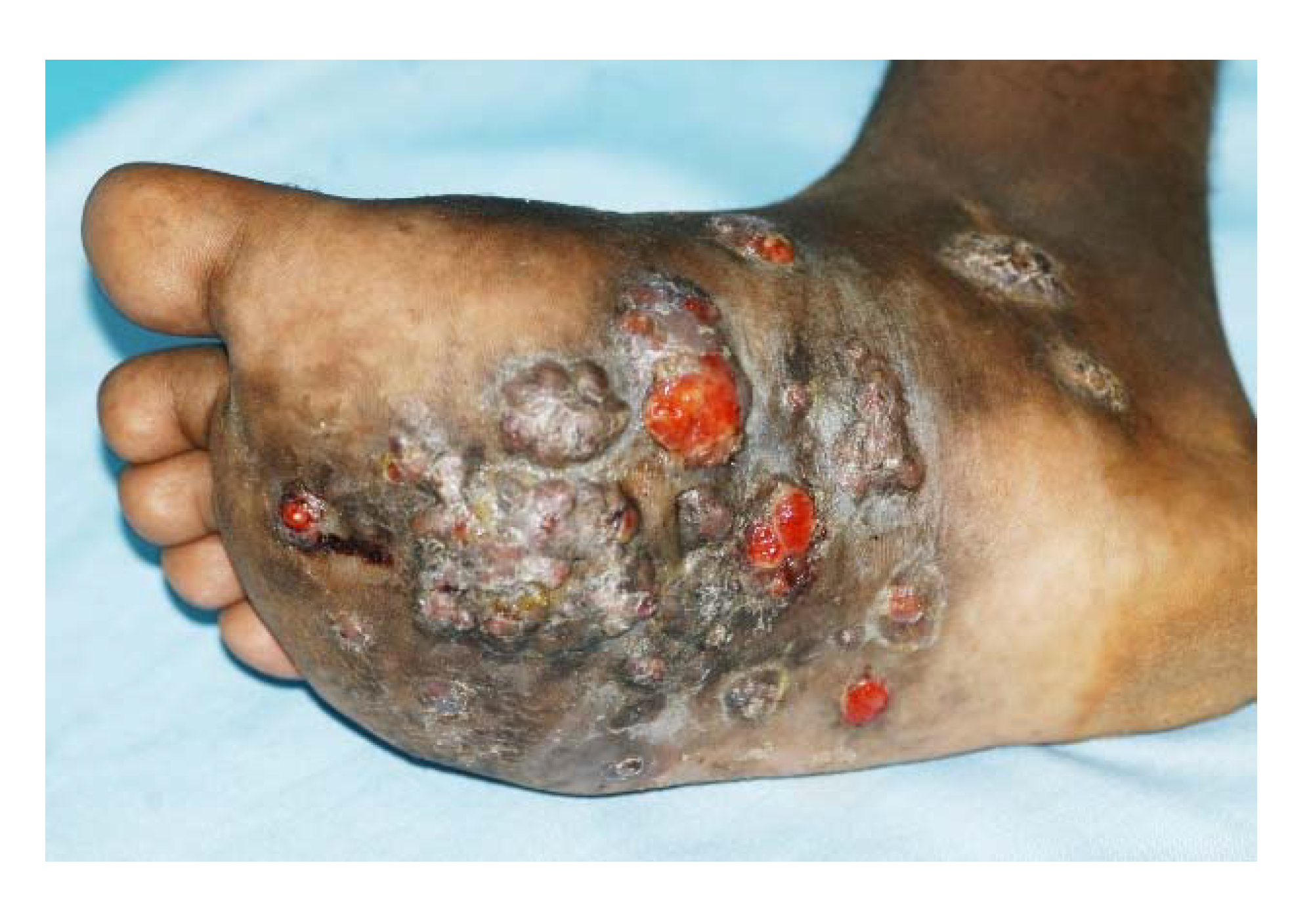
4. The Site of Mycetoma
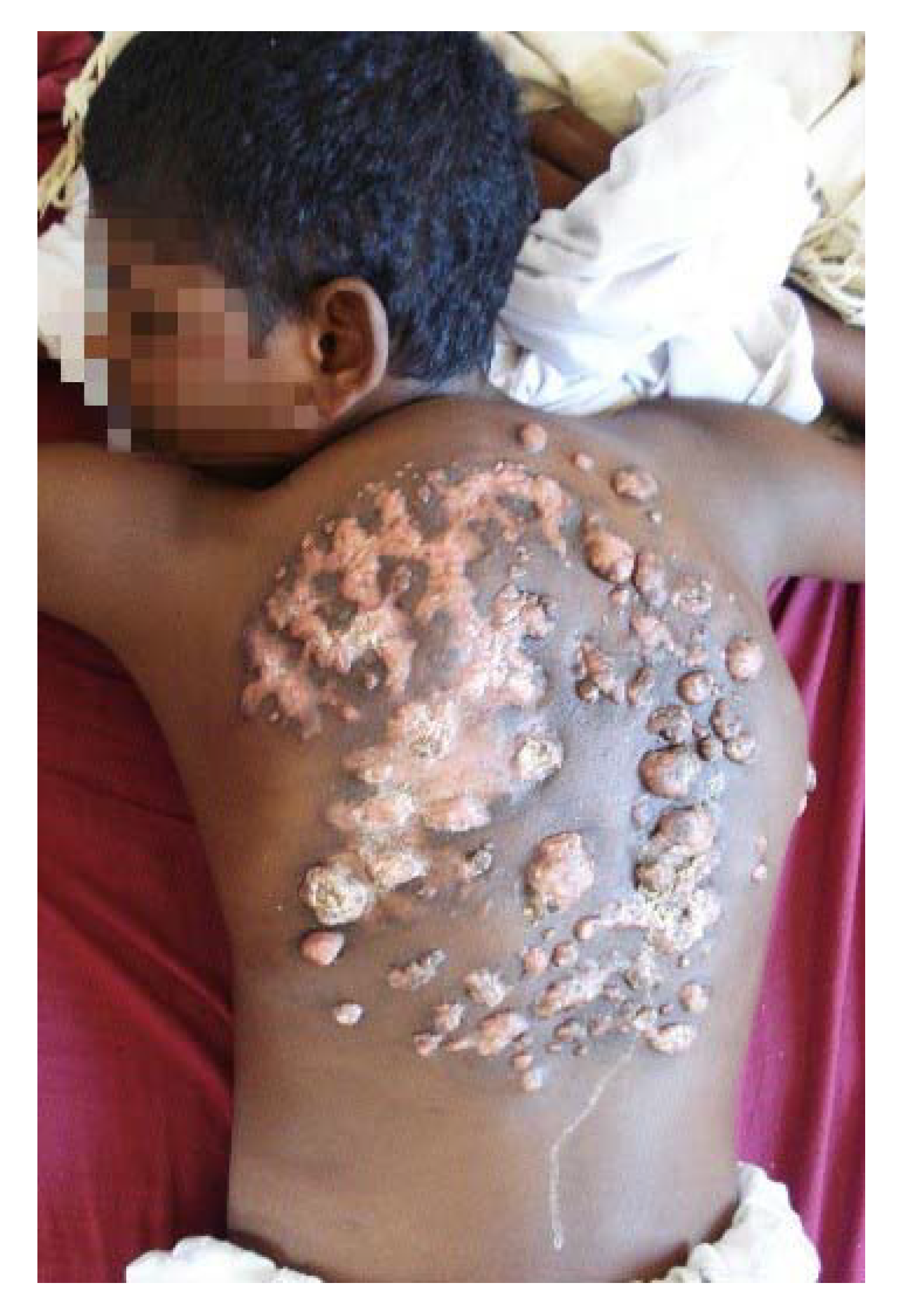
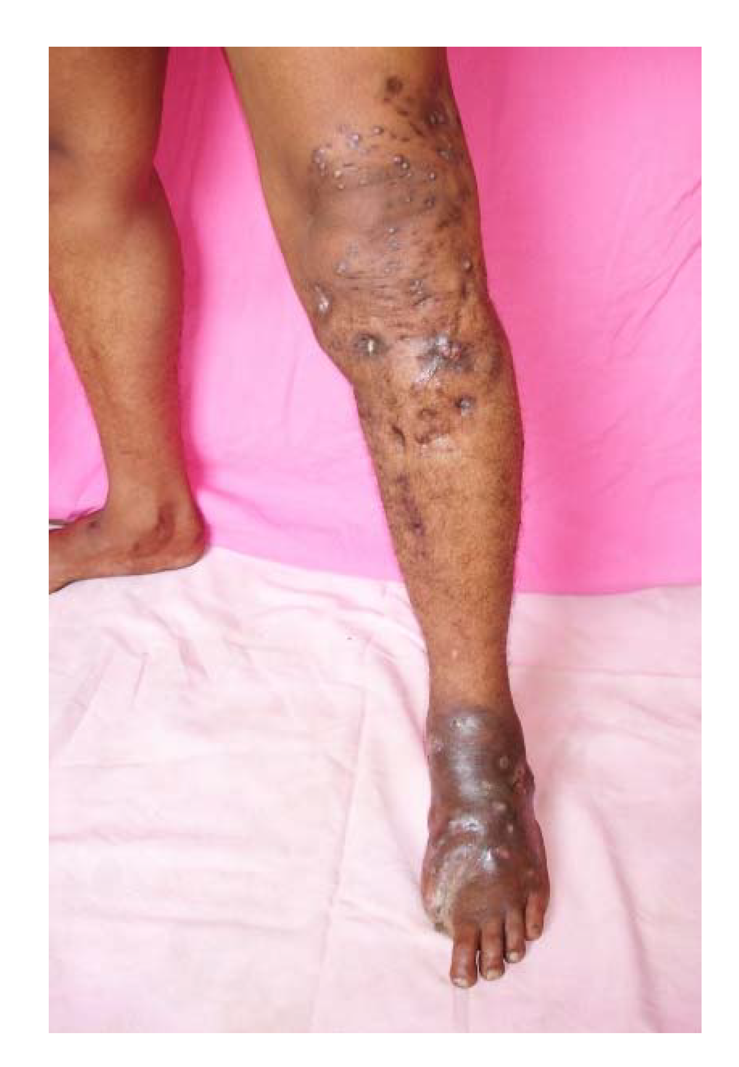
The commonest site for mycetoma is the foot (79.2%); most of the lesions are seen on the dorsal aspect of the forefoot and the left foot is affected slightly more often [39]. The hand ranks as the second commonest site (6.6%), with the right hand is more affected. This may imply a traumatic basis of the infection in this site [40]. In endemic areas, other parts of the body may be involved but less frequently, and these include the knee, arm, leg, head and neck, thigh and the perineum [41,42,43,44,45]. Rarer sites for eumycetomas are the chest and abdominal walls, facial bones, mandible, paranasal sinuses, eyelid, vulva, orbit and scrotum (Figure 9, Figure 10 and Figure 11) [46,47,48,49,50]. Nocardia actinomycetomas are often found on the chest or abdominal wall. Bilateral limb mycetomas, although described, are very rare.

5. Mycetoma Spread

Spread along the lymphatics to the regional lymph nodes is reported and this is more frequent with actinomycetoma [12]. At the regional lymph nodes, the disease progresses; a new disease satellite lesion develops and progresses to form a massive mycetoma, and in some cases, this can cause lymphatic obstruction and lymphedema (Figure 11) [51]. Bloodstream spread in mycetoma is a rare event. It has been reported in some cases, where there was spinal cord compression and paraplegia. In these cases, the skin and subcutaneous tissue were normal and the mycetoma was an unexpected finding at surgery upon opening the spinal dura matter [52]. In these cases, the mycetoma grains were actually seen in the lumen of the intact blood vessel (Figure 12).

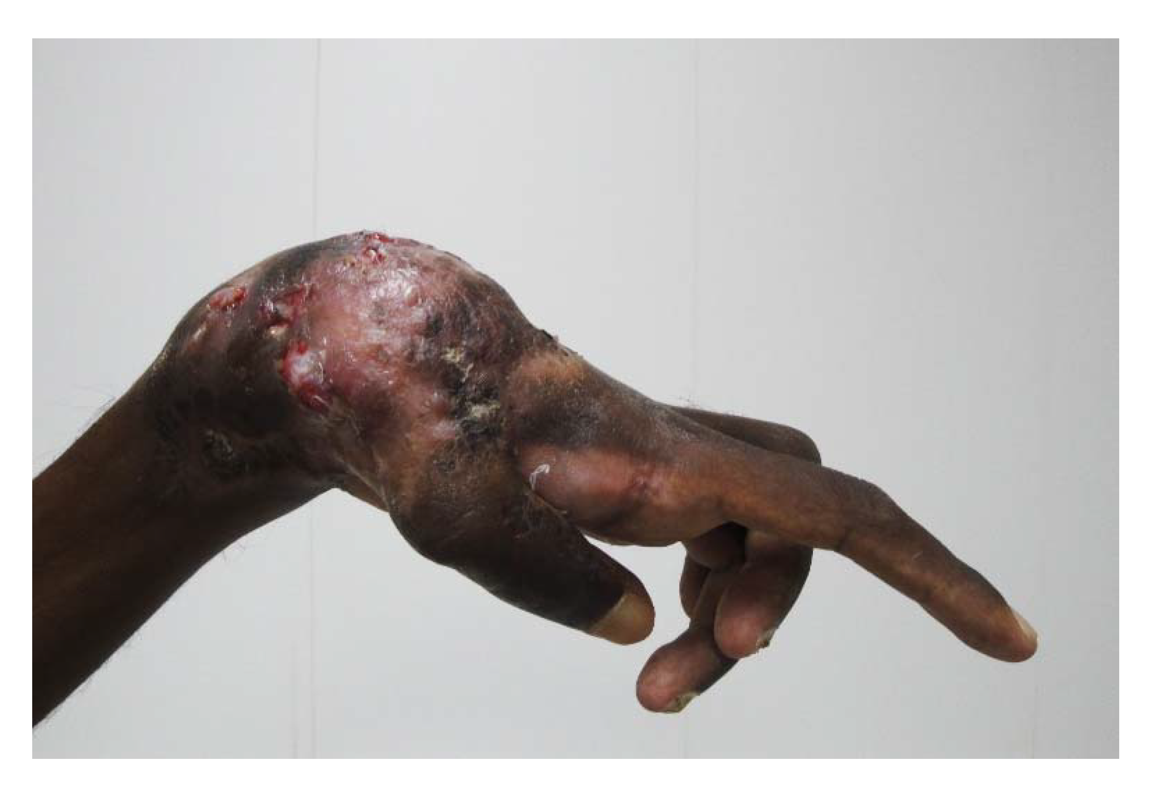
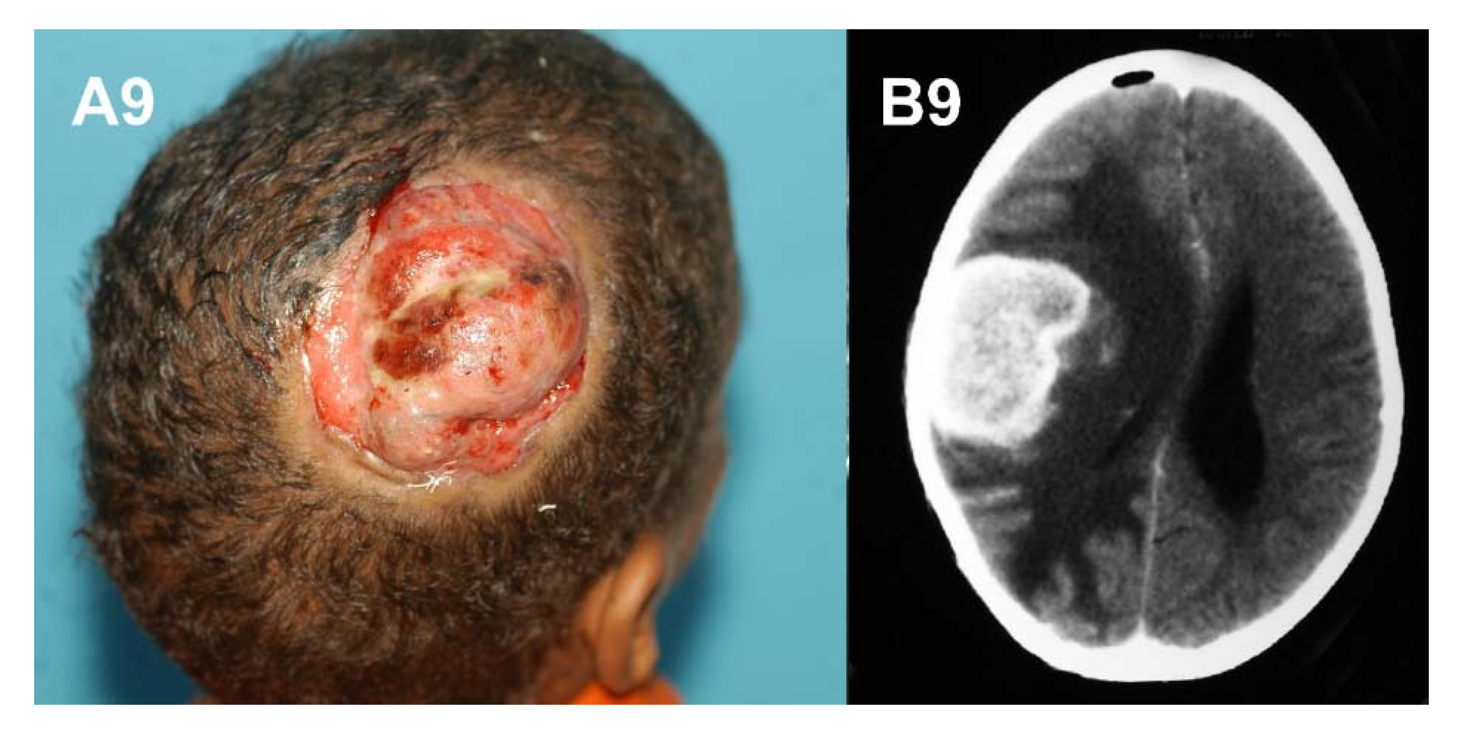
6. Aggressive Mycetoma

Although mycetoma is primarily a localised disease of gradual onset and slow progress, some patients present with massive, aggressive, uncontrolled disease and most of these are fatal. In these patients, the disease progressed wildly and aggressively from the subcutaneous tissue to involve the deep organs such as the urinary bladder, pelvic organs, spinal cord, lung and other structures and resulted in fatal outcome [53,54,55,56].
Two patients with fatal eumycetoma presented with pulmonary secondaries were reported; one from the knee and the second from gluteal eumycetoma. In these patients, Madurella mycetomatis had progressed widely without response to the different medical and surgical treatment modalities. Vascular spread, which is a rare phenomenon in mycetoma, may explain the secondary lung lesions encountered in these patients [52,54,55,56].
Several cases of mycetoma of the head and neck region have reported. They proved to be serious medical and health problem, with low cure rate and were associated with grave complications and poor outcome due to intra-cranial spread [57].
Pelvic mycetoma spread has also been reported. In these cases, the urinary bladder, rectum, hip bones and other local structures were involved. The disease was complicated by multiple urinary and rectal fistulae, pathological fractures and all patients died from massive sepsis (Figure 13) [52,53,54,55,56].

7. The Differential Diagnosis

In endemic areas, any subcutaneous mass is considered as mycetoma unless proved otherwise. The differential diagnosis of mycetoma includes thorn and foreign body granulomas, particularly in mycetoma endemic areas [57]. Also, it includes many soft tissue tumours such as Kaposi’s sarcoma, fibroma, malignant melanoma or fibrolipoma and keloids [1,58]. Many dermatological conditions, such as botryomycosis, sporotrichosis and plantar or acral psoriasis, can mimic mycetoma [59]. In very early mycetomas, before the first appearance of sinuses, the lesion simply resembles a firm subcutaneous nodule, which makes recognition of the earliest signs based on a visual appearance very difficult as the differential encompasses a variety of benign and inflammatory conditions, from dermatofibromas to hypertrophic scars. The radiological features of advanced mycetoma resemble osteogenic sarcoma and bone tuberculosis (Figure 14). Primary osseous mycetoma, without overlying sinuses, is uncommon but must be differentiated from chronic osteomyelitis, osteoclastoma, bone cysts and syphilitic osteitis.
This paper has concentrated on the variations in the clinical presentation of mycetomas. The currently available treatments for mycetoma are suboptimal and unsatisfactory, characterised by a low cure rate and high patient follow-up dropout and recurrence rates [49]. Furthermore, presently there are neither preventive nor control measures or programs for mycetoma. Moreover, mycetoma affects the most underprivileged population in remote areas and hence most patients present late with advanced disease. To overcome this, early case detection, which depends on detecting the visual warning signs of the disease and treatment is the only available means to prevent the numerous and serious complications of mycetoma. The first step in early case detection is meticulous clinical examination of the suspected patients, as other diagnostics tools, including direct microscopy, culture, histopathology and molecular identification, are commonly not available in mycetoma- endemic regions [60].

Author Contributions

The three authors contributed equally to this review article, and that included conceptualization, literature review, writing of original draft preparation, and review & editing.

Funding

This research received no external funding.

Conflicts of Interest

The authors declare no conflict of interest.

References

  1. Fahal, A.H. Mycetoma: A thorn in the flesh. Trans. R. Soc. Trop. Med. Hyg. 2004, 98, 3–11. [Google Scholar]
  2. Fahal, A.H.; Hassan, M.A. Mycetoma. Br. J. Surg. 1992, 79, 1138–1141. [Google Scholar] [CrossRef] [PubMed]
  3. Bonifaz, A.; Tirado-Sánchez, A.; Calderón, L.; Saúl, A.; Araiza, J.; Hernández, M.; González, G.M.; Ponce, R.M. Mycetoma: Experience of 482 cases in a single center in Mexico. PLoS Negl. Trop. Dis. 2014, 8, e3102. [Google Scholar]
  4. Ahmed, A.O.; van Leeuwen, W.; Fahal, A.; van de Sande, W.; Verbrugh, H.; van Belkum, A. Mycetoma caused by Madurella mycetomatis: A neglected infectious burden. Lancet Infect. Dis. 2004, 4, 566–574. [Google Scholar] [CrossRef]
  5. Ahmed, A.O.A.; van de Sande, W.W.; Fahal, A.; Bakker-Woudenberg, I.; Verbrugh, H.; van Belkum, A. Management of mycetoma: Major challenge in tropical mycoses with limited international recognition. Curr. Opin. Infect. Dis. 2007, 20, 146–151. [Google Scholar] [PubMed]
  6. Fahal, A.H.; van de Sandy, W.W. The epidemiology of mycetoma. Curr. Fungal Infect. Rep. 2012, 6, 320–326. [Google Scholar] [CrossRef]
  7. Fahal, A.H. Mycetoma. In Tropical Infectious Diseases. Principles, Pathogens and Practice, 3rd ed.; Guerrant, R.L., Walker, D.H., Weller, P.F., Eds.; Elsevier: Amsterdam, The Netherlands, 2011; pp. 565–568. [Google Scholar]
  8. Williams, N.S.; Bulstrode, C.; O’Connell, P.R. Bailey and Love’s Short Practice of Surgery 26E, 26th ed.; Taylor & Francis Ltd.: London, UK, 2013; pp. 84–88. [Google Scholar]
  9. Fahal, A.H. Mycetoma: Clinico-Pathological Monograph; University of Khartoum Press: Khurtum, Sudan, 2006; pp. 40–49. [Google Scholar]
  10. Hassan, M.A.; Fahal, A.H. Mycetoma: In Tropical Surgery; Kamil, R., Lumbly, J., Eds.; Westminster Publication Ltd.: London, UK, 2004; pp. 30–45. [Google Scholar]
  11. Fahal, A.H.; Suliman, S.H.; Gadir, A.F.A.; EL Hag, I.A.; EL Amin, F.I.; Gumaa, S.A.; Mahgoub, E.S. Abdominal wall mycetoma: An unusual presentation. Trans. R. Soc. Trop. Med. Hyg. 1994, 88, 78–80. [Google Scholar] [CrossRef]
  12. Fahal, A.H.; Suliman, S.H. Clinical presentation of mycetoma. Sudan Med. J. 1994, 32, 46–66. [Google Scholar]
  13. Fahal, A.H.; El Hassan, A.M.; Abdelalla, A.O.; Sheikh, H.E. Cystic mycetoma: An unusual clinical presentation of Madurella mycetomatis. Trans. R. Soc. Trop. Med. Hyg. 1998, 92, 66–67. [Google Scholar] [CrossRef]
  14. Fahal, A.H.; EL Toum, E.A.; EL Hassan, A.M.; Gumaa, S.A.; Mahgoub, E.S. A preliminary study on the ultrastructure of Actinomadura pelletieri and its host tissue reaction. J. Med. Vet. Mycol. 1994, 32, 343–348. [Google Scholar] [CrossRef] [PubMed]
  15. EL Hassan, A.M.; Fahal, A.H.; EL Hag, I.A.; Khalil, E.A.G. The pathology of mycetoma: Light microscopic and ultrastructural features. Sudan Med. J. 1994, 32, 23–45. [Google Scholar]
  16. Fahal, A.H.; Hassan, M.A.; Sanhouri, M. Surgical treatment of mycetoma. Sudan Med. J. 1994, 32, 98–104. [Google Scholar]
  17. Fahal, A.H.; EL Toum, E.A.; EL Hassan, A.M.; Gumaa, S.A.; Mahgoub, E.S. Host tissue reaction to Madurella mycetomatis: New classification. J. Med. Vet. Mycol. 1995, 33, 15–17. [Google Scholar] [CrossRef] [PubMed]
  18. Van de Sande, W.W.; Fahal, A.H.; Goodfellow, M.; Maghoub, E.S.; Welsh, O.; Zijlstra, E.E. The merits and pitfalls of the currently used diagnostic tools in mycetoma. PLoS Negl. Trop. Dis. 2014, 3, e2918. [Google Scholar] [CrossRef] [PubMed] [Green Version]
  19. Welsh, O.; Al-Abdely, H.M.; Salinas-Carmona, M.C.; Fahal, A.H. Mycetoma medical therapy. PLoS Negl. Trop. Dis. 2014, 8, e3218. [Google Scholar] [CrossRef] [PubMed]
  20. Welsh, O.; Vera-Cabrera, L.; Welsh, E.; Salinas, M.C. Actinomycetoma and advances in its treatment. Clin. Dermatol. 2012, 30, 372–381. [Google Scholar] [CrossRef] [PubMed]
  21. Mhmoud, N.A.; Ahmed, S.A.; Fahal, A.H.; de Hoog, G.S.; Gerrits van den Ende, A.H.; van de Sande, W.W. Pleurostomophora ochracea, a novel agent of human eumycetoma with yellow grains. J. Clin. Microbiol. 2012, 50, 2987–2994. [Google Scholar] [CrossRef] [PubMed]
  22. De Hoog, G.S.; Van Diepeningen, A.D.; Mahgoub, E.S.; Van de Sande, W.W. New species of Madurella, causative agents of black-grain mycetoma. Clin. Microbiol. 2012, 50, 988–994. [Google Scholar] [CrossRef] [PubMed]
  23. Mahgoub, E.S.; Gumaa, S.A. Ketoconazole in the treatment of eumycetoma due to Madurella mycetomi. Trans. R. Soc. Trop. Med. Hyg. 1984, 78, 376–379. [Google Scholar] [CrossRef]
  24. Fahal, A.H.; Rahman, I.A.; El-Hassan, A.M.; EL Rahman, M.E.; Zijlstra, E.E. The efficacy of itraconazole in the treatment of patients with eumycetoma due to Madurella mycetomatis. Trans. R. Soc. Trop. Med. Hyg. 2011, 105, 127–132. [Google Scholar] [CrossRef] [PubMed]
  25. Mahgoub, E.S.; Murray, I.G. Mycetoma; Medical Books Ltd.: London, UK, 1973; pp. 2–25. [Google Scholar]
  26. Abbott, P.H. Mycetoma in the Sudan. Trans. R. Soc. Trop. Med. Hyg. 1956, 50, 11–24. [Google Scholar] [CrossRef]
  27. Lynch, J.B. Mycetoma in the Sudan. Ann. R. Coll. Surg. Engl. 1964, 35, 319–340. [Google Scholar] [PubMed]
  28. EL Moghraby, I.M. Mycetoma in Gezira. Sudan Med. J. 1971, 9, 77. [Google Scholar]
  29. Mahgoub, E.S. Mycoses in the Sudan. Trans. R. Soc. Trop. Med. Hyg. 1977, 71, 184–188. [Google Scholar] [CrossRef]
  30. Fahal, A.H.; Mahgoub, E.S.; EL Hassan, A.M.; Abdel-Rahman, M.E.; Alshambaty, Y.; Hashim, A.; Hago, A.; Zijlstra, E.E. A new model for management of mycetoma in the Sudan. PLoS Negl. Trop. Dis. 2014, 8, e3271. [Google Scholar] [CrossRef] [PubMed]
  31. López-Martínez, R.; Méndez-Tovar, L.J.; Lavalle, P.; Welsh, O.; Saúl, A. Epidemiology of mycetoma in Mexico: Study of 2105 cases. Gac. Med. Mex. 1992, 128, 477–481. [Google Scholar] [PubMed]
  32. López-Martínez, R.; Méndez-Tovar, L.J.; Bonifaz, A.; Arenas, R.; Mayorga, J.; Welsh, O.; Vera-Cabrera, L.; Padilla-Desgarennes, M.C.; Contreras Pérez, C.; Chávez, G.; et al. Update on the epidemiology of mycetoma in Mexico. A Review of 3933 cases. Gac. Med. Mex. 2013, 149, 586–592. [Google Scholar] [PubMed]
  33. Ahmed, A.O.A.; Adelmann, D.; Fahal, A.H.; Verbrugh, H.A.; Van Belkum, A.; de Hoog, S.A. Environmental occurrence of Madurella mycetomatis, major agent of human eumycetoma in Sudan. J. Clin. Microbiol. 2002, 40, 1031–1036. [Google Scholar] [CrossRef] [PubMed]
  34. Ahmed, A.O.A.; van Vianen, W.; ten Kate, M.; van de Sande, W.W.J.; Van Belkum, A. A murine model of Madurella mycetomatis eumycetoma. FEMS Immunol. Med. Microbiol. 2003, 37, 29–36. [Google Scholar] [CrossRef]
  35. Fahal, A.H.; Azziz, K.A.A.; Suliman, S.H.; Galib, H.V.; Mahgoub, E.S. Dual infection with mycetoma and tuberculosis. East Afr. Med. J. 1995, 72, 749–750. [Google Scholar] [PubMed]
  36. Fahal, A.H.; Arbab, M.A.R.; EL Hassan, A.M. Aggressive clinical presentation of mycetoma due to Actinomadura pelletierii. Khartoum Med. J. 2012, 5, 699–702. [Google Scholar]
  37. Ibrahim, A.I.; EL Hassan, A.M.; Fahal, A.H.; van de Sande, W.W. A histopathological exploration of the Madurella mycetomatis grain. PLoS ONE 2013, 8, e57774. [Google Scholar] [CrossRef] [PubMed] [Green Version]
  38. Aounallah, A.; Boussofara, L.; Ben Saïd, Z.; Ghariani, N.; Saidi, W.; Denguezli, M.; Belajouza, C.; Nouira, R. Analysis of 18 Tunisian cases of mycetoma at the Sousse hospital (1974–2010). Bull. Soc. Pathol. Exot. 2013, 106, 5–8. [Google Scholar] [CrossRef] [PubMed]
  39. Dieng, M.T.; Niang, S.O.; Diop, B.; Ndiaye, B. Actinomycetomas in Senegal: Study of 90 cases. Bull. Soc. Pathol. Exot. 2005, 98, 18–20. [Google Scholar] [PubMed]
  40. Marc, S.; Meziane, M.; Hamada, S.; Hassam, B.; Benzekri, L. Clinical and epidemiological features of mycetoma in Morocco. Med. Mal. Infect. 2011, 41, 163–164. [Google Scholar] [CrossRef] [PubMed]
  41. Philippon, M.; Larroque, D.; Ravisse, P. Mycetoma in Mauritania: Species found, epidemiologic characteristics and country distribution. Report of 122 cases. Bull. Soc. Pathol. Exot. 1992, 85, 107–114. [Google Scholar] [PubMed]
  42. Fahal, A.H.; Sharfy, A.R.A. Vulval mycetoma: A rare cause of bladder outlet obstruction. Trans. R. Soc. Trop. Med. Hyg. 1998, 92, 652–653. [Google Scholar] [CrossRef]
  43. Mohamed, E.W.; Mohamed, E.N.A.; Yousif, B.M.; Fahal, A.H. Tongue actinomycetoma due to Actinomadura madurae: A rare clinical presentation. J. Oral. Maxillofac. Surg. 2012, 70, e622–e624. [Google Scholar] [CrossRef] [PubMed]
  44. Suleiman, A.M.; Fahal, A.H. Oral eumycetoma: A rare and unusual presentation. Oral. Surg. Oral. Med. Oral. Pathol. Oral. Radiol. 2013, 115, e23–e25. [Google Scholar] [CrossRef] [PubMed]
  45. Fahal, A.H.; Yagi, H.I.; EL Hassan, A.M. Mycetoma induced palatal deficiency and pharyngeal plexus dysfunction. Trans. R. Soc. Trop. Med. Hyg. 1996, 90, 676–677. [Google Scholar] [CrossRef]
  46. Fahal, A.H.; Sharfi, A.R.; Sheikh, H.E.; EL Hassan, A.M. Internal fistula formation: An unusual complication of mycetoma. Trans. R. Soc. Trop. Med. Hyg. 1996, 89, 550–552. [Google Scholar] [CrossRef]
  47. Mohamed, E.W.; Suleiman, H.S.; Fadella, A.I.; Fahal, A.H. Aggressive perineal and pelvic eumycetoma: An unusual and challenging problem to treat. Khartoum Med. J. 2012, 5, 771–774. [Google Scholar]
  48. Hussein, A.; Ahmed, A.E.M.; Fahal, A.H.; Mahmoud, A.; Sidig, A.; Awad, K. Cervical cord compression secondary to mycetoma infection. Sud. J. Public Health 2007, 2, 112–115. [Google Scholar]
  49. Zein, H.A.M.; Fahal, A.H.; Mahgoub, E.S.; EL Hassan, T.; Abdel Rahman, M.E. The predictors of cure, amputation & follow-up dropout among mycetoma patients as seen at the Mycetoma Research Centre, University of Khartoum. Trans R. Soc. Trop. Med. Hyg. 2012, 106, 639–644. [Google Scholar] [PubMed]
  50. Wendy, W.J.; van de Sande, W.W.; Mahgoub, E.S.; Fahal, A.H.; Goodfellow, M.; Welsh, O. The mycetoma knowledge gap: Identification of research priorities. PLoS Negl. Trop. Dis. 2014, 8, e2667. [Google Scholar]
  51. EL Hassan, A.M.; Mahgoub, E.S. Lymph node involvement in mycetoma. Trans. R. Soc. Trop. Med. Hyg. 1972, 66, 165–169. [Google Scholar] [CrossRef]
  52. Arbab, M.A.; El Hag, I.A.; Abdul Gadir, A.F.; Siddik, H.E.-R. Intraspinal mycetoma: Report of two cases. Am. J. Trop. Med. Hyg. 1997, 56, 27–29. [Google Scholar] [CrossRef] [PubMed]
  53. Saad, E.S.A.; Fahal, A.H. Broncho-pleuro-cutaneous fistula and pneumothorax: Rare challenging complications of chest wall eumycetoma. PLoS Negl. Trop. Dis. 2017, 11, e0005737. [Google Scholar] [CrossRef] [PubMed]
  54. Mohamed, N.A.; Fahal, A.H. Mycetoma pulmonary secondaries from a gluteal eumycetoma: An unusual presentation. PLoS Negl. Trop. Dis. 2016, 10, e0004945. [Google Scholar] [CrossRef] [PubMed]
  55. Mohamed, E.S.W.; Seif El Din, N.; Fahal, A.H. Multiple mycetoma lung secondaries from knee eumycetoma: An unusual complication. PLoS Negl. Trop. Dis. 2016, 10, e0004735. [Google Scholar] [CrossRef] [PubMed]
  56. Scolding, P.S.; Abbas, M.A.; Omer, R.F.; Fahal, A.H. Madurella mycetomatis-induced massive shoulder joint destruction: A management challenge. PLoS Negl. Trop. Dis. 2016, 10, e0004849. [Google Scholar] [CrossRef] [PubMed]
  57. Fahal, A.; Mahgoub, E.S.; El Hassan, A.M.; Jacoub, A.O.; Hassan, D. Head and neck mycetoma: The Mycetoma Research Centre experience. PLoS Negl. Trop. Dis. 2015, 9, e0003587. [Google Scholar] [CrossRef] [PubMed]
  58. Fahal, A.; Mahgoub, E.S.; El Hassan, A.M.; Abdel-Rahman, M.E. Mycetoma in the Sudan: An update from the Mycetoma Research Centre, University of Khartoum, Sudan. PLoS Negl. Trop. Dis. 2015, 9, e0003679. [Google Scholar] [CrossRef] [PubMed]
  59. Mitjà, O.; Marks, M.; Bertran, L.; Kollie, K.; Argaw, D.; Fahal, A.H. Integrated control and management of neglected tropical skin diseases. PLoS Negl. Trop. Dis. 2017, 11, e0005136. [Google Scholar] [CrossRef] [PubMed]
  60. Bakhiet, S.M.; Fahal, A.H.; Musa, A.M.; Mohamed, E.S.W.; Omer, R.F.; Ahmed, E.S. A holistic approach to the mycetoma management. PLoS Negl. Trop. Dis. 2018, 12, e0006391. [Google Scholar] [CrossRef] [PubMed]
Figure 1. (A) A small mycetoma swelling engulfing the middle part of the left ring finger with a scar of previous surgical excision. (B) Multiple small nodules of Nocardia actinomycetoma at an earlier stage.
Tropicalmed 03 00097 g001
Figure 2. Massive actinomycetoma foot with multiple sinuses and discharge.
Tropicalmed 03 00097 g002
Figure 3. Surgical specimen with multiple black grains.
Tropicalmed 03 00097 g003
Figure 4. Foot eumycetoma with skin hyperpigmentation, multiple nodules and active sinuses.
Tropicalmed 03 00097 g004
Figure 5. Foot eumycetoma with skin hyperpigmentation, multiple nodules, active sinuses discharging black grains discharge and local hyperhidrosis.
Tropicalmed 03 00097 g005
Figure 6. Massive back actinomycetoma with multiple nodules and sinuses.
Tropicalmed 03 00097 g006
Figure 7. Massive mycetoma foot with dilated tortuous veins at and proximal to the mycetoma site.
Tropicalmed 03 00097 g007
Figure 8. Hand eumycetoma with massive deformity.
Tropicalmed 03 00097 g008
Figure 9. (A) massive head eumycetoma. (B) CT scan showing intra-cranial extension.
Tropicalmed 03 00097 g009
Figure 10. Massive gluteal, perineal and scrotal eumycetoma.
Tropicalmed 03 00097 g010
Figure 11. Massive foot actinomycetoma with leg and knee inguinal satellite lesions.
Tropicalmed 03 00097 g011
Figure 12. Photomicrograph showing A. madurae grains within the lumen of the intact blood vessel (H&E X 200).
Tropicalmed 03 00097 g012
Figure 13. Massive anterior abdominal eumycetoma spreading to both inguinal regions and scrotum.
Tropicalmed 03 00097 g013
Figure 14. X-ray of thigh and knee eumycetoma showing massive soft tissue shadow, periosteal reaction and multiple bone cavities resembling osteogenic sarcoma.
Tropicalmed 03 00097 g014

