Article

Ethanolic Leaf Extract of Annona muricata Pauses Plasmodium knowlesi Schizogony and Reduces Binding of Infected Red Blood Cells to Endothelial Cells


by
Yi-Jun Lim
1,
Gordon Xue-Zhen Chong
1,
Joo-Yie Chin
1,
Muhammed-Nur-Iman Mohammed-Syafiei
1,
Muhammad-Nasreen Suhaimi
1,
Siti-Nursyazziana Nordin
1,
Shin-Yee Fung
2,
Hazel Anne Tabo
3,
Polrat Wilairatana
4,
Tadesse Hailu
5,
Veeranoot Nissapatorn
6,* and
Wenn-Chyau Lee
1,7,*
1
Department of Parasitology, Faculty of Medicine, Universiti Malaya, Kuala Lumpur 50603, Malaysia
2
Department of Molecular Medicine, Faculty of Medicine, Universiti Malaya, Kuala Lumpur 50603, Malaysia
3
Biological Sciences Department, College of Science and Computer Studies, De La Salle University-Dasmariñas City, Cavite 4115, Philippines
4
Department of Clinical Tropical Medicine, Faculty of Tropical Medicine, Mahidol University, Bangkok 10140, Thailand
5
Department of Medical Parasitology and Entomology, College of Medicine and Health Sciences, Bahir Dar University, Bahir Dar P.O. Box 79, Ethiopia
6
Futuristic Science Research Center, School of Science, World Union for Herbal Drug Discovery (WUHeDD), Research Excellence Center for Innovation and Health Products (RECIHP), Walailak University, 222 Thaiburi, Thasala District, Nakhon Si Thammarat 80160, Thailand
7
A*STAR Infectious Diseases Labs (A*IDL), Agency for Science, Technology and Research (A*STAR), Singapore 138648, Singapore
*
Authors to whom correspondence should be addressed.
Trop. Med. Infect. Dis. 2026, 11(7), 184; https://doi.org/10.3390/tropicalmed11070184
Submission received: 14 May 2026 / Revised: 30 June 2026 / Accepted: 4 July 2026 / Published: 6 July 2026
(This article belongs to the Special Issue Advances in Tools for Battling Malaria)

Abstract

Plasmodium knowlesi infections can progress rapidly to life-threatening complications, such as acute respiratory distress syndrome (ARDS) and acute kidney injury (AKI), which are driven by the ability of the zoonotic parasite to rapidly replicate towards high parasitemia and the cytoadherence properties of the infected erythrocytes (IRBCs). This study evaluated the anti-parasitic and vasoprotective potential of Annona muricata (soursop) leaf ethanol extract against P. knowlesi. In vitro assays using the P. knowlesi A1-H.1 reference strain revealed significant blood stage schizogony inhibition (IC50: 9.65 µg/mL), specifically targeting the trophozoites. The anti-parasitic activity was concentrated in the <30 kDa subfraction of the extract. Washout assays confirmed that the effect was parasitostatic rather than parasiticidal, where the malaria parasites underwent developmental arrest but remained morphologically normal and resumed growth post-removal. Furthermore, priming human endothelial cell lines with the extract significantly reduced IRBC–endothelial binding. These results demonstrate that A. muricata extract exerts a dual-action effect by arresting P. knowlesi asexual replication and inhibiting IRBC–endothelial cytoadherence. While clinical translation would require exhaustive standardization and chemotypic profiling due to the natural variability of plant compositions, these findings provide a foundational academic framework for the potential of A. muricata leaf extract in mitigating severe knowlesi malaria complications.

1. Introduction

Plasmodium knowlesi, a zoonotic parasite naturally found in long-tailed macaques, has emerged as the primary cause of human malaria in Southeast Asia [1], particularly in the forested areas that harbor simio-anthropophagic anopheline vectors [2,3,4,5]. Characterized by a rapid 24 h asexual cycle, P. knowlesi can lead to hyperparasitemia and life-threatening complications, most notably acute respiratory distress syndrome (ARDS) and acute kidney injury (AKI) [6,7,8,9,10]. These severe manifestations are increasingly linked to the sequestration of infected red blood cells (IRBCs) within the microvasculature, driven by cytoadherence to endothelial cells in target organs [8,10]. For at-risk populations in remote forested regions, the rapid progression from symptom onset to multi-organ failure, as well as the difficulty for these patients to get access to the well-resourced healthcare centers in towns, presents a critical clinical challenge [11].
While artemisinin-based combination therapies (ACTs) remain the gold standard [12,13], there is an urgent need for adjunctive therapies that can specifically target the vascular pathogenesis of the disease. Annona muricata (soursop) is a promising candidate; its leaf extracts have demonstrated potent anti-plasmodial activity against Plasmodium in traditional and laboratory settings [14,15,16]. In addition to its anti-parasitic effects, the extract exhibits anti-inflammatory and vasoprotective properties that could mitigate organ damage [15,17,18]. Nevertheless, the effect of A. muricata leaf extracts on the unique virulence factors of P. knowlesi remains unexplored. This study evaluated the efficacy of an ethanolic leaf extract of A. muricata against P. knowlesi growth and its potential to inhibit IRBC binding to human pulmonary microvascular (HPMEC) and renal glomerular (HRGEC) endothelial cells.

2. Materials and Methods

2.1. Materials Used and General Experimental Conditions

Detailed information on materials used in the experiments is available in Supplementary Materials Table S1. All experimental procedures were conducted in strict accordance with the guidelines of the Universiti Malaya Institutional Biosafety and Biosafety Committee (IBBC; ref: UMIBBC/PA/R/FOM/PARA-025/2022) and the University of Malaya Medical Centre (UMMC) Medical Research Ethics Committee (MREC ID: 2024312-13526). Unless otherwise specified, all experiments were performed using eight biological replicates.

2.2. In Vitro Cultures of Malaria Parasites and Human Endothelial Cell Lines

Reference strains of P. knowlesi (A1-H.1) were maintained in RPMI 1640 medium supplemented with AlbuMAX II and 10% (v/v) heat-inactivated human serum (complete parasite culture medium) [8,19,20]. Cultures were incubated under a tri-gas atmosphere (5% CO2, 5% O2, and 90% N2) at 37 °C. Before experiments, parasites were stage-synchronized using magnetic-activated cell sorting (MACS) LD columns (Miltenyi Biotec, Bergisch Gladbach, Germany), where the late-stage parasites with high accumulation of hemozoin were retained by the magnetic field of the sorting system [21]. For human endothelial cell cultivation, culture flasks were pre-coated with 0.5% gelatin solution for 6 h at 37 °C. Subsequently, the human pulmonary microvascular (HPMEC) and human renal glomerular (HRGEC) endothelial cell lines were thawed and inoculated into the prepared flasks under in vitro cultivation conditions supplied with 5% CO2 [10].

2.3. Drug Plate Preparation and Parasite Susceptibility Evaluation

The A. muricata leaves were subjected to ethanolic extraction using established extraction protocols described elsewhere [22,23]. Briefly, fresh, mature, and healthy A. muricata leaves were collected from Laguna, the Philippines. Species identification was confirmed by Dr. Sandra Yap, a botanist from the Far Eastern University Herbarium (FEUH), the Philippines, as A. muricata L., family Annonaceae, with specimen accession number 2025-104A. The collected leaves were cleaned with distilled water to remove debris prior to air-drying and pulverization. The powdered leaves (200 g) were soaked in 800 mL of 95% ethanol for 5 days at 25 ± 2 °C with occasional stirring. The crude extract was formed following filtration and rotary evaporation.
Drug assays were performed in flat-bottom, 96-well microplates with a total culture volume of 200 µL per well [24,25,26]. Two types of drug plates were prepared: a dihydroartemisinin (DHA)-seeded plate and an A. muricata leaf extract-seeded plate. To prepare the DHA plates, the drug was dissolved in absolute, non-denatured ethanol to achieve final test concentrations ranging from 0 to 703.4 nM. The solution was added to the wells, air-dried under sterile conditions, and stored at −20 °C until use [27,28]. Similarly, the A. muricata leaf extract was prepared in absolute ethanol and added to separate plates to achieve final working concentrations of 0 to 640 µg/mL, based on the previous study against P. falciparum [14]. For untreated control wells, an equal volume of absolute ethanol was added and dried under identical conditions.
The schizont maturation inhibition assay was conducted with stringently stage-synchronized P. knowlesi, adapted from protocols previously described [24,28,29]. Briefly, the parasite (IRBCs with at least 80% of the parasite population in the fine, early (ring) stage, parasitemia adjusted to 1%, prepared as a 2% hematocrit suspension in the complete parasite culture medium) was subjected to the exposure of DHA and A. muricata leaf extract and cultured in the tri-gas atmosphere until the parasites in the drug-free control wells reached the mature schizont stage (consisting of at least eight merozoites per schizont under the Giemsa-stained blood smear). Thick and thin smears were made from each tested well, stained with 5% Giemsa solution, and examined with a light microscope under immersion oil magnification. The percentage of parasite population that reached the mature schizont stage was determined by recruiting 200 parasites.
We also compared the susceptibility of P. knowlesi early and late erythrocytic stages to the leaf extract. Based on the inhibitory concentration of the leaf extract obtained from the schizont maturation inhibition assay, a separate experiment was conducted, where the ring stage parasites were incubated with A. muricata leaf extract (320 µg/mL) for two hours under in vitro cultivation conditions. Subsequently, the leaf extract was removed via centrifugation and removal of supernatant, followed by suspending the pelleted cells with fresh Plasmodium culture medium and culturing them under the in vitro cultivation conditions until the parasites in the drug-free controls reached the mature schizont stage. The experiment was repeated with late-stage parasites (mid- to late-stage trophozoite) to investigate the susceptibility of the parasite early and late stages to the A. muricata leaf extract [28].

2.4. Molecular Weight-Based Fractionation of A. muricata Leaf Extract

The A. muricata leaf extract starting solution (320 µg/mL) was separated based on the molecular weight of solutes within it, using a Vivaspin20 twin PES membrane concentrator (30,000 MWCO) (Sartorius, Göttingen, Germany) centrifuged at 3000× g for 20 min [30]. This process yielded a filtrate (<30 kDa) and a retentate (≥30 kDa) fraction. To assess their relative contribution to the crude extract’s potency, both fractions were tested in the schizont maturation inhibition assay at volumes equivalent to their respective yields from the initial 320 µg/mL extract. In a separate assay tracking the growth kinetics post-exposure, parasites were treated with an untreated control, a solvent control (200 µL absolute ethanol per well, dried completely under the same conditions as the drug plate preparation), 703.4 nM DHA, and the volumetric equivalents of the <30 kDa and ≥30 kDa fractions. Following incubation, the parasite suspension from each well was collected, centrifuged, and replenished with fresh culture medium. The cultures were maintained for an additional 72 h at 37 °C under the tri-gas atmosphere. Parasitemia was monitored every 24 h using the Giemsa-stained thin blood smears to observe the long-term parasite population recovery.

2.5. Effect of A. muricata Leaf Extract on P. knowlesi-Infected Erythrocyte (IRBC)–Endothelial Cytoadherence

The P. knowlesi–IRBCs have been demonstrated to cytoadhere to certain types of human endothelial cells (particularly the pulmonary microvascular and renal glomerular endothelial cells), which aligns with the commonly reported severe complications of knowlesi malaria [8,10]. To test the effect of A. muricata leaf extract on the cytoadherence dynamics of P. knowlesi–IRBCs with endothelial cells, each of the chamber slide-seeded endothelial cell lines (HPMECs and HRGECs) was divided into two groups. One group was co-incubated with the leaf extract (320 µg/mL) for 24 h under in vitro cultivation conditions, and the other group served as the “unprimed control”. Subsequently, the cell lines were co-incubated with P. knowlesi culture suspension (at least 70% of the parasite population being the late-stage parasites) for two hours prior to gentle washing steps to remove unbound cells from the surface of the endothelial cells. After that, the cells were fixed with ice-cold absolute methanol for ten minutes, followed by staining with Giemsa, and they were examined via light microscopy with immersion oil magnification. The IRBC–endothelial binding was calculated by determining the number of IRBCs attached to endothelial cells per 100 fields (equivalent to the coverage of approximately 8000 cells) [8].

2.6. Statistical Analyses

GraphPad Prism 11.0 was used for data analyses. The normality of the data was determined with the Shapiro–Wilk test. For unpaired comparison of two sets of parametric data, Welch’s t-test was used. For the comparison of multiple matched, non-parametric datasets, the Friedman test with Dunn’s multiple comparison analysis was used. For the comparison of multiple independent, non-parametric data, the Kruskal–Wallis test with Dunn’s multiple comparison analysis was used. To determine the IC50 of the compound, non-linear X-Y analysis was used. For the comparison of two categorical independent factors (duration and types of compound exposed) on a continuous dependent variable (parasitemia), two-way ANOVA with Tukey’s test was applied.

3. Results

3.1. Ethanol Extract of A. muricata Leaf Significantly Suppressed Erythrocytic Schizogony of P. knowlesi

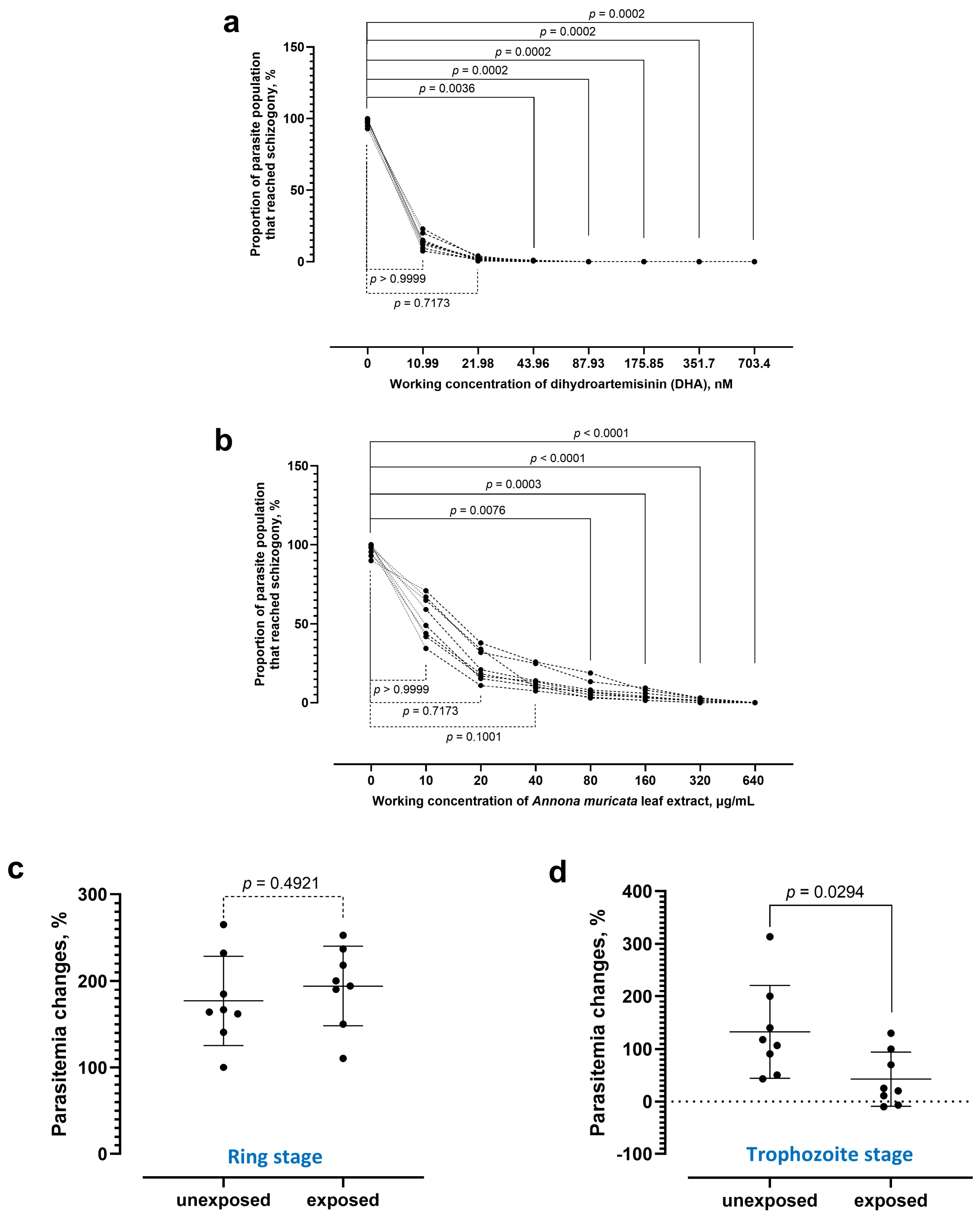
From the schizont maturation inhibition assay, the schizogony of P. knowlesi was significantly hampered by DHA (Figure 1a), with an IC50 of 1.70 nM (95% CI 1.40–2.02 nM). The asexual replication of P. knowlesi was also hampered by A. muricata leaf extract (Figure 1b), where the proportion of the parasite population that reached schizogony was significantly reduced at working concentrations of 80 µg/mL and above, as compared to the drug-free control. The IC50 of the A. muricata leaf extract against P. knowlesi was 9.65 µg/mL (95% CI 7.86–11.78 µg/mL). The leaf extract at working concentrations of 320 µg/mL and 640 µg/mL demonstrated a similar inhibitory effect against the parasites. Subsequent testing with 320 µg/mL of A. muricata leaf extract revealed that the leaf extract was ineffective against the ring stages of the parasites (Figure 1c). Instead, the leaf extract was potent against the trophozoite stages of P. knowlesi (Figure 1d). Succinctly, the ethanol extract of A. muricata leaf was potent against the asexual replication of P. knowlesi blood stages.

3.2. Schizogony-Halting Effect Attributed to Extract Compounds Smaller than 30 kDa

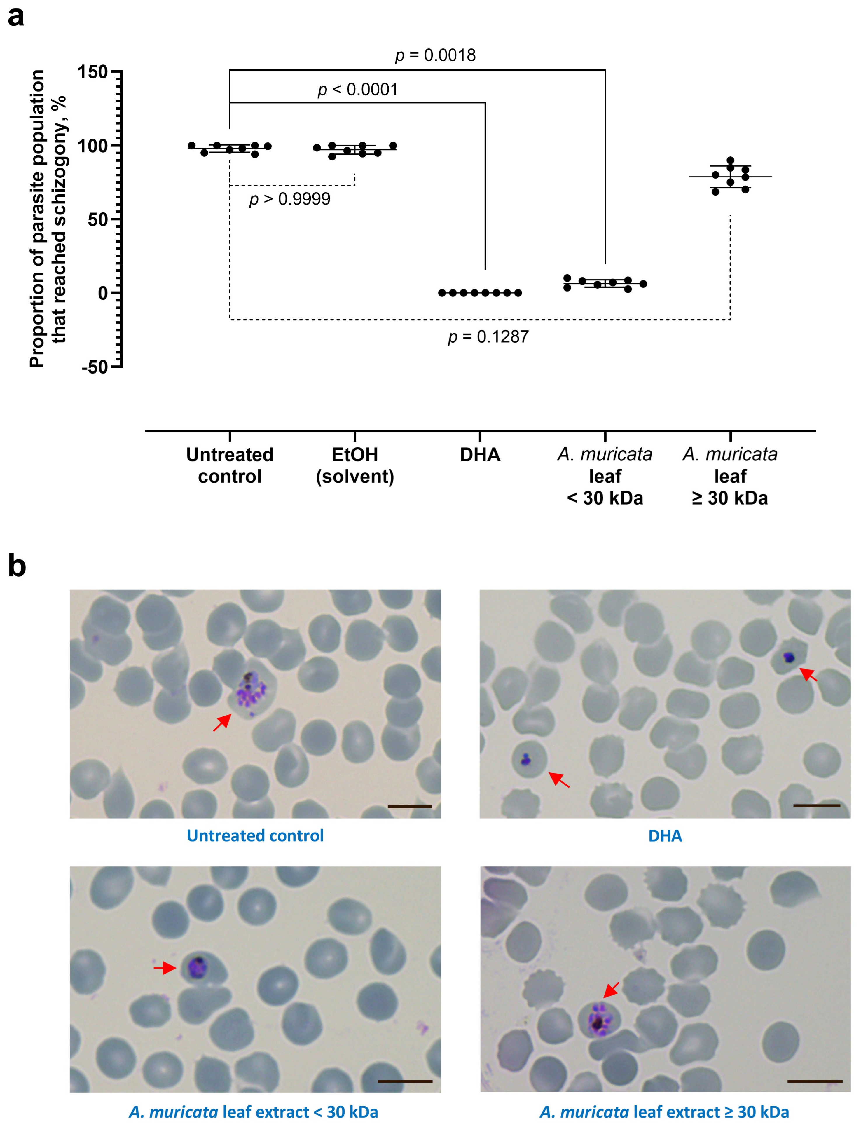
The A. muricata leaf extract was further fractionated based on the molecular weight of the compounds within. We found that the anti-parasitic effect of the A. muricata leaf extract was attributed to the fraction of <30 kDa (Figure 2a). The A. muricata leaf extract of ≥30 kDa caused insignificant changes to the proportion of parasite population that reached schizogony, as compared with the untreated control. Notably, parasites exposed to the active fraction of A. muricata displayed a distinct morphology compared to DHA-treated parasites. While DHA-treated parasites were pyknotic (indicating cell death), those exposed to the <30 kDa extract showed arrested development without chromatin pyknosis (Figure 2b). This suggests that the extract induces a reversible growth inhibition rather than immediate parasiticidal action.

3.3. A. muricata Leaf Extract Was Parasitostatic Instead of Parasitocidal

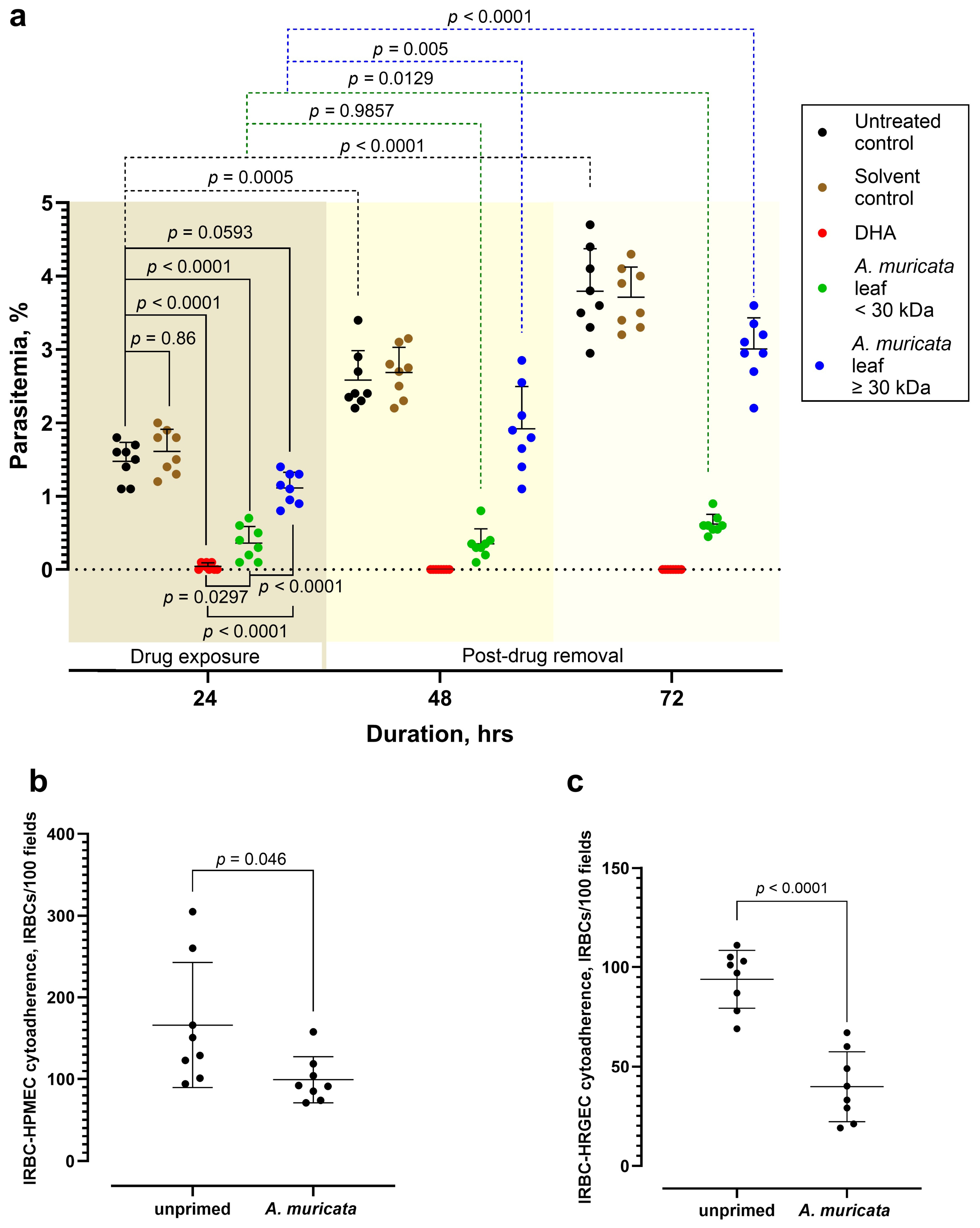
To validate the parasitostatic nature of the extract, we exposed the parasites to both fractions of the A. muricata leaf extract, along with DHA and drug-free controls for 24 h, which demonstrated a significant growth inhibitory effect against the parasites by DHA and the A. muricata leaf extract fraction of <30 kDa (Figure 3a). Subsequently, the tested materials were removed from the system, replenished with fresh parasite culture medium, and monitored closely. After removing the A. muricata leaf extract fraction of ≥30 kDa, the parasites continued to replicate, causing a significant increment in parasitemia 24 h post-removal of the tested material (i.e., the 48th hour of the experiment). For the group exposed to the A. muricata leaf extract fraction of <30 kDa, the parasites were “revived” at a slower pace, with a significant increment in parasitemia recorded 48 h post-removal of the A. muricata leaf extract fraction of <30 kDa from the system. Of note, the parasites appeared morphologically normal. The experimental group exposed to DHA showed no parasite growth after the drug removal from the system, indicating successful killing of the parasites by DHA. In short, the active ingredients within the A. muricata leaf extract exerted growth arrest on P. knowlesi instead of killing the parasites.

3.4. A. muricata Leaf Extract Reduced IRBC–Endothelial Binding

The lung-derived HPMEC primed with the A. muricata leaf extract prior to exposure to P. knowlesi demonstrated significantly lower IRBC–endothelial binding, as compared to the unprimed HPMEC (Figure 3b). Similarly, prior exposure to the A. muricata leaf extract caused a significant reduction in P. knowlesi–IRBC cytoadhesion to the kidney-derived HRGEC (Figure 3c).

4. Discussion

Our findings of the P. knowlesi schizogony-blocking effect by the A. muricata leaf ethanol extract agreed well with the earlier studies with P. falciparum [14]. We further showed that the schizogony-hampering effect by the A. muricata leaf extract was potent against the mature forms of the parasite erythrocytic stages (i.e., trophozoite) but ineffective against the younger forms (i.e., ring). Based on studies conducted with P. falciparum, several parasite-derived proteins play crucial roles during the transformation of trophozoite to schizont. These include the ribosomal P2 protein [31], SUN domain proteins [32], merozoite organizing protein [33], cell cycle regulatory proteins [34,35], and farnesyltransferase [36,37]. The A. muricata leaf-derived potent players may block the maturation of trophozoite to schizont via interactions with these parasite-derived proteins. Notably, the potent anti-schizogony compounds in the A. muricata leaf extract were of a molecular weight smaller than 30 kDa. The filtering of ethanol leaf extract with a PES membrane column of a 30 kDa cutoff value yielded a “cleaner” extract subfraction without large proteins and enzymes, complex polysaccharides, and large condensed tannins. The A. muricata leaf extract subfraction of <30 kDa contains smaller components such as alkaloids, flavonoids, phenolic compounds, and small tannin oligomers [38,39,40,41]. These metabolites are known for their anti-inflammatory and enzyme-inhibitory properties, which likely mediate the observed growth arrest [17,39,42]. The molecular weight-based fractionation using a 30 kDa cutoff membrane served as an initial chemical characterization barrier to differentiate the biological impact of macro-structural components from low-molecular-weight secondary metabolites. Many of the herbal extract-derived metabolites with therapeutic potential (such as acetogenins, flavonoids, and alkaloids) possess molecular weights well below the 30 kDa threshold [13,14]. However, there are larger molecular weight components (such as tannins, polyphenolic polymers, and certain polysaccharides) that contribute to antimicrobial and oxidation-modulating effects [14,37,38]. The fundamental purpose of utilizing a 30,000 MWCO membrane filter was not to partition individual micro-molecules but rather to isolate the active low-molecular-weight fractions from these large, complex botanical macro-aggregates, condensed tannins, or proteins (>30 kDa) that frequently cause non-specific mechanical interference in the in vitro microplate bioassays. This step confirmed that the observed anti-schizogony effect is driven explicitly by the small-molecule phytochemical matrix. Further characterization with techniques such as ultra-high-performance liquid chromatography (UHPLC) and direct chemical comparisons is warranted to further decipher the potent components against Plasmodium schizogony, followed by subsequent specific mechanism-deciphering investigations.
Importantly, the A. muricata leaf extract did not kill the malaria parasites. Instead, it induced a state of developmental arrest in the parasites, preventing them from completing the asexual replication that releases numerous merozoites, each of which is capable of invading uninfected erythrocytes [43]. This parasitostatic effect may serve as a critical “buy time” strategy for knowlesi malaria patients from remote, hard-to-reach forested areas, allowing them a window to reach urban healthcare facilities for the gold standard management of malaria [1]. Hence, the leaf extract of this widely available plant in the tropics may lower the risk of developing severe malaria complications among malaria patients. In addition to the parasitostatic effect, the A. muricata leaf extract also demonstrated a malaria-specific vasoprotective effect by reducing the P. knowlesi–IRBC cytoadherence to the endothelial cells. The lowering of IRBC–endothelial cytoadherence reduces endothelial activation, hence vascular injury, which is the hallmark of severe malaria pathogenesis [43]. In this study, cytotoxicity evaluation of the A. muricata leaf extract on the endothelial cell lines was not performed, as earlier studies have demonstrated the low toxicity of this extract on different human cell lines, including the endothelial cell lines [44,45,46]. Nevertheless, a formal, parallel cell line cytotoxicity assay would be a valuable addition to evaluate the extract’s therapeutic potential. Of note, brain-derived endothelial cell lines were not used in our study, as cerebral malaria or neurological complications are rare among the knowlesi malaria patients [47,48]. Our findings suggest that the application of the A. muricata leaf extract may reduce the risk of knowlesi malaria patients from developing severe complications such as ARDS and AKI. Although the A. muricata leaf extract has been shown to possess good tolerability and safety [49], further experimentation is needed to translate this into clinical application. In addition, the potential of this extract to reverse artemisinin resistance by the artemisinin-resistant P. falciparum isolates deserves to be investigated in the future.

5. Conclusions

The A. muricata leaf ethanol extract demonstrated a parasitostatic effect against P. knowlesi and significantly reduced the cytoadherence of IRBCs to lung- and kidney-derived human endothelial cells. These dual anti-parasitic and vasoprotective properties suggest that the extract has the potential to serve as an adjunctive therapy to mitigate the risk of severe complications, such as ARDS and AKI, in knowlesi malaria patients.

Supplementary Materials

The following supporting information can be downloaded at: https://www.mdpi.com/article/10.3390/tropicalmed11070184/s1, Table S1: Information on reagents and biological materials used in this study.

Author Contributions

Conceptualization, W.-C.L.; methodology, W.-C.L., S.-Y.F., V.N., T.H., G.X.-Z.C., J.-Y.C., H.A.T., P.W. and Y.-J.L.; validation, Y.-J.L., G.X.-Z.C. and W.-C.L.; formal analysis, Y.-J.L., G.X.-Z.C. and W.-C.L.; investigation, Y.-J.L., G.X.-Z.C., J.-Y.C., M.-N.-I.M.-S., M.-N.S. and S.-N.N.; resources, W.-C.L. and V.N.; data curation, Y.-J.L. and G.X.-Z.C.; writing—original draft preparation, Y.-J.L., J.-Y.C. and W.-C.L.; writing—review and editing, V.N. and W.-C.L.; supervision, W.-C.L.; project administration, J.-Y.C.; funding acquisition, W.-C.L. All authors have read and agreed to the published version of the manuscript.

Funding

This research was funded by the Universiti Malaya Research Excellence Grant (UMREG) (UMREG007-2023) awarded to W.-C.L.

Institutional Review Board Statement

The study was conducted in accordance with the Declaration of Helsinki and was approved by the Institutional Biosafety and Biosafety Committee (IBBC) of Universiti Malaya (UMIBBC/PA/R/FOM/PARA-025/2022; approval: 15 November 2022; renewal: 30 March 2026) and the University of Malaya Medical Centre (UMMC) Medical Research Ethics Committee (MREC) (MRECID#2024312-13526; approval: 1 August 2024).

Informed Consent Statement

Informed consent was obtained from all subjects involved in the study.

Data Availability Statement

The data presented in this study are available in this article and its Supplementary Materials.

Acknowledgments

J.-Y.C. was a candidate under the graduate research assistant Universiti Malaya Scholarship Scheme (UMSS) via the UMREG grant (UMREG007-2023) awarded to W.-C.L. The authors have reviewed and edited the output and take full responsibility for the content of this publication. Also, we acknowledged the International Mobility Fund for Research Publication (WU-CIA-01803/2024), Walailak University, Thailand for the support.

Conflicts of Interest

The authors declare no conflicts of interest.

Abbreviations

The following abbreviations are used in this manuscript:
DHADihydroartemisinin
HPMECsHuman Pulmonary Microvascular Endothelial Cells
HRGECsHuman Renal Glomerular Endothelial Cells
IRBCsInfected Red Blood Cells

References

  1. Lee, W.C.; Cheong, F.W.; Amir, A.; Lai, M.Y.; Tan, J.H.; Phang, W.K.; Shahari, S.; Lau, Y.L. Plasmodium knowlesi: The game changer for malaria eradication. Malar. J. 2022, 21, 140. [Google Scholar] [CrossRef] [PubMed]
  2. Tan, C.H.; Vythilingam, I.; Matusop, A.; Chan, S.T.; Singh, B. Bionomics of Anopheles latens in Kapit, Sarawak, Malaysian Borneo in relation to the transmission of zoonotic simian malaria parasite Plasmodium knowlesi. Malar. J. 2008, 7, 52. [Google Scholar] [CrossRef] [PubMed]
  3. Jiram, A.I.; Vythilingam, I.; NoorAzian, Y.M.; Yusof, Y.M.; Azahari, A.H.; Fong, M.Y. Entomologic investigation of Plasmodium knowlesi vectors in Kuala Lipis, Pahang, Malaysia. Malar. J. 2012, 11, 213. [Google Scholar] [CrossRef] [PubMed]
  4. Braima, K.A.; Sum, J.S.; Ghazali, A.R.; Muslimin, M.; Jeffery, J.; Lee, W.C.; Shaker, M.R.; Elamin, A.E.; Jamaiah, I.; Lau, Y.L.; et al. Is there a risk of suburban transmission of malaria in Selangor, Malaysia? PLoS ONE 2013, 8, e77924. [Google Scholar] [CrossRef] [PubMed]
  5. Lau, Y.L.; Lee, W.C.; Chen, J.; Zhong, Z.; Jian, J.; Amir, A.; Cheong, F.W.; Sum, J.S.; Fong, M.Y. Draft genomes of Anopheles cracens and Anopheles maculatus: Comparison of simian malaria and human malaria vectors in Peninsular Malaysia. PLoS ONE 2016, 11, e0157893. [Google Scholar] [CrossRef] [PubMed]
  6. Lee, W.C.; Chin, P.W.; Lau, Y.L.; Chin, L.C.; Fong, M.Y.; Yap, C.J.; Supramaniam, R.R.; Mahmud, R. Hyperparasitaemic human Plasmodium knowlesi infection with atypical morphology in Peninsular Malaysia. Malar. J. 2013, 12, 88. [Google Scholar] [CrossRef] [PubMed]
  7. Singh, B.; Daneshvar, C. Human infections and detection of Plasmodium knowlesi. Clin. Microbiol. Rev. 2013, 26, 165–184. [Google Scholar] [CrossRef] [PubMed]
  8. Lee, W.C.; Shahari, S.; Nguee, S.Y.T.; Lau, Y.L.; Renia, L. Cytoadherence properties of Plasmodium knowlesi-infected erythrocytes. Front. Immunol. 2022, 12, 804417. [Google Scholar] [CrossRef] [PubMed]
  9. Anstey, N.M.; Grigg, M.J.; William, T.; Rajahram, G.S.; Cooper, D.J.; Barber, B.E. Acute kidney injury in non-falciparum malaria. Semin. Nephrol. 2025, 45, 151615. [Google Scholar] [CrossRef] [PubMed]
  10. Phong, Z.Y.; Chin, J.Y.; Ng, Y.L.; Zakaria, N.I.; Athirah-Azman, S.N.; Kosaisavee, V.; Renia, L.; Lee, W.C. The role of periostin (OSF-2) in the cytoadherence phenomena mediated by malaria parasites. Front. Cell. Infect. Microbiol. 2025, 15, 1599872. [Google Scholar] [CrossRef] [PubMed]
  11. Naserrudin, N.A.; Hod, R.; Jeffree, M.S.; Ahmed, K.; Hassan, M.R. The emerging threat of Plasmodium knowlesi malaria infection: A concept paper on the vulnerable factors in human. Int. J. Environ. Res. Public Health 2022, 19, 4419. [Google Scholar] [CrossRef] [PubMed]
  12. Kokori, E.; Olatunji, G.; Akinboade, A.; Akinoso, A.; Egbunu, E.; Aremu, S.A.; Okafor, C.E.; Oluwole, O.; Aderinto, N. Triple artemisinin-based combination therapy (TACT): Advancing malaria control and eradication efforts. Malar. J. 2024, 23, 25. [Google Scholar] [CrossRef] [PubMed]
  13. Barber, B.E.; Grigg, M.J.; William, T.; Yeo, T.W.; Anstey, N.M. The treatment of Plasmodium knowlesi malaria. Trends Parasitol. 2017, 33, 242–253. [Google Scholar] [CrossRef] [PubMed]
  14. Leesombun, A.; Boonmasawai, S.; Nishikawa, Y. Ethanol extracts from Thai plants have anti-Plasmodium and anti-Toxoplasma activities in vitro. Acta Parasitol. 2019, 64, 257–261. [Google Scholar] [CrossRef] [PubMed]
  15. Mutakin, M.; Fauziati, R.; Fadhilah, F.N.; Zuhrotun, A.; Amalia, R.; Hadisaputri, Y.E. Pharmacological activities of soursop (Annona muricata Lin.). Molecules 2022, 27, 1201. [Google Scholar] [CrossRef] [PubMed]
  16. Sarakul, O.; Boonhok, R.; Somsak, V. Enhanced antimalarial efficacy of Annona muricata leaf extract combined with artesunate, chloroquine, and pyrimethamine in Plasmodium berghei-infected ICR mice. Adv. Pharmacol. Pharm. Sci. 2025, 2025, 8736555. [Google Scholar] [CrossRef] [PubMed]
  17. Orak, H.H.; Bahrisefit, I.S.; Sabudak, T. Antioxidant activity of extracts of Soursop (Annona muricata L.) leaves, fruit pulps, peels, and seeds. Pol. J. Food Nutr. Sci. 2019, 69, 359–366. [Google Scholar] [CrossRef]
  18. Nsor, O.O.; Alabi, B.A.; Badejo, J.A.; Afolabi, F.; Nku-Ekpang, O.A.; Iwalewa, E.O. Soursop leaf extract and fractions protects against L-NAME-induced hypertension and hyperlipidemia. Front. Nutr. 2024, 11, 1437101. [Google Scholar] [CrossRef] [PubMed]
  19. Moon, R.W.; Hall, J.; Rangkuti, F.; Ho, Y.S.; Almond, N.; Mitchell, G.H.; Pain, A.; Holder, A.A.; Blackman, M.J. Adaptation of the genetically tractable malaria pathogen Plasmodium knowlesi to continuous culture in human erythrocytes. Proc. Natl. Acad. Sci. USA 2013, 110, 531–536. [Google Scholar] [CrossRef] [PubMed]
  20. Ng, Y.L.; Lee, W.C.; Lau, Y.L.; Fong, M.Y. The impact of geographical variation in Plasmodium knowlesi apical membrane protein 1 (PkAMA-1) on invasion dynamics of P. knowlesi. Trop. Med. Infect. Dis. 2023, 8, 56. [Google Scholar] [CrossRef] [PubMed]
  21. Chin, J.Y.; Mohammed-Syafiei, M.N.; Lim, Y.J.; Chong, G.X.; Suhaimi, M.N.; Phong, Z.Y.; Ng, Y.L.; Lau, Y.L.; Sam, I.C.; Renia, L.; et al. Effect of periostin (OSF-2) on phagocytosis of Plasmodium-infected erythrocytes. Front. Microbiol. 2025, 16, 1728562. [Google Scholar] [CrossRef] [PubMed]
  22. Mitsuwan, W.; Sin, C.; Keo, S.; Sangkanu, S.; de Lourdes Pereira, M.; Jimoh, T.O.; Salibay, C.C.; Nawaz, M.; Norouzi, R.; Siyadatpanah, A.; et al. Potential anti-Acanthamoeba and anti-adhesion activities of Annona muricata and Combretum trifoliatum extracts and their synergistic effects in combination with chlorhexidine against Acanthamoeba triangularis trophozoites and cysts. Heliyon 2021, 7, e06976. [Google Scholar] [CrossRef] [PubMed]
  23. Kulnanan, P.; Chuprom, J.; Thomrongsuwannakij, T.; Romyasamit, C.; Sangkanu, S.; Manin, N.; Nissapatorn, V.; de Lourdes Pereira, M.; Wilairatana, P.; Kitpipit, W.; et al. Antibacterial, antibiofilm, and anti-adhesion activities of Piper betle leaf extract against avian pathogenic Escherichia coli. Arch. Microbiol. 2021, 204, 49. [Google Scholar] [CrossRef] [PubMed]
  24. Russell, B.; Malleret, B.; Suwanarusk, R.; Anthony, C.; Kanlaya, S.; Lau, Y.L.; Woodrow, C.J.; Nosten, F.; Renia, L. Field-based flow cytometry for ex vivo characterization of Plasmodium vivax and P. falciparum antimalarial sensitivity. Antimicrob. Agents Chemother. 2013, 57, 5170–5174. [Google Scholar] [CrossRef] [PubMed]
  25. Suwanarusk, R.; Russell, B.; Ong, A.; Sriprawat, K.; Chu, C.S.; PyaePhyo, A.; Malleret, B.; Nosten, F.; Renia, L. Methylene blue inhibits the asexual development of vivax malaria parasites from a region of increasing chloroquine resistance. J. Antimicrob. Chemother. 2015, 70, 124–129. [Google Scholar] [CrossRef] [PubMed]
  26. Lee, W.C.; Russell, B.; Lau, Y.L.; Nosten, F.; Rénia, L. Rosetting responses of Plasmodium-infected erythrocytes to antimalarials. Am. J. Trop. Med. Hyg. 2022, 106, 1670–1674. [Google Scholar] [CrossRef] [PubMed]
  27. Rutvisuttinunt, W.; Chaorattanakawee, S.; Tyner, S.D.; Teja-Isavadharm, P.; Se, Y.; Yingyuen, K.; Chaichana, P.; Bethell, D.; Walsh, D.S.; Lon, C.; et al. Optimizing the HRP-2 in vitro malaria drug susceptibility assay using a reference clone to improve comparisons of Plasmodium falciparum field isolates. Malar. J. 2012, 11, 325. [Google Scholar] [CrossRef] [PubMed]
  28. Lee, W.C.; Russell, B.; Lee, B.; Chu, C.S.; Phyo, A.P.; Sriprawat, K.; Lau, Y.L.; Nosten, F.; Renia, L. Plasmodium falciparum rosetting protects schizonts against artemisinin. eBioMedicine 2021, 73, 103680. [Google Scholar] [CrossRef] [PubMed]
  29. Rieckmann, K.H.; Campbell, G.H.; Sax, L.J.; Mrema, J.E. Drug sensitivity of Plasmodium falciparum: An in-vitro microtechnique. Lancet 1978, 1, 22–23. [Google Scholar] [CrossRef] [PubMed]
  30. Lee, W.C.; Russell, B.; Sobota, R.M.; Ghaffar, K.; Howland, S.W.; Wong, Z.X.; Maier, A.G.; Dorin-Semblat, D.; Biswas, S.; Gamain, B.; et al. Plasmodium-infected erythrocytes induce secretion of IGFBP7 to form type II rosettes and escape phagocytosis. eLife 2020, 9, e51546. [Google Scholar] [CrossRef] [PubMed]
  31. Das, S.; Basu, H.; Korde, R.; Tewari, R.; Sharma, S. Arrest of nuclear division in Plasmodium through blockage of erythrocyte surface exposed ribosomal protein P2. PLoS Pathog. 2012, 8, e1002858. [Google Scholar] [CrossRef] [PubMed]
  32. Kandelis-Shalev, S.; Goyal, M.; Elam, T.; Assaraf, S.; Dahan, N.; Farchi, O.; Berenshtein, E.; Dzikowski, R. SUN-domain proteins of the malaria parasite Plasmodium falciparum are essential for proper nuclear division and DNA repair. mBio 2025, 16, e0021625. [Google Scholar] [CrossRef] [PubMed]
  33. Absalon, S.; Robbins, J.A.; Dvorin, J.D. An essential malaria protein defines the architecture of blood-stage and transmission-stage parasites. Nat. Commun. 2016, 7, 11449. [Google Scholar] [CrossRef] [PubMed]
  34. Ganter, M.; Goldberg, J.M.; Dvorin, J.D.; Paulo, J.A.; King, J.G.; Tripathi, A.K.; Paul, A.S.; Yang, J.; Coppens, I.; Jiang, R.H.; et al. Plasmodium falciparum CRK4 directs continuous rounds of DNA replication during schizogony. Nat. Microbiol. 2017, 2, 17017. [Google Scholar] [CrossRef] [PubMed]
  35. Voss, Y.; Klaus, S.; Guizetti, J.; Ganter, M. Plasmodium schizogony, a chronology of the parasite’s cell cycle in the blood stage. PLoS Pathog. 2023, 19, e1011157. [Google Scholar] [CrossRef] [PubMed]
  36. Chakrabarti, D.; Da Silva, T.; Barger, J.; Paquette, S.; Patel, H.; Patterson, S.; Allen, C.M. Protein farnesyltransferase and protein prenylation in Plasmodium falciparum. J. Biol. Chem. 2002, 277, 42066–42073. [Google Scholar] [CrossRef] [PubMed]
  37. Fletcher, S.; Cummings, C.G.; Rivas, K.; Katt, W.P.; Horney, C.; Buckner, F.S.; Chakrabarti, D.; Sebti, S.M.; Gelb, M.H.; Van Voorhis, W.C.; et al. Potent, Plasmodium-selective farnesyltransferase inhibitors that arrest the growth of malaria parasites: Structure-activity relationships of ethylenediamine-analogue scaffolds and homology model validation. J. Med. Chem. 2008, 51, 5176–5197. [Google Scholar] [CrossRef] [PubMed]
  38. Awad, A.M.; Kumar, P.; Ismail-Fitry, M.R.; Jusoh, S.; Ab Aziz, M.F.; Sazili, A.Q. Green extraction of bioactive compounds from plant biomass and their application in meat as natural antioxidant. Antioxidants 2021, 10, 1465. [Google Scholar] [CrossRef] [PubMed]
  39. Gil-Martin, E.; Forbes-Hernandez, T.; Romero, A.; Cianciosi, D.; Giampieri, F.; Battino, M. Influence of the extraction method on the recovery of bioactive phenolic compounds from food industry by-products. Food Chem. 2022, 378, 131918. [Google Scholar] [CrossRef] [PubMed]
  40. Lee, J.E.; Jayakody, J.T.M.; Kim, J.I.; Jeong, J.W.; Choi, K.M.; Kim, T.S.; Seo, C.; Azimi, I.; Hyun, J.M.; Ryu, B.M. The influence of solvent choice on the extraction of bioactive compounds from Asteraceae: A comparative review. Foods 2024, 13, 3151. [Google Scholar] [CrossRef] [PubMed]
  41. Tanambell, H.; Danielsen, M.; Freund, S.; Nissen, S.H.; Møller, A.H.; Corredig, M.; Dalsgaard, T.K. Partitioning of proteins and small molecular weight compounds from Alfalfa juice during ultrafiltration. Food Biosci. 2024, 62, 105044. [Google Scholar] [CrossRef]
  42. Rady, I.; Bloch, M.B.; Chamcheu, R.N.; Banang Mbeumi, S.; Anwar, M.R.; Mohamed, H.; Babatunde, A.S.; Kuiate, J.R.; Noubissi, F.K.; El Sayed, K.A.; et al. Anticancer properties of Graviola (Annona muricata): A comprehensive mechanistic review. Oxid. Med. Cell. Longev. 2018, 2018, 1826170. [Google Scholar] [CrossRef] [PubMed]
  43. Lee, W.C.; Russell, B.; Renia, L. Sticking for a cause: The falciparum malaria parasites cytoadherence paradigm. Front. Immunol. 2019, 10, 1444. [Google Scholar] [CrossRef] [PubMed]
  44. Cardenas, C.; Torres-Vargas, J.A.; Cardenas-Valdivia, A.; Jurado, N.; Quesada, A.R.; Garcia-Caballero, M.; Martinez-Poveda, B.; Medina, M.A. Non-targeted metabolomics characterization of Annona muricata leaf extracts with anti-angiogenic activity. Biomed. Pharmacother. 2021, 144, 112263. [Google Scholar] [CrossRef] [PubMed]
  45. Marrero, A.D.; Quesada, A.R.; Martinez-Poveda, B.; Medina, M.A.; Cardenas, C. A proteomic study of the bioactivity of Annona muricata leaf extracts in HT-1080 fibrosarcoma cells. Int. J. Mol. Sci. 2023, 24, 12021. [Google Scholar] [CrossRef] [PubMed]
  46. Zubaidi, S.N.; Qadi, W.S.M.; Maarof, S.; Mohmad Misnan, N.; Mohammad Noor, H.S.; Hamezah, H.S.; Baharum, S.N.; Rosli, N.; Jam, F.A.; Al-Olayan, E.; et al. Assessing the acute toxicological effects of Annona muricata leaf ethanol extract on rats: Biochemical, histopathological, and metabolomics analyses. Toxics 2023, 11, 688. [Google Scholar] [CrossRef] [PubMed]
  47. Daneshvar, C.; Davis, T.M.; Cox-Singh, J.; Rafa’ee, M.Z.; Zakaria, S.K.; Divis, P.C.; Singh, B. Clinical and laboratory features of human Plasmodium knowlesi infection. Clin. Infect. Dis. 2009, 49, 852–860. [Google Scholar] [CrossRef] [PubMed]
  48. Gunawan, C.A. Severe knowlesi malaria with acute neurocognitive impairment: A case report. IJID Reg. 2025, 14, 100513. [Google Scholar] [CrossRef] [PubMed]
  49. Chan, W.J.; McLachlan, A.J.; Hanrahan, J.R.; Harnett, J.E. The safety and tolerability of Annona muricata leaf extract: A systematic review. J. Pharm. Pharmacol. 2020, 72, 1–16. [Google Scholar] [CrossRef] [PubMed]
Figure 1. Susceptibility of P. knowlesi via in vitro assays. (a) Schizont inhibition assay with dihydroartemisinin (DHA). Based on the Friedman test with Dunn’s multiple comparison test, the schizogony of P. knowlesi was significantly halted at concentration points of 43.96 nM DHA and above (p < 0.0001; Q = 54.06, n = 8, df = 7), as compared to the drug-free control. (b) Schizont inhibition assay with A. muricata leaf extract. Based on the Friedman test with Dunn’s multiple comparison test, the schizogony of P. knowlesi was significantly halted at concentration points of 80 µg/mL and above (p < 0.0001; Q = 55.87, n = 8, df = 7), as compared to the drug-free control. Additionally, there was no significant difference in the proportion of parasite population that reached schizogony between experiment groups 320 µg/mL and 640 µg/mL A. muricata leaf extract (p > 0.9999). (c) Welch’s t-test revealed that the two-hour exposure of the P. knowlesi ring stage to 320 µg/mL A. muricata leaf extract did not significantly alter the parasite population growth, as compared to the unexposed control (Welch-corrected t = 0.7057, df = 13.82). Error bars represent mean and SD. (d) Welch’s t-test revealed that the two-hour exposure of the P. knowlesi trophozoite stage to 320 µg/mL A. muricata leaf extract significantly reduced the parasite population growth, as compared to the unexposed control (Welch-corrected t = 2.492, df = 11.28). Error bars represent mean and SD.
Tropicalmed 11 00184 g001
Figure 2. Deciphering subfractions of the A. muricata leaf extract with potent hampering effect on P. knowlesi growth. (a) A Kruskal-Wallis test with Dunn’s multiple comparison test revealed that the subfraction of <30 kDa demonstrated a significant blocking effect on P. knowlesi schizogony, as compared to the untreated control (p < 0.0001; H = 35.75, df = 4). Error bars represent means and SD. (b) Morphology of P. knowlesi upon the end of the schizont maturation inhibition assay under different experimental settings, visualized with 5% Giemsa-stained thin blood smears and examined with light microscopy under immersion oil magnification. The IRBC is indicated by red arrow. Scale bar = 10 µm.
Tropicalmed 11 00184 g002
Figure 3. Further characterization of the effect of A. muricata leaf extract on pathobiology of P. knowlesi infection. (a) The monitoring of parasitemia changes following 24 h of incubation with drugs (DHA, A. muricata leaf extract <30 kDa subfraction, A. muricata leaf extract ≥30 kDa subfraction, solvent control, and untreated control), and an additional 48 h after removal of the tested components, as indicated by the background colors of the graph. Based on the two-way ANOVA with Tukey’s test, significance was found with regard to the duration monitored (F1.72,60.28 = 211.4, p < 0.0001) and the types of compounds exposed to the parasites (F4,35 = 229.6, p < 0.0001). There was a significant interaction between the duration monitored and the types of compounds exposed to the parasites (F6.89,60.28 = 30.65, p < 0.0001). Comparisons between the data collected right after drug exposure and the data collected post-drug removal were indicated with dotted lines. (b) Priming of HPMECs with the A. muricata leaf extract significantly reduced the binding of P. knowlesi–IRBCs to the endothelial cells (Welch-corrected t = 2.317, df = 8.884). (c) Priming of HRGECs with the A. muricata leaf extract significantly reduced the binding of P. knowlesi-IRBCs to the endothelial cells (Welch-corrected t = 6.695, df = 13.50). Error bars represent means and SD.
Tropicalmed 11 00184 g003
Disclaimer/Publisher’s Note: The statements, opinions and data contained in all publications are solely those of the individual author(s) and contributor(s) and not of MDPI and/or the editor(s). MDPI and/or the editor(s) disclaim responsibility for any injury to people or property resulting from any ideas, methods, instructions or products referred to in the content.