Article

Brucella abortus Infection Promotes Mesenchymal Stem Cell Differentiation Toward Adipogenesis, Enhancing the Proinflammatory Profile


by
Rosa Nicole Freiberger
1,
Cynthia Alicia Marcela López
1,
María Belén Palma
2,3,
Cintia Cevallos
1,
Franco Agustin Sviercz
1,
Patricio Jarmoluk
1,
Marcela Nilda García
2,
Jorge Quarleri
1 and
M. Victoria Delpino
1,*
1
Consejo Nacional de Investigaciones Científicas y Tecnológicas (CONICET), Laboratorio de Inmunopatología Viral, Instituto de Investigaciones Biomédicas en Retrovirus y Sida (INBIRS), Universidad de Buenos Aires (UBA), Buenos Aires 1121, Argentina
2
Cátedra de Citología, Histología y Embriología, Facultad de Ciencias Médicas, Universidad Nacional de La Plata, La Plata 1900, Argentina
3
Laboratorio de Investigación Aplicada a Neurociencias (LIAN), Fleni, Consejo de Investigaciones Científicas y Técnicas (CONICET), Escobar 1625, Argentina
*
Author to whom correspondence should be addressed.
Trop. Med. Infect. Dis. 2026, 11(5), 112; https://doi.org/10.3390/tropicalmed11050112
Submission received: 26 February 2026 / Revised: 17 April 2026 / Accepted: 18 April 2026 / Published: 23 April 2026
(This article belongs to the Special Issue Advances in Brucella Infections)

Abstract

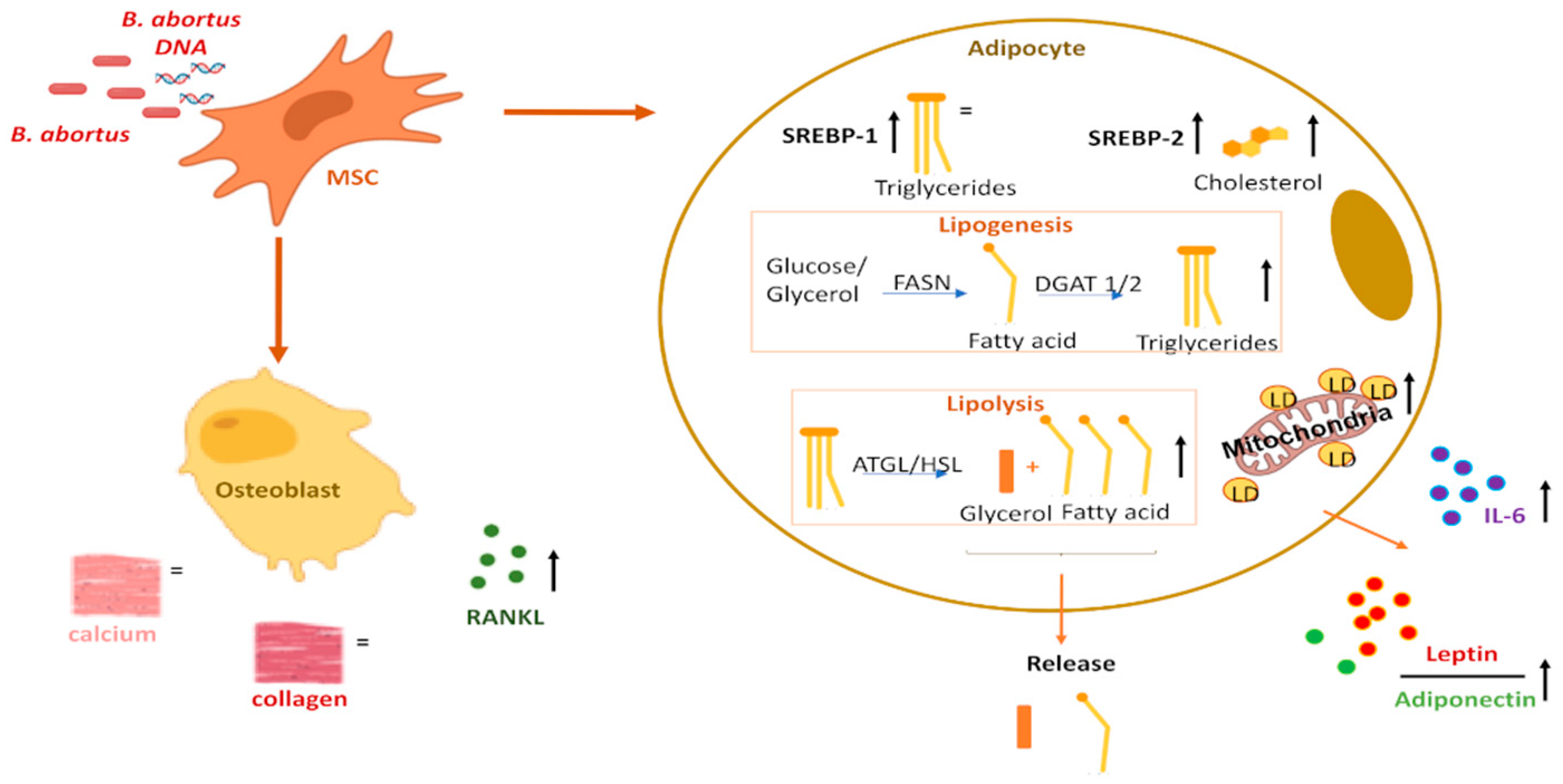
The most common complication of active brucellosis in humans is osteoarticular injury. In the bone marrow microenvironment, mesenchymal stem cells (MSCs) can differentiate into either adipocytes or osteoblasts, and this balance is tightly regulated because an increase in adipogenesis may negatively affect bone formation and favor bone loss. The differentiation of MSCs into adipocytes or osteoblasts is tightly regulated by mechanisms that promote cell fate toward one lineage while repressing the other. Our study demonstrated that Brucella abortus infects MSCs but does not affect the deposition of organic and mineral matrix during osteoblast differentiation. However, the infection upregulates Receptor Activator of Nuclear Factor Kappa-B Ligand (RANKL) expression in osteoblasts, which may contribute to osteoclast activation and bone resorption. Conversely, B. abortus infection significantly influences adipocyte differentiation by modulating lipolysis, lipogenesis, and interactions between lipid droplets and mitochondria. This leads to increased cellular cholesterol levels and reduced intracellular triglycerides, accompanied by glycerol release. These changes result in more differentiated adipocytes and larger lipid droplets. Consequently, we observed increased IL-6 secretion and a higher leptin/adiponectin ratio. Importantly, these effects were independent of a functional type IV secretion system (T4SS), as purified Brucella DNA fully reproduced the adipogenic phenotype. Moreover, inhibition of TLR9—the primary sensor of bacterial DNA—significantly reduced the DNA-induced adipogenic response, demonstrating that adipocyte modulation is at least in part mediated through TLR9 signaling. In summary, B. abortus promotes MSC differentiation toward an inflammatory adipocyte phenotype. It involves a TLR-9-mediated DNA detection. It may contribute to osteoarticular injury and infection-associated bone resorption.

Graphical Abstract

1. Introduction

Brucellosis is a widespread zoonotic infection worldwide, although its impact is markedly higher in developing countries [1]. The most common clinical complication of active human brucellosis is osteoarticular disease [2,3,4,5,6,7,8].
In addition to other sites, Brucella can persist in the human bone marrow [9,10]. In most patients, cellular alterations in the bone marrow improve or disappear after antibiotic treatment [9]. Moreover, transmission of brucellosis through bone marrow transplantation from apparently healthy donors has been reported [11]. These findings indicate that even when the bacterium is not detected in the bone marrow, it may persist, concealed within cells [12,13]. This observation suggests that the ability of Brucella to cause chronic infection may be linked to its persistence in the bone marrow.
Mesenchymal stem cells (MSCs) are among the key cellular components of the bone marrow microenvironment. They are multipotent precursor cells capable of differentiating into various stromal lineage cell types, including chondrocytes, adipocytes, and osteoblasts [14,15]. The differentiation of MSCs into adipocytes or osteoblasts is tightly regulated by mechanisms that promote commitment to one lineage while repressing the other [16]. For instance, osteoporosis, the most prevalent bone remodeling disorder globally, often exhibits heightened marrow fat content [17,18]. Indeed, increased bone marrow adiposity is commonly observed in many conditions associated with bone loss, such as aging and various pathological states [19,20,21].
The commitment of MSCs to either the osteoblast or adipocyte lineage depends on specific transcriptional regulators [22]. Among these, peroxisome proliferator-activated receptor gamma (PPAR-γ) and CCAAT/enhancer-binding protein (C/EBP) play critical roles in adipogenesis [23]. In contrast, Runt-related transcription factor 2 (RUNX2) acts as the master transcription factor for osteoblast differentiation [24]. This process is further regulated by PPAR-γ, which inhibits osteoblastogenesis by suppressing the transcription of RUNX2 [25].
Previous findings using murine cells indicate that Brucella abortus can modulate responses of adipocytes and osteoblasts, suggesting that both cell types could be involved in bone damage [26,27,28].
Therefore, we hypothesize that B. abortus interacts with MSCs and may promote adipocyte differentiation while reducing osteoblast differentiation; however, this process remains unclear and requires further investigation.
Here, we explore the interaction between B. abortus and MSCs during their differentiation into osteoblasts and adipocytes.

2. Materials and Methods

2.1. Bacterial Culture

Brucella abortus S2308, DsRed-expressing B. abortus S2308, or the isogenic B. abortus virB10 polar mutants (kindly provided by Diego Comerci, Universidad Nacional de San Martín (UNSAM), Buenos Aires, Argentina) were cultured for 18 h in 10 mL of tryptic soy broth supplemented with yeast extract (Merck, Darmstadt, Germany), under constant agitation (150 rpm) at 37 °C. Bacteria were harvested, and inocula were prepared as previously described [29].
To prepare heat-killed Brucella abortus (HKBA), the bacteria were washed five times with sterile phosphate-buffered saline (PBS), with each wash lasting 10 min. Subsequently, they were heat-inactivated at 70 °C for 20 min, then aliquoted and stored at −70 °C until use. The absence of bacterial growth on tryptose soy agar confirmed the complete loss of B. abortus viability. All manipulations involving live B. abortus were performed in biosafety level 3 facilities.

2.2. DNA from B. abortus

Brucella abortus DNA was isolated using the Wizard® Genomic DNA Kit (Promega, Madison, WI, USA) in accordance with the manufacturer’s protocol. The concentration of the extracted DNA was determined using a spectrophotometer. Following the extraction process, a 100 µg aliquot of the DNA was treated with DNase I (1 U/mg DNA) (ZymoResearch, Beijing, China), as per the manufacturer’s guidelines. HKBA (2.5 × 10⁹ bacteria) was treated with 1 U of DNase in a final volume of 50 µL.

2.3. Isolation of Mesenchymal Stem Cells (MSCs)

The umbilical cords were preserved in α-Minimal Essential Medium (α-MEM, Gibco, Waltham, MA, USA) and handled following previously outlined procedures [30]. Essentially, each umbilical cord underwent a 5 mm fragmentation, with a sagittal incision exposing Wharton’s jelly. Removal of the umbilical blood vessels was meticulously conducted using clamps. Subsequently, the fragments underwent 2 or 3 washes with Dulbecco’s phosphate-buffered saline from Sigma-Aldrich (St. Louis, MO, USA) to eliminate residual blood. The section with the exposed jelly was then positioned face down at the bottom of a culture plate, and a minimum essential medium (α-MEM, Gibco) supplemented with 10% platelet lysate was added. The plates were incubated at 37 °C in a humid atmosphere containing 5% CO2, and medium was refreshed every 2 to 3 days 2 to 3 days. Expansion of umbilical cord-derived MSCs was typically observed between 10 and 14 days post-explantation, and cells were further amplified until reaching passage 2/3. MSCs were characterized by the expression of CD105, CD73, and CD90, and the absence of CD45, CD34, CD14, CD19, and HLA-DR molecules. For experimental purposes, MSCs were cultured in DMEM supplemented with 10% heat-inactivated fetal bovine serum (Gibco-BRL, Life Technologies, Grand Island, NY, USA), 100 U/mL of penicillin, and 100 mg/mL of streptomycin (complete medium) and were utilized until passage 5. This study obtained approval from the Comité de Bioética y Ética, Facultad de Ciencias Médicas, Universidad de Buenos Aires, Argentina (RESCD-2023-1291). Written informed consent was obtained from each mother before normal cesarean birth, and human umbilical cords were collected from discarded placentas.

2.4. Adipocyte Differentiation

Initially, MSCs were seeded at a density of 5 × 10⁴ cells per well in 24-well plates and allowed to reach confluence. Following this, the culture medium was replaced with adipocyte differentiation medium, comprising DMEM supplemented with 0.5 mM 3-isobutyl-1-methylxanthine (IBMX), 0.01 µM dexamethasone, 50 µM indomethacin, and 10 µg/mL human insulin, all sourced from Sigma Aldrich, St. Louis, MO, USA. Complete differentiation was achieved within 7 days.

2.5. Osteoblast Differentiation

Mesenchymal stem cells were seeded at a density of 5 × 10⁴ cells per well in 24-well plates and cultured until reaching confluence using a complete medium. Subsequently, complete differentiation of osteoblasts was achieved between day 14 and day 21 by culturing with a specific differentiation medium. This medium consisted of a complete medium supplemented with 10 mM β-glycerophosphate, 0.1 µM dexamethasone, and 50 µM ascorbic acid, all obtained from Sigma.

2.6. Brucella Infection of Cultured MSCs

Mesenchymal stem cells were seeded in 24-well plates and infected with B. abortus S2308, DsRed-expressing B. abortus S2308, or its isogenic virB10 mutant at a multiplicity of infection (MOI) equal to 100. After adding the bacterial suspension, the plates were incubated for 2 h at 37 °C in a 5% CO2 atmosphere. The cells were then thoroughly washed with DMEM to remove extracellular bacteria and incubated in a medium supplemented with 100 µg/mL gentamicin and 50 µg/mL streptomycin to eliminate any remaining extracellular bacteria. For the osteoblast and adipocyte differentiation experiments, the culture medium was replaced with osteoblast differentiation medium or adipocyte differentiation medium three days post-infection.

2.7. Stimulation with HKBA and DNA

Mesenchymal stem cells were stimulated with B. abortus DNA (0.1–1000 ng/mL), corresponding to DNA obtained from 1 × 10⁴ to 1 × 10⁸ bacteria, or with HKBA (1 × 10⁶ bacteria/mL). Three days after stimulation, the culture medium was replaced with adipocyte differentiation medium.

2.8. Preparation of Cellular mRNA and RT-qPCR

Real-time PCR was performed using SYBR Green as a DNA-binding fluorescent dye on a StepOne Real-Time PCR System (Applied Biosystems, Waltham, MA, USA). The primer sequences used for amplification are listed in Table 1.
All primer sets yielded a single product of the correct size. The amplification cycle for PPARγ, CEBPα, CEBPβ, HSL, DGAT1, DGAT2, and FASN was 95 °C for 15 s, 59 °C for 30 s, and 72 °C for 60 s. For LPL, ATGL, SREBP1, SREBP2, Leptin, and AdipoQ the cycle was 95 °C for 15 s, 60 °C for 30 s, and 72 °C for 60 s. Relative transcript levels were calculated using the 2−ΔΔCt method using β-actin as a normalizer gene.

2.9. Measurement of Activator of Nuclear Factor Kappa-B Ligand (RANKL) and IL-6 Protein Expression

The expression of RANKL and IL-6 was assessed in culture supernatants using an ELISA kit (R&D Systems, Minneapolis, MN, USA, and BD Pharmingen, San Diego, CA, USA, respectively), following the manufacturer’s instructions.

2.10. Assessment of Calcium Deposition via Alizarin Red S Staining

After 14 and 21 days of osteoblast differentiation, cells were fixed with 4% paraformaldehyde (PFA) and stained with 2% (w/v) Alizarin Red S to assess calcium deposition, as previously performed in our laboratory [27]. The stained cells were then examined by light microscopy.
For quantitative analysis, the monolayers were detached using 10% (v/v) acetic acid heated to 85 °C for 10 min. The supernatants were then neutralized with 10% (v/v) ammonium hydroxide, and absorbance was measured at 405 nm using a microplate reader, following procedures previously established in our laboratory [27].

2.11. Assessment of Collagen Deposition by Sirius Red Staining

Collagen deposition was assessed at 14 and 21 days post-differentiation using Sirius red dye (Sigma-Aldrich, Buenos Aires, Argentina), as was previously described [27]. Before fixation, the cells were stained with the Sirius red dye reagent dissolved in saturated aqueous picric acid (0.1%) and observed under light microscopy. For quantitative analysis, the stained material was dissolved in 0.2 mL of 0.1 N sodium hydroxide, and the optical density (OD) was measured at 550 nm using a microplate reader.

2.12. Assessment of Adipocyte Differentiation Through Lipid Droplet Accumulation

To assess adipocyte differentiation, we used Bodipy 493/503 (Life Technologies, Carlsbad, CA, USA). Cells were cultured in 24-well plates, fixed with 10% formalin for one hour, and permeabilized with 0.3% Triton X100. Subsequently, lipid droplets were stained with 1 µg/mL of Bodipy 493/503 from Invitrogen. For nuclear visualization, DAPI (Thermo Scientific, Waltham, MA, USA) was applied as a counterstain. The prepared coverslips were mounted in a PBS-glycerin solution (9:1 v/v) and examined using a Zeiss LSM 800 confocal microscope (Jena, Germany). For each experimental set, ten microscopic fields per well were quantified, from three wells per condition.

2.13. Mitochondria Staining

To identify mitochondria, cells were incubated in the dark at 37 °C and 5% CO2 for 35 min with 100 nM MitoTracker™ Deep Red (MTDR, Invitrogen, CA, USA), which reflects live mitochondria presence [31]. Cells were fixed with 4% paraformaldehyde (PFA) in phosphate-buffered saline (PBS) for 10 min at room temperature. Nuclei were counterstained using DAPI (Thermo Scientific) for visualization. Confocal imaging was performed using a Zeiss LSM 800 confocal microscope (Zeiss, Jena, Germany). Mitochondrial mass was assessed by analyzing changes in the fluorescence intensity ratio.

2.14. Confocal Image Analysis

Images were acquired with a Zeiss LSM 800 confocal microscope, utilizing Zen System software version 3.6 for image capture. The FIJI software version 2.9.0 (ImageJ, National Institutes of Health, Bethesda, MD, USA) was employed to analyze and quantify both the number and size of lipid droplets (LD) present in the captured images. To assess the colocalization between LDs labeled with Bodipy 493/503 and mitochondria stained with the MitoTracker Deep Red probe, the Manders’ correlation coefficient was calculated from the confocal images using FIJI software [32].

2.15. Glycerol Determination

Glycerol release was determined in differentiated adipocytes according to Garland and Randle’s protocol with some modifications [33]. The cell culture medium was aspirated and replaced with phosphate-buffered saline (PBS) supplemented with 2% fatty-acid-free bovine serum albumin (BSA). The cells were incubated for 6 h at 37 °C. Following the incubation period, the supernatants were collected and analyzed using the TG Colour GPO/PAP AA enzymatic assay kit (Wiener, Buenos Aires, Argentina) in accordance with the manufacturer’s protocol. Absorbance readings were performed at 505 nm using a microplate reader.

2.16. Intracellular Concentration of Triglycerides and Cholesterol

The concentration of triglycerides was determined in a lysate of differentiated adipocytes treated with 1% Triton X-100 in PBS. The triglycerides were measured using a commercial enzymatic assay kit TG Colour GPO/PAP AA (Wiener, Buenos Aires, Argentina). The extraction buffer was used to extract the lipid droplets, and triglycerides were then converted to glycerol and fatty acids using the lipase enzyme. The OD value at 505 nm was used to measure the released glycerol. The intracellular cholesterol level was quantified by a cholesterol assay kit according to the manufacturer’s protocol using the enzymatic kit Colestat (Wiener, Buenos Aires, Argentina). The OD value at 505 nm was used to measure the presence of cholesterol.
The relative concentration was normalized with the concentration of total proteins, according to the Bradford method, using a Bio-Rad Protein assay kit.

2.17. TLR9 Inhibition

To determine whether the adipogenic effect induced by Brucella DNA was mediated through TLR9 signaling, MSCs were treated with the inhibitory oligodeoxynucleotide INH-ODN-18 during the adipocyte differentiation protocol.
Mesenchymal stem cells were pre-incubated with INH-ODN-18 (Sigma-Aldrich, St. Louis, MO, USA) at final concentrations of 0.1, 1, and 10 μM for 40 min at 37 °C before stimulation. After pre-incubation, cells were stimulated with purified B. abortus DNA (1000 ng/mL) or HKBA in a concentration comparable to a MOI of 100 (1 × 10⁶ bacteria/mL). The inhibitor was maintained throughout the differentiation period by adding INH-ODN-18 at each medium refresh.
The selected concentration range was used to assess the dose-dependent inhibitory effect on TLR9-mediated adipocyte differentiation. Vehicle controls (cells receiving medium ± vehicle without INH-ODN) and stimulus controls (stimulated in the absence of inhibitor) were included in all assays. All experimental conditions were processed in parallel and repeated independently.
The sequence of inhibitory oligonucleotides (INH-ODN)-18 used was: 5′-CCTGGATGGGAACTTACCGCTGCA-3′ (linear oligodeoxynucleotide with full phosphorothioate backbone).

2.18. Statistical Analysis

Statistical analysis was performed using one-way ANOVA. Multiple comparisons among all pairs of groups were performed using Tukey’s post hoc test, and comparisons against two groups were performed using Student’s t-test and Mann–Whitney test. Normality was assessed using the Shapiro–Wilk test. Each experiment was replicated in triplicate using distinct culture preparations across 3–7 independent occasions. Graphical and statistical analyses were performed using GraphPad Prism 8.0. Data were expressed as mean ± SD, with significance levels indicated as follows: * p < 0.01, ** p < 0.001, *** p < 0.0001 vs. NI.

3. Results

3.1. Intracellular Replication of B. abortus in MSCs Is Impaired by Osteogenic Differentiation and Reduced During Adipogenesis

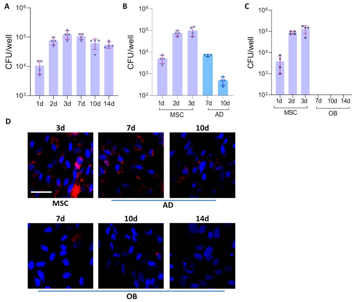
Mesenchymal stem cells (MSCs) were infected with B. abortus. After 72 h of infection, some cells were cultured in osteoblast or adipocyte differentiation medium. The colony-forming units (CFU) of intracellular bacteria were determined at different times post-infection. As shown in Figure 1A, B. abortus infects and replicates in MSCs, reaching the highest level at 3 days post-infection, and these values did not differ significantly for at least 14 days post-infection. However, the levels of CFU declined after the initiation of adipocyte differentiation (Figure 1B). The osteoblast differentiation process completely abolished B. abortus to replicate in these cells (Figure 1C). These results were confirmed by confocal microscopy using B. abortus expressing DsRed (Figure 1D). These findings indicate that adipocyte and osteoblast differentiation modulate the intracellular survival of B. abortus.

3.2. Brucella abortus Was Unable to Modulate Matrix Deposition During Osteoblast Differentiation

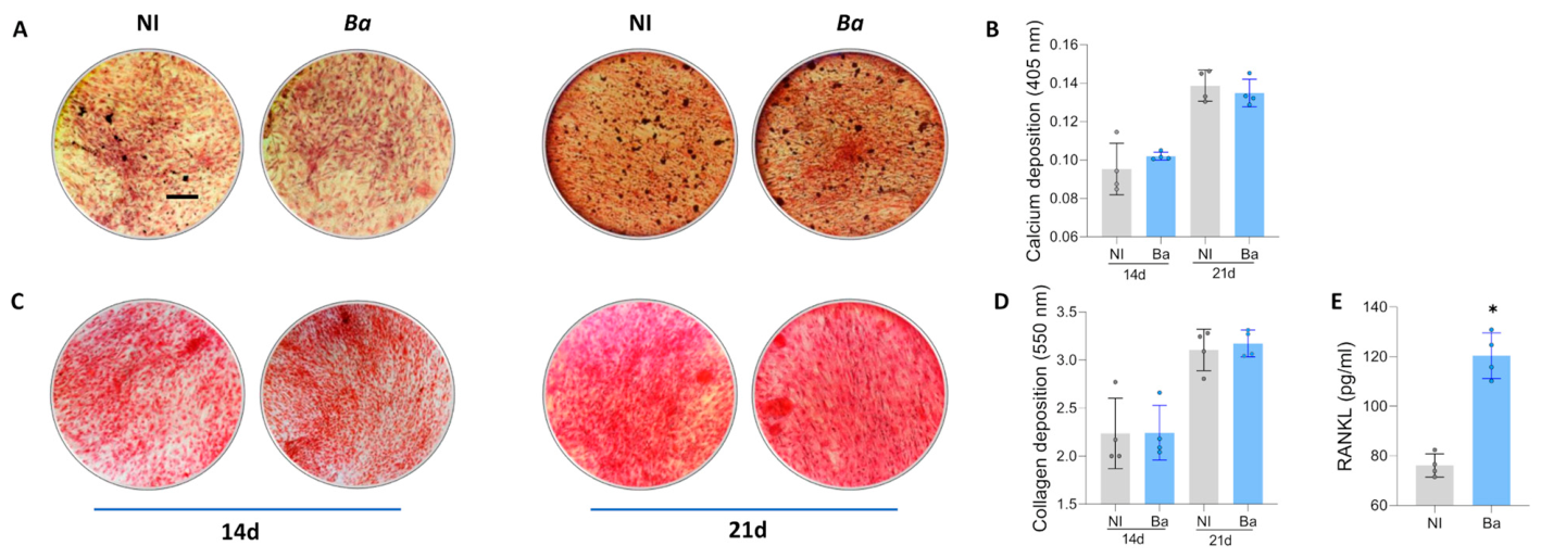
Proper placement of the mineral matrix and the formation of the bone’s structural architecture depend critically on the accurate deposition of the organic matrix [34]. To investigate the impact of B. abortus infection on the deposition of organic and mineral matrices, MSCs were infected with B. abortus. Three days post-infection, the culture medium was switched to an osteoblast differentiation medium. The deposition of the mineral matrix, indicated by calcium deposits, and the organic matrix, indicated by collagen deposits, were evaluated at 14 and 21 days post-differentiation using Alizarin Red S (Figure 2A,B) and Sirius Red (Figure 2C,D) staining, respectively. The results revealed that calcium or collagen deposition among osteoblasts obtained from Brucella-infected MSCs remained unaltered at the observed time points.

3.3. Brucella abortus Induces Activator of Nuclear Factor Kappa-B Ligand (RANKL) Expression by Osteoblasts

Bone homeostasis is maintained through the balance between bone resorption by osteoclasts and bone formation by osteoblasts. RANKL is essential for the differentiation and activation of osteoclasts, playing a critical role in bone remodeling [35].
Our results indicated that osteoblasts derived from B. abortus-infected MSCs secrete higher levels of RANKL than osteoblasts derived from non-infected cells 21 days after differentiation (Figure 2E). Thus, B. abortus infection appears to disrupt bone homeostasis by promoting increased RANKL secretion, which could induce osteoclastogenesis.

3.4. Brucella abortus Infection Modulates Adipocyte Differentiation

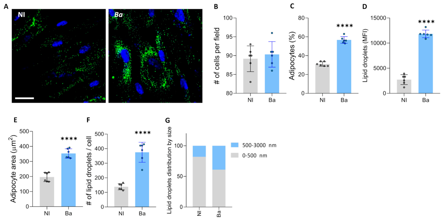
The differentiation of MSCs into osteoblasts or adipocytes is meticulously controlled by a combination of biological signals and chemical influences [20,21]. Therefore, to determine the effect of B. abortus infection on adipocyte differentiation, MSCs were infected with B. abortus, and after 3 days, the culture medium was replaced with adipocyte differentiation medium. A significant increase in adipocyte differentiation compared to uninfected cells was observed, as shown by the increases in adipocyte number, adipocyte size, and lipid droplets size evaluated at 7 days after differentiation (Figure 3).
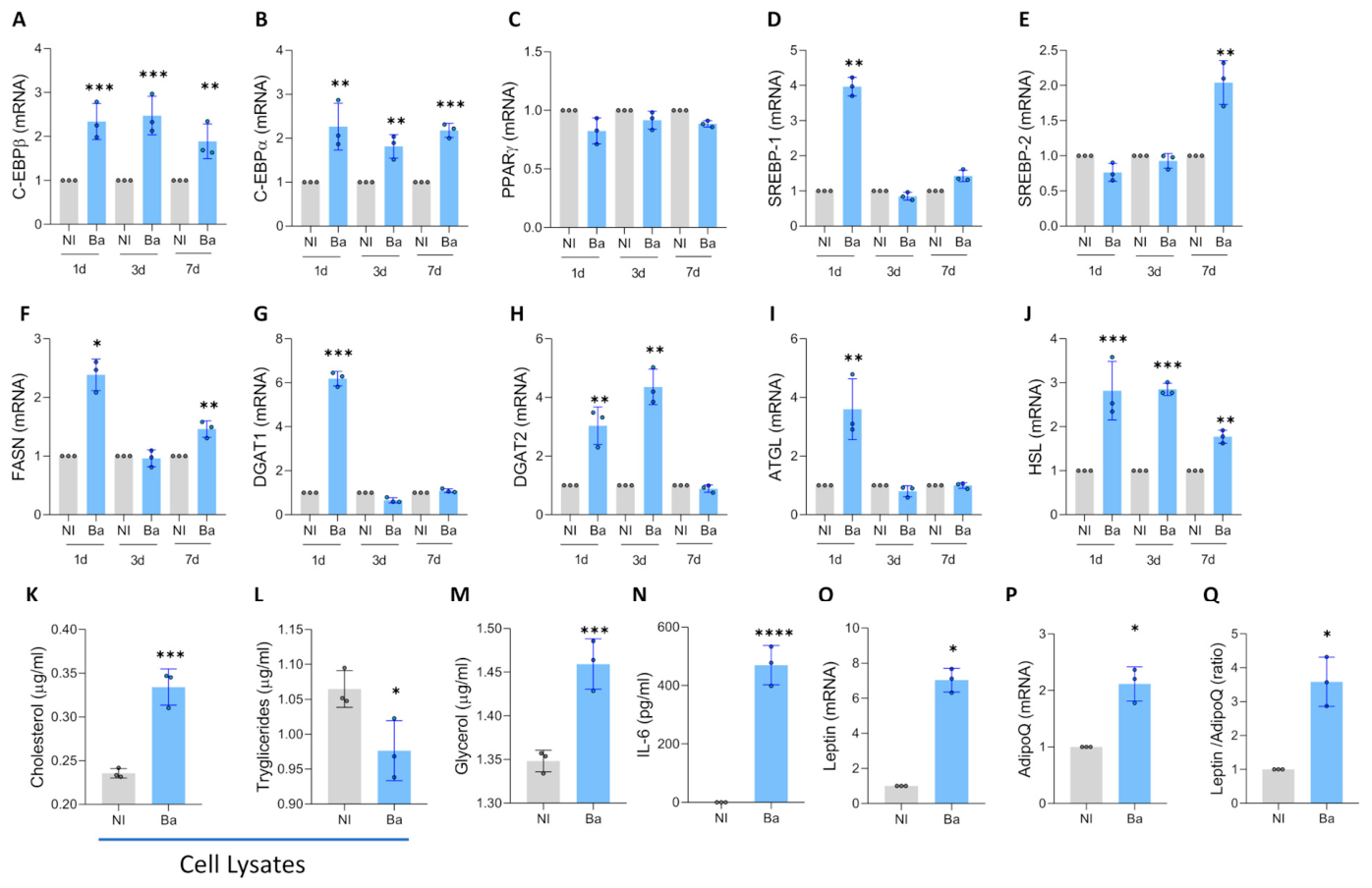
CCAAT/enhancer-binding protein (C/EBP)β is expressed in the initial stages of differentiation and enhances the transcription of C/EBPα and Peroxisome proliferator-activated receptor gamma (PPARγ). Mesenchymal stem cells were infected with B. abortus for 3 days and cultured in adipocyte differentiation medium. At 1, 3, and 7 days post-differentiation, the transcription of C/EBPβ, C/EBPα, and PPARγ was evaluated by RT-qPCR. An increase in C/EBPβ expression, along with concurrent transcription of C/EBPα, was observed (Figure 4A,B) without significant changes in PPARγ levels (Figure 4C).

3.5. Brucella abortus Infection Modulates Lipogenesis and Lipolysis During Adipocyte Differentiation

Adipocyte enlargement results from an increase in the size of lipid droplets. The enlarged lipid droplets demand more protein production to support the growth of plasma and lipid droplet membranes. Additionally, hypertrophic adipocytes encounter heightened demands for lipogenic, lipolytic, and endocrine activities, which increase cellular stress and necessitate the activation of growth signaling pathways [36].
Sterol regulatory element-binding proteins (SREBP)-1 and SREBP-2 are crucial transcription factors that regulate lipid metabolism during the differentiation of mesenchymal stem cells into adipocytes. SREBP-1 is essential for activating genes involved in lipogenesis, including those coding for key enzymes in fatty acid and triglyceride synthesis. Its expression increases significantly during the first 24 to 48 h of differentiation. On the other hand, SREBP-2 is involved in regulating cholesterol metabolism [37]. Our results indicate that B. abortus infection increases the transcription of SREBP-1 on day 1 of differentiation and SREBP-2 on day 7 post-differentiation (Figure 4D,E).
Accordingly, B. abortus infection induces an increase in lipogenesis, as revealed by the upregulation of FASN transcription at 1 and 3 days post-differentiation, and DGAT1 and DGAT2 at 1 and 3 days post-differentiation, respectively (Figure 4F–H). This is accompanied by lipolysis, evidenced by the increase in ATGL at day 1 post-differentiation and HSL at 1, 3, and 7 days post-differentiation (Figure 4I,J). As a result, at 7 days post-differentiation, B. abortus induces an increase in intracellular cholesterol compared to uninfected controls (Figure 4K). Additionally, a reduction in intracellular triglycerides was observed in cell lysates from cells infected with B. abortus, along with an increase in glycerol presence in culture supernatants (Figure 4L,M).
Adipocyte hypertrophy is associated with an inflammatory profile. Our results indicate that B. abortus infection induces an increase in IL-6 secretion (Figure 4N) and an increase in the leptin/adiponectin ratio (Figure 4O–Q). Together, these findings indicate that B. abortus modulates lipogenesis and lipolysis, leading to adipocyte hypertrophy with an inflammatory profile.

3.6. Brucella abortus Infection Increases Lipid Droplet-Mitochondria Interaction During Adipocyte Differentiation

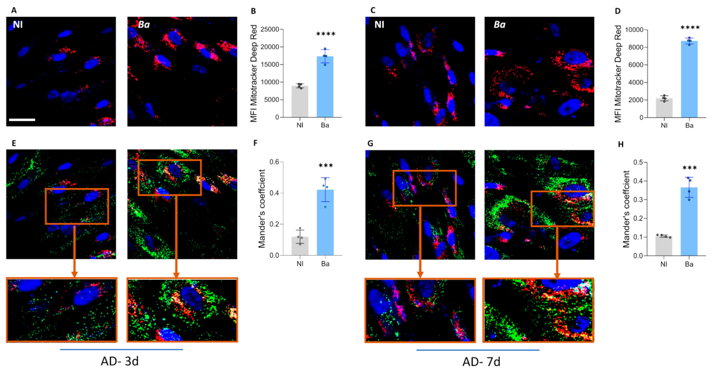
Mitochondria bind to lipid droplets and regulate lipid storage and utilization [38]. Therefore, experiments were conducted to determine whether B. abortus infection increases interaction between mitochondria and lipid droplets. To this end, lipid droplets were stained with Bodipy 493/503, and mitochondria were labeled with MitoTracker Deep Red at 3 and 7 days after adipocyte differentiation. Our results indicate that B. abortus infection increases mitochondrial mass measured at 3 and 7 days post-differentiation (Figure 5 A–D). As expected and consistent with the increase in lipid droplet size in B. abortus-infected cells, a significant colocalization between lipid droplets and mitochondria was measured using Manders’ overlap analysis at 3 and 7 days post differentiation (Figure 5E–H). Our results indicate that B. abortus infection increases mitochondrial mass and the interaction between mitochondria and lipid droplets.

3.7. Brucella abortus Induces an Increase in Lipid Droplet Size Through a VirB- Independent Mechanism

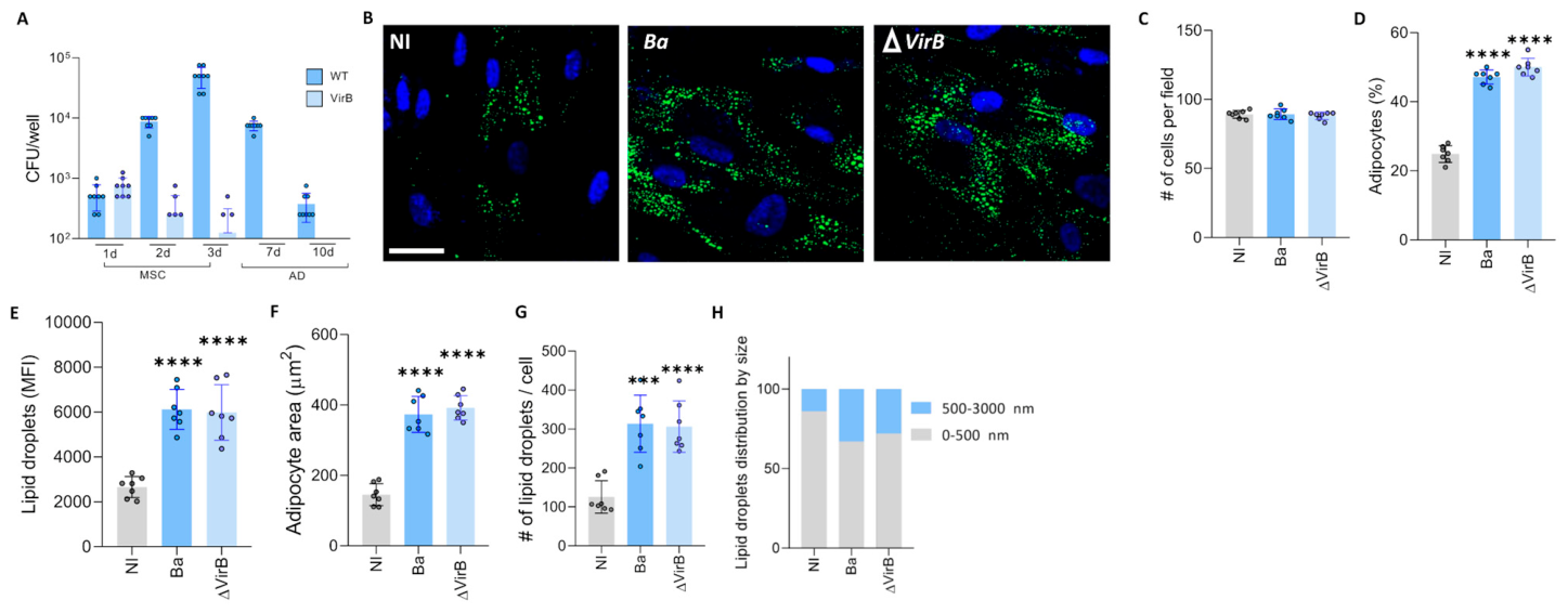
The type IV secretion system (T4SS) VirB is involved in the ability of different Brucella species to establish an intracellular replication niche and in the induction of inflammatory responses during B. abortus infection [39,40,41,42]. To this end, MSCs were infected with B. abortus and an isogenic VirB mutant. Three days post-infection, the medium was replaced with adipocyte differentiation medium. The presence of intracellular bacteria was evaluated at 1, 2, 3, 7, and 10 days post-infection. These data indicate that the ability of B. abortus to replicate in these cells depends on the presence of a functional T4SS (Figure 6A). However, the modulation of adipocyte differentiation may not require a functional T4SS, as evidenced by the similar increase in adipocyte number, as well as in lipid droplets number and size, induced by both wild-type B. abortus and the VirB mutant 7 days after differentiation (Figure 6B–H). These results indicate that B. abortus infection increases adipocyte number and size in a VirB-independent manner.

3.8. Brucella DNA Recapitulates the Adipogenic Modulation Induced by B. abortus Infection

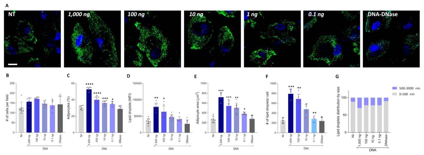
Bacterial DNA has been linked to adipocyte hypertrophy and an inflammatory profile [43,44]. As shown in Figure 7, B. abortus DNA influences adipocyte differentiation similarly to live bacteria, leading to an increase in adipocyte number, size, and lipid droplet accumulation observed seven days after differentiation. This effect was proportional to the amount of DNA present in the culture (Figure 7). Notably, when the highest dose of B. abortus DNA (1000 ng/mL) was treated with DNase I, the effect was abolished, confirming that bacterial DNA plays a direct role in modulating adipocyte differentiation. These results indicated that B. abortus-DNA modulates adipocyte differentiation in a dose-dependent manner.

3.9. Heat-Killed B. abortus (HKBA) Modulate Adipocyte Differentiation Through DNA

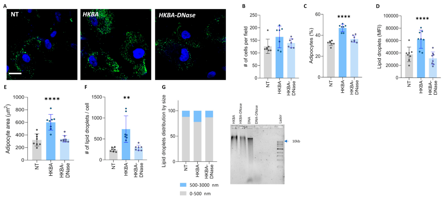
To determine whether viable bacteria are necessary for modulating adipocyte differentiation from mesenchymal stem cells, we tested the ability of HKBA to mimic the effects of live bacteria. Similar to live B. abortus, HKBA induced an increase in adipocyte differentiation and lipid droplet size (Figure 8). The fact that HKBA also influenced the adipocyte differentiation process suggests that this phenomenon is mediated by a structural component of B. abortus.
To further investigate the role of Brucella genomic DNA in adipocyte differentiation, we treated HKBA with DNase I before using it to stimulate adipocyte precursors. Our results showed that DNase I-treated HKBA was unable to modulate adipocyte differentiation, as indicated by the comparable adipocyte number, size, and lipid droplet accumulation observed between treated and untreated cells. Our findings demonstrate that Brucella DNA plays a crucial role in modulating adipocyte differentiation, highlighting its potential impact on host metabolism.

3.10. Brucella DNA Modulates Adipogenesis Through TLR9 Signaling

It has been reported that TLR9 -the receptor responsible for recognizing bacterial DNA—is one of the TLRs most critically involved in Brucella infection [45]. To determine whether the adipogenic effect induced by bacterial DNA in our model depended on this pathway, MSCs were differentiated into adipocytes in the presence of the TLR9 antagonist INH-ODN-18.
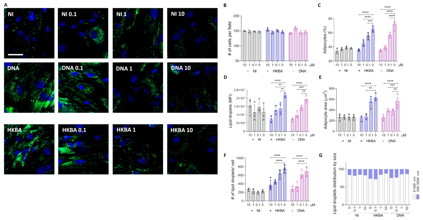
As shown above, stimulation with B. abortus DNA markedly increased adipocyte number, cell size, and lipid droplet accumulation. However, preincubation with INH-ODN-18 significantly reduced these effects in a dose-dependent manner. The highest inhibitor concentration (10 μM) nearly abolished the adipogenic response to bacterial DNA, whereas intermediate (1 μM) and low (0.1 μM) concentrations produced partial inhibition (Figure 9A–F).
Importantly, INH-ODN-18 alone did not affect basal adipocyte differentiation, suggesting that the effects observed were related to TLR9 blockade. Similar results were obtained when cells were stimulated with HKBA, supporting the involvement of bacterial DNA in the adipogenic response. Taken together, these findings indicate that B. abortus DNA may promote adipocyte differentiation through a TLR9-dependent pathway, although additional bacterial components may also contribute.

4. Discussion

Osteoarticular disease is the most common clinical complication of active human brucellosis [2,3,4,5,6,7,8]. Bone marrow, located within the bone cavities, is a specialized tissue that plays essential roles in hematopoiesis, regulation of skeletal remodeling, and immune modulation [46]. Because Brucella can be detected in bone marrow during active infection [9,11], it is plausible that, in osteoarticular brucellosis, the bacterium may affect mesenchymal stem cell (MSC) differentiation and modulate the production of soluble mediators, thereby contributing to bone loss.
In the present study, we found that B. abortus infection of MSCs did not affect the deposition of organic or mineral matrix during osteoblast differentiation. This result differs from our previous observations, in which B. abortus inhibited osteoblast differentiation in murine preosteoblasts derived from calvaria and in the MC3T3-E1 cell line [27]. One possible explanation is that umbilical cord-derived MSCs have a high osteogenic potential [47]. In addition, MSCs are increasingly recognized as heterogeneous populations composed of distinct subpopulations with different cell surface marker profiles and functional properties [48]. Thus, one possibility is that B. abortus does not preferentially infect the MSC subpopulation most committed to osteogenic differentiation. Alternatively, the bacterium may infect and replicate before these cells enter the osteogenic program.
Although the deposition of mineral and protein matrix during osteoblast differentiation was preserved, B. abortus may still contribute to bone damage through another mechanism, namely by promoting osteoclastogenesis.
Activator of Nuclear Factor Kappa-B Ligand (RANKL), a homotrimeric molecule expressed on osteoblasts, is a central regulator of osteoclast differentiation and bone resorption [49]. Similar to other infections that increase RANKL expression [50,51,52,53], B. abortus infection of MSCs resulted in osteoblasts expressing higher RANKL levels than those differentiated in the absence of bacteria. Therefore, even in the absence of a direct effect on matrix deposition by differentiated osteoblasts, B. abortus may favor bone destruction by shifting the balance toward osteoclast activation.
A second major finding of this study is that B. abortus modulates adipocyte differentiation, increasing both adipocyte number and cell size. CCAAT/enhancer-binding protein (C/EBP)β is a key regulator of adipocyte development and lipid accumulation [54,55]. In agreement with this role, we observed increased C/EBPβ expression, together with concomitant induction of C/EBPα, in adipocytes differentiated from B. abortus-infected MSCs compared with uninfected controls. Likewise, adipocytes derived from infected MSCs showed an early increase in SREBP-1, a transcription factor that regulates lipid synthesis and uptake [56,57,58], along with increased expression of fatty acid synthase (FASN), Diglyceride acyltransferase (DGAT)1, and DGAT2. These findings indicate that B. abortus enhances the transcriptional program associated with adipogenesis and lipid accumulation.
Interestingly, B. abortus infection also increased lipolysis. Hypertrophic adipocytes are associated with increased lipolytic activity and reduced insulin responsiveness, which in turn lead to decreased glucose uptake, elevated basal lipolysis, and impaired stimulated lipolysis. As adipocytes enlarge through lipid droplet expansion, the demand for membrane and protein synthesis increases to sustain cell growth. Consequently, coordinated lipogenic and lipolytic activities are required to support these processes [59]. In this context, the increase in lipolysis observed in infected cells may contribute to the metabolic remodeling associated with adipocyte hypertrophy.
Mitochondrial mass is closely linked to the rate of lipid droplet expansion and may contribute to de novo lipogenesis and lipid droplet formation during early adipocyte differentiation [38]. Consistent with this, after Brucella-induced lipid droplet enlargement, we observed increased mitochondrial mass and a closer association between lipid droplets and mitochondria in adipocytes differentiated in the presence of B. abortus compared with uninfected controls. In parallel, glycerol derived from lipolysis can be recycled through phosphorylation by glycerol kinase and used as a substrate for triacylglycerol synthesis, suggesting an important role for glycerol recycling in adipocyte expansion [60]. In line with this mechanism, B. abortus induced glycerol release without significantly altering intracellular triglyceride levels at the time analyzed.
Sterol regulatory element-binding protein (SREBP)-2 is primarily involved in cholesterol metabolism [57]. In enlarged adipocytes, this transcription factor can be activated in response to membrane cholesterol depletion to regulate genes involved in cholesterol biosynthesis [61]. Hypertrophic adipocytes are also characterized by a proinflammatory phenotype, including increased IL-6 production and a higher leptin/adiponectin ratio [62,63]. Therefore, the adipogenic program induced by B. abortus may not only promote cellular hypertrophy but also contribute to a local inflammatory environment that could further affect bone homeostasis.
Regarding the bacterial components involved in this response, the type IV secretion system (T4SS) encoded by the virB genes is essential for intracellular replication of Brucella [39] and has also been implicated in the inflammatory response during B. abortus infection [41,64,65]. However, in our model, the T4SS was not involved in adipocyte differentiation. Instead, the effect of Brucella infection on adipogenesis was reproduced by HKBA, indicating that bacterial DNA is sufficient to trigger this response. This is consistent with previous studies showing that bacterial DNA contributes to adipose tissue inflammation and hypertrophy [43,44]. TLR9 has been identified as the most important TLR involved in Brucella infection, likely through recognition of bacterial DNA [45]. Although Brucella lipopolysaccharide (LPS) and lipoproteins also contribute to the immune response [66], their role in adipocyte differentiation was not determined until now. Nonetheless, we cannot exclude synergistic or context-dependent effects of these structural components. Further studies will be needed to determine whether TLR2-, TLR4-, or other DNA-independent pathways also participate in adipogenesis alongside TLR9. In agreement with this interpretation, inhibition of TLR9 with INH-ODN-18 markedly reduced the increase in adipocyte number, hypertrophy, and lipid droplet accumulation induced by HKBA and B. abortus DNA, supporting a key role for this receptor in the observed phenotype.
Overall, our results indicate that B. abortus infection does not impair osteoblast differentiation or function under the conditions tested. In contrast, it promotes adipogenesis and generates cells with a proinflammatory profile that may influence both osteoblast and osteoclast activity, thereby contributing to bone damage. In vitro models have provided valuable insights into the intracellular mechanisms involved [67], and the present study adds new information on Brucella-host interactions and advances our understanding of the cellular basis of bone involvement in brucellosis.
This study has some limitations. We used umbilical cord mesenchymal stem cells (UC-MSCs) instead of bone marrow mesenchymal stem cells (BM-MSCs) because UC-MSCs are more readily available and can be obtained non-invasively from tissue usually discarded after birth. In addition, we evaluated only B. abortus DNA as a bacterial stimulus involved in adipocyte hypertrophy, so the contribution of other Brucella components cannot be excluded. Further studies are needed to determine whether additional bacterial factors and Toll-like receptor pathways participate in this response.

Author Contributions

Conceptualization, R.N.F., J.Q. and M.V.D.; data curation, R.N.F. and C.A.M.L.; formal analysis R.N.F., C.A.M.L. and F.A.S.; methodology, R.N.F., C.A.M.L., M.B.P., C.C., F.A.S. and P.J.; investigation, R.N.F., C.A.M.L., M.B.P., C.C., F.A.S., M.N.G. and P.J.; resources M.B.P. and M.N.G.; validation R.N.F. and C.C. writing—original draft, M.V.D.; writing—review and editing, J.Q. and M.V.D.; funding acquisition, J.Q. and M.V.D. All authors have read and agreed to the published version of the manuscript.

Funding

This work was supported by grants from Agencia Nacional de Promoción Científica y Tecnológica (ANPCYT, Argentina), PICT 2020-0691 to MVD. Funding agencies had no role in the study design, data collection and analysis, decision to publish, or preparation of the manuscript.

Institutional Review Board Statement

The study was conducted in accordance with the Declaration of Helsinki and was approved by the Comité de Bioética y Ética, Facultad de Ciencias Médicas, Universidad de Buenos Aires, Argentina (RESCD-2023-1291).

Informed Consent Statement

Written informed consent was obtained from each mother before cesarean birth, and human umbilical cords were obtained from discarded placentas.

Data Availability Statement

The raw data supporting the conclusions of this article will be made available by the authors, without undue reservation.

Acknowledgments

Graphical abstract was created with BioRender.com. https://www.biorender.com/ (accessed on day 1 February 2026). All individuals included in this section have consented to the acknowledgement.

Conflicts of Interest

The authors declare no conflicts of interest. The funders had no role in the design of this study; in the collection, analyses, or interpretation of data; in the writing of the manuscript; or in the decision to publish the results.

Abbreviations

The following abbreviations are used in this manuscript:
MSCsmesenchymal stem cells
α-MEMα-Minimal Essential Medium
DMEMDulbecco’s Modified Eagle Medium
PBSphosphate-buffered saline
IBMX3-isobutyl-1-methylxanthine
RUNX2runt-related transcription factor 2
IL-6interleukin-6
RANKLreceptor activator of nuclear factor kappa B ligand
ALPalkaline phosphatase
CFUcolony-forming units
C/EBPCCAAT enhancer binding protein
HSLhormone-sensitive lipase
ATGLadipose triglyceride lipase
FASNfatty acid synthase
DGATdiglyceride acyltransferase
PPARperoxisome proliferator-activated receptor

References

  1. Liu, Z.; Gao, L.; Wang, M.; Yuan, M.; Li, Z. Long ignored but making a comeback: A worldwide epidemiological evolution of human brucellosis. Emerg. Microbes Infect. 2024, 13, 2290839. [Google Scholar] [CrossRef]
  2. Esmaeilnejad-Ganji, S.M.; Esmaeilnejad-Ganji, S.M.R. Osteoarticular manifestations of human brucellosis: A review. World J. Orthop. 2019, 10, 54–62. [Google Scholar] [CrossRef]
  3. Aydin, M.; Fuat Yapar, A.; Savas, L.; Reyhan, M.; Pourbagher, A.; Turunc, T.Y.; Ziya Demiroglu, Y.; Yologlu, N.A.; Aktas, A. Scintigraphic findings in osteoarticular brucellosis. Nucl. Med. Commun. 2005, 26, 639–647. [Google Scholar] [CrossRef]
  4. Gotuzzo, E.; Alarcon, G.S.; Bocanegra, T.S.; Carrillo, C.; Guerra, J.C.; Rolando, I.; Espinoza, L.R. Articular involvement in human brucellosis: A retrospective analysis of 304 cases. Semin. Arthritis Rheum. 1982, 12, 245–255. [Google Scholar] [CrossRef] [PubMed]
  5. Khateeb, M.I.; Araj, G.F.; Majeed, S.A.; Lulu, A.R. Brucella arthritis: A study of 96 cases in Kuwait. Ann. Rheum. Dis. 1990, 49, 994–998. [Google Scholar] [CrossRef] [PubMed]
  6. Pourbagher, A.; Pourbagher, M.A.; Savas, L.; Turunc, T.; Demiroglu, Y.Z.; Erol, I.; Yalcintas, D. Epidemiologic, clinical, and imaging findings in brucellosis patients with osteoarticular involvement. AJR Am. J. Roentgenol. 2006, 187, 873–880. [Google Scholar] [CrossRef] [PubMed]
  7. Buzgan, T.; Karahocagil, M.K.; Irmak, H.; Baran, A.I.; Karsen, H.; Evirgen, O.; Akdeniz, H. Clinical manifestations and complications in 1028 cases of brucellosis: A retrospective evaluation and review of the literature. Int. J. Infect. Dis. 2010, 14, e469–e478. [Google Scholar] [CrossRef]
  8. Madkour, M.M. Osteoarticular brucellosis. In Madkour’s Brucellosis, 2nd ed.; Madkour, M.M., Ed.; Springer: Berlin, Germany, 2001; pp. 74–84. [Google Scholar]
  9. Demir, C.; Karahocagil, M.K.; Esen, R.; Atmaca, M.; Gonullu, H.; Akdeniz, H. Bone marrow biopsy findings in brucellosis patients with hematologic abnormalities. Chin. Med. J. 2012, 125, 1871–1876. [Google Scholar]
  10. El-Koumi, M.A.; Afify, M.; Al-Zahrani, S.H. A prospective study of brucellosis in children: Relative frequency of pancytopenia. Mediterr. J. Hematol. Infect. Dis. 2013, 5, e2013011. [Google Scholar] [CrossRef]
  11. Ertem, M.; Kurekci, A.E.; Aysev, D.; Unal, E.; Ikinciogullari, A. Brucellosis transmitted by bone marrow transplantation. Bone Marrow Transplant. 2000, 26, 225–226. [Google Scholar] [CrossRef]
  12. Atluri, V.L.; Xavier, M.N.; de Jong, M.F.; den Hartigh, A.B.; Tsolis, R.M. Interactions of the human pathogenic Brucella species with their hosts. Annu. Rev. Microbiol. 2011, 65, 523–541. [Google Scholar] [CrossRef]
  13. Gonzalez-Espinoza, G.; Arce-Gorvel, V.; Memet, S.; Gorvel, J.P. Brucella: Reservoirs and Niches in Animals and Humans. Pathogens 2021, 10, 186. [Google Scholar] [CrossRef]
  14. Fink, T.; Zachar, V. Adipogenic differentiation of human mesenchymal stem cells. Methods Mol. Biol. 2011, 698, 243–251. [Google Scholar] [CrossRef]
  15. Song, L.; Tuan, R.S. Transdifferentiation potential of human mesenchymal stem cells derived from bone marrow. FASEB J. 2004, 18, 980–982. [Google Scholar] [CrossRef]
  16. Berendsen, A.D.; Olsen, B.R. Osteoblast-adipocyte lineage plasticity in tissue development, maintenance and pathology. Cell Mol. Life Sci. 2014, 71, 493–497. [Google Scholar] [CrossRef]
  17. Rosen, C.J.; Bouxsein, M.L. Mechanisms of disease: Is osteoporosis the obesity of bone? Nat. Clin. Pract. Rheumatol. 2006, 2, 35–43. [Google Scholar] [CrossRef]
  18. Ali, D.; Tencerova, M.; Figeac, F.; Kassem, M.; Jafari, A. The pathophysiology of osteoporosis in obesity and type 2 diabetes in aging women and men: The mechanisms and roles of increased bone marrow adiposity. Front. Endocrinol. 2022, 13, 981487. [Google Scholar] [CrossRef] [PubMed]
  19. Verma, S.; Rajaratnam, J.H.; Denton, J.; Hoyland, J.A.; Byers, R.J. Adipocytic proportion of bone marrow is inversely related to bone formation in osteoporosis. J. Clin. Pathol. 2002, 55, 693–698. [Google Scholar] [CrossRef] [PubMed]
  20. Moerman, E.J.; Teng, K.; Lipschitz, D.A.; Lecka-Czernik, B. Aging activates adipogenic and suppresses osteogenic programs in mesenchymal marrow stroma/stem cells: The role of PPAR-gamma2 transcription factor and TGF-beta/BMP signaling pathways. Aging Cell 2004, 3, 379–389. [Google Scholar] [CrossRef] [PubMed]
  21. Yeung, D.K.; Griffith, J.F.; Antonio, G.E.; Lee, F.K.; Woo, J.; Leung, P.C. Osteoporosis is associated with increased marrow fat content and decreased marrow fat unsaturation: A proton MR spectroscopy study. J. Magn. Reson. Imaging 2005, 22, 279–285. [Google Scholar] [CrossRef]
  22. Gimble, J.M.; Zvonic, S.; Floyd, Z.E.; Kassem, M.; Nuttall, M.E. Playing with bone and fat. J. Cell Biochem. 2006, 98, 251–266. [Google Scholar] [CrossRef] [PubMed]
  23. Rosen, E.D.; MacDougald, O.A. Adipocyte differentiation from the inside out. Nat. Rev. Mol. Cell Biol. 2006, 7, 885–896. [Google Scholar] [CrossRef]
  24. Komori, T. Regulation of osteoblast differentiation by Runx2. Adv. Exp. Med. Biol. 2010, 658, 43–49. [Google Scholar] [CrossRef]
  25. Jeon, M.J.; Kim, J.A.; Kwon, S.H.; Kim, S.W.; Park, K.S.; Park, S.W.; Kim, S.Y.; Shin, C.S. Activation of peroxisome proliferator-activated receptor-gamma inhibits the Runx2-mediated transcription of osteocalcin in osteoblasts. J. Biol. Chem. 2003, 278, 23270–23277. [Google Scholar] [CrossRef]
  26. Freiberger, R.N.; Lopez, C.A.M.; Sviercz, F.A.; Cevallos, C.; Guano, A.D.; Jarmoluk, P.; Quarleri, J.; Delpino, M.V.B. abortus Infection Promotes an Imbalance in the Adipocyte-Osteoblast Crosstalk Favoring Bone Resorption. Int. J. Mol. Sci. 2023, 24, 5617. [Google Scholar] [CrossRef]
  27. Scian, R.; Barrionuevo, P.; Fossati, C.A.; Giambartolomei, G.H.; Delpino, M.V. Brucella abortus invasion of osteoblasts inhibits bone formation. Infect. Immun. 2012, 80, 2333–2345. [Google Scholar] [CrossRef]
  28. Pesce Viglietti, A.I.; Giambartolomei, G.H.; Quarleri, J.; Delpino, M.V. Brucella abortus Infection Modulates 3T3-L1 Adipocyte Inflammatory Response and Inhibits Adipogenesis. Front. Endocrinol. 2020, 11, 585923. [Google Scholar] [CrossRef]
  29. Delpino, M.V.; Fossati, C.A.; Baldi, P.C. Proinflammatory response of human osteoblastic cell lines and osteoblast-monocyte interaction upon infection with Brucella spp. Infect. Immun. 2009, 77, 984–995. [Google Scholar] [CrossRef]
  30. Palma, M.B.; Luzzani, C.; Andrini, L.B.; Riccillo, F.; Buero, G.; Pelinski, P.; Inda, A.M.; Errecalde, A.L.; Miriuka, S.; Carosella, E.D.; et al. Wound Healing by Allogeneic Transplantation of Specific Subpopulation From Human Umbilical Cord Mesenchymal Stem Cells. Cell Transplant. 2021, 30, 963689721993774. [Google Scholar] [CrossRef]
  31. Xiao, B.; Deng, X.; Zhou, W.; Tan, E.K. Flow Cytometry-Based Assessment of Mitophagy Using MitoTracker. Front. Cell Neurosci. 2016, 10, 76. [Google Scholar] [CrossRef] [PubMed]
  32. Manders, E.M.M.; Verbeek, F.J.; Aten, J.A. Measurement of co-localization of objects in dual-colour confocal images. J. Microsc. 1993, 169, 375–382. [Google Scholar] [CrossRef]
  33. Garland, P.B.; Randle, P.J. A rapid enzymatic assay for glycerol. Nature 1962, 196, 987–988. [Google Scholar] [CrossRef] [PubMed]
  34. Sroga, G.E.; Vashishth, D. Effects of bone matrix proteins on fracture and fragility in osteoporosis. Curr. Osteoporos. Rep. 2012, 10, 141–150. [Google Scholar] [CrossRef] [PubMed]
  35. Yasuda, H. Discovery of the RANKL/RANK/OPG system. J. Bone Miner. Metab. 2021, 39, 2–11. [Google Scholar] [CrossRef] [PubMed]
  36. Sakers, A.; De Siqueira, M.K.; Seale, P.; Villanueva, C.J. Adipose-tissue plasticity in health and disease. Cell 2022, 185, 419–446. [Google Scholar] [CrossRef]
  37. Jarc, E.; Petan, T. Lipid Droplets and the Management of Cellular Stress. Yale J. Biol. Med. 2019, 92, 435–452. [Google Scholar]
  38. Benador, I.Y.; Veliova, M.; Liesa, M.; Shirihai, O.S. Mitochondria Bound to Lipid Droplets: Where Mitochondrial Dynamics Regulate Lipid Storage and Utilization. Cell Metab. 2019, 29, 827–835. [Google Scholar] [CrossRef]
  39. Comerci, D.J.; Martinez-Lorenzo, M.J.; Sieira, R.; Gorvel, J.P.; Ugalde, R.A. Essential role of the VirB machinery in the maturation of the Brucella abortus-containing vacuole. Cell Microbiol. 2001, 3, 159–168. [Google Scholar] [CrossRef]
  40. Arriola Benitez, P.C.; Rey Serantes, D.; Herrmann, C.K.; Pesce Viglietti, A.I.; Vanzulli, S.; Giambartolomei, G.H.; Comerci, D.J.; Delpino, M.V. The Effector Protein BPE005 from Brucella abortus Induces Collagen Deposition and Matrix Metalloproteinase 9 Downmodulation via Transforming Growth Factor beta1 in Hepatic Stellate Cells. Infect. Immun. 2016, 84, 598–606. [Google Scholar] [CrossRef]
  41. Arriola Benitez, P.C.; Pesce Viglietti, A.I.; Gomes, M.T.R.; Oliveira, S.C.; Quarleri, J.F.; Giambartolomei, G.H.; Delpino, M.V. Brucella abortus Infection Elicited Hepatic Stellate Cell-Mediated Fibrosis Through Inflammasome-Dependent IL-1beta Production. Front. Immunol. 2019, 10, 3036. [Google Scholar] [CrossRef]
  42. de Jong, M.F.; Starr, T.; Winter, M.G.; den Hartigh, A.B.; Child, R.; Knodler, L.A.; van Dijl, J.M.; Celli, J.; Tsolis, R.M. Sensing of bacterial type IV secretion via the unfolded protein response. mBio 2013, 4. [Google Scholar] [CrossRef] [PubMed]
  43. Massier, L.; Chakaroun, R.; Tabei, S.; Crane, A.; Didt, K.D.; Fallmann, J.; von Bergen, M.; Haange, S.B.; Heyne, H.; Stumvoll, M.; et al. Adipose tissue derived bacteria are associated with inflammation in obesity and type 2 diabetes. Gut 2020, 69, 1796–1806. [Google Scholar] [CrossRef] [PubMed]
  44. Liu, W.; Yang, G.; Liu, P.; Jiang, X.; Xin, Y. Modulation of adipose tissue metabolism by microbial-derived metabolites. Front. Microbiol. 2022, 13, 1031498. [Google Scholar] [CrossRef]
  45. Campos, P.C.; Gomes, M.T.; Guimaraes, G.; Costa Franco, M.M.; Marim, F.M.; Oliveira, S.C. Brucella abortus DNA is a major bacterial agonist to activate the host innate immune system. Microbes Infect. 2014, 16, 979–984. [Google Scholar] [CrossRef]
  46. Gesta, S.; Tseng, Y.H.; Kahn, C.R. Developmental origin of fat: Tracking obesity to its source. Cell 2007, 131, 242–256. [Google Scholar] [CrossRef]
  47. Shen, C.; Yang, C.; Xu, S.; Zhao, H. Comparison of osteogenic differentiation capacity in mesenchymal stem cells derived from human amniotic membrane (AM), umbilical cord (UC), chorionic membrane (CM), and decidua (DC). Cell Biosci. 2019, 9, 17. [Google Scholar] [CrossRef]
  48. Wang, X.; Wang, Y.; Gou, W.; Lu, Q.; Peng, J.; Lu, S. Role of mesenchymal stem cells in bone regeneration and fracture repair: A review. Int. Orthop. 2013, 37, 2491–2498. [Google Scholar] [CrossRef]
  49. Xu, H.; Wang, W.; Liu, X.; Huang, W.; Zhu, C.; Xu, Y.; Yang, H.; Bai, J.; Geng, D. Targeting strategies for bone diseases: Signaling pathways and clinical studies. Signal Transduct. Target. Ther. 2023, 8, 202. [Google Scholar] [CrossRef]
  50. Kelesidis, T.; Currier, J.S.; Yang, O.O.; Brown, T.T. Role of RANKL-RANK/osteoprotegerin pathway in cardiovascular and bone disease associated with HIV infection. AIDS Rev. 2014, 16, 123–133. [Google Scholar]
  51. Kajiya, M.; Giro, G.; Taubman, M.A.; Han, X.; Mayer, M.P.; Kawai, T. Role of periodontal pathogenic bacteria in RANKL-mediated bone destruction in periodontal disease. J. Oral. Microbiol. 2010, 2, 10. [Google Scholar] [CrossRef] [PubMed]
  52. Chen, W.; Foo, S.S.; Rulli, N.E.; Taylor, A.; Sheng, K.C.; Herrero, L.J.; Herring, B.L.; Lidbury, B.A.; Li, R.W.; Walsh, N.C.; et al. Arthritogenic alphaviral infection perturbs osteoblast function and triggers pathologic bone loss. Proc. Natl. Acad. Sci. USA 2014, 111, 6040–6045. [Google Scholar] [CrossRef] [PubMed]
  53. Granata, V.; Possetti, V.; Parente, R.; Bottazzi, B.; Inforzato, A.; Sobacchi, C. The osteoblast secretome in Staphylococcus aureus osteomyelitis. Front. Immunol. 2022, 13, 1048505. [Google Scholar] [CrossRef] [PubMed]
  54. Darlington, G.J.; Ross, S.E.; MacDougald, O.A. The role of C/EBP genes in adipocyte differentiation. J. Biol. Chem. 1998, 273, 30057–30060. [Google Scholar] [CrossRef]
  55. Payne, V.A.; Au, W.S.; Gray, S.L.; Nora, E.D.; Rahman, S.M.; Sanders, R.; Hadaschik, D.; Friedman, J.E.; O’Rahilly, S.; Rochford, J.J. Sequential regulation of diacylglycerol acyltransferase 2 expression by CAAT/enhancer-binding protein beta (C/EBPbeta) and C/EBPalpha during adipogenesis. J. Biol. Chem. 2007, 282, 21005–21014. [Google Scholar] [CrossRef]
  56. Yokoyama, C.; Wang, X.; Briggs, M.R.; Admon, A.; Wu, J.; Hua, X.; Goldstein, J.L.; Brown, M.S. SREBP-1, a basic-helix-loop-helix-leucine zipper protein that controls transcription of the low density lipoprotein receptor gene. Cell 1993, 75, 187–197. [Google Scholar] [CrossRef]
  57. Brown, M.S.; Goldstein, J.L. The SREBP pathway: Regulation of cholesterol metabolism by proteolysis of a membrane-bound transcription factor. Cell 1997, 89, 331–340. [Google Scholar] [CrossRef]
  58. Hua, X.; Yokoyama, C.; Wu, J.; Briggs, M.R.; Brown, M.S.; Goldstein, J.L.; Wang, X. SREBP-2, a second basic-helix-loop-helix-leucine zipper protein that stimulates transcription by binding to a sterol regulatory element. Proc. Natl. Acad. Sci. USA 1993, 90, 11603–11607. [Google Scholar] [CrossRef]
  59. Laurencikiene, J.; Skurk, T.; Kulyte, A.; Heden, P.; Astrom, G.; Sjolin, E.; Ryden, M.; Hauner, H.; Arner, P. Regulation of lipolysis in small and large fat cells of the same subject. J. Clin. Endocrinol. Metab. 2011, 96, E2045–E2049. [Google Scholar] [CrossRef]
  60. Hibuse, T.; Maeda, N.; Funahashi, T.; Yamamoto, K.; Nagasawa, A.; Mizunoya, W.; Kishida, K.; Inoue, K.; Kuriyama, H.; Nakamura, T.; et al. Aquaporin 7 deficiency is associated with development of obesity through activation of adipose glycerol kinase. Proc. Natl. Acad. Sci. USA 2005, 102, 10993–10998. [Google Scholar] [CrossRef]
  61. Boizard, M.; Le Liepvre, X.; Lemarchand, P.; Foufelle, F.; Ferre, P.; Dugail, I. Obesity-related overexpression of fatty-acid synthase gene in adipose tissue involves sterol regulatory element-binding protein transcription factors. J. Biol. Chem. 1998, 273, 29164–29171. [Google Scholar] [CrossRef] [PubMed]
  62. Friedman, J.M.; Halaas, J.L. Leptin and the regulation of body weight in mammals. Nature 1998, 395, 763–770. [Google Scholar] [CrossRef]
  63. Ghanemi, A.; St-Amand, J. Interleukin-6 as a “metabolic hormone”. Cytokine 2018, 112, 132–136. [Google Scholar] [CrossRef]
  64. Gomes, M.T.; Campos, P.C.; Oliveira, F.S.; Corsetti, P.P.; Bortoluci, K.R.; Cunha, L.D.; Zamboni, D.S.; Oliveira, S.C. Critical role of ASC inflammasomes and bacterial type IV secretion system in caspase-1 activation and host innate resistance to Brucella abortus infection. J. Immunol. 2013, 190, 3629–3638. [Google Scholar] [CrossRef]
  65. Roux, C.M.; Rolan, H.G.; Santos, R.L.; Beremand, P.D.; Thomas, T.L.; Adams, L.G.; Tsolis, R.M. Brucella requires a functional Type IV secretion system to elicit innate immune responses in mice. Cell Microbiol. 2007, 9, 1851–1869. [Google Scholar] [CrossRef] [PubMed]
  66. Gomes, M.T.; Campos, P.C.; de Almeida, L.A.; Oliveira, F.S.; Costa, M.M.; Marim, F.M.; Pereira, G.S.; Oliveira, S.C. The role of innate immune signals in immunity to Brucella abortus. Front. Cell Infect. Microbiol. 2012, 2, 130. [Google Scholar] [CrossRef] [PubMed]
  67. Jamil, T.; Iqbal, S.; Sandalakis, V. Exploring in vivo and in vitro infection models in brucellosis research: A mini-review. Ger. J. Vet. Res. 2024, 4, 32–38. [Google Scholar] [CrossRef]
Figure 1. Brucella abortus replication in MSCs, adipocytes, and osteoblasts. Infection with B. abortus was performed at a multiplicity of infection (MOI) of 100, and CFU was determined at different times post-infection in mesenchymal stem cells (MSCs) (A), during adipocyte (AD) differentiation (B), and during osteoblast (OB) differentiation (C). Intracellular DsRed-expressing B. abortus in MSCs and during adipocyte and osteoblast differentiation was assessed by confocal microscopy. Nuclei were stained with DAPI (D). d (days post-infection). Data are expressed as mean ± SD obtained from 4 independent experiments.
Tropicalmed 11 00112 g001
Figure 2. Brucella abortus was unable to modulate matrix deposition, but increased RANKL expression during osteoblast differentiation. Effect of B. abortus (Ba) infection on osteoblast differentiation. Representative microscopy images reveal calcium deposition by Alizarin Red S staining (A) and collagen deposition by Sirius Red staining (C) at 14 and 21 days post-differentiation. Spectrophotometric quantification of calcium (B) and collagen deposition (D). RANKL expression was determined in culture supernatants by ELISA at 21 days post-differentiation. NI (non-infected), d (days post-differentiation) (E). Ten microscopic fields per condition were quantified for each experiment. Scale bar: 100 µm. Data are expressed as mean ± SD measured in duplicate from 4 independent experiments. * p < 0.01 vs. NI.
Tropicalmed 11 00112 g002
Figure 3. Brucella abortus modulates adipocyte differentiation. Effect of B. abortus (Ba) infection on adipocyte differentiation. Representative images of lipid droplets stained with Bodipy 493/503 at 7 days post differentiation (A). Quantification of the experiment performed in A (BG). Number of cells per field (B). Percentage of adipocytes per field (C). Quantification of lipid droplets by mean fluorescence intensity (MFI) (D). Adipocyte area (E). Number of lipid droplets per cell (F). Lipid droplets size distribution (G). NI (non-infected). d (days post differentiation). Ten microscopic fields per condition were quantified for each experiment. Scale bar: 50 µm. Data are expressed as mean ± SD measured in duplicate from 6 independent experiments. **** p < 0.00001 vs. NI.
Tropicalmed 11 00112 g003
Figure 4. Brucella abortus modulates adipocyte mediators during its differentiation. Effect of B. abortus infection on C/EBPβ (A), C/EBPα (B), PPARγ (C), SREBP1 (D), SREBP2 (E), FASN (F), DGAT1 (G), DGAT2 (H), ATGL (I), and HSL (J) determined by RT-qPCR at day 1, 3, and 7 post differentiation. Cholesterol (K) and triglycerides (L) were measured in cell lysates. Glycerol release was measured in culture supernatants (M). IL-6 secretion was measured in culture supernatant by ELISA (N). Leptin (O) and AdipoQ (P) transcription were measured by RT-qPCR. Leptin/AdipoQ ratio (Q). Determinations from panels (KQ) were performed at 7 days post-differentiation. Data are expressed as mean ± SD measured in triplicate from 3 independent experiments. * p < 0.01, ** p < 0.001, *** p < 0.0001, **** p < 0.00001 vs. NI.
Tropicalmed 11 00112 g004
Figure 5. Brucella abortus infection increases lipid droplets and mitochondria interaction during adipocyte differentiation. Representative images showing mitochondria staining at 3 (A) and 7 (C) days post-differentiation. Quantification of median fluorescence intensity (MFI) of mitochondria amount using Image–J from images in A and C, at 3 (B) and 7 (D) days post differentiation. Lipid droplets stained with Bodipy 493/503 and mitochondria stained with MitoTracker Deep Red at 3 (E) and 7 (G) days post differentiation. Lipid droplets-mitochondria colocalization was quantified from 10 images using Mander’s overlap analysis at 3 (F) and 7 (H) days post-differentiation. Data are given as the mean ± SD measured in duplicate from 4 independent experiments. *** p < 0.0001, **** p < 0.00001 vs. NI.
Tropicalmed 11 00112 g005
Figure 6. Brucella abortus increased lipid droplet size was independent of VirB. Infection with B. abortus (Ba) and B. abortus VirB10 mutant (ΔVirB) was performed at a multiplicity of infection (MOI) of 100, and colony-forming units (CFU) were determined at different times of mesenchymal stem cells (MSCs) and adipocyte (AD) differentiation (A). Effect of B. abortus and ΔVirB infection on adipocyte differentiation (BH). Representative images of lipid droplets stained with Bodipy 493/503 at 7 days post differentiation (B). Quantification of the experiment performed in A (BG). Number of cells per field (C). Percentage of adipocytes per field (D). Quantification of lipid droplets by mean fluorescence intensity (MFI) (E). Adipocyte area (F). Number of lipid droplets per cell (G). Lipid droplet size distribution (H). NI (non-infected). d (days post differentiation). Ten microscopic fields per condition were quantified for each experiment. Scale bar: 25 µm. Data are expressed as mean ± SD measured in duplicate from 7 independent experiments. *** p < 0.0001, **** p < 0.00001 vs. NI.
Tropicalmed 11 00112 g006
Figure 7. Brucella abortus DNA mimics the effects of B. abortus infection on adipocyte differentiation. Effect of stimulation with B. abortus DNA (1000 to 0.1 ng/mL) or 1000 ng/mL of DNA treated with DNase on adipocyte differentiation. Representative images of lipid droplets stained with Bodipy 493/503 at 7 days post differentiation (A). Quantification of the experiment performed in A (BG). Number of cells per field (B). Percentage of adipocytes per field (C). Quantification of lipid droplets by mean fluorescence intensity (MFI) (D). Adipocyte area (E). Number of lipid droplets per cell (F). Lipid droplet size distribution (G). NT (non-treated). Ten microscopic fields per condition were quantified for each experiment. Scale bar: 25 µm. Data are expressed as mean ± SD measured in duplicate from 7 independent experiments. * p < 0.01, ** p < 0.001, *** p < 0.0001, **** p < 0.00001 vs. NI.
Tropicalmed 11 00112 g007
Figure 8. HKBA modulates adipocyte differentiation via DNA. Stimulation with heat-killed B. abortus (HKBA) at a concentration of 100 bacteria per cell. Representative images of lipid droplets stained with Bodipy 493/503 at 7 days post differentiation (A). Quantification of the experiment performed in A (BG). Number of cells per field (B). Percentage of adipocytes per field (C). Quantification of lipid droplets by mean fluorescence intensity (MFI) (D). Adipocyte area (E). Number of lipid droplets per cell (F). Lipid droplet size distribution (G). NT (non-treated). Ten microscopic fields per condition were quantified for each experiment. Scale bar: 25 µm. Data are expressed as mean ± SD measured in duplicate from 7 independent experiments. ** p < 0.001, **** p < 0.00001 vs. NI.
Tropicalmed 11 00112 g008
Figure 9. Brucella DNA modulates adipogenesis through TLR9 signaling. Stimulation with heat-killed B. abortus (HKBA), at a concentration of 100 bacteria per cell or B. abortus DNA (1000 ng/mL), was performed after pretreatment for 1 h with the TLR9 antagonist INH-ODN-18 at concentrations of 10, 1, and 0.1 µM. To determine if INH-ODN-18 per se affects adipocyte differentiation, non-treated (NT) cells were pretreated with INH-ODN-18. Representative images of lipid droplets stained with Bodipy 493/503 at 7 days post differentiation (A). Quantification of the experiment performed in A (BG). Number of cells per field (B). Percentage of adipocytes per field (C). Quantification of lipid droplets by mean fluorescence intensity (MFI) (D). Adipocyte area (E). Number of lipid droplets per cell (F). Lipid droplet size distribution (G). NT (non-treated). Ten microscopic fields per condition were quantified for each experiment. Scale bar: 25 µm. Data are expressed as mean ± SD measured in duplicate from 7 independent experiments. ** p < 0.001, *** p < 0.0001, **** p < 0.00001 vs. NI.
Tropicalmed 11 00112 g009
Table 1. Primer sequences.
Gene IDLength (bp)Sense Primer Antisense Primer Product Size (bp)
Beta-actin (β-actin)18125′-CCTGGCACCCAGCACAAT-3′5′-CGGGATCCACACGGAGTACT-3′70
Peroxisome proliferator-activated receptor gamma (PPARγ)18565′-GGCCGCAGATTTGAAAGAAG-3′5′-GTTTGAGAAAATGGCCTTGTTGT-3′64
CCAAT/enhancer-binding protein (C-EBP)α26015′-CCAAGAAGTCGGTGGACAAGA-3′5′-ATTGTCACTGGTCAGCTCCA-3′143
C-EBPβ21135′-TACTACGAGGCGGACTGCTT-3′5′-CTGGTAGCCGAGGTAAGCG-3′555
Hormone-sensitive lipase (HSL)27915′-CATCTCCATTGGGCTGGTGT-3′5′-ATCTCAAAGGCTTCGGGTGG-3′279
Adipose triglyceride lipase (ATGL)24165′-CAAGCGGAGGATTACTCGCA-3′5′-CAAGCGGATGGTGAAGGACA-3′210
Fatty acid synthase (FASN)84595′-GCGTGGCCGGCTACTCCTAC-3′5′-GTGTAGGCCAGTACGTAGGT-3′136
Diglyceride acyltransferase (DGAT)138425′-CCGGACAATCTGACCTACCG-3′5′-GGGATGTTCCAGTTCTGCCA-3′410
DGAT238485′-GCCTGTGTTGAGGGAGTACC-3′5′-CAGGGCCAGTTTCACAAAGC-3′199
Sterol regulatory element-binding proteins (SREBP)145205′-GGGACCACTGTCACTTCCAG-3′5′-TTCAAAGCTTCGACGCAGG-3′105
SREBP248625′-ATGGGCAGCAGAGTTCCTTC-3′5′-CGACAGTAGCAGGTCACAGG-3′137
Leptin34275′-GCTGTGCCCATCCAAAAAGTCC-3′5′-CCCAGGAATGAAGTCCAAACCG-3′135
Adiponectin (AdipoQ)45935′-CAGGCCGTGATGGCAGAGATG-3′5′-GGTTTCACCGATGTCTCCCTTAG-3′92
Disclaimer/Publisher’s Note: The statements, opinions and data contained in all publications are solely those of the individual author(s) and contributor(s) and not of MDPI and/or the editor(s). MDPI and/or the editor(s) disclaim responsibility for any injury to people or property resulting from any ideas, methods, instructions or products referred to in the content.