Case Report

Emergence of Multidrug-Resistant Campylobacter jejuni in a Common Variable Immunodeficiency Patient: Evolution of Resistance Under the Selective Antibiotic Pressure

by
Tajana Juzbašić
1,*,
Nataša Andrijašević
2,
Ivana Ferenčak
1,
Dragan Jurić
1,
Silvija Šoprek
2,
Vlatka Poje Janeš
3,
Ljiljana Žmak
1,4,
Arjana Tambić Andrašević
2,5 and
Ana Gverić Grginić
1
1
Department of Microbiology, Croatian Institute of Public Health, 10000 Zagreb, Croatia
2
Department for Clinical Microbiology, University Hospital for Infectious Diseases, 10000 Zagreb, Croatia
3
Department for Microbiology, Public Health Institute of Koprivnica Križevci County, 48000 Koprivnica, Croatia
4
School of Medicine, University of Zagreb, 10000 Zagreb, Croatia
5
School of Dental Medicine, University of Zagreb, 10000 Zagreb, Croatia
*
Author to whom correspondence should be addressed.
Trop. Med. Infect. Dis. 2025, 10(6), 165; https://doi.org/10.3390/tropicalmed10060165
Submission received: 4 April 2025 / Revised: 29 May 2025 / Accepted: 9 June 2025 / Published: 12 June 2025

Abstract

:
Campylobacter jejuni is a leading cause of bacterial gastroenteritis worldwide which usually presents as mild, and self-limiting disease in immunocompetent individuals. However, in immunocompromised patients, such as those with common variable immunodeficiency, C. jejuni can cause severe recurrent infections requiring antibiotic treatment. Our study reports a case of a 37-year-old male patient with CVID, who had multiple episodes of C. jejuni intestinal infections over a 3.5-year period. A total of 27 stool samples were collected and analyzed between December 2020 and July 2024 during acute febrile diarrheal episodes, with C. jejuni isolated in 15 samples. Antimicrobial susceptibility testing (AST) during the course of the disease revealed three different antimicrobial resistance profiles including multi-drug-resistant phenotype. Whole genome sequencing was performed on three representative isolates, all identified as MLST type 367, ST-257 complex, with minimal genetic divergence, indicating a clonal origin. Genes and point mutations conferring resistance to macrolides, fluoroquinolones, beta-lactams, and tetracycline were identified in different C. jejuni isolates, along with key virulence factors linked to adherence, invasion, motility, and immune evasion. The genetic analysis of macrolide phenotypic resistance revealed different resistance mechanisms. Genotypic and phenotypic analyses of the same C. jejuni clone from single patient, and identified multidrug resistance pattern, present the first documented case of in vivo resistance development of C. jejuni in Croatia. This case highlights the role of prolonged antibiotic pressure in driving resistance evolution and underscores the need for careful antimicrobial stewardship and genomic monitoring in immunocompromised patients. Further research is needed to correlate phenotypic resistance with genetic determinants in Campylobacter spp.

1. Introduction

Campylobacter (C) species (spp.), in particular Campylobacter jejuni and Campylobacter coli, are the major cause of bacterial gastroenteritis worldwide. Since 2005 in the European Union (EU) campylobacteriosis is the most reported zoonosis in humans [1,2,3]. It is mostly associated with the consumption of undercooked poultry, raw milk, and contaminated water. Campylobacteriosis in healthy individuals usually presents as a mild, self-limited disease, lasting five to seven days, while immunocompromised patients are at risk for severe, prolonged and recurrent infections that require antimicrobial treatment, which can contribute to the development of multidrug resistant (MDR) Campylobacter strains and treatment failure [4,5,6,7,8].
If Campylobacter spp. infection requires treatment, macrolides, and fluoroquinolones are the preferred first-line antibiotics [5]. Although tetracyclines are considered an alternative treatment option, they are infrequently used in clinical practice. In cases of severe or recurrent infections, multiple courses of antibiotics may be required, including the use of aminoglycosides or even carbapenems [4,9,10,11]. Fluoroquinolone-antimicrobial resistance among human C. jejuni isolates rapidly increases. Despite the evidence of stable resistance rate of C. jejuni to macrolides in many countries, recently some countries report an increase in erythromycin resistance levels. The widespread use of fluoroquinolones and macrolides in clinical treatments, and as growth promoters in food-producing animals, created the selective pressure for the emergence of fluoroquinolone-resistant and macrolide-resistant Campylobacter strains [12]. Due to the worldwide emergence of strains with a high level of resistance to fluoroquinolones, the World Health Organization (WHO) classifies Campylobacter spp. as a high-priority pathogen and includes it on the list of bacteria for which new antibiotics are urgently needed [13]. Therefore, campylobacteriosis requires constant active surveillance and diagnostic methods that can ensure adequate response to this emerging pathogen.
The main bacterial resistance mechanisms to fluoroquinolones, macrolides and tetracyclines in Campylobacter spp. are related to altered targets and efflux pumps. Fluoroquinolone resistance is chromosomally encoded and mostly mediated by point mutations in the quinolone resistance-determining region (QRDR) of DNA gyrase subunit A (gyrA). The most frequently reported mutation is the C257T change in the gyrA gene, which leads to the T86I substitution in the DNA gyrase and contributes to high level resistance to fluoroquinolones [14]. Other, less commonly reported mutations, include T86K, D90N and A70T, which in comparison to the T86I, rarely contribute to high level resistance to fluoroquinolones [14,15]. Macrolide resistance is mainly associated with target modification due to a point mutation in the genes for 23S rRNA and/or ribosome proteins L4 and L22 [14]. The most frequently reported mutations in Campylobacter spp. are A2075G, A2074C and A2074G that are found to confer a high-level resistance to macrolide antibiotics (for example erythromycin minimal inhibitory concentrations (MIC) > 128 μg/mL) while modifications of L4 and L22 contribute to low-level macrolide resistance [14,16,17]. Tetracycline resistance is mainly associated with the acquired tet(O) gene, encoding a ribosomal protection protein (RPR) [14,18].
Multidrug efflux pump, cmeABC, contributes to resistance to fluoroquinolones, macrolides and tetracyclines by expelling them from the bacterial cell via an energy dependent mechanism and thus reducing their intracellular concentrations [14].
The whole genome sequencing (WGS) has become a powerful and achievable tool allowing affordable, real-time, large-scale analysis of foodborne pathogens. This enables studying genetic variations that contribute to infection dynamics, virulence, and antimicrobial resistance [19]. This is especially important in infections that tend to persist in immunocompromised patients where pathogenic bacteria are subjected to prolonged environmental pressure from extended antibiotic or immunotherapy treatments.
WGS enables comprehensive profiling of resistance-associated genes, single nucleotide polymorphisms (SNPs), and mobile genetic elements, allowing for precise characterization of multidrug-resistant strains. Also, WGS allows for the identification and analysis of virulence genes that may influence a pathogen’s ability to colonize the host, evade immune responses, or persist in the host environment [20]. The relatedness of strains and the spread of specific lineages can be assessed by analyzing genomes, which provide insights into how specific Campylobacter strains evolve and adapt within a host over time [21]. By comparing WGS-derived and phenotypic antimicrobial resistance profile of C. jejuni, studies have shown that WGS is highly concordant with phenotypic antimicrobial susceptibility testing (AST) and therefore can be a good tool for predicting antimicrobial resistance (AMR) phenotypes for surveillance purposes [22,23].
This case report describes a patient with common variable immunodeficiency (CVID) who experienced multiple episodes of severe acute exacerbations of chronic colitis, accompanied with the formation of microabscesses in the colon, and persistent C. jejuni isolation in stool samples. CVID is a rare immunological disorder, with an estimated frequency of approximately 1 in 20,000 people. It is characterized by decreased serum levels of Immunoglobulin (Ig) G antibodies, in combination with IgA and/or IgM produced against various antigens. Hypogammaglobulinemia leads to compromised local immune response in the intestinal mucosa, which is a pathophysiological basis for recurrent infections caused by Campylobacter spp. [24]. Our research aimed to identify genetic characteristics of C. jejuni strains isolated in subsequent diarrheal episodes that followed different antibiotic treatment. These strains exhibit different antimicrobial susceptibility patterns. An additional aim to define was if it was a single, persistent sequence type that developed different AMR profiles over time under the pressure of antibiotics or it was persistent intestinal mucosa colonization with possible in vivo biofilm formation and occasional shedding off novel sessile genotypes.

2. Case Presentation

The patient, diagnosed with CVID in 2010, experienced the first manifestation of gastrointestinal symptoms in 2019. The detailed clinical course of the disease with the symptoms, and treatments is presented in Table 1. Between the dates in the table, the patient experienced short-term clinical improvements.
He was on continuous Ig replacement therapy from 2019 to 2024, initially receiving 80 g/800 mL monthly, increased to 100 g/1000 mL in 2020. Episodes of toxin-producing C. difficile colitis (2020, 2022) and repeated C. jejuni intestinal infections (from 2020 to 2024) complicated course of the disease, particularly following the emergence of multidrug-resistant strains in 2024. Ig therapy dosage remained stable since 2020, and no significant modifications were made aside from transient antibiotic pauses.
Despite initial responsiveness to antibiotics, treatment efficacy diminished as resistance developed, culminating in the use of broad-spectrum agents (meropenem, gentamicin) and consideration of fecal microbiota transplantation.

3. Materials and Methods

3.1. Sample Collection and Processing

3.1.1. Sample Collection and Preparation

A total of 27 stool samples were collected and analyzed between December 2020 and July 2024 during the patient’s treatment and recurrent episodes of colitis. Fifteen samples were processed at the Public Health Institute of Koprivnica-Križevci County (PHIKC), eight at the University Hospital for Infectious Diseases Zagreb (UHID), and four at the University Hospital Centre Zagreb (UHC). Stool samples were stored at 4 °C if not processed immediately, and all samples were analyzed within 24 h of collection.

3.1.2. Bacteriological Cultivation and Identification

All stool samples were cultured on campylobacter selective media: in PHIKC on Campylosel agar plates (bioMerieux, Marcy-l’Étoile, France), in UHID on CASA plates (bioMerieux, Marcy-l’Étoile, France), and in UHC on Columbia Skirrow plates (Oxoid, Basingstoke, Hampshire, UK). The cultures were incubated under microaerobic conditions at 42 °C for 48 h using CampyGen 2.5 L gas-generating kits (Thermo Fisher Scientific, Oxoid Limited, Hants, UK). After incubation, colonies were identified using classical biochemical tests (catalase, oxidase, hippurate hydrolysis test) and confirmed using matrix-assisted laser desorption/ionization time-of-flight mass spectrometry (MALDI-TOF) with the VITEK® MS system (bioMérieux, Marcy-l’Étoile, France).

3.1.3. Antimicrobial Susceptibility Testing (AST)

AST was performed on thirteen C. jejuni isolates following the European Committee on Antimicrobial Susceptibility Testing (EUCAST) guidelines [25]. The disk diffusion method was used to test susceptibility to erythromycin, ciprofloxacin, tetracycline, and gentamicin, while the gradient test was employed for assessing meropenem susceptibility. To meet the quality standards for susceptibility testing by the disk diffusion method, the C. jejuni ATCC 33560 reference strain was used.
Interpretation for erythromycin, ciprofloxacin, and tetracycline, was conducted according to EUCAST clinical breakpoints [25] while for gentamicin and meropenem was done according to EUCAST protocol for harmonized monitoring of antimicrobial resistance in Salmonella and Campylobacter and interpreted as susceptible (wild-type) or resistant (non-wild-type with potential resistance mechanisms) using epidemiological cut-off values (ECOFFs) [26].
From thirteen isolates with AST results, three isolates of interest were sent for conformational AST and molecular analysis to the Croatian Institute of Public Health (CIPH).

3.2. Genomics

3.2.1. Library Preparation and Sequencing

DNA was extracted from the bacterial isolate using the NucleoSpin Tissue kit (Macherey-Nagel, Dueren, Germany), following the manufacturer’s instructions, which included a three-hour digestion with proteinase K at 56 °C. The WGS library with 2 × 150-bp reads, was prepared with the Illumina DNA prep kit and sequenced on the Illumina Nextseq 550 instrument (Illumina, San Diego, CA, USA). Quality control of the reads was performed with FastQC v0.12.1 [27] followed by trimming using Fastp v0.23.4 [28]. De novo genome assembly was carried out with the SPAdes genome assembler v3.10.1 under default settings and the “careful” option [29]. The assembled genome was further assessed for quality and G + C content using Quast software v5.2.0 [30]. Acceptable quality control (QC) parameters were maintained throughout the analytical procedure, and the data for this study have been deposited in the European Nucleotide Archive (ENA).

3.2.2. Bioinformatic Tools

To detect the presence of C. jejuni/coli and for species confirmation of all samples, Kraken2 v2.1.2 was initially used with the minikraken2_v2_8GB_201904_UPDATE database [31]. Although Kraken2 predicted C. jejuni/coli, further analysis was conducted to achieve precise species clarification. Mash v2.3 was used to screen de novo assembled genomes against a custom sketch, which included reference genomes for C. coli (NZ_CP046317.1) and C. jejuni (NC_002163.1). This additional step allowed for accurate species differentiation, enhancing the reliability of the results [32]. We determined the similarity of the three C. jejuni strains with the dRep software tool v3.4.5 for genome comparison and clustering [33]. This analysis calculates the essential metrics for assessing genomic relatedness: MinHash Alignment for SHort reads (MASH) distances and Average Nucleotide Identity (ANI) values. DNA-DNA hybridization (DDH), which measures the degree of genetic similarity between organisms, was calculated using the Genome-to-Genome Distance Calculator v3.0 [34].
The Multi-Locus Sequence Type (MLST) and the clonal complex (CC) were determined using mlst v2.23.0 [35] and an online tool from https://pubmlst.org/ (accessed online on 25 March 2025) [36]. The core genome multi-locus sequence typing (cgMLST) that is used for high-resolution bacterial strain typing and evolutionary analysis by comparing genetic variations across the core genome is performed with the SeqSphere + software v10.5 (Ridom Gmbh, Münster, Germany). The antibiotic resistance genetic mechanisms were identified using RGI 6.0.3 software which scans the de novo genome assemblies against the CARD database 3.3.0 [37]. The analysis was conducted with the criteria set to select perfect, strict and loose hits, but with ≥95% identity of loose hits to strict which are stringent enough to ensure reliable detection and confirmation of the presence or absence of resistance genes.
The online tool Plasmid Finder v2.1 with the database v2023-01-18 was used to find possible plasmids [38,39]. To find genes that encode virulence factors Abricate v1.0.1 that scans VFDB databases was used [40,41].

4. Results

4.1. Phenotypic Results

4.1.1. Species Identification/Confirmation

From December 2020 to July 2024, 27 stool samples were analyzed, of which 15 were positive for C. jejuni, and 12 were negative.

4.1.2. Antimicrobial Susceptibility Testing

Out of fifteen positive cultures, AST was successfully performed on thirteen C. jejuni isolates (KC01–KC06 and ZG01–ZG07). Two isolates were excluded due to failed subcultivation. Three distinct AST profiles were identified, with two alternating during the final episodes of C. jejuni colitis. The timeline of the isolated trains and their resistance profile is presented in Figure 1.
Four isolates collected between March and May 2024, exhibited an MDR phenotype, including KC05, KC06, ZG02, and ZG03, respectively. Subsequently, an isolated ZG04 showed restored susceptibility to macrolides. Identification and susceptibility profiles were confirmed by the national reference laboratory for selected isolates: KC05 (RefZG01), KC06 (RefZG02), and ZG04 (RefZG03). Detailed AST results of the samples analyzed are reported in Table 2.

4.2. WGS Results

4.2.1. Species Identification/Confirmation

All three sequences were matched to C. jejuni subspecies jejuni NCBI: txid1248402, with distances of less than 0.008. The genomic similarity metrics between isolates is shown in Table 3.
All three isolates were MLST type 367, ST-257 complex and cgMLST for performing evolutionary analysis gave results presented in Table 4.

4.2.2. Antimicrobial Resistance Genomic Profile

The genetic resistance profile confirmed the presence of MDR C. jejuni. The analysis of isolate RefZG01 revealed the presence of blaOXA-461 gene from OXA-61 family class D beta-lactamase that harbors beta-lactam resistance, tetracycline resistance ribosomal protection protein-tet(O) gene. Also, distinctive point mutations were found on 23S ribosomal RNA that are responsible for resistance to macrolides (23S_A2074C), on DNA gyrase subunit A that confers resistance to quinolones (gyrA_T86I), and on the blaOXA-61 promoter region, giving resistance to beta-lactams (blaOXA-61_G-57T) [42]. All isolates have genes for the cmeABC multidrug efflux system which is responsible for expelling the active substances out of the bacteria cell and thus reducing their intracellular concentration.
Isolates RefZG02 and RefZG03 have the same genetic resistance profile that differs from RefZG01 in that they have not got 23S_A2074C mutation and thus should be sensitive to macrolides. The acquired antimicrobial resistance genes and point mutations are presented in Table 5.

4.2.3. Virulence Genes

All three sequenced isolates showed identical virulence profiles when their sequence data were run against VFDB. Overall, 61 genes encoding adherence factors (5), invasion (2), motility (51), exotoxin (3), and immune modulation (1) were determined (Table 6).

5. Discussion

In this paper we presented a patient diagnosed with CVID who experienced repeated episodes of severe acute colitis and persistent isolation of C. jejuni. Such frequent episodes of campylobacteriosis occur in patients with underlying CVID, and are often present in severe forms [24]. As Janssen et al. reported in their study, an effective immune system (innate, cell-mediated, and humoral) is crucial for the host defense against Campylobacter spp. infection. Cellular immunity mechanisms involving decreased levels of CD4+ T-cell, CD8+ T-cell, B-cell, and NK-cell production are well known established risk factors [6,43]. An additional protective mechanism is the acidity of the stomach, and it is crucial in early stages of infection. Its impairment due to the pharmacological effect of gastritis treatment can promote infection by Campylobacter spp. [43].
Low or undetectable levels of IgA were recognized in different studies as risk factors for chronic infections caused by Campylobacter spp. Our patient was constantly receiving a regular application of Ig therapy, in the nine-year period, but he never reached the preferred reference levels of IgA, IgM, and IgG, as presumed for healthy individuals [44].
Successive isolation of Campylobacter spp. strains with different phenotypic antimicrobial susceptibility characteristics have raised several questions. The most important one was whether it involved the same clone colonizing the intestinal mucosa and inducing sporadic acute exacerbations of colitis or infections with novel strains. Chronic Campylobacter spp. intestinal carriage, recurrent infections, and the development of severe clinical manifestations are possible complications during CVID. Reported cases of chronic Campylobacter infection in patients with hypogammaglobulinemia, range from several months to six years with additional reports of potential cases lasting for 17 and 25 years [6].
Genomic relatedness confirmed by cgMLST indicates the origin of all three sequenced isolates from within the same genetic clone. We used cut-offs presented in a recent study on isolates from recurring infections [6,45]. These findings confirm clonal expansion and strongly suggest a persistent infection with the same strain rather than repeated reinfections from external sources. Despite the phenotypic variation in antibiotic susceptibility, the low genomic divergence in core genes points to in vivo in-host evolution rather than the introduction of new strains. This observation underscores the role of selective pressure in shaping resistance within a single clonal background.
The strain’s genetic capacity for infection has been demonstrated by the identified genes associated with virulence mechanisms and pathogenesis, including genes that facilitate disease development and those that promote extended bacterial survival and host colonization. As shown in Table 6, all three sequenced strains had the same genetic virulence profile with predominance of genes encoding flagellar motility (83.6%, 51/61). The prevalence of motility-associated genes suggests a significant capacity for mucosal colonization.
Aside from abundant motility genes, genetic virulence patterns included genes associated with the initial phase of invasion, particularly adhesion, specifically cadF and JlpA. Proteins encoded by these genes are crucial in illness development, since Campylobacter spp. infiltrates the epithelial cells by adhering to fibronectin via these proteins. The genes caiB and caiC have been linked to intracellular invasion, the survival of Campylobacter spp. in Campylobacter-containing vacuoles (CCV), and persistent infections within epithelial cells. Recurrent pathohistological findings in colon biopsy specimens taken during colonoscopies showed the presence of microabscesses, suggesting the potential for a protracted infection. The formation of these microabscesses may be associated with the presence of genes encoding cytolethal distending toxin which contributes to inflammation and the formation of microabscesses in the mucosa [6,46]. These toxins stimulate IL-8 release and attract neutrophils, hence enhancing the inflammatory response [47].
The persistence of the uniform virulence factors across phenotypically different but clonally related isolates suggests that resistance developed without loss of pathogenicity. This presents a serious clinical concern in immunocompromised patients, where such strains can sustain colonization and resist treatment.
It was also important to address the question of how and through which genetic mechanism antimicrobial resistance developed. The emergence of MDR isolates during later stages of infection is clinically significant, suggesting in vivo evolution of resistance that was likely driven by repeated antibiotic exposure over time. While this shift in resistance may indicate selection of a less-resistant subpopulation, further genomic analysis was necessary to distinguish between reversion, mixed colonization, or re-infection. Fluoroquinolone resistance was consistently detected in all isolates tested, indicating that this resistance trait was stable and not subject to variation during the observed period. Resistance to tetracycline was present in most isolates and often co-occurred with macrolide resistance in MDR phenotypes.
These results highlight a dynamic resistance landscape, with significant clinical implications. The detection of resistance shifts, particularly the development of MDR profiles, emphasizes the need for cautious and targeted antimicrobial use, especially in immunocompromised patients where chronic colonization can drive resistance evolution.
As was previously described, the main bacterial resistance mechanisms to fluoroquinolones, macrolides, and tetracyclines in Campylobacter spp. are related to altered targets and efflux pumps [14,15,16,17,18,42]. In this study, the genetic determinants of AMR to the mentioned classes of antimicrobials were analyzed through WGS sequencing in three C. jejuni strains (RefZG01, RefZG02 and RefZG03). The genetic resistance mechanisms were found as follows: gyrA_T86I point mutation (RefZG01, RefZG02 and RefZG03) contributing to a high-level resistance to ciprofloxacin, 23S_A2074C point mutation (RefZG01) contributing to a high-level resistance to erythromycin, tet(O) gene (RefZG01, RefZG02, and RefZG03) contributing to resistance to tetracycline, as well as cmeABC, genes for multidrug efflux pump (RefZG01, RefZG02 and RefZG03) contributing to resistance to fluoroquinolones, macrolides and tetracyclines. This AMR gene profile backs up phenotypic findings. The presence of fluoroquinolone resistance in all three isolates attributed to gyrA mutation, according to Alfredson et al., is the most common worldwide, except Australia, while induction of fluoroquinolone resistance during treatment is also well recognized and has been reported to arise under selective pressure but not to withdraw [42].
The presence of macrolide resistance attributed to 23S_A2074C mutation in RefZG01, but was not confirmed in RefZG02, along with detection of cmeABC multidrug efflux system genes in all three isolates, raises the possibility of induced gene expression in RefZG02 that lacks distinct genes or point mutations responsible for macrolide resistance. Macrolide resistance in RefZG01 and RefZG02 is a possible result of selective pressure imposed by azithromycin therapies (point mutation and induction of gene expression of multidrug efflux pump). The RefZG02 and RefZg03 either lost or possibly did not acquire the 23S_A2074C mutation under the therapy with macrolide antibiotic. However, RefZG02 expresses phenotypic resistance, possibly due to the induction of an efflux mechanism under macrolide antimicrobial pressure. When treatment with azithromycin is discontinued, gene expression is suppressed resulting in the restoration of sensitivity. Even though WGS is a good tool for predicting AMR phenotypes for surveillance purposes, in certain cases gene expression analyses are necessary to confirm the connection between genotype and expressed phenotype [22,23]. These types of analyses were not conducted in this study, but future research could focus on exploring this aspect further as without the proven induction of genomic expression we cannot exclude the possibility that the mutation was present unevenly within the bacterial population, and therefore different results were produced depending upon the colony chosen for phenotypic testing.
This paper presents the first documented case of in vivo evolution of Campylobacter jejuni MDR in Croatia. Clinical outcomes, such as this, lead to difficult-to-treat and frequently recurring infections, illustrate the complexity of managing chronic infections in immunocompromised patients and raise the question of treatment options in the future. In recent years, hematopoietic stem cell transplantation has emerged as a potential therapeutic option as well as fecal transplantation [44]. Further research is needed to evaluate their efficacy in this context and to develop effective approaches for managing antimicrobial resistance in vulnerable patient populations.

6. Conclusions

Repeated courses of different antibiotic regimens resulted in the development of an MDR strain, driven by the selective pressure from antibiotic treatments. Notably, this case reports the first documented case of in vivo resistance development in Croatia and highlights the potential for in vivo development of antibiotic resistance in C. jejuni because of prolonged chronic infection and repeated exposure to various classes of antimicrobial agents. Our aim was to underscore the risk of resistance emergence during long-term antibiotic therapy in immunocompromised patients, where treatment often becomes increasingly complex. Importantly, a favorable clinical outcome depends not only on the appropriate selection of antimicrobial therapy, but also on the delicate balance between the host’s immune system and the pathogen. Future strategies should consider both the optimization of antimicrobial regimens and the modulation of host immune responses to more effectively counteract the development of antibiotic resistance.

Author Contributions

Conceptualization, T.J., I.F. and A.G.G. Data curation, methodology, and formal analysis: T.J., I.F., A.G.G. and N.A. Software: D.J. Validation: A.G.G., S.Š. and L.Ž. Investigation: T.J., V.P.J. and A.G.G. Resources I.F. and A.G.G. Writing—original draft: T.J., A.G.G. and I.F. Writing—review and editing: all authors. Supervision: L.Ž. and A.T.A. Project administration and funding acquisition: I.F. All authors have read and agreed to the published version of the manuscript.

Funding

This research was conducted as a part of the up-scaling WGS capacities under the HERA2 project which received funding from the European Union’s Health and Digital Executive Agency (HaDEA) under Grant Agreement No 101102440.

Institutional Review Board Statement

Not applicable.

Informed Consent Statement

Written informed consent has been obtained from the patient to publish this paper.

Data Availability Statement

The original data presented in the study are openly available in European Nucleotide Archive at https://www.ebi.ac.uk/ena/browser/view/PRJEB64612 (accessed on 25 March 2025). The experiment accession numbers are ERX13008384, ERX13008385, and ERX13008386.

Conflicts of Interest

The authors declare no conflicts of interest.

References

  1. World Health Organization. WHO Estimates of the Global Burden of Foodborne Diseases: Foodborne Disease Burden Epidemiology Reference Group 2007–2015; World Health Organization: Geneva, Switzerland, 2015; ISBN 978-92-4-156516-5. [Google Scholar]
  2. The European Food Safety Authority (EFSA); European Centre for Disease Prevention and Control (ECDC). The European Union One Health 2022 Zoonoses Report. EFSA J. 2023, 21, e8442. [Google Scholar] [CrossRef]
  3. Campylobacteriosis Overtakes Salmonellosis as the Most Reported Animal Infection Transmitted to Humans in the EU | EFSA. Available online: https://www.efsa.europa.eu/en/news/campylobacteriosis-overtakes-salmonellosis-most-reported-animal-infection (accessed on 25 March 2025).
  4. Fischer, G.H.; Hashmi, M.F.; Paterek, E. Campylobacter Infection. In StatPearls; StatPearls Publishing: Treasure Island, FL, USA, 2024. [Google Scholar]
  5. Javid, M.H. Medscape: Campylobacter Infections Treatment & Management. Available online: https://emedicine.medscape.com/article/213720-treatment (accessed on 25 March 2025).
  6. Barker, C.R.; Painset, A.; Swift, C.; Jenkins, C.; Godbole, G.; Maiden, M.C.J.; Dallman, T.J. Microevolution of Campylobacter Jejuni during Long-Term Infection in an Immunocompromised Host. Sci. Rep. 2020, 10, 10109. [Google Scholar] [CrossRef]
  7. Nunes, A.; Oleastro, M.; Alves, F.; Liassine, N.; Lowe, D.M.; Benejat, L.; Ducounau, A.; Jehanne, Q.; Borges, V.; Gomes, J.P.; et al. Recurrent Campylobacter Jejuni Infections with In Vivo Selection of Resistance to Macrolides and Carbapenems: Molecular Characterization of Resistance Determinants. Microbiol. Spectr. 2023, 11, e0107023. [Google Scholar] [CrossRef] [PubMed]
  8. Gharamti, A.A.; Moukalled, N.; Taher, A.; Kanafani, Z.A. Recurrent Campylobacter Bacteremia as the First Manifestation of Hypogammaglobulinemia: A Case Report and Literature Review. Infect. Chemother. 2020, 52, 415–420. [Google Scholar] [CrossRef]
  9. Shin, E.; Hong, H.; Oh, Y.; Lee, Y. First Report and Molecular Characterization of a Campylobacter Jejuni Isolate with Extensive Drug Resistance from a Travel-Associated Human Case. Antimicrob. Agents Chemother. 2015, 59, 6670–6672. [Google Scholar] [CrossRef] [PubMed]
  10. Moore, J.E.; Barton, M.D.; Blair, I.S.; Corcoran, D.; Dooley, J.S.G.; Fanning, S.; Kempf, I.; Lastovica, A.J.; Lowery, C.J.; Matsuda, M.; et al. The Epidemiology of Antibiotic Resistance in Campylobacter. Microbes Infect. 2006, 8, 1955–1966. [Google Scholar] [CrossRef]
  11. Burch, K.L.; Saeed, K.; Sails, A.D.; Wright, P.A. Successful Treatment by Meropenem of Campylobacter Jejuni Meningitis in a Chronic Alcoholic Following Neurosurgery. J. Infect. 1999, 39, 241–243. [Google Scholar] [CrossRef]
  12. Portes, A.B.; Panzenhagen, P.; Pereira dos Santos, A.M.; Junior, C.A.C. Antibiotic Resistance in Campylobacter: A Systematic Review of South American Isolates. Antibiotics 2023, 12, 548. [Google Scholar] [CrossRef]
  13. Tacconelli, E.; Carrara, E.; Savoldi, A.; Harbarth, S.; Mendelson, M.; Monnet, D.L.; Pulcini, C.; Kahlmeter, G.; Kluytmans, J.; Carmeli, Y.; et al. Discovery, Research, and Development of New Antibiotics: The WHO Priority List of Antibiotic-Resistant Bacteria and Tuberculosis. Lancet Infect. Dis. 2018, 18, 318–327. [Google Scholar] [CrossRef]
  14. Luangtongkum, T.; Jeon, B.; Han, J.; Plummer, P.; Logue, C.M.; Zhang, Q. Antibiotic Resistance in Campylobacter: Emergence, Transmission and Persistence. Future Microbiol. 2009, 4, 189–200. [Google Scholar] [CrossRef]
  15. Payot, S.; Bolla, J.-M.; Corcoran, D.; Fanning, S.; Mégraud, F.; Zhang, Q. Mechanisms of Fluoroquinolone and Macrolide Resistance in Campylobacter spp. Microbes Infect. 2006, 8, 1967–1971. [Google Scholar] [CrossRef] [PubMed]
  16. Gibreel, A.; Kos, V.N.; Keelan, M.; Trieber, C.A.; Levesque, S.; Michaud, S.; Taylor, D.E. Macrolide Resistance in Campylobacter Jejuni and Campylobacter Coli: Molecular Mechanism and Stability of the Resistance Phenotype. Antimicrob. Agents Chemother. 2005, 49, 2753–2759. [Google Scholar] [CrossRef]
  17. Hull, D.M.; Harrell, E.; van Vliet, A.H.M.; Correa, M.; Thakur, S. Antimicrobial Resistance and Interspecies Gene Transfer in Campylobacter Coli and Campylobacter Jejuni Isolated from Food Animals, Poultry Processing, and Retail Meat in North Carolina, 2018–2019. PLoS ONE 2021, 16, e0246571. [Google Scholar] [CrossRef]
  18. Gharbi, M.; Béjaoui, A.; Ben Hamda, C.; Ghedira, K.; Ghram, A.; Maaroufi, A. Distribution of Virulence and Antibiotic Resistance Genes in Campylobacter Jejuni and Campylobacter Coli Isolated from Broiler Chickens in Tunisia. J. Microbiol. Immunol. Infect. 2022, 55, 1273–1282. [Google Scholar] [CrossRef] [PubMed]
  19. Rokney, A.; Valinsky, L.; Vranckx, K.; Feldman, N.; Agmon, V.; Moran-Gilad, J.; Weinberger, M. WGS-Based Prediction and Analysis of Antimicrobial Resistance in Campylobacter Jejuni Isolates From Israel. Front. Cell. Infect. Microbiol. 2020, 10, 365. [Google Scholar] [CrossRef]
  20. Redondo, N.; Carroll, A.; McNamara, E. Molecular Characterization of Campylobacter Causing Human Clinical Infection Using Whole-Genome Sequencing: Virulence, Antimicrobial Resistance and Phylogeny in Ireland. PLoS ONE 2019, 14, e0219088. [Google Scholar] [CrossRef]
  21. Llarena, A.-K.; Taboada, E.; Rossi, M. Whole-Genome Sequencing in Epidemiology of Campylobacter Jejuni Infections. J. Clin. Microbiol. 2017, 55, 1269–1275. [Google Scholar] [CrossRef]
  22. Painset, A.; Day, M.; Doumith, M.; Rigby, J.; Jenkins, C.; Grant, K.; Dallman, T.J.; Godbole, G.; Swift, C. Comparison of Phenotypic and WGS-Derived Antimicrobial Resistance Profiles of Campylobacter Jejuni and Campylobacter Coli Isolated from Cases of Diarrhoeal Disease in England and Wales, 2015–2016. J. Antimicrob. Chemother. 2020, 75, 883–889. [Google Scholar] [CrossRef] [PubMed]
  23. Šoprek, S.; Duvnjak, S.; Kompes, G.; Jurinović, L.; Tambić Andrašević, A. Resistome Analysis of Campylobacter Jejuni Strains Isolated from Human Stool and Primary Sterile Samples in Croatia. Microorganisms 2022, 10, 1410. [Google Scholar] [CrossRef]
  24. Roa-Bautista, A.; Brown, L.-A.K.; Tadros, S.; Burns, S.O.; Godbole, G.; Lowe, D.M. Clinical Features, Immunological Characteristics, and Treatment Outcomes of Campylobacter Spp. Infections in Patients With Common Variable Immunodeficiency. J. Allergy Clin. Immunol. Pract. 2023, 11, 3493–3501.e4. [Google Scholar] [CrossRef]
  25. Eucast: MIC and Zone Distributions and ECOFFs. Available online: https://www.eucast.org/mic_and_zone_distributions_and_ecoffs (accessed on 7 August 2024).
  26. European Centre for Disease Prevention and Control. Antimicrobial-Resistance-Salmonella-Campylobacter-Harmonised-Monitoring-Annex-Aug-2021; European Centre for Disease Prevention and Control: Solna, Sweden, 2016. [Google Scholar]
  27. Babraham Bioinformatics—FastQC A Quality Control Tool for High Throughput Sequence Data. Available online: https://www.bioinformatics.babraham.ac.uk/projects/fastqc/ (accessed on 10 September 2024).
  28. Chen, S. Ultrafast One-Pass FASTQ Data Preprocessing, Quality Control, and Deduplication Using Fastp. iMeta 2023, 2, e107. [Google Scholar] [CrossRef]
  29. Prjibelski, A.; Antipov, D.; Meleshko, D.; Lapidus, A.; Korobeynikov, A. Using SPAdes De Novo Assembler. Curr. Protoc. Bioinforma. 2020, 70, e102. [Google Scholar] [CrossRef]
  30. Mikheenko, A.; Prjibelski, A.; Saveliev, V.; Antipov, D.; Gurevich, A. Versatile Genome Assembly Evaluation with QUAST-LG. Bioinforma. Oxf. Engl. 2018, 34, i142–i150. [Google Scholar] [CrossRef] [PubMed]
  31. Lu, J.; Rincon, N.; Wood, D.E.; Breitwieser, F.P.; Pockrandt, C.; Langmead, B.; Salzberg, S.L.; Steinegger, M. Metagenome Analysis Using the Kraken Software Suite. Nat. Protoc. 2022, 17, 2815–2839. [Google Scholar] [CrossRef]
  32. Ondov, B.D.; Treangen, T.J.; Melsted, P.; Mallonee, A.B.; Bergman, N.H.; Koren, S.; Phillippy, A.M. Mash: Fast Genome and Metagenome Distance Estimation Using MinHash. Genome Biol. 2016, 17, 132. [Google Scholar] [CrossRef] [PubMed]
  33. Olm, M.R.; Brown, C.T.; Brooks, B.; Banfield, J.F. dRep: A Tool for Fast and Accurate Genomic Comparisons That Enables Improved Genome Recovery from Metagenomes through de-Replication. ISME J. 2017, 11, 2864–2868. [Google Scholar] [CrossRef] [PubMed]
  34. Meier-Kolthoff, J.P.; Carbasse, J.S.; Peinado-Olarte, R.L.; Göker, M. TYGS and LPSN: A Database Tandem for Fast and Reliable Genome-Based Classification and Nomenclature of Prokaryotes. Nucleic Acids Res. 2022, 50, D801–D807. [Google Scholar] [CrossRef]
  35. Seeman, T. Mlst, Github. Available online: https://Github.Com/Tseemann/Mlst (accessed on 20 March 2025).
  36. Jolley, K.A.; Maiden, M.C. BIGSdb: Scalable Analysis of Bacterial Genome Variation at the Population Level. BMC Bioinform. 2010, 11, 595. [Google Scholar] [CrossRef]
  37. Alcock, B.P.; Huynh, W.; Chalil, R.; Smith, K.W.; Raphenya, A.R.; Wlodarski, M.A.; Edalatmand, A.; Petkau, A.; Syed, S.A.; Tsang, K.K.; et al. CARD 2023: Expanded Curation, Support for Machine Learning, and Resistome Prediction at the Comprehensive Antibiotic Resistance Database. Nucleic Acids Res. 2023, 51, D690–D699. [Google Scholar] [CrossRef]
  38. Carattoli, A.; Zankari, E.; García-Fernández, A.; Voldby Larsen, M.; Lund, O.; Villa, L.; Møller Aarestrup, F.; Hasman, H. In Silico Detection and Typing of Plasmids Using PlasmidFinder and Plasmid Multilocus Sequence Typing. Antimicrob. Agents Chemother. 2014, 58, 3895–3903. [Google Scholar] [CrossRef]
  39. Clausen, P.T.L.C.; Aarestrup, F.M.; Lund, O. Rapid and Precise Alignment of Raw Reads against Redundant Databases with KMA. BMC Bioinform. 2018, 19, 307. [Google Scholar] [CrossRef] [PubMed]
  40. Chen, L.; Zheng, D.; Liu, B.; Yang, J.; Jin, Q. VFDB 2016: Hierarchical and Refined Dataset for Big Data Analysis--10 Years On. Nucleic Acids Res. 2016, 44, D694–D697. [Google Scholar] [CrossRef] [PubMed]
  41. Seeman, T. Abricate, Github. Available online: https://Github.Com/Tseemann/Abricate (accessed on 20 March 2025).
  42. Alfredson, D.A.; Korolik, V. Antibiotic Resistance and Resistance Mechanisms in Campylobacter Jejuni and Campylobacter Coli. FEMS Microbiol. Lett. 2007, 277, 123–132. [Google Scholar] [CrossRef]
  43. Janssen, R.; Krogfelt, K.A.; Cawthraw, S.A.; van Pelt, W.; Wagenaar, J.A.; Owen, R.J. Host-Pathogen Interactions in Campylobacter Infections: The Host Perspective. Clin. Microbiol. Rev. 2008, 21, 505–518. [Google Scholar] [CrossRef]
  44. Ferenc, T.; Vujica, M.; Mayer, M. Long Term Management of Complex Patient with Common Variable Immunodeficiency—A Case Report. Reumatizam 2023, 69, 103–109. [Google Scholar] [CrossRef]
  45. Schürch, A.C.; Arredondo-Alonso, S.; Willems, R.J.L.; Goering, R.V. Whole Genome Sequencing Options for Bacterial Strain Typing and Epidemiologic Analysis Based on Single Nucleotide Polymorphism versus Gene-by-Gene–Based Approaches. Clin. Microbiol. Infect. 2018, 24, 350–354. [Google Scholar] [CrossRef] [PubMed]
  46. Tikhomirova, A.; McNabb, E.R.; Petterlin, L.; Bellamy, G.L.; Lin, K.H.; Santoso, C.A.; Daye, E.S.; Alhaddad, F.M.; Lee, K.P.; Roujeinikova, A. Campylobacter Jejuni Virulence Factors: Update on Emerging Issues and Trends. J. Biomed. Sci. 2024, 31, 45. [Google Scholar] [CrossRef]
  47. Callahan, S.M.; Dolislager, C.G.; Johnson, J.G. The Host Cellular Immune Response to Infection by Campylobacter spp. and Its Role in Disease. Infect. Immun. 2021, 89, e0011621. [Google Scholar] [CrossRef]
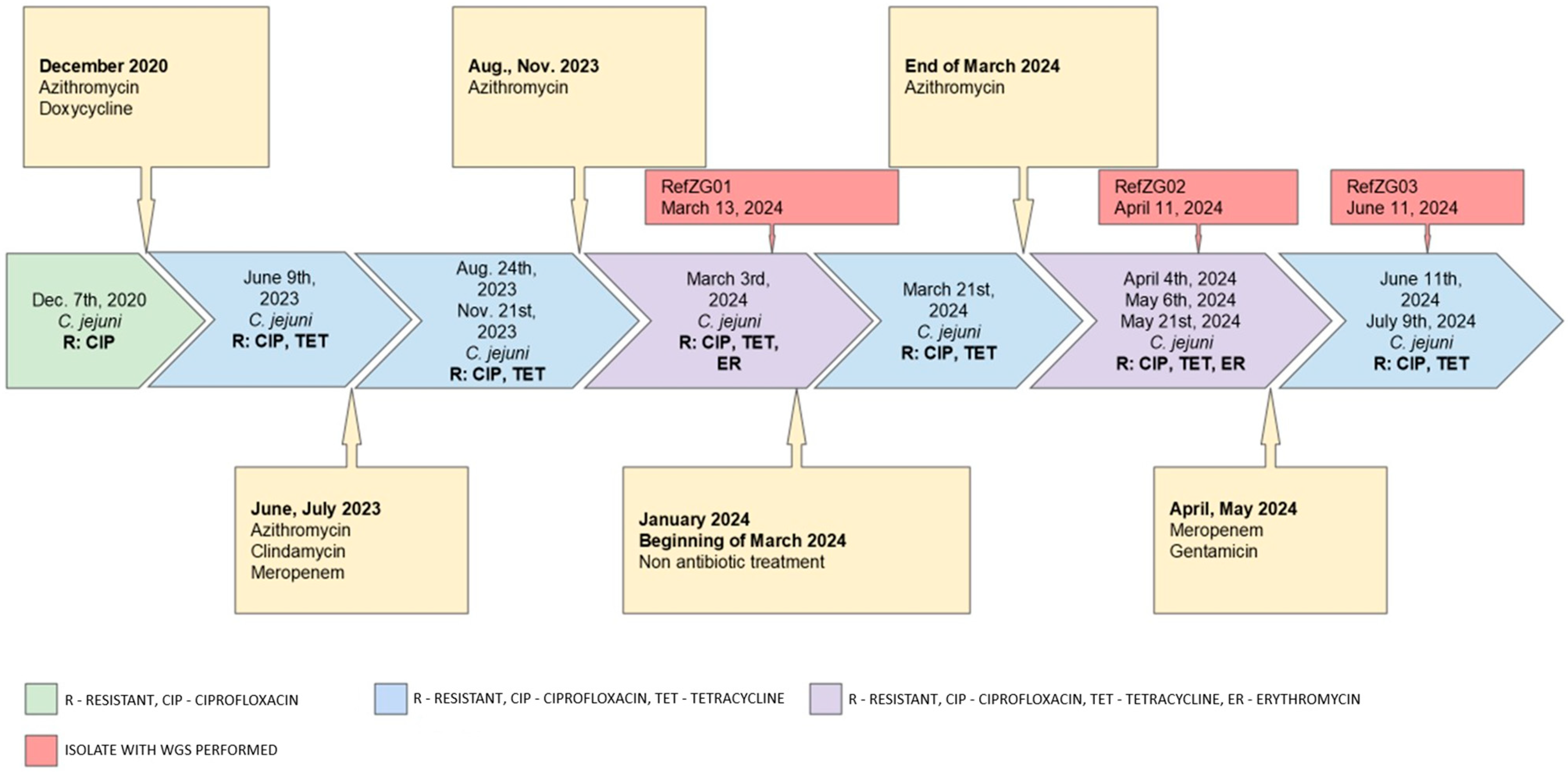
Figure 1. The timeline of isolates with their resistance profile.
Tropicalmed 10 00165 g001
Table 1. Clinical course of the disease.
Date Range SymptomsIsolatesTherapy
2019abdominal cramps, diarrhea, bloating and weight lossC. jejuni negativeoral ciprofloxacin (CIP) (0.5 g twice daily) and subcutaneous IG therapy (80 g/800 mL per month)
01/2020worsening GI symptoms
(diarrhea, abdominal cramps and fever)
Toxin-producing C. difficileoral vancomycin (0.125 g four times daily), metronidazole (0.4 g three times daily), corticosteroids (0.024 g/day), and mesalazine (1 g three times daily), along with an increased Ig dose to 100 g/1000 mL per month
12/2020worsening GI symptomsC. jejunioral azithromycin (0.5 g once daily)
2021–2022intermittent diarrheaC. jejuni negativeno reported treatment
12/2022intermittent diarrheaToxin-producing C. difficileoral vancomycin (0.125 g four times daily)
02/2023diarrhea (25 watery stools/day), fever of 38 °C, ICU admission for treatment of malabsorption, electrolyte imbalance, and subsequent pneumoniaC. jejuni negativeoral vancomycin (0.125 g four times daily), levofloxacin for pneumonia
06/2023diarrhea, feverC. jejuni R: CIP 1, TET 2oral azithromycin (0.5 g once daily) followed by oral clindamycin (0.3 g four times daily). Hospitalization with fever and treated with intravenous (IV) meropenem (1 g three times daily)
08/2023diarrheaC. jejuni R: CIP, TEToral azithromycin (0.5 g once daily)
11/2023diarrheaC. jejuni R: CIP, TEToral azithromycin (0.5 g once daily)
03/2024diarrhea with 3–5 s stools per day, abdominal discomfort, and low-grade feverC. jejuni R: CIP, TET, ER 3
C. jejuni R: CIP, TET
oral azithromycin (0.5 g once daily)
04–05/2024persistent diarrheaC. jejuni R: CIP, TET, ERsupportive therapy only
05/2024fever, 15 diarrheal stools daily, and abdominal crampsC. jejuni R: CIP, TET, ER
C. jejuni R: CIP, TET
IV meropenem (1 g three times daily)- discontinued after 10 days due to leukopenia.
IV gentamicin (0.24 g once daily) for four days, with prophylactic vancomycin.
06–07/2024abdominal pain, spasms, and flatulenceC. jejuni R: CIP, TETSupportive therapy, nutritional supplementation, probiotics
1 CIP—Ciprofloxacin. 2 TET—tetracycline. 3 ER—Erythromycin.
Table 2. The phenotypic results determined by the disk diffusion method (S—susceptible, R—resistant, WT—wild-type).
Antimicrobial Agent (Class)
Isolate/
Ref. Isolate
Erythromycin
(Macrolide)
Ciprofloxacin (Fluoroquinolone)Tetracycline (Tetracycline)Gentamycin (Aminoglycoside)Meropenem (Beta-Lactam)
KC01SRS--
KC02SRS--
KC03SRR--
KC04SRR--
KC05/RefZG01R/RR/RR/R-/WT-
ZG01SRR--
KC06/RefZg02R/RR/RR/R-/WT
ZG02RRRWTWT
ZG03RRRWTWT
ZG04/RefZG03S/SR/RR/R--
ZG05SRR--
ZG06SRR--
ZG07SRR--
Table 3. Genomic similarity metrics: MASH distance, average nucleotide identity and DNA-DNA hybridization.
RefZG01 vs RefZG02RefZG01 vs RefZG03RefZG02 vs RefZG03
MASH 1 distance0.0000240.0000240.000024
ANI 2 (%)99.999.999.9
DDH 3 (%)10099.999.9
1 MASH—MinHash alignment for SHort reads. 2 ANI—average nucleotide identity. 3 DDH—DNA-DNA hybridization.
Table 4. cgMLST genetic distance between isolates.
RefZG01RefZG02RefZG03
RefZG01099
RefZG02903
RefZG03930
Table 5. Genetic resistance profiles and point mutations.
IsolatesAMR GenesPoint Mutations Associated with AMR
RefZG01blaOXA-461blaOXA-61_G-57T
tet(O)gyrA_T86I
cmeA, cmeB, cmeC, cmeR23S_A2074C
RefZG02blaOXA-461blaOXA-61_G-57T
tet(O)
cmeA, cmeB, cmeC, cmeR
gyrA_T86I
RefZG03blaOXA-461blaOXA-61_G-57T
tet(O)
cmeA, cmeB, cmeC, cmeR
gyrA_T86I
Table 6. Virulence-related genes grouped by virulence category.
IsolateAdherence Related GenesInvasion Related GenesMotility Related GenesExotoxin Related GenesImmune Modulation Related Genes
RefZG01cadF, jlpA, pebA, porA, cj1279cciaB, ciaCfla(C, -G, -A), flg(B, -C, -D, -E, -G, -G2, -H, -I, -J, -K, -L, -M, -P, -Q, -R, -S),
flh(A, -B, -F, -G),
fli(A, -D, -E, -F, -G, -H, -I, -K, -L, -M, -N, -P, -Q, -R, -S, -W, -Y)
pse(B, -C, -F)
rpoN
che(A, -V, -W, -Y)
eptC
Cj0371, Cj0883c
cdtA, cdtB, cdtCCj1135
RefZG02cadF, jlpA, pebA, porA, cj1279cciaB, ciaCfla(C, -G, -A), flg(B, -C, -D, -E, -G, -G2, -H, -I, -J, -K, -L, -M, -P, -Q, -R, -S),
flh(A, -B, -F, -G),
fli(A, -D, -E, -F, -G, -H, -I, -K, -L, -M, -N, -P, -Q, -R, -S, -W, -Y)
pse(B, -C, -F)
rpoN
che(A, -V, -W, -Y)
eptC
Cj0371, Cj0883c
cdtA, cdtB, cdtCCj1135
RefZG03cadF, jlpA, pebA, porA, cj1279cciaB, ciaCfla(C, -G, -A), flg(B, -C, -D, -E, -G, -G2, -H, -I, -J, -K, -L, -M, -P, -Q, -R, -S),
flh(A, -B, -F, -G),
fli(A, -D, -E, -F, -G, -H, -I, -K, -L, -M, -N, -P, -Q, -R, -S, -W, -Y)
pse(B, -C, -F)
rpoN
che(A, -V, -W, -Y)
eptC
Cj0371, Cj0883c
cdtA, cdtB, cdtCCj1135
Disclaimer/Publisher’s Note: The statements, opinions and data contained in all publications are solely those of the individual author(s) and contributor(s) and not of MDPI and/or the editor(s). MDPI and/or the editor(s) disclaim responsibility for any injury to people or property resulting from any ideas, methods, instructions or products referred to in the content.