1. Introduction
Understanding how neural activity propagates through space and time, and how that propagation changes across operating conditions, remains a central problem in computational neuroscience. At one end of the modelling spectrum, biophysically detailed descriptions such as the Hodgkin–Huxley formalism provide a mechanistic account of membrane currents and spike generation, but their computational cost grows rapidly when extended to large or spatially distributed networks [
1]. This scaling difficulty arises because each neuron is represented by multiple coupled state variables and typically requires small numerical integration steps, while the network-level simulation must also process large numbers of synaptic events and connectivity interactions at each update. For many questions concerned with network organization, signal spread, and collective dynamics, reduced spiking models provide a more tractable alternative. Leaky integrate-and-fire [
2,
3], adaptive exponential integrate-and-fire [
4], and related reduced formulations [
5,
6] preserve thresholded excitation, refractoriness, and spike timing structure at substantially lower computational cost, making them suitable for repeated simulations, parameter sweeps, and mechanistic comparison across regimes. Spike-timing-dependent plasticity further highlights the importance of timing-dependent interaction rules in neural systems [
7,
8]. In parallel, neuronal oscillation and network neuroscience studies have introduced population-level and graph-based descriptors, including oscillatory coordination, efficiency, modularity, and centrality, as standard summaries of large-scale structural and functional organization [
9,
10,
11]. Together, these modelling traditions motivate computational frameworks that are expressive enough to capture propagation and regime-level structure, while remaining transparent and scalable.
Agent-based modelling (ABM) offers one such framework by representing systems as collections of locally interacting entities whose macroscopic behaviour emerges from explicit rules [
12,
13]. In neural contexts, this perspective is particularly attractive because it supports heterogeneity, spatial embedding, asynchronous interactions, and rule-level interpretability, and NetLogo has become a widely used environment for specifying and reproducing ABMs [
14,
15]. From the classical McCulloch–Pitts neuron [
16] to more recent work showing that delayed pulse-like interactions can generate logic-like behaviour in self-organized neural communities [
17], the broader modelling literature has repeatedly demonstrated that local interaction rules can produce nontrivial collective computation. However, in many neural simulations, whether agent-based or otherwise, signal transmission is still encoded implicitly within neuron updates, synaptic events, or aggregate coupling terms. This makes it difficult to isolate the respective roles of neuronal excitability, transport, and the local medium in shaping observed activity patterns. In particular, when transmission is hidden inside neuron state updates, propagation paths, wavefront structure, and delay accumulation are difficult to inspect directly, even when they are central to the interpretation of the model’s behaviour [
2,
3,
4,
5,
6,
16,
17].
A second challenge concerns the role of the surrounding medium. Experimental and theoretical work on local field potentials [
18], extracellular currents [
19], magnetoencephalography and electrophysiological imaging [
20] and mesoscopic neural activity [
21] indicates that the extracellular environment is not merely a passive background, but is shaped by distributed neural activity and, in turn, can influence population-level coordination and responsiveness. At a more abstract level, normalization and gain control principles suggest that local activity can modulate effective responsiveness through bounded feedback mechanisms without requiring a fully detailed biophysical reconstruction of ionic or extracellular processes [
22]. This motivates a modelling choice that is especially relevant for computationally tractable ABMs: the surrounding medium can be represented as an explicit dynamic field that accumulates recent activity, spreads locally, decays over time, and feeds back on neuronal effectiveness. Such a field is necessarily phenomenological rather than fully biophysical, but it can still provide a transparent mesoscopic bridge between local interactions and system-level modulation, consistent with mesoscopic field descriptions [
21] and normalization-based gain control formulations [
22].
Within this context, we introduce LANA (Local Adaptive Neural Agents), a dual-agent neural agent-based model in which neurons and propagating signals are represented as distinct interacting entities embedded in a dynamic environmental field. The central contribution of LANA is not the generic use of agents itself, but the operational separation of three processes that are often combined within neural simulations: neuronal integration and excitability, spatial transport of activity, and environment-mediated modulation of neuronal responsiveness. In LANA, neuron agents implement thresholded integration and refractory dynamics; signal agents are mobile computational carriers of activity; and the environmental field accumulates recent activity, diffuses locally, decays over time, and feeds back through a bounded divisive normalization rule. Here, “explicit” means that these processes are represented by separate state variables and update rules that can be logged during simulation. By contrast, an implicit formulation would absorb transport or feedback effects into the neuron update or into an aggregate coupling term without representing the transport layer or environmental field as separately updated model components. This decomposition allows first-spike timing, cumulative recruitment, propagation radius, and related spatial recruitment summaries to be computed from explicitly represented model states rather than reconstructed only from aggregate spike-count trajectories. The aim of the present study is therefore not to claim a fully biophysical reconstruction of extracellular neural physics, nor to argue that agent-based modelling is new in neuroscience in a general sense. Rather, the goal is to develop and evaluate a normalized mesoscopic framework for studying how excitability, activity transport, delayed coupling, and environmental feedback jointly shape emergent network behaviour under controlled simulation conditions. To achieve this, this manuscript is structured around four complementary analyses. First, we provide a compact internal verification block for the implemented operators, including delay propagation, environmental decay and diffusion, threshold activation, and refractory enforcement. Second, we compare the full LANA model against a matched neuron-only baseline in order to test whether representing signal transport and environmental feedback as separate model components produces different network-level behaviour under the same network realization and stimulation protocol. Third, we summarize spatial recruitment structure through first-spike maps, cumulative recruitment times, and wavefront speed as a secondary descriptive metric. Fourth, we compare two controlled operating regimes, a resting regime (S1) and a hyperexcitable regime (S2), under fixed network size, fixed stimulation schedule, and matched random seeds and examine robustness through local and factorial sensitivity analyses.
By combining rule-based model specification, compact internal verification, matched-seed baseline comparison, spatial recruitment analysis, and controlled regime testing, this work positions LANA as a neural ABM designed for mechanistic transparency rather than as a calibrated reconstruction of a specific biological circuit. The resulting framework is intended to support controlled analysis of emergent neural activity while remaining computationally tractable for systematic experimentation and reproducible comparison.
2. Contributions and Research Questions
In this manuscript, the term mesoscopic denotes an intermediate modelling level between detailed single-neuron biophysics and purely aggregate population-level descriptions. Individual neurons are represented as discrete agents with thresholded dynamics, but ionic currents, membrane channel kinetics, and detailed extracellular electrodynamics are not resolved. The environmental field should therefore be interpreted as a coarse-grained activity-coupling variable, not as a direct biophysical simulation of the extracellular medium. This definition limits the scope of the claims made below: LANA is used here to study mechanism-level interactions among excitability, transport, delayed coupling, and environmental feedback, rather than to provide a calibrated reconstruction of a specific biological circuit. This study makes four principal contributions.
First, it introduces LANA as an explicitly specified rule-based dual-agent neural agent-based model in which neuronal integration, signal transport, and environmental feedback are represented as distinct but interacting dynamical processes. Within this architecture, neuron agents are responsible for thresholded integration and refractoriness, signal agents carry transport state information through space, synaptic links implement weighted directed coupling with explicit delays, and a dynamic environmental field accumulates and redistributes recent activity while modulating effective neuronal functionality.
Second, this manuscript provides a compact internal verification block for the implemented operators. Rather than relying solely on verbal model description, the study evaluates whether the core mechanisms behave as intended under controlled conditions. These verification tests target delay propagation, environmental decay, environmental diffusion, threshold activation, and refractory enforcement, thereby establishing that the principal update operators are functioning consistently before higher-level behavioural interpretations are made.
Third, this manuscript evaluates the explanatory value of the dual-agent architecture through a matched-seed comparison between the full LANA model and a neuron-only baseline. This comparison is complemented by a dedicated spatial recruitment benchmark based on first-spike timing, cumulative recruitment, propagation radius, and wavefront speed as a secondary descriptive metric. Together, these analyses are intended to determine whether representing transport and environmental feedback as separate model components provides spatial recruitment and delay-related summaries that are not available in the same form when transmission is represented only through neuron-level updates.
Fourth, the paper analyses controlled regime differences between a resting regime (S1) and a hyperexcitable regime (S2) under a fixed network size, a fixed stimulation schedule, and matched random seeds and complements these comparisons with local one-at-a-time and focused factorial sensitivity analyses. This design allows regime-level differences to be interpreted as consequences of controlled parameter variation rather than as artefacts of changing network size, input schedule, or unrelated implementation settings.
These contributions are organized around three research questions.
RQ1. Does representing signal transport and environmental feedback as separate model components generate different network dynamics than a matched neuron-only baseline under the same network realization and stimulation protocol?
RQ2. What spatial recruitment and delay-related summaries can be computed when transmitted activity is represented through signal agents rather than only through neuron-level updates?
RQ3. Are the differences between resting and hyperexcitable regimes robust under controlled parameter perturbations and matched-seed comparisons?
In summary, this paper presents LANA as a normalized mesoscopic neural agent-based model and evaluates it through rule-based model specification, compact internal verification, matched-seed baseline comparison, spatial recruitment benchmarking, and controlled regime analysis. The resulting claims concern comparative mechanism-level behaviour within the proposed normalized framework rather than direct quantitative prediction for a named anatomical preparation or physiological time scale.
3. Materials and Methods
3.1. Model Overview
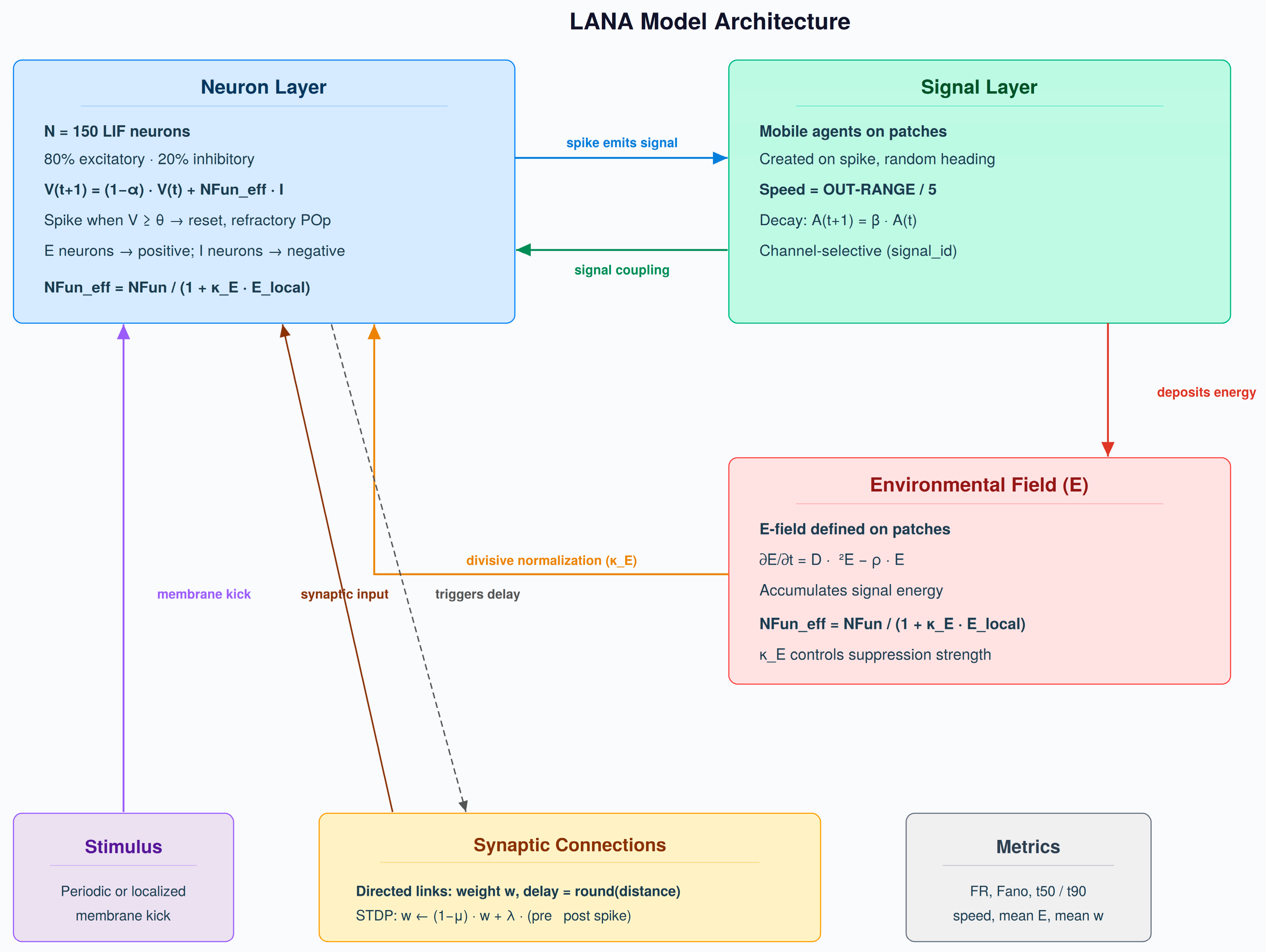
LANA (Local Adaptive Neural Agents) is implemented in NetLogo as a spatially embedded dual-agent neural agent-based model. The framework comprises four interacting components: neuron agents, mobile signal agents, directed synaptic links, and a dynamic environmental field defined on patches. Neuron agents implement discrete leaky integrate-and-fire dynamics with thresholding and refractoriness; signal agents transport activity through space and decay over time; synaptic links provide weighted directed coupling with explicit distance-dependent delays; and the environmental field accumulates recent activity, diffuses locally, decays over time, and feeds back on effective neuronal functionality through bounded divisive normalization. The model is therefore designed to represent neuronal integration, signal transport, and environment-mediated feedback as separately updated model components within a single computational framework. The overall LANA architecture is summarized in
Figure 1.
The purpose of the model is not to reproduce detailed ionic physiology or to reconstruct a specific anatomical neural circuit. In the present experiments, the spatial network is used as a synthetic spatial substrate: neurons are placed in a two-dimensional domain to provide a controlled setting in which excitability, activity transport, delayed coupling, and environment-mediated feedback can be varied and compared under matched random seeds. The model parameters are expressed in normalized computational units and are not calibrated to empirical membrane conductances, extracellular potentials, or a named biological preparation. Consequently, the results should be interpreted as mechanism-level evidence about the behaviour of the proposed rule-based framework, rather than as quantitative physiological predictions for a specific neural tissue or circuit.
3.2. Entities and State Variables
LANA contains four principal components: neurons, signals, synaptic links, and the environmental field. Neuron agents carry membrane potential V, refractory counter R, threshold θ, baseline neuronal functionality NFun, effective functionality NFuneff, neuron type (excitatory or inhibitory), and bookkeeping variables for spike timing and activity. Signal agents are mobile computational carriers emitted by spiking neurons. Each signal agent has a position, direction of motion, amplitude A, decay rule, and signal identity label. They should not be interpreted as separate biological particles or as a detailed representation of extracellular currents; rather, they are transport state variables used to represent propagating activity within the spatial simulation. Directed synaptic links connect neuron pairs through weights w and explicit delays δ. The environmental field E is represented on the patch lattice as a scalar activity trace that diffuses, decays, and contributes to local gain modulation.
This assignment of different dynamical roles to different model components is central to the model design. Neurons encode excitability and thresholded response, signals encode transport and local transmission structure, synapses encode weighted delayed connectivity, and the environmental field encodes mesoscopic accumulation and feedback. As a result, propagation, delay, and environmental modulation are represented through separate model components rather than being absorbed into a single aggregate neuron update.
3.3. Network Generation and Initialization
At setup, neurons are placed on a two-dimensional spatial domain and assigned excitatory or inhibitory identity according to the specified inhibitory fraction. Each neuron is assigned a target out-degree, and directed synaptic links are formed by connecting that neuron to its nearest spatial neighbours until the target out-degree is reached. Each synaptic link is initialized with a common dimensionless normalized coupling weight of w = 1.0, after which synaptic weights evolve during the simulation under the bounded plasticity rule described below. The value w = 1.0 defines the reference coupling strength within the normalized simulation. It is not a calibrated biological conductance, current, voltage, or physical SI quantity. Instead, postsynaptic synaptic input is computed relative to this reference coupling scale, and later values of w are interpreted as dimensionless multiplicative changes relative to the initial reference strength. Transmission delay is set as the rounded Euclidean link length, with a minimum delay of one tick. The default network used in the main experiments contains N = 150 neurons; all matched-seed comparisons preserve the same network realization within each seed pair. Signal identities and local field interactions are initialized consistently with the implemented NetLogo configuration, and the environmental field is initialized at zero. This design does not by itself guarantee reproducibility, but it makes paired comparisons reproducible and interpretable: within each matched seed pair, the compared conditions share the same network realization, initialization logic, and stimulation schedule, so differences between conditions can be attributed to the tested mechanism changes rather than to uncontrolled variation in topology or initial conditions.
3.4. Core Update Rules
For reproducibility, the principal model dynamics are stated as explicit discrete-time update rules applied at each simulation tick.
Environmental field. The environmental field is stored on the patch lattice and updated through diffusion, decay, and local signal deposition:
where ρ corresponds to the NetLogo parameter RHO, D is the diffusion coefficient, and S(x,t) denotes the set of signal agents located at patch x at time t.
Environmental feedback on neurons. The environmental field modulates neuronal functionality through bounded divisive normalization:
where
corresponds to KAPPA-E and Ē
i(t) denotes the local mean environmental activity around neuron i. In the present model, this term is used as a mesoscopic gain control mechanism rather than as a detailed ephaptic or extracellular biophysical law. The feedback is described as explicit because the environmental state E(x,t) is represented, updated, and logged as a separate model component before being mapped back to neuronal functionality. In an implicit formulation, the same qualitative influence could be absorbed into a neuron-level gain parameter or fitted coupling coefficient without representing the environmental field as a separate dynamical state. For neuron i, local environmental activity is computed as the mean field value over patches within radius 1 around the neuron position.
Signal emission, transport, and decay. When a neuron spikes, it emits a single mobile signal agent at its own spatial location. The emitted signal amplitude is determined by the effective neuronal functionality of the spiking neuron and by neuron type: excitatory neurons emit positive-amplitude signals, whereas inhibitory neurons emit negative-amplitude signals. Signal motion is implemented as constant per-tick displacement with a random heading assigned at emission:
where
and u
s(t) is the unit direction of motion. Signal amplitude decays exponentially:
Signals are removed once their amplitude falls below a small cutoff to prevent negligible residual contributions.
Signal coupling is channel-selective: neurons respond only to signal agents carrying the same signal_id label as the receiving neuron. This introduces a discrete interaction channel structure into the local transport layer and prevents indiscriminate coupling among all nearby signals and neurons. Here, β denotes the per-tick signal retention factor, γ denotes the spatial attenuation coefficient in inverse patch units, and u_s(t) denotes the unit direction vector of signal motion.
Signal-to-neuron coupling. The total spatially mediated drive to neuron i is computed by summing all signal contributions within the interaction radius, restricted to signal agents with matching signal identity:
where
, and
. Excitatory and inhibitory influences are carried by the sign of
Delayed synaptic input is implemented as a parallel directed pathway. When a neuron spikes, each outgoing synapse is assigned its predefined eligibility countdown. Once this countdown reaches zero, the synapse delivers input to the postsynaptic neuron. The contribution of synapse is proportional to its weight and to the effective functionality of the presynaptic neuron, with excitatory neurons contributing positively and inhibitory neurons negatively. In this way, synaptic plasticity affects forward dynamics through the delayed synaptic pathway rather than through signal emission amplitude.
For synapse
, delayed synaptic delivery contributes
where
for excitatory presynaptic neurons and
for inhibitory presynaptic neurons, and delivery occurs only when the synaptic eligibility countdown reaches zero.
For compactness, we define the total input to neuron
i as the sum of spatial signal-mediated input and delayed synaptic delivery:
where
denotes the set of presynaptic synapses delivering input to neuron
i at tick
t. Thus, neuron integration combines both local signal transport and delayed synaptic transmission within a single net input term.
Neuron dynamics. Each neuron
i carries membrane potential V
i(t) and refractory counter R
i(t). If the neuron is refractory, it does not integrate new input:
Otherwise, the membrane state follows a discrete leaky integrate-and-fire-like update:
where α denotes the per-tick membrane update fraction and I_i^{tot}(t) denotes the total input to neuron i at tick t. In the NetLogo implementation, α corresponds to ALPHA and Δt denotes one computational update step.
where
denotes the local signal-mediated input and
the delayed synaptic input.
After spiking, the neuron is reset and enters a refractory state of duration POp:
In the default network, synaptic delay is set as the rounded Euclidean link length with a minimum of one tick:
In dedicated chain verification experiments, this default rule is replaced by a fixed delay parameter (FIXED-DELAY) to permit exact operator testing.
Plasticity. Synaptic weights evolve according to a bounded delayed Hebbian-like update:
where η
p controls potentiation, η
d provides mild decay, and
enforces numerical bounds. This rule ensures that synaptic plasticity enters the forward dynamics through the delayed synaptic pathway rather than remaining a purely descriptive variable.
At each simulation tick, the implemented update order is: (i) external stimulation, (ii) environmental field update, including diffusion and local signal deposition, (iii) signal update, (iv) update of effective neuronal functionality, (v) neuron update, (vi) plasticity update, and (vii) logging. This order is part of the model specification. In a continuous-time limit with sufficiently small time steps and commuting operators, update order may have negligible influence. However, LANA is a finite-step, event-based agent model that includes thresholding, reset, refractory counters, signal deposition, and delayed synaptic delivery. These operations do not necessarily commute within a single tick. The update order is therefore reported to support exact reproducibility of the implemented simulation rather than to imply that a different ordering would be mathematically equivalent.
3.5. Relation to Established Model Classes and Modelling Scope
Although LANA is implemented as an agent-based model, its core update rules are related to familiar reduced dynamical motifs. The neuron update has the form of a normalized discrete-time leaky integrate-and-fire-type rule; the environmental field follows a diffusion–decay–source structure analogous to mesoscopic neural field formulations [
21,
23] and classical excitatory–inhibitory population dynamics [
24]; and the signal motion rule represents a discrete transport process [
25]. These correspondences are used to clarify the modelling lineage of LANA, not to present it as a high-accuracy numerical solver for detailed conductance-based or extracellular electrodynamic equations. In particular, the forward-Euler analogy should be understood as a simple discrete-time interpretation of the leaky update, not as a claim that the model resolves stiff biophysical dynamics. LANA is therefore best interpreted as a computationally tractable agent-based instantiation of reduced motifs—leak, thresholding, delayed transmission, transport, diffusion, decay, and bounded gain control—whose separate representation is central to the present framework. To keep excitability, transport, and field feedback assigned to distinct model components, the baseline LANA configuration adopts several simplifying assumptions. Signal agents move deterministically at constant speed once emitted; signal influences combine through linear superposition at the level of the transport input; stochastic variation in the reported experiments arises from randomized network realization and initialization rather than from ongoing additive membrane potential noise; the extracellular medium is represented by a single scalar field rather than by detailed electrodynamics; and environmental feedback on neurons is implemented as bounded gain control rather than as a direct ephaptic biophysical law. These assumptions define the intended validity scope of the model. LANA is designed to study propagation, recruitment, and feedback structure in a normalized mesoscopic ABM setting; it is not intended to predict detailed spike waveforms, ionic currents, membrane channel kinetics, or circuit-specific extracellular potentials.
At the intended level of description, LANA should therefore be understood as an agent-based model operating on a mesoscopic scale, where the goal is not literal biological reconstruction but explicit mechanism-level analysis. The neuron, signal, synaptic, and environmental components are interpreted as coarse-grained computational analogues of biological processes rather than as one-to-one physiological entities. In this sense, the framework is designed to clarify how excitability, transport, delayed coupling, and environment-mediated feedback interact under controlled conditions, not to provide a direct anatomical or biophysical reconstruction of a named neural circuit.
For interpretive clarity, the principal model components can be mapped onto coarse-grained biological analogues as follows: neuron agents correspond to excitable units, signal agents to transport state carriers of propagated activity, directed synaptic links to delayed coupling pathways, and the environmental field to a phenomenological activity-dependent modulatory field. The two-dimensional network used here is likewise intended as a synthetic spatial substrate for controlled mechanism-level comparison rather than as a reconstruction of a named anatomical circuit. These analogies delimit the intended interpretive scope of the framework rather than implying one-to-one physiological equivalence with membrane biophysics, extracellular currents, or circuit-specific anatomy. A compact summary of these intended biological analogues and explicit non-claims is provided in
Table S7 in the Supplementary Material.
Because the model is presented as an explicit computational framework, it is important to state the bounded conditions under which its discrete-time updates remain well posed. In the absence of input, the membrane update is contractive for α ∈ (0,1). Signal amplitudes decay monotonically for β ∈ (0, 1), preventing unbounded signal persistence. With bounded source terms and ρ ∈ [0, 1], the environmental field remains bounded, and synaptic clipping further prevents runaway weight growth. These constraints define the admissible parameter regime within which the model exhibits bounded state evolution. The model combines linear and nonlinear update components. Conditional on fixed source terms, the environmental diffusion–decay update is linear in the field variable, and signal amplitude A decay is linear in signal amplitude. Signal contributions are also summed linearly before entering the neuron update. However, the full model is nonlinear because of threshold crossing, spike generation, reset, refractory enforcement, bounded divisive normalization of neuronal functionality, signal removal at the amplitude cutoff, and bounded synaptic clipping. Consequently, the reported regime differences should be interpreted as outcomes of a hybrid linear–nonlinear discrete dynamical system rather than as consequences of a purely linear propagation process.
Potentials, thresholds, synaptic weights, signal amplitudes, stimulus amplitudes, and field values are expressed in normalized computational units. These normalized scales are used to preserve internal consistency across simulation experiments and to support controlled comparisons, rather than to claim one-to-one correspondence with electrophysiological measurement units. Specifically, V and θ are normalized membrane state quantities, A is a normalized signal amplitude, E is a normalized environmental activity trace, STIM-AMP is a normalized external input amplitude, and w is a dimensionless normalized synaptic coupling coefficient. Spatial positions and distances are expressed in patch units, while delays, refractory periods, and simulation horizons are expressed in ticks. Within these normalized units, α is interpreted as a per-tick membrane update fraction, β as a per-tick signal retention factor, ρ as a per-tick environmental decay parameter, D as a patch lattice diffusion coefficient per update step, γ as a spatial attenuation coefficient in inverse patch units, and as a dimensionless environment-to-neuron feedback strength. In the no-input limit, V* = 0 and E* = 0 constitute trivial fixed points of the membrane and field subsystems.
3.6. NetLogo Implementation
LANA is implemented in NetLogo 7.0.2 (Center for Connected Learning and Computer-Based Modeling, Northwestern University, Evanston, IL, USA) using turtles for neurons and signals, directed links for synapses, and patch-own variables for the environmental field. The main control procedures are setup and step, with additional dedicated procedures for verification experiments, baseline mode, localized stimulation, and regime comparison. Interface parameters correspond directly to the model variables reported in this manuscript, including THRESHOLD, POp, ALPHA, BETA, GAMMA, D, RHO, and KAPPA-E.
To support reproducibility, all main experiments were executed using fixed protocol settings and consistent output logging. Logged quantities include firing rate, active fraction, spike-count variability, environmental field statistics, first-spike timing and spatial recruitment summaries derived from first-spike timing. Additional modes implemented in the codebase but not treated as central analyses in the present manuscript are documented in the
Supplementary Material.
3.7. Experimental Design and Parameterization
All main network experiments were performed under a fixed protocol with N = 150 neurons, REPEAT-K = 10, a simulation horizon of 1000 ticks, and matched random seeds for paired comparisons. One tick corresponds to one normalized computational update step, not to a fixed empirical time unit such as one millisecond. The 1000-tick horizon was therefore used as a standardized observation window for matched model comparisons rather than as a direct physiological duration. To verify that the principal qualitative findings were not an artefact of this window length, we additionally performed a supplementary longer-horizon control at 3000 ticks for the key full-versus-baseline and S1-versus-S2 comparisons (
Table S5). This horizon was selected because the primary recruitment measures in the full model were reached well before the end of the window, whereas conditions that failed to reach the required recruitment threshold were explicitly reported as not reached rather than assigned artificial numerical values. The experimental program comprised seven blocks: E0, compact internal verification; E1, full model versus neuron-only baseline; E2, propagation benchmarking under localized stimulation; E3, resting versus hyperexcitable regime comparison; E4, one-at-a-time sensitivity analysis; E5, focused
factorial screening analysis; and E6, additional robustness checks at
N = 300.
The resting regime (S1) and hyperexcitable regime (S2) differed only in four parameters: spike threshold θ, environment coupling strength
, inhibitory fraction INHIB-FRAC, and environmental decay rate ρ. All other principal parameters were held fixed. This design was chosen to ensure that regime differences could be interpreted as consequences of controlled mechanism changes rather than of varying network size, runtime, or stimulation protocol. In baseline mode, external stimulation is applied directly to the membrane potential of neurons located within the same spatial stimulation radius used in the full model. In the full model, the corresponding stimulus is delivered through an emitted signal agent at the same spatial input location. This design preserves the spatial placement and schedule of stimulation while removing explicit signal-mediated and environmental coupling in the baseline condition. The neuron-only baseline should therefore be interpreted as an operational comparator within the present implementation rather than as a clean single-component ablation. The complete parameter settings for the S1 and S2 regimes are summarized in
Table 1.
3.8. Experimental Blocks
E0 comprised compact internal verification of five operator classes: delayed propagation along a chain, environmental field decay, threshold activation, refractory enforcement, and a chain control test under baseline mode. E1 compared the full model against a matched neuron-only baseline. E2 summarized spatial recruitment under localized stimulation using first-spike timing, cumulative recruitment times (t50, t90), and wavefront speed as a secondary descriptive metric. E3 compared the resting (S1) and hyperexcitable (S2) regimes under matched seeds. E4 performed a one-at-a-time perturbation of eight principal parameters around the nominal operating point. E5 implemented a focused factorial screening design across the regime-defining parameters. E6 repeated the principal baseline and regime comparisons at N = 300 as a supplementary robustness check.
Supplementary calibrated baseline control. To test whether the nominally silent baseline reflected trivial under-stimulation rather than a substantive architectural difference, we performed an additional localized stimulation control in baseline mode. In this supplementary analysis, all baseline conditions were kept unchanged except for STIM-AMP, which was swept across a predefined range to identify the minimal amplitude sufficient to produce stable local stimulation site activation. STIM-AMP denotes the dimensionless normalized amplitude of the externally applied stimulation input in the baseline calibration procedure. In the calibration sweep, STIM-AMP = 1.2 represented the minimal level at which stimulation site activation first emerged, whereas STIM-AMP = 1.3 was used for the calibrated baseline comparison to ensure stable and near-complete local source activation across matched runs. The selected calibrated baseline amplitude was then evaluated under matched seeds and compared descriptively against the full model.
Supplementary component-wise ablation control. To further disentangle the architectural sources of the full-versus-baseline difference, we performed an additional supplementary component-wise ablation analysis. Under the same localized-stimulation protocol and matched-seed logic used in the principal comparison, we evaluated the intact full model together with two intermediate ablation variants: one in which environmental feedback on neuronal functionality was disabled while environmental field dynamics were retained, and one in which direct signal-to-neuron transport input was removed while signal emission, signal dynamics, and environmental deposition were retained. This analysis was intended to identify separable contributions of explicit transport and environment-mediated feedback to distributed recruitment (
Table S8).
3.9. Statistical Analysis
Matched-seed comparisons were analysed primarily with paired Wilcoxon signed-rank tests. Effect sizes for paired comparisons were reported as rank-biserial correlations (rrb), together with Hodges–Lehmann median differences and bootstrap 95% confidence intervals. Cohen’s d was retained as a secondary descriptive effect size measure when informative. For the focused factorial design, main effects and selected interactions were summarized using relative main-effect contributions and standardized contrasts.
Primary network metrics included mean firing rate (FR), active neuron fraction, Fano factor, spike-count coefficient of variation (CV), final mean synaptic weight across all existing synaptic links at the final logged simulation tick of each run (reported as Final mean w) and mean environmental field value. Because all synaptic links are initialized with a shared starting value of 1.0 and then evolve under the bounded plasticity rule, this metric reflects the post-adaptation synaptic state rather than the common initialization value. Propagation metrics were evaluated from first-spike timing and cumulative recruitment structure. For the descriptive visualization of propagantion we also computed a propagation radius r(t). At tick t, r(t) was defined as the maximum Euclidean distance from the stimulation centre among neurons that had emitted at least one spike by tick t. This quantity was used as a descriptive trajectory to complement cumulative recruitment and first-spike timing analyses, but it was not treated as a primary tabulated summary metric. Specifically, t50 and t90 were defined as the first simulation ticks at which 50% and 90%, respectively, of all neurons that eventually activated in each run had emitted their first spike. Wavefront speed was treated as a secondary descriptive propagation metric derived from a run-wise linear fit relating first-spike timing to distance from the stimulation centre. Because this estimate can become highly skewed when early recruitment is clustered or step-like rather than smoothly radial, it was interpreted together with t50, t90, active fraction, and first-spike maps rather than used as the sole summary of propagation. For the neuron-only baseline, propagation-related summaries that did not reach the required recruitment threshold within the 1000-tick simulation horizon were reported as not reached (NR) rather than assigned artificial numeric values.
All summary statistics reported for the main network experiments correspond to the fixed protocol described above. Supplementary robustness analyses at N = 300 were evaluated separately to confirm that the principal qualitative conclusions did not depend trivially on network size.
3.10. Reproducibility and Availability
To clarify the computational scope of the simulations, the
Supplementary Material reports approximate runtime and memory requirements for the principal experimental configurations (
Table S4). These measurements are intended to document computational tractability rather than to define a biological time scale. The NetLogo implementation, fixed experimental protocol, parameter files, seed lists, output tables, and analysis scripts used to generate the reported figures and tables are made publicly available in the project repository and archived with a persistent DOI. This package is intended to support direct reproduction of the principal analyses reported in the manuscript. The repository and archive are also intended to support FAIR data-management and reproducible computational research principles [
26,
27].
4. Results
Unless otherwise stated, all main network experiments (E1–E5) were performed with a fixed network size (N = 150), a fixed stimulation period (REPEAT-K = 10), a simulation horizon of 1000 ticks, and matched random seeds for paired comparisons. Additional network size robustness analyses were carried out separately at N = 300 and are reported in the
Supplementary Material.
4.1. Compact Internal Verification of Implemented Operators (E0)
Before interpreting network-level behaviour, we first verified the correctness of the principal implemented operators under controlled conditions. All five verification tests satisfied their predefined pass criteria (
Table 2), indicating that the core update mechanisms behave consistently with their intended design.
The delayed propagation along a chain verification confirmed exact propagation through the directed synaptic pathway. Across all tested delay values (1–5 ticks), the measured mean inter-neuron first-spike delay matched the configured transmission delay exactly, with mean absolute error equal to zero and propagation speed following the expected inverse delay relationship. The environmental decay verification reproduced the analytical exponential trajectory of the E-field with R2 = 1.000, confirming numerical consistency of the decay operator. The threshold verification showed a sharp silent-to-firing transition near the predicted stimulus amplitude, while the refractory verification confirmed that the minimum inter-spike interval was always consistent with the imposed refractory duration across all tested POp values.
Finally, the chain control test demonstrated that enabling baseline mode does not alter the core synaptic delay mechanism itself. Under both full-model and baseline settings, chain propagation remained exact when tested in isolation, indicating that later differences between the full model and the neuron-only baseline are attributable to the removal of signal-mediated and environmental coupling rather than to unintended modification of the basic delayed synaptic pathway.
4.2. Full Model Versus Neuron-Only Baseline (E1)
We next compared the full LANA model against a matched neuron-only baseline under identical network realizations, stimulation schedules, and random seeds. In the baseline configuration, mobile signal agents, environmental field updates, and environmental feedback on neurons were disabled, while the synaptic graph, delays, neuronal update rules, and external stimulation protocol were preserved.
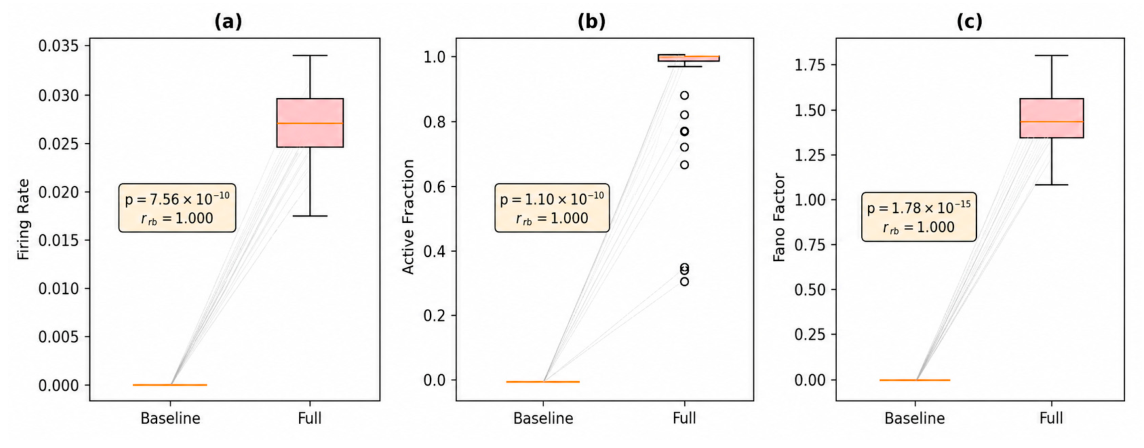
This comparison revealed a strong divergence between the two configurations (
Table 3,
Figure 2). In the neuron-only baseline, the network did not sustain distributed activity within the 1000-tick observation window: firing rate remained at zero across all matched runs, active neuron fraction remained zero, and no environmental activity was generated. In contrast, the full model produced sustained firing with a median firing rate of 0.027 and near-complete neuronal recruitment (median active fraction = 1.000). Variability-based metrics were also clearly separated, with the full model exhibiting a non-zero Fano factor and spike-count CV, whereas the baseline remained silent. All paired comparisons were highly significant and showed maximal or near-maximal rank-biserial effect sizes. Under the bounded plasticity rule, Final mean w also relaxed far below the common initialization value, remaining effectively zero in the silent baseline but non-zero in the full model.
These results indicate that, under the same network realization and stimulation protocol, the neuron-only configuration is insufficient to sustain or spread activity in the manner observed in the full model. In LANA, explicit signal transport and environmental feedback are therefore not cosmetic extensions of neuron dynamics but key components governing whether activity can recruit the network as a distributed process.
To test whether the nominally silent baseline merely reflected insufficient local stimulation, we additionally performed a supplementary calibrated baseline control under localized stimulation (
Tables S2 and S3). Increasing the baseline stimulus amplitude was sufficient to produce near-complete stimulation site activation, yet distributed recruitment remained minimal relative to the full model. This supplementary result confirms that the central difference from the full LANA architecture lies in propagation and network-wide recruitment rather than in threshold crossing at the stimulation site alone. The same qualitative separation between the full model and the baseline was preserved in the supplementary 3000-tick control (
Table S5), indicating that the absence of distributed recruitment in the baseline is not a consequence of the 1000-tick endpoint.
The supplementary component-wise ablation control further clarified that the full-versus-baseline contrast does not arise from a single undifferentiated architectural change (
Table S8). Relative to the intact full model, removal of environmental feedback increased firing activity (FR median 0.0880 vs. 0.0271), increased environmental accumulation (Mean E median 13.500 vs. 1.959), and accelerated cumulative recruitment (t50/t90 = 13/22 vs. 17/32), indicating that the environmental field acts primarily as a suppressive gain control mechanism under the present parameterization. By contrast, removal of direct signal-to-neuron transport input abolished distributed recruitment entirely, leaving firing rate and active fraction at zero and t50/t90 unreached, closely resembling the neuron-only baseline. Taken together, these ablation results indicate that explicit transport and environment-mediated feedback make separable, non-redundant contributions to distributed recruitment in LANA.
4.3. Spatial Recruitment and Delay Structure (E2)
To move beyond aggregate firing summaries, we summarized spatial recruitment under localized stimulation using first-spike timing and cumulative recruitment measures. For each run, we recorded the first-spike time of each activated neuron, derived cumulative recruitment times (t50 and t90), and estimated wavefront speed from the relationship between first-spike timing and distance from the stimulation centre.
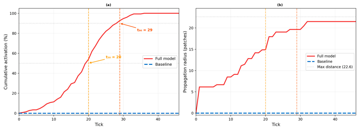
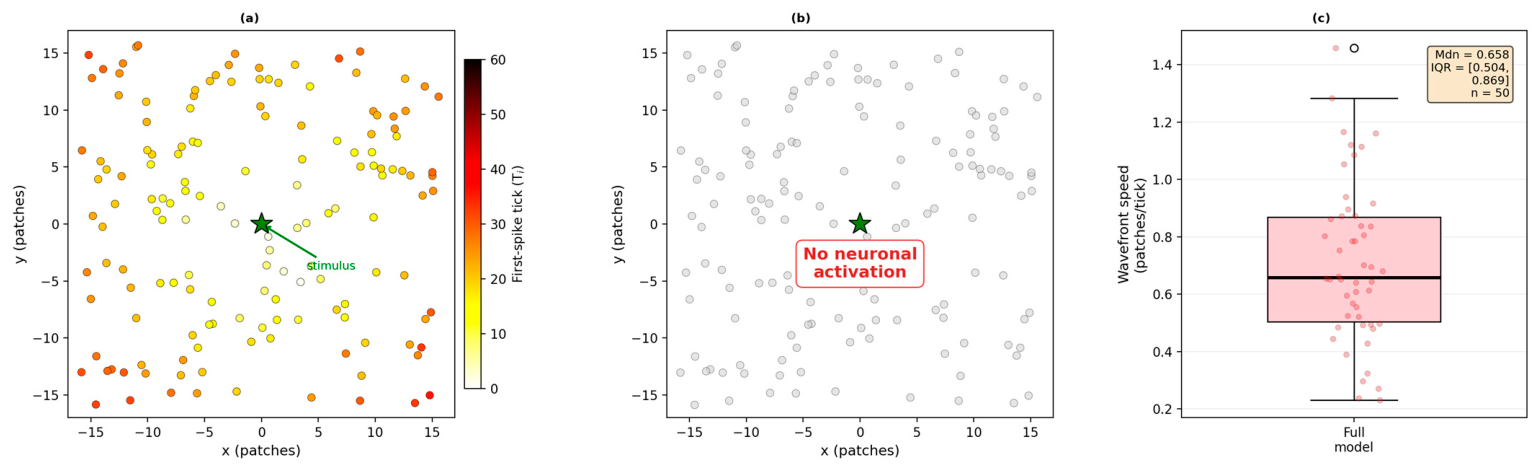
In the full model, localized stimulation produced clear outward recruitment across the network (
Table 4,
Figure 3 and
Figure 4). The median time for 50% cumulative recruitment was 17.0 ticks, and 90% cumulative recruitment was reached by 31.5 ticks. The wavefront speed distribution was highly skewed, with a median that rounded to 0.0 patches per tick and an upper-quartile value of approximately 10 patches per tick. We therefore interpret wavefront speed as a secondary descriptive indicator and rely primarily on cumulative recruitment times, active fraction, and first-spike maps in comparing propagation across conditions. First-spike maps showed that activity spread outward from the stimulated region in an organized manner, allowing spatial recruitment structure to be summarized from first-spike timing rather than only from aggregate spike-count trajectories.
Figure 3 complements these summary statistics by showing both cumulative recruitment and the time-varying propagation radius r(t) for a representative matched seed.
In the baseline configuration, the same stimulation protocol did not produce distributed propagation. Cumulative recruitment did not reach the 50% or 90% thresholds within the 1000-tick simulation horizon, and wavefront speed was therefore not defined in the same sense as in the full model. This contrast is central to the interpretability advantage claimed here: when signal transport is represented as a separate transport layer, first-spike maps, cumulative recruitment times, and propagation radius summaries can be computed from explicitly represented spatial states. In the baseline configuration used here, the same summaries either do not reach the required recruitment thresholds or are not meaningful in the same way because distributed transport is not represented as a separate model component.
4.4. Resting Versus Hyperexcitable Regime Comparison (E3)
We then compared two controlled operating regimes under a fixed network size, a fixed stimulation schedule, and matched random seeds: a resting regime (S1) and a hyperexcitable regime (S2). The two regimes differed only in four parameters: spike threshold (θ), environment coupling strength (), inhibitory fraction (INHIB-FRAC), and environmental decay rate (ρ). This design ensures that regime differences are not confounded by changes in network size, runtime, or stimulation protocol.
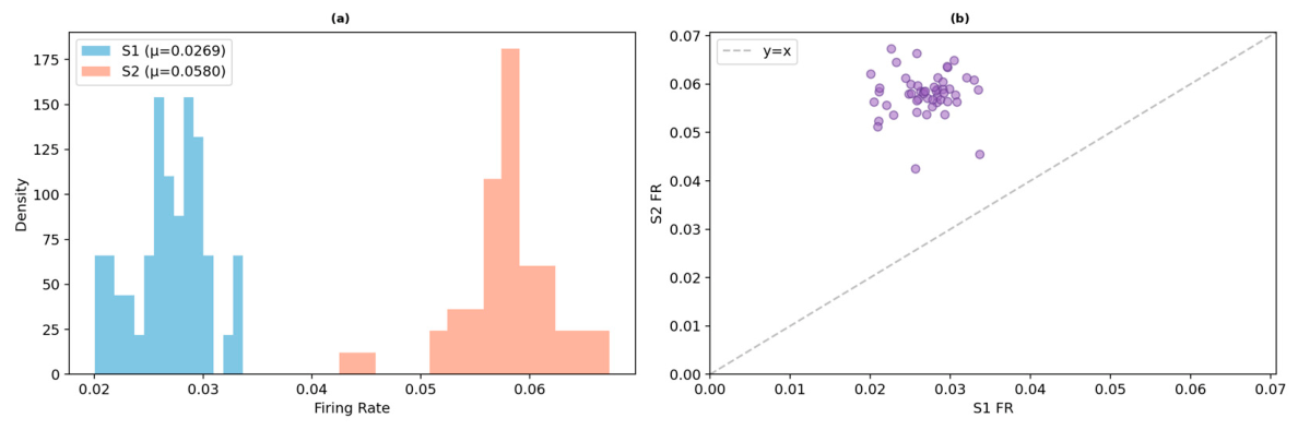
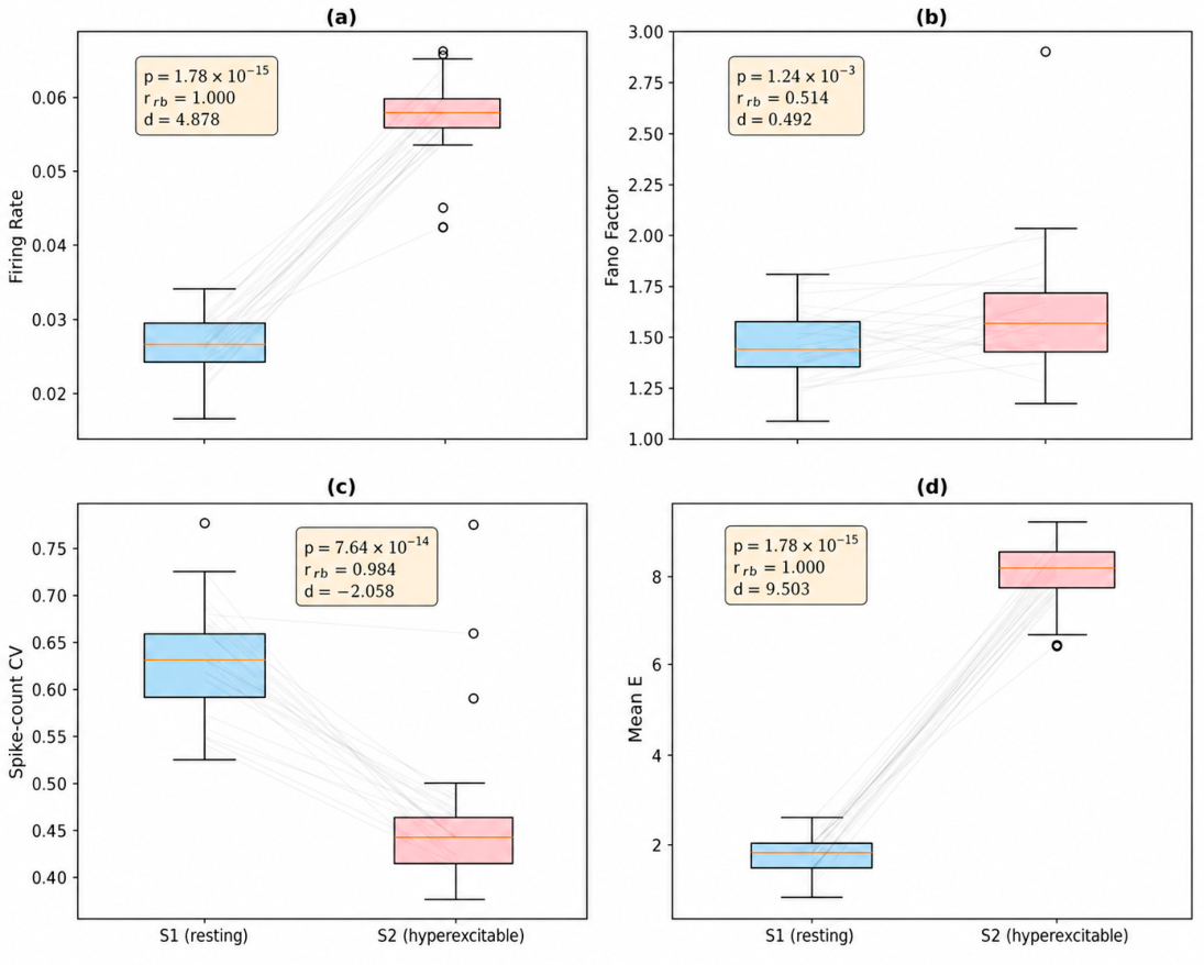
Across all matched comparisons, S2 produced substantially stronger and earlier network activation than S1 (
Table 5,
Figure 5 and
Figure 6). Median firing rate approximately doubled from 0.027 in S1 to 0.058 in S2, while the mean environmental field increased from 1.947 to 8.375, indicating much stronger accumulation of activity in the hyperexcitable setting. S2 also exhibited a higher Final mean w than S1, consistent with stronger sustained recruitment under the same bounded plasticity rule. Propagation was also faster in S2: the median cumulative recruitment time decreased from 16.0 to 9.0 ticks for t50 and from 28.5 to 13.5 ticks for t90. These results are mechanistically consistent with the intended interpretation of S2 as a regime with lower effective resistance to activation and reduced feedback suppression.
The variability structure also changed across regimes. Spike-count CV was lower in S2 than in S1, indicating that the hyperexcitable regime produced earlier and more regular recruitment once activation was established. Together with the shorter cumulative recruitment times and stronger environmental accumulation observed in S2, this result suggests that the most robust regime-level differences are expressed through faster recruitment and more sustained network-wide activation rather than through any single early-time summary alone.
Taken together, these findings show that the S1/S2 contrast is not limited to a vertical shift in spike-count trajectories. The hyperexcitable regime alters multiple aspects of the dynamics simultaneously: network recruitment begins earlier, spreads faster, reaches a larger active fraction sooner, and is accompanied by substantially stronger environmental accumulation. This regime ordering was also retained in the supplementary 3000-tick control, where S2 continued to exhibit stronger activity and earlier recruitment than S1 (
Table S5).
The dual-agent architecture preserves the identity and sign of each neuron’s contribution: excitatory neurons emit positive-amplitude signals and provide positive synaptic drive, whereas inhibitory neurons emit negative-amplitude signals and suppress postsynaptic targets. In the present S1/S2 design, the inhibitory fraction is itself one of the controlled regime-defining parameters (
Table 1). Regime differences should therefore be interpreted as arising from a specified combination of threshold, environment-mediated coupling, inhibition, and environmental decay, rather than from uncontrolled variation in network composition.
4.5. Robustness to Parameter Perturbations (E4–E5)
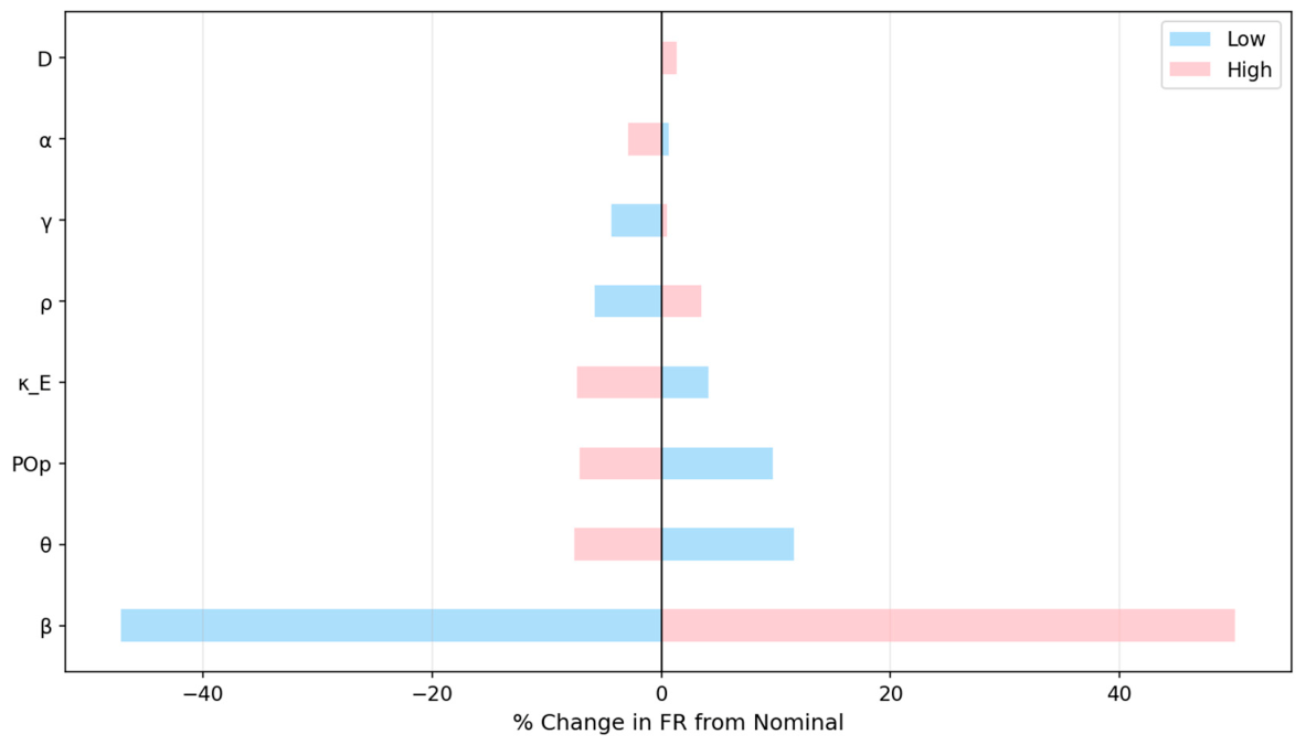
To assess the local robustness of the nominal operating point and to identify the parameters most responsible for regime variation, we performed both a one-at-a-time (OAT) perturbation analysis (E4) and a focused factorial screening analysis (E5). The OAT analysis revealed that signal decay (β) was the most perturbation-sensitive parameter in the tested neighbourhood of the nominal configuration (
Table 6,
Figure 7). A bounded perturbation of β between 0.90 and 0.99 produced approximately −47.0% to +55.7% change in firing rate, indicating that transport persistence strongly governs how far and how effectively activity can spread before dissipating. Other parameters showed moderate but still interpretable sensitivity. Threshold (θ) and refractory period (POp) produced changes on the order of 7–12%, while environment coupling (
) also showed a clear influence, with reduced coupling increasing firing rate and stronger coupling suppressing it. By contrast, D, γ, and α had relatively small effects in the local neighbourhood tested here.
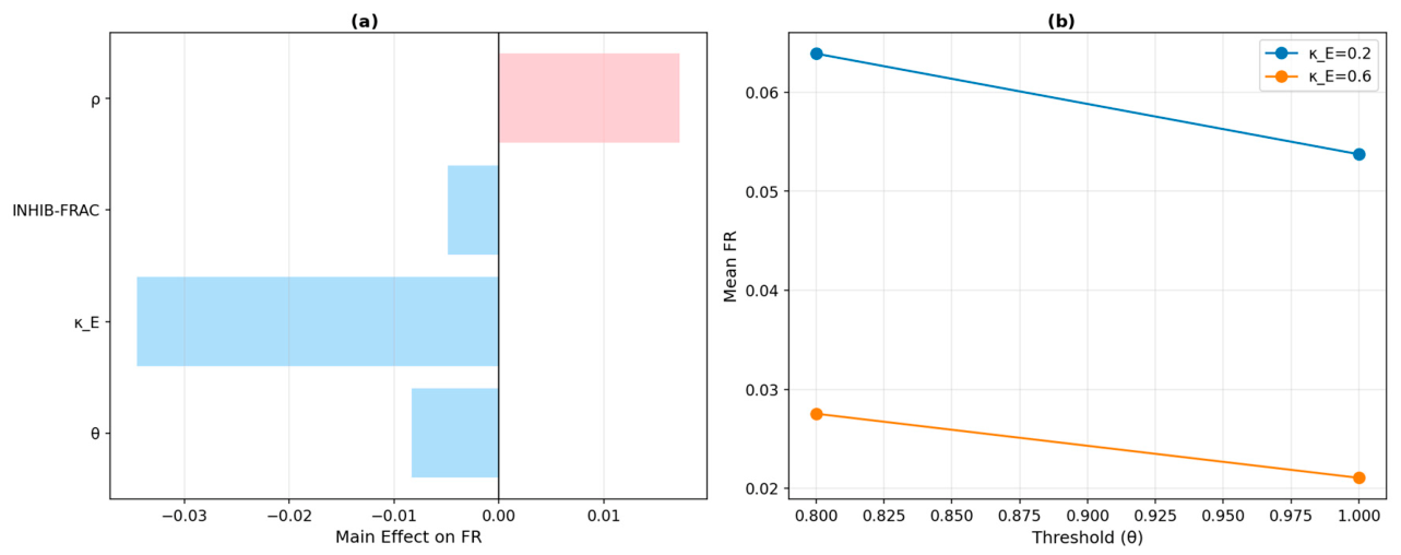
The factorial screening analysis provided a broader view of the regime-defining parameter set than the one-at-a-time perturbations. Across the
design,
emerged as the dominant control factor, accounting for 52.4% of the total absolute main effect magnitude across the tested factorial parameter set, followed by ρ (26.4%), θ (13.4%), and INHIB-FRAC (7.9%) (
Table 7,
Figure 8a). This relative contribution is a normalized summary of absolute main effect magnitudes, not an ANOVA variance decomposition. Interaction analysis further revealed a strong
interaction (
Figure 8b): lowering both threshold and environmental suppression generated a larger increase in firing rate than would be expected from either factor considered alone. This result supports the interpretation that the hyperexcitable regime is not driven by a single isolated parameter but by the coupled action of reduced activation threshold and weakened environment-mediated gain control. The
design was used as a focused screening design rather than as a complete response surface exploration. Testing three or four levels per factor would be useful for mapping nonlinear response surfaces in more detail, but that broader optimization analysis is beyond the scope of the present manuscript. The present design was chosen to test whether the S1/S2 regime differences were structured and attributable to the four regime-defining parameters under matched simulation conditions. To complement this two-level screening design, we also evaluated a supplementary three-level grid over
and
, which supported the same directional interpretation of the threshold–environment coupling relationship (
Table S6). Overall, the combined OAT and factorial analyses indicate that the main regime differences reported above are robust and mechanistically structured rather than arbitrary consequences of a single ad hoc parameter choice.
4.6. Additional Robustness to Network Size (E6)
To assess whether the main findings depended trivially on network size, we repeated the principal baseline and regime comparisons at
N = 300. The qualitative conclusions were preserved (
Table S1). The neuron-only baseline remained silent, the full model continued to sustain distributed activity, and the hyperexcitable regime again produced higher firing rates than the resting regime. Although absolute values changed modestly with network size, the direction and approximate magnitude of the principal effects remained stable, supporting the robustness of the reported findings beyond the nominal
N = 150 configuration.
5. Discussion
This study advances LANA as a normalized mesoscopic agent-based framework in which neuronal integration, signal transport, synaptic delay, and environmental feedback are represented as distinct but interacting model components. The central motivation is methodological: rather than absorbing transport and environmental effects into a single neuron state update, LANA represents them as separate state variables and update rules. This allows recruitment timing, delay structure, propagation radius, and environmental accumulation to be logged and summarized from the corresponding model components. The resulting quantities should not be interpreted as direct physiological measurements, but as mechanism-level summaries within a controlled computational framework. The compact internal verification block is important in this regard. The present manuscript does not rely solely on verbal model description; it also verifies that the core implemented operators behave as intended under controlled conditions. Exact delayed propagation along a chain, analytical environmental decay, threshold bifurcation, and correct refractory enforcement together establish that the principal update mechanisms are numerically and algorithmically consistent. This does not eliminate all modelling limitations, but it substantially strengthens the interpretability of the subsequent baseline, propagation, and regime analyses.
The matched-seed comparison with a neuron-only baseline further clarifies the role of the explicit coupling architecture. Under the same network realization and nominal stimulation protocol, the baseline configuration did not sustain distributed activity, whereas the full model did. This result should not be interpreted as a universal claim about all neuron-only models, but as a controlled statement about the present implementation: within LANA, explicit signal transport and environmental feedback are not peripheral additions, but major determinants of whether activity recruits the network as a distributed process. The spatial recruitment benchmark sharpens this interpretation by showing that first-spike maps, cumulative recruitment times, and propagation radius summaries can be computed from explicitly represented spatial states in the full-model configuration. This mechanism is broadly consistent with the canonical computation of divisive normalization described by Carandini and Heeger [
22], here instantiated through a spatially grounded environmental field rather than an abstract gain control circuit. The neuron-only baseline should therefore be interpreted as an operational comparator within the present implementation, not as an exhaustive representative of all reduced neural models without explicit transport or environmental state variables.
At the same time, the baseline and full configurations differ not only in signal propagation and environmental feedback, but also in the mechanism of external stimulation. In full mode, the periodic stimulus creates a signal agent that propagates spatially and may influence neurons beyond the immediate source site. In baseline mode, stimulus energy is injected directly into the membrane potential of neurons located within the stimulation radius, without an explicit transport layer. The silence of the nominal baseline therefore reflects the joint absence of signal-mediated input spreading and environmental amplification, rather than the removal of a single coupling component in isolation. Nonetheless, the central result remains unchanged: synaptic connectivity alone, even under direct local stimulation, is insufficient to generate the sustained distributed activity observed in the full LANA architecture under the tested parameterization. The supplementary component-wise ablation control further sharpens this interpretation by showing that explicit transport and environment-mediated feedback contribute separably to distributed recruitment within the full architecture (
Table S8).
Supplementary calibrated baseline control strengthens this interpretation further. When the baseline stimulus amplitude was increased sufficiently to produce near-complete stimulation site activation, distributed recruitment nevertheless remained minimal relative to the full model. This shows that the principal difference between the two configurations is not simply whether neurons at the stimulation site can cross thresholds, but whether the architecture supports propagation and sustained network-wide recruitment once activity is initiated. In this sense, the added signal layer and environmental feedback are not merely amplifying a local effect; they restructure the way activity spreads through the network.
The S1/S2 regime comparison shows that the framework captures structured operating differences under controlled parameter variation. Because the two regimes differ only in threshold, environmental coupling, inhibitory fraction, and environmental decay, the observed differences can be interpreted as consequences of the specified regime-defining parameters rather than as consequences of changes in network size, stimulation schedule, or unrelated implementation settings. The hyperexcitable regime activates earlier, recruits the network more rapidly, and produces stronger environmental accumulation. Further sensitivity analysis shows that these regime differences are not arbitrary: they are strongly governed by the interaction between threshold and environment-mediated suppression, while signal decay remains the most locally perturbation-sensitive transport parameter.
These findings position LANA as a phenomenological, normalized, mesoscopic framework rather than as a calibrated biological circuit model. The two-dimensional spatial network used here is a synthetic testbed designed to isolate mechanism-level effects under controlled conditions. It should not be interpreted as a reconstruction of a specific cortical column, hippocampal microcircuit, or other named anatomical preparation. The environmental field is likewise a coarse-grained activity-coupling variable, not a detailed model of extracellular electrodynamics or ephaptic coupling. The value of the framework lies in making excitability, transport state dynamics, delayed coupling, and environment-mediated gain control separable within a computationally tractable ABM setting. Importantly, the principal qualitative conclusions were unchanged in the supplementary 3000-tick control, arguing against the interpretation that the reported effects are artefacts of an arbitrarily short observation window (
Table S5). Circuit-specific calibration, anatomically constrained connectivity, and empirical comparison with electrophysiological recordings are therefore important future extensions rather than claims made by the present study. Several limitations should be acknowledged. The environmental medium is represented as a single scalar activity field rather than as a detailed extracellular biophysical process. The baseline comparison is operational and implementation-specific rather than a claim about all neural agent models without explicit signals. The present study is focused on propagation, recruitment, and regime structure rather than on direct empirical fitting, task-level cognition, or detailed oscillatory classification. In addition, although the model includes plasticity and explicit delay structure, the present manuscript emphasizes regime analysis and spatial recruitment summaries rather than long-horizon learning behaviour. These limitations also define the most relevant directions for future work. The current framework can be extended toward anatomically constrained spatial topologies, circuit-specific connectivity, empirically calibrated parameter ranges, longer-horizon simulations, stochastic membrane or transport perturbations, stronger synchronization and oscillation analyses, and comparison with mesoscopic experimental recordings. A particularly important next step would be to instantiate the same modelling architecture on a named biological circuit or experimentally constrained network, where connectivity, cell-type composition, spatial embedding, and parameter ranges can be linked to empirical data. Even in its present form, however, LANA demonstrates how separating neuronal processing, transport state dynamics, delayed coupling, and environmental feedback into interacting model components can support controlled mechanism-level analysis of emergent neural dynamics.





 ,
,