Article

Isolation and Characterization of a Novel Bacteriophage KpCCP1, Targeting Multidrug-Resistant (MDR) Klebsiella Strains

by
Boris Parra
1,2,3,*,
Maximiliano Matus-Köhler
1,3,
Fabiola Cerda-Leal
4,
Elkin Y. Suárez-Villota
2,
Matias I. Hepp
5,
Andrés Opazo-Capurro
1,3 and
Gerardo González-Rocha
1,3,*
1
Laboratorio de Investigación en Agentes Antibacterianos (LIAA), Departamento de Microbiología, Facultad de Ciencias Biológicas, Universidad de Concepción, Concepción 4070409, Chile
2
Facultad de Medicina Veterinaria y Agronomía, Instituto de Ciencias Naturales, Universidad de Las Américas, Av. Jorge Alessandri 1160, Campus El Boldal, Concepción 4100000, Chile
3
Grupo de Estudio en Resistencia Antimicrobiana (GRAM), Universidad de Concepción, Concepción 4070409, Chile
4
Departamento de Ingeniería en Alimentos, Universidad Del Bío-Bío, Chillán 3800708, Chile
5
Centro de Vigilancia de Aguas Residuales, Centinela Biobío, Facultad de Medicina, Universidad Católica de la Santísima Concepción, Concepción 4090541, Chile
*
Authors to whom correspondence should be addressed.
Sci 2025, 7(4), 157; https://doi.org/10.3390/sci7040157 (registering DOI)
Submission received: 29 August 2025 / Revised: 17 October 2025 / Accepted: 27 October 2025 / Published: 2 November 2025

Abstract

Antimicrobial resistance (AMR) is a major public health threat that urgently requires alternative strategies to address this challenge. Klebsiella spp. are among the most important clinical pathogens and a leading cause of opportunistic nosocomial infections, with high morbidity and mortality associated with strains resistant to last-line antimicrobials such as carbapenems. Bacteriophages are considered a promising therapeutic option for treating infections caused by Klebsiella strains. Hence, the aim of this work was to isolate and characterize a phage capable of infecting carbapenem-resistant Klebsiella strains. The phage KpCCP1 was isolated using the double layer agar method (DLA), from the influent of a wastewater treatment plant, which was characterized through phenotypic and genomic analyses. Morphological characteristics were determined using TEM, and its host range was evaluated against a collection of 133 Klebsiella strains. Its whole genome was sequenced using the Illumina NovaSeq X Plus platform and then assembled and annotated. VICTOR was used for phylogenetic analysis of the isolated phage, and VIRIDIC to compare its genome with those of its closest relatives. KpCCP1 is a tailed dsDNA lytic phage with a genome size of 177,276 bp and a GC content of 41.82%. It encodes 292 ORFs, including two tRNA genes. Phage KpCCP1 is a member of the Slopekvirus genus in the Straboviridae family. It is capable of infecting 22 carbapenem-resistant Klebsiella strains, including K. pneumoniae and K. michiganensis. Notably, it does not contain virulence or antibiotic resistance genes and harbors putative anti-CRISPR genes, therefore representing a promising candidate for phage therapy against clinically critical Klebsiella strains.

1. Introduction

Antimicrobial resistance (AMR) in clinical settings is widely recognized as a consequence of natural selection [1]. The relevance of AMR has increased in the last decades due to the rise of multidrug-resistant (MDR) pathogens and the lack of innovation in developing new treatments [2,3]. In 2019, an estimated 4.95 million deaths worldwide were associated with resistant bacteria [4]. In May 2024, the WHO updated its list of priority bacterial pathogens, in which carbapenem-resistant Klebsiella pneumoniae ranks at the top. This species represents the greatest public health threat because of limited therapeutic options, high disease burden, and increasing acquisition and dissemination of AMR [5]. K. pneumoniae is primarily associated with nosocomial infections, including respiratory tract, urinary tract, and bloodstream infections, particularly affecting immunocompromised patients; it is also a major cause of neonatal sepsis [6,7,8]. The acquisition of resistance by K. pneumoniae, especially to carbapenems, severely restricts therapeutic alternatives, increases healthcare costs and exacerbates morbidity [9,10,11]. Infections caused by carbapenem-resistant Enterobacterales have been demonstrated to be associated with higher mortality rates compared with infections caused by carbapenem-susceptible strains [12,13,14,15].
In Chile, carbapenemase-producing K. pneumoniae has increased markedly since the introduction of KPC-producing strains in 2012 [16] and NDM-producing strains in 2014 [17]. Data from the Chilean Institute of Public Health (ISP) report an increase in carbapenem-resistant K. pneumoniae, with 46.3%, 25.7% and 7.1% of isolates resistant to ertapenem, meropenem, and imipenem in 2020 [18]. More recently, significant diversity of lineages and mobile genetic platforms facilitating the dissemination of carbapenemase-encoding genes has been documented in Chilean K. pneumoniae strains [19,20,21,22]. For instance, the hypervirulent phenotype, typically associated with aggressive community-acquired infections and more prevalent in Southeast Asian countries [23], has been recently reported in Chile [24].
The necessity for alternative strategies to control MDR pathogens is becoming increasingly apparent in order to avoid dramatic repercussions [25,26], including bacteriophages, nanoparticles, and natural compounds. Bacteriophages, viruses that infect and kill bacteria, have been proposed in a strategy known as “phage therapy” [27]. The first independent descriptions were made in the early 20th century by Frederick Twort and Félix d’Hérelle, who coined the term bacteriophage [28]. During the 1920s–1930s, phages were explored as therapeutic agents to treat bacterial infections. However, the discovery and mass production of antibiotics shifted medical focus away from phages, leading to their decline in Western medicine [29].
Phages are characterized by their high specificity, which enables them to infect only the target bacteria without causing harm to the microbiota [30]. Moreover, phages often penetrate and destroy biofilms more effectively than antibiotics [31]. Together, these advantages result in fewer side effects, effective treatment of AMR bacteria, and minimal environmental impact. Additionally, phage therapy can be personalized, tailoring treatments to individual infections, thereby enhancing therapeutic effectiveness [32].
One of the main bacterial defence systems against the invasion of mobile genetic elements (MGE, such as phages and plasmids), is the CRISPR-Cas system [33,34,35]. In K. pneumoniae, diverse CRISPR-Cas systems have been identified [36,37,38], and an inverse association between the presence of the CRISPR-Cas systems and AMR has been reported (antibiotic resistance is higher in the absence of the CRISPR-Cas system) [39]. On the other hand, to overcome the bacterial protection afforded by CRISPR-Cas systems, MGEs have evolved Anti-CRISPR (Acr) systems [40]. In Klebsiella MGEs, Acrs have mainly been described in plasmids [41,42,43], although several Klebsiella phages have also been identified.
Hence, in this study, we isolated a novel Klebsiella phage and characterized it in terms of host range, morphology, and genomic features. We searched for deleterious genes, such as those associated with antibiotic resistance or virulence factors and putative Acr genes. Phage KpCCP1 is a Slopekvirus with broad-host range, infecting MDR K. pneumoniae and K. michiganensis. We demonstrated that KpCCP1 does not carry deleterious genes but harbors putative Acr genes that deserve further study.

2. Material and Methods

2.1. Bacterial Strains

The MDR K. pneumoniae strain UCO-545 was used for isolation of phage KpCCP1. This strain was isolated from a urine sample in December 2020. In addition, we used a collection of 131 K. pneumoniae and 2 K. michiganensis strains, isolated between 2012 and 2024, with no direct involvement of patients in the study. These strains were non-susceptible to carbapenems due to carbapenemase production (KPC, NDM and VIM). All strains are stored in the biorepository of the Laboratorio de Investigación en Agentes Antibacterianos (LIAA), Facultad de Ciencias Biológicas, Universidad de Concepción, Concepción, Chile. All bacterial strains were preserved in Luria–Bertani (LB) broth containing 20% glycerol and stored at −80 °C, and were routinely grown in LB media at 37 °C.
Moreover, we included the reference strains Escherichia coli ATCC 25922, Salmonella enterica subsp. enterica ATCC 25928 and Pseudomonas aeruginosa ATCC 27853.

2.2. Sample Collection and Processing

We searched for phages in wastewater because it has been reported as one of the most prominent sources for isolating phages against Klebsiella spp. [44,45,46,47]. Samples from the influent of a wastewater treatment plant (WWTP) in Concepción, Chile [48], were collected in January 2024. Each sample consisted of 200 mL of 24 h composite, collected in sterile 50 mL tubes and stored at 4 °C, to be processed within 2 days. Samples were centrifuged for 15 min at 8000× g and 4 °C, and the supernatant was filtered (0.22 µm).

2.3. Phage Isolation and Propagation

Phages were isolated by DLA [49] using 10 Klebsiella spp. strains. Aliquots of 100 µL of each overnight culture were mixed with 100 μL of filtered samples and 3 mL of molten (50 °C) soft LB agar (0.5%) supplemented with CaCl2 (final concentration 5 mM), and incubated at 37 °C. Single plaques were picked and suspended in 500 µL SM buffer (100 mM NaCl, 8 mM MgSO4, 50 mM Tris-HCl, pH 7.5). The isolate was then purified three times by DLA.
For the propagation of the phage, it was inoculated into 15 mL of LB broth containing strain UCO-545 at multiplicity of infection (MOI) 0.01, and incubated overnight at 37 °C with shaking at 120 rpm. Then, the suspension was centrifuged, filtered (0.22 µm) and transferred to Amicon tubes (100 kDa) for centrifugation at 3000× g for 20 min at 4 °C. The titer was determined by DLA and expressed as plaque-forming units per milliliter (PFU mL−1).
Precipitation with polyethylene glycol 8000 (PEG) was also performed. Briefly, a phage suspension and bacterial culture were mixed, and incubated overnight at 37 °C, then filtered (0.22 µm) and precipitated with PEG at 10% and 1 M NaCl. The mixtures were then incubated at 4 °C overnight and centrifuged 1 h at 16,000× g. Phage pellet was resuspended in 1 mL of SM buffer and the titer was determined by DLA.

2.4. Transmission Electron Microscopy

An aliquot of 15 µL from a pure phage suspension was added to a glow-discharged 200 mesh copper-coated grid. The grid was incubated 30 s before blotting off liquid. Then, suspension was fixed with 5 µL of glutaraldehyde, incubated for 10 s, and blotted off excess liquid. Sample was stained with 3 µL of 2% uranyl acetate and incubated for 30 s. TEM imaging was performed using a JEM-2011 microscope (Jeol, Tokyo, Japan) at the Centro de Espectroscopía y Microscopía (CESMI), Universidad de Concepción.

2.5. DNA Extraction

A volume of 447 μL of phage suspension (1011 PFU mL−1) was mixed with 50 μL of DNase buffer and 3 μL of DNase (AMPD1–1KT) (Sigma-Aldrich, Burlington, MA, USA), and incubated 1 h at 37 °C [48]. DNase was inactivated by adding 10 μL of DNase stop solution and 40 μL of EDTA at 50 mM.
DNA extraction was performed using the Purelink Viral DNA/RNA kit (Invitrogen) according to the manufacturer’s instructions. The purified DNA was then visualized by 0.8% agarose gel electrophoresis and stored at −20 °C. The concentration was quantified using the Qubit 1x dsDNA High Sensitivity Assay Kit (Thermo Fisher Scientific) and a Nanodrop spectrophotometer (Thermo Fisher Scientific).

2.6. Sequencing and Assembly

Sequencing was performed at SeqCenter Inc. (USA) on an Illumina NovaSeq X Plus sequencer (Illumina, San Diego, CA, USA), producing paired-end reads of 2 × 151 bp.
Reads were quality-checked using FastQC v0.12.1 [50] and the adapter trimming was performed with BCL-convert v4.2.4. Reads were randomly subsampled following Shen and Millard [51] using seqtk v1.4 [52] to obtain 2% of total reads. Then, SPAdes v3.14 [53,54] was used at Bacterial and Viral Bioinformatics Resource Center (BV-BRC) [55]. Assembly was evaluated using Quast v5.2.0 [56] and Bandage [57] and error correction was performed with Pilon [58].

2.7. Phylogenetic and Genomic Analysis

A BLASTn v2.14.1 was run to determine the similarity of phage KpCCP1 with other described viruses. Nucleotide sequences of Slopekvirus, according to the International Committee on Taxonomy of Viruses (ICTV), were downloaded from GenBank in May 2025. Pairwise comparisons between KpCCp1 and each Slopekvirus were conducted using VIRIDIC [59]. To classify phages and construct phylogenetic trees, we used Virus Classification and Tree Building Online Resource (VICTOR) [60] and taxmyPHAGE [61]. Moreover, we used ViPTree v4.0 [62] to analyze the relationship between KpCCP1 and related viruses. We used as a reference the guidelines described by the International Committee on Taxonomy of Viruses (ICTV) [63,64].
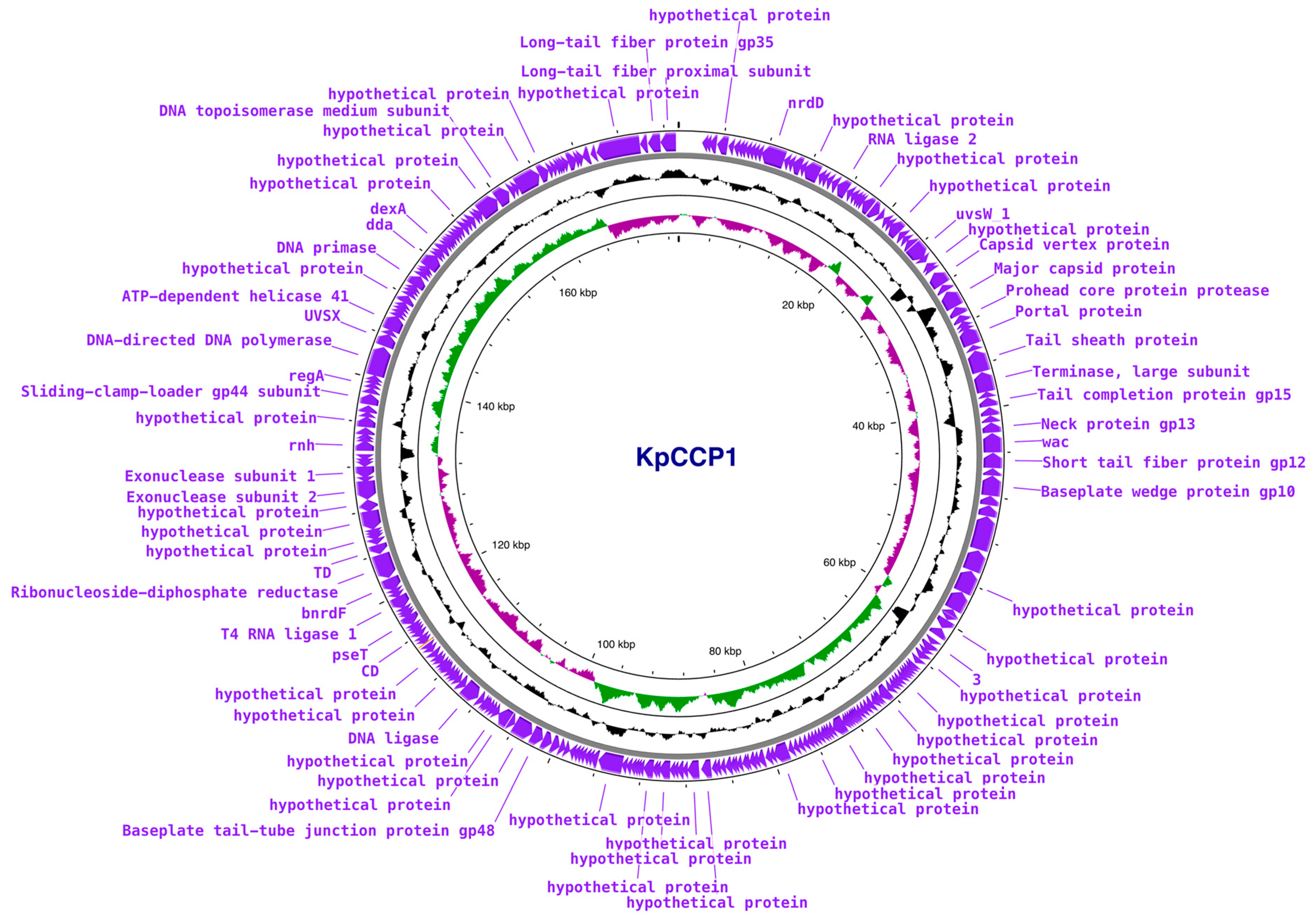
Open reading frames (ORFs) of phage KpCCP1 were predicted using Phanotate [65] in Pharokka v1.3.2 [66] at Phage Galaxy [67] and tRNAs were predicted using ARAGORN v2.36 [68]. Genome map of phage KpCCP1 was obtained using Proksee [69]. PhaTYP [70] and PhageAI [71] were used to predict the lifestyle of KpCCP1.
Antibiotic Resistance Genes (ARGs) were identified using Resfinder 4.0 [72] and Comprehensive Antibiotic Resistance Database (CARD) [73]. Virulence factors were assessed with PhageLeads [74] and ABRicate with Virulence Factor Database (VFDB) [75].
To identify putative Acrs we used PaCRISPR [76] and AcRanker [77] with default parameters. These programs allow the novo prediction of Acr-encoding genes with minimal prior knowledge.

2.8. Host Range

The host range was determined against diverse bacterial strains including K. pneumoniae, K. michiganensis, E. coli, Salmonella enterica and Pseudomonas aeruginosa (see Section 2.1). For this, mixtures of overnight bacterial cultures and phage suspension were incubated 15 min and then 20 μL were spotted onto LB agar plates by triplicate.
Antibiotic susceptibility of KpCCP1-susceptible strains was determined by the disk diffusion method in Mueller-Hinton agar according to the Clinical and Laboratory Standards Institute (CLSI) M100-2024 guidelines [78]. We used E. coli ATCC 25922 as quality control strain. The zones of growth inhibition around each of the antibiotic disks were measured and interpreted as according to CLSI criteria, considering both the susceptibility of the isolate and the diffusion rate of the drug through the agar medium. The test panel included the following antibiotics: imipenem (IMP), meropenem (MEM), ertapenem (ETP), ceftriaxone (CRO), cefotaxime (CTX), ceftazidime (CAZ), ceftazidime/avibactam (CZA), aztreonam (ATM), gentamicin (GEN), amikacin (AMK), trimethoprim/sulfamethoxazole (SXT) and ciprofloxacin (CIP).

2.9. Carbapenemase-Encoding Genes and Clonality of Phage-Sensitive Strains

For strains susceptible to phage infection, the Blue-Carba test was performed as described by Pires et al. [79], along with conventional PCR for blaKPC, blaNDM, blaVIM, blaIMP and blaOXA-48-like genes (Supplementary Materials).
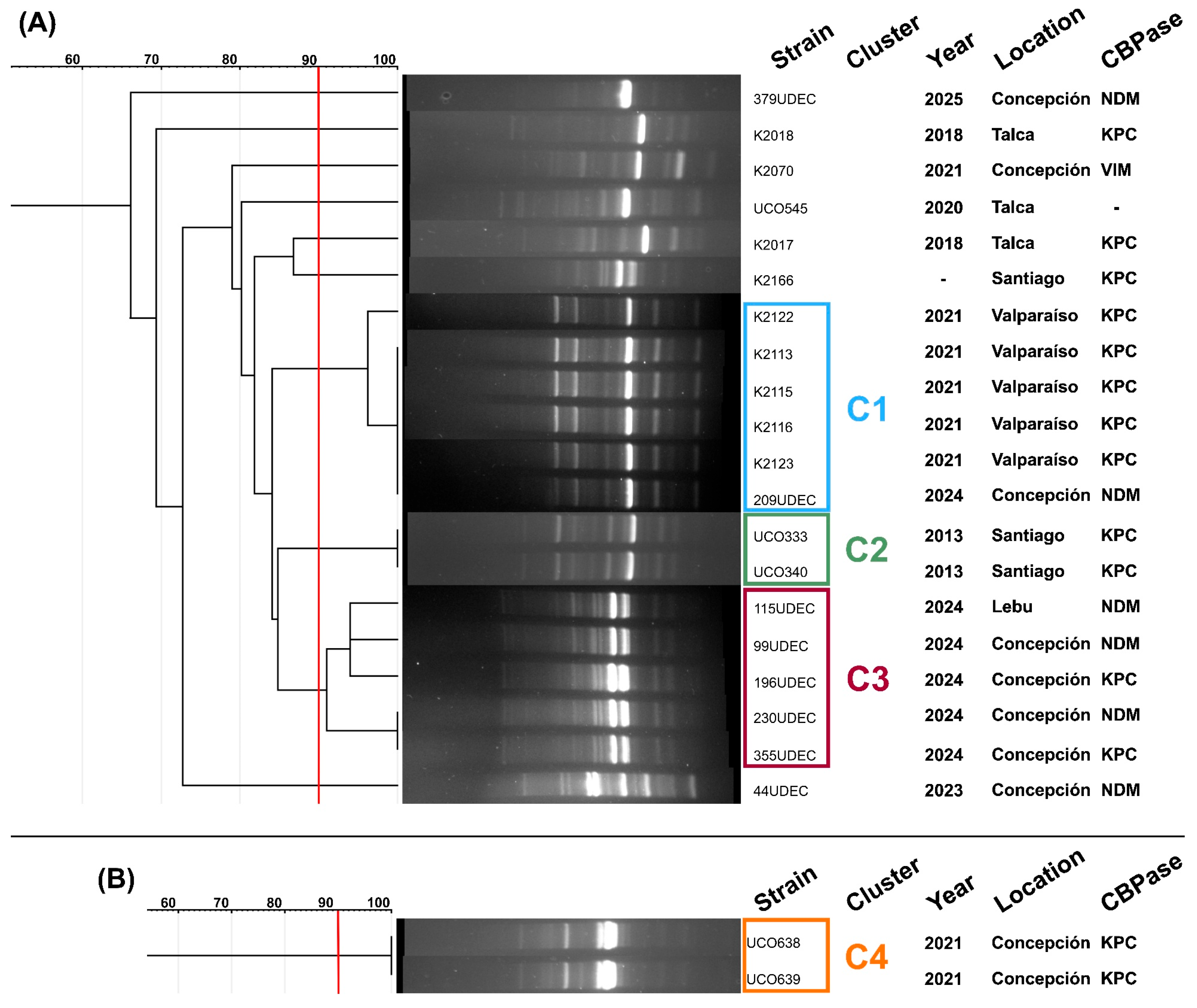
ERIC-PCR typing was applied to assess the genetic relatedness of the phage-sensitive strains. For this, the strains were grown on TS broth overnight at 37 °C and DNA extraction was performed using a 5% w/v Chelex resin matrix. Subsequently, DNA concentrations were adjusted to 50 ng/μL. For PCR, the ERIC-2 primer (5′-AAGTAAGTGACTGGGGGGGGGTGAGCG-3′) was used to amplify the Enterobacterial Repetitive Intergenic Consensus (ERIC) regions, following the protocol described in [80]. Amplification products were separated on a 1.5% agarose gel at 90 V for 2 h. Banding patterns were analyzed using GelJ v2.0 software [81], applying the unweighted pair group method with arithmetic mean (UPGMA) together with the Dice-Sørensen coefficient (DICE) and a band position tolerance of 2%. Two strains were considered genetically related when they came from a node with ≥90% similarity.

3. Results and Discussion

A lytic phage capable of infecting an MDR K. pneumoniae strain was isolated and designated KpCCP1. TEM analysis revealed a tailed morphology featuring an icosahedral head and long tail (Figure 1A). It produced clear, circular plaques 2–3 mm in diameter (Figure 1B).
The genome of phage KpCCP1 consists of a 177,276 bp double-stranded DNA molecule, with a GC content of 41.82% (accession no. PV402122) (Figure 2). The phage was categorized as lytic and belongs to the genus Slopekvirus, which includes lytic myoviruses capable of infecting Klebsiella strains and related to T4-like phages at the family level [82] (Table 1). After the abolishment of the family Myoviridae [83], two new families were created to include phages that have previously been referred to as T4-like phages, families Straboviridae and Kyanoviridae (ICTV proposal 2021.082B).
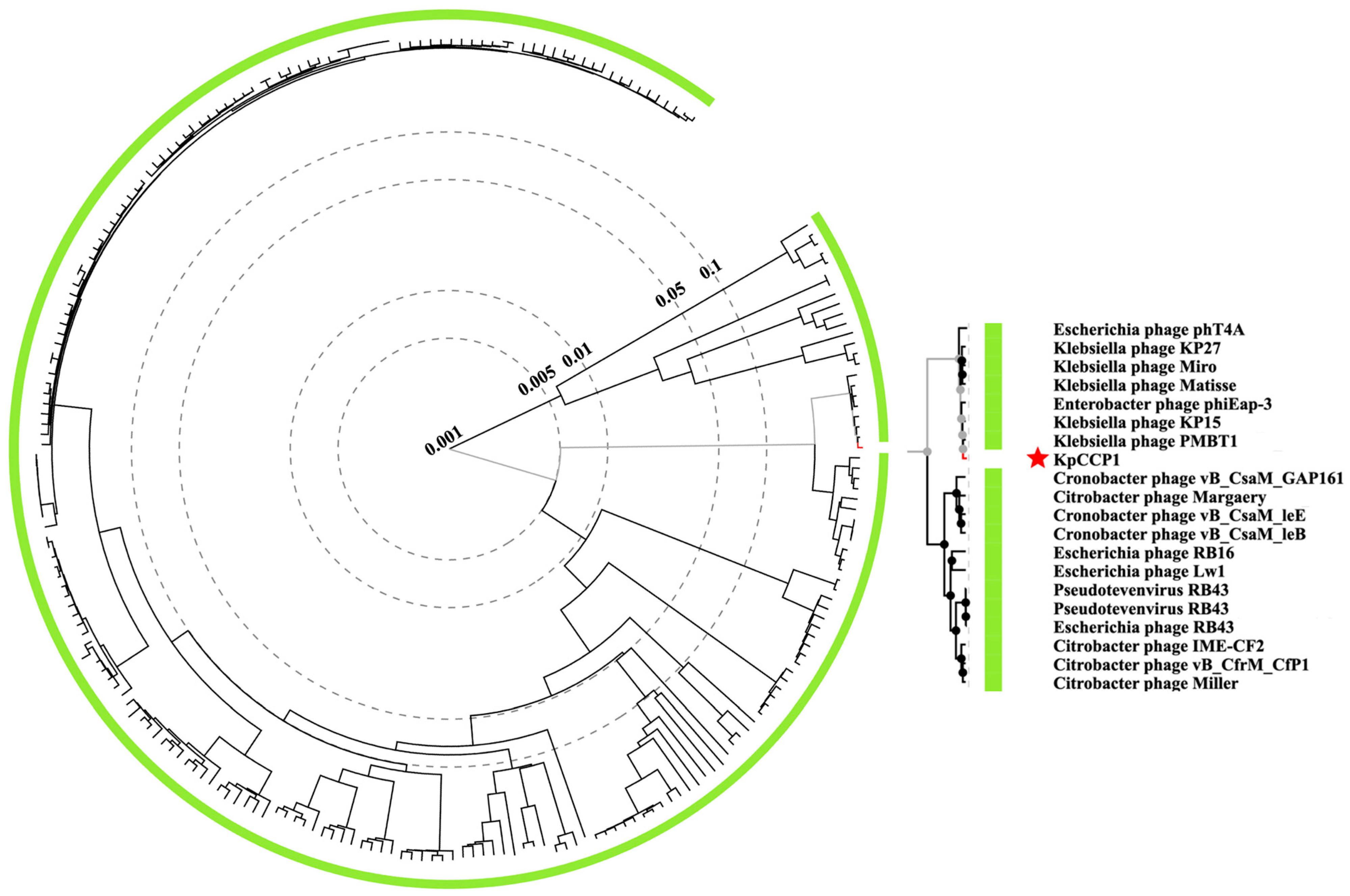
The genus Slopekvirus was created in 2016 by ICTV (proposal 2016.022a-dB) and was formerly known as Kp15virus. Originally, the genus included five species and belonged to the subfamily Tevenvirinae. According to ICTV proposal 2021.082B, the subfamily Tevenvirinae is no longer monophyletic, and several genera, including Slopekvirus, were reassigned as unclassified genera within the family Straboviridae. Currently, the family Straboviridae belongs to the recently created order Pantevenvirales (Proposal: 2024.026B), ratified by Turner et al. in July 2025 [84]. The genus most closely related to Slopekvirus is Pseudotevenvirus, which includes Escherichia, Cronobacter and Citrobacter phages (gap161, imecf2, leb, lee, lw1, margaery, miller) (Figure 3).
Slopekvirus phages have been isolated from Poland, USA and China [85,86,87]. On average, the genomes of members of the genus are ≈175 kb (41.8% GC content). The genus originally included five species, KP15, KP27, Matisse, Miro and phiEap3 (subsequently renamed eap3). Phage PMBT1 (accession no. OQ267591), isolated from raw sewage in Germany [86], was included in genus Slopekvirus, but it was later removed because it showed >95% nucleotide similarity to phage Eap3, indicating they represent the same species. Similarly, phage Miro was removed because it was >95% similar to Matisse. Currently, the genus includes five species: KP15, KP27, Matisse, Eap3 and pht4A. The latter is an Escherichia coli phage [88] that was incorporated into the genus Slopekvirus in 2021 (ICTV proposal 2021.082B).
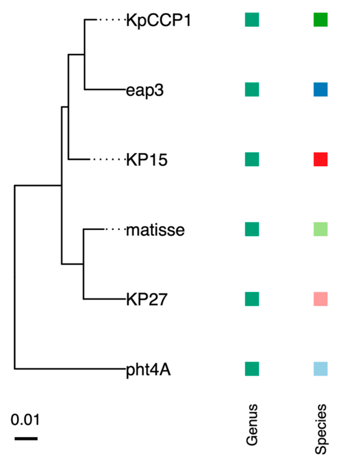
According to TaxMyPhage, phage KpCCP1 belongs to the same species as phage Kp15, according to VIRIDIC, phage KpCCP1 belongs to the same species as phage Eap3 (Table 1) and according to VICTOR, phage KpCCP1 is a novel species (Figure 4).
Phage Eap3 is the most similar to our isolated phage KpCCP1, with an intergenomic identity of 96% based on VIRIDIC. Phage Eap3 was isolated in Beijing, China [85]. It has a genome of 175,814 bp, a CG content of 42% and encodes 278 ORFs (Figure 5). Its host range does not include K. pneumoniae. Originally, it was described as Enterobacter phage, because it was isolated using E. aerogenes. However, E. aerogenes was renamed Klebsiella aerogenes [89].
Klebsiella phages have been described as biotechnologically relevant because they contain a battery of interesting enzymes. For instance, phage KP27 possesses two unique endonucleases, likely involved in uncharacterized mechanism of DNA modification and resistance to restriction digestion. Additionally, KP15 and KP27 were found to encode a complete set of lysis genes, including holin, antiholin, spanin, and endolysin [90].
Recently, Yoo et al. (2024) [91] described the Slopekvirus-like phage phi_KPN_S3 (S3) (accession no. OQ267591). They constructed phage cocktails and demonstrated their ability to suppress phage resistance in MDR K. pneumoniae. Likewise, Duarte et al., (2024) [92] described the Slopekvirus-like phage KP1LMA (accession no. PP002985), isolated from sewage in Portugal. This phage is capable of infecting both K. pneumoniae and E. coli ATCC13706 strains, two of the most common causatives of urinary tract infections (UTIs). By contrast, our phage KpCCP1 was unable to infect or propagate in E. coli. Moreover, our results indicated that phage KpCCP1 was also unable to use Salmonella enterica or Pseudomonas aeruginosa as host.
Phage KP13MC5-1 (accession no. OP617746), a Slopekvirus-like phage, was used together with other phages, to treat primary sclerosing cholangitis (SPF) [93]. The oral administration of the cocktail has been demonstrated to reduce Kp levels in Kp-colonized germ-free mice and SPF mice, without resulting in off-target dysbiosis. In addition, the oral and intravenous administration of phages has been shown to be an effective method of suppressing Kp levels and attenuating liver inflammation and disease severity in hepatobiliary injury-prone SPF mice, suggesting a promising potential for targeting Kp in PSC.
Our host range analysis was performed using carbapenemase-producing Klebsiella strains (131 K. pneumoniae and 2 K. michiganensis) because of the challenge they pose to current clinical management. We established that strains UCO638 and UCO639 are K. michiganensis by whole genome sequencing, which belong to the K. oxytoca complex but rather than the K. pneumoniae complex [94]. In this regard, Townsend et al. [95] described 64 Klebsiella phages, including seven Slopekvirus-like phages. They evaluated the host range of these phages, demonstrating that 6 of the 7 Slopekvirus were capable of forming plaques in K. michiganensis ATCC 25444. Moreover, these phages exhibited a broad host range, as most were able to infect other Klebsiella species (K. pneumoniae, K. oxytoca, K. variicola, K. quasipneumoniae and K. aerogenes). The phage with the broadest host range was a Slopekvirus (KoM-MeTiny), which showed lawn clearance in 79% and produced plaques in 42% of the 24 Klebsiella strains tested. Although our isolated phage KpCCP1 could be considered a broad-host-range phage, we did not evaluate its ability to infect Klebsiella species other than K. pneumoniae and K. michiganensis.
Phage vB_KpnM_M1 (M1) (accession no. MW448170), a Slopekvirus-like phage, was used to treat a patient with a pandrug-resistant K. pneumoniae infection [96]. This therapy results in improvement of the patient’s wounds and overall long-term condition. Phage M1 was isolated from sewage in Tbilisi (Georgia) and showed a broad host range (~65%) against clinical isolates of Klebsiella species (K. pneumoniae, K. oxytoca and K. terrigena).
Phage mtp5 (accession no. MZ612130), a Slopekvirus-like phage, displays the widest host range of Klebsiella phages isolated so far. This phage targeted most of the noncapsulated strains that were tested [97].
To our knowledge, no anti-CRISPR proteins or genes have been investigated in previous studies of Slopekvirus. As we mentioned earlier, in Klebsiella MGEs, Acrs have so far been described and studied only in plasmids. Therefore, using two complementary tools for cross-validation, PaCRISPR [76] and AcRanker [77], we identified putative Acr-encoding genes in phage KpCCP1 (Table 2). Because Acr proteins usually lack conserved sequences, their discovery is challenging [40,41]; however, recent machine learning–based programs have shown excellent performance. In phage KpCCP1, ninety-nine candidate genes were identified with PaCRISP and seventy-six with AcRanker. By merging the results from both predictors, thirty-eight genes were identified in common. Among them, we proposed CDS 104, 160 and 205 as putative Acr-encoding genes because they had the highest combined scores. These genes encode small proteins (95, 66 and 58 amino acids, respectively), which is typical for Acrs, usually ranging between 50 and 150 aa [98]. In addition, these genes encode hypothetical proteins whit no predicted function. The validation of these candidate Acr-encoding genes requires experimental confirmation, which we plan to address this in future studies.
Phage KpCCP1 was able to infect and propagate in 20 K. pneumoniae and 2 K. michiganensis strains out of 133 Klebsiella spp. strains tested (Table 3). The original host of KpCCP1 (UCO545) can be classified as MDR because it showed resistance to at least one agent from three antimicrobial categories [99]. Among the remaining 19 KpCCP1-susceptible K. pneumoniae strains, 12 were KPC-producers, 6 NDM and 1 VIM. Interestingly, the KPC-producing strain K2116 was susceptible to imipenem, resistant to meropenem and showed intermediate susceptibility to ertapenem, with a greenish shift in the Blue-Carba test. This suggests incomplete hydrolysis of the carbapenem, possibly reflecting a lower gene expression, either due to its genomic context or to the copy number of the plasmid on which it is located. The KPC-producing strain K2018 showed intermediate susceptibility to ertapenem, ceftriaxone, and ceftazidime, while being resistant to imipenem and meropenem, with a positive Blue-Carba test. Regarding the resistance to ceftazidime/avibactam, only strains K2113 and K2116 were resistant to this combination. Gentamicin and amikacin showed activity against only one and three strains, respectively. All other strains displayed a classical resistance profile associated with carbapenemase production, showing resistance to all carbapenems and third-generation cephalosporins tested.
Both K. michiganensis strains (UCO638 and UCO639) were KPC-producers and tested positive in the Blue-Carba assay. Additionally, they showed intermediate susceptibility to meropenem and ertapenem, while remaining susceptible to the ceftazidime/avibactam combination. These results underscore the importance of searching for new treatment options for carbapenem-resistant K. pneumoniae and K. michiganensis strains, given the limited number of therapeutic alternatives available to manage infections caused by these pathogens.
The genetic relationship among the 22 KpCCP1-infected strains indicated that, in K. pneumoniae (Figure 6A), three clusters were formed: cluster 1 (C1), consisting of K2122, K2113, K2115, K216, K2123 and 209UDEC; cluster 2 (C2), consisting of UCO333 and UCO340; and cluster 3 (C3), consisting of 115UDEC, 99UDEC, 196UDEC, 230UDEC and 355UDEC. Additionally, 7 strains did not group into clusters. Therefore, KpCCP1 can infect at least 10 genetically unrelated K. pneumoniae strains. As for K. michiganensis strains (Figure 6B), UCO638 and UCO639 formed a single cluster (C4) and were thus genetically related.
Among the various emerging strategies to combat antimicrobial resistance, such as the use of natural compounds and nanoparticles [100], phage therapy is promising due to its high specificity, its ability to self-replicate, and its capacity to adapt to bacterial resistance mechanisms [101]. However, each of these strategies has its own advantages and limitations, which is why continued, multifaceted efforts are essential.
A major barrier for phage therapy is the lack of standardized regulatory frameworks, which delays approval and large-scale application [27]. The host immune response is another obstacle, since repeated exposure may lead to the development of anti-phage antibodies that reduce therapeutic effectiveness [28]. Safety concerns are also relevant, as bacterial lysis can cause endotoxin release with inflammatory responses [30]. Overcoming these regulatory, immunological, and safety issues will be essential for integrating phage therapy into modern medicine [31,32].

4. Conclusions

The emergence of MDR K. pneumoniae and its evolution toward carbapenem-resistant strains have created an urgent need for alternative therapies to replace or complement antibiotics. In this study, we isolated, characterized, and performed a comprehensive genomic analysis of the lytic phage KpCCP1. This phage is able to infect multiple carbapenem-resistant Klebsiella strains, lacks deleterious genes, and possesses putative Anti-CRISPR systems. Therefore, phage KpCCP1 represents a promising therapeutic candidate for the treatment of MDR K. pneumoniae infections, a major human opportunistic pathogen responsible for a wide spectrum of diseases. However, further studies are needed to evaluate its in vivo efficacy, its potential in combination with antibiotics, and experimental validation of candidate Acr-encoding genes.

Supplementary Materials

The following supporting information can be downloaded at: https://www.mdpi.com/article/10.3390/sci7040157/s1, Table S1: Conventional PCR primers and programs used to amplify carbapenemases-encoding genes.

Author Contributions

Conceptualization, B.P. and G.G.-R.; methodology, B.P. and M.M.-K.; software, B.P. and M.M.-K.; validation, B.P. and G.G.-R.; formal analysis, B.P.; E.Y.S.-V. and F.C.-L.; investigation, B.P.; resources, B.P.; M.I.H.; E.Y.S.-V.; F.C.-L. and G.G.-R.; data curation, B.P.; writing—original draft preparation, B.P. and G.G.-R.; writing—review and editing, B.P., E.Y.S.-V. and F.C.-L.; M.I.H.; A.O.-C. and G.G.-R.; visualization, B.P.; supervision, G.G.-R.; project administration, B.P.; funding acquisition, B.P. All authors have read and agreed to the published version of the manuscript.

Funding

This project received funding from the grant “VRID Postdoctorado from the Vicerrectoría de Investigación y Desarrollo, Universidad de Concepción” (project CO 002200000279).

Data Availability Statement

The sequencing data for bacteriophage KpCCP1 are available in GenBank under accession no. PV402122.

Acknowledgments

We thank the CENTINELA group at the Universidad de la Santísima Concepción for providing the raw sewage. We express our gratitude to the Centro de Espectroscopía y Microscopía (CESMI) for their invaluable assistance in providing electron microscopy images.

Conflicts of Interest

The authors declare that they have no known competing financial interests or personal relationships that could have appeared to influence the work reported in this paper.

References

  1. Baquero, F.; Martinez, J.L.; FLanza, V.; Rodríguez-Beltrán, J.; Galán, J.C.; San Millán, A.; Cantón, R.; Coque, T.M. Evolutionary pathways and trajectories in antibiotic resistance. Clin. Microbiol. Rev. 2021, 34, e00050-19. [Google Scholar] [CrossRef] [PubMed]
  2. Salam, M.A.; Al-Amin, M.Y.; Salam, M.T.; Pawar, J.S.; Akhter, N.; Rabaan, A.A.; Alqumber, M.A. Antimicrobial resistance: A growing serious threat for global public health. Healthcare 2023, 11, 1946. [Google Scholar] [CrossRef] [PubMed]
  3. Allel, K.; Peters, A.; Haghparast-Bidgoli, H.; Spencer-Sandino, M.; Conejeros, J.; Garcia, P.; Pouwels, K.B.; Yakob, L.; Munita, J.M.; Undurraga, E.A. Excess burden of antibiotic-resistant bloodstream infections: Evidence from a multicentre retrospective cohort study in Chile, 2018–2022. Lancet 2024, 40, 1–15. [Google Scholar] [CrossRef] [PubMed]
  4. Murray, C.J.; Ikuta, K.S.; Sharara, F.; Swetschinski, L.; Aguilar, G.R.; Gray, A.; Han, C.; Bisignano, C.; Rao, P.; Wool, E.; et al. Global burden of bacterial antimicrobial resistance in 2019: A systematic analysis. Lancet 2022, 399, 629–655. [Google Scholar] [CrossRef]
  5. World Health Organization (WHO). WHO Bacterial Priority Pathogens List; World Health Organization: Geneva, Switzerland, 2024; pp. 12–13. [Google Scholar]
  6. Navon-Venezia, S.; Kondratyeva, K.; Carattoli, A. Klebsiella pneumoniae: A major worldwide source and shuttle for antibiotic resistance. FEMS Microbiol. Rev. 2017, 41, 252–275. [Google Scholar] [CrossRef]
  7. Wyres, K.L.; Lam, M.M.; Holt, K.E. Population genomics of Klebsiella pneumoniae. Nat. Rev. Microbiol. 2020, 18, 344–359. [Google Scholar] [CrossRef]
  8. Martin, R.M.; Bachman, M.A. Colonization, infection, and the accessory genome of Klebsiella pneumoniae. Front. Cell Infect. Microbiol. 2018, 8, 4. [Google Scholar]
  9. Paczosa, M.K.; Mecsas, J. Klebsiella pneumoniae: Going on the offense with a strong defense. Microbiol. Mol. Biol. Rev. 2016, 80, 629–661. [Google Scholar] [CrossRef]
  10. Munoz-Price, L.S.; Poirel, L.; Bonomo, R.A.; Schwaber, M.J.; Daikos, G.L.; Cormican, M.; Cornaglia, G.; Garau, J.; Gniadkowski, M.; Hayden, M.K.; et al. Clinical epidemiology of the global expansion of Klebsiella pneumoniae carbapenemases. Lancet Infect. Dis. 2013, 13, 785–796. [Google Scholar] [CrossRef]
  11. Lam, M.M.; Wick, R.R.; Watts, S.C.; Cerdeira, L.T.; Wyres, K.L.; Holt, K.E. A genomic surveillance framework and genotyping tool for Klebsiella pneumoniae and its related species complex. Nat. Commun. 2021, 12, 4188. [Google Scholar] [CrossRef]
  12. Zhou, R.; Fang, X.; Zhang, J.; Zheng, X.; Shangguan, S.; Chen, S.; Shen, Y.; Liu, Z.; Li, J.; Zhang, R.; et al. Impact of carbapenem resistance on mortality in patients infected with Enterobacteriaceae: A systematic review and meta-analysis. BMJ Open 2021, 11, e054971. [Google Scholar] [CrossRef] [PubMed]
  13. Hovan, M.R.; Narayanan, N.; Cedarbaum, V.; Bhowmick, T.; Kirn, T.J. Comparing mortality in patients with carbapenemase-producing carbapenem resistant Enterobacterales and non-carbapenemase-producing carbapenem resistant Enterobacterales bacteremia. Diagn. Microbiol. Infect. Dis. 2021, 101, 115505. [Google Scholar] [CrossRef] [PubMed]
  14. Baek, M.S.; Kim, J.H.; Park, J.H.; Kim, T.W.; Jung, H.I.; Kwon, Y.S. Comparison of mortality rates in patients with carbapenem-resistant enterobacterales bacteremia according to carbapenemase production: A multicenter propensity-score matched study. Sci. Rep. 2024, 14, 597. [Google Scholar]
  15. Tamma, P.D.; Goodman, K.E.; Harris, A.D.; Tekle, T.; Roberts, A.; Taiwo, A.; Simner, P.J. Comparing the outcomes of patients with carbapenemase-producing and non-carbapenemase-producing carbapenem-resistant Enterobacteriaceae bacteremia. Clin. Infect. Dis. 2017, 64, 257–264. [Google Scholar] [CrossRef]
  16. Cifuentes, M.; Garcia, P.; San Martin, P.; Silva, F.; Zuniga, J.; Reyes, S.; Rojas, R.; Ponce, R.; Quintanilla, R.; Delpiano, L.; et al. First isolation of KPC in Chile: From Italy to a public hospital in Santiago. Rev. Chil. Infectol. 2012, 29, 224–228. [Google Scholar] [CrossRef]
  17. Carrasco-Anabalón, S.; Neto, C.O.C.; Carvalho-Assef, A.P.D.A.; Lima, C.A.; Cifuentes, M.; Silva, F.; Barrera, B.; Domínguez, M.; González-Rocha, G.; Bello-Toledo, H. Introduction of NDM-1 and OXA-370 from Brazil into Chile in strains of Klebsiella pneumoniae isolated from a single patient. Int. J. Infect. Dis. 2019, 81, 28–30. [Google Scholar] [CrossRef]
  18. Ministerio de Salud. Instituto de Salud Pública de Chile. Boletín de Resistencia Microbiana. Available online: https://www.ispch.cl/wp-content/uploads/2022/09/BoletinRAM_FINAL-1-1.pdf (accessed on 12 May 2024).
  19. Quezada-Aguiluz, M.; Opazo-Capurro, A.; Lincopan, N.; Esposito, F.; Fuga, B.; Mella-Montecino, S.; Riedel, G.; Lima, C.A.; Bello-Toledo, H.; Cifuentes, M.; et al. Novel megaplasmid driving NDM-1-mediated carbapenem resistance in Klebsiella pneumoniae ST1588 in South America. Antibiotics 2022, 11, 1207. [Google Scholar] [CrossRef]
  20. Quesille-Villalobos, A.M.; Solar, C.; Martínez, J.R.; Rivas, L.; Quiroz, V.; González, A.M.; Riquelme-Neira, R.; Ugalde, J.A.; Peters, A.; Ortega-Recalde, O.; et al. Multispecies emergence of dual bla KPC/NDM carbapenemase-producing Enterobacterales recovered from invasive infections in Chile. Antimicrob. Agents Chemother. 2025, 69, e01205–e01224. [Google Scholar]
  21. Gálvez-Silva, M.; Arros, P.; Berríos-Pastén, C.; Villamil, A.; Rodas, P.I.; Araya, I.; Iglesias, R.; Araya, P.; Hormazábal, J.C.; Bohle, C.; et al. Carbapenem-resistant hypervirulent ST23 Klebsiella pneumoniae with a highly transmissible dual-carbapenemase plasmid in Chile. Biol. Res. 2024, 57, 7. [Google Scholar]
  22. Veloso, M.; Arros, P.; Acosta, J.; Rojas, R.; Berríos-Pastén, C.; Varas, M.; Araya, P.; Hormazábal, J.C.; Allende, M.L.; Chávez, F.P.; et al. Antimicrobial resistance, pathogenic potential, and genomic features of carbapenem-resistant Klebsiella pneumoniae isolated in Chile: High-risk ST25 clones and novel mobile elements. Microbiol. Spectr. 2023, 11, e00399-23. [Google Scholar]
  23. Russo, T.A.; Marr, C.M. Hypervirulent Klebsiella pneumoniae. Clin. Microbiol. Rev. 2019, 32, 10–1128. [Google Scholar] [CrossRef] [PubMed]
  24. Morales-León, F.; Matus-Köhler, M.; Araya-Vega, P.; Aguilera, F.; Torres, I.; Vera, R.; Ibarra, C.; Venegas, S.; Bello-Toledo, H.; González-Rocha, G.; et al. Molecular characterization of the convergent carbapenem-resistant and hypervirulent Klebsiella pneumoniae strain K1-ST23, collected in Chile during the COVID-19 pandemic. Microbiol. Spectr. 2023, 11, e00540-e23. [Google Scholar] [CrossRef] [PubMed]
  25. Naghavi, M.; Vollset, S.E.; Ikuta, K.S.; Swetschinski, L.R.; Gray, A.P.; Wool, E.E.; Aguilar, G.R.; Mestrovic, T.; Smith, G.; Han, C.; et al. Global burden of bacterial antimicrobial resistance 1990–2021: A systematic analysis with forecasts to 2050. Lancet 2024, 404, 1199–1226. [Google Scholar] [CrossRef] [PubMed]
  26. Mahoney, A.R.; Safaee, M.M.; Wuest, W.M.; Furst, A.L. The silent pandemic: Emergent antibiotic resistances following the global response to SARS-CoV-2. iScience 2021, 24, 102304. [Google Scholar] [CrossRef]
  27. Principi, N.; Silvestri, E.; Esposito, S. Advantages and limitations of bacteriophages for the treatment of bacterial infections. Front. Pharmacol. 2019, 10, 457104. [Google Scholar] [CrossRef]
  28. Strathdee, S.A.; Hatfull, G.F.; Mutalik, V.K.; Schooley, R.T. Phage therapy: From biological mechanisms to future directions. Cell 2023, 186, 17–31. [Google Scholar] [CrossRef]
  29. Cisek, A.A.; Dąbrowska, I.; Gregorczyk, K.P.; Wyżewski, Z. Phage therapy in bacterial infections treatment: One hundred years after the discovery of bacteriophages. Curr. Microbiol. 2017, 74, 277–283. [Google Scholar] [CrossRef]
  30. Kortright, K.E.; Chan, B.K.; Koff, J.L.; Turner, P.E. Phage therapy: A renewed approach to combat antibiotic-resistant bacteria. Cell Host Microbe 2019, 25, 219–232. [Google Scholar] [CrossRef]
  31. Gordillo Altamirano, F.L.; Barr, J.J. Phage therapy in the postantibiotic era. Clin. Microbiol. Rev. 2019, 32, 10–1128. [Google Scholar] [CrossRef]
  32. Lin, D.M.; Koskella, B.; Lin, H.C. Phage therapy: An alternative to antibiotics in the age of multi-drug resistance. World J. Gastrointest. Pharmacol. Ther. 2017, 8, 162. [Google Scholar] [CrossRef]
  33. Doudna, J.A.; Charpentier, E. The new frontier of genome engineering with CRISPR-Cas9. Science 2014, 346, 1258096. [Google Scholar] [CrossRef]
  34. Anzalone, A.V.; Koblan, L.W.; Liu, D.R. Genome editing with CRISPR–Cas nucleases, base editors, transposases and prime editors. Nat. Biotechnol. 2020, 38, 824–844. [Google Scholar] [CrossRef]
  35. Wang, J.Y.; Doudna, J.A. CRISPR technology: A decade of genome editing is only the beginning. Science 2023, 379, eadd8643. [Google Scholar] [CrossRef]
  36. Li, H.Y.; Kao, C.Y.; Lin, W.H.; Zheng, P.X.; Yan, J.J.; Wang, M.C.; Teng, C.H.; Tseng, C.C.; Wu, J.J. Characterization of CRISPR-Cas systems in clinical Klebsiella pneumoniae isolates uncovers its potential association with antibiotic susceptibility. Front. Microbiol. 2018, 9, 1595. [Google Scholar] [CrossRef] [PubMed]
  37. Mackow, N.A.; Shen, J.; Adnan, M.; Khan, A.S.; Fries, B.C.; Diago-Navarro, E. CRISPR-Cas influences the acquisition of antibiotic resistance in Klebsiella pneumoniae. PLoS ONE 2019, 14, e0225131. [Google Scholar] [CrossRef] [PubMed]
  38. Wang, G.; Song, G.; Xu, Y. Association of CRISPR/Cas system with the drug resistance in Klebsiella pneumoniae. Infect. Drug Resist. 2020, 13, 1929–1935. [Google Scholar] [CrossRef] [PubMed]
  39. Jwair, N.A.; Al-Ouqaili, M.T.; Al-Marzooq, F. Inverse association between the existence of CRISPR/Cas systems with antibiotic resistance, extended spectrum β-lactamase and carbapenemase production in multidrug, extensive drug and pandrug-resistant Klebsiella pneumoniae. Antibiotics 2023, 12, 980. [Google Scholar] [CrossRef]
  40. Pinilla-Redondo, R.; Shehreen, S.; Marino, N.D.; Fagerlund, R.D.; Brown, C.M.; Sørensen, S.J.; Fineran, P.C.; Bondy-Denomy, J. Discovery of multiple anti-CRISPRs highlights anti-defense gene clustering in mobile genetic elements. Nat. Commun. 2020, 11, 5652. [Google Scholar] [CrossRef]
  41. Wang, C.; Sun, Z.; Hu, Y.; Li, D.; Guo, Q.; Wang, M. A novel anti-CRISPR AcrIE9. 2 is associated with dissemination of bla KPC plasmids in Klebsiella pneumoniae sequence type 15. Antimicrob. Agents Chemother. 2023, 67, e01547-e22. [Google Scholar] [CrossRef]
  42. Jiang, C.; Yu, C.; Sun, S.; Lin, J.; Cai, M.; Wei, Z.; Feng, L.; Li, J.; Zhang, Y.; Dong, K.; et al. A new anti-CRISPR gene promotes the spread of drug-resistance plasmids in Klebsiella pneumoniae. Nucleic Acids Res. 2024, 52, 8370–8384. [Google Scholar] [CrossRef]
  43. Jiang, J.; Cienfuegos-Galletd, A.V.; Long, T.; Peirano, G.; Chu, T.; Pitout, J.D.; Kreiswirth, B.N.; Chen, L. Intricate interplay of CRISPR-Cas systems, anti-CRISPR proteins, and antimicrobial resistance genes in a globally successful multi-drug resistant Klebsiella pneumoniae clone. Genome Med. 2025, 17, 9. [Google Scholar] [CrossRef]
  44. Herridge, W.P.; Shibu, P.; O’Shea, J.; Brook, T.C.; Hoyles, L. Bacteriophages of Klebsiella spp., their diversity and potential therapeutic uses. J. Med. Microbiol. 2020, 69, 176–194. [Google Scholar]
  45. Li, N.; Zeng, Y.; Bao, R.; Zhu, T.; Tan, D.; Hu, B. Isolation and characterization of novel phages targeting pathogenic Klebsiella pneumoniae. Front. Cell. Infect. Microbiol. 2021, 11, 792305. [Google Scholar] [CrossRef] [PubMed]
  46. Zurabov, F.; Zhilenkov, E. Characterization of four virulent Klebsiella pneumoniae bacteriophages, and evaluation of their potential use in complex phage preparation. Virol. J. 2021, 18, 1–20. [Google Scholar] [CrossRef] [PubMed]
  47. Ballesté, E.; Blanch, A.R.; Muniesa, M.; García-Aljaro, C.; Rodríguez-Rubio, L.; Martín-Díaz, J.; Pascual-Benito, M.; Jofre, J. Bacteriophages in sewage: Abundance, roles, and applications. FEMS Microbes 2022, 3, xtac009. [Google Scholar] [CrossRef] [PubMed]
  48. Parra, B.; Sandoval, M.; Arriagada, V.; Amsteins, L.; Aguayo, C.; Opazo-Capurro, A.; Dechesne, A.; González-Rocha, G. Isolation and Characterization of Lytic Bacteriophages Capable of Infecting Diverse Multidrug-Resistant Strains of Pseudomonas aeruginosa: PaCCP1 and PaCCP2. Pharmaceuticals 2024, 17, 1616. [Google Scholar] [CrossRef]
  49. Kropinski, A.M.; Mazzocco, A.; Waddell, T.E.; Lingohr, E.; Johnson, R.P. Enumeration of bacteriophages by double agar overlay plaque assay. In Bacteriophages: Methods and Protocols; Springer: Berlin/Heidelberg, Germany, 2009; Volume 1, pp. 69–76. [Google Scholar]
  50. Andrews, S. FastQC: A Quality Control Tool for High Throughput Sequence Data. 2010. Available online: www.bioinformatics.babraham.ac.uk/projects/fastqc/ (accessed on 12 December 2024).
  51. Shen, A.; Millard, A. Phage genome annotation: Where to begin and end. Phage 2021, 2, 183–193. [Google Scholar] [CrossRef]
  52. Shen, W.; Le, S.; Li, Y.; Hu, F. SeqKit: A cross-platform and ultrafast toolkit for FASTA/Q file manipulation. PLoS ONE 2016, 11, e0163962. [Google Scholar] [CrossRef]
  53. Bankevich, A.; Nurk, S.; Antipov, D.; Gurevich, A.A.; Dvorkin, M.; Kulikov, A.S.; Lesin, V.M.; Nikolenko, S.I.; Pham, S.; Prjibelski, A.D.; et al. SPAdes: A new genome assembly algorithm and its applications to single-cell sequencing. J. Comput. Biol. 2012, 19, 455–477. [Google Scholar] [CrossRef]
  54. Turner, D.; Adriaenssens, E.M.; Tolstoy, I.; Kropinski, A.M. Phage annotation guide: Guidelines for assembly and high-quality annotation. Phage 2021, 2, 170–182. [Google Scholar] [CrossRef]
  55. Olson, R.D.; Assaf, R.; Brettin, T.; Conrad, N.; Cucinell, C.; Davis, J.J.; Dempsey, D.M.; Dickerman, A.; Dietrich, E.M.; Kenyon, R.W.; et al. Introducing the bacterial and viral bioinformatics resource center (BV-BRC): A resource combining PATRIC, IRD and ViPR. Nucleic Acids Res. 2023, 51, D678–D689. [Google Scholar] [CrossRef] [PubMed]
  56. Gurevich, A.; Saveliev, V.; Vyahhi, N.; Tesler, G. QUAST: Quality assessment tool for genome assemblies. Bioinformatics 2013, 29, 1072–1075. [Google Scholar] [CrossRef] [PubMed]
  57. Wick, R.R.; Schultz, M.B.; Zobel, J.; Holt, K.E. Bandage: Interactive visualization of de novo genome assemblies. Bioinformatics 2015, 31, 3350–3352. [Google Scholar] [CrossRef] [PubMed]
  58. Walker, B.J.; Abeel, T.; Shea, T.; Priest, M.; Abouelliel, A.; Sakthikumar, S.; Cuomo, C.A.; Zeng, Q.; Wortman, J.; Young, S.K.; et al. Pilon: An integrated tool for comprehensive microbial variant detection and genome assembly improvement. PLoS ONE 2014, 9, e112963. [Google Scholar] [CrossRef]
  59. Moraru, C.; Varsani, A.; Kropinski, A.M. VIRIDIC—A novel tool to calculate the intergenomic similarities of prokaryote-infecting viruses. Viruses 2020, 12, 1268. [Google Scholar] [CrossRef]
  60. Meier-Kolthoff, J.P.; Göker, M. VICTOR: Genome-based phylogeny and classification of prokaryotic viruses. Bioinformatics 2017, 33, 3396–3404. [Google Scholar] [CrossRef]
  61. Millard, A.; Denise, R.; Lestido, M.; Thomas, M.T.; Webster, D.; Turner, D.; Sicheritz-Pontén, T. taxmyPHAGE: Automated taxonomy of dsDNA phage genomes at the genus and species level. Phage 2025, 6, 5–11. [Google Scholar] [CrossRef]
  62. Nishimura, Y.; Yoshida, T.; Kuronishi, M.; Uehara, H.; Ogata, H.; Goto, S. ViPTree: The viral proteomic tree server. Bioinformatics 2017, 33, 2379–2380. [Google Scholar] [CrossRef]
  63. Turner, D.; Kropinski, A.M.; Adriaenssens, E.M. A roadmap for genome-based phage taxonomy. Viruses 2021, 13, 506. [Google Scholar] [CrossRef]
  64. Adriaenssens, E.M.; Brister, J.R. How to name and classify your phage: An informal guide. Viruses 2017, 9, 70. [Google Scholar] [CrossRef]
  65. McNair, K.; Zhou, C.; Dinsdale, E.A.; Souza, B.; Edwards, R.A. PHANOTATE: A novel approach to gene identification in phage genomes. Bioinformatics 2019, 35, 4537–4542. [Google Scholar] [CrossRef]
  66. Bouras, G.; Nepal, R.; Houtak, G.; Psaltis, A.J.; Wormald, P.J.; Vreugde, S. Pharokka: A fast scalable bacteriophage annotation tool. Bioinformatics 2023, 39, btac776. [Google Scholar] [CrossRef] [PubMed]
  67. Ramsey, J.; Rasche, H.; Maughmer, C.; Criscione, A.; Mijalis, E.; Liu, M.; Hu, J.C.; Young, R.; Gill, J.J. Galaxy and Apollo as a biologist-friendly interface for high-quality cooperative phage genome annotation. PLoS Comput. Biol. 2020, 16, e1008214. [Google Scholar] [CrossRef] [PubMed]
  68. Laslett, D.; Canback, B. ARAGORN, a program to detect tRNA genes and tmRNA genes in nucleotide sequences. Nucleic Acids Res. 2004, 32, 11–16. [Google Scholar] [CrossRef] [PubMed]
  69. Grant, J.R.; Enns, E.; Marinier, E.; Mandal, A.; Herman, E.K.; Chen, C.Y.; Graham, M.; Van Domselaar, G.; Stothard, P. Proksee: In-depth characterization and visualization of bacterial genomes. Nucleic Acids Res. 2023, 51, W484–W492. [Google Scholar] [CrossRef]
  70. Shang, J.; Tang, X.; Sun, Y. PhaTYP: Predicting the lifestyle for bacteriophages using BERT. Brief. Bioinform. 2023, 24, bbac487. [Google Scholar] [CrossRef]
  71. Tynecki, P.; Guziński, A.; Kazimierczak, J.; Jadczuk, M.; Dastych, J.; Onisko, A. PhageAI-bacteriophage life cycle recognition with machine learning and natural language processing. bioRxiv 2020. [Google Scholar] [CrossRef]
  72. Bortolaia, V.; Kaas, R.S.; Ruppe, E.; Roberts, M.C.; Schwarz, S.; Cattoir, V.; Philippon, A.; Allesoe, R.L.; Rebelo, A.R.; Florensa, A.F.; et al. ResFinder 4.0 for predictions of phenotypes from genotypes. J. Antimicrob. Chemother. 2020, 75, 3491–3500. [Google Scholar] [CrossRef]
  73. Alcock, B.P.; Huynh, W.; Chalil, R.; Smith, K.W.; Raphenya, A.R.; Wlodarski, M.A.; Edalatmand, A.; Petkau, A.; Syed, S.A.; Tsang, K.K.; et al. CARD 2023: Expanded curation, support for machine learning, and resistome prediction at the Comprehensive Antibiotic Resistance Database. Nucleic Acids Res. 2023, 51, D690–D699. [Google Scholar] [CrossRef]
  74. Yukgehnaish, K.; Rajandas, H.; Parimannan, S.; Manickam, R.; Marimuthu, K.; Petersen, B.; Clokie, M.R.; Millard, A.; Sicheritz-Pontén, T. PhageLeads: Rapid assessment of phage therapeutic suitability using an ensemble machine learning approach. Viruses 2022, 14, 342. [Google Scholar] [CrossRef]
  75. Chen, L.; Zheng, D.; Liu, B.; Yang, J.; Jin, Q. VFDB 2016: Hierarchical and refined dataset for big data analysis—10 years on. Nucleic Acids Res. 2016, 44, D694–D697. [Google Scholar] [CrossRef] [PubMed]
  76. Wang, J.; Dai, W.; Li, J.; Xie, R.; Dunstan, R.A.; Stubenrauch, C.; Zhang, Y.; Lithgow, T. PaCRISPR: A server for predicting and visualizing anti-CRISPR proteins. Nucleic Acids Res. 2020, 48, W348–W357. [Google Scholar] [CrossRef] [PubMed]
  77. Eitzinger, S.; Asif, A.; Watters, K.E.; Iavarone, A.T.; Knott, G.J.; Doudna, J.A.; Minhas, F.U.A.A. Machine learning predicts new anti-CRISPR proteins. Nucleic Acids Res. 2020, 48, 4698–4708. [Google Scholar] [CrossRef] [PubMed]
  78. Clinical and Laboratory Standards Institute (CLSI). Performance Standards for Antimicrobial Susceptibility Testing, 34th ed.; CLSI Supplement M100; Clinical and Laboratory Standards Institute: Malvern, PA, USA, 2024. [Google Scholar]
  79. Pires, J.; Novais, A.; Peixe, L. Blue-carba, an easy biochemical test for detection of diverse carbapenemase producers directly from bacterial cultures. J. Clin. Microbiol. 2013, 51, 4281–4283. [Google Scholar] [CrossRef]
  80. Vila, J.; Marcos, M.A.; Jimenez de Anta, M.T. A comparative study of different PCR-based DNA fingerprinting techniques for typing of the Acinetobacter calcoaceticus-A. baumannii complex. J. Med. Microbiol. 1996, 44, 482–489. [Google Scholar] [CrossRef]
  81. Heras, J.; Domínguez, C.; Mata, E.; Pascual, V.; Lozano, C.; Torres, C.; Zarazaga, M. GelJ–a tool for analyzing DNA fingerprint gel images. BMC Bioinform. 2015, 16, 1–8. [Google Scholar] [CrossRef]
  82. Zhao, J.; Zhang, Z.; Tian, C.; Chen, X.; Hu, L.; Wei, X.; Li, H.; Lin, W.; Jiang, A.; Feng, R.; et al. Characterizing the biology of lytic bacteriophage vB_EaeM_φEap-3 infecting multidrug-resistant Enterobacter aerogenes. Front. Microbiol. 2019, 10, 420. [Google Scholar] [CrossRef]
  83. Turner, D.; Shkoporov, A.N.; Lood, C.; Millard, A.D.; Dutilh, B.E.; Alfenas-Zerbini, P.; Van Zyl, L.J.; Aziz, R.K.; Oksanen, H.M.; Poranen, M.M.; et al. Abolishment of morphology-based taxa and change to binomial species names: 2022 taxonomy update of the ICTV bacterial viruses subcommittee. Arch. Virol. 2023, 168, 74. [Google Scholar] [CrossRef]
  84. Turner, D.; Adriaenssens, E.M.; Amann, R.I.; Bardy, P.; Bartlau, N.; Barylski, J.; Błażejak, S.; Bouzari, M.; Briegel, A.; Briers, Y.; et al. Summary of taxonomy changes ratified by the International Committee on Taxonomy of Viruses (ICTV) from the Bacterial Viruses Subcommittee, 2025. J. Gen. Virol. 2025, 106, 002111. [Google Scholar] [CrossRef]
  85. Kęsik-Szeloch, A.; Drulis-Kawa, Z.; Weber-Dąbrowska, B.; Kassner, J.; Majkowska-Skrobek, G.; Augustyniak, D.; Łusiak-Szelachowska, M.; Żaczek, M.; Górski, A.; Kropinski, A.M. Characterising the biology of novel lytic bacteriophages infecting multidrug resistant Klebsiella pneumoniae. Virol. J. 2013, 10, 1–12. [Google Scholar] [CrossRef]
  86. Provasek, V.E.; Lessor, L.E.; Cahill, J.L.; Rasche, E.S.; Kuty Everett, G.F. Complete genome sequence of carbapenemase-producing Klebsiella pneumoniae myophage Matisse. Genome Announc. 2015, 3, 10–1128. [Google Scholar] [CrossRef] [PubMed]
  87. Koberg, S.; Brinks, E.; Fiedler, G.; Hüsing, C.; Cho, G.S.; Hoeppner, M.P.; Heller, K.J.; Neve, H.; Franz, C.M. Genome sequence of Klebsiella pneumoniae bacteriophage PMBT1 isolated from raw sewage. Genome Announc. 2017, 5, 10–1128. [Google Scholar] [CrossRef] [PubMed]
  88. Pereira, C.; Moreirinha, C.; Lewicka, M.; Almeida, P.; Clemente, C.; Romalde, J.L.; Nunes, M.L.; Almeida, A. Characterization and in vitro evaluation of new bacteriophages for the biocontrol of Escherichia coli. Virus Res. 2017, 227, 171–182. [Google Scholar] [CrossRef] [PubMed]
  89. Wesevich, A.; Sutton, G.; Ruffin, F.; Park, L.P.; Fouts, D.E.; Fowler, V.G., Jr.; Thaden, J.T. Newly named Klebsiella aerogenes (formerly Enterobacter aerogenes) is associated with poor clinical outcomes relative to other Enterobacter species in patients with bloodstream infection. J. Clin. Microbiol. 2020, 58, 1–9. [Google Scholar] [CrossRef]
  90. Maciejewska, B.; Roszniowski, B.; Espaillat, A.; Kęsik-Szeloch, A.; Majkowska-Skrobek, G.; Kropinski, A.M.; Briers, Y.; Cava, F.; Lavigne, R.; Drulis-Kawa, Z. Klebsiella phages representing a novel clade of viruses with an unknown DNA modification and biotechnologically interesting enzymes. Appl. Microbiol. Biotechnol. 2017, 101, 673–684. [Google Scholar] [CrossRef]
  91. Yoo, S.; Lee, K.M.; Kim, N.; Vu, T.N.; Abadie, R.; Yong, D. Designing phage cocktails to combat the emergence of bacteriophage-resistant mutants in multidrug-resistant Klebsiella pneumoniae. Microbiol. Spectr. 2024, 12, e01258-23. [Google Scholar] [CrossRef]
  92. Duarte, J.; Máximo, C.; Costa, P.; Oliveira, V.; Gomes, N.C.; Romalde, J.L.; Pereira, C.; Almeida, A. Potential of an isolated bacteriophage to inactivate Klebsiella pneumoniae: Preliminary studies to control urinary tract infections. Antibiotics 2024, 13, 195. [Google Scholar] [CrossRef]
  93. Ichikawa, M.; Nakamoto, N.; Kredo-Russo, S.; Weinstock, E.; Weiner, I.N.; Khabra, E.; Ben-Ishai, N.; Inbar, D.; Kowalsman, N.; Mordoch, R.; et al. Bacteriophage therapy against pathological Klebsiella pneumoniae ameliorates the course of primary sclerosing cholangitis. Nat. Commun. 2023, 14, 3261. [Google Scholar] [CrossRef]
  94. Yang, J.; Long, H.; Hu, Y.; Feng, Y.; McNally, A.; Zong, Z. Klebsiella oxytoca complex: Update on taxonomy, antimicrobial resistance, and virulence. Clin. Microbiol. Rev. 2022, 35, e00006-21. [Google Scholar] [CrossRef]
  95. Townsend, E.M.; Kelly, L.; Gannon, L.; Muscatt, G.; Dunstan, R.; Michniewski, S.; Sapkota, H.; Kiljunen, S.J.; Kolsi, A.; Skurnik, M.; et al. Isolation and characterization of Klebsiella phages for phage therapy. Ther. Appl. Res. 2021, 2, 26–42. [Google Scholar] [CrossRef]
  96. Eskenazi, A.; Lood, C.; Wubbolts, J.; Hites, M.; Balarjishvili, N.; Leshkasheli, L.; Askilashvili, L.; Kvachadze, L.; van Noort, V.; Wagemans, J.; et al. Combination of pre-adapted bacteriophage therapy and antibiotics for treatment of fracture-related infection due to pandrug-resistant Klebsiella pneumoniae. Nat. Commun. 2022, 13, 302. [Google Scholar] [CrossRef]
  97. Lourenço, M.; Osbelt, L.; Passet, V.; Gravey, F.; Megrian, D.; Strowig, T.; Rodrigues, C.; Brisse, S. Phages against noncapsulated Klebsiella pneumoniae: Broader host range, slower resistance. Microbiol. Spectr. 2023, 11, e04812-22. [Google Scholar] [CrossRef] [PubMed]
  98. Pawluk, A.; Bondy-Denomy, J.; Cheung, V.H.; Maxwell, K.L.; Davidson, A.R. A new group of phage anti-CRISPR genes inhibits the type IE CRISPR-Cas system of Pseudomonas aeruginosa. mBio 2014, 5, e00896. [Google Scholar] [CrossRef] [PubMed]
  99. Magiorakos, A.P.; Srinivasan, A.; Carey, R.B.; Carmeli, Y.; Falagas, M.E.; Giske, C.G.; Harbarth, S.; Hindler, J.F.; Kahlmeter, G.; Olsson-Liljequist, B.J.C.M.; et al. Multidrug-resistant, extensively drug-resistant and pandrug-resistant bacteria: An international expert proposal for interim standard definitions for acquired resistance. Clin. Microbiol. Infect. 2012, 18, 268–281. [Google Scholar] [CrossRef] [PubMed]
  100. Peng, Q.; Fang, M.; Liu, X.; Zhang, C.; Liu, Y.; Yuan, Y. Isolation and characterization of a novel phage for controlling multidrug-resistant Klebsiella pneumoniae. Microorganisms 2020, 8, 542. [Google Scholar] [CrossRef]
  101. El-Demerdash, A.S.; Alfaraj, R.; Farid, F.A.; Yassin, M.H.; Saleh, A.M.; Dawwam, G.E. Essential oils as capsule disruptors: Enhancing antibiotic efficacy against multidrug-resistant Klebsiella pneumoniae. Front. Microbiol. 2024, 15, 1467460. [Google Scholar] [CrossRef]
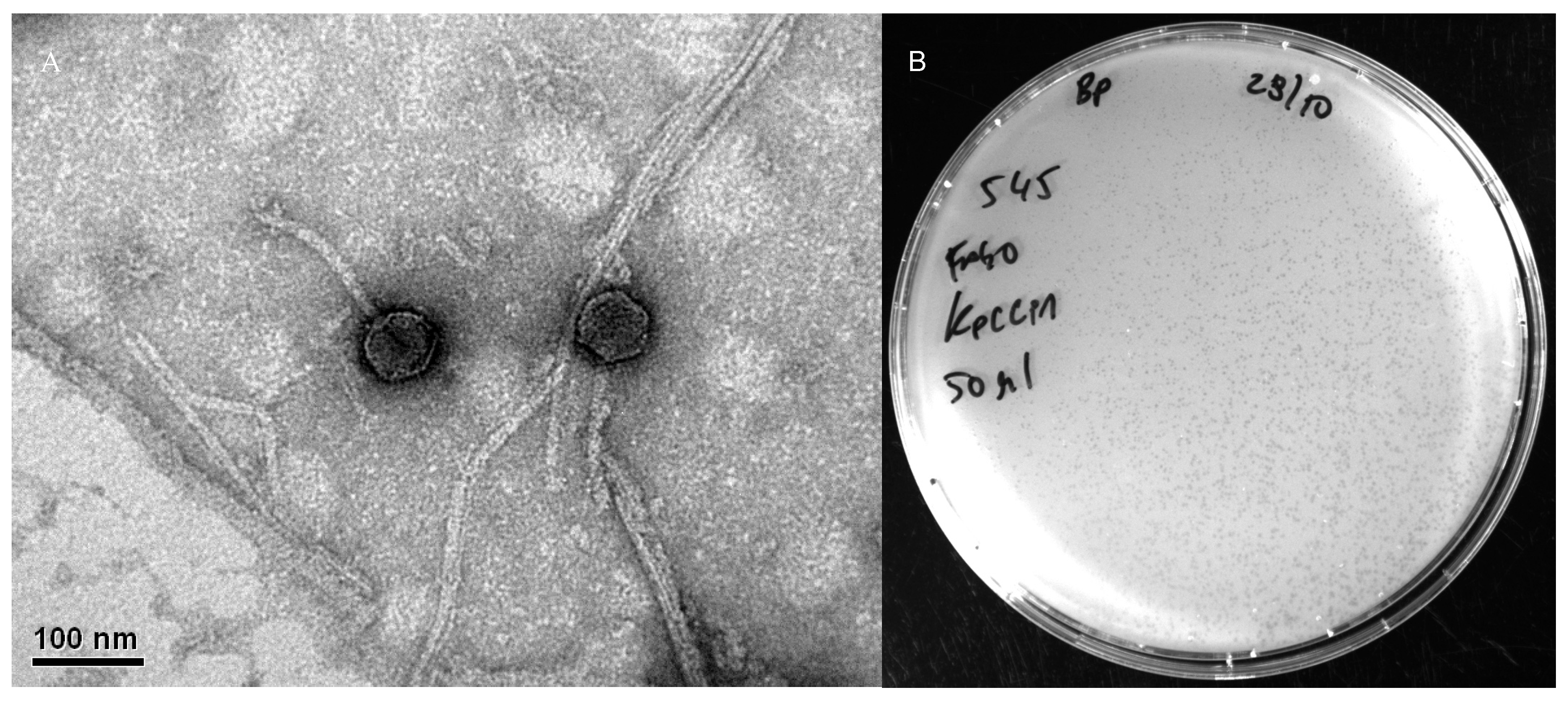
Figure 1. Transmission electron micrograph of phage KpCCP1 (A) and lysis plaques of phage KpCCP1 on the lawn of K. pneumoniae UCO-545 (B).
Sci 07 00157 g001
Figure 2. Genome map of phage KpCCP1 obtained using Proksee. Open reading frames (ORFs), GC content and GC skew are indicated.
Sci 07 00157 g002
Figure 3. Proteomic-based phylogenetic analysis of phage KpCCP1 generated using ViPTree. The global tree (left) shows the position of KpCCP1 within the family Straboviridae (red star), while the detailed view (right) highlights its relationship with members of the genera Slopekvirus and Pseudotevenvirus. Branch lengths represent genomic distance based on normalized tBLASTx (v2.14.1) scores.
Sci 07 00157 g003
Figure 4. Phylogenetic tree of phage KpCCP1 within the genus Slopekvirus, constructed using the Virus Classification and Tree Building Online Resource (VICTOR) based on whole-genome sequence analysis. Bootstrap values are shown at branch nodes, and branch lengths indicate genomic distance. The results support the classification of KpCCP1 as a novel species within the genus.
Sci 07 00157 g004
Figure 5. Genomic comparison of phage KpCCP1 and its closest relative, phage Eap3. Arrow colors represent the gene clusters encoding homologous proteins, and the connecting lines indicate proteins that share >80% sequence identity.
Sci 07 00157 g005
Figure 6. Dendrogram of ERIC-PCR profiles of carbapenem-resistant strains of K. pneumoniae (A) and K. michiganensis (B). Strains grouped on a branch arising from a node with a DICE similarity index ≥ 90% were considered genetically related.
Sci 07 00157 g006
Table 1. Comparative genomic characteristics of Slopekvirus phages and their intergenomic similarity (IS) with phage KpCCP1. Values were calculated using VIRIDIC. The table includes the assigned species cluster, genome size, GC content, number of coding sequences (CDSs), and host species for each phage.
PhageIS with KpCCP1Species ClusterGenome Size (bp)GC ContentNo of CDSsHostAccession no.
KpCCP1--1177,27641.8292K. pneumoniaePV402122
Eap396.01175,81442278K. aerogenesNC_041980
KP1595.92174,43642258K. pneumoniaeNC_014036
Matisse94.83176,08142280K. pneumoniaeNC_028750
KP2793.03174,41342276K. pneumoniaeNC_020080
pht4A914171,59841.5257Escherichia coliNC_055712
Table 2. Putative Acr-encoding genes detected in phage KpCCP1.
CDSLength (AA)Sequence
16095MITIVYWEEVESEMIDGELAEVDGESEMLTATETIENAFDRIKAHQKEMEGRDIQYMMSFWVGGEALCHAVIAESTSVDTCREKIEQYVKSFLH
10466MNMYEMLKSENSETQGEKDYNKWKRSVQHMLGSSDYDEDLMYALYSDGCTPEDAVCEYYAQGDEE
20558MQIKITKTFFRDITEGKVYTAKKDKEGLWIENDWNRDIWLNLTNEYNKELIEYEIVK
Table 3. Antibiotic resistance profiles of KpCCP1-susceptible Klebsiella strains.
Klebsiella spp. StrainAntibiotic Resistance Profile *
K2017IMP, MEM, ETP, CRO, CTX, ATM, AMK, SXT, CIP
K2018IMP, MEM, CTX, ATM, SXT, CIP
K2070IMP, MEM, ETP, CRO, CAZ, CTX, CZA, ATM, GEN, AMK, CIP
K2113IMP, MEM, ETP, CRO, CAZ, CTX, CZA, ATM, GEN, SXT, CIP
K2115IMP, MEM, ETP, CRO, CAZ, CTX, ATM, GEN, SXT, CIP
K2116MEM, CRO, CAZ, CTX, CZA, SXT, CIP
K2122IMP, MEM, ETP, CRO, CAZ, CTX, ATM, GEN, SXT, CIP
K2123IMP, MEM, ETP, CRO, CAZ, CTX, ATM, GEN, SXT, CIP
K2166IMP, MEM, ETP, CRO, CAZ, CTX, ATM, GEN, AMK, CIP
44UDECIMP, MEM, ETP, CRO, CAZ, CTX, CZA, GEN, AMK
99UDECIMP, MEM, ETP, CRO, CAZ, CTX, CZA, ATM, GEN, AMK, SXT, CIP
115UDECIMP, MEM, ETP, CRO, CAZ, CTX, CZA, ATM, GEN, AMK, SXT, CIP
196UDECIMP, MEM, ETP, CRO, CAZ, CTX, ATM, AMK, CIP
209UDECIMP, MEM, ETP, CRO, CAZ, CTX, CZA, ATM, GEN, AMK, SXT, CIP
230UDECIMP, MEM, ETP, CRO, CAZ, CTX, CZA, ATM, GEN, AMK, CIP
355UDECIMP, MEM, ETP, CRO, CAZ, CTX, ATM, AMK
379UDECIMP, MEM, ETP, CRO, CAZ, CTX, CZA, AMK
UCO333IMP, MEM, ETP, CRO, CAZ, CTX, ATM, GEN, AMK, CIP
UCO340IMP, MEM, ETP, CRO, CAZ, CTX, ATM, GEN, AMK, CIP
UCO545ETP, CRO, CAZ, CTX, ATM, AMK, SXT, CIP
UCO638CRO, CTX, SXT
UCO639CRO, CTX, SXT
* Abbreviations: IMP, imipenem; MEM, meropenem; ETP, ertapenem; CRO, ceftriaxone; CTX, cefotaxime; CAZ, ceftazidime; CZA, ceftazidime/avibactam; ATM, aztreonam; GEN, gentamicin; AMK, amikacin; SXT, trimethoprim/sulfamethoxazole; CIP, ciprofloxacin.
Disclaimer/Publisher’s Note: The statements, opinions and data contained in all publications are solely those of the individual author(s) and contributor(s) and not of MDPI and/or the editor(s). MDPI and/or the editor(s) disclaim responsibility for any injury to people or property resulting from any ideas, methods, instructions or products referred to in the content.