Article

Antibacterial Properties of Submerged Cultivated Fomitopsis pinicola, Targeting Gram-Negative Pathogens, Including Borrelia burgdorferi

1
Department of Chemistry and Biotechnology, Tallinn University of Technology, 12618 Tallinn, Estonia
2
Institute of Microbial Biotechnology, Agricultural University of Georgia, 0131 Tbilisi, Georgia
*
Author to whom correspondence should be addressed.
Sci 2025, 7(3), 104; https://doi.org/10.3390/sci7030104 (registering DOI)
Submission received: 6 June 2025 / Revised: 27 July 2025 / Accepted: 30 July 2025 / Published: 2 August 2025
(This article belongs to the Section Biology Research and Life Sciences)

Abstract

The rise in multidrug-resistant bacterial strains and persistent infections such as Lyme disease caused by Borrelia burgdorferi highlights the need for novel antimicrobial agents. The present study explores the antioxidant, antibacterial, and cytotoxic properties of extracts from submerged mycelial biomass of Fomitopsis pinicola, cultivated in synthetic and lignocellulosic media. Four extracts were obtained using hot water and 80% ethanol. The provided analysis of extracts confirmed the presence of various bioactive compounds, including flavonoids, alkaloids, and polyphenols. All extracts showed dose-dependent antioxidant activity (IC50: 1.9–6.7 mg/mL). Antibacterial tests revealed that Klebsiella pneumoniae was most sensitive, with the L2 extract producing the largest inhibition zone (15.33 ± 0.47 mm), while the strongest bactericidal effect was observed against Acinetobacter baumannii (MBC as low as 0.5 mg/mL for L1). Notably, all extracts significantly reduced the viability of stationary-phase B. burgdorferi cells, with L2 reducing viability to 42 ± 2% at 5 mg/mL, and decreased biofilm mass, especially with S2. Cytotoxicity assays showed minimal effects on NIH 3T3 cells, with slight toxicity in HEK 293 cells for S2 and L1. These results suggest that F. pinicola extracts, particularly ethanolic L2 and S2, may offer promising natural antimicrobial and antioxidant agents for managing resistant infections.

1. Introduction

The global rise in multidrug-resistant (MDR) bacteria and persistent bacterial infections represents one of the most urgent challenges in public health today. MDR pathogens, particularly those classified as ESKAPE pathogens (Enterococcus faecium, Staphylococcus aureus, Klebsiella pneumoniae, Acinetobacter baumannii, Pseudomonas aeruginosa, and Enterobacter species), are considered major agents of healthcare-associated infections (HAIs) and are notable for their resistance to multiple antibiotic classes. Recognized as critical targets by the Infectious Diseases Society of America and the World Health Organization, these pathogens are the principal causative agents of hospital-acquired infections that are difficult to treat and frequently affect immunocompromised patients [1,2].
Klebsiella pneumoniae, Acinetobacter baumannii, and Enterobacter cloacae, three Gram-negative members of the ESKAPE group, are often implicated in severe respiratory, urinary tract, and bloodstream infections, especially in hospitalized patients with weakened immune systems. K. pneumoniae, a Gram-negative, encapsulated, non-motile bacterium, is associated with respiratory infections such as bronchopneumonia and bronchiectasis, as well as urinary tract infections and sepsis, especially in intensive care units [3]. A. baumannii is similarly linked to high rates of morbidity and mortality due to its role in respiratory and bloodstream infections in critical-care settings and its extensive resistance profile [4]. E. cloacae is a significant pathogen due to its intrinsic resistance to multiple antibiotics, including penicillins and cephalosporins, which complicates treatment in healthcare-associated bloodstream and urinary tract infections [5,6]. With a high prevalence of pan-resistant strains among these pathogens, K. pneumoniae, A. baumannii, and E. cloacae represent a particularly severe threat, necessitating novel antimicrobial development and improved infection control measures [7,8].
Similarly challenging is Borrelia burgdorferi, the spirochete responsible for Lyme Borreliosis (LB), the most prevalent tick-borne bacterial infection across temperate regions in Europe, North America, and Asia [9]. Although many patients recover fully with antibiotic treatment, approximately 10–20% experience persistent symptoms, such as fatigue, musculoskeletal pain, and cognitive impairment, often attributed to the bacterium’s ability to form dormant cystic and biofilm states, complicating treatment and enabling chronic infection in some cases [10,11]
With the increasing challenges posed by MDR pathogens and the persistence of Borrelia infections, naturally occurring compounds have emerged as potential alternatives or adjuncts to traditional antibiotics. Various plant-derived polyphenolic compounds, such as quercetin, luteolin, baicalein, and resveratrol, have demonstrated significant activity against B. burgdorferi in vitro, particularly against both its spirochete and rounded forms, and often exhibit synergy with other phytochemicals [12,13,14,15,16,17,18]. Beyond plants, medicinal mushrooms, particularly Fomitopsis pinicola, have shown a broad spectrum of bioactive compounds, including antioxidants, antimicrobials, and immunomodulators [19,20,21].
Fomitopsis pinicola P. Karst., also known as the red-belted bracket fungus or red-banded polypore, is a ubiquitous brown-rot nontoxic medicinal mushroom belonging to the class Agaricomycetes, the family Fomitopsidaceae, and the division Basidiomycota [21,22,23]. F. pinicola have been reported in several studies to be one of the most promising sources for obtaining natural compounds with a broad spectrum of pharmaceutical and/or medicinal properties [22,24,25]. Various extracts of F. pinicola are being investigated by multiple research groups for their therapeutic potential, with particular emphasis on their anticancer and antibacterial activities [25,26,27]. F. pinicola is recognized as a rich source of diverse bioactive compounds. The fruiting body, submerged mycelium, and culture filtrate of this mushroom contain a wide range of active constituents, such as phenolic compounds, flavonoids, triterpenes, esters, lactones, steroids, amino acids, ergosterol, polysaccharides, and dietary fiber, along with a variety of micro- and macroelements [19,21,22,24,27,28]. Comprehensive investigations have substantiated that due to the content of various bioactive compounds, extracts derived from F. pinicola exhibit medicinally relevant potential, encompassing antioxidant, anti-inflammatory, antitumor, and antimicrobial properties [19,24,29,30]. The diverse bioactive constituents of F. pinicola make it a promising candidate for combating multidrug-resistant (MDR) pathogens. Moreover, despite the extensive literature on the antibacterial activity of various basidiomycete mushrooms, no data are currently available on the inhibition of B. burgdorferi, the causative agent of Lyme disease, by mushroom-derived compounds or extracts. Although F. pinicola has been the subject of a substantial number of studies, it remains less extensively investigated than other well-known basidiomycetes, and its full medicinal and pharmaceutical potential is yet to be fully elucidated. Notably, the present study is the first to evaluate the properties of a F. pinicola strain isolated in Georgia. Two fundamentally different growth substrates, glycerol and mandarin pomace (MP), were used for cultivating F. pinicola. Glycerol, an easily metabolizable carbon source, supports rapid fungal growth and primary metabolism. In contrast, mandarin pomace, a complex lignocellulosic substrate, closely resembles the natural habitat of wood-decaying basidiomycetes and is known to induce ligninolytic and stress-related secondary metabolism, thereby enhancing the production of phenolics, terpenoids, and other bioactive compounds with antioxidant and antimicrobial properties. In this study, the in vitro anticancer and bactericidal activities of water and ethanol extracts derived from F. pinicola mycelial biomass were evaluated against critical multidrug-resistant (MDR) Gram-negative pathogens from the ESKAPE group, K. pneumoniae, A. baumannii, and E. cloacae, as well as B. burgdorferi.

2. Materials and Methods

2.1. Microorganisms and Cell Lines

Fomitopsis pinicola fruiting bodies were collected in western Georgia in 2022. Molecular confirmation of the fungal species used in the study was carried out at the Biodiversité et Biotechnologie Fongiques INRAE-Aix Marseille Université (France). To identify the fungal species, DNA was extracted from the sample using the Nucleospin Plant II® kit, followed by amplification of the internal transcribed spacer (ITS) region using a protocol adapted from [31]. Sequencing was performed on both forward and reverse strands, and due to overlapping signals, multiple sequence variants were analyzed individually using BLASTn against the GenBank database. The resulting sequences exhibited high similarity (98.8–99.78%) with reference sequences of F. pinicola. This molecular analysis verifies the taxonomic affiliation of the isolate as F. pinicola. A pure culture of F. pinicola was preserved in the culture collection of the Institute of Microbial Biotechnology at the Agricultural University of Georgia. The culture was preserved on malt extract agar (MEA) slants within glass tubes and on Petri dishes and stored at 5 °C.
Human embryonic kidney cell HEK 293 and mouse embryonic fibroblast NIH 3T3 cell lines were obtained from the American Type Culture Collection. The cells were propagated in Dulbecco’s modified Eagle’s medium (DMEM) (Gibco), supplemented with 10% bovine calf serum (Gibco) and 5% penicillin/streptomycin. All cell lines were incubated at 37 °C in a humidified 5% CO2 and 95% air atmosphere.
Antibacterial activity of F. pinicola extracts was determined by two methods, MBC assessment in the case of K. pneumoniae, A. baumannii, and E. cloacae and SYBR Green I/PI Assay in the case of B. burgdorferi. A clinical isolate of K. pneumoniae HUMB 01336 was obtained from the Estonian Electronic Microbial database (https://eemb.ut.ee accessed on 5 September 2024), and A. baumannii DSM25645 and E. cloacae DSM109592 were obtained from the German Collection of Microorganisms and Cell Cultures (DSMZ). Bacteria were routinely cultivated on TSA medium (g/L distilled water) (papaic digest of soybean meal—3, dextrose—2.5, pancreatic digest of casein—17, NaCl—5, K2HPO4—2.5, and agar—15). Low-passage (≤8) isolates of Borrelia burgdorferi strain B31 were acquired from the American Type Culture Collection (Manassas, VA, USA). The bacteria were cultured in BSK-H medium supplemented with 6% rabbit serum. All media were sterilized by filtration through 0.2 µm pore-size filters. Cultures were maintained in sterile 50 mL closed conical tubes at 33 °C in a 5% CO2 atmosphere, without the addition of antibiotics. After 7 days of incubation, the cultures reached the stationary growth phase (approximately 107 spirochetes/mL), after which they were transferred to 96-well tissue culture microplates for fraction screening.

2.2. Mushroom Inoculum Preparation and Biomass Growth Parameters

Mushroom inocula were cultivated in 250 mL Erlenmeyer flasks containing 100 mL of synthetic nutrient medium and incubated on a rotary shaker (New Brunswick Scientific, Edison, NJ, USA) at 150 rpm and 27 °C for 8 days. The composition of the medium was (g/L distilled water): K2HPO4—0.6, KH2PO4—0.8, MgSO4·7H2O—0.5, glucose—15.0, peptone—3.0, and yeast extract—3.0, with the initial pH adjusted to 6.0 before sterilization. Following incubation, the resulting fungal pellets were homogenized twice for 20 s using a Waring laboratory blender (Waring Commercial, Torrington, CT, USA) and employed as a 10% inoculum.
To obtain sufficient mycelial biomass for extract preparation, submerged cultivation was carried out in 2 L Erlenmeyer flasks containing 1 L of basal medium. Two nutrient media were used: a synthetic medium supplemented with glycerol (10 g/L) as the carbon source and a lignocellulosic medium containing mandarin pomace (MP, 60 g/L). The basal medium composition was as follows (g/L distilled water): yeast extract—3.0; K2HPO4—0.6, KH2PO4—0.8, and MgSO4·7H2O—0.5, with the pH adjusted to 6.0 before sterilization. Cultures were incubated at 27 °C for 8 days on a refrigerated rotary shaker (MRC, Holon, Israel) set at 130 rpm.

2.3. Preparation of Mushroom Extracts

After 8 days of submerged cultivation, mycelial biomass was harvested, filtered, dried at 45 °C to constant weight, and ground to a fine powder using a laboratory mill (KM-1500, MRC, Israel). The dried biomass was extracted with distilled water and 80% ethanol. Hot water extraction was performed at 80 °C for 3 h (1 g/10 mL) in a shaking water bath, while ethanolic extraction was carried out at 27 °C for 3 h on a rotary shaker at 150 rpm. Insoluble material was removed by centrifugation (Eppendorf 5417R, Hamburg, Germany) at 6000 rpm and 4 °C for 15 min, followed by filtration through Whatman® No. 1 paper. Crude extracts were concentrated under a vacuum to yield resin or powder, transferred to pre-weighed tubes, and stored at −20 °C. Stock solutions were prepared in 99.9% DMSO (Sigma-Aldrich, Saint Louis, MO, USA) [32,33].

2.4. Scavenging Activity on 2,2-Diphenyl-1-Picrylhydrazyl (DPPH) Radicals

The free radical scavenging activity of mushroom extracts was evaluated by a DPPH assay [32]. A 5.9 mg/100 mL DPPH ethanolic solution was prepared. Then, 0.1 mL of extract at concentrations from 0.05 to 15.0 mg/mL was mixed with 1.9 mL of DPPH solution, vortexed for 5 s, and incubated in the dark at room temperature for 30 min. Absorbance was measured at 517 nm against a blank control. BHA and α-tocopherol served as positive controls. Radical scavenging activity (%) was calculated as follows: % inhibition = [(A blank − A sample)/A blank] × 100, where A blank is the control absorbance and A sample is the sample absorbance.

2.5. Qualitative Analysis of Bioactive Constituents in Fomitopsis Pinicola Extracts

Qualitative profiling of bioactive constituents in mushroom extracts obtained using water and ethanol was conducted following standard analytical protocols. This assessment aimed to identify major classes of bioactive constituents, including phenols, saponins, flavonoids, alkaloids, reducing sugars, and glycosides [33,34]. The following provides a brief description of the methods:
Fehling test for reducing sugars: Equal volumes of Fehling’s A and Fehling’s B media are mixed with a few milliliters of the test solution and boiled. The brick-red precipitate indicates a positive result for reducing sugars.
Biuret test for peptide bond: A few milliliters of the test solution is placed in a test tube. Then, an equal volume of Biuret reagent (copper (II) sulfate + sodium hydroxide) is added to the solution. If proteins or peptides are present in the sample, the solution changes color from blue to violet.
Foam test for saponins: A small amount of extract is mixed with distilled water and vigorously shaken. A stable, persistent froth or foam indicates the presence of saponins.
Mayer’s test for alkaloids: A few drops of Mayer’s reagent are added to the acidified extract. If alkaloids are present, a cream-colored or white precipitate appears.
Iodine test for phenols: A few drops of iodine solution (iodine dissolved in potassium iodide) are added to the sample and mixed. If phenols are present, a brown- or yellow-colored precipitate forms.
Braymer’s test for tannins: A few drops of Braymer’s reagent are added to the solution and mixed. The reaction is positive if blue-black or green precipitate forms.
Salkowski’s test for glucosides: A small amount of the extract is dissolved in chloroform. Then, a few drops of concentrated sulfuric acid are carefully added along the sides of the test tube. The presence of a reddish-brown, red, or violet ring indicates a positive result.
Lead acetate test for flavonoids: A few drops of 10% lead acetate solution are added to a small amount of the sample and mixed. If flavonoids are present, a yellow-colored precipitate forms.

2.6. Cell Culture Assays

2.6.1. Cell Treatment Procedure

Cells were seeded in 96-well plates at a density of 2.5 × 10⁵ cells per well using the Countess Automated Cell Counter (Invitrogen) and incubated overnight. After 24 h, 100 µL of either fresh medium or fresh medium containing test extracts at a final concentration of 50 µg/mL (diluted in 3% DMSO) was added to each well, followed by an additional 48 h incubation period. Wells treated with 3% DMSO alone served as the solvent control.

2.6.2. Cell Viability by WST-1 Assay

Cell viability was assessed using the WST-1 assay (Roche), which enables a colorimetric evaluation based on the reduction of tetrazolium salts to water-soluble formazan by metabolically active cells. The amount of formazan produced is directly proportional to the number of viable cells. Measurements were performed 48 h following treatment. Wells treated with 5 µL of 3% DMSO served as the solvent control. Subsequently, 5 µL of WST-1 reagent was added to each well containing 100 µL of culture medium, and the plates were incubated at 37 °C for 2 h. Absorbance was then measured at 450 nm using a TECAN GENios Pro Microplate Reader (Switzerland) [18].

2.7. Bacterial Culture Assays

The disk diffusion method is used to evaluate the antimicrobial activity of each mushroom extract. The disk diffusion test was performed with Trypton Soya Agar (TSA) and disks from Oxoid®-England by the Kirby–Bauer technique. The mushroom extracts at a concentration of 100 mg/mL were sterilized through a Millipore filter (0.22 µm) and then loaded onto sterile filter paper disks (6 mm in diameter). A single bacterial colony was selected from overnight culture plates and inoculated into 5 mL of tryptic soy (TS) broth, followed by incubation at 37 °C with shaking at 150 rpm for 16 h. The resulting culture was then diluted 1:50 in fresh TS broth and incubated for an additional 2 h to achieve exponential growth. The optical density at 600 nm was adjusted to 0.1, and 100 µL of the resulting bacterial suspension was evenly spread onto TSA plates. Plates were allowed to air dry for 5 min. Sterile filter paper disks impregnated with mushroom extract were then placed onto the surface of the TSA plates. The plates were incubated at 35 °C for 24 h. Antibacterial activity was evaluated by measuring the diameter of the inhibition zones using a Vernier caliper. All experiments were conducted in three biological replicates.

2.8. Minimal Bactericidal Concentration (MBC)

A single colony from a TSA plate was inoculated in TS broth and incubated for 16 h at 37 °C with shaking at 150 rpm. The resulting culture was then diluted 1:50 with fresh medium and further incubated under the same conditions until reaching the exponential growth phase (OD600 = 0.6). Cells were harvested by centrifugation at 5000× g for 10 min at 4 °C, and the pellet was resuspended in an equal volume of sterile water. The washing step was repeated twice, and the final pellet was resuspended in sterile water to achieve a target cell density of OD600 = 0.2.
Test compounds were prepared at defined concentrations using 3% DMSO as a solvent. This concentration was selected based on preliminary experiments showing that a 1:1 dilution of 3% DMSO (resulting in a final concentration of 1.5%) had no significant effect on the viability of any of the tested bacterial strains after 24 h of exposure. The maximum concentration tested in this format was 5 mg/mL.
For the assay, 100 µL of bacterial suspension was mixed with 100 µL of mushroom extract solution and incubated at 37 °C for 24 h. After the exposure period, 3 µL of each cell suspension was spot-inoculated onto TSA plates and incubated at 37 °C for an additional 24 h. The MBC was defined as the lowest compound concentration at which no visible colony formation was observed from the 3 µL spot on TSA. All MBC determinations were performed in three independent biological replicates [35].

2.9. SYBR Green I/PI Assay

Standard methods such as disk diffusion and MBC determination on agar are unsuitable for B. burgdorferi because it does not form colonies on solid media, grows slowly under strict microaerophilic conditions, and can form persister cells and biofilms that are not detected by conventional assays. Therefore, we employed the SYBR Green I/PI viability assay, a well-established and validated method specifically optimized for B. burgdorferi, which enables sensitive and accurate discrimination between live and dead cells.
To assess the viability of Borrelia burgdorferi, a SYBR Green I/propidium iodide (PI) dual-staining assay was performed as previously described by Feng et al. [36]. In brief, 5 µL of SYBR Green I (100× stock; Invitrogen, Waltham, MA, USA) and 5 µL of PI (0.5 mM; Sigma, St. Louis, MO, USA) were added to each well of the microplate and mixed thoroughly. The plates were then incubated in the dark at room temperature for 15 min. Fluorescence was measured using a TECAN Genios Pro microplate reader (Männedorf, Switzerland) with excitation at 450 nm and emission recorded at 535 nm (green fluorescence) and 635 nm (red fluorescence) for each well. In parallel, B. burgdorferi suspensions with defined ratios of live to dead cells (0:10, 2:8, 5:5, 8:2, 10:0), with dead cells prepared by treatment with 70% isopropyl alcohol, were mixed and added to separate wells of a 96-well plate. The SYBR Green I/PI staining solution was applied to each of these samples, and the green-to-red fluorescence ratios were recorded using the same instrument settings. A standard curve correlating the percentage of live cells with the green/red fluorescence ratio was generated using least-squares regression analysis. The resulting regression equation was then used to calculate the percentage of viable B. burgdorferi cells in each experimental well of the screening plate.

2.10. Evaluation of Antibacterial Effect of Fomitopsis pinicola Extracts Against B. burgdorferi

To evaluate the possible activity of mushroom extracts against stationary-phase B. burgdorferi, probes at concentrations of 2 mg/mL and 5 mg/mL were added to 100 µL of the seven-day-old B. burgdorferi culture in the 96-well plate. The entire experiment was repeated in triplicate for each concentration of the tested compounds. All plates were incubated at 33 °C in 5% CO2 for the next 7 days. Control cultures were treated with DMSO and a control solution with a mix of the antibiotics doxycycline, daptomycin, and cefoperazone, with a final concentration of 50 µM. The live and dead cells were evaluated using the SYBR Green I/PI assay, and the viability % was calculated through the regression equation.

2.11. Crystal Violet Biofilm Assay

The anti-biofilm activity of the tested mushroom extracts was evaluated by quantifying total biofilm biomass using crystal violet staining, as previously described [17]. After incubation, the culture media were carefully removed to retain the surface-attached biofilms. Plates were then gently washed by submerging them in phosphate-buffered saline (PBS) to remove non-adherent cells, followed by heat fixation at 60 °C for 1 h. Biofilms were stained with 2% crystal violet solution for 5 min, then washed twice by immersion in deionized water to remove excess dye. Bound crystal violet was solubilized with 33% glacial acetic acid, and biofilm biomass was quantified by measuring the optical density at 570 nm using a TECAN GENios Pro microplate reader.

2.12. Statistical Analysis

Statistical analysis was conducted using one-way analysis of variance (ANOVA), followed by Dunnett’s post hoc multiple comparison test. All data represent the mean ± standard deviation (SD) from a minimum of three independent experiments, each performed in triplicate. The optical density of the solvent-treated control group was defined as 100% cell viability. Statistical significance was indicated as follows: p < 0.05 (*), p < 0.01 (**), p < 0.001 (***), and p < 0.0001 (****). All statistical analyses were performed using GraphPad Prism version 9.

3. Results

3.1. Yield and Abbreviation of Extracts Obtained from Fomitopsis pinicola Mycelial Biomass

To acquire submerged mycelial biomass, F. pinicola was cultivated for 8 days following the protocol described in the Methods section, using two-liter Erlenmeyer flasks incubated on a rotary shaker at 150 rpm and 27 °C. The harvested biomass was then dried at 45 °C to a constant weight and ground into a fine powder.
Table 1 displays the yields and corresponding abbreviations for the extracts derived from the submersed mycelia of the examined mushroom. The abbreviations S1 and S2 represent extracts obtained from submersed mushroom biomass cultivated in a synthetic medium containing glycerol. Conversely, the abbreviations L1 and L2 denote extracts obtained from submersed mushroom biomass cultivated in a medium containing lignocellulose (MP—mandarin pomace). The dry powdered biomass of the tested mushroom was subjected to extraction using distinct solvents, including distilled water (via hot extraction at 80 °C) and ethanol (80%). Minimal quantitative variances in yield were observed between the aqueous and ethanolic extracts derived from the submersed biomass of F. pinicola on various growth substrates (Table 1).

3.2. DPPH Free Radical Scavenging Activity of Fomitopsis pinicola Extracts

Antioxidants are vital in neutralizing reactive oxygen species (ROS), which are highly detrimental to biological systems and have been implicated in the pathogenesis of numerous diseases, including multiple types of cancer. The ability of antioxidant compounds, such as ascorbic acid, flavonoids, and diverse polyphenols, to scavenge free radicals is essential for protecting cells from oxidative stress. Although synthetic antioxidants like butylated hydroxyanisole (BHA), butylated hydroxytoluene (BHT), and tert-butylhydroquinone (TBHQ) are commonly employed as food additives, studies have demonstrated their potential toxicity, including tumorigenic effects [37,38]. Consequently, there is a growing demand for natural sources of potent antioxidants. Notably, approximately 90% of known mushroom species have yet to be investigated for their antioxidant and anticancer properties [28]. A wide range of mushroom species, including F. pinicola, are recognized as valuable sources of antioxidant compounds. In the present study, the antioxidant activity of unrefined extracts obtained from F. pinicola was assessed using the DPPH radical scavenging assay, with the corresponding results illustrated in Figure 1.
The free radical scavenging activity of the tested extracts exhibited a clear concentration dependency. Notably, the highest antioxidant activities were recorded at concentrations of 5, 10, and 15 mg/mL, with scavenging effects ranging between 67 and 77%, 69 and 79%, 68 and 79%, and 47 and 74%, respectively (Figure 1). The IC50 values representing the concentration required to neutralize 50% of DPPH radicals were relatively low, ranging from 1.9 to 6.7 mg/mL, indicating considerable antioxidant efficacy. Despite the variation in antioxidant capacity among the extracts, their activity levels did not match those of the reference standards, BHA, and α-tocopherol, which demonstrated significantly higher scavenging potential. As expected, both positive controls exhibited strong antioxidant effects even at the minimal concentration of 0.05 mg/mL. The findings confirm that the tested extracts possess notable antioxidant potential, which is likely to contribute to their overall bioactivity.

3.3. Qualitative Analysis of Bioactive Constituents in Fomitopsis pinicola Extracts

The phytochemical profiles of mushroom extracts obtained using various solvents are presented in Table 2, revealing the presence of multiple bioactive constituents. Extracts from submerged F. pinicola mycelial biomass investigated as S and L samples demonstrated the presence of reducing sugars, glycosides, saponins, polyphenols, tannins, flavonoids, and alkaloids. Saponins, a class of glycosides commonly found in both plants and fungi, are known for their broad spectrum of pharmacological effects, including antiviral, anti-inflammatory, and anticancer activities [39]. Notably, saponins were detected across all solvent extracts analyzed in the present study. Mushrooms are recognized for their high nutritional value and are abundant sources of polyphenols and flavonoids, compounds extensively documented for their anti-inflammatory, antimicrobial, and antioxidant activities [40,41,42]. These bioactive constituents, including tannins, were consistently detected across all tested extracts. Alkaloids, which exhibit potent pharmacological properties such as antimicrobial, anti-inflammatory, anticancer, antiviral, and anti-aging effects [43], were exclusively identified in the ethanolic extracts. Their absence in water extracts is likely attributable to the predominantly non-polar nature of these compounds, limiting their solubility in water. All extracts tested positive for the presence of reducing sugars (carbohydrates) and glucosides. Reducing sugars play a crucial role as an energy source supporting mushroom growth and metabolic processes. Certain glucosides derived from mushrooms have been shown to confer positive health benefits, including antioxidant, anticancer, antimicrobial, and neuroprotective activities [44,45]. However, it is important to note that some glucosides may hydrolyze to release toxic aglycones. Comparative analysis of the results revealed no evident correlation between the major classes of bioactive compounds detected and the type of cultivation medium (lignocellulosic (L) or synthetic (S)) employed. Among the extracts, S2 and L2 exhibited the most pronounced bioactivity, which may be attributed to the presence of alkaloids, suggesting that these compounds could be the primary contributors to the observed effects. Nonetheless, other bioactive constituents were present in all extracts, albeit likely at varying concentrations, which may also influence their overall bioactivity.
Interestingly, the comparative analysis of the results demonstrates that the cultivation medium (synthetic vs. lignocellulosic) did not strongly correlate with the qualitative presence of the major classes of bioactive compounds, indicating that solvent choice plays a more decisive role in extracting certain compounds than the growth substrate. Nonetheless, the substrate likely influences the quantity and specific profile of the metabolites produced, as reflected in the differing bioactivities of the extracts. Based on the qualitative analysis of F. pinicola extracts, it is hypothesized that the antibacterial effects against B. burgdorferi and Gram-negative pathogens are primarily driven by the presence of alkaloids, flavonoids, and polyphenolic compounds. Alkaloids, found exclusively in the ethanolic extracts, are known for their potent antimicrobial activities, including disruption of bacterial membranes and inhibition of key enzymes, which likely contribute significantly to the observed bactericidal effects. Flavonoids and polyphenols, abundant in all extracts, possess strong antioxidant and antimicrobial properties through mechanisms such as membrane destabilization and interference with microbial metabolism. Additionally, saponins and tannins may enhance these effects by compromising bacterial cell integrity and exerting anti-inflammatory actions. The combined presence and possible synergistic interactions of these bioactive compounds likely underpin the broad-spectrum antibacterial activity of F. pinicola extracts observed in this study.

3.4. Antibacterial Efficiency of Extracts from Fomitopsis pinicola Mycelial Biomass

The antibacterial activity of Fomitopsis pinicola extracts was evaluated by measuring the zone of inhibition (ZOI) on semisolid agar medium, while their bactericidal effect was assessed in aqueous media and expressed as MBC. Preliminary experiments demonstrated that DMSO toxicity did not interfere with the ZOI assay; however, in the MBC assay, the maximum permissible DMSO concentration was limited to 1.5% [35]. Among three tasted pathogenic bacteria, K. pneumoniae was the most sensitive to the tested mushroom extracts S1, L1, and L2, as evidenced by the largest zones of growth inhibition, measuring 6.87 ± 0.26, 14.00 ± 0.82, and 15.33 ± 0.47 mm, respectively (Figure 2). Interestingly, K. pneumoniae was also the only tested bacteria for which no inhibition zone was observed with the S2 extract. In the case of the bacteria A. baumannii, extracts S1 and S2 demonstrated a more pronounced antibacterial effect, as evidenced by their respective inhibition zones of 11.67 ± 0.24 and 8.45 ± 0.41 mm, in contrast to the antibacterial properties of extracts L1 and L2, whose growth inhibition zones corresponded to 7.33 ± 0.62 and 6.87 ± 0.19 mm. The mushroom extracts exhibited the least pronounced antibacterial activity against E. cloacae, with inhibition zone diameters measuring 6.8 ± 0.28 mm, 6.83 ± 0.24 mm, 7.00 ± 0.41 mm, and 6.97 ± 0.45 mm for the S1, S2, L1, and L2 extracts, respectively.
However, differences in the average inhibition zone diameters observed in the F. pinicola extract paper disk assays were not emphasized, as these tests served primarily as preliminary confirmation of the extracts’ inhibitory effects against K. pneumoniae, A. baumannii, and E. cloacae.

3.5. Bactericidal Activity of Extracts from Fomitopsis pinicola Mycelial Biomass

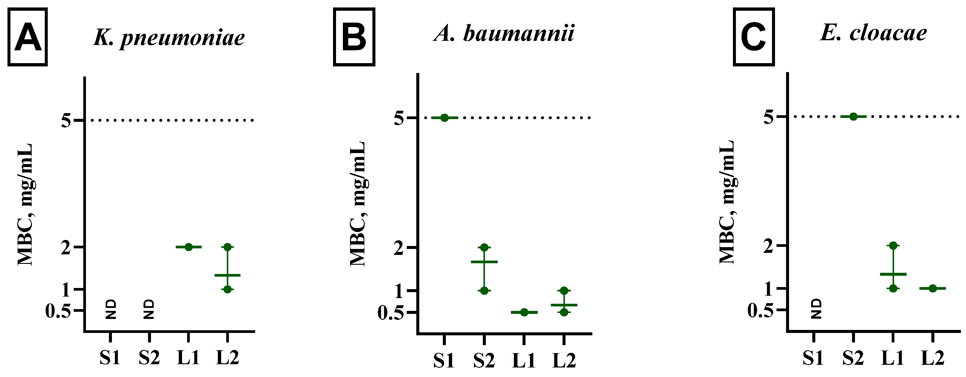
The MBC was noted as the lowermost concentration of F. pinicola extracts where no visible growth of bacteria was observed (Figure 3). The maximum bactericidal effect inflicted by the extracts after 24 h of exposure (MBC of 0.5 and 0.67 mg/mL for extracts L1 and L2, respectively) was found against A. baumannii. Interestingly, the extracts cultivated on glycerol, S1 and S2, also exhibited the lowest MBC against A. baumannii; however, it was higher than for extracts grown on the lignocellulose medium, namely 5 and 1.67 mg/mL, respectively. The S1 extract exhibited bactericidal activity at the highest tested concentration of 5 mg/mL exclusively against A. baumannii. In contrast, the S2 extract demonstrated bactericidal activity at 5 mg/mL against E. cloacae, while for A. baumannii, the MBC was determined to be 1.67 mg/mL. The strongest inhibitory effect on the growth of E. cloacae was observed with the L1 and L2 extracts, with their MBC values being 1.3 mg/mL and 1 mg/mL, respectively. A similar effect of these extracts was noted for K. pneumoniae, although the MBC values were higher, at 2 mg/mL and 1.35 mg/mL for the L1 and L2 extracts, respectively.

3.6. Anti-B. burgdorferi Activity of Extracts from Fomitopsis pinicola Mycelial Biomass

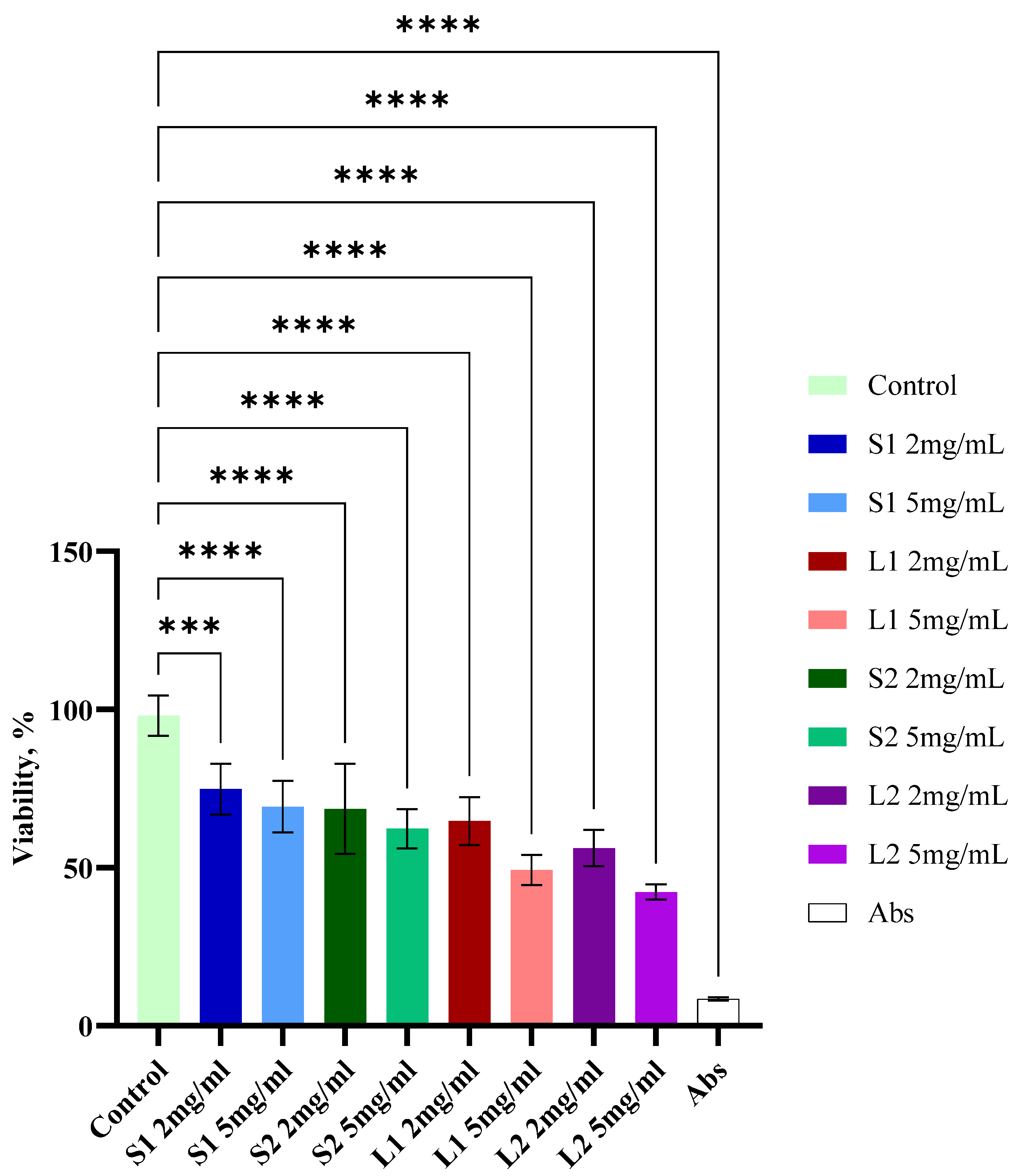
In the present investigation, the antibacterial activity of F. pinicola extracts against the used B. burgdorferi was evaluated in 96-well plates incubated for seven days (Figure 4). All extracts demonstrated considerable activity (p < 0.001) against the stationary-phase B. burgdorferi culture, outperforming the control mix of antibiotics, including doxycycline, daptomycin, and cefoperazone. According to the SYBR Green I/PI assay, treatment with the S1 extract at concentrations of 2 mg/mL and 5 mg/mL significantly (p = 0.0007 and p < 0.0001) reduced the viability of the tested bacteria compared to the control (74 ± 8% and 69 ± 8%, respectively). B. burgdorferi cells’ incubation with the S2 extract at 2 mg/mL and 5 mg/mL concentrations resulting in 68 ± 14% and 62 ± 6% residual viability, respectively, when compared to the DMSO-treated control (p value < 0.0001). The most significant results were achieved with the L1 and L2 extracts. The L1 extract at both 2 mg/mL and 5 mg/mL concentrations resulted in a significant decrease in B. burgdorferi viability (65 ± 8% and 49 ± 5%, respectively, p < 0.0001) compared to the DMSO-treated negative control samples. Moreover, the extract L2 proved to be the most toxic to bacterial cells; it caused a statistically significant decrease (p value < 0.0001) in viability at both 2 mg/mL and 5 mg/mL concentrations: 56 ± 6% and 42 ± 2%, respectively. Thus, treatment with mushroom extracts showed a significant reduction in the viability of seven-day-old B. burgdorferi in the stationary phase compared to the DMSO-treated control group (p < 0.001). The effects of all extracts on viability were clearly concentration-dependent.
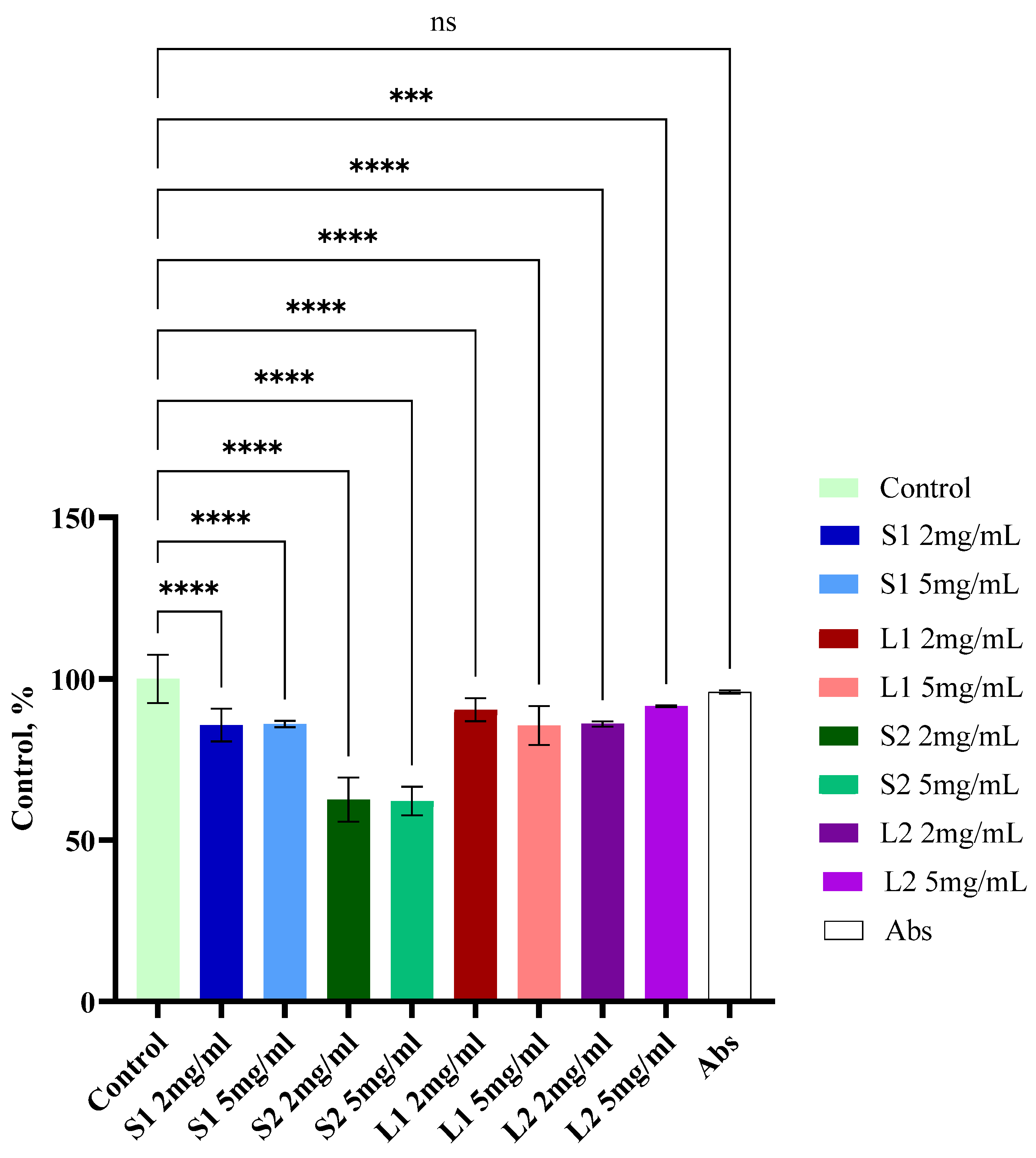
To evaluate the in vitro effect of antimicrobial agents on Borrelia biofilms grown on plastic surfaces, the biofilms were stained with crystal violet and the effectiveness of the different antimicrobial agents was quantified (Figure 5). Borrelia biofilms treated with the three-antibiotic combination did not show any significant effect on biofilm mass compared to the drug-free control. The treatment with all mushroom extracts at all tested concentrations, however, reduced Borrelia biofilm mass compared to the control. The most significant effect on biofilm formation was caused by the ethanolic mushroom extract S2—~37% biofilm reduction at both concentrations used. Interestingly, there were no significant changes in the Borrelia biofilm mass depending on the applied extract concentration.

3.7. Cytotoxic Activity of Extracts from Submerged Fomitopsis pinicola Mycelial Biomass

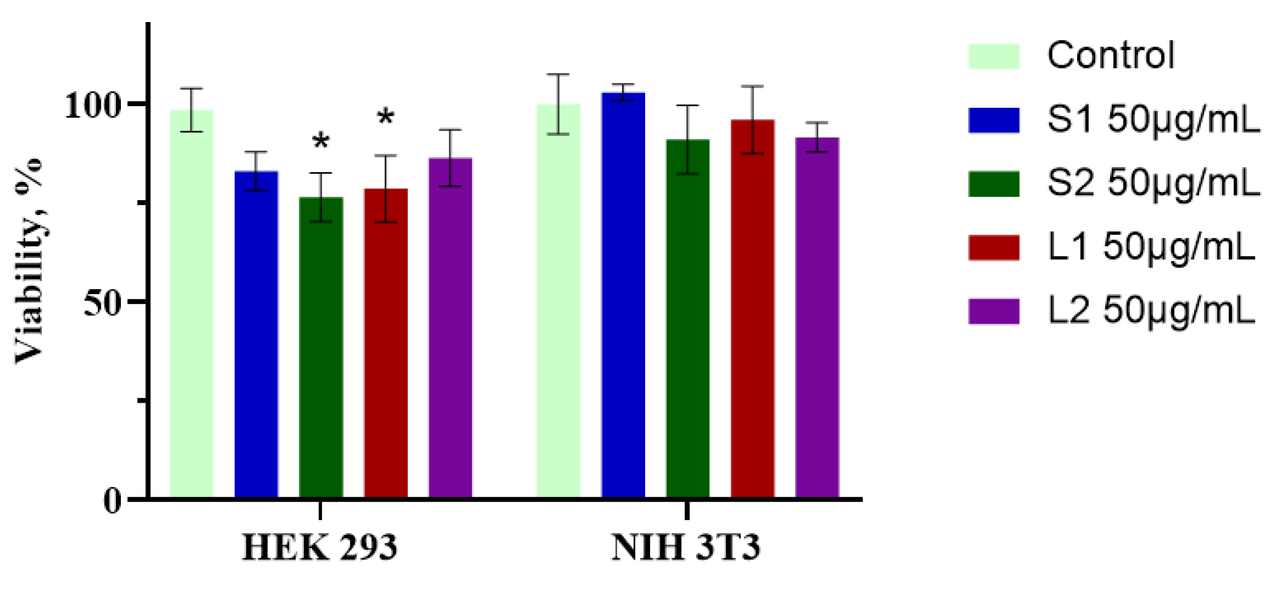
Cell viability was assessed using the WST-1 assay. The cytotoxic effects of all extracts were evaluated in HEK 293 and NIH 3T3 cell lines (Figure 6). It is important to note that HEK 293 cells are not typical human cells; they are immortalized by a well-characterized oncogene but have not undergone malignant transformation. In contrast, NIH 3T3 cells are derived from mouse embryonic fibroblasts (MEFs) and are capable of bypassing cellular senescence to spontaneously immortalize under specific culture conditions. All extracts were prepared at a concentration of 50 mg/mL and incubated with the cells for 48 h. None of the extracts exhibited statistically significant cytotoxic effects on NIH 3T3 cells. Cell viability was detected as 102.9 ± 2%; 91.1 ± 8%; 96.0 ± 8%; and 91.6 ± 3% for extracts S1, S2, L1, and L2, respectively. However, extracts S2 and L1 demonstrated cytotoxicity towards 293 HEK cells, with cell viability recorded at 76.5 ± 6% and 78.6 ± 8%, respectively (p < 0.05). The S1 and L2 extracts did not exhibit statistically significant effects on the growth of 293 HEK cells, with cell viability recorded at 83.1 ± 5% and 86.4 ± 7%, respectively.

4. Discussion

This study explored the bioactive potential of water and ethanol extracts derived from submerged F. pinicola mycelial biomass cultivated on distinct substrates, synthetic (S) and lignocellulosic (L). The yields were consistent across solvents and nutrient media, confirming the feasibility of submerged fermentation for producing bioactive fungal biomass. Ethanol extracts generally exhibited higher biological activity, which may be attributable to their ability to solubilize more diverse and potent non-polar secondary metabolites such as alkaloids.
The growth substrate influenced the biological activity of F. pinicola extracts, particularly their antibacterial properties. While both glycerol-based (synthetic) and lignocellulosic (mandarin pomace-based) media produced comparable extraction yields, lignocellulosic substrates generally enhanced antibacterial activity. Extracts from lignocellulosic media (L1 and L2) showed stronger bactericidal effects, especially against K. pneumoniae, A. baumannii, E. cloacae, and stationary-phase B. burgdorferi, along with greater biofilm inhibition. In contrast, antioxidant activity was moderate across all samples and showed no strong correlation with substrate type, indicating that it was more influenced by the extraction solvent. Overall, the substrate had a more significant effect on antibacterial activity than antioxidant activity, likely due to differences in the concentration or profile of bioactive metabolites.
The evaluation of antioxidant potential using the DPPH radical scavenging assay demonstrated that all extracts exhibited dose-dependent activity, though none surpassed the synthetic antioxidants BHA or α-tocopherol. The relatively low IC50 values (1.9–6.7 mg/mL) indicate that F. pinicola extracts possess significant radical scavenging capabilities, aligning with the literature, which highlights mushrooms as a source of potent antioxidant compounds [42]. These effects are likely due to the presence of polyphenols, flavonoids, and tannins, confirmed in the screening of bioactive compounds.
Several classes of compounds detected in the extracts may contribute to antibacterial activity through different pathways. Flavonoids, for instance, are known to disrupt bacterial membranes and interfere with intracellular targets. In a study conducted by Wu et al. (2013), the antibacterial properties of various particular flavonoids were assessed against E. coli, with a particular focus on their membrane interaction effects [46].
However, in a comprehensive review article, it was summarized that flavonoids exhibit antibacterial activity primarily through disruption of bacterial membranes and inhibition of key enzymes such as DNA gyrase and ATP synthase [47].
Different classes of alkaloids exhibit diverse antibacterial mechanisms, including membrane disruption [48], inhibition of nucleic acid [49], and protein synthesis [50]. These mechanisms are effective not only against a broad spectrum of Gram-negative bacteria [51], but also against antibiotic-resistant strains, as described in the review [52]. The presence of alkaloids exclusively in ethanolic extracts may partly explain the higher activity observed in the S2 and L2 samples, which exhibited the most pronounced antibacterial and anti-Borrelia effects. These extracts showed anti-bacterial activity primarily against K. pneumoniae and A. baumannii and moderate effects against E. cloacae, while their bactericidal concentrations were within the range of 0.5–5 mg/mL depending on the strain and extract type. Extracts L1 and L2, obtained from biomass grown on lignocellulose-based media, were more effective in terms of bactericidal activity, suggesting that the growth substrate may modulate the biosynthesis of specific antimicrobial metabolites. Our findings are consistent with those reported by Dresch et al. (2015) [53], who investigated the antimicrobial properties of F. pinicola alongside other European medicinal polypores. In their study, the authors were unable to determine minimal inhibitory concentrations (MICs) or MBCs for F. pinicola extracts against Gram-negative bacteria such as Escherichia coli and Pseudomonas aeruginosa, as the highest tested concentration (1 mg/mL) was insufficient to inhibit bacterial growth [53]. In contrast, their extracts exhibited strong activity against Gram-positive strains, with MICs ranging from 31 to 125 μg/mL for Bacillus subtilis and 31 to 500 μg/mL for Staphylococcus aureus.
Compared to that study, our investigation offers a more detailed and comprehensive evaluation of F. pinicola’s activity specifically against Gram-negative clinical pathogens, using a broader concentration range (up to 5 mg/mL). This allowed us to determine MBCs and identify potent bactericidal activity—particularly in the L1 and L2 ethanolic extracts, which showed MBC values as low as 0.5 mg/mL against A. baumannii and 1.35–2 mg/mL against K. pneumoniae.
Moreover, when compared with recent reports on other medicinal mushrooms, F. pinicola extracts appear comparable or even superior in efficacy. For example, ethanolic extracts of Lentinula edodes and Agaricus bisporus demonstrated MICs of 5–9.5 mg/mL against K. pneumoniae and A. baumannii, which are notably higher (i.e., less potent) than the MBCs we observed for F. pinicola (0.5–5 mg/mL) [54]. Similarly, Trametes versicolor methanolic and hydrolysate extracts exhibited moderate antibacterial activity, with MICs ranging from 0.8 to 1.2 mg/mL against certain Gram-negative pathogens, though with less consistent and narrower-spectrum activity overall [55].
Importantly, our study also demonstrates the anti-Borrelia activity of F. pinicola extracts. The L2 extract was particularly effective, showing the greatest reduction in stationary-phase B. burgdorferi viability (down to 42%) and reducing biofilm biomass by approximately 37%, outperforming even a triple antibiotic combination. This result underscores the therapeutic potential of F. pinicola as a natural anti-persister agent, addressing a critical need in the management of chronic Lyme disease. Together, these results significantly broaden our understanding of F. pinicola’s antibacterial potential, especially against Gram-negative ESKAPE pathogens, and support its relevance as a multifunctional therapeutic source.
Cytotoxicity assays indicated no significant toxic effects on NIH 3T3 cells, supporting the extracts’ general biosafety. However, the S2 and L1 extracts showed moderate cytotoxicity against HEK 293 cells. This selective toxicity may be desirable if the goal is targeted anticancer activity, though further investigation is warranted. Finally, while many medicinal mushrooms display antioxidant and antimicrobial properties, F. pinicola stands out for combining these effects with low cytotoxicity toward non-malignant mammalian cells, making it a particularly promising candidate for further pharmaceutical development. Our data suggest that F. pinicola submerged extracts, particularly those obtained with ethanol and cultivated on lignocellulose, possess significant bioactive potential due to their antioxidant, antibacterial, anti-Borrelia, and possibly cytotoxic properties. The presence and synergism of diverse bioactive metabolites such as alkaloids, flavonoids, and polyphenols are likely contributors to the observed effects.

5. Conclusions

This study demonstrated that extracts derived from submerged Fomitopsis pinicola mycelial biomass possess notable multifunctional bioactivities, particularly antioxidant and antibacterial properties. The ethanolic extracts, especially S2 and L2, were enriched with pharmacologically relevant compounds, including alkaloids, polyphenols, and flavonoids. Among these, the L2 extract, obtained from biomass cultivated on a lignocellulosic substrate, exhibited the most potent bactericidal activity against Gram-negative ESKAPE pathogens and stationary-phase Borrelia burgdorferi, as well as significant biofilm inhibition—surpassing the efficacy of a clinically employed antibiotic combination. While the type of growth substrate did not substantially alter the qualitative profile of bioactive compounds, it did affect the intensity of the observed biological activities. Notably, lignocellulosic substrates enhanced both antibacterial and anti-Borrelia effects, supporting their potential utility in optimizing F. pinicola cultivation for the production of bioactive extracts. Cytotoxicity assays further confirmed the relative safety of the extracts on non-malignant cell lines. These findings underscore the critical role of both the cultivation substrate and extraction solvent in modulating the biological efficacy of mushroom-derived compounds and highlight F. pinicola as a promising and sustainable source of bioactive metabolites. Future investigations should aim to purify and structurally characterize the active constituents, validate their efficacy and safety in vivo, and explore potential synergistic interactions with conventional antibiotics to fully realize their therapeutic potential.

Author Contributions

Conceptualization, O.B., M.K. (Maria Kulp) and M.K. (Maria Kuhtinskaja); methodology, O.B., V.E. and M.A.; software, O.B.; validation, O.B., M.K. (Maria Kulp)., M.A. and M.K. (Maria Kuhtinskaja); formal analysis, O.B. and M.A.; investigation, O.B. and M.A.; resources, M.K. (Maria Kulp) and M.A.; data curation, O.B. and M.K. (Maria Kulp); writing—original draft preparation, O.B., M.A. and M.K. (Maria Kulp).; writing—review and editing, O.B., M.K. (Maria Kulp), M.A., V.E. and M.K. (Maria Kuhtinskaja); project administration, M.K. (Maria Kulp); funding acquisition, M.K. (Maria Kulp) and M.A. All authors have read and agreed to the published version of the manuscript.

Funding

This research was funded by the Shota Rustaveli National Science Foundation of Georgia (Grant STEM-22-309).

Institutional Review Board Statement

Not applicable.

Informed Consent Statement

Not applicable.

Data Availability Statement

Data are contained within the article.

Conflicts of Interest

The authors declare no conflicts of interest.

Abbreviations

The following abbreviations are used in this manuscript:
MDRMultidrug-resistant bacteria
MBCMinimal bactericidal concentration
MPMandarin pomace (juice production waste)
DWDry weight

References

  1. Rice, L.B. Federal funding for the study of antimicrobial resistance in nosocomial pathogens: No ESKAPE. J. Infect. Dis. 2008, 197, 1079–1081. [Google Scholar] [CrossRef]
  2. Tacconelli, E.; Carrara, E.; Savoldi, A.; Harbarth, S.; Mendelson, M.; Monnet, D.L.; Pulcini, C.; Kahlmeter, G.; Kluytmans, J.; Carmeli, Y.; et al. Discovery, research, and development of new antibiotics: The WHO priority list of antibiotic-resistant bacteria and tuberculosis. Lancet Infect. Dis. 2018, 18, 318–327. [Google Scholar] [CrossRef]
  3. Effah, C.Y.; Sun, T.; Liu, S.; Wu, Y. Klebsiella pneumoniae: An increasing threat to public health. Ann. Clin. Microbiol. Antimicrob. 2020, 19, 1. [Google Scholar] [CrossRef]
  4. Pendleton, J.N.; Gorman, S.P.; Gilmore, B.F. Clinical relevance of the ESKAPE pathogens. Expert Rev. Anti. Infect. Ther. 2013, 11, 297–308. [Google Scholar] [CrossRef]
  5. Davin-Regli, A.; Lavigne, J.P.; Pages, J.M. Enterobacter spp.: Update on Taxonomy, Clinical Aspects, and Emerging Antimicrobial Resistance. Clin. Microbiol. Rev. 2019, 32, 10–1128. [Google Scholar] [CrossRef] [PubMed]
  6. Intra, J.; Carcione, D.; Sala, R.M.; Siracusa, C.; Brambilla, P.; Leoni, V. Antimicrobial Resistance Patterns of Enterobacter cloacae and Klebsiella aerogenes Strains Isolated from Clinical Specimens: A Twenty-Year Surveillance Study. Antibiotics 2023, 12, 775. [Google Scholar] [CrossRef] [PubMed]
  7. de Man, T.J.B.; Lutgring, J.D.; Lonsway, D.R.; Anderson, K.F.; Kiehlbauch, J.A.; Chen, L.; Walters, M.S.; Sjolund-Karlsson, M.; Rasheed, J.K.; Kallen, A.; et al. Genomic Analysis of a Pan-Resistant Isolate of Klebsiella pneumoniae, United States 2016. MBio 2018, 9, 10–1128. [Google Scholar] [CrossRef] [PubMed]
  8. Dandachi, I.; Chaddad, A.; Hanna, J.; Matta, J.; Daoud, Z. Understanding the Epidemiology of Multi-Drug Resistant Gram-Negative Bacilli in the Middle East Using a One Health Approach. Front. Microbiol. 2019, 10, 1941. [Google Scholar] [CrossRef]
  9. O’Connell, S.; Wolfs, T.F. Lyme borreliosis. Pediatr. Infect Dis. J. 2014, 33, 407–409. [Google Scholar] [CrossRef]
  10. Zajkowska, J.; Lewczuk, P.; Strle, F.; Stanek, G. Lyme borreliosis: From pathogenesis to diagnosis and treatment. Clin. Dev. Immunol. 2012, 2012, 231657. [Google Scholar] [CrossRef]
  11. Stanek, G.; Wormser, G.P.; Gray, J.; Strle, F. Lyme borreliosis. Lancet 2012, 379, 461–473. [Google Scholar] [CrossRef] [PubMed]
  12. Brorson, O.; Brorson, S.H. Grapefruit seed extract is a powerful in vitro agent against motile and cystic forms of Borrelia burgdorferi sensu lato. Infection 2007, 35, 206–208. [Google Scholar] [CrossRef]
  13. Feng, J.; Leone, J.; Schweig, S.; Zhang, Y. Evaluation of Natural and Botanical Medicines for Activity Against Growing and Non-growing Forms of B. burgdorferi. Front. Med. 2020, 7, 6. [Google Scholar] [CrossRef]
  14. Heggers, J.P.; Cottingham, J.; Gusman, J.; Reagor, L.; McCoy, L.; Carino, E.; Cox, R.; Zhao, J.G. The effectiveness of processed grapefruit-seed extract as an antibacterial agent: II. Mechanism of action and in vitro toxicity. J. Altern. Complement Med. 2002, 8, 333–340. [Google Scholar] [CrossRef]
  15. Liebold, T.; Straubinger, R.K.; Rauwald, H.W. Growth inhibiting activity of lipophilic extracts from Dipsacus sylvestris Huds. roots against Borrelia burgdorferi ss in vitro. Pharmazie 2011, 66, 628–630. [Google Scholar]
  16. Rauwald, H.W.; Liebold, T.; Grotzinger, K.; Lehmann, J.; Kuchta, K. Labdanum and Labdanes of Cistus creticus and C. ladanifer: Anti-Borrelia activity and its phytochemical profiling. Phytomedicine 2019, 60, 152977. [Google Scholar] [CrossRef]
  17. Laanet, P.R.; Bragina, O.; Joul, P.; Vaher, M. Plantago major and Plantago lanceolata Exhibit Antioxidant and Borrelia burgdorferi Inhibiting Activities. Int. J. Mol. Sci. 2024, 25, 7112. [Google Scholar] [CrossRef] [PubMed]
  18. Saar-Reismaa, P.; Bragina, O.; Kuhtinskaja, M.; Reile, I.; Laanet, P.R.; Kulp, M.; Vaher, M. Extraction and Fractionation of Bioactives from Dipsacus fullonum L. Leaves and Evaluation of Their Anti-Borrelia Activity. Pharmaceuticals 2022, 15, 87. [Google Scholar] [CrossRef] [PubMed]
  19. Ravikumar, K.S.; Ramya, H.; Ajith, T.A.; Shah, M.A.; Janardhanan, K.K. Bioactive extract of Fomitopsis pinicola rich in 11-α-acetoxykhivorin mediates anticancer activity by cytotoxicity, induction of apoptosis, inhibition of tumor growth, angiogenesis and cell cycle progression. J. Funct. Foods 2021, 78, 104372. [Google Scholar] [CrossRef]
  20. Grienke, U.; Zoll, M.; Peintner, U.; Rollinger, J.M. European medicinal polypores—A modern view on traditional uses. J. Ethnopharmacol. 2014, 154, 564–583. [Google Scholar] [CrossRef]
  21. Yu, H.; Chen, Q.; Xu, T.C.; Wang, Y.; Xu, W.F.; Li, M.; Zhang, X.; Zhao, C.; Zhang, D.L.; Jin, P.F.; et al. Bioactive terpenoids and sterols from the fruiting bodies of Fomitopsis pinicola. Phytochemistry 2025, 236, 114510. [Google Scholar] [CrossRef] [PubMed]
  22. Bishop, K.S. Characterisation of Extracts and Anti-Cancer Activities of Fomitopsis pinicola. Nutrients 2020, 12, 609. [Google Scholar] [CrossRef] [PubMed]
  23. Liu, S.; Han, M.L.; Xu, T.M.; Wang, Y.; Wu, D.M.; Cui, B.K. Taxonomy and Phylogeny of the Fomitopsis pinicola Complex With Descriptions of Six New Species From East Asia. Front. Microbiol. 2021, 12, 644979. [Google Scholar] [CrossRef] [PubMed]
  24. Krupodorova, T.; Barshteyn, V.; Dzhagan, V.; Pluzhnyk, A.; Zaichenko, T.; Blume, Y. Enhancement of antioxidant activity and total phenolic content of Fomitopsis pinicola mycelium extract. Fungal. Biol. Biotechnol. 2024, 11, 18. [Google Scholar] [CrossRef]
  25. Kozarski, M.; Klaus, A.; Spirovic-Trifunovic, B.; Miletic, S.; Lazic, V.; Zizak, Z.; Vunduk, J. Bioprospecting of Selected Species of Polypore Fungi from the Western Balkans. Molecules 2024, 29, 314. [Google Scholar] [CrossRef]
  26. Wang, Y.; Cheng, X.; Wang, P.; Wang, L.; Fan, J.; Wang, X.; Liu, Q. Investigating migration inhibition and apoptotic effects of Fomitopsis pinicola chloroform extract on human colorectal cancer SW-480 cells. PLoS ONE 2014, 9, e101303. [Google Scholar] [CrossRef]
  27. Peng, X.R.; Su, H.G.; Liu, J.H.; Huang, Y.J.; Yang, X.Z.; Li, Z.R.; Zhou, L.; Qiu, M.H. C30 and C31 Triterpenoids and Triterpene Sugar Esters with Cytotoxic Activities from Edible Mushroom Fomitopsis pinicola (Sw. Ex Fr.) Krast. J. Agric. Food Chem. 2019, 67, 10330–10341. [Google Scholar] [CrossRef]
  28. Blagodatski, A.; Yatsunskaya, M.; Mikhailova, V.; Tiasto, V.; Kagansky, A.; Katanaev, V.L. Medicinal mushrooms as an attractive new source of natural compounds for future cancer therapy. Oncotarget 2018, 9, 29259–29274. [Google Scholar] [CrossRef]
  29. Liu, Y.; Liu, W.; Li, M.; Yuan, T. Lanostane triterpenoids from the fruiting bodies of Fomitopsis pinicola and their anti-inflammatory activities. Phytochemistry 2022, 193, 112985. [Google Scholar] [CrossRef]
  30. Badalyan, S.M.; Shnyreva, A.V.; Barkhudaryan, A. Antimicrobial Activity of Different Collections of Medicinal Polypore Fungus Fomitopsis pinicola (Agaricomycetes). Int. J. Med. Mushrooms 2024, 26, 33–48. [Google Scholar] [CrossRef]
  31. Lomascolo, A.; Cayol, J.L.; Roche, M.; Guo, L.; Robert, J.L.; Record, E.; Lesage-Meessen, L.; Ollivier, B.; Sigoillot, J.C.; Asther, M. Molecular clustering of Pycnoporus strains from various geographic origins and isolation of monokaryotic strains for laccase hyperproduction. Mycol. Res. 2002, 106, 1193–1203. [Google Scholar] [CrossRef]
  32. Asatiani, M.D.; Sharvit, L.; Barseghyan, G.S.; Chan, J.S.L.; Elisashvili, V.; Wasser, S.P. Cytotoxic Activity of Medicinal Mushroom Extracts on Human Cancer Cells. SF J. Biotechnol. Biomed. Eng. 2018, 1, 1–7. [Google Scholar]
  33. Huang, C.W.; Hung, Y.C.; Chen, L.Y.; Asatiani, M.; Elisashvili, V.I.; Klarsfeld, G.; Melamed, D.; Fares, B.; Wasser, S.P.; Mau, J.L. Chemical Composition and Antioxidant Properties of Different Combinations of Submerged Cultured Mycelia of Medicinal Mushrooms. Int. J. Med. Mushrooms 2021, 23, 1–24. [Google Scholar] [CrossRef]
  34. Xu, B.J.; Chang, S.K. A comparative study on phenolic profiles and antioxidant activities of legumes as affected by extraction solvents. J. Food Sci. 2007, 72, S159–S166. [Google Scholar] [CrossRef]
  35. Shaikh, J.R.; Patil, M. Qualitative tests for preliminary phytochemical screening: An overview. Int. J. Chem. Stud. 2020, 8, 603–608. [Google Scholar] [CrossRef]
  36. Rao, A.; Kumari, S.; Laura, J.S.; Dhania, G. Qualitative Phytochemical Screening of Medicinal Plants Using Different Solvent Extracts. Orient. J. Chem. 2023, 39, 621–626. [Google Scholar] [CrossRef]
  37. Mohan, M.K.; Kaur, H.; Rosenberg, M.; Duvanova, E.; Lukk, T.; Ivask, A.; Karpichev, Y. Synthesis and Antibacterial Properties of Novel Quaternary Ammonium Lignins. ACS Omega 2024, 9, 39134–39145. [Google Scholar] [CrossRef]
  38. Feng, J.; Wang, T.; Zhang, S.; Shi, W.; Zhang, Y. An optimized SYBR Green I/PI assay for rapid viability assessment and antibiotic susceptibility testing for Borrelia burgdorferi. PLoS ONE 2014, 9, e111809. [Google Scholar] [CrossRef] [PubMed]
  39. Wang, W.; Kannan, K. Quantitative identification of and exposure to synthetic phenolic antioxidants, including butylated hydroxytoluene, in urine. Environ. Int. 2019, 128, 24–29. [Google Scholar] [CrossRef] [PubMed]
  40. Thu, Z.M.; Myo, K.K.; Aung, H.T.; Clericuzio, M.; Armijos, C.; Vidari, G. Bioactive Phytochemical Constituents of Wild Edible Mushrooms from Southeast Asia. Molecules 2020, 25, 1972. [Google Scholar] [CrossRef]
  41. Vyshnavi, D.V.; Swaroop, S.S.; Sudheer, A.; Kanala, S.R.; Naik, R.M.; Varalakshmi, O. Plant-Derived Selected Bioactive Saponins and Tannins: An Overview of their Multi-Target Mechanisms and Diverse Biological Activities. Pharmacogn. Res. 2023, 15, 623–635. [Google Scholar] [CrossRef]
  42. Roy, A.; Khan, A.; Ahmad, I.; Alghamdi, S.; Rajab, B.S.; Babalghith, A.O.; Alshahrani, M.Y.; Islam, S.; Islam, M.R. Flavonoids a Bioactive Compound from Medicinal Plants and Its Therapeutic Applications. Biomed Res. Int. 2022, 5445291. [Google Scholar] [CrossRef]
  43. Yu, W.B.; Zhang, Y.F.; Lu, Y.; Ouyang, Z.W.; Peng, J.H.; Tu, Y.Y.; He, B. Recent research on the bioactivity of polyphenols derived from edible fungi and their potential in chronic disease prevention. J. Funct. Foods 2025, 124, 106627. [Google Scholar] [CrossRef]
  44. Zorrilla, J.G.; Evidente, A. Structures and Biological Activities of Alkaloids Produced by Mushrooms, a Fungal Subgroup. Biomolecules 2022, 12, 1025. [Google Scholar] [CrossRef]
  45. Bhambri, A.; Srivastava, M.; Mahale, V.G.; Mahale, S.; Karn, S.K. Mushrooms as Potential Sources of Active Metabolites and Medicines. Front. Microbiol. 2022, 13, 837266. [Google Scholar] [CrossRef] [PubMed]
  46. Yim, H.S.; Akowuah, G.A.; Chye, F.Y.; Sia, C.M.; Okechukwu, P.N.; Ho, C.W. Identification of Apigenin-7-Glucoside and Luteolin-7-Glucoside in Pleurotus porrigens and Schizophyllum commune Mushrooms by Liquid Chromatography—Ion Trap Tandem Mass Spectrometry. Curr. Bioact. Compd. 2015, 11, 202–208. [Google Scholar] [CrossRef]
  47. Wu, T.; He, M.Y.; Zang, X.X.; Zhou, Y.; Qiu, T.F.; Pan, S.Y.; Xu, X.Y. A structure-activity relationship study of flavonoids as inhibitors of E. coli by membrane interaction effect. Biochim. Biophys. Acta-Biomembr. 2013, 1828, 2751–2756. [Google Scholar] [CrossRef]
  48. Shamsudin, N.F.; Ahmed, Q.U.; Mahmood, S.; Shah, S.A.A.; Khatib, A.; Mukhtar, S.; Alsharif, M.A.; Parveen, H.; Zakaria, Z.A. Antibacterial Effects of Flavonoids and Their Structure-Activity Relationship Study: A Comparative Interpretation. Molecules 2022, 27, 1149. [Google Scholar] [CrossRef] [PubMed]
  49. Li, N.; Tan, S.N.; Cui, J.; Guo, N.; Wang, W.; Zu, Y.G.; Jin, S.; Xu, X.X.; Liu, Q.; Fu, Y.J. PA-1, a novel synthesized pyrrolizidine alkaloid, inhibits the growth of Escherichia coli and Staphylococcus aureus by damaging the cell membrane. J. Antibiot. 2014, 67, 689–696. [Google Scholar] [CrossRef] [PubMed]
  50. Larghi, E.L.; Bracca, A.B.; Arroyo Aguilar, A.A.; Heredia, D.A.; Pergomet, J.L.; Simonetti, S.O.; Kaufman, T.S. Neocryptolepine: A Promising Indoloisoquinoline Alkaloid with Interesting Biological Activity. Evaluation of the Drug and its Most Relevant Analogs. Curr. Top. Med. Chem. 2015, 15, 1683–1707. [Google Scholar] [CrossRef]
  51. Kelley, C.; Lu, S.F.; Parhi, A.; Kaul, M.; Pilch, D.S.; LaVoie, E.J. Antimicrobial activity of various 4-and 5-substituted 1-phenylnaphthalenes. Eur. J. Med. Chem. 2013, 60, 395–409. [Google Scholar] [CrossRef]
  52. Yan, Y.; Li, X.; Zhang, C.; Lv, L.; Gao, B.; Li, M. Research Progress on Antibacterial Activities and Mechanisms of Natural Alkaloids: A Review. Antibiotics 2021, 10, 318. [Google Scholar] [CrossRef]
  53. Dresch, P.; MN, D.A.; Rosam, K.; Grienke, U.; Rollinger, J.M.; Peintner, U. Fungal strain matters: Colony growth and bioactivity of the European medicinal polypores Fomes fomentarius, Fomitopsis pinicola and Piptoporus betulinus. AMB Express 2015, 5, 4. [Google Scholar] [CrossRef] [PubMed]
  54. Erdogan Eliuz, E.A. Antibacterial activity and antibacterial mechanism of ethanol extracts of Lentinula edodes (Shiitake) and Agaricus bisporus (button mushroom). Int. J. Environ. Health Res. 2022, 32, 1828–1841. [Google Scholar] [CrossRef] [PubMed]
  55. Michalak, K.; Winiarczyk, S.; Adaszek, L.; Kosikowska, U.; Andrzejczuk, S.; Garbacz, K.; Dobrut, A.; Jarosz, L.; Czupryna, W.; Pietras-Ozga, D. Antioxidant and antimicrobial properties of an extract rich in proteins obtained from Trametes versicolor. J. Vet. Res. 2023, 67, 209–218. [Google Scholar] [CrossRef] [PubMed]
Figure 1. DPPH free radical scavenging capacity of F. pinicola extracts.
Sci 07 00104 g001
Figure 2. Growth inhibition of K. pneumoniae, A. baumannii and E. cloacae caused by F. pinicola extracts. Mean and standard deviation of three biological replicates are shown.
Sci 07 00104 g002
Figure 3. Minimal bactericidal concentration (MBC) of F. pinicola extracts against K. pneumoniae (A), A. baumannii (B), and E. cloacae (C). Median and range of three biological replicates are shown. Highest concentration (5 mg/mL) used in MBC assay is shown as a gray dotted line on the Y-axis. ND (not determined) means that the highest concentration used did not affect the bacterial growth.
Sci 07 00104 g003
Figure 4. Effect of F. pinicola extracts on the seven-day-old stationary-phase B. burgdorferi, determined by SYBR Green I/PI assay. Statistical significance was indicated as follows: p < 0.001 (***), and p < 0.0001 (****).
Sci 07 00104 g004
Figure 5. Effect of F. pinicola extracts on the seven-day-old stationary-phase B. burgdorferi biofilms determined by crystal violet assay. Statistical significance was indicated as follows: p < 0.001 (***), and p < 0.0001 (****).
Sci 07 00104 g005
Figure 6. Effect of F. pinicola extracts after 48 h of incubation on the cell’s viability by WST-1 assay. Statistical significance was indicated as follows: p < 0.05 (*).
Sci 07 00104 g006
Table 1. Extraction yields and abbreviations of extracts derived from submerged biomass of Fomitopsis pinicola.
Extracts from Biomass, Cultivated in a Glycerol-Containing Synthetic Medium
ExtractsAbbreviations Yield of Extracts
(g/g DW)
WaterS10.386
EthanolS20.342
Extracts from biomass, cultivated in a lignocellulose (MP)-based medium
WaterL10.391
EthanolL20.369
MP—mandarin pomace (juice production waste); DW—dry weight.
Table 2. Detection of biologically active compounds in F. pinicola extracts.
Class of CompoundTestExtract-S1Extract-S2Extract-L1Extract-L2
Peptide bondBiuret +
SaponinsFoam+*+*
PhenolsIodine++++
AlkaloidsMayer’s++
TanninsBraymer’s++++
Reducing sugarsFehling++++
GlycosidesSalkowski’s++++
FlavonoidsLead acetate++++
*—foam test could not be performed due to low surface tension of ethanol.
Disclaimer/Publisher’s Note: The statements, opinions and data contained in all publications are solely those of the individual author(s) and contributor(s) and not of MDPI and/or the editor(s). MDPI and/or the editor(s) disclaim responsibility for any injury to people or property resulting from any ideas, methods, instructions or products referred to in the content.