Article

Development and Application of DNA-Based Tools to Authenticate Marketed Salvia officinalis Products

by
Teresa Maria Rosaria Regina
* and
Elisa Calabrese
Dipartimento di Biologia, Ecologia e Scienze della Terra (DiBEST), Università della Calabria, via Ponte P. Bucci, Arcavacata di Rende, 87036 Cosenza, Italy
*
Author to whom correspondence should be addressed.
Submission received: 17 March 2025 / Revised: 1 May 2025 / Accepted: 20 May 2025 / Published: 1 June 2025
(This article belongs to the Special Issue Feature Papers—Multidisciplinary Sciences 2025)

Abstract

:
Salvia officinalis (common or medicinal sage) is a highly valued member of the genus Salvia. Due to its wide range of applications in various fields, including medicine, pharmacy, cosmetics, and food, S. officinalis is a common target for economic fraud. It is imperative to implement rigorous quality control measures to ensure that fraudulent practices are prevented. Such measures should include fast and simple diagnostic tools that can also be used in the field. The objective of the present study was to ascertain the true plant composition of several Salvia-based products. This was accomplished by using PCR-RFLP and LAMP assays. In both procedures, the chloroplast trnL (UAA)-trnF (GAA) intergenic spacer served as the target analyte. The findings demonstrated the reliability and validity of the two DNA-based methods for the unequivocal identification of S. officinalis as the principal component in various sage products, as well as for the detection of irregularities (mainly the presence of adulterating species) in the production and marketing of some of these products. Nonetheless, the LAMP assay offers a more straightforward, rapid, efficient, and cost-effective approach that facilitates the authentication process for sage. The adoption of this method by quality control laboratories could then ensure safety and protect consumers from potential health risks associated with adulterated sage products.

1. Introduction

Salvia is one of the largest genera of plants. Indeed, it contains more than 900 species, thus representing the most numerous genus within the Lamiaceae family [1]. The genus Salvia is distributed across all continents (except Australia), with approximately 250 spontaneous species primarily growing in the Mediterranean region and Southeast Asia [2]. Members of the Salvia genus are widely reputed to possess a plethora of pharmacological properties, encompassing anti-inflammatory, anti-hypertensive, and anti-hyperglycemic effects [3]. Beyond these well-known properties, Salvia also exhibits remarkable antimicrobial and anti-oxidative activities [4]. Salvia officinalis L., commonly known as common sage, is one of the most commercially exploited species of Salvia due to its use in food, medicine, pharmacy, and cosmetics [5]. Historically, sage has been employed as a popular condiment for meat and fish and served as the fundamental ingredient in the preparation of sage tea, which is recognized for its diuretic properties. Various studies have also documented the utilization of sage infusions for the management of conditions such as depression, anxiety, and liver disease [5]. Furthermore, sage leaves have been employed in various applications, including as an antiseptic, as a component in gargles to address laryngitis and tonsillitis, as a mouthwash, and for dental hygiene. In addition to these uses, sage is also the source of an essential oil used in perfumery and cosmetics [5]. The global marketing of sage is driven by its multifaceted applications and diverse health benefits. However, the high demand for sage frequently exceeds the available supply, leading to fraudulent activities, including adulteration and/or mislabelling. As with many herbs, sage is typically traded in either chopped or ground form, which facilitates the addition of other cheaper ingredients without the supply chain or the consumer detecting this modification. As with oregano, it has been reported that a significant proportion of sage products on the European market have been adulterated with olive leaves [6,7,8]. Illegal (voluntary or not) manipulation of sage foodstuffs can occur during industrial processing and is most often perpetrated by food companies in order to reduce significant production costs. However, adulteration has the potential to compromise the quality of the sage product, thereby posing a health risk to consumers and triggering adverse consequences for producers and suppliers. A number of different approaches have been developed to assist in the identification of Salvia in products and to detect adulteration, when it occurs, in order to ensure consumer protection and food safety [9]. In addition to the simple morphological identification of the plant material, a process that requires high levels of training and expertise, many analytical methods have been recently described. These include infrared spectroscopy and sophisticated chemistry approaches, such as liquid or gas chromatography coupled with quadrupole time-of-flight (LC/Q-ToF or GC/Q-ToF) or triple quadrupole mass spectrometry (LC/TQ), often combined with chemometrics [10,11,12]. However, due to the cost of the instrument and the technical skills required to develop and operate such tools, LC or GC/MS/MS instruments have not been widely adopted by the botanical industry for quality control purposes.
Conversely, DNA-based methods and, particularly, polymerase chain reaction (PCR)-based assays like amplified fragment length polymorphism (AFLP) [13], random amplification of polymorphic DNA (RAPD) [14], single sequence repeat (SSR) [15], PCR-restriction fragment length polymorphism (PCR-RFLP) [16], and DNA barcoding [17] appear to offer a more efficient, accurate, and cost-effective way of distinguishing between different plant species by analyzing polymorphisms in standard DNA sequences.
Among the aforementioned methods, PCR amplification of particular nuclear or chloroplast DNA regions followed by subsequent restriction enzyme analyses (i.e., PCR-RFLP) is a technique that has found wide application in the analysis of processed and semi-processed foods. Thus, it plays a pivotal role in distinguishing their composition and verifying their authenticity [18,19,20,21,22]. PCR-RFLP associated with the nuclear ribosomal internal transcribed spacer sequences (ITS1 and ITS2 regions) or the chloroplast sequences (including inter- and intragenic regions such as psbA-trnH, trnL-trnF, trnL, or rps16 introns) has also been successfully applied in several plants to detect interspecific variation and/or to differentiate between cultivars [22,23,24,25,26,27,28]. Particularly, the trnL-trnF intergenic region has become a prominent marker in plant taxonomy due to its high rate of single-nucleotide polymorphism (SNP) and indel accumulation [29,30,31].
PCR-RFLP, along with all previously mentioned PCR-based assays, however, is a time-consuming and labour-intensive procedure. It requires extensive and complex sample preparation, as well as sophisticated and costly instruments that are not currently accessible in spice-processing plants and along the supply chain. These factors are crucial in hindering the implementation of these analyses in routine operations.
The recently emerged loop-mediated isothermal amplification (LAMP) has become a powerful tool for botanical origin screening and a model for rapid authentication of various food products as well as detection of pathogens directly from infected commodities [32,33,34,35,36]. LAMP technique is based on strand displacement DNA synthesis using a particular DNA polymerase, typically the Bst DNA polymerase from Bacillus stearothermophilus [37,38,39]. The LAMP assay has been shown to be highly specific, efficient, and rapid. It requires only four to six primers to amplify the target gene within one hour at a single temperature, ranging from 60 to 66 °C, depending on the length and sequence of the primer set used. LAMP can also be used directly on crude DNA extracted from infected or infested raw materials as it is highly tolerant of sample inhibitors.
It does not require special machinery such as a thermocycler but rather simple incubators and/or small portable devices sufficient to provide and maintain the amplification temperature. Furthermore, gel electrophoresis is not a prerequisite for the separation of the resulting amplified fragments as LAMP products can be detected by changes in turbidity, pH, reactive dyes, intercalating fluorescent dyes, or bioluminescence [37,38,39]. The aim of this study was to evaluate the potential of the PCR-RFLP and LAMP methods, both of which are based on the trnL-trnF spacer sequence, for the accurate identification of S. officinalis and then to determine which of these approaches was the most effective and rapid for the authentication of various sage-related products available in Italian food and herbal markets.

2. Materials and Methods

2.1. Sample Collection

Sage products, encompassing dried leaves, teas, and infusions, were collected from various Italian retail and herbal markets (Table 1). To safeguard the confidentiality of the manufacturers, all samples were anonymized by assigning them acronyms (Table 1). Additionally, fresh leaves of S. officinalis (hereafter also referred to as the reference species) were collected from the Botanical Garden of the University of Calabria.

2.2. Sample DNA Preparation and Analysis

Total DNA (totDNA) was isolated from 100 mg of ground-frozen green tissues of the reference plant species, as well as from all commercial sage products, using the DNeasy Plant Mini kit (Qiagen GmbH, Hilden, Germany) and the DNeasy Mericon Food kit (Qiagen GmbH, Hilden, Germany), respectively, following the manufacturer’s protocol. Following extraction, the DNA concentrations of all samples were determined by spectrophotometry (NanoDrop 2000, Thermo Fisher Scientific, Waltham, MA, USA). The DNA isolates were used for assays either immediately or stored at −20 °C.

2.3. PCR-RFLP Assay

The trnL-trnF intergenic spacer sequence from S. officinalis (KT003607.1), S. sclarea (KP326397.1), S. purpurea (MF664049.1), S. plebeia (MW381779.1), S. divinorum (MF663976.1), Origanum vulgare (AJ505543), Laurus nobilis (FJ490794), Ocimum basilicum (OQ706275.1), Mentha spicata (NC_037247), Olea europaea (FJ490798.1), Corylus avellana (FJ012026.1), Cistus creticus (EU684567.1), and Myrtus communis (NC_066006.1) were retrieved from the National Center for Biotechnology Information (NCBI) database (https://www.ncbi.nlm.nih.gov/nuccore, accessed on 18 October 2024). All sequences were aligned using Clustal Omega v. 1.0.2 [40] and manually refined by Genedoc v. 2.7 [41]. For PCR amplification of the chloroplast trnL-trnF spacer region from the reference plant species and all sage commercial samples, the primer combination was LFfw: 5′-GGTTCAAGTCCCTCTATCCC-3′ and LFrev: 5′-ATTTGAACTGGTGACACGAG-3′. Primers were taken from Taberlet et al. [42]. The specific target was amplified in a 50 μL total reaction mixture containing 10–20 ng of each template DNA, 10.0 μL of 5X Wonder Taq Reaction buffer (consisting of 5 mM dNTPs and 15 mM MgCl2), 1.5 μL of LFfw (10 pm/μL), 1.5 μL of LFrev (10 pm/μL), and 0.25 μL of Wonder Taq (1.25 U) (Euroclone S.p.A., Pero, Milano, Italy). The thermal cycling conditions comprised an initial denaturation at 95 °C for 3 min, followed by 30 cycles of denaturation at 95 °C for 30 s, annealing at 55 °C for 30 s, elongation at 72 °C for 30 s, and final elongation at 72 °C for 5 min. The amplification reaction was performed using the AB2720 thermocycler (Applied Biosystems, Waltham, MA, USA). Restriction enzyme digestions were performed in a total volume of 30 μL composed of 10 μL of each trnL-trnF PCR product (hereafter referred to as the “amplicon”), 3 μL of 10x digestion buffer, and 7.5 U of HindIII restriction enzyme (New England BioLabs Inc., Ipswich, MA, USA). Digestions were incubated at 37 °C for 1 h. Next, the resulting restriction fragments were subjected to electrophoresis on a 2% agarose gel, and subsequent visualization was facilitated through the utilization of ethidium bromide staining in a UV transilluminator (Bio-Rad Laboratories; Milano, Italy). All sage trnL-trnF amplicons were purified directly from the PCR reaction mixture using the QIAquick PCR purification kit (Qiagen, Milano, Italy) and subsequently sequenced on both strands by the Eurofins Genomics DNA sequencing service (https://www.eurofinsgenomics.eu, accessed on 18 October 2024) using the same primer pairs as employed during the PCR stage. A BLAST (Basic Local Alignment Search Tool) v. 2.16.0 analysis [43] was performed on the resulting sequences to ascertain the botanical composition of each sage product sampled.

2.4. LAMP Primers and Reaction

For the specific identification of S. officinalis, a set of LAMP primers was designed using PrimerExplorer V4 software (http://primerexplorer.jp, accessed on 18 October 2024; Eiken Chemical Co., Ltd., Tokyo, Japan), based on the trnL-trnF sequence of sage obtained from Genbank (KT003607.1). The LAMP primers consisted of two outer primers (F3 and B3) and two inner primers (forward inner primer, FIP, and backward inner primer, BIP), whose respective annealing positions on the target and sequence are shown in Figure 1 and Table 1, respectively.
LAMP reactions proceeded with a total volume of 25 μL containing 2.5-μL 10× Bst DNA polymerization buffer, 0.2 mM dNTPs, 8 U of Bst 2.0 WarmStart® DNA polymerase (New England BioLabs Inc., Ipswich, MA, USA), 0.2 μM outer primer (F3 and B3), 1.6 μM inner primer (FIP and BIP), and 100 ng of S. officinalis genomic DNA. The mixtures were reacted at 63 °C for 30 min in a heating block (Thermomixer, Eppendorf, Milano, Italy), and the resulting LAMP products were detected by 2% agarose gel electrophoresis. Sterile deionized water was used as the template for the negative control. The specificity of the selected primer set for potential cross-reactions with other plant species was also tested by in silico analysis using the NCBI Primer-BLAST online tool (https://www.ncbi.nlm.nih.gov/tools/primer-blast/, accessed on 18 October 2024). The sensitivity of the LAMP method was checked using a 10-fold serial dilution of the totDNA of S. officinalis with sterile double-distilled water from 100 ng to 1 pg. Moreover, the relative limit of detection of the LAMP reaction was determined through the combination of known amounts of powdered S. officinalis (50%, 10%, 5%, 1%, 0.1%, 0.01%, 0.001%) and O. europaea as an adulterating species.

3. Results and Discussion

3.1. PCR-RFLP Analysis for S. officinalis Authentication

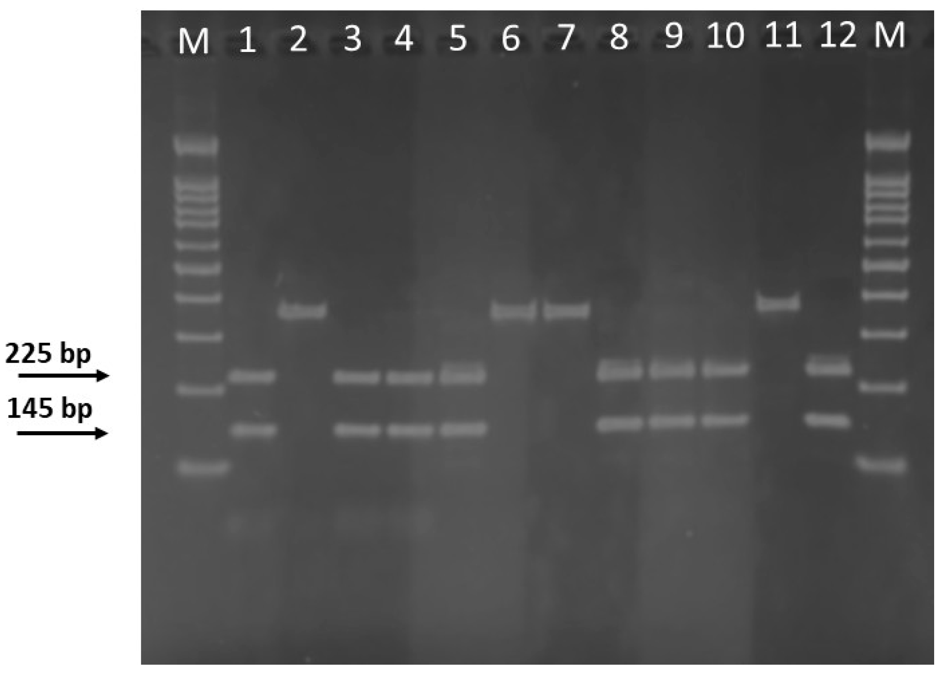
To develop a PCR-RFLP assay for efficient discrimination of S. officinalis in commercial products, the trnL-trnF intergenic spacer sequence was used as a specific DNA marker. This chloroplast region is one of the most popular and widely used molecular markers. Because of its high mutation rate, it has often been chosen to distinguish plants into species and subspecies levels [30,31,32]. The prediction of the restriction profile for the S. officinalis trnL-trnF spacer region [370 base pairs (bp) long], retrieved from Genbank (accession number KT003607.1), was performed using NEB cutter (http://nc2.neb.com/NEBcutter2/, accessed on 15 September 2024). This analysis revealed a single restriction site, HindIII (AAGCTT), which was predicted to generate two fragments of 225 and 145 bp, respectively. The unique restriction pattern identified in S. officinalis was confirmed by an in silico comparative analysis of the trnL-trnF sequences from other herbs and plant species, as well as various Salvia species (both edible and ornamental) (Figure S1). Then, it is highly probable that a mutation in the HindIII restriction site resulted in the trnL-trnF homologous amplicon of all the aforementioned plants remaining undigested. From a taxonomic perspective, this finding is rather interesting, since the restriction pattern produced by the HindIII-digested trnL-trnF amplicon clearly distinguishes S. officinalis from other Salvia species (Figure S1). The effectiveness of the PCR-RFLP assay was then investigated on a collection of 30 sage products of varying typology (Table 1), purchased from diverse Italian retailers and herbalists. Aliquots of totDNA isolated from both fresh green leaves and commercial preparations of S. officinalis, as described in the Section 2, were first amplified using universal primers for the trnL-trnF spacer sequence [42] to produce approximately 370 bp-long amplicons as expected, which were then HindIII digested. Electrophoresis of the resulting restriction products on a 2.5% agarose gel showed the typical RFLP pattern for almost all sage samples, confirming that they were authentic S. officinalis products (Figure 2). It is intriguing to note that the trnL-trnF amplified product from 7 samples claiming to be or containing sage (So5, So9, So14, So15, So23, So26, and So28 in Table 1) did not yield a clear or consistent match with that of S. officinalis, suggesting a high probability of mislabelling. The selected PCR-RFLP results from this study are shown in Figure 2.
However, the potential for contaminants (salts, glycerol) in some amplifications to inhibit the activity of the restriction enzyme and affect the true result cannot be excluded. In view of the results of the PCR-RFLP analysis, it was necessary to undertake sequencing in order to verify the identity of the PCR products. Therefore, the trnL-trnF amplicon obtained from some Salvia samples, with a particular focus on those that had not been digested, was subjected to direct sequencing using the same primers that had been employed for the initial genomic amplification. BLAST analysis at NCBI (https://www.ncbi.nlm.nih.gov, accessed on 18 October 2024) revealed, curiously, a striking similarity between the trnL-trnF spacer region of So14, So15, So26, and So28 and the homologous sequence from S. sclarea or S. purpurea, both of which are devoid of a HindIII cleavage site (Figure S1). Furthermore, the comparative analysis revealed the presence of trnL-trnF sequences belonging to Olea europaea in So5 and So9 and Cistus ladanifer in So23, indicating that the latter sage samples were adulterated preparations of S. officinalis. This result was not particularly unexpected, as previous studies have already demonstrated that a significant proportion of sage products, as well as other spices (mainly oregano), on the European Union market were found to be adulterated with olive, cistus, hazelnut, or myrtle leaves [8].
As a result, the PCR-RFLP technique has been demonstrated to be a valuable tool for the authentication of S. officinalis and the detection of its potential adulteration. However, the process is notably laborious, necessitating extensive preparation (i.e., amplification and sequencing of the target DNA sequence and selection of appropriate restriction enzymes) and is consequently a relatively costly and time-consuming procedure. Alternative molecular methods may be used to help overcome such drawbacks.

3.2. Development of LAMP Assay for the Identification of S. officinalis DNA

LAMP is recognized as a simple, rapid, and sensitive technique for the identification and detection of plant DNA and is becoming increasingly popular [36,37,38]. It has been shown to exhibit numerous distinctive advantages in comparison with other DNA amplification-based procedures. Firstly, due to its high sensitivity and specificity, LAMP requires only low concentrations of target nucleic acids to be detected. Secondly, as LAMP operates at a constant temperature, there is no requirement to adjust the temperature during nucleic acid amplification. Consequently, LAMP may be performed without the necessity for expensive thermocyclers, which are generally required for regular PCR. The simplification of the equipment required has the effect of facilitating the accessibility of LAMP in resource-limited settings.
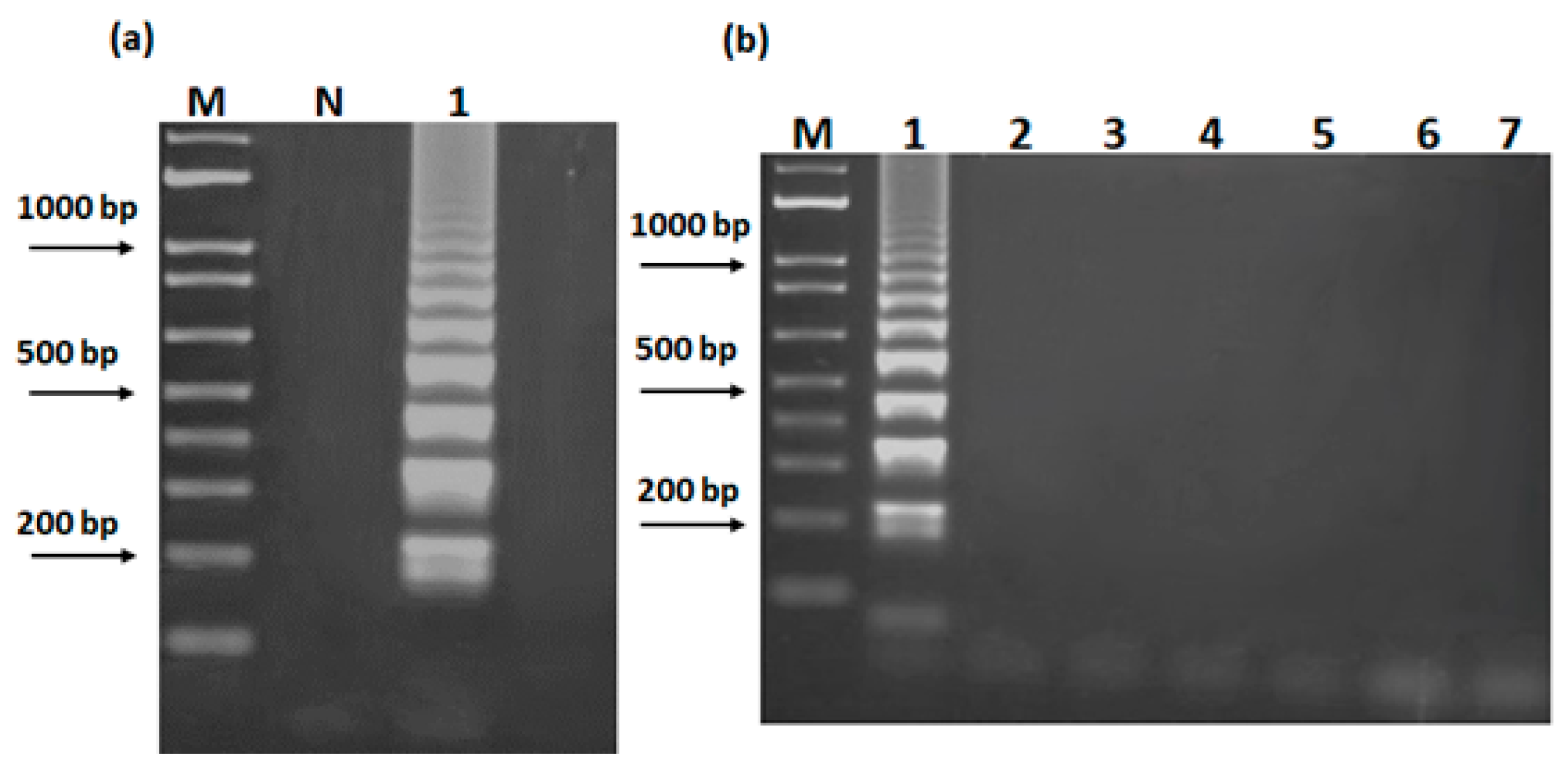
To the best of our knowledge, no report has yet been published on the use of LAMP for rapid and reliable identification of S. officinalis, and in the present study, we aimed to fill this gap. Therefore, four primers, including two outer primers (F3 and B3) and two inner primers (FIP and BIP), based on the trnL-trnF sequence of common sage (GenBank KT003607.1) (Figure 1, Table 2), were designed to specifically bind to six different regions on the target gene. An initial amplification of S. officinalis genomic DNA isolated from fresh leaves was carried out in order to optimize the isothermal conditions that were to be applied. The objective of optimizing reaction parameters was, firstly, to suppress the occurrence of non-specific amplification and, secondly, to achieve a positive result in a short time. A positive LAMP product with a typical pattern of ladder-like DNA fragments on an electrophoresed agarose gel was obtained for the common sage DNA sample within 30 min from the beginning of the amplification reaction at 63 °C in a simple heating block (Figure 3a). To evaluate the specificity of the LAMP assay, the process was also performed using totDNA from other species of the genus Salvia, some herbs, and, mainly, plant representatives that are considered to be sage-adulterating species (e.g., olive, rock rose) [8]. The LAMP assay yielded a positive result only for common sage, while all other aforementioned plants were found to be negative (Figure 3b).
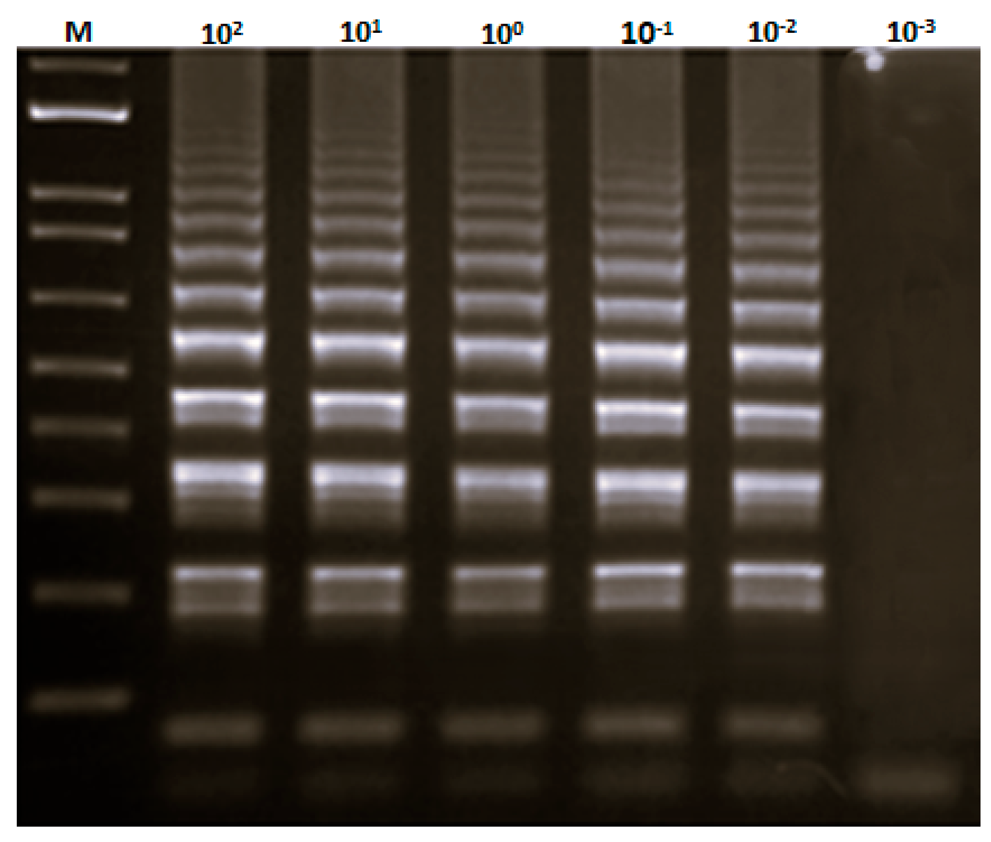
This finding thus confirmed that the novel set of trnL-trnF LAMP primers developed here can be used specifically and rapidly in practical applications. These include the identification of S. officinalis DNA and, interestingly, the discrimination between Salvia species. In order to verify the sensitivity of the LAMP assay in authenticating S. officinalis genomic DNA, different amounts of extracted sage genomic DNA, ranging from 100 ng to 1 pg, were used as template DNA. The ladder-like DNA pattern was observed when at least 10 pg of S. officinalis DNA was added to the LAMP reaction mixture (Figure 4).
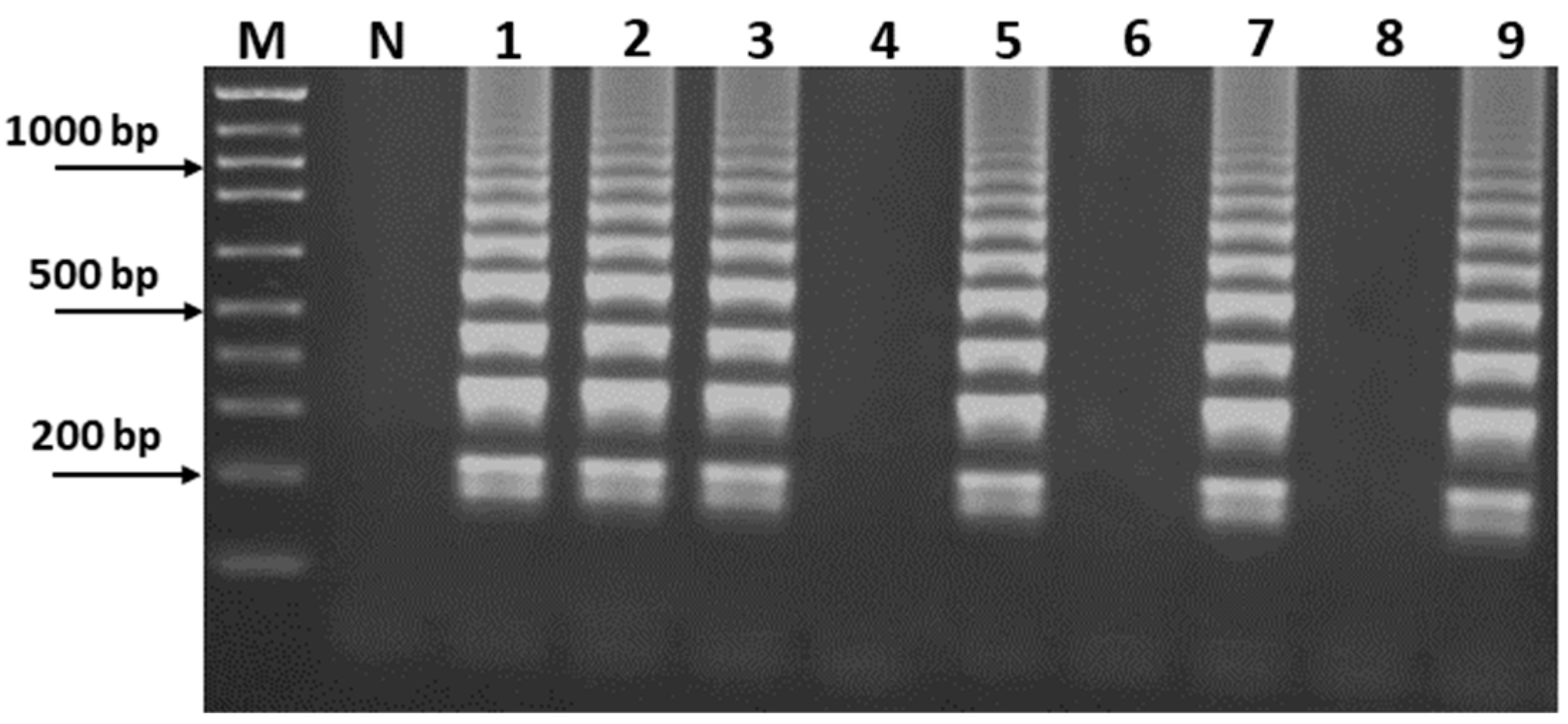
The ultimate objective of this study was to verify the ability of this isothermal amplification technique to authenticate commercial sage products. Agarose gel electrophoresis of LAMP products revealed a ladder-like pattern in 23 out of 30 sage samples examined. Conversely, no LAMP products were identified in the remaining 7 commercial samples (Figure 5), thereby confirming the absence of S. officinalis DNA and indicating that these samples (in particular, So9, So14, and So23) were, in fact, adulterated sage preparations. These results were consistent with those from PCR-RFLP detection, thus demonstrating the efficacy of the LAMP assay in identifying fraudulent practices (whether voluntary or not), including adulteration through the substitution of plant material.
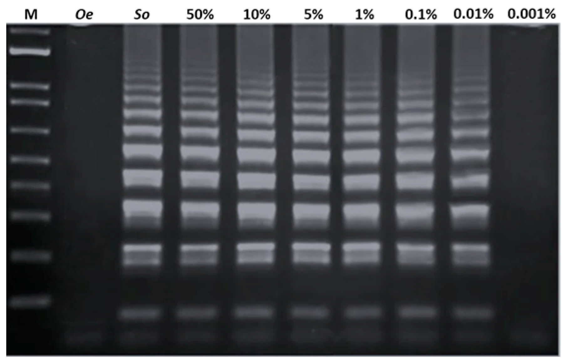
The high sensitivity of our LAMP assay was also confirmed by the low level of detection (LOD) of 0.01% observed after amplification of the DNA extracted from a mixed sample of S. officinalis and one of its adulterants (O. europaea) (Figure 6).

4. Conclusions

The findings of this study demonstrated that both of the two tested DNA-based methods contributed to the proper identification of S. officinalis and irregularities (e.g., accidental/deliberate presence of undeclared contaminating plant material) in the production and marketing of sage-based products, thereby ensuring the health and safety of consumers. However, compared to PCR-RFLP and other molecular (as well as physical, chemical, biochemical) methods used to detect food fraud, the LAMP assay has greater sensitivity, specificity, and speed, and it can be performed using inexpensive equipment. As a consequence, the LAMP assay is a valuable tool for manufacturers in the field of quality control, particularly for the screening of herbal materials, which are usually found in powdered or pulverized form, making counterfeits easy. However, the LAMP technique is not without its drawbacks. For example, the formation of primer dimmers or other mismatched structures in LAMP assays can increase the chance of non-specific amplification and, thus, false positive results. In addition, great care must be taken when performing LAMP to avoid product contamination. These issues can be addressed in a number of ways, including the design of a new set of LAMP primers and the optimization of amplification conditions, the use of restriction enzymes to cleave the LAMP amplicons at a specific site, or the addition of uracil DNA glycosylase (UDG), fluorescent, or pH-sensitive dyes to the reaction. These represent more advantageous detection methods that do not require opening of the reaction vessels [44,45,46]. The implementation of LAMP technology for quality control in various industries, and particularly its ability to be combined with a variety of naked-eye detection strategies, will primarily help reduce the costs associated with product defects and recalls, legal costs, and protecting the company’s brand reputation. These costs include the financial implications of ensuring that food products meet quality and safety standards as well as customer expectations.

Supplementary Materials

The following supporting information can be downloaded at: https://www.mdpi.com/article/10.3390/sci7020070/s1, Figure S1: trnL-trnF multiple sequence alignment.

Author Contributions

T.M.R.R.: conceptualization, supervision, resources, writing—review and editing; E.C.: methodology, investigation, formal analysis, writing—original draft preparation. All authors have read and agreed to the published version of the manuscript.

Funding

This work was supported by grants from the Università della Calabria and the Italian Ministero dell’Istruzione, Università e Ricerca (M.I.U.R.).

Data Availability Statement

Data will be made available upon request.

Conflicts of Interest

The authors declare no conflicts of interest.

References

  1. Jakovljević, M.; Jokić, S.; Molnar, M.; Jašić, M.; Babić, J.; Jukić, H.; Banjari, I. Bioactive profile of various Salvia officinalis L. preparations. Plants 2019, 8, 55. [Google Scholar] [CrossRef] [PubMed]
  2. Kintzios, S.E. Sage: The Genus Salvia; Harwood Academic Publishers: Amsterdam, The Netherlands, 2000. [Google Scholar]
  3. Ghorbani, A.; Esmaeilizadeh, M. Pharmacological properties of Salvia officinalis and its components. J. Tradit. Complement. Med. 2017, 7, 433–440. [Google Scholar] [CrossRef] [PubMed]
  4. Topçu, G. Bioactive triterpenoids from Salvia species. J. Nat. Prod. 2006, 69, 482–487. [Google Scholar] [CrossRef] [PubMed]
  5. Akacha, B.B.; Kačániová, M.; Mekinić, I.G.; Kukula-Koch, W.; Koch, W.; Orhan, I.E.; Čmiková, N.; Taglieri, I.; Venturi, F.; Samartin, C.; et al. Sage (Salvia officinalis L.): A botanical marvel with versatile pharmacological properties and sustainable applications in functional foods. S. Afr. J. Botany 2024, 169, 361–382. [Google Scholar] [CrossRef]
  6. Black, C.; Haughey, S.A.; Chevallier, O.P.; Galvin-King, P.; Elliott, C.T. A comprehensive strategy to detect the fraudulent adulteration of herbs: The oregano approach. Food Chem. 2016, 210, 551–557. [Google Scholar] [CrossRef]
  7. Bononi, M.; Tateo, F. LC-ESI-MS/MS identification of oleuropein as marker of Olea europaea L. leaves used as a bulking agent in ground oregano and sage. Ital. J. Food Sci. 2011, 23, 245–251. [Google Scholar]
  8. Tomčić, N.; Jankov, M.; Ristivojevic, P.; Trifkovic, J.; Andrić, F. Assessment of adulteration of sage (Salvia sp.) with olive leaves using high-performance thin-layer chromatography, image analysis, and multivariate linear modeling. J. Chemom. 2024, 38, e3533. [Google Scholar] [CrossRef]
  9. Velázquez, R.; Rodríguez, A.; Hernández, A.; Casquete, R.; Benito, M.J.; Martín, A. Spice and herb frauds: Types, incidence, and detection: The state of the art. Foods 2023, 12, 3373. [Google Scholar] [CrossRef]
  10. Avula, B.; Bae, J.Y.; Chittiboyina, A.G.; Wang, Y.H.; Wang, M.; Srivedavyasasri, R.; Ali, Z.; Li, J.; Wu, C.; Khan, I.A. Comparative analysis of five Salvia species using LC-DAD-QToF. J. Pharm. Biomed. Anal. 2022, 209, 114520. [Google Scholar] [CrossRef]
  11. Lee, J.; Wang, M.; Zhao, J.; Avula, B.; Chittiboyina, A.G.; Li, J.; Wu, C.; Khan, I.A. Chemical authentication and speciation of Salvia botanicals: An investigation utilizing GC/Q-TOF and chemometrics. Foods 2022, 11, 2132. [Google Scholar] [CrossRef]
  12. de Aguiar, T.R.; Dorocz, E.L.; do Santos, L.D.; Tanamati, A.A.C.; Gozzo, A.M.; Bona, E. Mid-infrared spectroscopy and chemometrics in the detection of adulteration in chia oil (Salvia hispanica L) and α-linolenic acid content prediction. Food Control 2024, 165, 110687. [Google Scholar] [CrossRef]
  13. Braglia, L.; Casabianca, V.; De Benedetti, L.; Pecchioni, N.; Mercuri, A.; Cervelli, C.; Ruffoni, B. Amplified fragment length polymorphism markers for DNA fingerprinting in the genus Salvia. Plant Biosyst. 2011, 145, 274–277. [Google Scholar] [CrossRef]
  14. Sunar, S.; Korkmaz, M.; SiĞmaz, B.; AĞar, G. Determination of the genetic relationships among Salvia species by RAPD and ISSR analyses. Turk. J. Pharm. Sci. 2020, 17, 480–485. [Google Scholar] [CrossRef]
  15. Bahadirli, N.P.; Ayanoglu, F. Genetic diversity of Salvia species from Turkey assessed by microsatellite markers. J. Appl. Res. Med. Aromat. Plants 2021, 20, 100281. [Google Scholar] [CrossRef]
  16. Ibrahim, R.I.H.; Sakamoto, M.; Azuma, J.I. PCR-RFLP and genetic diversity analysis of cpDNA in some species of the genus Salvia L. Chromosome Bot. 2012, 7, 1–8. [Google Scholar] [CrossRef]
  17. Bielecka, M.; Pencakowski, B.; Stafiniak, M.; Jakubowski, K.; Rahimmalek, M.; Gharibi, S.; Matkowski, A.; Ślusarczyk, S. Metabolomics and DNA-based authentication of two traditional Asian medicinal and aromatic species of Salvia subg. Perovskia. Cells 2021, 10, 112. [Google Scholar] [CrossRef]
  18. Lo, Y.-T.; Shaw, P.-C. DNA-based techniques for authentication of processed food and food supplements. Food Chem. 2018, 240, 767–774. [Google Scholar] [CrossRef]
  19. Madesis, P.; Ganopoulos, I.; Sakaridis, I.; Argiriou, A.; Tsaftaris, A. Advances of DNA-based methods for tracing the botanical origin of food products. Food Res. Int. 2014, 60, 163–172. [Google Scholar] [CrossRef]
  20. Zeng, L.; Wen, J.; Fan, S.; Chen, Z.; Xu, Y.; Sun, Y.; Chen, D.; Zhao, J.; Xu, L.; Li, Y. Identification of sea cucumber species in processed food products by PCR-RFLP method. Food Control 2018, 90, 166–171. [Google Scholar] [CrossRef]
  21. Griffiths, A.M.; Sotelo, C.G.; Mendes, R.; Pérez-Martín, R.I.; Schröder, U.; Shorten, M.; Silva, H.A.; Verrez-Bagnis, V.; Mariani, S. Current methods for seafood authenticity testing in Europe: Is there a need for harmonisation? Food Control 2014, 45, 95–100. [Google Scholar] [CrossRef]
  22. Wu, K.; Liu, Y.; Yang, B.; Kung, Y.; Chang, K.; Lee, M. Rapid discrimination of the native medicinal plant Adenostemma lavenia from its adulterants using PCR-RFLP. Peer J. 2022, 10, e13924. [Google Scholar] [CrossRef] [PubMed]
  23. Yang, B.C.; Lee, M.S.; Sun, F.C.; Chao, H.H.; Chang, W.T.; Lin, M.K.; Chen, H.J.; Lee, M.S. Rapid identification of the indigenous medicinal crop Cinnamomum osmophloeum from various adulterant Cinnamomum species by DNA polymorphism analysis. Pharmacogn. Mag. 2020, 16, 64–68. [Google Scholar]
  24. Spaniolas, S.; May, S.T.; Bennett, M.J.; Tucker, G.A. Authentication of coffee by means of PCR-RFLP analysis and lab-on-a-chip capillary electrophoresis. J. Agric. Food Chem. 2006, 54, 7466–7470. [Google Scholar] [CrossRef] [PubMed]
  25. Souframanien, J.; Joshi, A.; Gopalakrishna, T. Intraspecific variation in the internal transcribed region of rDNA in black gram (Vigna mungo (L.) Hepper). Curr. Sci. 2003, 85, 798–802. [Google Scholar]
  26. Kim, O.T.; Bang, K.H.; In, D.S.; Lee, J.W.; Kim, Y.C.; Shin, Y.S.; Hyun, D.Y.; Lee, S.S.; Cha, S.W.; Seong, N.S. Molecular authentication of ginseng cultivars by comparison of internal transcribed spacer and 5.8S rDNA sequences. Plant Biotechnol. Rep. 2007, 1, 163–167. [Google Scholar] [CrossRef]
  27. Lee, J.H.; Lee, J.W.; Sung, J.S.; Bang, K.H.; Moon, S.G. Molecular authentication of 21 Korean Artemisia species (Compositae) by polymerase chain reaction-restriction fragment length polymorphism based on trnL-F region of chloroplast DNA. Biol. Pharm. Bull. 2009, 32, 1912–1916. [Google Scholar] [CrossRef]
  28. Murphy, T.M.; Bola, G. DNA identification of Salvia divinorum samples. Forensic Sci. Int. Genet. 2013, 7, 189–193. [Google Scholar] [CrossRef]
  29. Lee, S.C.; Wang, C.H.; Yen, C.E.; Chang, C. DNA barcode and identification of the varieties and provenances of Taiwan’s domestic and imported made teas using ribosomal internal transcribed spacer 2 sequences. J. Food Drug Anal. 2016, 25, 260–274. [Google Scholar] [CrossRef]
  30. Ng, A.E.; Sandoval, E.; Murphy, T.M. Identification and individualization of Lophophora using DNA analysis of the trnL/trnF region and rbcL gene. J. Forensic Sci. 2016, 61, S226–S229. [Google Scholar] [CrossRef]
  31. Sen, F.; Uncu, A.O.; Uncu, A.T.; Erdeger, S.N. The trnL (UAA)-trnF (GAA) intergenic spacer is a robust marker of green pea (Pisum sativum L.) adulteration in economically valuable pistachio nuts (Pistacia vera L.). J. Sci. Food Agric. 2020, 100, 3056–3061. [Google Scholar] [CrossRef]
  32. Kitamura, M.; Kazato, A.; Yamamuro, T.; Ando, H.; Sasaki, Y.; Suzuki, R.; Shirataki, Y. Rapid identification of Aconitum plants based on loop-mediated isothermal amplification assay. BMC Res. Notes 2019, 12, 408. [Google Scholar] [CrossRef] [PubMed]
  33. Focke, F.; Haase, I.; Fischer, M. Loop-mediated isothermal amplification (LAMP): Methods for plant species identification in food. J. Agric. Food Chem. 2013, 61, 2943–2949. [Google Scholar] [CrossRef] [PubMed]
  34. Holz, N.; Illarionov, B.; Wax, N.; Schmidt, C.; Fischer, M. Point-of-care suitable identification of the adulterants Carthamus tinctorius and Curcuma longa in Crocus sativus based on loop-mediated isothermal amplification (LAMP) and lateral-flow-assay (LFA). Food Control 2023, 148, 109637. [Google Scholar] [CrossRef]
  35. Holz, N.; Wax, N.; Illarionov, B.A.; Iskhakova, M.; Fischer, M. Food Authentication: The detection of Arbutus unedo and Olea europaea leaves as an admixture of oregano using LAMP- and Duplex LAMP-based test systems with Lateral-Flow Assays. Agriculture 2024, 14, 597. [Google Scholar] [CrossRef]
  36. Niessen, L.; Bechtner, J.; Fodil, S.; Taniwaki, M.H.; Vogel, R.F. LAMP-based group specific detection of aflatoxin producers within Aspergillus section Flavi in food raw materials, spices, and dried fruit using neutral red for visible-light signal detection. Int. J. Food Microbiol. 2018, 266, 241–250. [Google Scholar] [CrossRef]
  37. Notomi, T.; Okayama, H.; Masubuchi, H.; Yonekawa, T.; Watanabe, K.; Amino, N.; Hase, T. Loop-mediated isothermal amplification of DNA. Nucleic Acids Res. 2000, 28, E63. [Google Scholar] [CrossRef]
  38. Mori, Y.; Kitao, M.; Tomita, N.; Notomi, T. Real-time turbidimetry of LAMP reaction for quantifying template DNA. J. Biochem. Biophys. Methods 2004, 59, 145–157. [Google Scholar] [CrossRef]
  39. Li, J.; Macdonald, J. Advances in isothermal amplification: Novel strategies inspired by biological processes. Biosens. Bioelectron. 2015, 64, 196–211. [Google Scholar] [CrossRef]
  40. Sievers, F.; Wilm, A.; Dineen, D.; Gibson, T.J.; Karplus, K.; Li, W.; Lopez, R.; McWilliam, H.; Remmert, M.; Söding, J.; et al. Fast, scalable generation of high-quality protein multiple sequence alignments using Clustal Omega. Mol. Syst. Biol. 2011, 7, 539. [Google Scholar] [CrossRef]
  41. Nicholas, K.B.; Nicholas, H.B., Jr.; Deerfield, D.W. GeneDoc: Analysis and visualization of genetic variation. Embnew News 1997, 4, 14. [Google Scholar]
  42. Taberlet, P.; Gielly, L.; Pautou, G.; Bouvet, J. Universal primers for amplification of three non-coding regions of chloroplast DNA. Plant Mol. Biol. 1991, 17, 1105–1109. [Google Scholar] [CrossRef] [PubMed]
  43. Altschul, S.F.; Gish, W.; Miller, W.; Myers, E.W.; Lipman, D.J. Basic local alignment search tool. J. Mol. Biol. 1990, 215, 403–410. [Google Scholar] [CrossRef] [PubMed]
  44. Hsieh, K.; Mage, P.L.; Csordas, A.T.; Eisenstein, M.; Soh, H.T. Simultaneous elimination of carryover contamination and detection of DNA with uracil-DNA-glycosylase-supplemented loop-mediated isothermal amplification (UDG-LAMP). Chem. Commun. 2014, 50, 3747–3749. [Google Scholar] [CrossRef]
  45. Notomi, T.; Mori, Y.; Tomita, N.; Kanda, H. Loop-mediated isothermal amplification (LAMP): Principle, features, and future prospects. J. Microbiol. 2015, 53, 1–5. [Google Scholar] [CrossRef]
  46. Tanner, N.A.; Zhang, Y.; Evans, T.C. Visual detection of isothermal nucleic acid amplification using pH-sensitive dyes. Biotechniques 2015, 58, 59–68. [Google Scholar] [CrossRef]
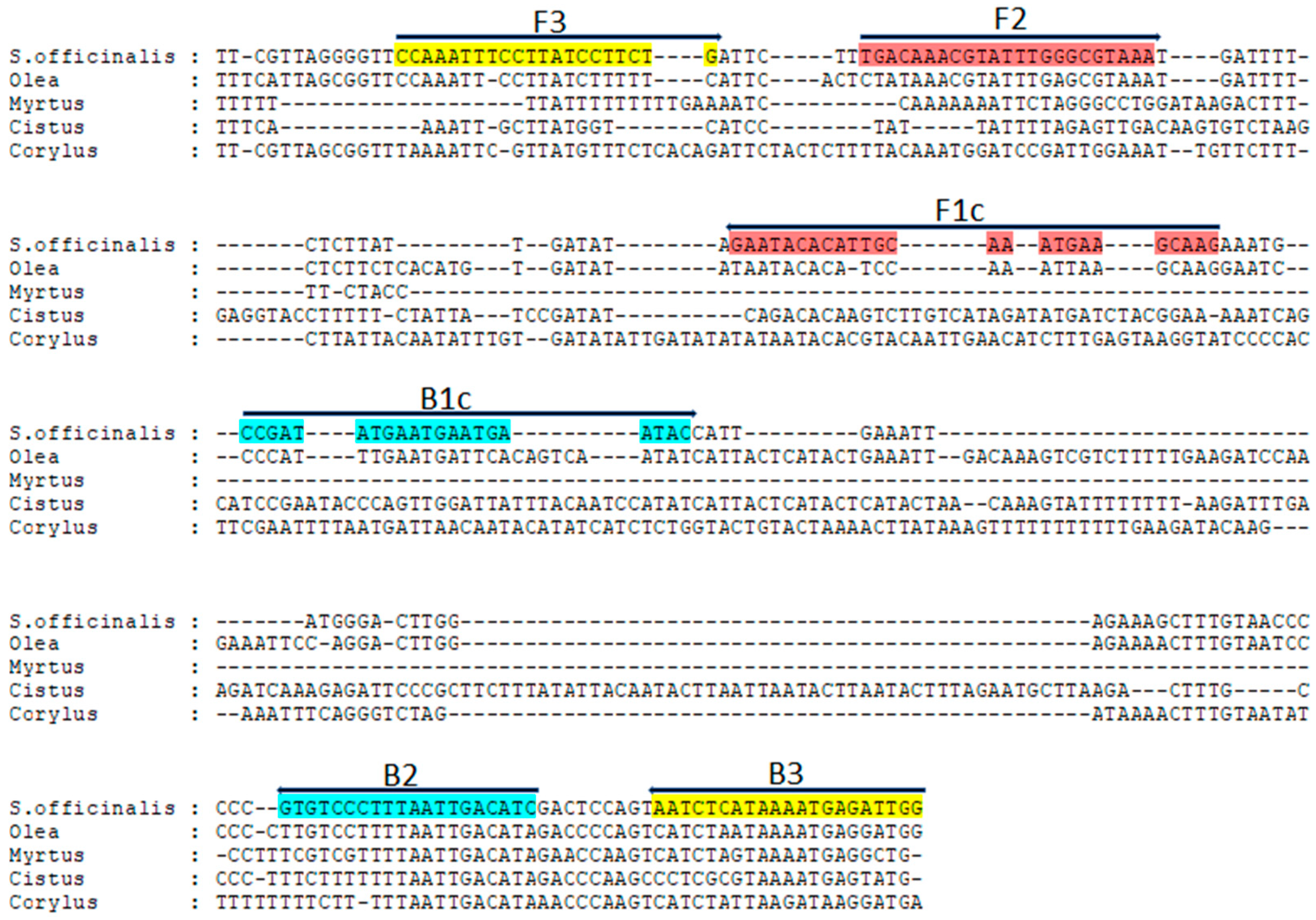
Figure 1. Multiple sequence alignment of the trnL-trnF spacer sequence of S. officinalis (KT003607.1), Olea europaea (FJ490798.1), Myrtus communis (NC_066006.1), Cistus creticus (EU684567.1), and Corylus avellana (FJ012026.1). The annealing position and orientation of the LAMP primers specific to S. officinalis (Table 2) are highlighted in different colours.
Sci 07 00070 g001
Figure 2. Agarose gel electrophoresis of HindIII-restricted trnL-trnF amplicon from totDNA of some marketed S. officinalis samples analyzed in this study. M, 100 bp DNA ladder (Thermo Fisher Scientific, Waltham, MA, USA). Samples: So4 (lane 1), So5 (lane 2), So6 (lane 3), So7 (lane 4), So13 (lane 5), So14 (lane 6), So15 (lane 7), So16 (lane 8), So21 (lane 9), So22 (lane 10), So23 (lane 11), and So24 (lane 12).
Sci 07 00070 g002
Figure 3. (a) Electrophoretic analysis of trnL-trnF LAMP products obtained after isothermal amplification of genomic DNA from S. officinalis (lane 1). Lanes M and N represent 100 bp of the DNA ladder (Thermo Fisher Scientific, Waltham, MA, USA) and the negative control, respectively. (b) Primer specificity of the LAMP assay for the authentication of S. officinalis DNA. Lane M represents 100 bp of the DNA ladder (Thermo Fisher Scientific, Waltham, MA, USA). Lanes 1–7 represent different genomic DNAs: 1, S. officinalis.; 2, S. sclarea; 3, S. purpurea.; 4, Origanum vulgare; 5, Mentha spicata; 6, Olea europaea; 7, Cistus creticus.
Sci 07 00070 g003
Figure 4. Sensitivity analysis of the LAMP assay for the detection of S. officinalis DNA using a 10-fold serial dilution of sage DNA, ranging from 100 ng⋅to 1 pg. M: M: 100 bp DNA ladder.
Sci 07 00070 g004
Figure 5. Electrophoretic analysis of LAMP products from some commercial sage preparations analyzed in this study (Table 1). Lanes M and N represent 100 bp of the DNA ladder marker (Thermo Fisher Scientific, Waltham, MA, USA) and the negative control, respectively. Samples: So6 (lane 1), So7 (lane 2), So8 (lane 3), So9 (lane 4), So13 (lane 5), So14 (lane 6), So22 (lane 7), So23 (lane 8), and So24 (lane 9).
Sci 07 00070 g005
Figure 6. Reactivity of LAMP primers for the detection of sage following amplification of DNA isolated from mixtures containing known amounts of S. officinalis (50%, 10%, 5%, 1%, 0.1%, 0.01%, and 0.001%) and Olea europaea (Oe). M: 100 bp DNA ladder. So: S. officinalis.
Sci 07 00070 g006
Table 1. Sage products with the sampling area analyzed in this study. Sage samples found to be adulterated are in bold.
S. officinalis Samples Province
Manufacturing
Dried LeavesTeasInfusions
So1So11So21Torino, Piemonte, Italy
So2So12So22Torino, Piemonte, Italy
So3So13So23Verona, Veneto, Italy
So4So14So24Verona, Veneto, Italy
So5So15So25Verona, Veneto, Italy
So6So16So26Roma, Lazio, Italy
So7So17So27Roma, Lazio, Italy
So8So18So28Roma, Lazio, Italy
So9So19So29Cosenza, Calabria, Italy
So10So20So30Cosenza, Calabria, Italy
Table 2. Sequence of the LAMP primers used in this study for the identification of S. officinalis.
PrimerLength (bp)Sequence (5′–3′)
F321CCAAATTTCCTTATCCTTCTG
B321CCAATCTCATTTTATGAGATT
FIP (F2 + F1c)48TGACAAACGTATTTGGGCGTAAA-CTTGCTTCATTTGCAATGTGTATTC
BIP (B2 + B1c)41GATGTCAATTAAAGGGACAC-
CCGATATGAATGAATGAATAC
Disclaimer/Publisher’s Note: The statements, opinions and data contained in all publications are solely those of the individual author(s) and contributor(s) and not of MDPI and/or the editor(s). MDPI and/or the editor(s) disclaim responsibility for any injury to people or property resulting from any ideas, methods, instructions or products referred to in the content.