Article

Proteomic Comparison between Periodontal Pocket Tissue and Other Oral Samples in Severe Periodontitis: The Meeting of Prospective Biomarkers

1
Proteomic Laboratory, Department of Surgery, Medicine, Dentistry and Morphological Sciences with Transplant Surgery, Oncology and Regenerative Medicine Relevance, University of Modena and Reggio Emilia, Via G. Campi 287, 41125 Modena, Italy
2
Unit of Dentistry and Oral-Maxillofacial Surgery, Periodontology Section, Department of Surgery, Medicine, Dentistry and Morphological Sciences with Transplant Surgery, Oncology and Regenerative Medicine Relevance, University-Hospital of Modena and Reggio Emilia, Via del Pozzo 71, 41124 Modena, Italy
*
Author to whom correspondence should be addressed.
Submission received: 17 July 2024 / Revised: 21 August 2024 / Accepted: 25 September 2024 / Published: 27 September 2024
(This article belongs to the Special Issue Feature Papers—Multidisciplinary Sciences 2024)

Abstract

:
Periodontitis is characterized by gingival regression, alveolar bone resorption and the development of deep periodontal pockets that, if left untreated, can lead to tooth loss. Currently, specific biomarkers are needed for the early, objective diagnosis, monitoring, and management of periodontal patients. In this proteomic study, periodontal pocket tissues from patients with severe periodontitis were analyzed in comparison to periodontally healthy sites with the aim of discovering distinctive protein targets. Gingival tissues were fragmented using a motorized mechanical method and mixture protein was separated via mono-dimensional gel electrophoresis. The examination of protein bands using definite 1D image analysis software allowed for the detection of 22 differentially expressed proteins between pathological and healthy samples that were identified through mass spectrometry. A comparative assessment of these proteins with those previously reported in other studies conducted on periodontal diseases in various types of oral specimens, such as gingival crevicular fluid, dentin, tooth pulp, root canal content, salivary gland secretions, saliva, periodontal ligament cells, and dental stem cells, highlighted a great number of significant common matches. The discovery of a selective cluster of periodontitis-related biomarkers could become particularly important before the clinical manifestation of the disease to promptly stop its progression for a timely preventive diagnosis.

1. Introduction

Periodontal diseases (PDs) comprise inflammatory disorders such as gingivitis, characterized by limited inflammation of the periodontal soft structure, which could evolve towards periodontitis, along with the progression of local inflammation [1]. Periodontitis is a chronic inflammation of the periodontium, the teeth-supporting tissues, accompanied by the loss of the periodontal ligament attachment and bony support, that can, if left untreated, lead to gradual resorption and consequent teeth loss [2]. It is affected by both local, as well as systemic etiological factors. It is caused by a complex interplay between bacterial infection (microorganisms adhering and growing on the tooth’s surface) and the host response, often modified by behavioral factors [3]. The primary characteristic of the disease is the chronic inflammation of the periodontal tissue, leading to the formation of deep periodontal pockets (i.e., the pathognomonic lesion characterizing periodontitis) [4]. The classification of periodontitis is based on the emphasis on the individual features of the disease and, therefore, on phenotype differences [5]. These resulted from various considerations, including the identification of typical bacteria as etiologic agents, the presence of several risk factors (smoking, genetic predisposition, systemic diseases, and low socioeconomic status), the recognition of genetic susceptibility and targeted polymorphisms related to the periodontitis severity [5]. The general classification system led to four different grades of periodontitis: two more common forms, chronic and aggressive, representing destructive periodontal disease with slow progression, or highly destructive forms characterized by rapid progression, respectively, including unusual necrotizing conditions of the gingival or periodontal tissues, rare periodontitis as a manifestation of genetic defects, and acquired deficiencies of the host defense network or direct systemic diseases [6]. Two further forms of periodontitis were added to the latest international classification scheme: periodontal abscesses, acute lesions with pus formation inside the periodontal pocket, characterized by rapid tissue destruction and a high risk of systemic diffusion, and endodontic–periodontal lesions, which involve pathological interactions between the periodontal tissue and the pulpal of a tooth [7]. Moreover, based on the extent, periodontitis can be classified as localized or generalized, while, according to the severity, it can be categorized into four different stages (from stage I, corresponding to early/mild severity, to stage IV, corresponding to very severe forms) [5].
Currently, PDs are increasing globally, with an estimated prevalence ranging from 20% to 50% worldwide [8,9]. Particularly, periodontitis represents one of the most common diseases of the oral cavity, and its incidence significantly rises with age [10,11]. Furthermore, as suggested by a growing body of literature, periodontitis may be associated with several systemic diseases [12], including cardiovascular diseases [13,14,15], diabetes [16,17], acute and chronic respiratory diseases [18,19], cancer [20], negative pregnancy outcomes [21], and a greater risk of developing dementia [22]. To date, the diagnosis of periodontitis is only established by clinical criteria based on the use of a periodontal probe to detect attachment loss and pocket depth. These periodontal defects are the clinical expressions of periodontitis, and the diagnosis is made on a quite subjective basis, considering periodontal damage that has already occurred [5,23]. However, the etiopathogenesis of periodontitis is not yet well defined, and reliable markers for early diagnosis and increasingly accurate therapy are still lacking.
In the last few years, proteomic research has shed new light on modern dentistry, helping the identification of candidate protein biomarkers and alterations in expression patterns, leading to the discovery of potential targets for the diagnosis, prognosis, and treatment of PDs, thus encouraging the advancement of personalized medicine [24]. Specifically, oral clinical proteomics represents an attractive examination approach in periodontics; therefore, it can be considered the future of periodontal diagnostics [25,26]. Different oral specimens, such as periodontal pocket tissue [27,28], GCF [29,30], and tooth surface collected material (TSCM) [31], have been progressively employed in our studies with the aim of differentiating the proteomic profiles of these prominent samples.
The first purpose of this work was to analyze pocket-associated tissue samples obtained from patients with severe periodontitis in comparison with tissue collected from healthy sites, employing a novel method of protein extraction in an attempt to optimize and increase protein recovery yields. Proteins were separated using sodium dodecyl sulfate–polyacrylamide gel electrophoresis (SDS-PAGE) and identified via liquid chromatography/tandem mass spectrometry (LC-MS/MS). The results were compared with those obtained both from our previous proteomic studies and from other proteomic investigations conducted on periodontitis in various oral samples, in addition to periodontal gingival tissue, such as GCF, dentin, tooth pulp, root canal content, salivary gland secretion, saliva, periodontal ligament cells/fibroblasts, and human dental stem cells, searching for significant matches to strengthen and confirm the periodontitis-associated biomarkers proposed up to now.

2. Materials and Methods

2.1. Chemicals and Reagents

Dithiothreitol (DTT), 2X Laemmli sample buffer, Coomassie Blue G-250, and Precision Plus Protein™ Standards Dual Blue were obtained from Bio-Rad Laboratories (Milan, Italy). High-purity phosphate-buffer saline (PBS), urea, thiourea, iodoacetamide, Tris, ammonium bicarbonate, trifluoroacetic acid, formic acid, and 3-[(3-Cholamidopropyl)-dimethylammonio]-propane-sulfonate (CHAPS) were purchased from Merck KGaA (Milan, Italy). Roche cOmpleteTM Protease inhibitors EDTA-free tablets were obtained from Roche Italia (Milan, Italy). Precast gel Bolt™ 4–12% Bis-Tris Plus and MES SDS 10X running buffer were obtained from Life Technologies Italia (Monza, MB, Italy). The trypsin used for protein digestion was of gold mass spectrometry-grade and obtained from Promega Corporation (Milan, Italy). The water used for electrophoresis and MS protocols was Versylene sterile water (Fresenius Kabi, Verona, Italy). MS-purity solvents (acetone, methanol, glacial acetic acid, and acetonitrile) were obtained from Carlo Erba Reagents (Milan, Italy).

2.2. Study Design and Patient Selection

This study was designed according to the last edition of the principles of the Helsinki Declaration and was subjected to the appraisal of the local Ethic Committee of the Health Service of the Emilia-Romagna region, which approved and registered the research project (protocol number 3968/2017, registration number 315/2017) and supervised all procedures. Patient enrollment was managed by a specialized periodontist at the Periodontology Unit of Dentistry and Oral-Maxillofacial Surgery (Modena University Hospital, Italy). Eligible subjects were asked to sign an informed consent form after receiving comprehensive information about the study procedures. Subjects referred for dental and periodontal therapy were selected, suffering from periodontitis (stage III or IV) [5], with the presence of at least one pocket associated with a shallow intrabony defect suitable for treatment via osseous resective surgery next to a healthy interproximal site with a normal probing depth (PD ≤ 3 mm). At screening, the following inclusion criteria were carefully considered: aged between 18 and 70 years, non-smoker, no history of alcohol abuse, non-pregnant or lactating, and a general medical history of good health. The exclusion criteria were diabetes and/or other critical systemic diseases (i.e., cardiovascular diseases), bone-related diseases, pregnancy, drug or alcohol abuse, and current drug and/or antibiotic prophylaxis use. Upon the completion of cause-related therapy, the inclusion criteria were a full-mouth plaque score (FMPS) and a full-mouth bleeding score (FMBS) of ≤15%, no bleeding on probing (BoP) at experimental pathological and healthy sites, and high levels of patient compliance and motivation [32]. A total of 12 subjects that met the above-mentioned criteria, matched for age and gender (mean 52.6 years ± 8.4, SD; 7 females/5 males), were definitively enrolled in this study.

2.3. Dental–Periodontal Procedures and Samples Collection

Three months after completion of the preliminary dental and cause-related therapy, the experimental area was treated with osseous resective surgery [33,34]. In compliance with surgical therapeutic needs, the flap included the periodontal defect-associated site, and the next gingival healthy tissue. Following elevation of the buccal and lingual split thickness flaps, the pocket-associated interproximal tissues and the healthy interproximal tissues at sites with normal probing depth (comprised in the resective secondary flap) were harvested in sterile tubes and immediately frozen at –80 °C to preserve protein integrity until proteomic analysis. Bone surgery was then performed, and the flaps were apically positioned using a periosteal anchorage. Patients were followed up with a post-operative supportive care program.

2.4. Tissue Proteins Extraction and Separation (SDS-PAGE)

Gingival tissue samples were first washed in PBS to remove any blood residue and then shattered with a motorized pestle (Argos Technologies Inc., London, UK) until a fine homogenate was obtained. This was further solubilized via incubation for 1 h at room temperature with constant shaking with 1 mL of lysis buffer (7 M urea, 2 M thiourea, 3% CHAPS, 40 mM Tris, pH 8.3, protease inhibitors) and then sonicated twice with an ultrasonic homogenizer (Sonoplus HD model 2070, Bandelin Electronic, Berlin, Germany). The samples were centrifuged at +4 °C for 10 min at 10,000× g and the supernatant was allowed to precipitate overnight with acetone at –20 °C. Finally, the protein pellet obtained via centrifugation at +4 °C for 15 min at 14,000× g was resuspended in rehydration buffer (6 M urea, 2 M thiourea, 4% CHAPS, 25 mM DTT). The samples were pooled (4 samples per pool), thus obtaining 3 different pools for the healthy tissue samples and 3 distinct pools for the pathological samples. The total protein content was quantified according to the Bradford assay. The measurements were repeated 4 times/each to prove the effective protein solubilization in the buffer and, at the same time, verify sample stability. Tissue protein separation was carried out using SDS-PAGE, as previously described [30]. Briefly, the tissue protein content (10 µg) was mixed with 2X Laemmli sample buffer and denatured by heating at +95 °C for 5 min (Thermomixer Comfort, Eppendorf, Milan, Italy). Proteins were then separated using precast gel BoltTM 4–12% Bis-Tris Plus and MES SDS 1X as running buffer. Each pool was run in duplicate. The gels were stained with Coomassie Blue G-250, and subsequently de-stained with a solution composed of 40% methanol/7% glacial acetic acid until the gel background became transparent, thus highlighting the protein bands.

2.5. Gel Image Acquisition and Analysis

Gel images were scanned and acquired using a calibrated imaging densitometer (GS-800 model, Bio-Rad, Hercules, CA, USA), and lanes and bands were analyzed through the use of integrated 1D image analysis software (QuantityOne, version 4.6.7, Bio-Rad), [accessed 29 May 2023]. The software automatically detects the bands in the defined lanes of the gel based on given parameters. Before band detection, lane-based background subtraction was performed to remove the background from the image, minimizing the noise density. Moreover, a normalization setting was applied to adjust for variability due to gel staining. The relative abundance of the detected bands in each lane was measured by calculating the area under their intensity profile curve, selecting the ‘Trace quantity’ attribute. The units were expressed as optical density (OD) × mm. Band density was further evaluated using QuantityOne software and reported as an intensity trace plot. The bands’ molecular weights were estimated according to the molecular weight standards (PrecisionPlus Dual Blue, Bio-Rad), which were run together with the tissue samples during SDS-PAGE separation.

2.6. Mass Spectrometry Identification

The bands of interest were sliced from the gels with a surgical scalpel and the inside proteins were digested with trypsin, as previously described [35]. Gel bands were de-stained with a solution of acetonitrile/ammonium bicarbonate and subsequently reduced using dithiothreitol and alkylated with iodoacetamide. Proteins were digested over night at +37 °C with Trypsin MS-grade (Trypsin Gold, Promega, Italy). Finally, the obtained peptides were extracted with a solution composed of acetonitrile/trifluoroacetic acid and concentrated in a Concentrator Plus (Eppendorf Italia, Milan, Italy). Dry samples were resuspended in acetonitrile/formic acid and then sonicated before MS analysis, which was performed using a UHPLC-MS QExactive Hybrid Quadrupole-Orbitrap™ mass spectrometer (Thermo Fisher Scientific, Reinach, Switzerland) (LC-ESI-QO-MS/MS System), as previously reported [36].

2.7. Data Analysis

The Mascot search engine (www.matrixscience.com) was used to analyze MS and MS/MS data by peptide mass fingerprint and MS/MS ion search (accessed 21 July 2023). Protein identification was based on raw MS/MS data from one or more peptides. The neXtProt database was accessed to detect peptide sequences, while the C-Rap database was selected to identify the presence of any contaminants. Mascot files (.mgf) were searched with Mascot, setting the following search parameters: taxonomy (Human), proteolytic enzyme (trypsin; no more than one missed cleavage), fixed modification (carbamidomethylation of Cysteine), variable modification (Methionine oxidation; Asparagine and Glutamine deamidation), mass tolerance for precursor ions (10 ppm), mass tolerance for product ions (0.05 Da), and false discovery rate (FDR) < 1%. Only proteins with a minimum of two unique peptides and a statistically significant p-value (<0.05) were considered differentially expressed. Protein identification was performed at least in duplicate using bands cut from replicated gel lanes. Unpaired Student’s t-test was used to compare the following: the different protein yields obtained from motorized and manual fragmentation, the protein concentration data between healthy and periodontally affected tissue samples, and the quantitative values of healthy and diseased-related protein bands (significance was set to a p-value of < 0.05).

3. Results

3.1. Protein Quantification

Gingival tissue disruption via the motorized mechanical method resulted in 7-fold greater total protein recovery than using a manual pestle (average concentration ± standard deviation, SD: 1.08 mg/mL ± 0.27 vs. 0.36 mg/mL ± 0.26, respectively, p-value = 0.0042). The quantification of the total protein content, performed for each tissue sample after protein extraction, showed a significantly higher protein amount in pocket-associated tissues than in healthy tissue samples (average concentration ± SD: 1.04 mg/mL ± 0.32 vs. 0.61 mg/mL ± 0.09, respectively, p-value = 0.0030).

3.2. Protein Separation and Bands Analysis

Protein separation via SDS-PAGE, and the subsequent densitometric analysis of the protein bands with the QuantityOne 1D image analysis software revealed five bands that were significantly overexpressed in periodontopathic samples vs. healthy tissue samples (p-values < 0.05), as shown in Figure 1. The bands indicated by arrows were cut from the gel for protein identification via MS analysis.
The relative quantities values of the selected bands (expressed as OD × mm and reported as average ± SD), as well as the statistical significance values resulting from the comparison between healthy and pathological samples, are shown in Table 1.
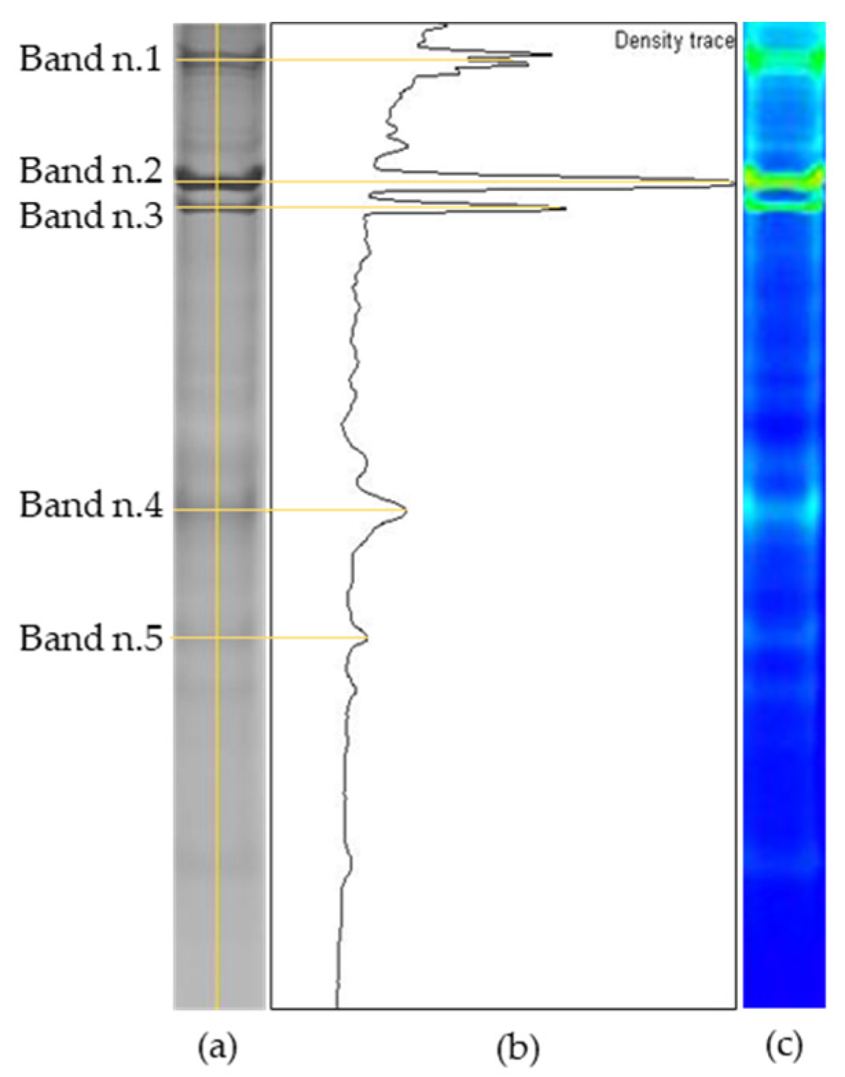
Further examination of the bands, considering the intensity trace, is illustrated in Figure 2. A representative lane from SDS-PAGE is shown for the periodontal tissue sample (Figure 2a), along with the corresponding vertical plot graphic of the band intensities (Figure 2b) and the inverted color image with respect to the original gray lane (Figure 2c). Figure 2b,c were obtained using QuantityOne 1D image analysis software (Bio-Rad). The five differential bands reflect most of the peaks with the highest intensity.

3.3. Protein Identification by LC-MS/MS

The protein mixtures extracted from the significantly increased bands were subjected to LC-MS/MS analysis, thus leading to the identification of 22 different proteins in periodontal pocket tissue samples. The complete list of the recognized proteins is shown in Table 2.
Column 1 reports the identification code of each analyzed protein band, as indicated in both Figure 1 and Figure 2, and in Table 1. The second column refers to the primary accession number of the identified protein provided via the neXtProt database. Column 3 shows the primary protein’s full name (from the neXtProt database), and next, the gene name is reported. Column 5 shows the theoretical protein mass derived from the MS analysis, expressed in Dalton, while column 6 refers to the highest score obtained via the analysis using the Mascot search engine. Finally, the last two columns display the number of significant matching peptides and the number of significant protein sequences, respectively.

3.4. Protein Comparisons

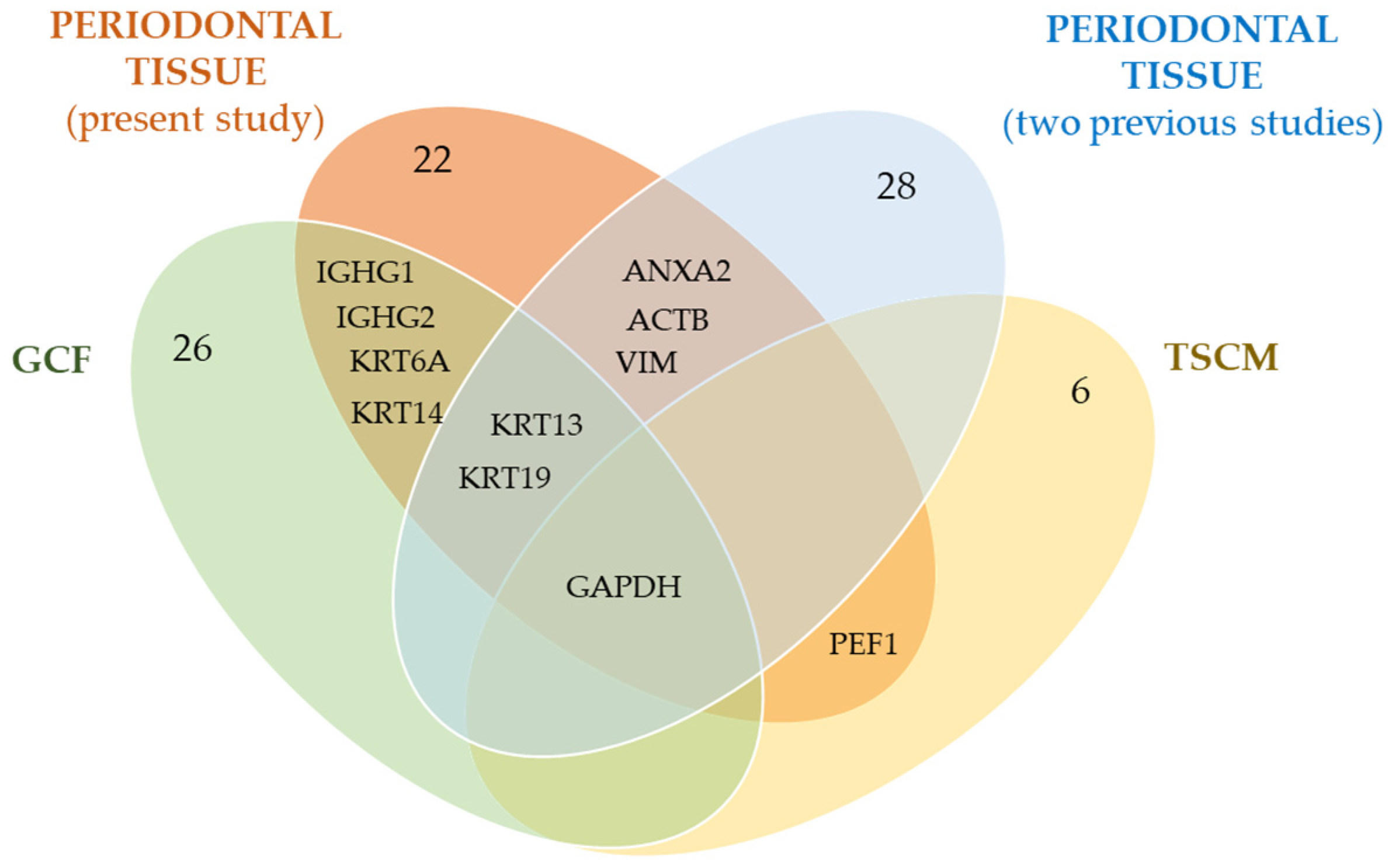
A comparative evaluation of the proteins detected in the present work with those characterized in our previous proteomic studies, conducted on patients with periodontitis, highlighted some common identifications. A graphical illustration of the comparisons, showing the overlapping proteins among the different oral samples analyzed, is provided in Figure 3.
As evidenced in the diagram (Figure 3), 11 proteins overlap with our previous proteomic studies. Of these proteins, ANXA2, ACTB, and VIM were identified in previous periodontal tissues [27,28], as well as KRT13 and KTR19 [28]. These latter two proteins were also detected in GCF [30]. Four additional proteins, namely two isoforms of immunoglobulin heavy constant gamma, IGHG1 and IGHG2, and other two types of keratins, KRT6A and KRT14, were previously identified in GCF [30], while PEF1 was previously found only in TSCM [31]. Finally, the enzymatic protein GAPDH was detected in all previously analyzed oral specimens from patients with periodontitis. The numbers reported in the ellipse indicate the total number of proteins identified in each mentioned study.
Further protein matches were found by extending the comparisons towards proteomics studies on PDs proposed by different authors and conducted on various types of oral samples, as shown in Table 3. The literature search was essentially carried out using the PubMed (MEDLINE), Scopus and Google Scholar databases using the following keywords, proteomic-proteomics, periodontal disease, and periodontitis, in association with the name of each protein identified in our present study. Table 3 reports the full names of proteins and their abbreviated names (in round brackets), the different types of oral samples analyzed (periodontal pocket gingival tissue, GCF, TSCM, dentin, tooth pulp, root canal content, salivary gland secretion, saliva, periodontal ligament cells/fibroblasts, and human dental stem cells), the electrophoretic method used for protein separation, the mass spectrometry platform employed for protein identification, and, finally, all the corresponding bibliographic references.

4. Discussion

At present, the clinical manifestation of periodontitis is usually recognized late, after the destruction of periodontal gingival tissue, which is basically caused by chronic inflammation, interference from host agents, and genetic and environmental factors. The diagnosis is performed considering periodontal damage that has already occurred, and, therefore, periodontal therapy is largely focused on the dysbiotic ecosystems that trigger the disease [65]. Furthermore, the treatment of periodontitis is not standardized, as it varies depending on each individual patient and on the subjective evaluation of the periodontist treating [66]. In this regard, the advanced concept of personalized medicine is becoming increasingly worthy of importance as it could provide better prognostic methods than conventional treatments. Markedly, an early diagnostic system based on selective biomarkers able to predict periodontitis and guide therapeutic planning is required for the timely identification and management of this disease.
In this study, the proteomic profile of periodontal pocket tissue was compared with that of a related healthy site with the purpose of identifying protein biomarkers associated with periodontitis. Tissue proteins were successfully extracted using an innovative motorized mechanical method that allowed for the disruption of hard tissues (such as connective tissue) and, consequently, the recovery of a greater protein number for a more complete protein cluster. Using SDS-PAGE coupled to QuantityOne 1D image analysis software (Figure 1, Table 1, and Figure 2), and through LC-MS/MS analysis, 22 upregulated proteins were found in periodontitis-affected sites in comparison to periodontally healthy gingival sites (Table 2). Each protein was first compared with our previous proteomic studies (Figure 3) and then with contemporary proteomics literature (Table 3), highlighting noteworthy protein correspondences.

4.1. Collagens and Fibronectin

The gingival connective tissue is primarily composed of collagens, fibronectin, proteoglycans, and osteonectin. During the development of periodontitis, several changes occur in the molecular composition of gingival connective tissue, as well as structural degenerative modifications, starting with significant supporting tissue destruction, inflammatory lesions, and collagens degradation, thus leading to the loss of connective tissue and bone support [67]. However, the junctional epithelium, acting as a barrier site, is able to maintain its regenerative capability in response to damage. Constructive changes have been established at the level of the fibrotic periodontal pocket wall, such as endothelial and connective tissue cell proliferation and new formations of capillaries, fibroblasts, and collagen fibers [68]. Higher expression levels of collagens found in pocket tissue samples may stimulate periodontal regeneration. In accordance with our results, Badovinac et al. reported the upregulation of COL1A1, COL1A2, COL12A1, and COL14A1 in pathological gingival tissue from severe generalized aggressive periodontitis, and, differently, the downregulation of COL6A1, COL6A2, and COL6A3 [37].
FN is a multifunctional glycoprotein that mediates inflammation, wound healing, and tissue repair (UniProt database). Moreover, it is implicated in mediating some cellular activities and interactions with the extracellular matrix, such as cell growth, differentiation, adhesion, and migration. It binds to a great variety of molecules, comprising heparin, fibrin, integrin, and collagen [69]. Cellular FN is produced in various tissues by resident cells, such as bone, connective, and periodontal tissues. While periodontal inflammation progresses, the gingival tissue deteriorates since the inflammation extends deep into the supporting connective tissues [67]. Hence, the uncovering of high levels of FN in periodontal pocket sites could be due to a tentative to restore the structure of the injured matrix tissue by the organization and regulation of cellular processes, in a cooperative interaction with collagens. In addition, in severe periodontitis, alterations in the fibrillar morphology and distribution of FN have been observed in the cementum, thus positively influencing the regeneration, homeostasis, and connective tissue attachment onto periodontally affected root surfaces [70].

4.2. Type I and Type II Cytoskeletal Keratins

Keratins are cytoskeletal filamentous proteins with a key role as mechano-protectors, showing mechanical and nonmechanical functions in epithelia, including the support and maintenance of epithelial cell structural integrity, gene transcription, regulation of cell growth and migration, protein synthesis, stress signaling, as well as protection from apoptosis and cellular injury (UniProt database). In our study, we found the overexpression of five different keratin forms in periodontal pocket tissue, namely KRT13, KRT14, KRT19, KRT5, and KRT6A. Type II keratins, KRT5 and KRT6A, are involved, respectively, in the conservation of the skin barrier in response to mechanical stress, forming intermediate filaments in the basal epidermis, and in wound healing, activating the follicular keratinocytes after injury (neXtProt database). Also in this case, the high presence of these proteins in the inflamed and injured periodontal gingival sulcus may function in replacing damaged cells and tissues. In a recent proteomic study, KRT14 and KRT19 were found to be upregulated in periodontal gingival tissue from the periodontitis group compared to the healthy group, which is in agreement with our findings [57].

4.3. Immunoglobulins Heavy Constant Gamma

IGHG1 and IGHG2 are membrane-bound or secreted glycoproteins produced by B-lymphocytes that participate in immune response versus pathological agents and have a role in tissue protection against bacteria (The Human Protein Atlas and UniProt databases). Therefore, the high expression of immunoglobulins in periodontally diseased tissue can be reasonably attributed to an intrinsic defense mechanism against the polymicrobial environment of the periodontal pocket, thus counteracting subgingival dysbiosis, which leads to tissue destruction. Similar results were found in a proteomic analysis of gingival tissue from aggressive periodontitis using label-free quantitative LC-MS/MS, where both IGHG1 and IGHG2 resulted in upregulation compared to healthy tissue samples [37].

4.4. Vimentin

VIM is a type III cytoskeletal intermediate filament protein expressed in mesenchymal cells, comprising fibroblasts, endothelial cells, neutrophils, and lymphocytes. Along with tubulin-based microtubules and actin-based microfilaments, ir constitutes the cytoskeleton and stabilizes cytoskeletal interactions. Essentially, VIM has relevant roles in various cell processes, including cell motility, adhesion, and migration, as well as being responsible for maintaining cell shape, plasticity, and integrity of the cytoplasm. Moreover, VIM filaments bind and stabilize type I collagen mRNAs for COL1A1 and COL2A2 (UniProt database). Recent studies have demonstrated that the implications of VIM go beyond structural and cytoskeletal functions as it is involved in several pathophysiological conditions, e.g., autoimmune and inflammatory diseases, infections, and immune response, where it acts as a ligand at the cell surface. The VIM signaling network can directly detect pathogens and mobilize antimicrobial and host responses [71]. Elevation of VIM in infected periodontal pocket tissue found in this study, and already reported in previous proteomic works [28,37], could be associated with VIM immune activity. Recently, the release of citrullinated VIM has been demonstrated in a mouse model of periodontitis, hypothesizing a correlation with the promotion of periodontal bone resorption [60].

4.5. Annexin A2

ANXA2 is a calcium-dependent multicompartmental membrane-binding protein with a wide range of important intracellular membrane-related functions, such as repair events, regulation of membrane fusion, vesicle trafficking, membrane organization, and the formation of ion channels (Transporter Classification Database). It may be involved in heat stress responses and osteogenesis and has been proposed to have a role in the exocytosis of intracellular proteins to the extracellular domain, as well as in endocytosis, via the recruitment of peripheral membrane proteins. Additionally, it is involved in epithelial cell motility, fibrinolysis, cell–matrix interactions and the linkage of protein–membrane complexes to the actin cytoskeleton (neXtProt database). ANXA2 is considered to be a pleiotropic protein: it can promote the adhesion, entry, and proliferation of bacteria into host cells. Then, in the later stages of acute inflammation, it contributes to starting the angiogenesis process, which, in turn, stimulates tissue repair; otherwise, if dysregulated, it can lead to chronic inflammation [72]. Since severe periodontitis is a condition presenting with a complex inflammatory microenvironment, we can assume that the overexpression of ANXA2 detected in the periodontal pocket tissue might be due to an attempt to resolve the inflammation status and heal the gingival tissue, namely a double function of defense and tissue repair.

4.6. Glyceraldehyde-3-Phosphate-Dehydrogenase

G3PDH is a pivotal enzyme in glycolysis. It also has a valuable function in innate immunity via the production of type I interferon and the promotion of TNF-induced NF-kB activation (UniProt DB). Furthermore, G3PDH modulates the organization and assembly of the cytoskeleton and participates in nuclear events, such as transcription, RNA transport, DNA replication, and apoptosis (The Human Protein Atlas DB). Interestingly, G3PDH was always found to be upregulated in our previous proteomic studies conducted on periodontal pocket tissue [27,28], GCF [30], and TSCM [31] from patients with severe periodontitis. This finding has been rewardingly confirmed in the present study.

4.7. Actin, Cytoplasmic 1

ACTB is a highly conserved scaffold protein present in various cell types as a component of the cytoskeleton and as a mediator of internal cell contraction and motility (InterPro database). ACTB polymerization produces filaments that form cross-linked networks in cell cytoplasm. It also localizes in the nucleus, playing a key role in gene transcription regulation and the repair of damaged DNA (UniProt database). The meaning of elevated amounts of ACTB in periodontally diseased sites, as well as the high presence of keratins and VIM, could indicate a dynamic, powerful activity of tissue retrieval and renovation through the relationships of different proteins with similar functions.

4.8. Asporin

ASPN, also known as periodontal ligament-associated protein 1 (PLAP-1), is an extracellular tissue-specific matrix protein belonging to the Class I small leucine-rich repeat proteoglycan family (UniProt database). It binds calcium and type I collagen, and is implicated in osteoblast-driven collagen biomineralization, osteoarthritis, cancer, and PDL mineralization [73]. Based on its functions, it is not surprising to observe an increased expression of ASPN in periodontitis, with it being a disease characterized by the destruction of the PDL and periodontal tissue. We can assume that the high presence of ASPN could have a role in collagen calcification and mineralization at the PDL level, with the purpose of restoring the homeostasis of the tooth-supporting system. Supporting this assumption, a recent study conducted using an animal model of periodontitis showed alterations to PDL structures and acceleration of bone loss in mice lacking PLAP-1/asporin. These results highlight the effectiveness of ASPN in maintaining the morphology and strength of collagen fibrils against mechanical stress and load forces (such as occlusal force) in PDL [74].

4.9. Mimecan

MIME, also called osteoglycin and osteoinductive factor (OIF), is a small leucine-rich proteoglycan capable of inducing bone formation in combination with TGF-b1 or TGF-b2. Its expression can be modulated by different cytokines and growth factors. MIME takes part in various biological processes, e.g., as a regulator of collagen fibrillogenesis, cellular growth, angiogenesis, and inflammation (InterPro database). Additionally, it acts as a significant regulator during odontogenesis [75]. In accordance with our outcomes, Liu et al. demonstrated increased levels of MIME in inflamed periodontal gingival tissue collected from patients with periodontitis when compared to healthy control subjects [57].

4.10. Putative Elongation Factor 1-Alpha-like 3 Isoform (PEF1)

PEF1 is a translation/elongation factor protein encoded in humans by the gene EEF1A1P5. During the elongation phase of protein biosynthesis, PEF1 mediates the GTP-dependent binding of aminoacyl-tRNA to the A-site of the 80S ribosome, allowing for the ubiquitous expression of the protein (NIH website). It putatively binds actin filaments and microtubules to modulate cytoskeleton organization. It is predicted to participate in apoptosis, contributing to the regulation of cell growth and immune response (UniProt). Increased levels of PEF1 were previously reported in TSCM samples of periodontopathic patients [31].

Limitations

This study has some limitations that need to be mentioned, such as the low number of initial cases. Moreover, the identified proteins demand further investigation (for example, by 2DE) and validation (such as by Western blot and/or enzymatic immunosorbent assay). On the other hand, as protein expression is a dynamic process, particularly in the periodontium, the complete understanding of functional changes at the periodontal tissue proteome level will require a combination of metabolic labeling through complementary approaches, like metabolomics. Additionally, it would be pertinent to also compare periodontally healthy subjects with those presenting with periodontitis. Future perspectives will be directed towards analyzing more accessible samples, such as saliva, the collection of which is totally non-invasive, safe, and quantitatively abundant, focusing on possible common identification and considering a larger number of cases. This would allow us to confirm and expand the panel of periodontitis biomarkers for a more comprehensive assessment of the disease.

5. Conclusions

In summary, most of the results are consistent with a defense-oriented inflammatory response and the regenerative tendency of the injured tissue. The identified proteins are essentially scaffold proteins (structural or regulatory proteins of the cytoskeleton) involved in tissue repair, bone regeneration/mineralization, osteogenesis, and immune response against infectious microorganisms. These proteins could be specific for periodontitis, while proteins with defensive/protective roles towards tissue lesions, implicated in wound healing and inflammation, might reasonably also be present in other inflammatory conditions involving the oral cavity (such as gingivitis). These findings contribute to increasing our understanding of periodontal tissue regeneration and repair at the molecular level. Collectively, the detected proteins could constitute a pattern of informative oral biomarkers, which could be applied to improve the diagnosis of the disease (such as through the development of an early clinical diagnostic test) or be of interest for targeted therapy. Constant advances in tissue isolation, as well as in protein separation and sequence analysis through proteomic technologies, promise to bring significant innovations to the field of dental sciences.

Author Contributions

Conceptualization and methodology, E.B., E.M., C.B. and S.B.; software and validation, E.B., E.M. and S.B.; formal analysis, E.B. and S.B.; investigation, E.B., C.B. and S.B.; data curation, E.B., E.M., C.B. and S.B.; writing—original draft preparation, E.B., C.B. and S.B.; writing—review editing, E.B., E.M., C.B. and S.B.; visualization, E.B., E.M., C.B. and S.B.; supervision, E.B., C.B. and S.B.; project administration, C.B., and S.B. All authors have read and agreed to the published version of the manuscript.

Funding

This research received no external funding.

Institutional Review Board Statement

This study was conducted in accordance with the Declaration of Helsinki and approved by the Ethics Committee of the Health Service of the Emilia-Romagna region (protocol code n. 3968/2017, registration n. 315/2017, date of approval 24 October 2017).

Informed Consent Statement

Informed consent was obtained from all subjects involved in this study.

Data Availability Statement

All relevant data and the original contributions presented in this study are contained within the article. Further inquiries can be directed to the corresponding authors.

Acknowledgments

We thank the staff of the CIGS (Interdepartmental Center for Big Instruments) at the University of Modena and Reggio Emilia, Italy, for their technical assistance during samples analysis with the LC-ESI-QO-MS/MS mass spectrometer.

Conflicts of Interest

The authors declare no conflicts of interest.

List of Referred Protein Databases/Websites

UniProt; The Human Protein Atlas; Transporter Classification Database; neXtProt; InterPro; NIH website (https://www.ncbi.nlm.nih.gov/).

Abbreviations

ACTBActin, cytoplasmic 1
ANXA2Annexin A2
ASPNAsporin
COL12A1Collagen alpha-1(XII) chain isoform Iso 1
COL6A3Collagen alpha-3(VI) chain isoform Iso 1
COL1A1Collagen alpha-1(I) chain isoform Iso 1
COL14A1Collagen alpha-1(XIV) chain isoform Iso 1
COL1A2Collagen alpha-2(I) chain isoform Iso 1
COL6A1Collagen alpha-1(VI) chain isoform 2C2
COL6A2Collagen alpha-2(VI) chain isoform 2C2
1DEMono-dimensional gel electrophoresis
2DETwo-dimensional gel electrophoresis
DFCsDental follicle cells
DPCsDental papilla cells
DPSCsDental pulp stem cells
DTTDithiothreitol
ESIElectro spray ionization
FMBSFull-mouth bleeding score
FMPSFull-mouth plaque score
FNFibronectin
GCFGingival crevicular fluid
G3PDHGlyceraldehyde-3-phosphate dehydrogenase
hPDLSCsHuman periodontal ligament stem cells
iDFCsImmortalized dental follicle cells
IGHG1Immunoglobulin heavy constant gamma 1
IGHG2Immunoglobulin heavy constant gamma 2
iTRAQIsobaric tag for relative and absolute quantitation
KRT13Keratin, type I cytoskeletal 13
KRT14Keratin, type I cytoskeletal 14
KRT19Keratin, type I cytoskeletal 19
KRT5Keratin, type II cytoskeletal 5
KRT6AKeratin, type II cytoskeletal 6A
LC-MS/MSLiquid chromatography/tandem mass spectrometry
MALDI-ToFMatrix-assisted laser desorption/ionization-time-of-flight
ODOptical density
MIMEMimecan
PBSPhosphate-buffered saline
PDLPeriodontal ligament
PDsPeriodontal diseases
PEF1Putative elongation factor 1-alpha-like 3
QOQuadrupole Orbitrap
SDS-PAGESodium Dodecyl Sulfate-PolyAcrylamide Gel Electrophoresis
SHEDsStem cells from human exfoliated deciduous teeth
TMT-tagTandem mass tag
TSCMTooth surface collected material
UPLC-MSEUltra-performance LC-MS in data-independent analysis mode
VIMVimentin

References

  1. Nocini, R.; Lippi, G.; Mattiuzzi, C. Periodontal disease: The portrait of an epidemic. J. Public Health Emerg. 2020, 4, 10. [Google Scholar] [CrossRef]
  2. Raittio, E.; Baelum, V. Justification for thr 2017 periodontitis classification in the light of the checklist for modifying disease definitions: A narrative review. Community Dent. Oral Epidemiol. 2023, 51, 1169–1179. [Google Scholar] [CrossRef] [PubMed]
  3. Bascones-Martinez, A.; Figuero-Ruiz, E. Periodontal diseases as bacterial infection. Med. Oral Patol. Oral Cir. Bucal. 2004, 9, 92–100. [Google Scholar] [CrossRef] [PubMed]
  4. Bosshardt, D.D. The periodontal pocket: Pathogenesis, histopathology and consequences. Periodontol. 2000 2018, 76, 43–50. [Google Scholar] [CrossRef]
  5. Tonetti, M.S.; Greenwell, H.; Kornman, K.S. Staging and grading of periodontitis: Framework and proposal of a new classification and case definition. J. Periodontol. 2018, 89 (Suppl. S1), S159–S172. [Google Scholar] [CrossRef]
  6. Armitage, G.C. Development of a classification system for periodontal diseases and conditions. Ann. Periodontol. 1999, 4, 1–6. [Google Scholar] [CrossRef] [PubMed]
  7. Papapanou, P.N.; Sanz, M.; Buduneli, N.; Dietrich, T.; Feres, M.; Fine, D.H.; Fleming, T.F.; Garcia, R.; Giannobile, W.V.; Graziani, F.; et al. Periodontitis: Consensus report of workgroup 2 of the 2017 World Workshop on the Classification of periodontal and peri-implant diseases and conditions. J. Periodontol. 2018, 89 (Suppl. S1), S173–S182. [Google Scholar] [CrossRef] [PubMed]
  8. Nazir, M.; Al-Ansari, A.; Al-Khalifa, K.; Alhareky, M.; Gaffar, B.; Almas, K. Global prevalence of periodontal disease lack of its surveillance. Sci. World J. 2020, 1, 2146160. [Google Scholar] [CrossRef]
  9. Balaram, S.K.B.; Galgali, S.R.; Santosh, A.B.R. Periodontal epidemiology. Eur. Dent. Res. Biomater. J. 2020, 1, 20–26. [Google Scholar] [CrossRef]
  10. Huang, Q.; Dong, X. Prevalence of periodontal disease in middle-aged and elderly patients and its influencing factors. Am. J. Transl. Res. 2022, 14, 5677–5684. [Google Scholar]
  11. Alvarez, G.M.; Rodriguez, K.A.; Romero, K.A. Progression of age-related periodontits: Literature review. World J. Adv. Res. Rev. 2023, 17, 657–671. [Google Scholar] [CrossRef]
  12. Bui, F.Q.; Almeida-da-Silva, C.L.C.; Huynh, B.; Trinh, A.; Liu, J.; Woodward, J.; Asadi, H.; Ojcius, D. Association between periodontal pathogens and systemic disease. Biomed. J. 2019, 42, 27–35. [Google Scholar] [CrossRef] [PubMed]
  13. Shetty, B.; Fazal, I.; Khan, S.F.; Nambiar, M.; Irfana, K.; Prasad, R.; Raj, A. Association between cardiovascular diseases and periodontal disease: More than what meets the eye. Drug Target Insights 2023, 17, 31–38. [Google Scholar] [CrossRef] [PubMed]
  14. Leng, Y.; Hu, Q.; Ling, Q.; Yao, X.; Liu, M.; Chen, J.; Yan, Z.; Dai, Q. Periodontal disease is associated with the risk of cardiovascular disease independent of sex: A meta-analysis. Front. Cardiovasc. Med. 2023, 10, 1114927. [Google Scholar] [CrossRef]
  15. Bertoldi, C.; Salvatori, R.; Pinti, M.; Mattioli, A.V. Could the periodontal therapy improve the cardiologic patient health? A narrative review. Curr. Probl. Cardiol. 2024, 49, 102699. [Google Scholar] [CrossRef]
  16. Preshaw, P.M.; Bissett, S.M. Periodontitis and diabetes. Br. Dent. J. 2019, 227, 577–584. [Google Scholar] [CrossRef]
  17. Paunica, I.; Giurgiu, M.; Dumitriu, A.S.; Paunica, S.; Stoian, A.M.P.; Martu, M.A.; Serafinceanu, C. The bidirectional relationship between periodontal disease and diabetes mellitus—A review. Diagnostics 2023, 13, 681. [Google Scholar] [CrossRef]
  18. Herrera, D.; Sanz, M.; Shapira, L.; Brotons, C.; Chapple, I.; Frese, T.; Graziani, F.; Hobbs, F.D.R.; Huck, O.; Hummers, E.; et al. Association between periodontal diseases and cardiovascular diseases, diabetes and respiratory diseases: Consensus report of the Joint Workshop by the European Federation of Periodontology (EFP) and the European arm of the World Organization of Family Doctors (WONCA Europe). J. Clin. Periodontol. 2023, 50, 819–841. [Google Scholar] [CrossRef]
  19. Zhang, Z.; Wen, S.; Liu, J.; Ouyang, Y.; Su, Z.; Chen, D.; Liang, Z.; Wang, Y.; Luo, T.; Jiang, Q.; et al. Advances in the relationship between periodontopathogens and respiratory diseases (Review). Mol. Med. Rep. 2024, 29, 42. [Google Scholar] [CrossRef]
  20. Baima, G.; Minoli, M.; Michaud, D.S.; Aimetti, M.; Sanz, M.; Loos, B.G.; Romandini, M. Periodontitis and risk of cancer: Mechanistic evidence. Periodontol. 2000 2023, 1–12. [Google Scholar] [CrossRef]
  21. Machado, V.; Ferreira, M.; Lopes, L.; Mendes, J.J.; Botelho, J. Adverse pregnancy outcomes and maternal periodontal disease: An overview on meta-analytic and methodological quality. J. Clin. Med. 2023, 12, 3635. [Google Scholar] [CrossRef] [PubMed]
  22. Lee, Y.T.; Lee, H.C.; Hu, C.J.; Huang, L.K.; Chao, S.P.; Lin, C.P.; Su, E.C.Y.; Lee, Y.C.; Chen, C.C. Periodontitis as a modifiable risk factor for dementia: A nationwide population-based cohort study. J. Am. Geriatr. Soc. 2017, 65, 301–305. [Google Scholar] [CrossRef] [PubMed]
  23. Page, R.C.; Eke, P.I. Case definitions for use in population-based surveillance of periodontitis. J. Periodontol. 2007, 78, 1387–1399. [Google Scholar] [CrossRef]
  24. Nambiar, P.S.; Kadam, M.; Kasoju, C. Proteomics—The new “-omic” era in periodontics. Int. J. Recent. Sci. Res. 2021, 12, 40920–40924. [Google Scholar] [CrossRef]
  25. Patil, V.A.; Wagh, P.; Patel, J.; Bhargavi, B.; George, B. Proteomics: A new diagnostic horizon in periodontics. IOSR J. Dent. Med. Sci. 2017, 16, 52–57. [Google Scholar] [CrossRef]
  26. Nisha, K.J.; Annie, K.G. Proteomics—The future of periodontal diagnosis. Biomed. J. Sci. Tech. Res. 2017, 1, 1402–1406. [Google Scholar] [CrossRef]
  27. Bertoldi, C.; Bellei, E.; Pellacani, C.; Ferrari, D.; Lucchi, A.; Cuoghi, A.; Bergamini, S.; Cortellini, P.; Tomasi, A.; Zaffe, D.; et al. Non-bacterial protein expression in periodontal pockets by proteome analysis. J. Clin. Periodontol. 2013, 40, 573–582. [Google Scholar] [CrossRef]
  28. Monari, E.; Cuoghi, A.; Bellei, E.; Bergamini, S.; Lucchi, A.; Tomasi, A.; Cortellini, P.; Zaffe, D.; Bertoldi, C. Analysis of protein expression in periodontal pocket tissue: A preliminary study. Proteome Sci. 2015, 13, 33. [Google Scholar] [CrossRef]
  29. Bertoldi, C.; Bergamini, S.; Ferrari, M.; Lalla, M.; Bellei, E.; Spinato, S.; Tomasi, A.; Monari, E. Comparative proteomic analysis between the gingival crevicular fluid and the corresponding periodontal pocket: A preliminary study. J. Biol. Regul. Homeost. Agents 2019, 33, 983–986. [Google Scholar]
  30. Bellei, E.; Bertoldi, C.; Monari, E.; Bergamini, S. Proteomics disclose the potential of gingival crevicular fluid (GCF) as source of biomarkers for severe periodontitis. Materials 2022, 15, 2161. [Google Scholar] [CrossRef]
  31. Bergamini, S.; Bellei, E.; Generali, L.; Tomasi, A.; Bertoldi, C. A proteomic analysis of discolored tooth surfaces after the use of 0.12% Chlorhexidine (CHX) mouthwash and CHX provided with an anti-discoloration system (ADS). Materials 2021, 14, 4338. [Google Scholar] [CrossRef] [PubMed]
  32. Bertoldi, C.; Venuta, M.; Guaraldi, G.; Lalla, M.; Guaitolini, S.; Generali, L.; Monzani, D.; Cortellini, P.; Zaffe, D. Are periodontal outcomes affected by personality patterns? A 18-month follow-up study. Acta Odontol. Scand. 2018, 76, 48–57. [Google Scholar] [CrossRef] [PubMed]
  33. Pontoriero, R.; Carnevale, G. Surgical crown lengthening: A 12-month clinical wound healing study. J. Periodontol. 2001, 72, 841–848. [Google Scholar] [CrossRef]
  34. Carnevale, G. Fibre retention osseous resective surgery: A novel conservative approach for pocket elimination. J. Clin. Periodontol. 2007, 34, 182–187. [Google Scholar] [CrossRef] [PubMed]
  35. Bellei, E.; Cuoghi, A.; Monari, E.; Bergamini, S.; Fantoni, L.I.; Zappaterra, M.; Guerzoni, S.; Bazzocchi, A.; Tomasi, A.; Pini, L.A. Proteomic analysis of urine in medication-overuse headache patients: Possible relation with renal damages. J. Headache Pain 2012, 13, 45–52. [Google Scholar] [CrossRef]
  36. Bellei, E.; Rustichelli, C.; Bergamini, S.; Monari, E.; Baraldi, C.; Lo Castro, F.; Tomasi, A.; Ferrari, A. Proteomic serum profile in menstrual-related and post menopause migraine. J. Pharm. Biomed. Anal. 2020, 184, 113165. [Google Scholar] [CrossRef] [PubMed]
  37. Badovinac, A.; Razdorov, G.; Grgurevic, L.; Puhar, I.; Plancak, D.; Bozic, D. The application of LC-MS/MS technology for proteomic analysis of gingival tissue: A pilot study. Acta Stomatol. Croat. 2013, 47, 10–20. [Google Scholar] [CrossRef] [PubMed]
  38. Park, E.-S.; Cho, H.-S.; Kwon, T.-G.; Jang, S.-N.; Lee, S.-H.; An, C.-H.; Shin, H.-I.; Kim, J.-Y.; Cho, J.-Y. Proteomics analysis of human dentin reveals distinct protein expression profiles. J. Proteome Res. 2009, 8, 1338–1346. [Google Scholar] [CrossRef] [PubMed]
  39. Jágr, M.; Eckhardt, A.; Pataridis, S.; Mikšík, I. Comprehensive proteomic analysis of human dentin. Eur. J. Oral Sci. 2012, 120, 259–268. [Google Scholar] [CrossRef]
  40. Eckhardt, A.; Jágr, M.; Pataridis, S.; Mikšík, I. Proteomic analysis of human tooth pulp: Proteomics of human tooth. J. Endod. 2014, 40, 1961–1966. [Google Scholar] [CrossRef]
  41. Pääkkönen, V.; Ohlmeier, S.; Bergmann, U.; Larmas, M.; Salo, T.; Tjäderhane, L. Analysis of gene and protein expression in healthy and carious tooth pulp with cDNA microarray and two-dimensional gel electrophoresis. Eur. J. Oral Sci. 2005, 113, 369–379. [Google Scholar] [CrossRef] [PubMed]
  42. Oliveira Silva, P.A.; de Freitas Lima, S.M.; de Souza Freire, M.; Melro Murad, A.; Franco, O.L.; Berto Rezende, T.M. Proteomic analysis of human dental pulp in different clinical diagnosis. Clin. Oral Investig. 2020, 25, 3285–3295. [Google Scholar] [CrossRef] [PubMed]
  43. Louriero, C.; Buzalaf, M.A.R.; Moraes, F.R.N.; Ventura, T.M.O.; Pelá, V.T.; Pessan, J.P.; Jacinto, R.C. Quantitative proteomic analysis in symptomatic and asymptomatic apical periodontitis. Int. Endod. J. 2021, 54, 834–847. [Google Scholar] [CrossRef] [PubMed]
  44. Li, Q.; Lou, T.; Lu, W.; Yi, X.; Zhao, Z.; Liu, J. Proteomic analysis of human periodontal ligament cells under hypoxia. Proteome Sci. 2019, 17, 3. [Google Scholar] [CrossRef]
  45. Reichenberg, E.; Redlich, M.; Cancemi, P.; Zaks, B.; Pitaru, S.; Fontana, S.; Pucci-Minafra, I.; Palmon, A. Proteomic analysis of protein components in periodontal ligament fibroblasts. J. Periodontol. 2005, 76, 1645–1653. [Google Scholar] [CrossRef]
  46. Li, J.; Wang, Z.; Huang, X.; Wang, Z.; Chen, Z.; Wang, R.; Chen, Z.; Liu, W.; Wu, B.; Fang, F.; et al. Dynamic proteomic profiling of human periodontal ligament stem cells during osteogenic differentiation. Stem Cell Res. Ther. 2021, 12, 98. [Google Scholar] [CrossRef]
  47. Xiong, J.; Menicanin, D.; Zilm, P.S.; Marino, V.; Bartold, P.M.; Gronthos, S. Investigation of the cell surface proteome of human periodontal ligament stem cells. Stem Cells Int. 2016, 2016, 1947157. [Google Scholar] [CrossRef]
  48. Taraslia, V.; Lymperi, S.; Pantazopoulou, V.; Anagnostopoulos, A.K.; Papassideri, I.S.; Basdra, E.K.; Bei, M.; Kontakiotis, E.G.; Tsangaris, G.T.; Stravopodis, D.J.; et al. A high-resolution proteomic landscaping of primary human dental stem cells: Identification of SHED- and PDLSC-specific biomarkers. Int. J. Mol. Sci. 2018, 19, 158. [Google Scholar] [CrossRef]
  49. Niehage, C.; Karbanová, J.; Steenblock, C.; Corbeil, D.; Hoflack, B. Cell surface proteome of dental pulp stem cells identified by label-free mass spectrometry. PLoS ONE 2016, 11, e0159824. [Google Scholar] [CrossRef] [PubMed]
  50. Dou, L.; Wu, Y.; Wang, J.; Zhang, Y.; Ji, P. Secretome profiles of immortalized dental follicle cells using iTRAQ-based proteomic analysis. Sci. Rep. 2017, 7, 7300. [Google Scholar] [CrossRef]
  51. Huynh, A.H.S.; Veith, P.D.; McGregor, N.R.; Adams, G.G.; Chen, D.; Reynolds, E.C.; Ngo, L.H.; Darby, I.B. Gingival crevicular fluid proteomes in health, gingivitis and chronic periodontitis. J. Periodontal Res. 2015, 50, 637–649. [Google Scholar] [CrossRef] [PubMed]
  52. Ngo, L.H.; Veith, P.D.; Chen, Y.Y.; Chen, D.; Darby, I.B.; Reynolds, E.C. Mass spectrometric analyses of peptides and proteins in human gingival crevicular fluid. J. Proteome Res. 2010, 9, 1683–1693. [Google Scholar] [CrossRef] [PubMed]
  53. Wu, Y.; Feng, Y.; Shu, R.; Chen, Y.; Feng, Y.; Li, H. Proteomic analysis of saliva obtained from patients with chronic periodontitis. Int. J. Clin. Exp. Med. 2016, 9, 15540–15546. [Google Scholar] [CrossRef]
  54. Tsuchida, S.; Satoh, M.; Kawashima, Y.; Sogawa, K.; Kado, S.; Sawai, S.; Nishimura, M.; Ogita, M.; Takeuchi, Y.; Kobyashi, H.; et al. Application of quantitative proteomic analysis using tandem mass tags for discovery and identification of novel biomarkers in periodontal disease. Proteomics 2013, 13, 2339–2350. [Google Scholar] [CrossRef]
  55. Silva-Boghossian, C.M.; Colombo, A.P.V.; Tanaka, M.; Rayo, C.; Xiao, Y.; Siqueira, W.L. Quantitative proteomic analysis of gingival crevicular fluid in different periodontal conditions. PLoS ONE 2013, 8, e75898. [Google Scholar] [CrossRef] [PubMed]
  56. Choi, Y.J.; Heo, S.H.; Lee, J.M.; Cho, J.Y. Identification of azurocidin as a potential periodontitis biomarker by a proteomic analysis of gingival crevicular fluid. Proteome Sci. 2011, 9, 42. [Google Scholar] [CrossRef]
  57. Liu, W.; Qiu, W.; Huang, Z.; Zhang, K.; Wu, K.; Deng, K.; Chen, Y.; Guo, R.; Wu, B.; Chen, T.; et al. Identification of nine signature proteins involved in periodontitis by integrated analysis of TMT proteomics and transcriptomics. Front. Immunol. 2022, 13, 963123. [Google Scholar] [CrossRef] [PubMed]
  58. Siqueira, W.L.; Salih, E.; Wan, D.L.; Helmerhorst, E.J.; Oppenheim, F.G. Proteome of human minor salivary gland secretion. J. Dent. Res. 2008, 87, 445–450. [Google Scholar] [CrossRef]
  59. Bostanci, N.; Heywood, W.; Mills, K.; Parkar, M.; Nibali, L.; Donos, N. Application of label-free absolute quantitative proteomics in human gingival crevicular fluid by LC/MSE (gingival exudatome). J. Proteome Res. 2010, 9, 2191–2199. [Google Scholar] [CrossRef]
  60. Shindo, S.; Pierrelus, R.; Ikeda, A.; Nakamura, S.; Heidari, A.; Pastore, M.R.; Leon, E.; Ruiz, S.; Chheda, H.; Khatiwala, R.; et al. Extracellular release of citrullinated vimentin directly acts on osteoclasts to promote bone resorption in a mouse model of periodontitis. Cells 2023, 12, 1109. [Google Scholar] [CrossRef]
  61. Bostanci, N.; Selevsek, N.; Wolski, W.; Grossmann, J.; Bao, K.; Wahlander, A.; Trachsel, C.; Schlapbach, R.; Özgen Öztürk, V.; Afcan, B.; et al. Targeted proteomics guided by label-free quantitative proteome analysis in saliva reveal transition signatures from health to periodontal disease. Mol. Cell. Proteomics 2018, 17, 1392–1409. [Google Scholar] [CrossRef] [PubMed]
  62. Patil, R.; Kumar, B.M.; Lee, W.J.; Jeon, R.H.; Jang, S.J.; Lee, Y.M.; Park, B.W.; Byun, J.H.; Ahn, C.S.; Kim, J.W.; et al. Multilineage potential and proteomic profiling of human dental stem cells derived from a single donor. Exp. Cell Res. 2014, 320, 92–107. [Google Scholar] [CrossRef] [PubMed]
  63. Guo, L.; Li, J.; Qiao, X.; Yu, M.; Tang, W.; Wang, H.; Guo, W.; Tian, W. Comparison of odontogenic differentiation of human dental follicle cells and human dental papilla cells. PLoS ONE 2013, 8, e62332. [Google Scholar] [CrossRef]
  64. Akpinar, G.; Kasap, M.; Aksoy, A.; Duruksu, G.; Gacar, G.; Karaoz, E. Phenotypic and proteomic characteristics of human dental pulp derived mesenchymal stem cells from a natal, an exfoliated deciduous, and an impacted third molar tooth. Stem Cells Int. 2014, 2014, 457059. [Google Scholar] [CrossRef]
  65. Kornman, K.S. Mapping the pathogenesis of periodontitis: A new look. J. Periodontol. 2008, 79, 1560–1568. [Google Scholar] [CrossRef] [PubMed]
  66. Kikuchi, T.; Hayashi, J.-I.; Mitani, A. Next-generation examination, diagnosis, and personalized medicine in periodontal disease. J. Pers. Med. 2022, 12, 1743. [Google Scholar] [CrossRef]
  67. Bartold, P.M.; Narayanan, A.S. Molecular and cell biology of healthy and diseased periodontal tissues. Periodontol. 2000 2016, 40, 29–49. [Google Scholar] [CrossRef]
  68. Kowsalya, S.; Kanakamamedala, A.K.; Mahendra, J.; Ambalavanan, N. A review on periodontal pocket—The pathologically deepened sulcus. Ann. Rom. Soc. Cell Biol. 2020, 24, 394–402. [Google Scholar]
  69. Pankov, R.; Yamada, K.M. Fibronectin at a glance. J. Cell Sci. 2022, 115, 3861–3863. [Google Scholar] [CrossRef]
  70. Komboli, M.G.; Kodovazenitis, G.J.; Katsorhis, T.A. Comparative immunohistochemical study of the distribution of fibronectin in healthy and diseased root surfaces. J. Periodontol. 2009, 80, 824–832. [Google Scholar] [CrossRef] [PubMed]
  71. Arrindel, J.; Desnues, B. Vimentin: From a cytoskeletal protein to a critical modulator of immune response and a target for infection. Front. Immunol. 2023, 14, 1224352. [Google Scholar] [CrossRef] [PubMed]
  72. Dellacasagrande, V.; Hajjar, K.A. Annexin A2 in inflammation and host defense. Cells 2020, 9, 1499. [Google Scholar] [CrossRef] [PubMed]
  73. Kalamajski, S.; Aspberg, A.; Lindblom, K.; Heinegard, D.; Oldberg, A. Asporin competes with decorin for collagen binding, binds calcium and promotes osteoblast collagen mineralization. Biochem. J. 2009, 423, 53–59. [Google Scholar] [CrossRef] [PubMed]
  74. Kinoshita, M.; Yamada, S.; Sasaki, J.; Suzuki, S.; Kajikawa, T.; Iwayama, T.; Fujihara, C.; Imazato, S.; Murakami, S. Mice lacking PLAP-1/asporin show alteration of periodontal ligament structures and acceleration of bone loss in periodontitis. Int. J. Mol. Sci. 2023, 24, 15989. [Google Scholar] [CrossRef] [PubMed]
  75. Hou, C.; Liu, Z.X.; Tang, K.L.; Wang, M.G.; Sun, J.; Wang, J.; Li, S. Developmental changes and regional localization of Dspp, Mepe, Mimecan and Versican in postnatal developing mouse teeth. J. Mol. Histol. 2012, 43, 9–16. [Google Scholar] [CrossRef]
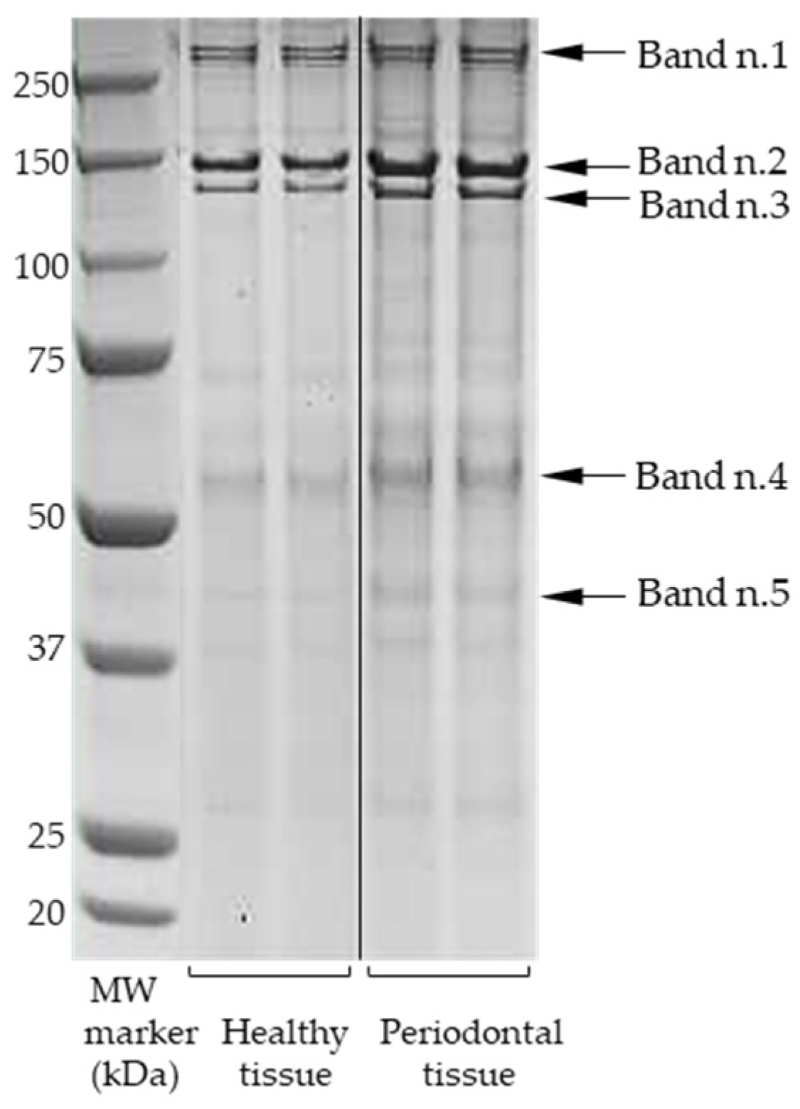
Figure 1. SDS-PAGE separation. Representative gel image showing the comparison between tissue samples from healthy and periodontal pocket-associated sites. Gradient gel 4–12% Bis-Tris, Coomassie Blue staining. Lane 1, molecular weight marker expressed in kilodalton (All Blue precision protein standard, Bio-Rad); Lanes 2–3, representative pools of healthy tissue sample, in duplicate; Lanes 4–5, representative pools of pathological tissue sample, in duplicate. All lanes originate from the same original gel image. Arrows indicate the significantly different bands between healthy and pathological samples.
Sci 06 00057 g001
Figure 2. Protein band analysis. Representative 1D gel lane obtained for periodontal tissue sample (a); vertical plot graph showing the peaks of bands intensities (b); gel image with inverted color (c). On the left are indicated the numbers of the differential bands. The density trace image (b) and the color image (c) were acquired using QuantityOne 1D image analysis software.
Sci 06 00057 g002
Figure 3. Protein overlap: Diagram showing the overlap of proteins identified in periodontal tissue in the present work with our previous proteomic studies conducted in GCF, TSCM, and periodontal pocket tissue samples from patients with periodontitis. The numbers in each ellipse refer to the total number of proteins identified in the corresponding study. Complete data are available from [30] for GCF, [31] for TSCM and [27,28] for previous periodontal pocket tissue.
Sci 06 00057 g003
Table 1. Quantitative statistical analysis of differential protein bands.
Band
ID a
Relative Quantity (OD × mm) bp-Value c
Healthy TissuePeriodontal Tissue
Band n.11.63 ± 0.021.90 ± 0.010.0004
Band n.20.29 ± 0.030.34 ± 0.040.0345
Band n.30.19 ± 0.010.24 ± 0.030.0067
Band n.40.13 ± 0.010.21 ± 0.030.0305
Band n.50.11 ± 0.010.14 ± 0.010.0349
a Band ID corresponding to those reported in Figure 1. b Relative quantity (OD × mm) of each significant band, expressed as mean ± SD for the two groups of samples. c p-values obtained via independent Student’ t-test (significant p-value < 0.05).
Table 2. Upregulated proteins identified using LC-MS/MS analysis in periodontal tissue.
Band a Acc. Number bProtein Name cGene dMass eScore fMat. gSeq. h
n.1NX_Q99715-1Collagen alpha-1(XII) chain isoform Iso 1COL12A1334,13812755
NX_P12111-1Collagen alpha-3(VI) chain isoform Iso 1COL6A3345,1671991717
NX_P02751-1Fibronectin isoform Iso 1FN1266,0523722
n.2NX_P02452-1Collagen alpha-1(I) chain isoform Iso 1COL1A1139,8838153627
NX_Q05707-1Collagen alpha-1(XIV) chain isoform Iso 1COL14A1194,4782541413
n.3NX_P12109-1Collagen alpha-1(VI) chain isoform 2C2COL6A1109,6023301312
NX_P08123-1Collagen alpha-2(I) chain isoform Iso 1COL1A2129,7494992617
NX_P12110-1Collagen alpha-2(VI) chain isoform 2C2COL6A2109,7095533
n.4NX_P13646-1Keratin, type I cytoskeletal 13KRT1349,90013866
NX_P02533-1Keratin, type I cytoskeletal 14KRT1451,8729155
NX_P013647-1Keratin, type II cytoskeletal 5KRT562,5681971313
NX_P02538-1Keratin, type II cytoskeletal 6AKRT6A60,2932801414
NX_P01857-1Immunoglobulin heavy constant gamma 1IGHG136,59612465
NX_P01859-1Immunoglobulin heavy constant gamma 2IGHG236,5055633
n.5NX_P08670-1VimentinVIM53,6766902926
NX_P07355-1Annexin A2ANXA238,8085182119
NX_P08727-1Keratin, type I cytoskeletal 19KRT1944,0794422622
NX_P04406-1Glyceraldehyde-3-phosphate dehydrogenaseG3PDH36,2013432114
NX_P60709-1Actin, cytoplasmic 1ACTB42,0523311913
NX_Q9BXN1-1AsporinASPN43,7889088
NX_P20774-1MimecanOGN34,2436355
NX_Q5VTE0Putative elongation factor 1-alpha-like 3 (PEF1)EEF1A1P550,4956133
a Identification code of protein band, as reported in Figure 1 and Figure 2, and in Table 1; b primary protein accession number (neXtProt database; c protein complete name (neXtProt database); d gene name; e monoisotopic mass from MS analysis (Da); f the highest score (Mascot search engine); g number of significant matching peptides; h number of significant protein sequences.
Table 3. Protein comparisons and matches with the current proteomic literature.
Full and Abbreviated Protein NameType of Oral SampleSeparation
Method
Identification/
Detection Platform
Bibliographic Reference
Collagen alpha-1(I) chain
(COL1A1)
Periodontal gingival tissue
Dentin
Dentin
Tooth pulp
Tooth pulp
Dental pulp
Root canal content
PDL cells
PDL fibroblasts
hPDLSCs
hPDLSCs
PDLSCs + SHEDs
DPSCs
SDS-PAGE
SDS-PAGE
SDS-PAGE + 2DE
2DE
2DE


2DE

2DE

SDS-PAGE
LC-MS/MS
LC-ESI-MS/MS
nano-LC-MS/MS
Q-ToF-MS/MS
MALDI-ToF
nano-UPLC-MSE
nano-LC-ESI-MS/MS
2D-LC-MS/MS
Ion-trap MS
TMT-tag
2-DE-MS
nano-LC-MS/MS
LC-MS/MS
[37]-Badovinac et al. (2013)
[38]-Park et al. (2009)
[39]-Jágr et al. (2012)
[40]-Eckhardt et al. (2014)
[41]-Pääkkönen et al. (2005)
[42]-Oliveira Silva et al. (2020)
[43]-Louriero et al. (2021)
[44]-Li et al. (2019)
[45]-Reichenberg et al. (2005)
[46]-Li et al. (2021)
[47]-Xiong et al. (2016)
[48]-Taraslia et al. (2018)
[49]-Niehage et al. (2016)
Collagen alpha-1(VI) chain
(COL6A1)
Dentin
Tooth pulp
Tooth pulp
hPDLSCs
PDLSCs + SHEDs
DPSCs
iDFCS
SDS-PAGE
2DE
2DE


SDS-PAGE
LC-ESI-MS/MS
Q-ToF-MS/MS
MALDI-ToF
TMT-tag
nano-LC-MS/MS
LC-MS/MS
iTRAQ + LC-MS/MS
[38]-Park et al. (2009)
[40]-Eckhardt et al. (2014)
[41]-Pääkkönen et al. (2005)
[46]-Li et al. (2021)
[48]-Taraslia et al. (2018)
[49]-Niehage et al. (2016)
[50]-Dou et al. (2017)
Collagen alpha-1(XII) chain
(COL12A1)
Periodontal gingival tissue
Dentin
Root canal content
PDL cells
PDLSCs + SHEDs
DPSCs
iDFCS
SDS-PAGE
SDS-PAGE


SDS-PAGE
LC-MS/MS
LC-ESI-MS/MS
nano-LC-ESI-MS/MS
2D-LC-MS/MS
nano-LC-MS/MS
LC-MS/MS
iTRAQ + LC-MS/MS
[37]-Badovinac et al. (2013)
[38]-Park et al. (2009)
[43]-Louriero et al. (2021)
[44]-Li et al. (2019)
[48]-Taraslia et al. (2018)
[49]-Niehage et al. (2016)
[50]-Dou et al. (2017)
Collagen alpha-1(XIV) chain
(COL14A1)
Periodontal gingival tissue
PDLSCs
iDFCS
SDS-PAGE

LC-MS/MS
nano-LC-MS/MS
iTRAQ + LC-MS/MS
[37]-Badovinac et al. (2013)
[48]-Taraslia et al. (2018)
[50]-Dou et al. (2017)
Collagen alpha-2(I) chain
(COL1A2)
Periodontal gingival tissue
Dentin
Dentin
Tooth pulp
Tooth pulp
Dental pulp
Root canal content
hPDLSCs
PDLSCs + SHEDs
DPSCs
SDS-PAGE
SDS-PAGE
SDS-PAGE + 2DE
2DE
2DE


SDS-PAGE
LC-MS/MS
LC-ESI-MS/MS
nano-LC-MS/MS
Q-ToF-MS/MS
MALDI-ToF
nano-UPLC-MSE
nano-LC-ESI-MS/MS
TMT-tag
nano-LC-MS/MS
LC-MS/MS
[37]-Badovinac et al. (2013)
[38]-Park et al. (2009)
[39]-Jágr et al. (2012)
[40]-Eckhardt et al. (2014)
[41]-Pääkkönen et al. (2005)
[42]-Oliveira Silva et al. (2020)
[43]-Louriero et al. (2021)
[46]-Li et al. (2021)
[48]-Taraslia et al. (2018)
[49]-Niehage et al. (2016)
Collagen alpha-2(VI) chain
(COL6A2)
Dentin
PDLSCs + SHEDs
DPSCs
iDFCS
SDS-PAGE

SDS-PAGE
LC-ESI-MS/MS
nano-LC-MS/MS
LC-MS/MS
iTRAQ + LC-MS/MS
[38]-Park et al. (2009)
[48]-Taraslia et al. (2018)
[49]-Niehage et al. (2016)
[50]-Dou et al. (2017)
Collagen alpha-3(VI) chain
(COL6A3)
Dentin
Tooth pulp
hPDLSCs
PDLSCs + SHEDs
DPSCs
iDFCS
SDS-PAGE
2DE
2DE

SDS-PAGE
LC-ESI-MS/MS
Q-ToF-MS/MS
2-DE-MS
nano-LC-MS/MS
LC-MS/MS
iTRAQ + LC-MS/MS
[38]-Park et al. (2009)
[40]-Eckhardt et al. (2014)
[47]-Xiong et al. (2016)
[48]-Taraslia et al. (2018)
[49]-Niehage et al. (2016)
[50]-Dou et al. (2017)
Fibronectin
(FN)
GCF
GCF
Saliva
PDL fibroblasts
PDLSCs + SHEDs
DPSCs
1DE
SDS-PAGE
SDS-PAGE
2DE

SDS-PAGE
LC-ESI-MS/MS
nano-LC-ESI-MS/MS
LC-MS/MS
Ion-trap MS
nano-LC-MS/MS
LC-MS/MS
[51]-Huynh et al. (2015)
[52]-Ngo et al. (2009)
[53]-Wu et al. (2016)
[45]-Reichenberg et al. (2005)
[48]-Taraslia et al. (2018)
[49]-Niehage et al. (2016)
Keratin, type I cytoskeletal 13
(KRT13)
Periodontal pocket tissue
GCF
GCF
GCF
GCF
2DE
SDS-PAGE


1D SDS-PAGE
LC-MS/MS
LC-ESI-QO-MS/MS
TMT-tag + LC-MS/MS
LC-ESI-MS/MS
nano-LC-ESI-MS/MS
[28]-Monari et al. (2015)
[30]-Bellei et al. (2022)
[54]-Tsuchida et al. (2013)
[55]-Silva-Boghossian et al. (2013)
[56]-Choi et al. (2011)
Keratin, type I cytoskeletal 14
(KRT14)
Periodontal gingival tissue
GCF
GCF
GCF
GCF
Dentin
Tooth pulp
PDLSCs + SHEDs
SDS-PAGE
SDS-PAGE


1D SDS-PAGE
SDS-PAGE + 2DE
2DE
LC-MS/MS
LC-ESI-QO-MS/MS
TMT-tag + LC-MS/MS
LC-ESI-MS/MS
nano-LC-ESI-MS/MS
nano-LC-MS/MS
Q-ToF-MS/MS
nano-LC-MS/MS
[57]-Liu et al. (2022)
[30]-Bellei et al. (2022)
[54]-Tsuchida et al. (2013)
[55]-Silva-Boghossian et al. (2013)
[56]-Choi et al. (2011)
[39]-Jágr et al. (2012)
[40]-Eckhardt et al. (2014)
[48]-Taraslia et al. (2018)
Keratin, type I cytoskeletal 19
(KRT19)
Periodontal gingival tissue
Periodontal pocket tissue
GCF
GCF
SDS-PAGE
2DE
SDS-PAGE
LC-MS/MS
LC-MS/MS
LC-ESI-QO-MS/MS
LC-ESI-MS/MS
[57]-Liu et al. (2022)
[28]-Monari et al. (2015)
[30]-Bellei et al. (2022)
[55]-Silva-Boghossian et al. (2013)
Keratin, type II cytoskeletal 5
(KRT5)
GCF
GCF
GCF
Dentin
Tooth pulp
PDLSCs + SHEDs


1D SDS-PAGE
SDS-PAGE + 2DE
2DE
TMT-tag + LC-MS/MS
LC-ESI-MS/MS
nano-LC-ESI-MS/MS
nano-LC-MS/MS
Q-ToF-MS/MS
nano-LC-MS/MS
[54]-Tsuchida et al. (2013)
[55]-Silva-Boghossian et al. (2013)
[56]-Choi et al. (2011)
[39]-Jágr et al. (2012)
[40]-Eckhardt et al. (2014)
[48]-Taraslia et al. (2018)
Keratin, type II cytoskeletal 6A
(KRT6A)
GCF
GCF
Dentin
Tooth pulp
SHEDs
SDS-PAGE

SDS-PAGE + 2DE
2DE
LC-ESI-QO-MS/MS
LC-ESI-MS/MS
nano-LC-MS/MS
Q-ToF-MS/MS
nano-LC-MS/MS
[30]-Bellei et al. (2022)
[55]-Silva-Boghossian et al. (2013)
[39]-Jágr et al. (2012)
[40]-Eckhardt et al. (2014)
[48]-Taraslia et al. (2018)
Immunoglobulin heavy constant gamma 1
(IGHG1)
Periodontal gingival tissue
GCF
GCF
GCF
GCF
Dentin
Tooth pulp
Tooth pulp
Dental pulp
Root canal content
Salivary gland secretion
PDLSCs + SHEDs
SDS-PAGE
SDS-PAGE
SDS-PAGE

1D SDS-PAGE
SDS-PAGE + 2DE
2DE
2DE


PAGE
LC-MS/MS
LC-ESI-QO-MS/MS
nano-LC-ESI-MS/MS
LC-ESI-MS/MS
nano-LC-ESI-MS/MS
nano-LC-MS/MS
Q-ToF-MS/MS
MALDI-ToF
nano-UPLC-MSE
nano-LC-ESI-MS/MS
LC-ESI-MS/MS
nano-LC-MS/MS
[37]-Badovinac et al. (2013)
[30]-Bellei et al. (2022)
[52]-Ngo et al. (2010)
[55]-Silva-Boghossian et al. (2013)
[56]-Choi et al. (2011)
[39]-Jágr et al. (2012)
[40]-Eckhardt et al. (2014)
[41]-Pääkkönen et al. (2005)
[42]-Oliveira Silva et al. (2020)
[43]-Louriero et al. (2021)
[58]-Siqueira et al. (2008)
[48]-Taraslia et al. (2018)
Immunoglobulin heavy constant gamma 2
(IGHG2)
Periodontal gingival tissue
GCF
GCF
Dentin
Tooth pulp
Dental pulp
Root canal content
Saliva
SDS-PAGE
SDS-PAGE

SDS-PAGE + 2DE
2DE


SDS-PAGE
LC-MS/MS
LC-ESI-QO-MS/MS
Label-free LC-MSE
nano-LC-MS/MS
Q-ToF-MS/MS
nano-UPLC-MSE
nano-LC-ESI-MS/MS
LC-MS/MS
[37]-Badovinac et al. (2013)
[30]-Bellei et al. (2022)
[59]-Bostanci et al. (2010)
[39]-Jágr et al. (2012)
[40]-Eckhardt et al. (2014)
[42]-Oliveira Silva et al. (2020)
[43]-Louriero et al. (2021)
[53]-Wu et al. (2016)
Vimentin
(VIM)
Periodontal gingival tissue
Periodontal pocket tissue
GCF
GCF
Dentin
Dentin
Tooth pulp
Tooth pulp
Root canal content
SDS-PAGE
2DE


SDS-PAGE
SDS-PAGE + 2DE
2DE
2DE
LC-MS/MS
LC-MS/MS
LC-ESI-MS/MS
ELISA and PCR
LC-ESI-MS/MS
nano-LC-MS/MS
Q-ToF-MS/MS
MALDI-TOF
nano-LC-ESI-MS/MS
[37]-Badovinac et al. (2013)
[28]-Monari et al. (2015)
[55]-Silva-Boghossian et al. (2013)
[60]-Shindo et al. (2023)
[38]-Park et al. (2009)
[39]-Jágr et al. (2012)
[40]-Eckhardt et al. (2014)
[41]-Pääkkönen et al. (2005)
[43]-Louriero et al. (2021)
Saliva
PDL fibroblasts
hPDLSCs
PDLSCs + SHEDs
DFCs + DPSCs + DPCs
DFCs + DPCs
DPSCs

2DE
2DE

2DE
2DE
2DE
LC-MS/MS
Ion-trap MS
2-DE-MS
nano-LC-MS/MS
MALDI-ToF-MS
MALDI-ToF/ToF
MALDI-ToF/ToF
[61]-Bostanci et al. (2018)
[45]-Reichenberg et al. (2005)
[47]-Xiong et al. (2016)
[48]-Taraslia et al. (2018)
[62]-Patil et al. (2014)
[63]-Guo et al. (2013)
[64]-Akpinar et al. (2014)
Annexin A2
(ANXA2)
Periodontal pocket tissue
GCF
Dentin
Dentin
Tooth pulp
Tooth pulp
Dental pulp
PDL fibroblasts
hPDLSCs
hPDLSCs
PDLSCs + SHEDs
DFCs + DPCs
2DE
2DE

SDS-PAGE
SDS-PAGE + 2DE
2DE
2DE

2DE

2DE

2DE
ESI-Q-ToF
LC-MS/MS
TMT-tag + LC-MS/MS
LC-ESI-MS/MS
nano-LC-MS/MS
Q-ToF-MS/MS
MALDI-TOF
nano-UPLC-MSE
Ion-trap MS
TMT-tag
2-DE-MS
nano-LC-MS/MS
MALDI-ToF/ToF
[27]-Bertoldi et al. (2013)
[28]-Monari et al. (2015)
[54]-Tsuchida et al. (2013)
[38]-Park et al. (2009)
[39]-Jágr et al. (2012)
[40]-Eckhardt et al. (2014)
[41]-Pääkkönen et al. (2005)
[42]-Oliveira Silva et al. (2020)
[45]-Reichenberg et al. (2005)
[46]-Li et al. (2021)
[47]-Xiong et al. (2016)
[48]-Taraslia et al. (2018)
[63]-Guo et al. (2013)
Glyceraldehyde-3-phosphate-dehydrogenase
(G3PDH)
Periodontal pocket tissue
GCF
GCF
GCF
TSCM
Dentin
Dentin
Tooth pulp
Tooth pulp
2DE
SDS-PAGE
SDS-PAGE
1D SDS-PAGE
SDS-PAGE
SDS-PAGE
SDS-PAGE + 2DE
2DE
2DE
LC-MS/MS
LC-ESI-QO-MS/MS
nano-LC-ESI-MS/MS
nano-LC-ESI-MS/MS
LC-ESI-QO-MS/MS
LC-ESI-MS/MS
nano-LC-MS/MS
Q-ToF-MS/MS
MALDI-TOF
[28]-Monari et al. (2015)
[30]-Bellei et al. (2022)
[52]-Ngo et al. (2010)
[56]-Choi et al. (2011)
[31]-Bergamini et al. (2021)
[38]-Park et al. (2009)
[39]-Jágr et al. (2012)
[40]-Eckhardt et al. (2014)
[41]-Pääkkönen et al. (2005)
Dental pulp
Root canal content
Saliva
PDL cells
PDL fibroblasts
PDLSCs + SHEDs
DFCs + DPSCs + DPCs


2DE

2DE
nano-UPLC-MSE
nano-LC-ESI-MS/MS
LC-MS/MS
2D-LC-MS/MS
Ion-trap MS
nano-LC-MS/MS
MALDI-ToF-MS
[42]-Oliveira Silva et al. (2020)
[43]-Louriero et al. (2021)
[61]-Bostanci et al. (2018)
[44]-Li et al. (2019)
[45]-Reichenberg et al. (2005)
[48]-Taraslia et al. (2018)
[62]-Patil et al. (2014)
Actin, cytoplasmic 1
(ACTB)
Periodontal pocket tissue
GCF
GCF
GCF
Dentin
Tooth pulp
Tooth pulp
Dental pulp
Root canal content
Salivary gland secretion
Saliva
PDL fibroblasts
PDLSCs + SHEDs
DPSCs
2DE
2DE
SDS-PAGE

1D SDS-PAGE
SDS-PAGE + 2DE
2DE
2DE


PAGE

2DE

2DE
ESI-Q-ToF
LC-MS/MS
nano-LC-ESI-MS/MS
LC-ESI-MS/MS
nano-LC-ESI-MS/MS
nano-LC-MS/MS
Q-ToF-MS/MS
MALDI-ToF
nano-UPLC-MSE
nano-LC-ESI-MS/MS
LC-ESI-MS/MS
LC-MS/MS
Ion-trap MS
nano-LC-MS/MS
MALDI-ToF/ToF
[27]-Bertoldi et al. (2013)
[28]-Monari et al. (2015)
[52]-Ngo et al. (2010)
[55]-Silva-Boghossian et al. (2013)
[56]-Choi et al. (2011)
[39]-Jágr et al. (2012)
[40]-Eckhardt et al. (2014)
[41]-Pääkkönen et al. (2005)
[42]-Oliveira Silva et al. (2020)
[43]-Louriero et al. (2021)
[58]-Siqueira et al. (2008)
[61]-Bostanci et al. (2018)
[45]-Reichenberg et al. (2005)
[48]-Taraslia et al. (2018)
[64]-Akpinar et al. (2014)
Asporin
(ASPN)
Dentin
Dentin
Tooth pulp
SDS-PAGE
SDS-PAGE + 2DE
2DE
LC-ESI-MS/MS
nano-LC-MS/MS
Q-ToF-MS/MS
[38]-Park et al. (2009)
[39]-Jágr et al. (2012)
[40]-Eckhardt et al. (2014)
Mimecan (or Osteoglycin)
(MIME)
Periodontal gingival tissue
Dentin
Dentin
Tooth pulp
SHEDs
SDS-PAGE
SDS-PAGE
SDS-PAGE + 2DE
2DE
LC-MS/MS
LC-ESI-MS/MS
nano-LC-MS/MS
Q-ToF-MS/MS
nano-LC-MS/MS
[57]-Liu et al. (2022)
[38]-Park et al. (2009)
[39]-Jágr et al. (2012)
[40]-Eckhardt et al. (2014)
[48]-Taraslia et al. (2018)
Putative elongation factor 1-alpha-like 3
(PEF1)
TSCMSDS-PAGELC-ESI-QO-MS/MS[31]-Bergamini et al. (2021)
PDL: periodontal ligament; TSCM: tooth surface collected material; hPDLSCs: human periodontal ligament stem cells; SHEDs: stem cells from human exfoliated deciduous teeth; DPSCs: dental pulp stem cells; iDFCs: immortalized dental follicle cells; DPCs: dental papilla cells; 2DE: two-dimensional gel electrophoresis; 1DE: mono-dimensional gel electrophoresis; LC: liquid chromatography; MS/MS: tandem mass spectrometry; ESI: electro spray ionization; MALDI-ToF: matrix-assisted laser desorption/ionization-time-of-flight; UPLC-MSE: ultra-performance LC-MS in data-independent analysis mode; TMT-tag: tandem mass tag; QO: Quadrupole Orbitrap; iTRAQ: isobaric tag for relative and absolute quantitation.
Disclaimer/Publisher’s Note: The statements, opinions and data contained in all publications are solely those of the individual author(s) and contributor(s) and not of MDPI and/or the editor(s). MDPI and/or the editor(s) disclaim responsibility for any injury to people or property resulting from any ideas, methods, instructions or products referred to in the content.