Review

A Time-of-Flight Neutron Backscattering Spectrometer at the China Spallation Neutron Source: Principle, Design, and Its Prospects


1
Institute of High Energy Physics, Chinese Academy of Sciences (CAS), Beijing 100049, China
2
Spallation Neutron Source Science Center, Dongguan 523803, China
3
University of Chinese Academy of Sciences, Beijing 100049, China
*
Authors to whom correspondence should be addressed.
Quantum Beam Sci. 2026, 10(3), 20; https://doi.org/10.3390/qubs10030020
Submission received: 23 June 2026 / Revised: 23 July 2026 / Accepted: 25 August 2026 / Published: 27 August 2026
(This article belongs to the Special Issue Neutron Instrumentation)

Abstract

The quasielastic neutron scattering technique is an indispensable tool for probing microscopic dynamics in condensed matter at the nanoscale. However, the lack of a high-resolution spectrometer has restricted comprehensive studies of dynamics in China. To address this gap, a new time-of-flight neutron backscattering spectrometer, NuBS, is currently under construction at the China Spallation Neutron Source. This review systematically introduces basic principles, instrumental design, and scientific opportunities of NuBS. NuBS is engineered to deliver high energy resolution with a broad dynamic range, based on the moderator pulse structure. Given the wide time window of NuBS, we highlight its research prospects in frontier areas, including energy storage materials and heterogeneous catalysis. NuBS is expected to provide new opportunities for studies of complex molecular dynamics and in situ investigations, significantly elevating the capabilities of the quasielastic neutron scattering community upon its scheduled completion.

1. Introduction

Understanding stochastic dynamics across multiple spatial and temporal scales remains a central challenge in condensed matter physics, soft matter science, energy materials, and heterogeneous catalysis. For many functional materials, macroscopic properties such as ionic conductivity, catalytic selectivity, proton transport, and molecular adsorption are governed by microscopic motions occurring on pico- to nanosecond timescales and length scales ranging from angstroms to nanometers. These dynamical processes often involve multiple coupled relaxation mechanisms, including localized vibrations, rotational reorientations, jump diffusion, and long-range translational transport [1,2,3,4]. Experimental techniques capable of simultaneously resolving both the temporal and spatial characteristics of such motions are therefore essential for establishing microscopic structure–dynamics–function relationships.
Quasielastic neutron scattering (QENS) has become one of the most powerful experimental techniques for probing nanoscale stochastic dynamics because it directly probes atomic motion through neutron energy and momentum transfer [5,6]. Owing to the exceptionally large incoherent scattering cross section of hydrogen, QENS is particularly sensitive to hydrogen-containing materials, including polymers, biological systems, porous materials, proton conductors, and catalytic adsorbates [7,8,9,10,11,12]. QENS provides direct access to relaxation times, diffusion coefficients, jump lengths, and geometrical constraints associated with molecular motion over characteristic timescales spanning approximately 10 13 10 9 s. Among QENS techniques, neutron backscattering spectroscopy (NBS) is uniquely suited for probing nanoscale dynamics within the pico- to nanosecond time regime. By exploiting Bragg reflection in exact or near-backscattering geometries, NBS achieves energy resolutions in the μeV range, enabling direct observation of stochastic motions such as translational diffusion, rotational relaxation, confined dynamics, and slow magnetic fluctuations [13,14]. Traditional reactor-based NBS instruments achieve sub-μeV energy resolution by combining monochromator and analyzer crystals in exact backscattering geometry [13]. Although this approach provides exceptional energy resolution, the accessible dynamic range is usually limited to tens of μeV. In contrast, time-of-flight backscattering spectrometers (ToF-BSs) at pulsed spallation neutron sources determine the incident neutron energy from the time-of-flight (ToF) over a long flight path while preserving analyzer-based final-energy selection for scattered neutrons. By selecting a broad incident wavelength band through chopper systems, ToF-BSs substantially extend the accessible dynamic range to hundreds or even thousands of μeV while maintaining μeV-scale energy resolution [15]. A broader dynamic range enables the observation of relaxation processes spanning different timescales, which would otherwise require multiple complementary measurements. This capability is increasingly important because many advanced functional materials exhibit hierarchical dynamics spanning multiple timescales. Accessing these hierarchical dynamics within a single experimental framework represents one of the major motivations for the development of modern ToF-BSs. It is also beneficial for in situ characterization of advanced functional materials, which meets the growing demand for bridging microscopic dynamics and macroscopic functional performance under realistic working conditions.
The development of ToF-BSs has accelerated in recent years with the construction of advanced pulsed neutron facilities worldwide. These developments illustrate that no single instrument can optimize all performance parameters simultaneously. Instead, different facilities adopt distinct design philosophies depending on source characteristics and targeted scientific applications. Within this international context, the China Spallation Neutron Source (CSNS) provides a significant opportunity for the development of high-resolution neutron spectroscopy in China. Since the beginning of user operations in 2018, CSNS has rapidly expanded its neutron-scattering functionality [16,17,18,19]. The ongoing CSNS-II upgrade, scheduled for completion by 2029, will increase the proton beam power from 100 kW to 500 kW and substantially enlarge the instrument suite. Among these instruments is a new ToF-BS, NuBS [20]. Benefiting from the long neutron flight path, the narrow pulse width of the decoupled poisoned hydrogen moderator (DPHM), and the 25 Hz repetition rate of CSNS, NuBS is expected to achieve an energy resolution of approximately 3 μeV using Si(111) analyzers while simultaneously providing a dynamic range approaching ±550 μeV in standard operating mode. The combination of a relatively broad dynamic range and μeV-scale energy resolution places NuBS in a distinctive operational regime among existing ToF-BSs, particularly for research requiring simultaneous access to localized and long-range relaxation processes, without combining multiple measurements from different spectrometers [21].
This paper reviews the basic principles, instrumental design, and scientific opportunities associated with NuBS at CSNS. Section 2 introduces the basic principles of ToF-BSs. Section 3 presents the design philosophy and major technical components of NuBS, including the moderator system, chopper configuration, neutron optics, analyzer geometry, detector arrangement, and sample environments. Section 4 discusses prospective scientific applications in energy materials and heterogeneous catalysis, highlighting the unique opportunities enabled by the wide time window of the instrument.

2. Principles of Neutron Backscattering Spectroscopy

2.1. QENS and Nanoscale Dynamics

NBS is primarily employed to study stochastic motions occurring on pico- to nanosecond timescales and over angstrom-to-nanometer length scales. Within this spatiotemporal regime, QENS provides direct access to microscopic relaxation processes such as translational diffusion, rotational relaxation, jump dynamics, and confined motion. In QENS experiments, these stochastic motions manifest as a broadening of the elastic scattering line centered at zero energy transfer [5,6,14]. The experimentally measured quantity in QENS is the dynamic structure factor S ( Q , ω ) , which describes the spatial and temporal correlations of atomic motions and is related to the intermediate scattering function and van Hove correlation function through Fourier transformation in time, and in time and space, respectively [22].
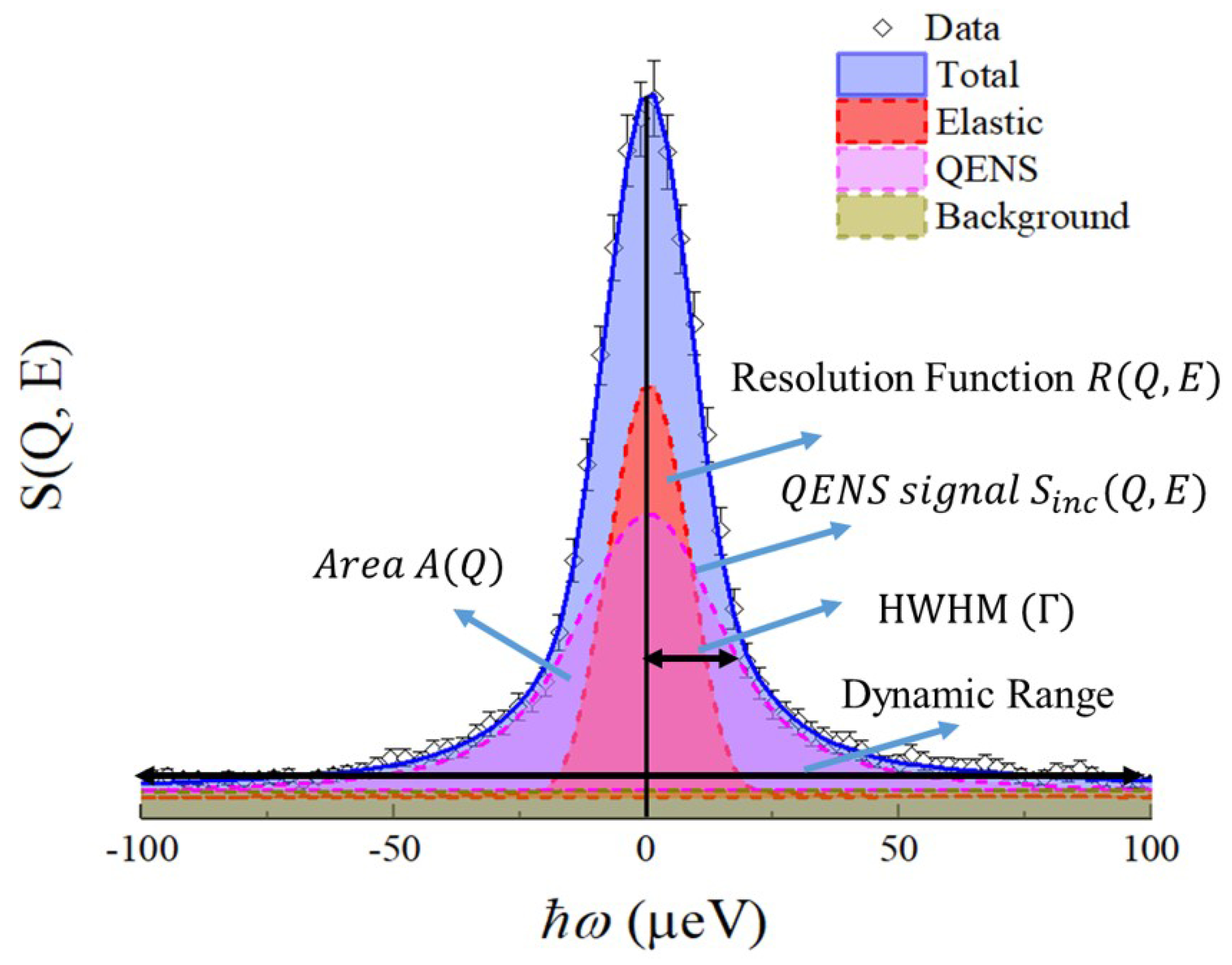
In many soft-matter, biological, and energy-related systems, the neutron signal is dominated by the large incoherent scattering cross section of hydrogen. Under such conditions, QENS primarily probes self-correlated atomic and molecular motions through the incoherent dynamic structure factor S inc ( Q , ω ) . The measured spectrum S meas ( Q , ω ) in the QENS regime is typically analyzed by convolving a model scattering function with the instrumental resolution function R ( Q , ω ) , together with an additional background contribution B ( Q , ω ) as illustrated schematically in Figure 1 [23,24,25,26],
S meas ( Q , ω ) = [ S inc ( Q , ω ) R ( Q , ω ) ] + B ( Q , ω )
The resolution function is commonly determined by measuring an elastic scatterer, such as vanadium or samples at low temperature where all dynamical processes are frozen within the instrumental time window. For continuous translational diffusion characterized by an exponential temporal relaxation, the quasielastic spectral component is commonly approximated by a Lorentzian function,
S inc ( Q , ω ) = 1 π Γ ( Q ) Γ ( Q ) 2 + ω 2
where Γ ( Q ) represents the half-width at half-maximum (HWHM) of the quasielastic broadening. For Fickian diffusion [27], the linewidth follows
Γ ( Q ) = D Q 2
where D is the translational diffusion coefficient. Deviations from this behavior often indicate jump diffusion, confined motion, or spatially restricted relaxation processes. Jump diffusion models, such as the Chudley–Elliott model, can be employed to extract residence times and jump lengths [28,29,30,31]. In addition to translational diffusion, QENS is also highly sensitive to localized and rotational motions. In such cases, the scattering intensity is frequently analyzed using the elastic incoherent structure factor (EISF), which reflects the geometry and spatial confinement of the motion [32]. The combined analysis of quasielastic linewidths and EISFs enables detailed characterization of microscopic relaxation mechanisms across a broad range of condensed matter systems. In many advanced functional materials, however, multiple dynamical processes coexist within overlapping time windows. For example, ions in solid electrolytes often exhibit fast localized vibrations within transient coordination cages together with slower long-range hopping processes [2]. Similarly, molecules confined in nanoporous catalysts may simultaneously undergo rotational relaxation, restricted rattling motions, and translational diffusion through the pore network [3]. These processes may generate quasielastic linewidths spanning more than three orders of magnitude, from approximately 1 μeV to over 1000 μeV. Recent developments in data-analysis methodologies, as well as molecular dynamics (MD) simulations, are increasingly combined with QENS experiments to assist the interpretation of complex relaxation mechanisms [33,34,35,36,37,38,39,40,41,42].
The characteristic timescale accessible in neutron spectroscopy is fundamentally linked to the instrumental energy resolution and dynamic range through the Fourier time–energy uncertainty relation,
τ Δ E
Consequently, resolving both localized picosecond motions and slower nanosecond transport processes requires instruments that combine μeV energy resolution with a sufficiently broad dynamic range. Achieving both objectives simultaneously has become one of the major challenges in modern neutron spectroscopy and has strongly motivated the development of ToF-BSs.

2.2. Basic Principles of ToF-BS

Among QENS techniques, NBS is one of the principal techniques for high-resolution measurements, typically achieving energy resolutions in the μeV range, whereas neutron spin echo spectroscopy can probe longer timescales by directly measuring the intermediate scattering function in the time domain. The high resolution of NBS arises from Bragg diffraction by monochromator and analyzer crystals operated in exact or near-backscattering geometries. This principle follows from Bragg’s law,
λ = 2 d sin θ B ,
where d is the spacing of the diffracting lattice planes and θ B is the Bragg angle. The relative wavelength uncertainty is
Δ λ λ = Δ d d + cot θ B Δ θ B .
As the Bragg angle approaches 90°, the angular term becomes negligible. This unique property enables extremely precise wavelength and energy selection using perfect crystal analyzers such as Si(111).
Traditional reactor-based backscattering spectrometers employ exact backscattering geometry for both the monochromator and analyzer crystals. Representative instruments include IN16B at ILL [43], HFBS at NIST [44], SPHERES at FRM II [45], and Emu at ANSTO [46]. A prototype GaAs(200) analyzer tested on IN16B at ILL even yielded an unprecedented energy resolution of 77–78 neV [47], but the resulting reduction in intensity remains a significant drawback. In these instruments, the incident neutron energy is scanned, typically using Doppler-driven monochromators, while the analyzer fixes the final neutron energy. Although this configuration provides exceptional energy resolution, the accessible dynamic range is intrinsically constrained by the limited Doppler velocity range and mechanical stability of the monochromator system. An alternative analyzer, CaF2 (422), adopted in IN13 at ILL, provides an extended dynamic range (±200 μeV), but at the expense of coarse resolution (∼8 μeV) [48].
The emergence of pulsed spallation neutron sources has enabled an alternative approach based on the ToF technique. A ToF-BS illuminates the sample using a pulsed polychromatic neutron beam and determines the incident neutron energy from the neutron flight time over a long flight path, while analyzer crystals operating in near-backscattering geometry provide final-energy selection for the scattered neutrons. This hybrid configuration preserves the high energy resolution characteristic of backscattering spectroscopy while substantially extending the accessible dynamic range. Furthermore, a ToF-BS determines the energy transfer directly from the ToF of each detected neutron, thereby eliminating the need for incident-energy scanning and enabling simultaneous measurements over a broad energy range. More recently, the BATS option (BATS: Backscattering and Time-of-flight Spectrometer) implemented on IN16B at ILL has demonstrated that a hybrid ToF approach can significantly extend the dynamic range of reactor-based backscattering spectrometers while maintaining μeV energy resolution [49,50].
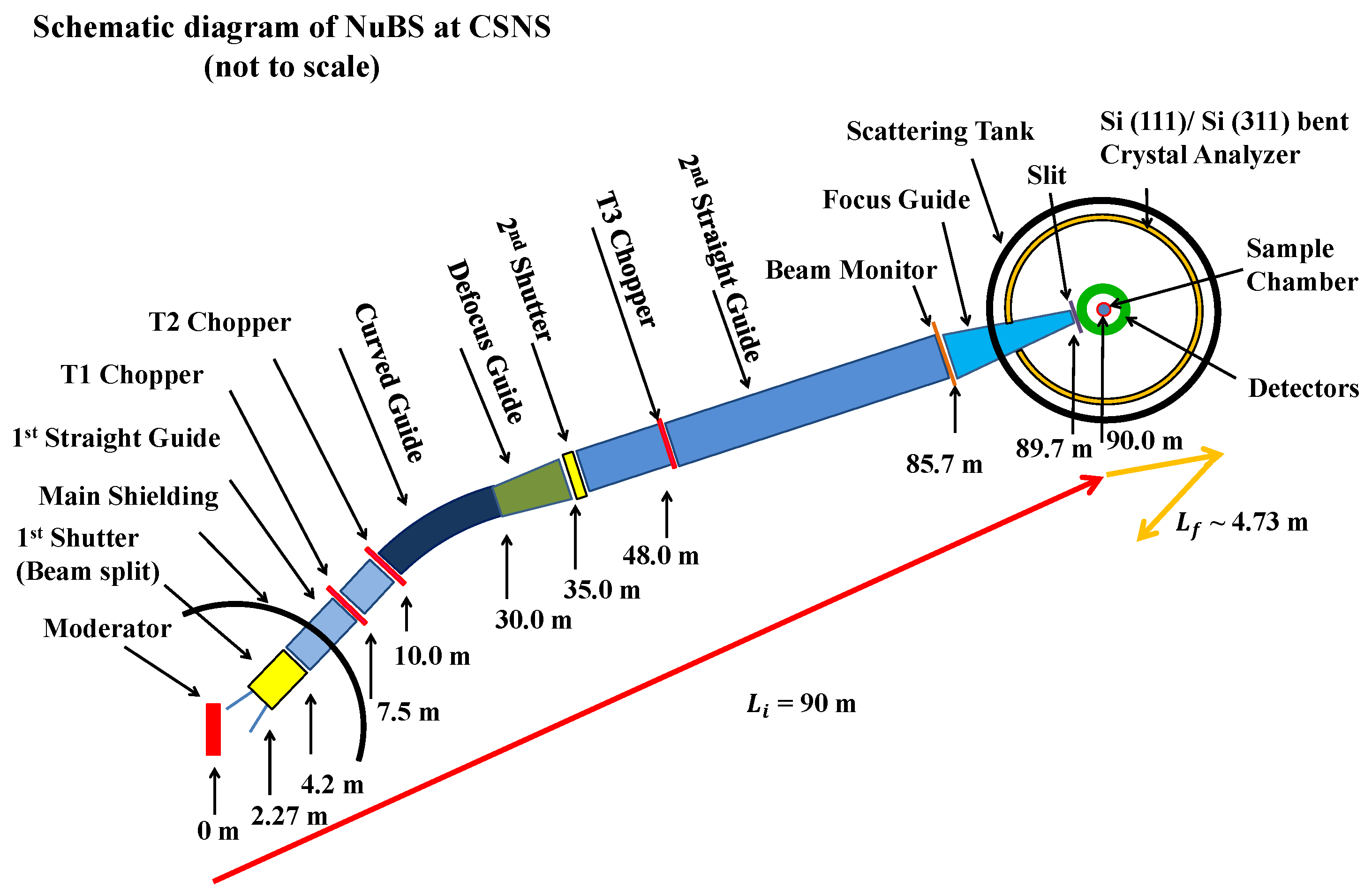
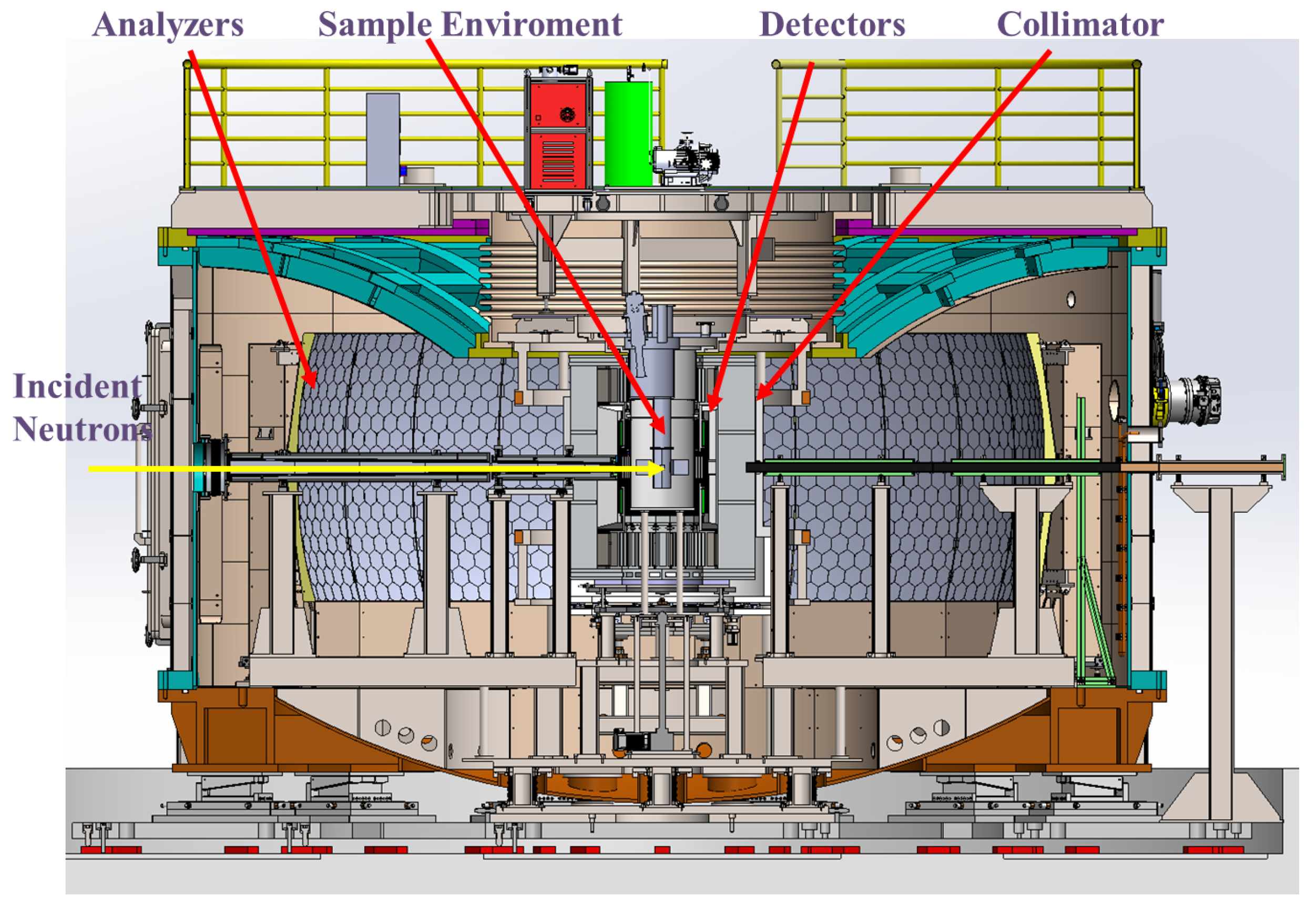
A typical ToF-BS is conventionally divided into two sections: the primary spectrometer, which transports and conditions the incident neutron beam upstream of the sample position, and the secondary spectrometer, which performs energy analysis and neutron detection downstream of the sample. The primary spectrometer principally consists of a neutron guide system that transports cold neutrons from the moderator to the sample over a long flight path, and a set of disc choppers to select the neutron bandwidth. The secondary spectrometer consists of large-area arrays of analyzer crystals and a corresponding bank of detectors, all housed within an evacuated scattering tank to minimize background and beam attenuation. The analyzer crystals are positioned at a fixed Bragg angle θ B , slightly offset from exact backscattering, thereby providing physical space for the sample environment. The detectors are arranged in cylindrical arrays that register the arrival position and ToF of each reflected neutron. A schematic layout of a ToF-BS (e.g., NuBS) is shown in Figure 2.
In this inverted-geometry configuration, the energy of the scattered neutrons is fixed by Bragg reflection from the analyzer crystals, as shown in Equation (5). The incident neutron energy E i spans the wavelength band defined by the primary spectrometer. Since the total flight time t total from the moderator to the detector is measured, and the flight time in the secondary spectrometer t f is fixed by the instrument geometry, the incident energy is determined from the flight time in the primary spectrometer t i over the path length L i , expressed as
E i = 1 2 m n L i t i 2 = 1 2 m n L i t total t 0 L f / v f 2 .
The energy transfer to the sample is therefore given by ω = E i E f . And the momentum transfer vector is defined as Q = k i k f , where k i and k f are the wavevectors of the incident and scattered neutrons, respectively.
The overall energy resolution of a ToF-BS can be derived from Equations (6) and (7). It arises from uncertainties in the flight path geometry, the finite width of the source pulse, the intrinsic properties of the analyzer crystal, and the Bragg angle divergence of the scattered neutron. The relative energy resolution at the elastic line ( ω = 0 ) is given by
Δ E E i = 2 Δ L t o t a l L i 2 + Δ t 0 t i 2 + Δ d d 2 + cot θ B Δ θ B 2 ,
where Δ E denotes the full width at half maximum (FWHM) of the elastic line. Δ L t o t a l represents the uncertainty of the total length of the flight path, mainly originating from the finite dimensions of the sample. The term Δ t 0 is the effective width of the neutron pulse emitted by the moderator or by the pulse-shaping choppers (PSCs). The contribution Δ d / d represents the spread of the lattice spacing of the analyzer crystal. The final term, proportional to cot θ B , is minimized by keeping θ B as close to 90 as space constraints permit, and Δ θ B originates predominantly from the finite dimensions of the sample and the pixel size of the position-sensitive detector.
This expression illustrates the fundamental design philosophy of modern ToF-BSs. High energy resolution requires narrow moderator pulses, long flight paths, highly perfect analyzer crystals, and analyzer geometries operating as close as possible to exact backscattering. Balancing these competing requirements for energy resolution, neutron intensity, and dynamic range has driven the evolution of ToF-BSs.

2.3. An Overview of Worldwide ToF-BSs

The development of ToF-BSs over the past several decades can be viewed as a succession of technological innovations aimed at overcoming successive performance bottlenecks in energy resolution, dynamic range, neutron efficiency, and operational flexibility. The first successful realization of the ToF-BS concept was achieved with IRIS at ISIS [51]. The principal objective of IRIS was to combine the intrinsically broad dynamic range available at pulsed neutron sources with the high energy resolution properties of indirect-geometry spectroscopy. IRIS views a liquid-hydrogen moderator that generates a pulse with a width of 22 λ (Å) μs at wavelengths near the peak of the Maxwellian. It employed pyrolytic graphite (PG) analyzers arranged in a circular geometry within the horizontal scattering plane, covering scattering angles from 15° to 165° to enlarge the Q range. The relatively large mosaic spread of PG crystals provided high neutron reflectivity and therefore high count rates, although at the expense of energy resolution, which is comparable to the contribution from the flight-time uncertainty. Depending on the analyzer reflection used, IRIS achieved an energy resolution of approximately 20 μeV. The IRIS mica option, which achieves an energy resolution of approximately 1 μeV, is often not viable due to the substantial loss of intensity and the restricted momentum transfer range for lower reflection orders. IRIS successfully demonstrated that ToF techniques can be combined with indirect-geometry energy analysis to provide a dynamic range substantially larger than that of conventional reactor-based backscattering spectrometers.
Although IRIS established the viability of the ToF-BS concept, its relatively limited analyzer solid-angle coverage restricted the overall neutron collection efficiency. This limitation motivated the development of OSIRIS, which retained the basic indirect-geometry architecture of IRIS while introducing a substantially redesigned secondary spectrometer [52,53]. The key innovation of OSIRIS at ISIS was the implementation of a vertically extended analyzer assembly consisting of approximately 9000 PG crystals mounted on an elliptically machined support structure. The analyzer geometry was designed such that one focal point coincided with the sample position and the other with the detector bank. This arrangement significantly increased the accepted solid angle without introducing large flight-path variations that would degrade the energy resolution. Consequently, OSIRIS achieved substantially higher neutron collection efficiency while maintaining an energy resolution comparable to that of IRIS. Another important improvement was the adoption of supermirror guides to enhance neutron transport efficiency. Currently, an update of OSIRIS is underway. A new guide with an elliptic defocusing and focusing geometry has been proposed, providing a 10-fold increase in flux [54]. In addition, the implementation of Si(111) analyzers is underway to improve the energy resolution to 11 μeV [55].
Another breakthrough in ToF-BS occurred with the commissioning of BASIS at the Spallation Neutron Source in 2007 [56,57]. BASIS was designed with the primary objective of achieving substantially higher energy resolution than IRIS and OSIRIS. To accomplish this goal, BASIS introduced two key innovations. First, the instrument utilizes a DPHM, which provides a significantly narrower neutron pulse width than the coupled moderators employed at ISIS. Second, the PG crystals were replaced by Si(111). The combination of these two developments simultaneously reduced both the moderator timing contribution and the analyzer contribution to the overall resolution function. As a result, BASIS achieved an energy resolution of approximately 3.5 μeV while maintaining a dynamic range of approximately ± 100 μeV. Operating the choppers at half the source frequency provides a flexible way to extend the bandwidth to ± 200 μeV at the expense of discarding alternate pulses. An alternative Si(311) analyzer configuration further extended the Q range to 3.8 Å−1 and the dynamic range to approximately ± 660 μeV. The silicon crystals are arranged in a simple spherical geometry and are coupled to position-sensitive 3He detectors. The position-sensitive 3He detectors can distinguish the neutrons arriving along different trajectories. In this arrangement, the uncertainties in cot θ B Δ θ B and Δ L f / L i depend only on the sizes of the detector pixel and sample. BASIS therefore marked a fundamental transition from graphite-based, flux-oriented instruments toward silicon-based, high-resolution ToF-BS.
The key innovation of the next instrument, DNA at J-PARC, was the incorporation of PSCs into the primary spectrometer [58,59]. Instead of relying exclusively on moderator characteristics to determine the pulse width, DNA offers flexible adjustment of the energy resolution and flux by utilizing PSCs. Narrow pulse-shaping settings provide energy resolutions as high as approximately 1.4 μeV, whereas wider settings increase neutron flux. The dynamic range in standard mode extends only from −20 μeV to 80 μeV. This design provides flexibility in balancing energy resolution and neutron flux, but at the expense of dynamic range. To overcome this limitation, DNA introduced a repetition-rate multiplication (RRM) mode, enabling multiple incident wavelength bands to be measured simultaneously within a single source period [60]. Through RRM mode and additional experiments with different phases of choppers, DNA can access an energy transfer range extending from approximately 500 μeV to + 1500 μeV. DNA therefore establishes a new paradigm in which instrument resolution and flux can be optimized for different scientific applications. Furthermore, to reduce background from the aluminum frame, the backside of the silicon wafer is covered by an absorbing layer. This design achieves an extremely low instrumental background with a signal-to-noise ratio of 10 5 . Similar approaches have subsequently been adopted by other ToF-BSs.
The next generation of ToF-BSs could be represented by MIRACLES at the European Spallation Source [61,62]. Unlike previous instruments developed for short-pulse sources, MIRACLES is specifically designed for the ESS long-pulse source. The instrument combines PSCs and advanced neutron-guide optics to exploit the unique characteristics of the ESS moderator system [63,64]. MIRACLES is designed to provide a tunable energy resolution between approximately 2 and 40 μeV while maintaining a dynamic range approaching ± 550 μeV [62]. The instrument is designed to fully exploit the ESS long-pulse source and is scheduled for completion in 2028.
From a historical perspective, the evolution of ToF-BSs can be interpreted as a sequence of technological innovations addressing successive performance bottlenecks. IRIS established the feasibility of the ToF-BS concept and demonstrated the advantages of combining the ToF technique with indirect geometry. OSIRIS improved neutron collection efficiency through advanced analyzer geometry. BASIS introduced silicon backscattering analyzers and narrow-pulse moderator technology to achieve substantially improved energy resolution. DNA demonstrated that the use of PSCs and RRM provides flexibility in balancing resolution and flux. MIRACLES further extends these concepts to long-pulse spallation sources. The principal characteristics of current ToF-BSs are summarized in Table 1.

3. Physical Design and Performance Characteristics of NuBS at CSNS

NuBS at CSNS is a ToF-BS currently under construction, with user operations expected to begin around 2029 [20]. As the first NBS instrument in China, NuBS is designed to provide a wide instrumental time window. Benefiting from the long neutron flight path, the narrow pulse width of DPHM, and the 25 Hz repetition rate of the source, NuBS is expected to achieve an energy resolution of approximately 3 μeV using Si(111) analyzers while simultaneously providing a dynamic range approaching ±550 μeV in standard operating mode. The combination of a relatively broad dynamic range and μeV-scale energy resolution places NuBS in a unique performance regime among existing ToF-BSs. The following subsection describes the detailed design of the spectrometer, together with the performance characteristics of NuBS.

3.1. Moderator

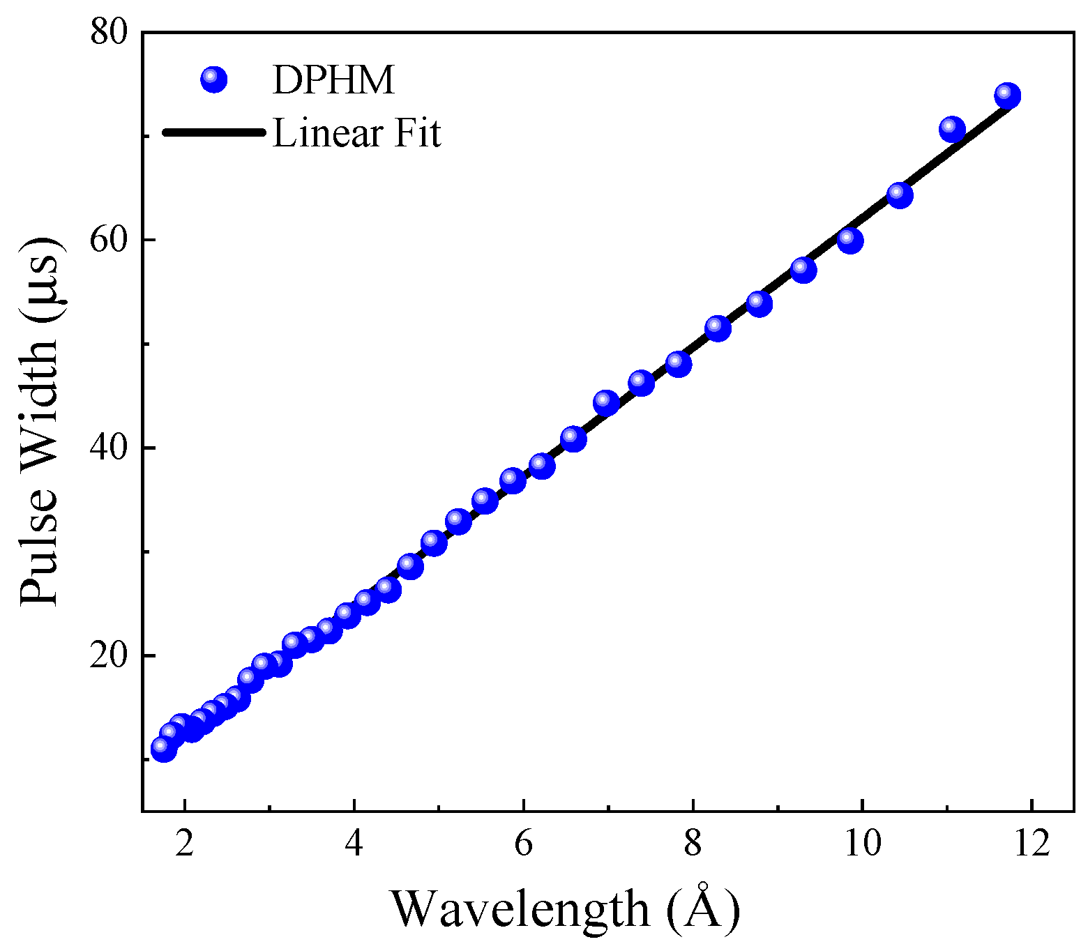
NuBS views the DPHM positioned above the spallation target, with outer dimensions of approximately 10 × 10 × 5 cm 3 . The center of the moderator is positioned 1935.5 mm above the spectrometer hall floor, and the orientation of the #10 neutron channel provides sufficient physical space for the long instrument layout [69]. The DPHM is specifically designed to deliver a narrow neutron pulse, which is essential to achieving high energy resolution. The characteristics of the moderator were obtained through MCNP simulations. The pulse width, as shown in Figure 3, varies approximately linearly with wavelength and measures only 39.7 μ s at λ = 6.267 Å , corresponding to a relative time uncertainty of Δ t 0 / t i 2.73 × 10 4 at the nominal incident energy. This narrow intrinsic pulse, combined with the long flight path, allows NuBS to achieve μeV-level resolution. The DPHM provides a considerable neutron flux within the wavelength range of 1– 10 Å , covering the operational requirements of Si(111) and Si(311) analyzer configurations.

3.2. Chopper System

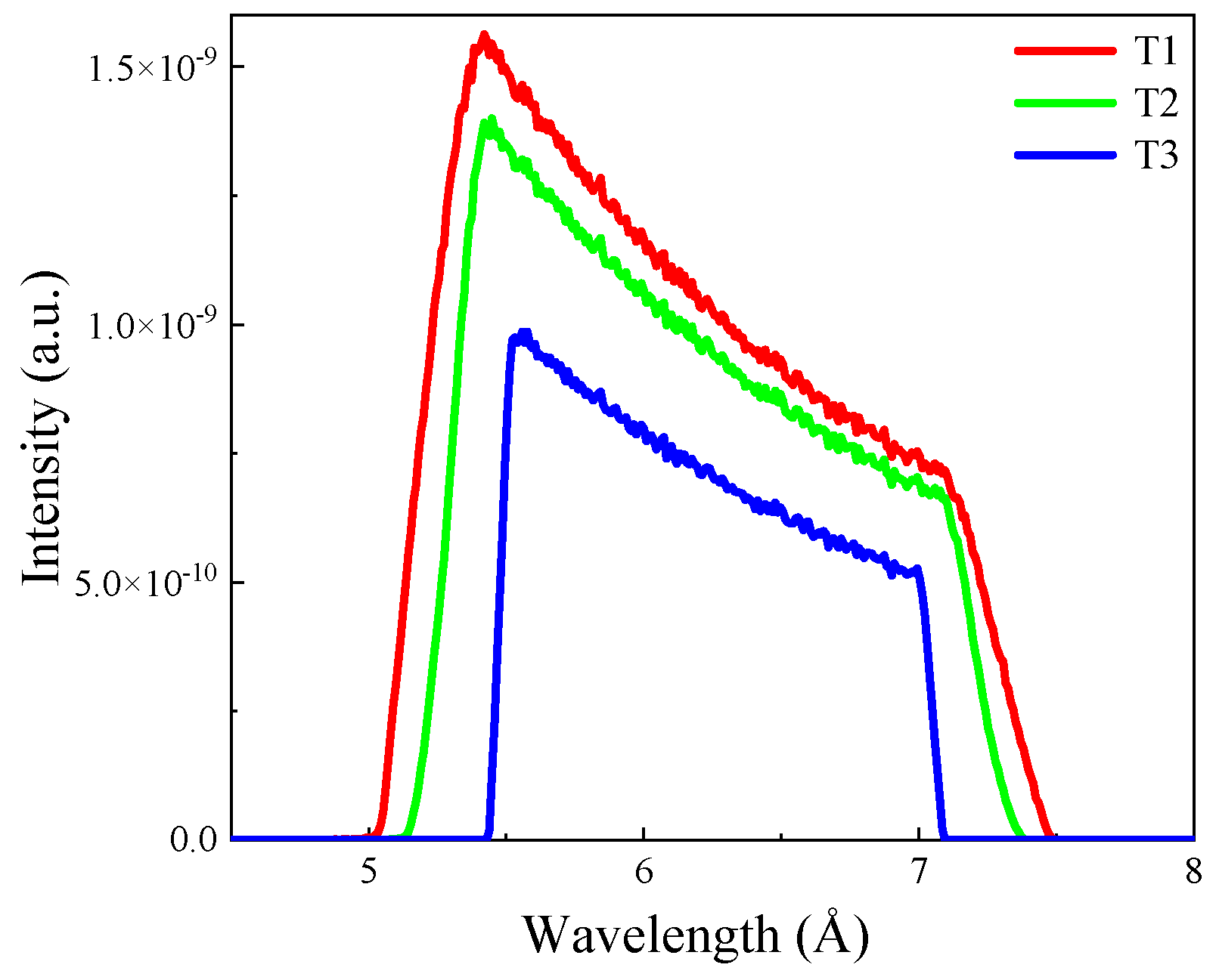
The chopper system of NuBS comprises three disc choppers designed to select a well-defined incident wavelength band while eliminating frame overlap and long-wavelength contamination [70,71]. The first two choppers, T1 and T2, are located at 7.5 m and 10 m from the moderator, respectively, and serve as frame-overlap suppression and background removal elements. The bandwidth selection is performed by the third chopper, T3, positioned at 48 m from the moderator. Each disc has a radius of 35.5 cm and is fabricated from aluminum alloy coated with 10B4C for neutron absorption [72]. The opening angles are 41.42 for T1, 50.81 for T2, and 180.33 for T3. The three choppers operate in synchrony to transmit a neutron bandwidth of approximately 1.65 Å centered at λ 0 = 6.267 Å when operating at the nominal frequency of 25 Hz , as shown in Figure 4. This yields a dynamic range of approximately 420 to + 680 μ eV for the Si(111) analyzer configuration. The central wavelength of the transmitted band can be adjusted by changing the chopper phases, allowing the instrument to operate at any desired wavelength within the 1– 10 Å range without affecting the bandwidth. For experiments requiring an even broader dynamic range, the choppers can be operated at 12.5 Hz , effectively eliminating alternate pulses and doubling the transmitted bandwidth to 3.30 Å , which extends the dynamic range to approximately 780 to + 1800 μ eV .

3.3. Neutron Optics

The neutron guide system of NuBS is designed to transport cold neutrons from the moderator to the sample over the 90 m flight path with maximum efficiency while suppressing high-energy background. Due to space constraints within the shared #10 channel, the guide center of NuBS is offset by 10 mm horizontally and 5 mm vertically from the moderator center, with a centerline deviation of 0.6 from the channel axis. A beam-splitting component within the first shutter, starting at 2.27 m from the moderator, separates neutrons between NuBS and the #10A beamline using a tapered guide section. The NuBS guide system begins with a straight guide of 60 × 90 mm 2 (W*H) cross-section, which is embedded in the wall insert and extends to the T2 chopper at 10 m . It is followed by a 20 m curved guide with a curvature radius of 1800 m and a supermirror coating factor of m = 2.5 on the left and right sides, which eliminates a direct line of sight to the moderator over a distance of 34 m and thereby removes fast neutrons and gamma rays. The characteristic wavelength of the curved guide is λ * = 1.89 Å , ensuring efficient transmission of cold neutrons in the operating range.
To compensate for reflection losses in the relatively narrow initial guide section, a defocusing guide section between 30 and 35 m expands the cross-section from 60 × 90 mm 2 to 90 × 90 mm 2 , resulting in a 30 % increase in flux at the sample position. A long straight guide of 90 × 90 mm 2 cross-section transports the neutrons to the vicinity of the sample, followed by an elliptical guide that focuses the beam onto the desired sample area. The focusing guide is shaped as a half-ellipse with a major axis of 4.235 m and a minor axis of 0.045 m , reducing the beam cross-section from 90 × 90 mm 2 to approximately 30 × 30 mm 2 at the sample center position. A 0.3 m gap between the exit of the guide and the sample provides space for sample environment equipment, and a four-blade slit is placed in this gap to suppress background. The supermirror coating factors are optimized along the guide to balance transmission efficiency against cost, where m = 2 for the long straight guide and ranges from 2.5 to 5 for the focusing guide. The guide parameters were optimized using the particle swarm optimization method to increase the neutron flux at the sample [73,74].
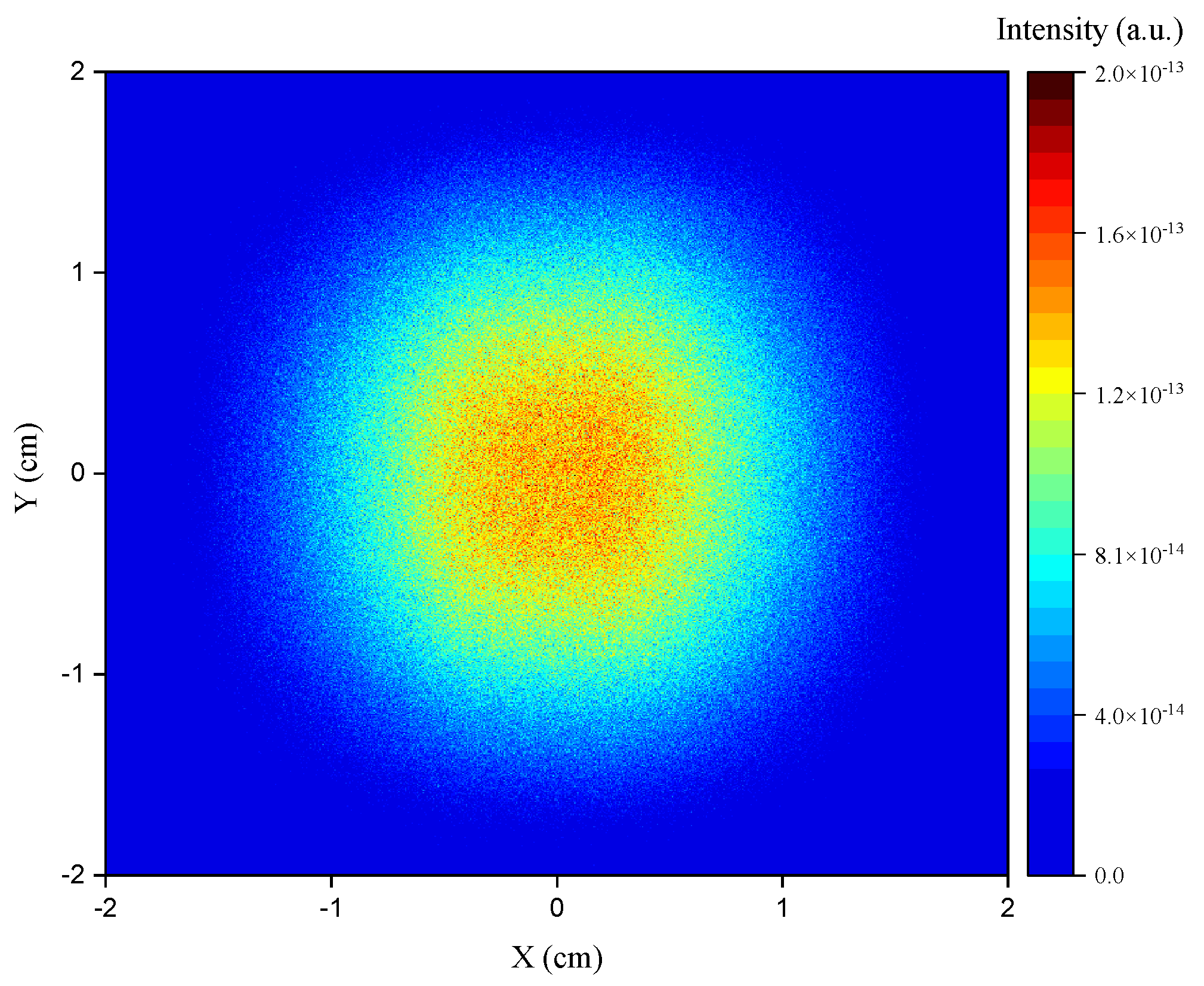
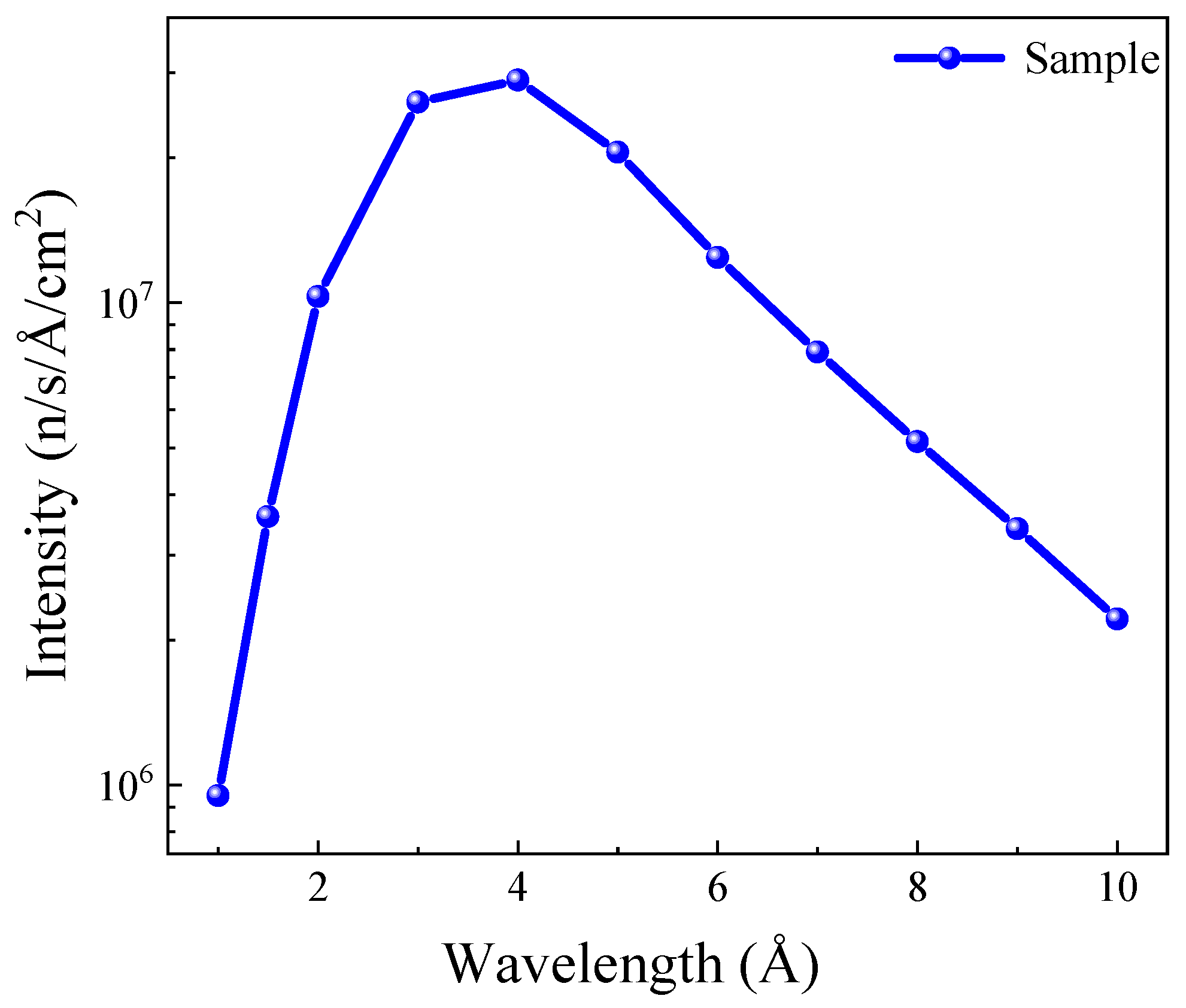
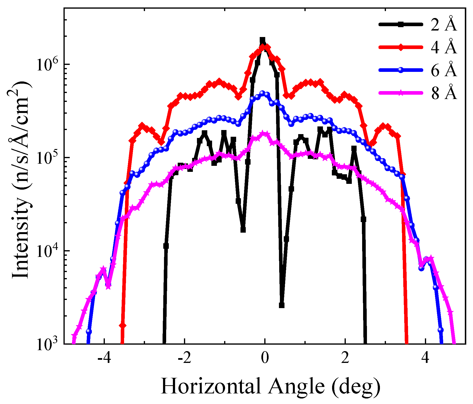
The beam spot at the sample position with a central wavelength of 6.267 Å and a bandwidth of 1.65 Å is shown in Figure 5. After the focusing guide, the neutrons form a nearly circular beam spot at the sample position with a diameter of around 3 cm. With a 500 kW target power after the CSNS-II upgrade, the average flux in the 30 × 30 mm 2 range is about 1.77 × 10 7 n / s / cm 2 , and is 3.47 × 10 7 n / s / cm 2 in the 10 × 10 mm 2 range. The flux at the sample position versus neutron wavelength is shown in Figure 6. The total thickness of the aluminum walls in the neutron flight path is estimated to be about 20 mm, and it causes a flux loss of approximately 10%. The divergences at selected wavelengths in the horizontal direction are shown in Figure 7. The intensity is integrated over the vertical direction. The divergence in the vertical direction is similar to that in the horizontal direction, with the FWHM of the beam divergence below ± 2 .

3.4. Secondary Spectrometer

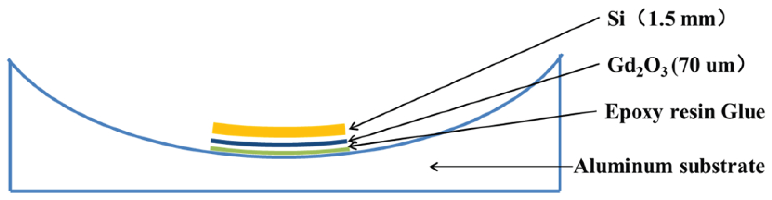
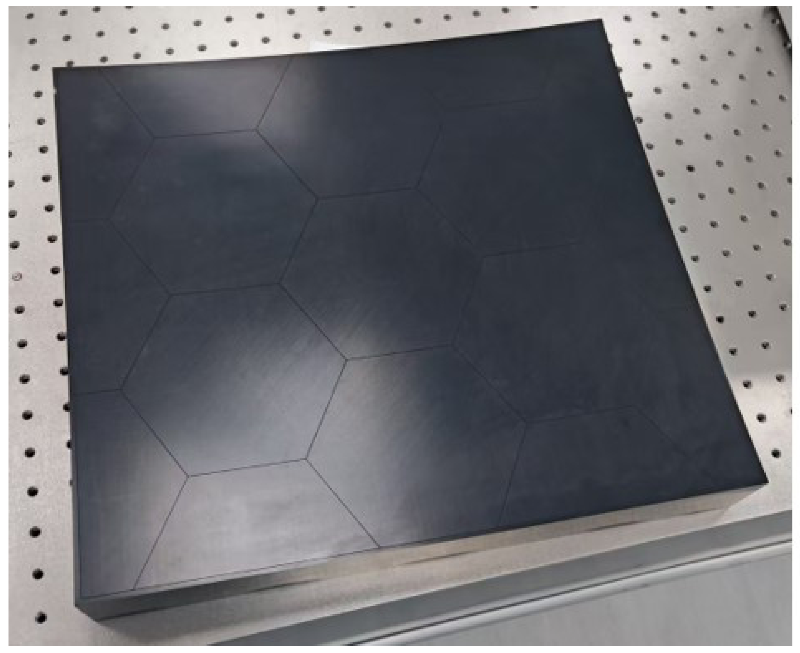
The secondary spectrometer of NuBS is housed within a vacuum scattering tank maintained at a pressure below 10 3 Pa , which reduces beam attenuation and provides the high-vacuum environment required by the detector electronics. The layout of the major components within the scattering tank is shown in Figure 8. The contribution from the intrinsic Darwin width of Si is negligible compared with that arising from the time uncertainty. For efficient neutron collection and to increase the reflected intensity, the silicon wafers are elastically bent to a controlled curvature radius. Thus, the analyzers are fabricated from silicon wafers of 1.5 mm thickness, elastically bent to a curvature radius of R c = 2.5 m and bonded to aluminum alloy substrates with epoxy resin. The effective lattice-spread contribution is Δ d / d = 2.68 × 10 4 , a value chosen to balance energy resolution against neutron reflectivity. The back surface of each wafer is coated with a 70 μ m gadolinium oxide film to absorb neutrons that are not Bragg-reflected, thereby reducing background. A schematic and a prototype of the crystal analyzer are shown in Figure 9 and Figure 10.
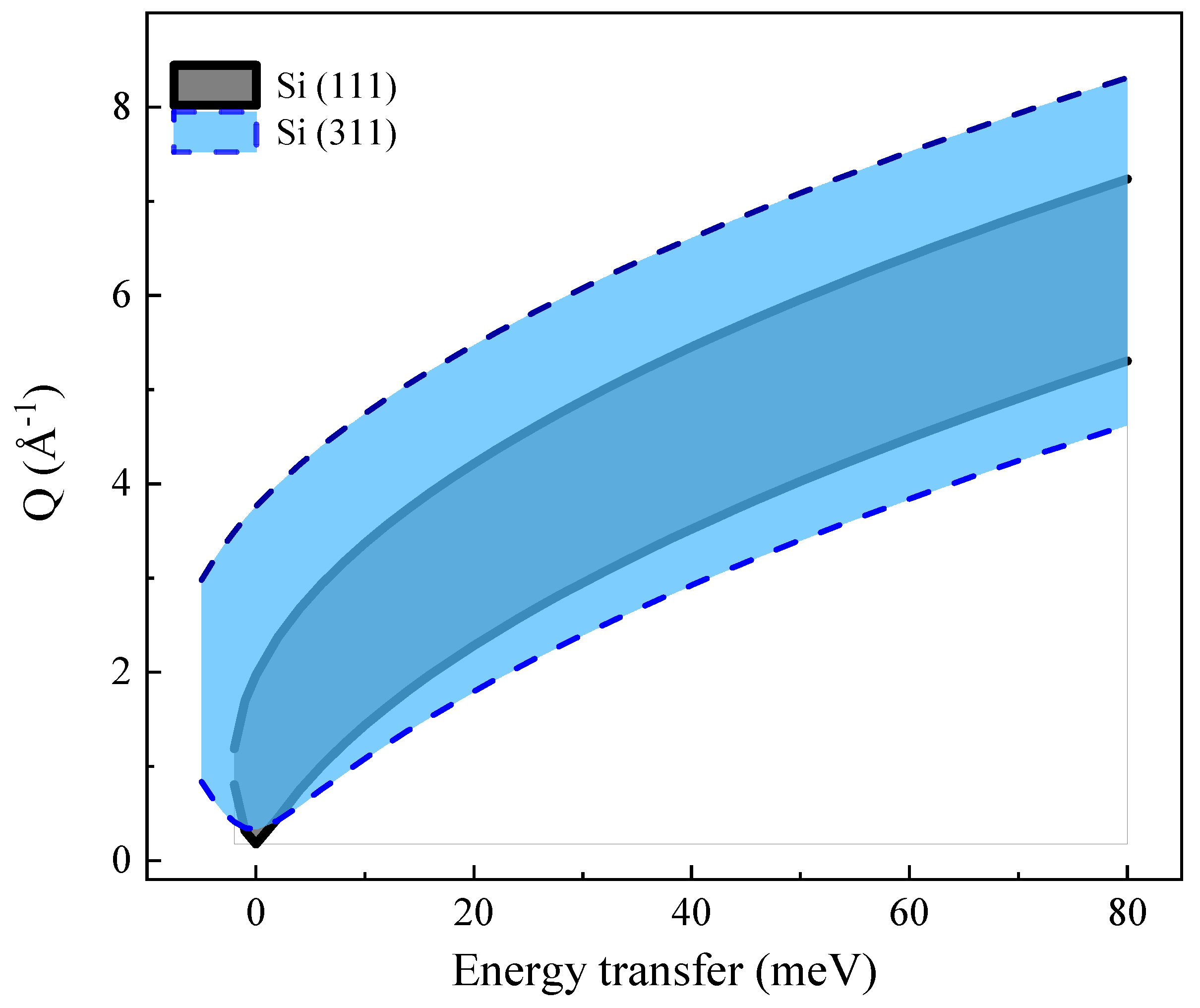
The analyzer panels are arranged on two vertically offset spherical surfaces whose centers are located 87.2 mm above and below the sample position, corresponding to a nominal Bragg angle θ B 88 . This geometry provides the necessary physical clearance for the sample environment while preserving the near-backscattering condition. Three quadrants of the analyzer array are equipped with Si(111) analyzers, covering a horizontal angular range of 160 to 10 and + 10 to + 160 , with a vertical acceptance of ± 20 . The remaining quadrant employs Si(311) analyzers to provide a complementary configuration with coarser energy resolution but extended momentum transfer range. For Si(111), the selected scattered wavelength is λ f 6.267 Å , providing a Q-range of 0.2 2.0 Å 1 at the elastic line. For Si(311), the scattered wavelength is λ f 3.272 Å , with an elastic Q-range of 0.4 4.0 Å 1 and a dynamic range of 2.7 to + 5.7 meV at 25 Hz . The accessible Q regions for different energy transfers in the Si(111) and Si(311) configurations are shown in Figure 11.
The detector system employs 3He-filled tubes arranged in two cylindrical arrays of radius 270 mm , corresponding to the upper and lower analyzer hemispheres. Each tube has a diameter of 0.5 inch and an effective length of approximately 13 cm , with a pixel size of 4 mm to resolve the vertical variation in Bragg angles and flight path lengths. The detector modules cover ± 10 to ± 160 horizontally. A radial collimator with an inner diameter of 300 mm , an outer diameter of 600 mm , and a height of 60 cm is mounted outside the detector modules. The collimator features 108 slits with a 2.8 opening angle, coated with a gadolinium oxide neutron-absorbing material, and is designed to oscillate with an amplitude of 2.8 to avoid systematic misalignment between the collimator slits and the 3He tubes.
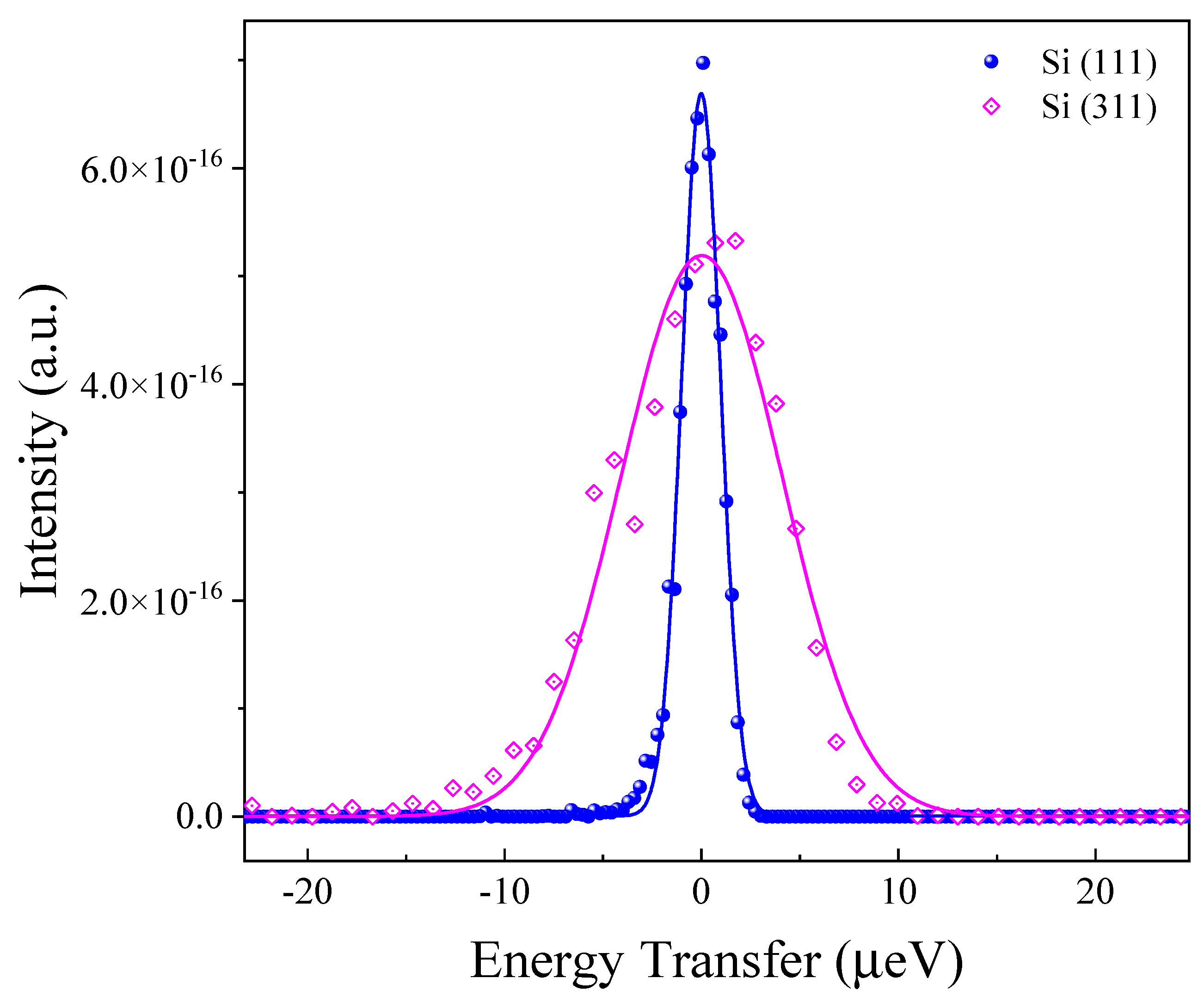
The energy resolution of NuBS was simulated using McStas with a 1 mm thick annular cylindrical vanadium sample with a diameter of 3 cm and a height of 3 cm, for Si (111) and Si (311) analyzer configurations. The neutron count and the total flight times are recorded by the detectors, and the ToF spectrum can be converted into the energy spectrum, which can be fitted with a Gaussian function, as shown in Figure 12. The fitted FWHM of the energy spectrum at Q = 1 Å−1 is about 2.3 μeV for Si (111) and 10.5 μeV for Si (311).

3.5. Sample Environment

NuBS will support a comprehensive suite of sample environments to enable studies across a wide range of scientific applications. These capabilities are essential for probing nanoscale dynamics under precisely controlled thermodynamic and electromagnetic conditions. A cryo-furnace capable of spanning the temperature range from 4 K to 700 K will serve as the primary thermal control system for most experiments. This wide temperature coverage is essential for the study of thermally activated processes across diverse material classes. A cryostat capable of reaching temperatures below 1 K will be available for studies of quantum materials, such as spin ices and quantum spin liquids [75]. A high-temperature furnace extending the accessible range up to 1000 K will be provided for studies of solid-state ionic conductors and nuclear materials [76,77].
In addition to temperature control, NuBS will offer devices for gas dosing, pressure, and humidity regulation. These functionalities are particularly relevant for catalysis studies involving nanoporous materials for gas separation and adsorption [11]. Humidity control is critical for understanding the role of water in biological materials and polymers [78,79]. Pressure control will further enable studies of water and aqueous solutions under varying pressures [80,81]. Recognizing the growing importance of in situ characterization in energy materials studies, NuBS is being designed to accommodate electrical contacts and electrochemical cells [82]. The implementation of a magnetic field environment on NuBS will open avenues for the study of field-dependent spin dynamics in quantum magnets, spin ices, and multiferroic materials [83].

4. The Prospects of NuBS for Probing Nanoscale Dynamics

The instrumental characteristics of NuBS make it a powerful tool for studying nanoscale dynamics across a broad range of scientific disciplines. The wide time window of NuBS enables direct investigation of complex relaxation phenomena that cannot be fully resolved by conventional backscattering spectrometers, thereby providing a unique opportunity to perform multiscale dynamical studies and in situ investigations. While the important scientific impact of NuBS will be shaped by the future user community, two rapidly expanding research directions, energy materials and heterogeneous catalysis, are expected to particularly benefit from its features.

4.1. Energy Materials

The development of sustainable energy technologies relies critically on understanding ionic and molecular transport in functional materials, including solid-state electrolytes, fuel-cell components, and hydrogen-storage media. In these systems, charge transport is governed by atomic-scale motions occurring on pico- to nanosecond timescales and over angstrom-to-nanometer length scales, a regime that is accessible within the capabilities of NuBS.
In solid-state battery materials, QENS can directly quantify ion-hopping processes through the momentum-transfer dependence of the quasielastic linewidth, yielding microscopic quantities such as jump lengths and residence times. Fast-ion conductors including Li 10 GeP 2 S 12 and Na 3 SbS 4 exhibit diffusion coefficients on the order of 10 6 10 7 cm 2 s 1 under practical operating conditions, corresponding to quasielastic broadenings well matched to the accessible time window of NuBS [84,85]. Beyond determining diffusion coefficients, simultaneous observation of localized ionic motion and long-range hopping provides direct insight into the microscopic origins of superionic conductivity. Such a multiscale perspective is essential for understanding the coupling between lattice dynamics and ionic transport in systems such as δ - Bi 2 O 3 [86] and Li 6 PS 5 Cl [12], where superionic transport emerges from highly anharmonic lattice dynamics and overdamped phonon excitations. The integration of electrical contacts and controlled atmospheres into the sample environment enables measurements under realistic battery operating conditions [87,88,89].
Fuel-cell materials present another class of systems where multiple transport mechanisms coexist and interact. In proton-exchange membrane fuel cells (PEMFCs), proton conduction arises from a combination of hydronium diffusion, proton transfer along hydrogen-bond networks, and the translational and rotational dynamics of hydration water [90,91,92,93]. Resolving these coupled motions remains a major challenge because they span a broad range of characteristic timescales. Furthermore, measurements performed under controlled hydration and operating conditions offer the possibility of correlating proton mobility with electrochemical performance [82]. Similar opportunities exist for solid oxide fuel cells (SOFCs), where QENS has been widely used to study oxygen-ion diffusion in doped ceria, lanthanum gallate perovskites, and bismuth oxide-based electrolytes [94,95]. For emerging proton-conducting perovskite oxides, the strong neutron sensitivity to hydrogen further enables direct observation of proton transport and its coupling to lattice dynamics, providing mechanistic information that is difficult to obtain through conventional electrochemical techniques [96].
Hydrogen-storage materials are also an important research area in which QENS can provide unique insights. The uptake and release of hydrogen in metal hydrides, complex hydrides, and porous sorbents are ultimately controlled by the mobility of atomic or molecular hydrogen within the host structure [97,98,99]. The large incoherent scattering cross section of hydrogen makes these systems particularly suitable for QENS investigations. Quantitative determination of diffusion coefficients, residence times, and jump geometries for hydrogen can therefore be achieved over a broad temperature range. In addition, extending measurements to higher momentum transfer through the Si(311) analyzer configuration enables more detailed characterization of the spatial aspects of hydrogen transport. Experiments performed under controlled pressures are expected to provide valuable insight into the relationship between hydrogen mobility, storage capacity, and kinetic limitations governing charging and discharging processes in practical storage systems [100,101].

4.2. Heterogeneous Catalysis

Heterogeneous catalysis underpins a large fraction of modern chemical manufacturing, from fuel production to environmental remediation. In many catalytic systems, overall activity and selectivity are determined not only by the intrinsic reactivity of active sites but also by the transport of reactants, intermediates, and products within complex pore networks and interfacial environments [102]. Understanding these molecular transport processes under realistic reaction conditions therefore remains a central challenge in catalysis studies [3].
Microporous catalysts such as zeolites provide a representative example in which molecular transport strongly influences catalytic performance. Because pore dimensions are comparable to molecular sizes, diffusion often occurs through activated jumps between adsorption sites, with residence times and jump lengths that depend sensitively on pore topology, molecular shape, and host–guest interactions [103,104]. QENS has revealed rich dynamical behavior in a variety of sorbate–zeolite systems, including methane and 1-octene in ZSM-5, methanol in H-ZSM-5, and phenolic molecules in zeolite Beta [105,106,107,108,109]. Polar molecules capable of hydrogen bonding, such as water and alcohols, frequently exhibit transient trapping and heterogeneous jump-diffusion behavior that differs markedly from the dynamics of nonpolar hydrocarbons [110]. Such systems often contain multiple dynamical populations simultaneously, ranging from localized librational and rotational motions to long-range translational diffusion.
Resolving these hierarchical dynamics is essential for understanding transport limitations in hierarchical and chemically heterogeneous catalytic materials [111]. Direct measurements of molecular transport within working catalysts therefore provide valuable insight into the relationship between pore-scale dynamics and catalytic performance. These considerations are directly relevant to the methanol-to-hydrocarbons (MTH) process, an industrially essential route for converting methanol into light olefins and liquid fuels over acidic zeolite catalysts. According to the hydrocarbon-pool mechanism, polymethylated aromatic species confined within zeolite pores act as co-catalytic intermediates through repeated methylation and olefin-elimination cycles [112], which play a critical role in determining product selectivity and catalyst deactivation through coke formation [107,113]. Similar transport-related challenges arise in NH3-SCR catalysts for NOx abatement, where ammonia diffusion through the zeolite framework strongly influences reaction efficiency [114]. Extending such research to realistic multicomponent gas environments will provide a more complete picture of transport limitations governing low-temperature catalytic activity.
Hydrogen-related catalysis represents another area where NuBS offers unique opportunities. QENS has been exploited to study hydrogen diffusion in iron-based Fischer–Tropsch catalysts, where multiple hydrogen populations with distinct mobilities were identified and linked to different local environments [115]. Similar approaches have also been applied to adsorption and hydrogenation catalysis, where molecular mobility is closely coupled to adsorption strength and surface reactivity. For example, combined inelastic neutron scattering and QENS studies of ethene adsorption on skeletal cobalt catalysts revealed pronounced changes in molecular motion upon adsorption and activation at the catalyst surface [116]. Extending these investigations to larger hydrocarbons and realistic hydrogenation conditions may provide essential mechanistic insight into the microscopic origins of catalytic activity and selectivity. By enabling such studies under in situ conditions with a wide time window, NuBS is expected to help establish the missing link between molecular transport dynamics and macroscopic catalytic performance, ultimately guiding the rational design of more efficient hydrogenation catalysts.

5. Conclusions and Outlook

In this review, we have comprehensively presented the basic principles, instrumental design, and prospective applications of NuBS currently under construction at CSNS. As a ToF-BS optimized for high energy resolution and broad dynamic range, NuBS is expected to fill a critical gap in the neutron infrastructure of China. This distinct technical advantage allows for the simultaneous extraction of fast local dynamics and slow translational motions under identical experimental conditions. The scientific examples discussed above illustrate only a fraction of the opportunities that will become accessible with the commissioning of NuBS. Across energy materials, catalysis, soft matter, and biological systems, functional properties are now recognized to emerge from the interplay of multiple dynamical processes spanning wide temporal and spatial scales. Resolving these hierarchical dynamics therefore represents one of the central challenges in materials science. A major trend in QENS is the growing demand for in situ and operando studies. Many technologically relevant phenomena occur in environments involving electrical bias, gas flow, pressure gradients, chemical reactions, or electrochemical cycling. The ability to directly observe microscopic dynamics during device operation or catalytic turnover offers the possibility of establishing quantitative links between molecular transport processes and macroscopic performance metrics such as ionic conductivity, catalytic activity, and selectivity.
An important opportunity lies in the integration of QENS with other complementary experimental techniques, such as diffraction, inelastic neutron scattering, X-ray spectroscopy, and electrochemical characterization. Such multimodal approaches will enable structure–dynamics–function relationships to be established under identical thermodynamic and operating conditions, providing a more complete picture of complex materials than any single technique can achieve. A particularly significant development in recent years has been the increasingly close coupling of QENS measurements with MD simulations. Because atomistic trajectories can be directly transformed into experimentally observable quantities such as the incoherent intermediate scattering function I s ( Q , t ) and the dynamic structure factor S ( Q , ω ) , comparisons between experiment and simulation can be performed without relying exclusively on simplified analytical diffusion models. Looking ahead, advances in machine learning and data-driven methodologies are expected to further accelerate spectral analysis, facilitate model selection, improve uncertainty quantification, and enable the extraction of microscopic transport parameters from increasingly complex datasets. Another emerging direction is the implementation of polarization analysis on ToF-BSs, although this remains technically challenging. This approach aims to separate coherent and incoherent scattering contributions, thereby providing independent access to collective and single-particle dynamics. Such selectivity is particularly promising for complex soft matter and biological systems, where coherent scattering can make a non-negligible contribution to the measured signal.
As the first dedicated ToF-BS in China, NuBS will significantly expand the resources available to the domestic scientific community and create new opportunities for addressing scientific questions worldwide. More significantly, by bridging the gap between localized motions and long-range transport, and by enabling studies under increasingly realistic conditions, NuBS is expected to contribute to frontier research in energy science, catalysis, soft matter, and beyond. It will provide a versatile platform for exploring the dynamical behaviors in complex materials. Scheduled for completion around 2029, NuBS will begin user operations as a competitive ToF-BS open to the global scientific community.

Author Contributions

Conceptualization, T.X. and H.G.; methodology, T.X. and H.Z.; validation, T.X., H.Z. and X.L.; writing—original draft preparation, T.X.; writing—review and editing, T.X. and H.G.; supervision, H.G.; funding acquisition, T.X. All authors have read and agreed to the published version of the manuscript.

Funding

This research was supported by the National Natural Science Foundation of China (Grant No. 12204269) and the Guangdong Basic and Applied Basic Research Foundation, China (Grant No. 2021A-1515111164).

Data Availability Statement

No new data were created or analyzed in this study. Data sharing is not applicable to this article.

Acknowledgments

We are grateful to the administration and professional groups of CSNS, including the neutron optics, choppers, detector and electronics, data acquisition, sample environment, neutron physics, and software groups.

Conflicts of Interest

The authors declare no conflicts of interest.

References

  1. Boothroyd, A.T. Principles of Neutron Scattering from Condensed Matter; Oxford University Press: Oxford, UK, 2020. [Google Scholar]
  2. Schwaighofer, B.; Gonzalez, M.A.; Johnson, M.R.; Evans, J.S.O.; Evans, I.R. Ionic Mobility in Energy Materials: Through the Lens of Quasielastic Neutron Scattering. Chem. Mater. 2025, 37, 3575–3593. [Google Scholar] [CrossRef] [Scilit]
  3. Yu, X.; Cheng, Y.; Li, Y.; Polo-Garzon, F.; Liu, J.; Mamontov, E.; Li, M.; Lennon, D.; Parker, S.F.; Ramirez-Cuesta, A.J.; et al. Neutron Scattering Studies of Heterogeneous Catalysis. Chem. Rev. 2023, 123, 8638–8700. [Google Scholar] [CrossRef] [Scilit] [PubMed]
  4. Guo, L.; Chu, X. Unraveling the Dynamics of Biological Systems through Neutron Scattering. Photon Sci. 2025, 1, 49–65. [Google Scholar] [CrossRef] [Scilit]
  5. Bee, M. Quasielastic Neutron Scattering: Principles and Applications in Solid State Chemistry, Biology, and Materials Science; Adam Hilger: Bristol, UK, 1988. [Google Scholar]
  6. Kruteva, M. Dynamics studied by Quasielastic Neutron Scattering (QENS). Adsorption 2021, 27, 875–889. [Google Scholar] [CrossRef] [Scilit]
  7. Ashkar, R. Selective dynamics in polymeric materials: Insights from quasi-elastic neutron scattering spectroscopy. J. Appl. Phys. 2020, 127, 151101. [Google Scholar] [CrossRef] [Scilit]
  8. Hassani, A.N.; Haris, L.; Appel, M.; Seydel, T.; Stadler, A.M.; Kneller, G.R. Multiscale relaxation dynamics and diffusion of myelin basic protein in solution studied by quasielastic neutron scattering. J. Chem. Phys. 2022, 156, 025102. [Google Scholar] [CrossRef] [Scilit] [PubMed]
  9. Tomiyasu, K.; Mizuta, Y.P.; Matsuura, M.; Aoyama, K.; Kawamura, H. Observation of topological Z 2 vortex fluctuations in the frustrated Heisenberg magnet NaCrO2. Phys. Rev. B 2022, 106, 054407. [Google Scholar] [CrossRef] [Scilit]
  10. Wang, A.; Breakwell, C.; Foglia, F.; Tan, R.; Lovell, L.; Wei, X.; Wong, T.; Meng, N.; Li, H.; Seel, A.; et al. Selective ion transport through hydrated micropores in polymer membranes. Nature 2024, 635, 353–358. [Google Scholar] [CrossRef] [Scilit] [PubMed]
  11. Han, Y.; Huang, W.; He, M.; An, B.; Chen, Y.; Han, X.; An, L.; Kippax-Jones, M.; Li, J.; Yang, Y.; et al. Trace benzene capture by decoration of structural defects in metal-organic framework materials. Nat. Mater. 2024, 23, 1531–1538. [Google Scholar] [CrossRef] [Scilit] [PubMed]
  12. Ding, J.; Gupta, M.K.; Rosenbach, C.; Lin, H.-M.; Osti, N.C.; Abernathy, D.L.; Zeier, W.G.; Delaire, O. Liquid-like dynamics in a solid-state lithium electrolyte. Nat. Phys. 2025, 21, 118–125. [Google Scholar] [CrossRef] [Scilit]
  13. Gardner, J.S.; Ehlers, G.; Faraone, A.; Sakai, V.G. High-resolution neutron spectroscopy using backscattering and neutron spin-echo spectrometers in soft and hard condensed matter. Nat. Rev. Phys. 2020, 2, 103–116. [Google Scholar] [CrossRef] [Scilit]
  14. Frick, B. Neutron backscattering spectroscopy. In Neutron and X-Ray Spectroscopy; Springer: Dordrecht, The Netherlands, 2006; pp. 483–527. [Google Scholar]
  15. Telling, M.T.F. A Practical Guide to Quasi-Elastic Neutron Scattering; Royal Society of Chemistry: London, UK, 2020. [Google Scholar]
  16. Jie, W.; Shi-Nian, F.; Jing-Yu, T.; Ju-Zhou, T.; Ding-Sheng, W.; Fang-Wei, W.; Sheng, W. China Spallation Neutron Source-an overview of application prospects. Chin. Phys. C 2009, 33, 1033–1042. [Google Scholar] [CrossRef] [Scilit]
  17. Jie, W.; Shi-Nian, F.; Jing-Yu, T.; Ju-Zhou, T.; Ding-Sheng, W.; Fang-Wei, W.; Sheng, W. China Spallation Neutron Source: Design, R&D, and outlook. Nucl. Instrum. Methods Phys. Res. A 2009, 600, 10–13. [Google Scholar] [CrossRef] [Scilit]
  18. Chen, H.; Wang, X.L. China’s first pulsed neutron source. Nat. Mater. 2016, 15, 689–691. [Google Scholar] [CrossRef] [Scilit] [PubMed]
  19. Chen, H.; Chen, Y.; Fu, S.; Ma, L.; Wang, S.; Chen, F.; Chen, Y.; Dong, H.; Dong, L.; Feng, G.; et al. Design and construction of the China Spallation Neutron Source. Nucl. Instrum. Methods Phys. Res. A 2025, 1078, 170431. [Google Scholar] [CrossRef] [Scilit]
  20. Xiong, T.; Zhou, H.; Wu, Y.; Lin, X.; Wu, S.; Li, D.; Yang, Y.; Guo, H. Physical design of NuBS, the new neutron backscattering spectrometer at China Spallation Neutron Source. Nucl. Instrum. Methods Phys. Res. A 2025, 1078, 170558. [Google Scholar] [CrossRef] [Scilit]
  21. Osti, N.C.; Jalarvo, N.H.; Mamontov, E. Backscattering silicon spectrometer (BASIS): Sixteen years in advanced materials characterization. Mater. Horiz. 2024, 11, 4535–4572. [Google Scholar] [CrossRef] [Scilit] [PubMed]
  22. Benedetto, A.; Kearley, G.J. Experimental demonstration of the novel “van-Hove integral method (vHI)” for measuring diffusive dynamics by elastic neutron scattering. Sci. Rep. 2021, 11, 14093. [Google Scholar] [CrossRef] [Scilit] [PubMed]
  23. Xiong, T.; Tyagi, M.; Zhou, W.; Guo, H.; Briber, R.M.; Wang, H. Quasi-Elastic Neutron Scattering on Bimodal Dynamics in Molecular Solutions of Cellulose. J. Phys. Chem. B 2025, 129, 6714–6720. [Google Scholar] [CrossRef] [Scilit] [PubMed]
  24. Price, D.L.; Borodin, O.; González, M.A.; Kofu, M.; Shibata, K.; Yamada, T.; Yamamuro, O.; Saboungi, M.-L. Relaxation in a prototype ionic liquid: Influence of water on the dynamics. J. Phys. Chem. Lett. 2017, 8, 715–719. [Google Scholar] [CrossRef] [Scilit] [PubMed]
  25. Deng, Z.D.; Mauritz, K.A. Dielectric relaxation studies of water-containing short side chain perfluorosulfonic acid membranes. Macromolecules 1992, 25, 2739–2745. [Google Scholar] [CrossRef] [Scilit]
  26. Popov, I.; Sacci, R.L.; Sanders, N.C.; Matsumoto, R.A.; Thompson, M.W.; Osti, N.C.; Kobayashi, T.; Tyagi, M.; Mamontov, E.; Pruski, M.; et al. Critical Role of Anion-Solvent Interactions for Dynamics of Solvent-in-Salt Solutions. J. Phys. Chem. C 2020, 124, 8457–8466. [Google Scholar] [CrossRef] [Scilit]
  27. Fick, A. On liquid diffusion. J. Membr. Sci. 1995, 100, 33–38. [Google Scholar] [CrossRef] [Scilit]
  28. Singwi, K.S.; Sjölander, A. Diffusive motions in water and cold neutron scattering. Phys. Rev. 1960, 119, 863. [Google Scholar] [CrossRef] [Scilit]
  29. Teixeira, J.; Bellissent-Funel, M.-C.; Chen, S.H.; Dianoux, A.J. Experimental determination of the nature of diffusive motions of water molecules at low temperatures. Phys. Rev. A 1985, 31, 1913–1917. [Google Scholar] [CrossRef] [Scilit] [PubMed]
  30. Chudley, C.T.; Elliott, R.J. Neutron Scattering from a Liquid on a Jump Diffusion Model. Proc. Phys. Soc. 1961, 77, 353–361. [Google Scholar] [CrossRef] [Scilit]
  31. Hall, P.L.; Ross, D.K. Incoherent neutron scattering functions for random jump diffusion in bounded and infinite media. Mol. Phys. 1981, 42, 673–682. [Google Scholar] [CrossRef] [Scilit]
  32. Bée, M. A physical insight into the elastic incoherent structure factor. Phys. B Condens. Matter 1992, 182, 323–336. [Google Scholar] [CrossRef] [Scilit]
  33. Chen, S.-H.; Gallo, P.; Sciortino, F.; Tartaglia, P. Molecular-dynamics study of incoherent quasielastic neutron-scattering spectra of supercooled water. Phys. Rev. E 1997, 56, 4231. [Google Scholar] [CrossRef] [Scilit]
  34. Zhang, Y.; Lagi, M.; Fratini, E.; Baglioni, P.; Mamontov, E.; Chen, S.-H. Dynamic susceptibility of supercooled water and its relation to the dynamic crossover phenomenon. Phys. Rev. E 2009, 79, 040201. [Google Scholar] [CrossRef] [Scilit] [PubMed]
  35. Howells, W.S. The use of Fourier transforms in the analysis of QENS data. Phys. B Condens. Matter 1996, 226, 78–81. [Google Scholar] [CrossRef] [Scilit]
  36. Lukichev, A. Physical meaning of the stretched exponential Kohlrausch function. Phys. Lett. A 2019, 383, 2983–2987. [Google Scholar] [CrossRef] [Scilit]
  37. Mukhopadhyay, S.; Hewer, B.; Howells, S.; Markvardsen, A. A modern approach to QENS data analysis in Mantid. Phys. B Condens. Matter 2019, 563, 41–49. [Google Scholar] [CrossRef] [Scilit]
  38. Mamontov, E.; Smith, R.; Billings, J.; Ramirez-Cuesta, A. Simple analytical model for fitting QENS data from liquids. Phys. B Condens. Matter 2019, 566, 50–54. [Google Scholar] [CrossRef] [Scilit]
  39. Kikuchi, T. Mode-distribution analysis of quasielastic neutron scattering and application to liquid water. Phys. Rev. E 2013, 87, 062314. [Google Scholar] [CrossRef] [Scilit] [PubMed]
  40. Jobic, H.; Theodorou, D.N. Quasi-elastic neutron scattering and molecular dynamics simulation as complementary techniques for studying diffusion in zeolites. Microporous Mesoporous Mater. 2007, 102, 21–50. [Google Scholar] [CrossRef] [Scilit]
  41. Goret, G.; Aoun, B.; Pellegrini, E. MDANSE: An interactive analysis environment for molecular dynamics simulations. J. Chem. Inf. Model. 2017, 57, 1–5. [Google Scholar] [CrossRef] [Scilit] [PubMed]
  42. Goychuk, I.; Panchenko, A.; Shahzad, A.; Steffen, J.; Neiss, C.; Götz, K.; Vogel, C.; Weisser, M.; Hakim, B.; Embs, J.P.; et al. Effects of isotope and chemical incoherence on self-diffusion of atoms in liquid alloys of gallium with isotopes of nickel: Experiments based on quasielastic neutron scattering versus molecular dynamics simulations. Phys. Rev. B 2025, 111, 064302. [Google Scholar] [CrossRef] [Scilit]
  43. Frick, B.; Gonzalez, M. Five years operation of the second generation backscattering spectrometer IN16—A retrospective, recent developments and plans. Phys. B Condens. Matter 2001, 301, 8–19. [Google Scholar] [CrossRef] [Scilit]
  44. Meyer, A.; Dimeo, R.M.; Gehring, P.M.; Neumann, D.A. The high-flux backscattering spectrometer at the NIST Center for Neutron Research. Rev. Sci. Instrum. 2003, 74, 2759. [Google Scholar] [CrossRef] [Scilit]
  45. Wuttke, J.; Budwig, A.; Drochner, M.; Kämmerling, H.; Kayser, F.J.; Kleines, H.; Ossovyi, V.; Pardo, L.C.; Prager, M.; Richter, D.; et al. SPHERES, Jülich’s high-flux neutron backscattering spectrometer at FRM II. Rev. Sci. Instrum. 2012, 83, 075109. [Google Scholar] [CrossRef] [Scilit] [PubMed]
  46. de Souza, N.R. EMU: High-Resolution Backscattering Spectrometer at ANSTO. Neutron News 2016, 27, 20. [Google Scholar] [CrossRef] [Scilit]
  47. Kuhlmann, K.; Appel, M.; Frick, B.; Magerl, A. Breakthrough in neutron backscattering spectroscopy: Energy resolution improved by one order of magnitude using the GaAs 200 reflection. Rev. Sci. Instrum. 2019, 90, 015119. [Google Scholar] [CrossRef] [Scilit] [PubMed]
  48. Francesca, N.; Peters, J.; Russo, D.; Barbieri, S.; Chiapponi, C.; Cupane, A.; Deriu, A.; Di Bari, M.T.; Farhi, E.; Gerelli, Y.; et al. IN13 backscattering spectrometer at ILL: Looking for motions in biological macromolecules and organisms. Neutron News 2008, 19, 14–18. [Google Scholar] [CrossRef] [Scilit]
  49. Appel, M.; Frick, B.; Magerl, A. A flexible high speed pulse chopper system for an inverted neutron time-of-flight option on backscattering spectrometers. Sci. Rep. 2018, 8, 13580. [Google Scholar] [CrossRef] [Scilit] [PubMed]
  50. Appel, M.; Frick, B.; Magerl, A. First results with the neutron backscattering and TOF spectrometer option BATS on IN16B. Phys. B Condens. Matter 2019, 562, 6–8. [Google Scholar] [CrossRef] [Scilit]
  51. Carlile, C.J.; Adams, M.A. The design of the IRIS inelastic neutron spectrometer and improvements to its analysers. Phys. B Condens. Matter 1992, 182, 431–440. [Google Scholar] [CrossRef] [Scilit]
  52. Telling, M.T.; Andersen, H. Spectroscopic characteristics of the OSIRIS near-backscattering crystal analyser spectrometer on the ISIS pulsed neutron source. Phys. Chem. Chem. Phys. 2005, 7, 1255–1261. [Google Scholar] [CrossRef] [Scilit] [PubMed]
  53. Demmel, F.; McPhail, D.; French, C.; Maxwell, D.; Harrison, S.; Boxall, J.; Rhodes, N.; Mukhopadhyay, S.; Silverwood, I.; Sakai, V.G.; et al. ToF-backscattering spectroscopy at the ISIS facility: Status and perspectives. J. Phys. Conf. Ser. 2018, 1021, 012027. [Google Scholar] [CrossRef] [Scilit]
  54. Perrichon, A.; Fernandez-Alonso, F.; Wolff, M.; Karlsson, M.; Demmel, F. A silicon analyser for the OSIRIS spectrometer: An analytical and Monte Carlo simulation study. Nucl. Instrum. Methods Phys. Res. A 2019, 947, 162740. [Google Scholar] [CrossRef] [Scilit]
  55. Perrichon, A.; Demmel, F. Upgrade of the OSIRIS primary spectrometer. Nucl. Instrum. Methods Phys. Res. A 2022, 1039, 167014. [Google Scholar] [CrossRef] [Scilit]
  56. Herwig, K.W.; Keener, W.S. Design and performance of the spallation neutron source backscattering spectrometer. Appl. Phys. A 2002, 74, s1592–s1594. [Google Scholar] [CrossRef] [Scilit]
  57. Mamontov, E.; Herwig, K.W. A time-of-flight backscattering spectrometer at the Spallation Neutron Source, BASIS. Appl. Phys. A 2011, 82, 085109. [Google Scholar] [CrossRef] [Scilit] [PubMed]
  58. Shibata, K.; Takahashi, N.; Kawakita, Y.; Matsuura, M.; Yamada, T.; Tominaga, T.; Kambara, W.; Kobayashi, M.; Inamura, Y.; Nakatani, T.; et al. The performance of TOF near backscattering spectrometer DNA in MLF, J-PARC. JPS Conf. Proc. 2015, 8, 036022. [Google Scholar]
  59. Kawakita, Y.; Matsuura, M.; Tominaga, T.; Yamada, T.; Tamatsukuri, H.; Nakagawa, H.; Ohuchi, K. Recent progress on DNA ToF backscattering spectrometer in MLF, J-PARC. EPJ Web Conf. 2022, 272, 02002. [Google Scholar] [CrossRef] [Scilit]
  60. Takahashi, N.; Shibata, K.; Kawakita, Y.; Nakajima, K.; Inamura, Y.; Nakatani, T.; Nakagawa, H.; Fujiwara, S.; Sato, T.J.; Tsukushi, I.; et al. Repetition Rate Multiplication: RRM, an Advanced Measuring Method Planed for the Backscattering Instrument, DNA at the MLF, J-PARC. J. Phys. Soc. Jpn. 2011, 80, SB007. [Google Scholar] [CrossRef] [Scilit]
  61. Takahashi, N.; Lechner, R.E.; Markó, M.; Bordallo, H.N. Conceptual design of the time-of-flight backscattering spectrometer, MIRACLES, at the European Spallation Source. Rev. Sci. Instrum. 2016, 87, 085118. [Google Scholar] [CrossRef] [Scilit] [PubMed]
  62. Villacorta, F.J.; Bordallo, H.N.; Conde, A.; Pereira, J.E.M.; Martínez, R.; Mazkiaran, I.; Aranda, I.; del Moral, O.G.; Zugazaga, A.; Harper, G.; et al. MIRACLES, the backscattering spectrometer of the European Spallation Source: Meeting scientific requirements with design. Rev. Sci. Instrum. 2025, 96, 045101. [Google Scholar] [CrossRef] [Scilit] [PubMed]
  63. Luna, P.; Bordallo, H.N.; Tsapatsaris, N.; Andersen, K.H.; Herranz, I.; Sordo, F.; Villacorta, F.J. Tailoring the energy resolution of MIRACLES, the time-of-flight–backscattering spectrometer at the ESS: An updated proposal for the chopper cascade. Phys. B Condens. Matter 2019, 564, 64–68. [Google Scholar] [CrossRef] [Scilit]
  64. Villacorta, F.J.; Rodríguez, D.M.; Bertelsen, M.; Bordallo, H.N. Optimization of the guide design of MIRACLES, the neutron time-of-flight backscattering spectrometer at the European Spallation Source. Quantum Beam Sci. 2021, 6, 3. [Google Scholar] [CrossRef] [Scilit]
  65. Available online: https://www.isis.stfc.ac.uk/instruments/iris/ (accessed on 24 August 2026).
  66. Available online: https://www.isis.stfc.ac.uk/instruments/osiris/ (accessed on 24 August 2026).
  67. Available online: https://neutrons.ornl.gov/basis (accessed on 24 August 2026).
  68. Available online: https://mlfinfo.jp/en/bl02/ (accessed on 24 August 2026).
  69. Wang, F.; Liang, T.; Yin, W.; Yu, Q.; He, L.; Tao, J.; Zhu, T.; Jia, X.; Zhang, S. Physical design of target station and neutron instruments for China Spallation Neutron Source. Sci. China-Phys. Mech. Astron. 2013, 56, 2410–2424. [Google Scholar] [CrossRef] [Scilit]
  70. Cai, W.; Wang, P.; Zhang, Q.; Geng, Y.; Zhang, W. Design method of bandwidth limiting neutron chopper system in CSNS. Nucl. Tech. 2018, 41, 120202. [Google Scholar]
  71. Wang, S.; Cai, W.; Wang, P.; Zhang, H.; Zuo, T.; Wang, S. Research on system performance of a bandwidth limiting neutron chopper at CSNS. Nucl. Tech. 2021, 44, 020204. [Google Scholar]
  72. Wang, P.; Yang, B.; Cai, W. Development of a bandwidth limiting neutron chopper for CSNS. Nucl. Instrum. Methods Phys. Res. A 2015, 792, 56–60. [Google Scholar] [CrossRef] [Scilit]
  73. Bentley, P.M.; Andersen, K.H. Optimization of focusing neutronic devices using artificial intelligence techniques. J. Appl. Crystallogr. 2009, 42, 217–224. [Google Scholar] [CrossRef] [Scilit]
  74. Rodriguez, D.M.; DiJulio, D.; Bentley, P. Systematic study on the performance of elliptic focusing neutron guides. Nucl. Instrum. Methods Phys. Res. Sect. A 2016, 808, 101–108. [Google Scholar] [CrossRef] [Scilit]
  75. Ehlers, G.; Mamontov, E.; Zamponi, M.; Kam, K.C.; Gardner, J.S. Direct Observation of a Nuclear Spin Excitation in Ho2Ti2O7. Phys. Rev. Lett. 2009, 102, 016405. [Google Scholar] [CrossRef] [Scilit] [PubMed]
  76. Cakmak, E.; Cinbiz, M.N.; Sundar, A.; Mamontov, E.; Yu, J.; Hu, X.; Linton, K.D. Hydrogen motion in near stoichiometric yttrium dihydride at elevated temperatures. J. Nucl. Mater. 2024, 593, 154972. [Google Scholar] [CrossRef] [Scilit]
  77. Du, P.; Chen, Q.; Fan, Z.; Pan, H.; Haibach, F.G.; Gomez, M.A.; Braun, A. Cooperative origin of proton pair diffusivity in yttrium substituted barium zirconate. J. Commun. Phys. 2020, 3, 200. [Google Scholar] [CrossRef] [Scilit]
  78. Miskowiec, A.; Kirkegaard, M.C.; Herwig, K.W.; Trowbridge, L.; Mamontov, E.; Anderson, B. Quasielastic neutron scattering with in-situ humidity control: Water dynamics in uranyl fluoride. J. Appl. Phys. 2016, 119, 094308. [Google Scholar] [CrossRef] [Scilit]
  79. Osti, N.C.; Dyatkin, B.; Thompson, M.W.; Tiet, F.; Zhang, P.; Dai, S.; Tyagi, M.; Cummings, P.T.; Gogotsi, Y.; Wesolowski, D.J.; et al. Influence of humidity on performance and microscopic dynamics of an ionic liquid in supercapacitor. Phys. Rev. Mater. 2017, 1, 035402. [Google Scholar] [CrossRef] [Scilit]
  80. Lundin, F.; Hansen, H.W.; Adrjanowicz, K.; Frick, B.; Rauber, D.; Hempelmann, R.; Shebanova, O.; Niss, K.; Matic, A. Pressure and temperature dependence of local structure and dynamics in an ionic liquid. J. Phys. Chem. B 2021, 125, 2719–2728. [Google Scholar] [CrossRef] [Scilit] [PubMed]
  81. Rescigno, M.; Toffano, A.; Ranieri, U.; Andriambariarijaona, L.; Gaal, R.; Klotz, S.; Koza, M.M.; Ollivier, J.; Martelli, F.; Russo, J.; et al. Observation of plastic ice VII by quasi-elastic neutron scattering. Nature 2025, 640, 662–667. [Google Scholar] [CrossRef] [Scilit] [PubMed]
  82. Martinez, N.; Morin, A.; Berrod, Q.; Frick, B.; Ollivier, J.; Porcar, L.; Gebel, G.; Lyonnard, S. Multiscale water dynamics in a fuel cell by operando quasi elastic neutron scattering. J. Phys. Chem. C 2018, 122, 1103–1108. [Google Scholar] [CrossRef] [Scilit]
  83. Turrini, A.A.; Ruminy, M.; Bourdarot, F.; Stuhr, U.; White, J.S.; Tucker, G.; Skoulatos, M.; Núñez-Valdez, M.; Fennell, T. Magnetic-field control of magnetoelastic coupling in the rare-earth pyrochlore Tb2Ti2O7. Phys. Rev. B 2021, 104, 224403. [Google Scholar] [CrossRef] [Scilit]
  84. Hori, S.; Kanno, R.; Kwon, O.; Kato, Y.; Yamada, T.; Matsuura, M.; Yonemura, M.; Kamiyama, T.; Shibata, K.; Kawakita, Y. Revealing the Ion Dynamics in Li10GeP2S12 by Quasi-Elastic Neutron Scattering Measurements. J. Phys. Chem. C 2022, 126, 9518–9527. [Google Scholar] [CrossRef] [Scilit]
  85. Zhang, Q.; Zhang, C.; Hood, Z.D.; Chi, M.; Liang, C.; Jalarvo, N.H.; Yu, M.; Wang, H. Abnormally Low Activation Energy in Cubic Na3SbS4 Superionic Conductors. Chem. Mater. 2020, 32, 2264–2271. [Google Scholar] [CrossRef] [Scilit]
  86. Schwaighofer, B.; Gonzalez, M.A.; Appel, M.; Koza, M.M.; Evans, I.R. Oxide Ion Mobility in V- and P-Doped Bi2O3-Based Solid Electrolytes: Combining Quasielastic Neutron Scattering with Ab Initio Molecular Dynamics. Chem. Mater. 2023, 35, 1125–1133. [Google Scholar] [CrossRef] [Scilit]
  87. Mahurin, S.M.; Mamontov, E.; Thompson, M.W.; Zhang, P.; Turner, C.H.; Cummings, P.T.; Dai, S. Relationship between pore size and reversible and irreversible immobilization of ionic liquid electrolytes in porous carbon under applied electric potential. Appl. Phys. Lett. 2016, 109, 143111. [Google Scholar] [CrossRef] [Scilit]
  88. Novak, E.; Daemen, L.; Jalarvo, N. Advanced Characterization of Solid-State Battery Materials Using Neutron Scattering Techniques. Materials 2024, 17, 6209. [Google Scholar] [CrossRef] [Scilit] [PubMed]
  89. Liu, J.; He, L.; Osti, N.C.; Zhang, Y. Neutron Scattering in Sodium-Ion Battery Research: Progress and Prospects. Adv. Energy Mater. 2026, e06774. [Google Scholar] [CrossRef] [Scilit]
  90. Burankova, T.; Hempelmann, R.; Fossog, V.; Ollivier, J.; Seydel, T.; Embs, J.P. Proton diffusivity in the protic ionic liquid triethylammonium triflate probed by quasielastic neutron scattering. J. Phys. Chem. B 2015, 119, 10643–10651. [Google Scholar] [CrossRef] [Scilit] [PubMed]
  91. Appel, M.; Borisov, G.; Holderer, O.; Appavou, M.-S.; Zorn, R.; Lehnert, W.; Richter, D. Proton diffusion in the catalytic layer for high temperature polymer electrolyte fuel cells. RSC Adv. 2019, 9, 37768–37777. [Google Scholar] [CrossRef] [Scilit] [PubMed]
  92. Chan, E.P.; Frieberg, B.R.; Ito, K.; Tarver, J.; Tyagi, M.; Zhang, W.; Coughlin, E.B.; Stafford, C.M.; Roy, A.; Rosenberg, S.; et al. Insights into the water transport mechanism in polymeric membranes from neutron scattering. Macromolecules 2020, 53, 1443–1450. [Google Scholar] [CrossRef] [Scilit]
  93. Osti, N.C.; Mamontov, E. Microscopic dynamics in room-temperature ionic liquids confined in materials for supercapacitor applications. Sustain. Energy Fuels 2020, 4, 1554–1576. [Google Scholar] [CrossRef] [Scilit]
  94. Ning, D.; Chen, S.; Wang, D.; Li, X.; Song, J.; Li, W.; Liu, X.; Wang, Q.; Li, Q. Applications of neutron scattering in solid oxide fuel cells. Mater. Futur. 2026, 5, 032102. [Google Scholar] [CrossRef] [Scilit]
  95. Chen, Q.; Banyte, J.; Zhang, X.; Embs, J.P.; Braun, A. Proton diffusivity in spark plasma sintered BaCe0.8Y0.2O3-δ: In-situ combination of quasi-elastic neutron scattering and impedance spectroscopy. Solid State Ion. 2013, 252, 2–6. [Google Scholar] [CrossRef] [Scilit]
  96. Naumovska, E.; Orstadius, J.; Perrichon, A.; Lavén, R.; Koza, M.M.; Evenson, Z.; Karlsson, M. Localized Proton Motions in the Proton-Conducting Perovskites BaZr1-xScxO3Hx (x=0.10 and 0.50) Investigated with Quasielastic Neutron Scattering. J. Phys. Chem. C 2023, 127, 24532–24541. [Google Scholar] [CrossRef] [Scilit]
  97. NaraseGowda, S.; Brown, C.M.; Tyagi, M.; Jenkins, T.A.; Dobbins, T.A. Quasi-elastic neutron scattering studies of hydrogen dynamics for nanoconfined NaAlH4. J. Phys. Chem. C 2016, 120, 14863–14873. [Google Scholar] [CrossRef] [Scilit]
  98. Jung, M.; Park, J.; Muhammad, R.; Kim, J.Y.; Grzimek, V.; Russina, M.; Moon, H.R.; Park, J.T.; Oh, H. Elucidation of diffusivity of hydrogen isotopes in flexible MOFs by quasi-elastic neutron scattering. Adv. Mater. 2021, 33, 2007412. [Google Scholar] [CrossRef] [Scilit] [PubMed]
  99. Novak, E.; Daemen, L.; Ramirez-Cuesta, A.J.; Cheng, Y.; Smith, R.; Egami, T.; Jalarvo, N. Uncovering the hydride ion diffusion pathway in barium hydride via neutron spectroscopy. Sci. Rep. 2022, 12, 6194. [Google Scholar] [CrossRef] [Scilit] [PubMed]
  100. Contescu, C.I.; Saha, D.; Gallego, N.C.; Mamontov, E.; Kolesnikov, A.I.; Bhat, V.V. Restricted dynamics of molecular hydrogen confined in activated carbon nanopores. Carbon 2012, 50, 1071–1082. [Google Scholar] [CrossRef] [Scilit]
  101. Contescu, C.I.; Zhang, H.; Olsen, R.J.; Mamontov, E.; Morris, J.R.; Gallego, N.C. Isotope effect on adsorbed quantum phases: Diffusion of H2 and D2 in nanoporous carbon. Phys. Rev. Lett. 2013, 110, 236102. [Google Scholar] [CrossRef] [Scilit] [PubMed]
  102. O’Malley, A.J.; Parker, S.F.; Catlow, C.R.A. Neutron spectroscopy as a tool in catalytic science. Chem. Commun. 2017, 53, 12164–12176. [Google Scholar] [CrossRef] [Scilit] [PubMed]
  103. Wu, D.; Yang, M.; Yu, J.; Dyballa, M.; Yang, P.; Li, M.; Hou, G.; Hunger, M.; Dai, W. The role of adsorption and diffusion in improving the selectivity and reactivity of zeolite catalysts. Chem. Soc. Rev. 2025, 54, 9192–9244. [Google Scholar] [CrossRef] [Scilit] [PubMed]
  104. Zhao, L.; Yuan, J.; Xing, Y.; Qi, J.; Peng, P.; Liu, Z.; Zheng, A. Confinement effects on molecular diffusion in zeolites: Mechanisms and perspectives. Chem. Soc. Rev. 2026, 55, 210–253. [Google Scholar] [CrossRef] [Scilit] [PubMed]
  105. Hawkins, A.P.; O’mAlley, A.J.; Zachariou, A.; Collier, P.; Ewings, R.A.; Silverwood, I.P.; Howe, R.F.; Parker, S.F.; Lennon, D. Investigation of the dynamics of 1-octene adsorption at 293 K in a ZSM-5 catalyst by inelastic and quasielastic neutron scattering. J. Phys. Chem. C 2019, 123, 417–425. [Google Scholar] [CrossRef] [Scilit]
  106. Hawkins, A.P.; Zachariou, A.; Silverwood, I.P.; Yong, C.; Collier, P.; Todorov, I.; Howe, R.F.; Parker, S.F.; Lennon, D. Combining quasielastic neutron scattering and molecular dynamics to study methane motions in ZSM-5. J. Chem. Phys. 2022, 157, 184702. [Google Scholar] [CrossRef] [Scilit] [PubMed]
  107. Matam, S.K.; Catlow, C.R.A.; Silverwood, I.P.; O’Malley, A.J. Methanol dynamics in H-ZSM-5 with Si/Al ratio of 25: A quasi-elastic neutron scattering (QENS) study. Top. Catal. 2021, 64, 699–706. [Google Scholar] [CrossRef] [Scilit]
  108. Hernandez-Tamargo, C.; O’MAlley, A.; Silverwood, I.P.; de Leeuw, N.H. Molecular behaviour of phenol in zeolite Beta catalysts as a function of acid site presence: A quasielastic neutron scattering and molecular dynamics simulation study. Catal. Sci. Technol. 2019, 9, 6700–6713. [Google Scholar] [CrossRef] [Scilit]
  109. Hernandez-Tamargo, C.; Silverwood, I.P.; O’mAlley, A.J.; de Leeuw, N.H. Quasielastic neutron scattering and molecular dynamics simulation study on the molecular behaviour of catechol in zeolite Beta. Top. Catal. 2020, 64, 707–721. [Google Scholar] [CrossRef] [Scilit]
  110. Anovitz, L.M.; Mamontov, E.; Chakoumakos, B.C.; Kolesnikov, A.I. Quasielastic and Inelastic Neutron Scattering Study of Ultraconfined Water in Natural Mordenite ((Ca, Na2, K2) Al2Si10O24·7H2O). Langmuir 2025, 41, 23488–23500. [Google Scholar] [PubMed]
  111. Morton, K.; Appel, M.; Woodward, C.; Armstrong, J.; O’mAlley, A. The effect of pore structure on the local and nanoscale mobility of anisole and guaiacol in commercial zeolite catalysts. Microporous Mesoporous Mater. 2025, 383, 113388. [Google Scholar] [CrossRef] [Scilit]
  112. Zachariou, A.; Hawkins, A.P.; Collier, P.; Howe, R.F.; Parker, S.F.; Lennon, D. Neutron scattering studies of the methanol-to-hydrocarbons reaction. Catal. Sci. Technol. 2023, 13, 1976–1990. [Google Scholar] [CrossRef] [Scilit]
  113. O’Malley, A.J.; Sakai, V.G.; Silverwood, I.P.; Dimitratos, N.; Parker, S.F.; Catlow, C.R.A. Methanol diffusion in zeolite HY: A combined quasielastic neutron scattering and molecular dynamics simulation study. Phys. Chem. Chem. Phys. 2016, 18, 17294–17302. [Google Scholar] [CrossRef] [Scilit] [PubMed]
  114. O’Malley, A.J.; Sarwar, M.; Armstrong, J.; Catlow, C.R.A.; Silverwood, I.P.; York, A.P.E.; Hitchcock, I. Comparing ammonia diffusion in NH3-SCR zeolite catalysts: A quasielastic neutron scattering and molecular dynamics simulation study. Phys. Chem. Chem. Phys. 2018, 20, 11976–11986. [Google Scholar] [CrossRef] [Scilit] [PubMed]
  115. Davidson, A.L.; Webb, P.B.; Silverwood, I.P.; Lennon, D. The application of quasi-elastic neutron scattering to investigate hydrogen diffusion in an iron-based Fischer-Tropsch synthesis catalyst. Top. Catal. 2020, 63, 378–385. [Google Scholar] [CrossRef] [Scilit]
  116. Jones, E.; Inns, D.R.; Dann, S.E.; Silverwood, I.P.; Kondrat, S.A. Characterisation of ethylene adsorption on model skeletal cobalt catalysts by inelastic and quasi-elastic neutron scattering. Catal. Commun. 2022, 163, 106409. [Google Scholar] [CrossRef] [Scilit]
Figure 1. Schematic diagram of QENS spectrum.
Qubs 10 00020 g001
Figure 2. Schematic layout of NuBS in top view. (Reprinted with permission from [20]). Copyright (2025) Elsevier.
Qubs 10 00020 g002
Figure 3. The neutron pulse width of DPHM. (Reprinted with permission from [20]. Copyright (2025) Elsevier.
Qubs 10 00020 g003
Figure 4. The bandwidth selected by the chopper system at 25 Hz. (Reprinted with permission from [20]). Copyright (2025) Elsevier.
Qubs 10 00020 g004
Figure 5. The beam spot at the sample position with a central wavelength of 6.267 Å and a bandwidth of 1.65 Å. (Reprinted with permission from [20]). Copyright (2025) Elsevier.
Qubs 10 00020 g005
Figure 6. The flux of the neutron beam at the sample position for each wavelength. (Reprinted with permission from ([20]). Copyright (2025) Elsevier.
Qubs 10 00020 g006
Figure 7. The divergence of the neutron beam at the sample position for each wavelength. (Reprinted with permission from ([20]). Copyright (2025) Elsevier.
Qubs 10 00020 g007
Figure 8. Assembly diagram of the secondary spectrometer of NuBS.
Qubs 10 00020 g008
Figure 9. Schematic of crystal analyzer.
Qubs 10 00020 g009
Figure 10. Prototype of the analyzer module.
Qubs 10 00020 g010
Figure 11. Accessible Q–E range for the Si(111) and Si(311) configurations of NuBS.
Qubs 10 00020 g011
Figure 12. The energy spectrum at Q = 1 Å−1 for Si (111) and Si (311).
Qubs 10 00020 g012
Table 1. The key characteristics of current ToF-BSs.
SpectrometerCrystalsBest Resolution
(μeV)
Dynamic
Range (μeV)
Q Range
−1)
IRIS@ISIS [65]PG(002)17.5 ± 400 0.42–1.85
PG(004)54.5−3500–40000.84–3.70
Mica(002)1.0 ± 20 0.13–0.62
Mica(004)4.5 ± 150 0.26–1.24
Mica(006)11.0 ± 400 0.40–1.87
OSIRIS@ISIS [66]PG(002)25.4 ± 400 0.18–1.8
PG(004)99−3000–40000.37–3.6
BASIS@SNS [67]Si(111)3.5 ± 100 or ± 200 0.2–2.0
Si(311)15 ± 660 or ± 1700 0.4–3.8
DNA@J-PARC [68]Si(111)1.4−20–80 *0.08–1.98
Si(311)11.0\ **1.79–3.39
MIRACLES@ESS [62]Si(111)2 ± 550 0.2–2.0
NuBS@CSNS [20]Si(111)2.3 ± 550 0.2–1.8
Si(311)10.5 ± 4000 0.4–3.6
* For fixed phase of PSCs at the best resolution setting. ** No formally reported dynamic range.
Disclaimer/Publisher’s Note: The statements, opinions and data contained in all publications are solely those of the individual author(s) and contributor(s) and not of MDPI and/or the editor(s). MDPI and/or the editor(s) disclaim responsibility for any injury to people or property resulting from any ideas, methods, instructions or products referred to in the content.