Article

Biological Properties of a Composite Polymer Material Based on Polyurea and Submicron-Sized Selenium Particles

by
Sergey A. Shumeyko
1,
Dmitriy E. Burmistrov
1,
Denis V. Yanykin
1,
Ilya V. Baimler
1,
Alexandr V. Simakin
1,
Maxim E. Astashev
1,
Mikhail V. Dubinin
2,
Roman Y. Pishchalnikov
1,
Ruslan M. Sarimov
1,
Valeriy A. Kozlov
1,3,*,
Alexey S. Dorokhov
4 and
Andrey Yu. Izmailov
4
1
Prokhorov General Physics Institute, Russian Academy of Sciences, Vavilov Str. 38, 119991 Moscow, Russia
2
Department of Biochemistry, Cell Biology and Microbiology, Mari State University, pl. Lenina 1, 424001 Yoshkar-Ola, Russia
3
Department of Fundamental Sciences, Bauman Moscow State Technical University, 2-nd Baumanskaya Str. 5, 105005 Moscow, Russia
4
Federal Scientific Agroengineering Center VIM, 109428 Moscow, Russia
*
Author to whom correspondence should be addressed.
Inventions 2025, 10(5), 82; https://doi.org/10.3390/inventions10050082
Submission received: 4 July 2025 / Revised: 8 September 2025 / Accepted: 15 September 2025 / Published: 19 September 2025
(This article belongs to the Section Inventions and Innovation in Biotechnology and Materials)

Abstract

Using the method of laser ablation in liquid, submicron-sized particles of zero-valent amorphous selenium (Se SMPs) were created. A number of composite polymer materials were manufactured based on polyurea and Se SMPs at concentrations ranging 0.1–2.5 wt.%. The manufactured materials showed no significant surface or internal defects at either the macro or micro level. It was found that the Se SMPs were not uniformly distributed inside the polymer, but formed ordered areas with slightly higher and lower concentrations of the particles. It was demonstrated that the manufactured materials did not generate a significant amount of active oxygen species, which could damage biological objects such as protein molecules and DNA, while also exhibiting pronounced bacteriostatic properties without significantly affecting the growth and reproduction of mammalian cells. Materials containing 0.25 and 1% Se SMPs, when added to soil, improved the morphometric parameters of radish plants (Raphanus sativus var. sativus). These polymer composite materials based on polyurea with the addition of Se SMPs are promising functional materials for agriculture due to their antibacterial activity.

1. Introduction

Polymer composite materials functionalized with nanoparticles have attracted considerable attention at present due to their unique properties and improved characteristics compared to traditional polymers [1,2,3]. Depending on the physicochemical and mechanical properties, these materials are actively introduced into such industries as mechanical engineering [4], microelectronics [5], construction [6], aircraft construction [7], and space [8]. A number of composite materials have outstanding biological properties and are introduced into such industries as agrotechnology [9], agricultural industry [10,11], food industry [12,13], and biomedicine [14,15,16]. In the context of agronomy application, the possibility of using polymer composites containing nanoparticles has been widely discussed recently [17,18,19]. The possibility of using polymer-containing nanocomposites to combat plant pests is being considered [9,20], as slow-release fertilizers [21], to improve the nutritional properties of crops [22], and to increase the resistance of agricultural crops to external stress factors [23,24], as well as increasing the stability of the crop for long-term storage [25,26]. Of course, the main requirements for such materials are environmental safety, low toxicity, and ease of manufacture [27]. Therefore, the selection of a suitable polymer matrix is an important aspect.
One of the well-studied and proven classes of polymers with the appropriate properties are polyureas (PU). PUs are synthetic polymers containing urea fragments -NH-CO-NH- in the main chain. Polyurea is usually obtained by transamidation of urea with aliphatic diamines or copolymerization of diisocyanates with oligomeric di- or polyamines [28]. Polyureas are elastomers, durable polymers that are hydrophobic and highly resistant to external chemical and biological factors [29]. Currently, polyurea is widely used as a building material [30], in engineering structures [31], and as an anti-corrosion coating [32]. Due to its excellent hydrophobic properties, this polymer is considered as a matrix for creating coatings resistant to bacterial adhesion [33], as well as for creating materials that prevent biofouling [34].
Various types of inorganic particles are used for the functionalization of polymer matrices, among which selenium submicron-sized particles (Se SMPs) are often considered [35]. It should be noted that Se SMPs are often used to affect living objects. Se SMPs have potential for the treatment of various forms of cancer [36], stroke [37], protect primary cortical neurons and astrocytes during oxygen–glucose deprivation and reoxygenation [38], and are used to diagnose number of diseases [39] and deliver drugs [40]. Se SMPs are used in a large number of applications as food additives [41] and antioxidants [42]. In agriculture, Se SMPs are used as fertilizers and additives that exhibit biostimulating, stress-protective and biofortification activities [43]. Se SMPs are known to have antibacterial properties, especially against bacteria that form biofilms [44]. Se SMPs exert an antibacterial effect due to several mechanisms associated with both direct action on the bacterial cell and the interaction of the resulting selenium ions with molecular targets [45]. A number of studies have noted the inhibitory effect of Se SMPs on Staphylococcus aureus, Escherichia coli, and Pseudomonas aeruginosa [46,47,48]. The advantages of Se SMPs are: relatively low toxicity compared to other NPs in comparable concentrations; excellent bioavailability; and high biological activity compared to traditional chemical forms of Se ingested with food [49]. It is assumed that material based on polyurea containing Se SMPs may have a significant portion of the advantages of both polyurea and selenium particles. In this regard, the main goal of this work is to obtain and characterize the number of composite materials based on polyurea polymer containing Se SMPs in different concentrations.

2. Materials and Methods

2.1. Synthesis of Se SMPs and Their Characterization

Se SMPs were synthesized by laser ablation of a massive Se target in deionized water using an Ekspla Nd:YAG laser (EKSPLA, Vilnius, Lithuania) with a wavelength of 532 nm, a pulse duration of 3–6 ns, and pulse energy of 1 mJ. The laser beam was moved over the target surface at a speed of 3 m/s, along a “grid”-type route with a spacing between the stripes of about 10 μm. Laser ablation was performed in a flow cell with flow rate of 1 mL/s, thereby achieving a combination of laser ablation and fragmentation of the resulting particles [40]. The concentration, hydrodynamic diameter, and electrokinetic potential of the laser-ablated Se SMPs were determined by dynamic light scattering using a Zetasizer Ultra Red Label (Malvern Panalytical Ltd., Malvern, UK). The spectra of colloids were recorded using a U2000 spectrometer (Ocean Optics, Orlando, FL, USA). The morphology of Se SMPs was studied using a 200FE (Carl Zeiss, Oberkochen, Germany) TEM.

2.2. Preparation and Characterization of Composite Materials Based on Polyurea and Se SMPs

The synthesized colloidal aqueous solution of Se SMPs was centrifuged for 40 min using a Sigma 3-16KL centrifuge (Sigma Laborzentrifugen GmbH, Osterode am Harz, Germany) with a 12,158 rotor for 40 min at 7000× g. The supernatant was removed and acetone was added. The procedure was repeated at least 3 times. The resulting colloidal solution of Se SMPs in acetone was thoroughly mixed and treated in an ultrasonic bath for 20 min. Two-component polyurea (Poly Max, St. Petersburg, Russia) was used to prepare the composite materials. The polyurea used is in two component solutions: component “A” and component “B”. Component “A” is polyetheramines with amine end amino groups. Component “B” is a mixture of diphenylmethane diisocyanate and polyisocyanate. The required volume of the pre-prepared colloidal solution of Se SMPs in acetone was added to component “A” to obtain a final concentration of Se SMPs of 0.1–2.5%. The control group samples were prepared without adding Se SMPs. The resulting colloid was thoroughly mixed using a V-1 plus vortex (Biosan, Riga, Latvia), then component “B” was added in a ratio of 64:100 (A:B). The final mixture was thoroughly mixed again using a vortex. The composite film samples were obtained by pouring 5 mL of the final mixture between two polished fluoroplastic bars separated by 250 µm thick silicon plates (spacers). The resulting structure was placed under a press for 5 h. Then the structure was disassembled and the resulting composite film samples were removed. The films were cut to the required sizes and used for further experiments. The surface topology of the obtained films was assessed using and morphology analysis complex (NT-MDT, Zelenograd, Russia), which allows studying the micro- and nanostructure of the surface in non-contact and semi-contact modes. To assess the orderliness of the distribution of Se SMPs in the polymer, a modulation interference microscope MIM-321 (Amphora Lab, Moscow, Russia) was used.

2.3. Measurement of Hydrogen Peroxide Concentration

Deionized water (10 mL) was incubated at 40 °C for 180 min with samples of composite materials in the form of 10 × 10 × 0.25 mm3 plates. Quantitative assessment of hydrogen peroxide in aqueous solutions was carried out using a highly sensitive chemiluminometer Biotox-7A-USE (ANO “Engineering Center—Ecology”, Moscow, Russia). The “counting solution” contained luminol-4-iodophenol-horseradish peroxidase. The calibration and recording procedure is described in detail in [50]. The sensitivity of this method made it possible to determine 0.1 nM hydrogen peroxide.

2.4. Measurement of Hydroxyl Radical Concentration

The concentration of OH radicals was determined by the reaction with coumarin-3-carboxylic acid (CCA), the product of which is hydroxycoumarin-3-carboxylic acid (7-OH-CCA). A 10 mL aqueous solution of CCA was incubated at 80 °C for 120 min with samples of composite materials in the form of 10 × 10 × 0.25 mm3 plates. The fluorescence of 7-OH-CCA was recorded on an 8300 spectrofluorimeter (JASCO, Tokyo, Japan) at λex = 400 nm and λem = 450 nm. The calibration and recording procedure is described in detail in [51]. The sensitivity of the method made it possible to determine 0.1 nM OH radicals.

2.5. Measurement of Level of Long-Lived Reactive Protein Species

A 10 mL aqueous solution of 0.1% bovine serum albumin (BSA) was incubated at 40 °C for 120 min with samples of composite materials in the form of 10 × 10 × 0.25 mm3 plates. The LRPS level was assessed using the induced luminescence method of protein solutions. Measurements were taken 30 min after incubation. Measurements were performed in 20 mL antistatic polypropylene vials (Beckman, Brea, CA, USA). Highly sensitive Biotox-7A chemiluminometer (Engineering Center—Ecology, Moscow, Russia) was used as an instrument. BSA solutions that were not exposed to heating were used as control. All experimental details were described previously [52].

2.6. Enzyme-Linked Immunosorbent Assay (ELISA)

For quantitative determination of 8-oxoguanine in DNA, non-competitive enzyme-linked immunosorbent assay (ELISA) was used using monoclonal antibodies specific to 8-oxoguanine. The optical density of the samples was measured using a Feyond-A400 plate photometer (Allsheng, Hangzhou, China) at λ = 405 nm. The method was described in more detail previously [53].

2.7. Analysis of Bacteriostatic Activity

The bacteriostatic efficiency of the obtained composite materials was assessed against planktonic cells of Gram-negative E. coli K-12 and Gram-positive S. aureus RN4220 bacteria. Sterile samples of films with an area of ~20 cm2, pre-soaked for several hours in 70% ethanol for sterilization and then completely dried, were put on a sterile hoop, onto which LB broth with a known initial concentration of planktonic bacterial cells was then placed. Samples of the fixed composite material (film) with the placed cell suspension in the broth was incubated in an ES-20 shaker-incubator (Biosan, Riga, Latvia) at 37 °C, ~150 rpm for 24 h. The optical density of the bacterial suspension was assessed using a UV5Nano Excellence drop spectrometer (Mettler Toledo, Columbus, OH, USA). The optical density measured at 600 nm (OD600) reflected the concentration of bacterial cells in the nutrient medium per unit volume.

2.8. Cytotoxicity Study

Primary mouse lung fibroblast cultures were used to study the cytotoxicity of PU/Se SMPs composites. All manipulations with animal tissues and cells were performed in clean rooms using a Laminar-C class II biological safety cabinet (Lamsystems, Chelyabinsk, Russia). Primary cell cultures of isolated mouse lung fibroblasts were obtained according to the standard protocol with minor modifications; lung tissue was obtained from BALB/c mice. A detailed description of the procedure for isolating primary mouse lung fibroblast cultures is contained in [54]. DMEM medium (Biolot, Moscow, Russia) containing 10% fetal bovine serum (Thermo Fisher Scientific, Waltham, MA, USA), L-glutamine (PanEco, Moscow, Russia), 100 U/mL penicillin, 100 μg/mL streptomycin (PanEco, Moscow, Russia) were used as a nutrient medium for culturing cell cultures. Cultivation was carried out in an S-Bt Smart Biotherm CO2 incubator (Biosan, Riga, Latvia) at a temperature of 37 °C and 5% CO2.

2.9. Study of the Effect of PU-Se SMPs Composites on the Growth and Development of Radish Plants

Radish plants (R. sativus var. sativus) were used in the experiment. Plant seedlings were grown in a vegetative vessel with diameter of 6 cm and height of 10 cm. All plants were germinated in soil substrate containing 1 mg/g of total nitrogen, 2 mg/g of P2O5, 2.5 mg/g of K2O, with a pH of 5.5–6.5. PU/Se SMPs composite material in the form of a rod with diameter of 0.5 cm was stuck into the soil to the full depth of the vessel with soil at the rate of one rod per 150 cm3 of soil area. The surface area of the leaves of plant seedlings was calculated using Green Image 1.0 software [55]. An MB23 analyzer (Oharus, Parsippany, NJ, USA) was used to estimate the dry mass of the root. The experiments were carried out in a climate chamber using the following method: plants were grown in soil, simulating standard conditions of the organogenesis environment: illumination 16 h a day, temperature 22 °C, soil moisture 25% SDW, duration of the experiment 30 days. As control experiments to exclude the effect of the polymer (PU) on plant growth, the effect of aqueous colloid Se SMPs on the area of radish leaf was studied when introduced into the soil at concentrations of about 1, 5, 10 and 25 μg/kg.

2.10. Statistical Data Analysis

For statistical analysis of differences between plant groups, one-way analysis of variance (ANOVA) was used, followed by a posteriori comparison using the Tukey criterion and Student’s t-test for independent samples. The data were preliminarily checked for normal distribution using the Shapiro–Wilk test. Statistical processing was performed in the GraphPad Prism 8.3.0 program. Differences were considered reliable at a significance level of p ≤ 0.05. The reported values are averaged across at least three separate experiments to ensure reproducibility (n ≥ 3).

3. Results

3.1. Physicochemical Characteristics of PU/Se SMPs Composites

The concentration of Se SMPs in the colloid, their size distribution and electrokinetic potential were determined using dynamic light scattering. The work was mainly carried out with colloids with particle concentration of about 1010 mL−1. It was shown that in deionized water, the size distribution of particles has one maximum (Figure 1a). Se SMPs have an average hydrodynamic diameter of about 100 nm, the distribution range from 60 to 150 nm. The distribution of particles by ζ-potential has one maximum (Figure 1b). Se SMPs have an average ζ-potential of about −23 mV, the distribution range from −40 to −10 mV. The absorption spectrum of Se SMPs colloids was studied using UV-visible spectroscopy. It was shown that the absorption spectrum of the aqueous colloid Se SMPs contains local maxima in the region of 200–210 and 580–600 nm (Figure 1c). The morphology of the particles was studied using transmission electron microscopy (Figure 1d). It was shown that all the studied Se SMPs had shape close to spherical. The most common size of the particles is slightly less than 100 nm.
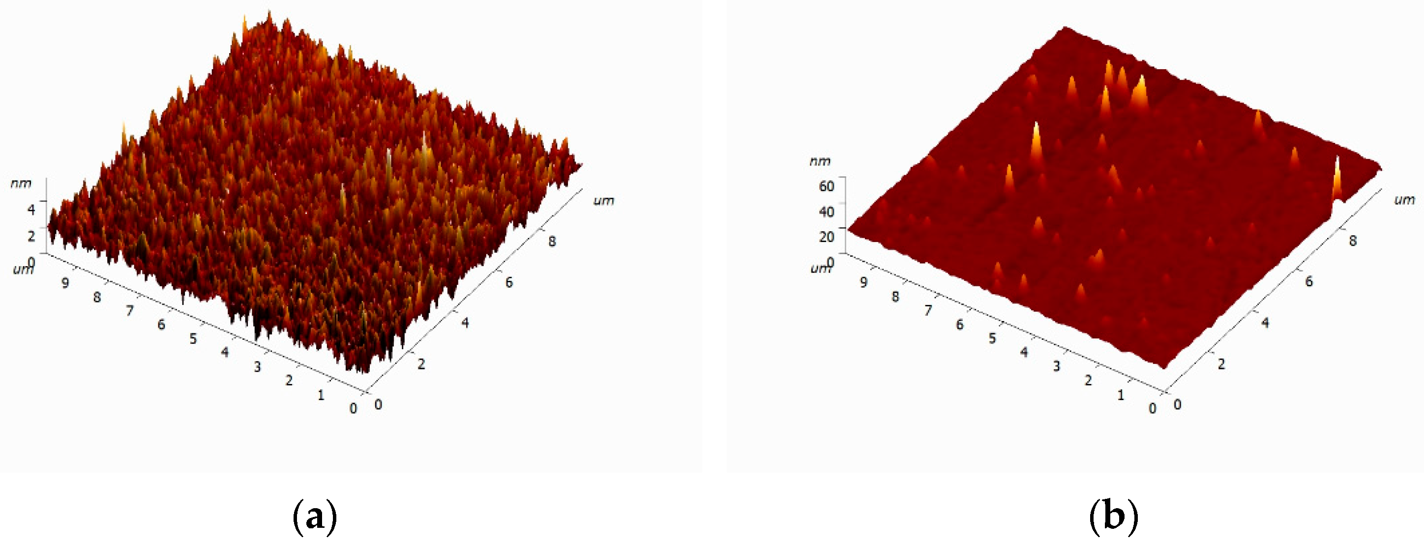
Se SMPs were added to the polymer, and film samples of about 250 μm thickness were obtained from the resulting mixture. The films had no visible defects. Preliminary experiments showed that the surface quality of the polyurea films is determined by the surface quality of the fluoroplastic plates used in the manufacturing process. The surface of the composite material samples was analyzed using atomic force microscopy. For the film control sample of polyurea, a homogeneous surface with a roughness below 5 mm was observed (Figure 2a). The addition of Se SMPs in all studied concentrations did not significantly affect the surface quality. We were unable to detect surface defects such as cavities, cracks. Even at the highest particle concentration of 2.5 wt.%, we managed to obtain composite film samples with maximum height difference over an area of 100 μm2 of less than 100 nm. The observed surface oscillations of the order of 100–200 μm and a height of about 60 nm may indicate the accumulation of Se SMPs and aggregates on the surface of the composite sample (Figure 2b).
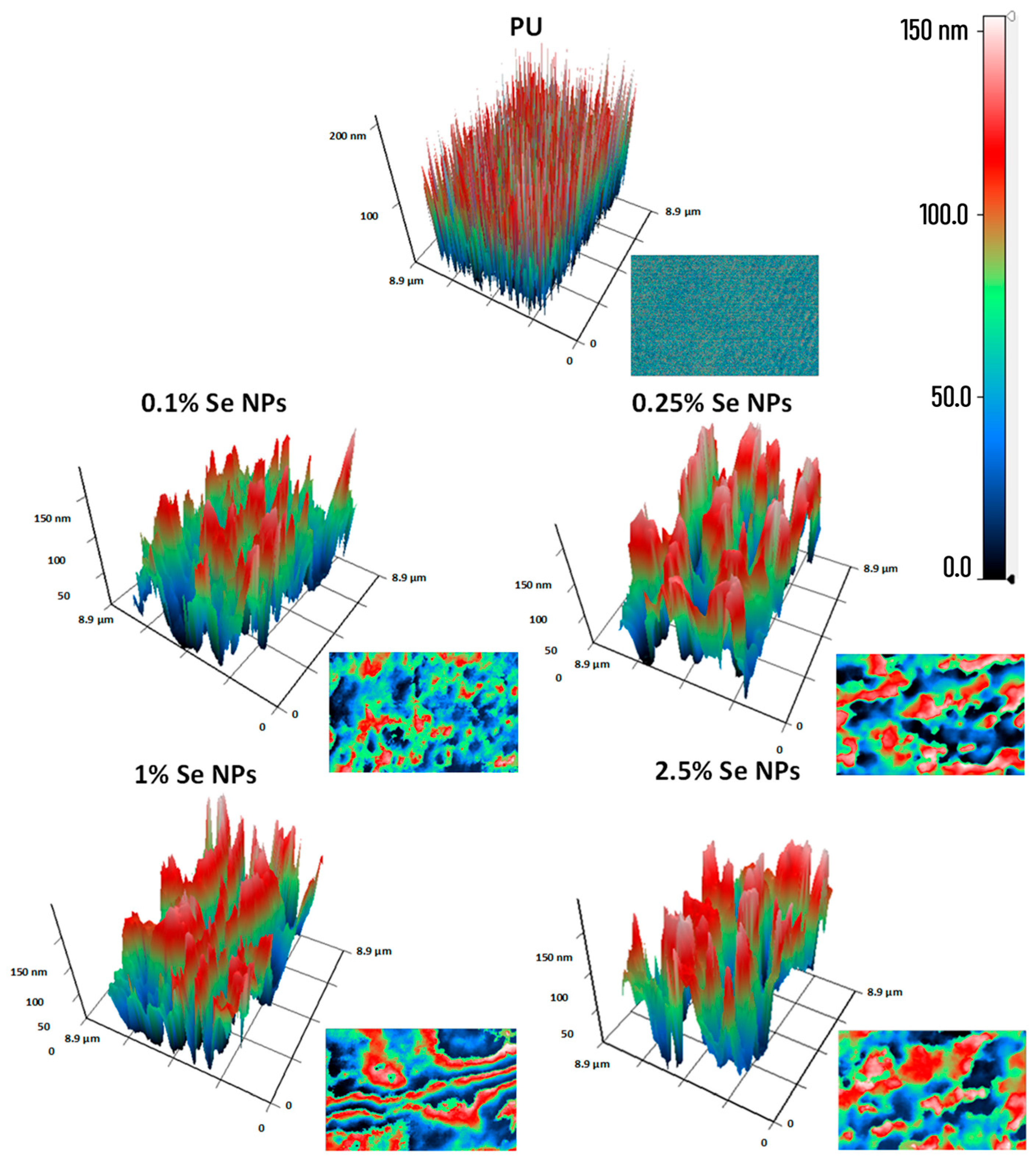
To assess the distribution of Se SMPs in the polyurea matrix, modulation interference microscopy (MIM) was used. It was found that polyurea films without Se SMPs are devoid of pronounced optical inhomogeneities (Figure 3). In polyurea films without Se SMPs, the areas of maximum and minimum phase incursions are distributed uniformly. The addition of Se SMPs contributed to the appearance of optical inhomogeneities inside the composite material. With an increase in the concentration of particles, the size of the contrast areas increases. For composite material containing 0.1% of Se SMPs, the inhomogeneities had sizes on the order of tens and hundreds of nanometers. For composite material containing 0.25% particles, the inhomogeneities had sizes in the range from hundreds of nanometers to a micrometer. At Se SMPs concentration of about 1%, optical inhomogeneities acquire the form of strands with length of about several micrometers. With a further increase in the concentration of particles to 2.5%, optical inhomogeneities of the order of several micrometers in diameter are formed. At the same time, in all the analyzed films, no inhomogeneities in the form of concentric rings are observed, which indicates the internal integrity of the polymer matrix and the absence of gas bubbles and other internal cavities.

3.2. Biological Activity of PU/Se SMPs Composites

The ability of the synthesized PU/Se SMPs composite materials to influence the generation of hydrogen peroxide in deionized water was assessed using the enhanced chemiluminescence method. (Figure 4a). It was found that under the given conditions, about 3.2 nM of hydrogen peroxide was formed in the control. It was shown that polyurea without Se SMPs did not lead to significant generation of hydrogen peroxide in water, compared to the control. Sample of the composite material with particle concentration of 0.1% also did not affect the concentration of hydrogen peroxide compared to the control. At the same time, an increase in the concentration of hydrogen peroxide in deionized water was noted, incubated with PU/Se SMPs samples containing 0.25–2.5% particles.
Using coumarin-3-carboxylic acid, a fluorescent sensor for hydroxyl radicals, the ability of the synthesized PU/Se SMPs composite materials to influence the generation of OH radicals in deionized water was assessed (Figure 4b). It was found that under the given conditions, slightly more than 20 nM hydroxyl radicals were formed in the control. It was shown that polyurea materials, both containing and not containing Se SMPs, did not significantly affect the generation of OH radicals, compared to the control. At the same time, with an increase in the concentration of particles, a tendency towards an increase in the concentration of hydroxyl radicals was observed.
The effect of the synthesized PU/Se SMPs composite materials on 8-oxoguanine in DNA in vitro was assessed using enzyme immunoassay (Figure 5a). It was found that under the given conditions, about 1.5 molecules of 8-oxoguanine per 105 guanine bases in DNA are formed in the control. It was shown that polyurea materials, both containing and not containing Se SMPs, did not significantly affect the level of oxidative DNA damage. At the same time, with an increase in the concentration of particles, tendency to an increase in the level of 8-oxoguanine in DNA was observed.
Using the method of induced luminescence of protein solutions, the effect of PU/Se SMPs composite materials on the formation of long-lived reactive protein species in vitro was studied (Figure 5b). It was found that under the given conditions in the control, the half-life of long-lived reactive protein species is about 4–5 h. It was shown that polyurea materials, both containing and not containing Se SMPs, did not significantly affect either the level or the half-life of long-lived reactive protein species. At the same time, with an increase in the concentration of particles, tendency to an increase in the level of long-lived reactive protein species in the protein solution was observed.
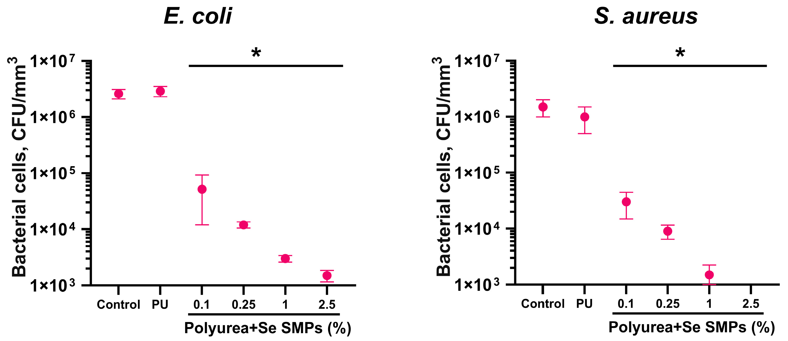
The effect of PU/Se SMPs composite materials on the number of planktonic cells of E. coli and S. aureus was studied (Figure 6). It was shown that polyurea without Se SMPs did not lead to a reliable change in the number of planktonic cells of either E. coli or S. aureus compared to the control. Samples of PU/Se SMPs composite materials contributed to reliable inhibition of planktonic cell growth of both bacterial species. Samples with Se SMPs concentrations of 0.1, 0.25, 1.0, and 2.5 wt.% inhibited the growth of planktonic cells of E. coli by approximately 50, 200, 800, and 1700 times, and S. aureus by approximately 50, 160, 1000, and 2300 times, respectively.
The effect of the synthesized composite materials on the viability of mouse lung fibroblast cell cultures in vitro was assessed (Figure 7). The percentage of nonviable cells in the control group cultures didn’t exceed 3%. The density of cell cultures in the control was 3.3 × 10⁴ cells/cm2. The median value of the area occupied by one cell was slightly more than 1.5 × 103 μm2. The median value of the area occupied by one nucleus was about 250 μm2. The percentage of nonviable cells in cultures growing on polyurea containing or not containing Se SMPs did not statistically differ from the percentage of nonviable cells in the control group. It should be noted that the range values in the PU/Se SMPs 2.5 wt.% group were 30% greater than in all other groups, i.e., the data obtained on PU/Se SMPs 2.5 wt.% were more heterogeneous. No statistically significant differences in the density of cell cultures growing on polyurea surfaces, PU/Se SMPs composite materials and control surfaces were found. At the same time, the density in the culture growing on the PU/Se SMPs 0.1 wt.% surface was 20% lower than in all other groups. The average area occupied by one cell differed significantly only in the culture growing on the PU/Se SMPs 0.1 wt.% composite material. In all other cases, the average area occupied by one cell did not differ from the control values. The area of cell nuclei was assessed, but no statistically significant changes in this parameter were found in any of the groups.
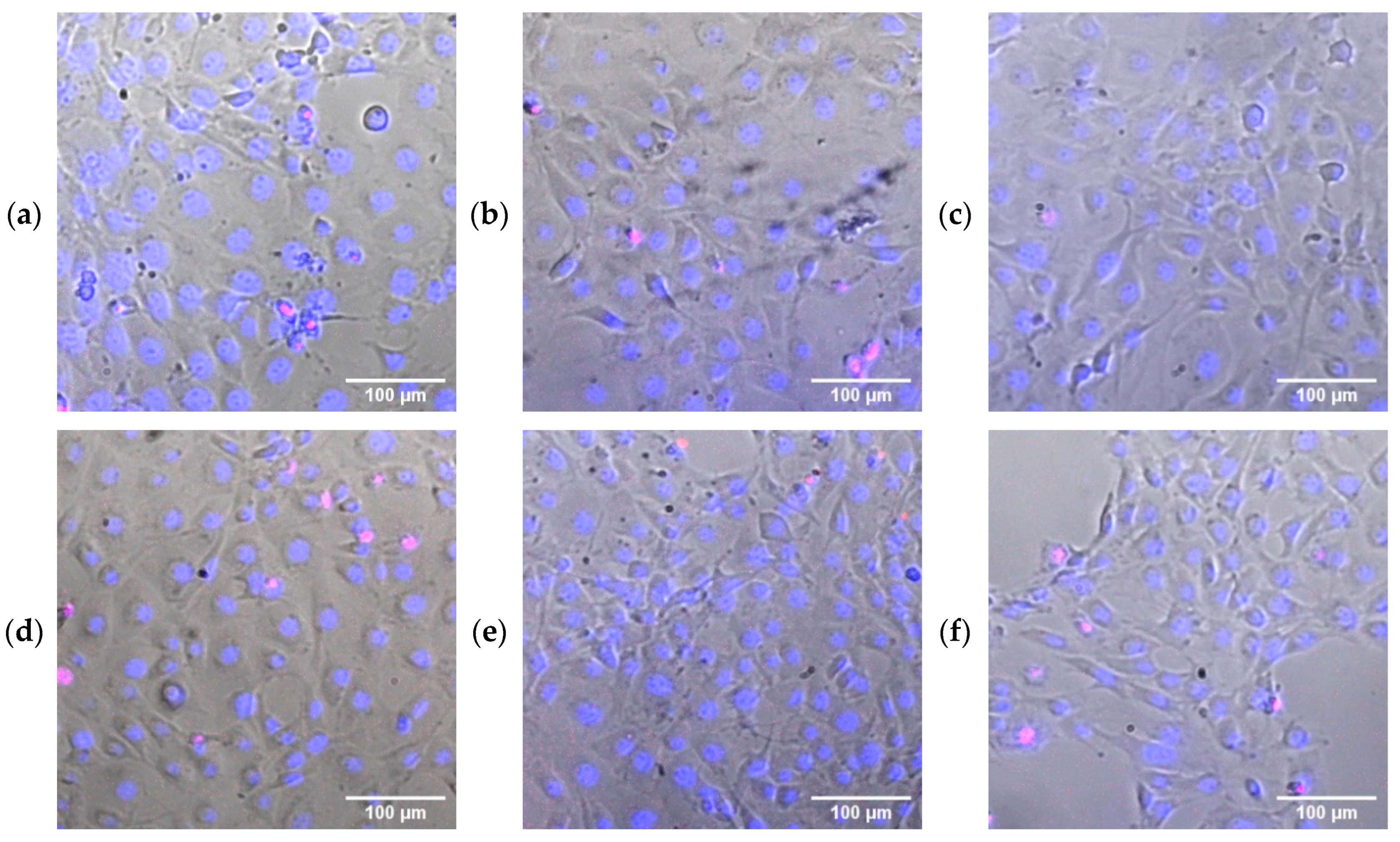
Figure 8 shows representative micrographs of cell cultures localized in the presence of the studied samples of composite materials. As can be seen from the micrographs, the pulmonary fibroblasts cultured in the presence of PU/Se SMPs composite materials have a correct morphology, not different from the cells of the control group.
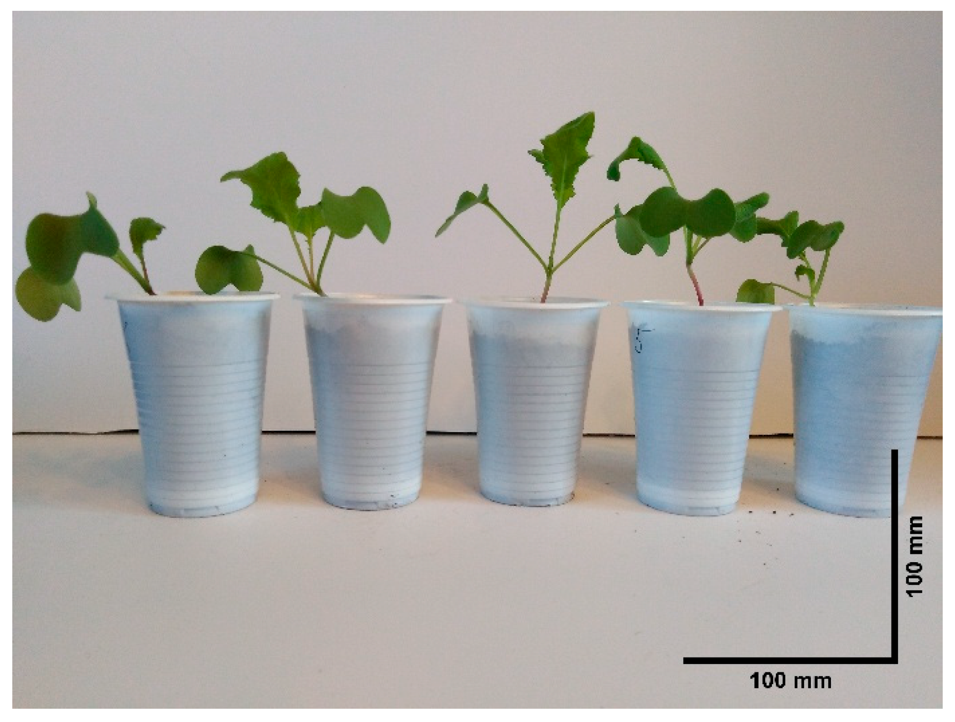
The effect of PU/Se SMPs composite materials on the rate of radish plant development (R. sativus var. sativus) was studied (Figure 9).
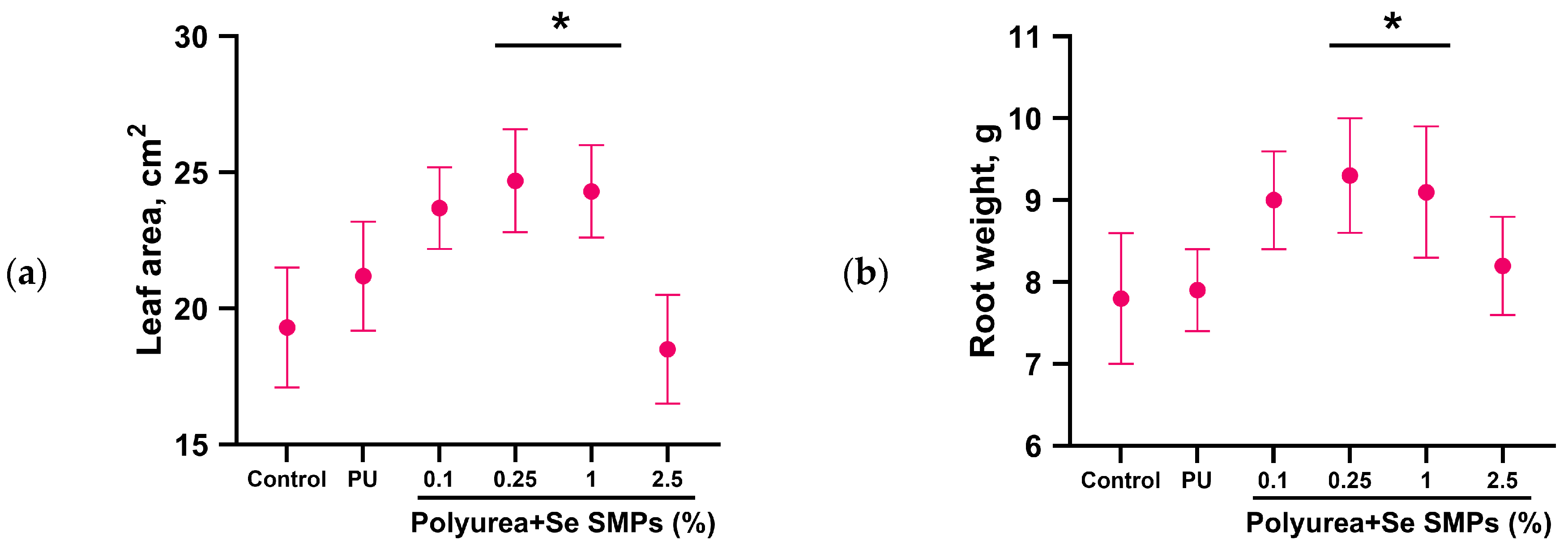
The following parameters were estimated: average leaf blade area (Figure 10a) and root dry weight (Figure 10b). It was shown that PU without particles and PU/Se SMPs 0.1 wt.% did not affect the analyzed parameters. The introduction of PU/Se SMPs composite materials containing 0.25 and 1% particles into the soil contributed to increase of the considered morphometric parameters of seedlings R. sativus var. sativus sprouts. At the same time, an increase in the root weight of radish plants by 15–20% and an increase in the leaf blade area by 25–30% were observed. It was found that the introduction of PU/Se SMPs composite materials containing 2.5% of particles into the soil tended to inhibit the growth of R. sativus var. sativus, compared with the PU/Se SMPs 0.25 and 1.0% groups; no differences were observed from the control group. The leaf area of radish plants with the addition of colloidal solutions of Se SMPs to the substrate as a control was about 32 ± 3 cm2, 37 ± 2 cm2, 38 ± 3 cm2 and 28 ± 4 cm2, respectively, for each concentration of added Se SMPs (1, 5, 10 and 25 µg/kg).

4. Discussion

Spherical submicron-sized selenium particles with an average hydrodynamic diameter of about 100 nm were synthesized using laser ablation in liquid (Figure 1). Method for adding Se SMPs to polyurea-based polymer was developed. Composite materials in the form of films with thickness of about 250 µm with Se SMPs content of 0.1 to 2.5 wt.% were obtained from the resulting colloid. It is known that the addition of particles often leads to the appearance of defects and deterioration of the properties of polymer surfaces [56,57,58]. The films obtained in this study had uniform surface without significant defects at the macro and micro levels (Figure 2). It is known that the refractive index for zero-valent selenium is 3.07 [59]. The refractive index for pure polyurea, depending on the ratio of components, lies in the range of 1.57–1.63 [60]. The difference in the refractive index of the materials by almost 2 times allows us to clearly distinguish these materials using the method of modulation-interference microscopy. It was found that the Se SMPs are non-uniformly distributed in the polymer matrices, probably forming areas with a slightly higher and slightly lower concentration of Se SMPs (Figure 3). Usually, the non-uniform distribution of particles in the polymer matrix is a consequence of the absence of ultrasonic treatment or mechanical activation [61]. In this work, we used quite effective means of both ultrasonic treatment and mechanical activation. Probably, the specific spatial distribution of particles in the polymer matrix that we discovered can be due to both the adhesive properties of the components and their surface energy, and the rheological behavior of the system as a whole [62].
Reactive oxygen species (ROS) are constantly formed in living organisms, both as a result of normal metabolism [63] and under the influence of various external factors [64]. An increase in the intracellular concentration of ROS above the level of antioxidant protection causes “oxidative stress”, which is accompanied by processes that are dangerous for the vital activity of cells, such as lipid peroxidation [65], oxidative modification of proteins [66] and nucleic acids [67]. Oxidative damage to nucleic acids is closely associated with the processes of mutagenesis [68] and carcinogenesis [69]. The level of oxidative stress is usually reduced with the help of antioxidants [70]. It is known that Se SMPs can exhibit both significant antioxidant [42] and prooxidant properties [71]. The materials obtained in this work didn’t affect the formation of hydroxyl radicals in aqueous solutions (Figure 4a), however, statistically significant increase in hydrogen peroxide was observed in solutions incubated with the samples of the obtained composite films (Figure 4b) containing 0.25, 1, 2.5% Se SMPs, compared to the control. In this case, the concentration of H2O2 was less than 10 nM upon exposure to samples of PU/Se SMPs composite materials containing 2.5% Se SMPs. According to the literature, these concentrations of hydrogen peroxide are not capable of causing significant oxidative damage [72,73,74]. It was found that the synthesized samples of PU/Se SMPs composite materials did not have significant oxidative effect on protein molecules and DNA in vitro (Figure 5). In the context of the biological activity of Se particles in the submicron range (100–1000 nm) in polymer functional composite materials, there is very little literature data. At the same time, Se NPs (<100 nm) are traditionally studied to obtain composite polymer materials for various applications [75,76,77,78,79,80,81,82].
It is noteworthy that the samples of PU/Se SMPs composite films containing the entire considered range of Se SMPs concentrations exerted significant bacteriostatic effect on E. coli and S. aureus cells, which increased with increasing concentration of particles (Figure 6). The most pronounced effect was revealed when cultivating bacterial suspension on the surface of PU/Se SMPs composite materials containing 2.5% Se SMPs; on these samples, the concentration of both types of bacterial cells was lower by more than 3 orders of magnitude compared to the control values. It is noteworthy that the bacteriostatic effect of the PU/Se SMPs composite materials against planktonic cells of S. aureus was more pronounced compared to E. coli cells. This result may be due to both the characteristics of the cell wall of Gram-positive microorganisms and differences in the metabolic activity of the strains studied. It is known that the cell wall of S. aureus contains a thick layer of peptide glycan, which can have a significant effect on the permeability of antimicrobial agents and their binding to cellular targets. At the same time, in E. coli, as a representative of Gram-negative bacteria, the presence of an outer membrane with a lipopolysaccharide layer serves as an additional barrier to the penetration of a number of substances, which potentially reduces the effectiveness of their action. A number of other studies examining the antibacterial activity of Se particles-containing polymer composites have also shown differential effects on bacteria depending on their Gram specificity. A recent study by ElSheikh et al. demonstrated a more pronounced (25%) inhibition of Streptococcus mutans growth compared to E. coli in the presence of polymeric materials based on photopolymer resins and Se NPs [83]. Also, Tran et al. demonstrated differential antibacterial activity of Polyvinyl alcohol-stabilized Se NPs; E. coli growth was not affected at all concentrations studied, while strong inhibition of S. aureus growth was observed at a NPs concentration of 1 ppm [84]. Also, the use of the ε-caprolactone matrix provided a prolonged release of Se NPs and contributed to a significant antibacterial effect against Gram-positive bacteria: S. aureus and Staphylococcus epidermidis [85]. In turn, Abdelaziz et al. proposed the use of a composite based on polyvinyl alcohol and chitosan containing 1% Se NPs as an edible coating of fruits; coating plum fruits with this nanocomposite ensured an extension of the shelf life after harvesting due to the antibacterial properties of the resulting composite material against a wide range of Gram-positive and Gram-negative bacteria [86]. Isolated SeMPs also exhibited antibacterial activity against various bacterial cell types. For example, Zhang et al. demonstrated antibacterial activity against both Gram-positive (Staphylococcus aureus, Bacillus cereus, Bacillus subtilis) and Gram-negative bacteria (Pseudomonas aeruginosa, Escherichia coli, Vibrio parahemolyticus) of Se particles with an average size of 120 nm [87]. Also, Shakibaie et al. demonstrated inhibition of biofilm formation of clinical isolates of Staphylococcus aureus, Pseudomonas aeruginosa and Proteus mirabilis by 42%, 34.3% and 53.4%, respectively, compared to untreated samples using Se particles of 80–200 nm size [88]. Huang et al. compared the antibacterial effect of Se particles depending on their size (43, 81, 124, 161, 205 nm) against methicillinsensitive and methicillin-resistant Staphylococcus aureus (MSSA and MRSA): the most pronounced antibacterial effect was observed when using 81-nm Se NPs [89].
The putative mechanisms of the antibacterial action of Se NPs are considered to be the initiation of the formation of reactive oxygen species (ROS) in prokaryotic cells [90], the destruction of the bacterial cell wall as a result of contact with Se NPs [91], and the inhibition of protein and DNA synthesis due to the formation of high concentrations of selenium cations [92]. Our results indicate significant antibacterial activity of the obtained composite materials against E. coli and S. aureus bacterial cells. It was found that the composite materials did not significantly affect the viability of eukaryotic cell cultures (Figure 7). No morphological changes in fibroblast cells growing in the presence of PU/Se SMPs composite materials were also detected (Figure 8). The presented results of cytotoxic studies are comparable with the data presented by other authors. In particular, in the work of Abdelaziz al. it was shown that for composites based on polyvinyl alcohol and chitosan containing Se NPs, the viability of Vero cells cultured in the presence of these materials was higher than 80% at all studied concentrations of Se NPs [86]. Also, in experiments on culturing hTERT BJ-5ta cell line fibroblasts on the surfaces of chitosan/Se NPs films, a high level of viability of cell cultures was demonstrated [93]. In the work of Hamdy et al. The cytotoxic and antibacterial properties of composites obtained by mixing Se NPs with cefepime, polyvinyl alcohol and polyhydroxybutyrate were studied; the absence of a significant effect on the growth and development of HEK-293 cell cultures and normal RPE-1 cells was demonstrated even at a Se NPs concentration of 50 μg/mL against the background of significant antibacterial properties of these materials [94].
It is known that selenium concentrations that cause toxicity in vitro can vary depending on several factors, including the specific cell type studied, the duration of exposure, and the chemical form of selenium used. Typically, Se NPs concentrations in the range of micrograms per milliliter (μg/mL) to milligrams per milliliter (mg/mL) are used in in vitro studies to induce cytotoxic effects, including cell death and damage to cellular components [95,96,97].
It is known that selenium is an element that affects plant growth, photosynthetic processes, and drought resistance [98]. The introduction of selenium-containing fertilizers has beneficial effect on the organoleptic characteristics and nutritional qualities of fruit crops [99,100]. The recent study by Chang et al. establishes a clear size criterion for Se particles for soil application (11–631 nm), as deviations from this range have been shown to make it impossible to obtain food products with regulated selenium content [101]. At the same time, improved digestibility by plants was observed for Se particles of smaller size [102].
The effect of adding the obtained PU/Se SMPs composite materials to the soil on the growth of R. sativus var. sativus plants was studied (Figure 9). Materials containing 0.25 and 1% of Se SMPs contributed to the improvement of morphometric growth parameters of radish plants (Figure 10). It is noteworthy that no statistically significant effect was observed with the addition of materials containing 0.1% Se SMPs, while materials containing 2.5% Se SMPs tended to inhibit the growth of radish plants. The obtained results are in good agreement with other reports on the efficiency of using Se SMPs in polymer matrices for agronomy applications. For example, algal carrageenan-based composites containing 2% Se NPs of a wide size range not only improved the morphometric parameters of potato plants, but also inhibited the growth of Clavibacter sepedonicus (bacteria that cause potato ring rot) [103]. Also, the introduction of Se NPs in the form of algal polysaccharide-based composites contributed to more efficient Se accumulation by rice plants, compared to the use of mineral forms of selenium (selenate and selenide) in experiments both in hydroponics and in pots [104]. In turn, the introduction of Se NPs/chitosan nanocomposites into the substrate not only enhanced the growth of rice seedlings, but also increased their tolerance to the effects of arsenic [105]. In the context of the use of synthetic polymer matrices, Siddiqui et al. proposed pre-sowing treatment of barley seeds with a composite based on polyvinylpyrrolidone, ascorbic acid and Se NPs (4.65 μg/mL); priming of seeds with the obtained composites contributed to a significant increase in the proportion of germinated seeds, as well as an improvement in the morphometric parameters of seedlings: the length of roots, shoots and the number of roots [106]. It is known that elevated concentrations of Se-containing compounds in the soil can have opposite effects (impair nutrient absorption, suppress photosynthesis, and cause oxidative damage) [107]. Finding optimal routes for controlled delivery of selenium, including the use of polymeric biodegradable composite materials functionalized with Se SMPs, is one of the promising strategies for the development of agricultural industry. The potential for using Se-containing composites based on synthetic slowly degradable polymers such as PU in agronomy is primarily associated with the possibility of controlled release of the microelement, increasing its bioavailability and reducing toxicity. Such composites stabilize Se SMPs for a long time, preventing aggregation and ensuring stability in soil and physiological solutions.

5. Conclusions

A composite material based on polyurea and Se SMPs has been developed. The obtained samples of composite material films have uniform surface without pronounced defects. The composite materials are biosafe and do not cause oxidative damage to DNA and proteins. At the same time, the developed composite materials have excellent bacteriostatic properties against Gram-negative E. coli and Gram-positive S. aureus bacteria, but don’t have acute cytotoxicity against primary cultures of mouse fibroblasts. It was found that the introduction of these materials into the soil has a beneficial effect on the growth of R. sativus radish plants. The obtained polymer composite materials based on polyurea with the addition of Se SMPs are of interest as promising materials for creating antibacterial biosafe coatings, for use in agriculture.

Author Contributions

Conceptualization, A.Y.I. and V.A.K.; methodology, D.V.Y.; software, R.Y.P. and M.E.A.; formal analysis, R.Y.P.; investigation, S.A.S., D.E.B., D.V.Y., I.V.B., A.V.S., M.V.D., R.M.S. and V.A.K.; resources, A.S.D. and A.Y.I.; data curation, M.E.A.; writing—original draft preparation, D.E.B. and S.A.S.; writing—review and editing, V.A.K., A.S.D. and A.Y.I.; funding acquisition, A.S.D. All authors have read and agreed to the published version of the manuscript.

Funding

This work was supported by grant of the Ministry of Science and Higher Education of the Russian Federation for large scientific projects in priority areas of scientific and technological development (grant number 075-15-2024-540).

Data Availability Statement

The raw data supporting the conclusions of this article will be made available by the authors on request.

Acknowledgments

The authors are grateful to the Center for Collective Use of VIM and GPI RAS.

Conflicts of Interest

The authors declare no conflicts of interest.

Abbreviations

The following abbreviations are used in this manuscript:
PU/Se SMPcomposite based on PolyUrea and Selenium SubMicron-sized particles
PUPolyUrea
Se NPsSelenium NanoParticles
Se SMPsSelenium SubMicron-sized particles
PIPropidium iodide

References

  1. Sharma, S.; Sudhakara, P.; Omran, A.A.B.; Singh, J.; Ilyas, R.A. Recent Trends and Developments in Conducting Polymer Nanocomposites for Multifunctional Applications. Polymers 2021, 13, 2898. [Google Scholar] [CrossRef]
  2. Gur’eva, L.L.; Kuzub, L.I.; Anokhin, D.V.; Badamshina, E.R. From synthesis of silver nanoparticles to polymer nanocomposites. Russ. Chem. Bull. 2025, 74, 281–304. [Google Scholar] [CrossRef]
  3. Shabalina, A.V.; Kozlov, V.A.; Popov, I.A.; Gudkov, S.V. A Review on Recently Developed Antibacterial Composites of Inorganic Nanoparticles and Non-Hydrogel Polymers for Biomedical Applications. Nanomaterials 2024, 14, 1753. [Google Scholar] [CrossRef]
  4. Folorunso, O.; Hamam, Y.; Sadiku, R.; Kupolati, W. Effects of Defects on the Properties of Polymer Nanocomposites: A Brief Review. J. Inorg. Organomet. Polym. Mater. 2024, 34, 5667–5690. [Google Scholar] [CrossRef]
  5. Oladele, I.O.; Adelani, S.O.; Taiwo, A.S.; AkinbamiyorinI, M.; Olanrewaju, O.F.; Orisawayi, A.O. Polymer-based nanocomposites for supercapacitor applications: A review on principles, production and products. RSC Adv. 2025, 15, 7509–7534. [Google Scholar] [CrossRef]
  6. Satdive, A.; Tayde, S.; Tonde, S.; Hazra, C.; Kundu, D.; Chatterjee, A. Bionanocomposites in the Construction and Building Applications. In Polymer Based Bio-Nanocomposites; Composites Science and Technology; Springer: Berlin/Heidelberg, Germany, 2022; pp. 293–310. [Google Scholar]
  7. Rajak, D.K.; Wagh, P.H.; Kumar, A.; Behera, A.; Pruncu, C.I. Advanced Polymers in Aircraft Structures. In Materials, Structures and Manufacturing for Aircraft; Sustainable Aviation; Springer: Berlin/Heidelberg, Germany, 2022; pp. 65–88. [Google Scholar]
  8. Bhat, A.; Budholiya, S.; Aravind Raj, S.; Sultan, M.T.H.; Hui, D.; Md Shah, A.U.; Safri, S.N.A. Review on nanocomposites based on aerospace applications. Nanotechnol. Rev. 2021, 10, 237–253. [Google Scholar] [CrossRef]
  9. Saberi Riseh, R.; Vatankhah, M.; Hassanisaadi, M.; Tamanadar, E.; Skorik, Y. Nanocomposites in agriculture as pesticides for plant protection: A review. Adv. Nat. Sci. Nanosci. Nanotechnol. 2024, 15, 023003. [Google Scholar] [CrossRef]
  10. Shabalina, A.V.; Kozlov, V.A.; Paskhin, M.O.; Popov, I.A.; Gudkov, S.V. A Mini-Review of Photoconversion Covers for Greenhouses: Assessment Parameters and Plant Experiment Results. Horticulturae 2025, 11, 680. [Google Scholar] [CrossRef]
  11. Gudkov, S.V.; Sarimov, R.M.; Astashev, M.E.; Pishchalnikov, R.Y.; Yanykin, D.V.; Simakin, A.V.; Shkirin, A.V.; Serov, D.A.; Konchekov, E.M.; Gusein-zade Namik Guseynaga, O.; et al. Modern physical methods and technologies in agriculture. Physics-Uspekhi 2023, 67, 194–210. [Google Scholar] [CrossRef]
  12. Smaoui, S.; Chérif, I.; Ben Hlima, H.; Khan, M.U.; Rebezov, M.; Thiruvengadam, M.; Sarkar, T.; Shariati, M.A.; Lorenzo, J.M. Zinc oxide nanoparticles in meat packaging: A systematic review of recent literature. Food Packag. Shelf Life 2023, 36, 101045. [Google Scholar] [CrossRef]
  13. Mishra, B.; Panda, J.; Mishra, A.K.; Nath, P.C.; Nayak, P.K.; Mahapatra, U.; Sharma, M.; Chopra, H.; Mohanta, Y.K.; Sridhar, K. Recent advances in sustainable biopolymer-based nanocomposites for smart food packaging: A review. Int. J. Biol. Macromol. 2024, 279, 135583. [Google Scholar] [CrossRef]
  14. Li, X.; Xu, W.; Xin, Y.; Yuan, J.; Ji, Y.; Chu, S.; Liu, J.; Luo, Q. Supramolecular Polymer Nanocomposites for Biomedical Applications. Polymers 2021, 13, 513. [Google Scholar] [CrossRef]
  15. Alosaimi, A.M.; Alorabi, R.O.; Katowah, D.F.; Al-Thagafi, Z.T.; Alsolami, E.S.; Hussein, M.A.; Qutob, M.; Rafatullah, M. Recent Biomedical Applications of Coupling Nanocomposite Polymeric Materials Reinforced with Variable Carbon Nanofillers. Biomedicines 2023, 11, 967. [Google Scholar] [CrossRef]
  16. Pourmadadi, M.; Rahmani, E.; Shamsabadipour, A.; Mahtabian, S.; Ahmadi, M.; Rahdar, A.; Díez-Pascual, A.M. Role of Iron Oxide (Fe2O3) Nanocomposites in Advanced Biomedical Applications: A State-of-the-Art Review. Nanomaterials 2022, 12, 3873. [Google Scholar] [CrossRef]
  17. Ray, A.; Das, P.; Chunduri, R.; Kumar, D.; Dulta, K.; Kaushal, A.; Gupta, S.; Rj, S.; Yadav, A.N.; Nagraik, R.; et al. Nanocomposite-based agricultural delivery systems: A sustainable approach to enhanced crop productivity and soil health. J. Nanoparticle Res. 2025, 27, 110. [Google Scholar] [CrossRef]
  18. Shafiq, F.; Mahmood, A.; Iqbal, M. Agricultural Use of Various Polymeric Nanocomposites and Future Avenues. In Specialty Polymers and Materials; Apple Academic Press: Palm Bay, FL, USA, 2024; pp. 21–52. [Google Scholar]
  19. Javed, F.; Hayat, S.; Aslam, B.; Saqalein, M.; Waseem, M.; Meklat, A.; Muzammil, S. Agricultural applications of bionanocomposites. In Advances in Bionanocomposites; Elsevier: Amsterdam, The Netherlands, 2024; pp. 327–350. [Google Scholar]
  20. Hoang, N.H.; Thanh, T.L.; Saengchan, C.; Sangpueak, R.; Thepbandit, W.; Zhou, X.; Kamkaew, A.; Buensanteai, K. Application of Nanocomposites-Based Polymers on Managing Fungal Diseases in Crop Production. Plant Pathol. J. 2025, 41, 437–455. [Google Scholar] [CrossRef]
  21. Rop, K.; Karuku, G.N.; Mbui, D. Polymer Nanocomposite Fertilizers: Effects on Maize, Capsicum, and Kale Growth and Yield. In Handbook of Nanofillers; Springer: Singapore, 2025; pp. 2821–2842. [Google Scholar]
  22. Prajapat, A.L.; Devre, P.V.; Bhavsar, P.S.; Nille, O.S.; Dongare, P.R.; Gore, A.H. Biopolymer-Based Nanocomposites for Adsorption/Recovery of Fertilizer Nutrients in Agricultural Applications. In Biopolymeric Nanoparticles for Agricultural Applications; Nanotechnology in the Life Sciences; Springer: Cham, Switzerland, 2024; pp. 59–77. [Google Scholar]
  23. Ghorbani, A.; Emamverdian, A.; Pehlivan, N.; Zargar, M.; Razavi, S.M.; Chen, M. Nano-enabled agrochemicals: Mitigating heavy metal toxicity and enhancing crop adaptability for sustainable crop production. J. Nanobiotechnol. 2024, 22, 91. [Google Scholar] [CrossRef]
  24. Sepehry Javan, Z.; Razavi, S.M.; Khalofah, A.; Ghorbani, A. The ameliorating effects of cinnamic acid-based nanocomposite against salt stress in peppermint. Environ. Sci. Pollut. Res. 2024, 31, 45055–45073. [Google Scholar] [CrossRef]
  25. Zhang, D.-D.; Zhao, J.-F.; Tan, L.-Q.; Wu, Q.; Lv, H.-X.; Zhang, Y.-R.; Zhang, M. Effects of zinc oxide nanocomposites on microorganism growth and protection of physicochemical quality during maize storage. Int. J. Food Microbiol. 2024, 411, 110552. [Google Scholar] [CrossRef]
  26. Asle-Mohammadi, Z.; Razavi, F.; Aghdam, M.S.; Ebrahimi, A. Employing Chitosan-coated Putrescine Nanocomposite for Preserving Kiwifruit Quality During Cold Storage. Appl. Fruit Sci. 2024, 66, 1505–1513. [Google Scholar] [CrossRef]
  27. Astashev, M.E.; Sarimov, R.M.; Serov, D.A.; Matveeva, T.A.; Simakin, A.V.; Ignatenko, D.N.; Burmistrov, D.E.; Smirnova, V.V.; Kurilov, A.D.; Mashchenko, V.I.; et al. Antibacterial behavior of organosilicon composite with nano aluminum oxide without influencing animal cells. React. Funct. Polym. 2022, 170, 105143. [Google Scholar] [CrossRef]
  28. Zhang, R.; Huang, W.; Lyu, P.; Yan, S.; Wang, X.; Ju, J. Polyurea for Blast and Impact Protection: A Review. Polymers 2022, 14, 2670. [Google Scholar] [CrossRef]
  29. Santana, J.S.; Cardoso, E.S.; Triboni, E.R.; Politi, M.J. Polyureas Versatile Polymers for New Academic and Technological Applications. Polymers 2021, 13, 4393. [Google Scholar] [CrossRef]
  30. Shojaei, B.; Najafi, M.; Yazdanbakhsh, A.; Abtahi, M.; Zhang, C. A review on the applications of polyurea in the construction industry. Polym. Adv. Technol. 2021, 32, 2797–2812. [Google Scholar] [CrossRef]
  31. Madelatparvar, M.; Hosseini, M.S.; Zhang, C. Polyurea micro-/nano-capsule applications in construction industry: A review. Nanotechnol. Rev. 2023, 12, 20220516. [Google Scholar] [CrossRef]
  32. Ding, L.; Wang, Y.; Lin, J.; Ma, M.; Hu, J.; Qiu, X.; Wu, C.; Feng, C. Recent advances in polyurea elastomers and their applications in blast protection: A review. J. Mater. Sci. 2024, 59, 14893–14923. [Google Scholar] [CrossRef]
  33. Zhong, J.; Hu, Y.; Wang, D.; Zhou, X.; Yuan, P.; Luo, B.; Li, Y. Enhancing Dental Material Performance: Tung Oil-Infused Polyurea Microcapsule Coatings for Self-Healing and Antimicrobial Applications. Polymers 2024, 16, 918. [Google Scholar] [CrossRef]
  34. Li, Y.; Woo, Y.; Sekar, M.; Narasimalu, S.; Dong, Z. Effect of Nano-Titanium Dioxide Contained in Titania-Polyurea Coating on Marina Biofouling and Drag Reduction. J. Biomed. Nanotechnol. 2020, 16, 1530–1541. [Google Scholar] [CrossRef]
  35. Mikhailova, E.O. Selenium Nanoparticles: Green Synthesis and Biomedical Application. Molecules 2023, 28, 8125. [Google Scholar] [CrossRef]
  36. Varlamova, E.G.; Turovsky, E.A.; Blinova, E.V. Therapeutic Potential and Main Methods of Obtaining Selenium Nanoparticles. Int. J. Mol. Sci. 2021, 22, 10808. [Google Scholar] [CrossRef]
  37. Turovsky, E.A.; Baryshev, A.S.; Plotnikov, E.Y. Selenium Nanoparticles in Protecting the Brain from Stroke: Possible Signaling and Metabolic Mechanisms. Nanomaterials 2024, 14, 160. [Google Scholar] [CrossRef]
  38. Turovsky, E.A.; Mal’tseva, V.N.; Sarimov, R.M.; Simakin, A.V.; Gudkov, S.V.; Plotnikov, E.Y. Features of the cytoprotective effect of selenium nanoparticles on primary cortical neurons and astrocytes during oxygen–glucose deprivation and reoxygenation. Sci. Rep. 2022, 12, 1710. [Google Scholar] [CrossRef]
  39. Hosnedlova, B.; Kepinska, M.; Skalickova, S.; Fernandez, C.; Ruttkay-Nedecky, B.; Peng, Q.; Baron, M.; Melcova, M.; Opatrilova, R.; Zidkova, J.; et al. Nano-selenium and its nanomedicine applications: A critical review. Int. J. Nanomed. 2018, 13, 2107–2128. [Google Scholar] [CrossRef]
  40. Varlamova, E.G.; Khabatova, V.V.; Gudkov, S.V.; Plotnikov, E.Y.; Turovsky, E.A. Cytoprotective Properties of a New Nanocomplex of Selenium with Taxifolin in the Cells of the Cerebral Cortex Exposed to Ischemia/Reoxygenation. Pharmaceutics 2022, 14, 2477. [Google Scholar] [CrossRef]
  41. González-Lemus, U.; Medina-Pérez, G.; Espino-García, J.J.; Fernández-Luqueño, F.; Campos-Montiel, R.; Almaraz-Buendía, I.; Reyes-Munguía, A.; Urrutia-Hernández, T. Nutritional Parameters, Biomass Production, and Antioxidant Activity of Festuca arundinacea Schreb. Conditioned with Selenium Nanoparticles. Plants 2022, 11, 2326. [Google Scholar] [CrossRef]
  42. Dawood, M.A.O.; Basuini, M.F.E.; Yilmaz, S.; Abdel-Latif, H.M.R.; Kari, Z.A.; Abdul Razab, M.K.A.; Ahmed, H.A.; Alagawany, M.; Gewaily, M.S. Selenium Nanoparticles as a Natural Antioxidant and Metabolic Regulator in Aquaculture: A Review. Antioxidants 2021, 10, 1364. [Google Scholar] [CrossRef]
  43. Burmistrov, D.E.; Shumeyko, S.A.; Semenova, N.A.; Dorokhov, A.S.; Gudkov, S.V. Selenium Nanoparticles (Se NPs) as Agents for Agriculture Crops with Multiple Activity: A Review. Agronomy 2025, 15, 1591. [Google Scholar] [CrossRef]
  44. Serov, D.A.; Khabatova, V.V.; Vodeneev, V.; Li, R.; Gudkov, S.V. A Review of the Antibacterial, Fungicidal and Antiviral Properties of Selenium Nanoparticles. Materials 2023, 16, 5363. [Google Scholar] [CrossRef]
  45. Sans-Serramitjana, E.; Obreque, M.; Muñoz, F.; Zaror, C.; Mora, M.d.L.L.; Viñas, M.; Betancourt, P. Antimicrobial Activity of Selenium Nanoparticles (SeNPs) against Potentially Pathogenic Oral Microorganisms: A Scoping Review. Pharmaceutics 2023, 15, 2253. [Google Scholar] [CrossRef]
  46. Tran, P.A.; Webster, T.J. Selenium nanoparticles inhibit Staphylococcus aureus growth. Int. J. Nanomed. 2011, 6, 1553–1558. [Google Scholar] [CrossRef]
  47. Salem, S.S.; Badawy, M.S.E.M.; Al-Askar, A.A.; Arishi, A.A.; Elkady, F.M.; Hashem, A.H. Green Biosynthesis of Selenium Nanoparticles Using Orange Peel Waste: Characterization, Antibacterial and Antibiofilm Activities against Multidrug-Resistant Bacteria. Life 2022, 12, 893. [Google Scholar] [CrossRef]
  48. Rangrazi, A.; Bagheri, H.; Ghazvini, K.; Boruziniat, A.; Darroudi, M. Synthesis and antibacterial activity of colloidal selenium nanoparticles in chitosan solution: A new antibacterial agent. Mater. Res. Express 2020, 6, 1250h3. [Google Scholar] [CrossRef]
  49. Au, A.; Mojadadi, A.; Shao, J.-Y.; Ahmad, G.; Witting, P.K. Physiological Benefits of Novel Selenium Delivery via Nanoparticles. Int. J. Mol. Sci. 2023, 24, 6068. [Google Scholar] [CrossRef]
  50. Ivanov, V.E.; Usacheva, A.M.; Chernikov, A.V.; Bruskov, V.I.; Gudkov, S.V. Formation of long-lived reactive species of blood serum proteins induced by low-intensity irradiation of helium-neon laser and their involvement in the generation of reactive oxygen species. J. Photochem. Photobiol. B Biol. 2017, 176, 36–43. [Google Scholar] [CrossRef]
  51. Sevostyanov, M.A.; Kolmakov, A.G.; Sergiyenko, K.V.; Kaplan, M.A.; Baikin, A.S.; Gudkov, S.V. Mechanical, physical–chemical and biological properties of the new Ti–30Nb–13Ta–5Zr alloy. J. Mater. Sci. 2020, 55, 14516–14529. [Google Scholar] [CrossRef]
  52. Gudkov, S.V.; Shtarkman, I.N.; Chernikov, A.V.; Usacheva, A.M.; Bruskov, V.I. Guanosine and inosine (riboxin) eliminate the long-lived protein radicals induced X-ray radiation. Dokl. Biochem. Biophys. 2007, 413, 50–53. [Google Scholar] [CrossRef]
  53. Chernikov, A.V.; Gudkov, S.V.; Shtarkman, I.N.; Bruskov, V.I. Oxygen effect in heat-induced DNA damage. Biophysics 2007, 52, 185–190. [Google Scholar] [CrossRef]
  54. Serov, D.; Burmistrov, D.; Simakin, A.; Astashev, M.; Uvarov, O.; Tolordava, E.; Semenova, A.; Lisitsyn, A.; Gudkov, S. Composite Coating for the Food Industry Based on Fluoroplast and ZnO-NPs: Physical and Chemical Properties, Antibacterial and Antibiofilm Activity, Cytotoxicity. Nanomaterials 2022, 12, 4158. [Google Scholar] [CrossRef]
  55. Gudkov, S.V.; Simakin, A.V.; Bunkin, N.F.; Shafeev, G.A.; Astashev, M.E.; Glinushkin, A.P.; Grinberg, M.A.; Vodeneev, V.A. Development and application of photoconversion fluoropolymer films for greenhouses located at high or polar latitudes. J. Photochem. Photobiol. B Biol. 2020, 213, 112056. [Google Scholar] [CrossRef]
  56. Cazan, C.; Enesca, A.; Andronic, L. Synergic Effect of TiO2 Filler on the Mechanical Properties of Polymer Nanocomposites. Polymers 2021, 13, 2017. [Google Scholar] [CrossRef]
  57. Ilyin, S.O.; Kotomin, S.V. Effect of Nanoparticles and Their Anisometry on Adhesion and Strength in Hybrid Carbon-Fiber-Reinforced Epoxy Nanocomposites. J. Compos. Sci. 2023, 7, 147. [Google Scholar] [CrossRef]
  58. Prorokova, N.P.; Odintsova, O.I.; Rumyantseva, V.E.; Rumyantsev, E.V.; Konovalova, V.S. Giving Improved and New Properties to Fibrous Materials by Surface Modification. Coatings 2023, 13, 139. [Google Scholar] [CrossRef]
  59. Khalid, A.; Tran, P.A.; Norello, R.; Simpson, D.A.; O’Connor, A.J.; Tomljenovic-Hanic, S. Intrinsic fluorescence of selenium nanoparticles for cellular imaging applications. Nanoscale 2016, 8, 3376–3385. [Google Scholar] [CrossRef]
  60. Tao, X.T.; Watanabe, T.; Zou, D.C.; Shimoda, S.; Sato, H.; Miyata, S. Polyurea with Large Positive Birefringence for Second Harmonic Generation. Macromolecules 2002, 28, 2637–2643. [Google Scholar] [CrossRef]
  61. Huo, S.; Zhao, P.; Shi, Z.; Zou, M.; Yang, X.; Warszawik, E.; Loznik, M.; Göstl, R.; Herrmann, A. Mechanochemical bond scission for the activation of drugs. Nat. Chem. 2021, 13, 131–139. [Google Scholar] [CrossRef]
  62. Bailly, M.; Kontopoulou, M.; El Mabrouk, K. Effect of polymer/filler interactions on the structure and rheological properties of ethylene-octene copolymer/nanosilica composites. Polymer 2010, 51, 5506–5515. [Google Scholar] [CrossRef]
  63. Sies, H.; Belousov, V.V.; Chandel, N.S.; Davies, M.J.; Jones, D.P.; Mann, G.E.; Murphy, M.P.; Yamamoto, M.; Winterbourn, C. Defining roles of specific reactive oxygen species (ROS) in cell biology and physiology. Nat. Rev. Mol. Cell Biol. 2022, 23, 499–515. [Google Scholar] [CrossRef]
  64. Lyakhov, G.A.; Man’ko, V.I.; Shcherbakov, I.A.; Suyazov, N.V. Impact of Classical Vibrations and Magnetic Fields on Quantum Objects. Phys. Wave Phenom. 2024, 32, 67–72. [Google Scholar] [CrossRef]
  65. Sokolov, A.V.; Kostevich, V.A.; Runova, O.L.; Gorudko, I.V.; Vasilyev, V.B.; Cherenkevich, S.N.; Panasenko, O.M. Proatherogenic modification of LDL by surface-bound myeloperoxidase. Chem. Phys. Lipids 2014, 180, 72–80. [Google Scholar] [CrossRef]
  66. Lankin, V.Z.; Sharapov, M.G.; Tikhaze, A.K.; Goncharov, R.G.; Antonova, O.A.; Konovalova, G.G.; Novoselov, V.I. Dicarbonyl-Modified Low-Density Lipoproteins Are Key Inducers of LOX-1 and NOX1 Gene Expression in the Cultured Human Umbilical Vein Endotheliocytes. Biochemistry 2023, 88, 2125–2136. [Google Scholar] [CrossRef]
  67. Kruchinin, A.A.; Kamzeeva, P.N.; Zharkov, D.O.; Aralov, A.V.; Makarova, A.V. 8-Oxoadenine: A «New» Player of the Oxidative Stress in Mammals? Int. J. Mol. Sci. 2024, 25, 1342. [Google Scholar] [CrossRef]
  68. Dushenko, M.V.; Abdullaev, S.A.; Ignatov, M.A.; Osipov, A.N. Comparative Study of Mitochondrial DNA Abnormalities in Mesenchymal Stem Cells after Exposure to X-Ray Radiation in Low and Medium Doses. Bull. Exp. Biol. Med. 2025, 178, 541–546. [Google Scholar] [CrossRef]
  69. Quezada-Maldonado, E.M.; Sánchez-Pérez, Y.; Chirino, Y.I.; García-Cuellar, C.M. Airborne particulate matter induces oxidative damage, DNA adduct formation and alterations in DNA repair pathways. Environ. Pollut. 2021, 287, 117313. [Google Scholar] [CrossRef]
  70. Karmanova, E.E.; Chernikov, A.V.; Popova, N.R.; Sharapov, M.G.; Ivanov, V.E.; Bruskov, V.I. Metformin mitigates radiation toxicity exerting antioxidant and genoprotective properties. Naunyn-Schmiedeberg’s Arch. Pharmacol. 2023, 396, 2449–2460. [Google Scholar] [CrossRef]
  71. Freire, B.M.; Lange, C.N.; Cavalcanti, Y.T.; Monteiro, L.R.; Pieretti, J.C.; Seabra, A.B.; Batista, B.L. The dual effect of Selenium nanoparticles in rice seedlings: From increasing antioxidant activity to inducing oxidative stress. Plant Stress 2024, 11, 100372. [Google Scholar] [CrossRef]
  72. Stone, J.R.; Yang, S. Hydrogen Peroxide: A Signaling Messenger. Antioxid. Redox Signal. 2006, 8, 243–270. [Google Scholar] [CrossRef]
  73. Lyublinskaya, O.; Antunes, F. Measuring intracellular concentration of hydrogen peroxide with the use of genetically encoded H2O2 biosensor HyPer. Redox Biol. 2019, 24, 101200. [Google Scholar] [CrossRef]
  74. Stepanov, E.V.; Shcherbakov, I.A. Physicochemical Methods of Studying Hydrogen Peroxide for Biomedical Applications. Phys. Wave Phenom. 2023, 31, 92–97. [Google Scholar] [CrossRef]
  75. Kopeikin, V.V.; Valueva, S.V.; Kipper, A.I.; Borovikova, L.N.; Nazarkina, Y.I.; Khlebosolova, E.N.; Filippov, A.I. Adsorption of Hydroxyethyl Cellulose Selenium Nanoparticles during Their Formation in Water. Russ. J. Appl. Chem. 2003, 76, 600–602. [Google Scholar] [CrossRef]
  76. Klechkovskaya, V.V.; Volkov, V.V.; Shtykova, E.V.; Arkharova, N.A.; Baklagina, Y.G.; Khripunov, A.K.; Smyslov, R.Y.; Borovikova, L.N.; Tkachenko, A.A. Network Model of Acetobacter Xylinum Cellulose Intercalated by Drug Nanoparticles. In Nanomaterials for Application in Medicine and Biology; NATO Science for Peace and Security Series B: Physics and Biophysics; Springer: Dordrecht, The Netherlands, 2008; pp. 165–177. [Google Scholar]
  77. Valueva, S.V.; Ivanov, I.V.; Volkov, A.Y.; Vylegzhanina, M.E.; Borovikova, L.N.; Kutin, A.A.; Yakimansky, A.V. Selenium Nanoparticles Stabilized by Amphiphilic Molecular Brushes with Varying Degrees of Polymerization of Side Chains: Spectral and Structural-Morphological Characteristics. Russ. J. Phys. Chem. A 2024, 98, 304–313. [Google Scholar] [CrossRef]
  78. Valueva, S.V.; Krasnopeeva, E.L.; Borovikova, L.N.; Morozova, P.Y.; Sokolova, M.P.; Melenevskaya, E.Y.; Yakimansky, A.V. Triple Nanosystems Based on Amphiphilic Molecular Brushes, Selenium Nanoparticles and Photosensitizer: Synthesis, Spectral, and Morphological Characteristics. Nanobiotechnol. Rep. 2024, 19, 108–115. [Google Scholar] [CrossRef]
  79. Panov, D.A.; Katsev, A.M.; Omel’chenko, A.V. Synthesis and Properties of Selenium Nanoparticles in a Natural Polysaccharide Matrix. Russ. J. Bioorg. Chem. 2023, 49, 1567–1576. [Google Scholar] [CrossRef]
  80. Valueva, S.V.; Morozova, P.Y.; Vylegzhanina, M.E.; Ivanov, I.V. Hybrid Nanosystems Based on Selenium Nanoparticles, Radachlorin, and Polymer Carriers (Graft Copolymers): Synthesis, Morphology, and Spectral Characteristics. Nanobiotechnol. Rep. 2025, 19, 959–965. [Google Scholar] [CrossRef]
  81. Valueva, S.V.; Vylegzhanina, M.E.; Mitusova, K.A.; Bezrukova, M.A.; Nazarova, O.V.; Zolotova, Y.I.; Panarin, E.F. Structural, Morphological, and Spectral Characteristics of Hybrid Bioactive Copper-, Selenium-, and Silver-Containing Nanosystems Based on Poly-4-Acryloylmorpholine. J. Surf. Investig. X-Ray Synchrotron Neutron Tech. 2021, 15, 110–120. [Google Scholar] [CrossRef]
  82. Perfileva, A.I.; Nozhkina, O.A.; Tretyakova, M.S.; Graskova, I.A.; Klimenkov, I.V.; Sudakov, N.P.; Alexandrova, G.P.; Sukhov, B.G. Biological Activity and Environmental Safety of Selenium Nanoparticles Encapsulated in Starch Macromolecules. Nanotechnol. Russ. 2020, 15, 96–104. [Google Scholar] [CrossRef]
  83. ElSheikh, S.K.; Eid, E.-S.G.; Abdelghany, A.M.; Abdelaziz, D. Physical/mechanical and antibacterial properties of composite resin modified with selenium nanoparticles. BMC Oral Health 2024, 24, 1245. [Google Scholar] [CrossRef]
  84. Tran, P.A.; O’Brien-Simpson, N.; Reynolds, E.C.; Pantarat, N.; Biswas, D.P.; O’Connor, A.J. Low cytotoxic trace element selenium nanoparticles and their differential antimicrobial properties against S. aureus and E. coli. Nanotechnology 2016, 27, 045101. [Google Scholar] [CrossRef]
  85. Filipović, N.; Veselinović, L.; Ražić, S.; Jeremić, S.; Filipič, M.; Žegura, B.; Tomić, S.; Čolić, M.; Stevanović, M. Poly (ε-caprolactone) microspheres for prolonged release of selenium nanoparticles. Mater. Sci. Eng. C 2019, 96, 776–789. [Google Scholar] [CrossRef]
  86. Abdelaziz, H.T.O.; Seif Mohamed, E.M.; Younis, S.K.A.; Ahmed, N.; Michaeel, M.N.; Abu-Hussien, S.H.; Bakry, A.; Ebeed, N.M.; Nasser, M.A.; El-Nasr, M.K.A.; et al. Selenium nanoparticle loaded on PVA/chitosan biofilm synthesized from orange peels: Antimicrobial and antioxidant properties for plum preservation. BMC Chem. 2025, 19, 245. [Google Scholar] [CrossRef]
  87. Zhang, H.; Li, Z.; Dai, C.; Wang, P.; Fan, S.; Yu, B.; Qu, Y. Antibacterial properties and mechanism of selenium nanoparticles synthesized by Providencia sp. DCX. Environ. Res. 2021, 194, 110630. [Google Scholar] [CrossRef]
  88. Shakibaie, M.; Forootanfar, H.; Golkari, Y.; Mohammadi-Khorsand, T.; Shakibaie, M.R. Anti-biofilm activity of biogenic selenium nanoparticles and selenium dioxide against clinical isolates of Staphylococcus aureus, Pseudomonas aeruginosa, and Proteus mirabilis. J. Trace Elem. Med. Biol. 2015, 29, 235–241. [Google Scholar] [CrossRef]
  89. Huang, T.; Holden, J.A.; Heath, D.E.; O’Brien-Simpson, N.M.; O’Connor, A.J. Engineering highly effective antimicrobial selenium nanoparticles through control of particle size. Nanoscale 2019, 11, 14937–14951. [Google Scholar] [CrossRef] [PubMed]
  90. Grudniak, A.; Folcik, J.; Szmytke, J.; Sentkowska, A. Mechanism of Antioxidant Activity of Selenium Nanoparticles Obtained by Green and Chemical Synthesis. Int. J. Nanomed. 2025, 20, 2797–2811. [Google Scholar] [CrossRef] [PubMed]
  91. Huang, X.; Chen, X.; Chen, Q.; Yu, Q.; Sun, D.; Liu, J. Investigation of functional selenium nanoparticles as potent antimicrobial agents against superbugs. Acta Biomater. 2016, 30, 397–407. [Google Scholar] [CrossRef]
  92. Hernández-Díaz, J.A.; Garza-García, J.J.O.; León-Morales, J.M.; Zamudio-Ojeda, A.; Arratia-Quijada, J.; Velázquez-Juárez, G.; López-Velázquez, J.C.; García-Morales, S. Antibacterial Activity of Biosynthesized Selenium Nanoparticles Using Extracts of Calendula officinalis against Potentially Clinical Bacterial Strains. Molecules 2021, 26, 5929. [Google Scholar] [CrossRef]
  93. Apryatina, K.V.; Murach, E.I.; Amarantov, S.V.; Erlykina, E.I.; Veselov, V.S.; Smirnova, L.A. Synthesis of a Bioactive Composition of Chitosan–Selenium Nanoparticles. Appl. Biochem. Microbiol. 2022, 58, 126–131. [Google Scholar] [CrossRef]
  94. Hamdy, S.M.; Alshaya, D.S.; Alomari, K.B.; Felemban, M.F.; Ashour, A.A.; Alqarni, A.; Attia, K.A.; El-Rab, S.M.F.G. Biofabrication, Cytotoxicity, and Biofunctionalities of Polyhydroxbutrate-Selenium-Cefepime as Controlled-Release Smart Nanodrug Delivery System. J. Inorg. Organomet. Polym. Mater. 2025. [Google Scholar] [CrossRef]
  95. Boroumand, S.; Safari, M.; Shaabani, E.; Shirzad, M.; Faridi-Majidi, R. Selenium nanoparticles: Synthesis, characterization and study of their cytotoxicity, antioxidant and antibacterial activity. Mater. Res. Express 2019, 6, 0850d8. [Google Scholar] [CrossRef]
  96. Webster, T.J.; Ramos, J.F. Cytotoxicity of selenium nanoparticles in rat dermal fibroblasts. Int. J. Nanomed. 2012, 7, 3907–3914. [Google Scholar] [CrossRef]
  97. Varlamova, E.G.; Goltyaev, M.V.; Mal’tseva, V.N.; Turovsky, E.A.; Sarimov, R.M.; Simakin, A.V.; Gudkov, S.V. Mechanisms of the Cytotoxic Effect of Selenium Nanoparticles in Different Human Cancer Cell Lines. Int. J. Mol. Sci. 2021, 22, 7798. [Google Scholar] [CrossRef]
  98. Liu, H.; Xiao, C.; Qiu, T.; Deng, J.; Cheng, H.; Cong, X.; Cheng, S.; Rao, S.; Zhang, Y. Selenium Regulates Antioxidant, Photosynthesis, and Cell Permeability in Plants under Various Abiotic Stresses: A Review. Plants 2022, 12, 44. [Google Scholar] [CrossRef] [PubMed]
  99. Xu, X.; Wang, J.; Wu, H.; Yuan, Q.; Wang, J.; Cui, J.; Lin, A. Effects of selenium fertilizer application and tomato varieties on tomato fruit quality: A meta-analysis. Sci. Hortic. 2022, 304, 111242. [Google Scholar] [CrossRef]
  100. Wang, L.; Gao, F.; Zhang, L.; Zhao, L.; Deng, Y.; Guo, H.; Qin, L.; Wang, C. Effects of Basal Selenium Fertilizer Application on Agronomic Traits, Yield, Quality, and Se Content of Dryland Maize. Plants 2022, 11, 3099. [Google Scholar] [CrossRef] [PubMed]
  101. Cheng, B.; Liu, J.; Li, X.; Yue, L.; Cao, X.; Li, J.; Wang, C.; Wang, Z. Bioavailability of selenium nanoparticles in soil and plant: The role of particle size. Environ. Exp. Bot. 2024, 220, 105682. [Google Scholar] [CrossRef]
  102. Samynathan, R.; Venkidasamy, B.; Ramya, K.; Muthuramalingam, P.; Shin, H.; Kumari, P.S.; Thangavel, S.; Sivanesan, I. A Recent Update on the Impact of Nano-Selenium on Plant Growth, Metabolism, and Stress Tolerance. Plants 2023, 12, 853. [Google Scholar] [CrossRef]
  103. Perfileva, A.; Nozhkina, O.; Graskova, I.; Sukhov, B.; Trofimov, B. Carrageenan as polymer matrix for selenium nanocomposites. Limnol. Freshw. Biol. 2020, 4, 876–878. [Google Scholar] [CrossRef]
  104. Yang, C.; Wang, C.; Khan, Z.; Duan, S.; Li, Z.; Shen, H. Algal polysaccharides–Selenium nanoparticles regulate the uptake and distribution of selenium in rice plants. Front. Plant Sci. 2023, 14, 1135080. [Google Scholar] [CrossRef]
  105. Khan, Z.; Chowdhury, D.; Upadhyaya, H. Application of the composite nanoparticles of selenium and chitosan for ameliorating arsenic stress in rice seedlings. Plant Physiol. Biochem. 2025, 220, 109470. [Google Scholar] [CrossRef]
  106. Siddiqui, S.A.; Blinov, A.V.; Serov, A.V.; Gvozdenko, A.A.; Kravtsov, A.A.; Nagdalian, A.A.; Raffa, V.V.; Maglakelidze, D.G.; Blinova, A.A.; Kobina, A.V.; et al. Effect of Selenium Nanoparticles on Germination of Hordéum Vulgáre Barley Seeds. Coatings 2021, 11, 862. [Google Scholar] [CrossRef]
  107. Qu, L.; Xu, J.; Dai, Z.; Elyamine, A.M.; Huang, W.; Han, D.; Dang, B.; Xu, Z.; Jia, W. Selenium in soil-plant system: Transport, detoxification and bioremediation. J. Hazard. Mater. 2023, 452, 131272. [Google Scholar] [CrossRef]
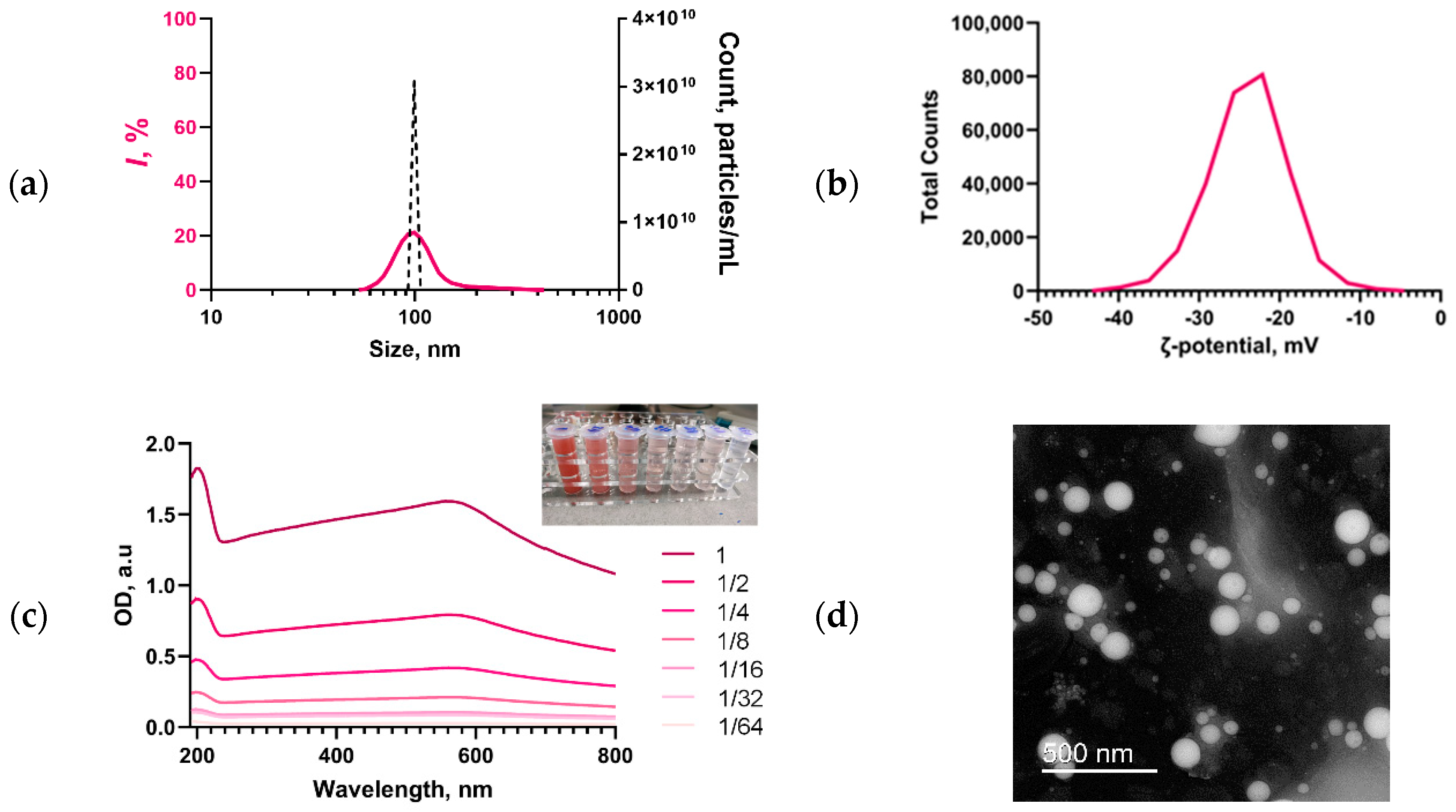
Figure 1. Characteristics of the aqueous colloid of Se SMPs synthesized by laser ablation. Concentration of Se SMPs (black dotted line), size distribution of Se SMPs (solid line) (a). Distribution of ζ-potential of Se SMPs (b). Optical absorption spectra of the aqueous colloid of diluted samples of Se SMPs; the photo in the upper right corner shows photographs of the corresponding samples, the legend indicates the corresponding dilution factors (c). TEM photograph of an individual Se SMPs (d).
Inventions 10 00082 g001
Figure 2. Reconstruction of the surface of the Control sample (a) and PU/Se SMPs composite film (b) obtained using AFM. The concentration of Se SMPs in the polymer is 2.5 wt.%.
Inventions 10 00082 g002
Figure 3. MIM micrographs and 3D reconstructions of phase incursion maps for the obtained material samples. MIM micrographs and 3D reconstructions are presented for PU films without particles and PU/Se SMPs composite materials with particle concentrations of 0.1, 0.25, 1 and 2.5 wt.%. 3D reconstructions of material sections measuring 8.9 μm2 are presented. The XY axes (plane) correspond to the real distance in μm, the Z axis displays the phase difference in nm, the greater the phase difference, the higher the value along the Z axis (red is the maximum value, blue is the minimum). The original data on the spatial distribution of the phase difference in the analyzed sample are shown in the lower corners of each panel.
Inventions 10 00082 g003
Figure 4. Effect of PU/Se SMPs composites on ROS generation in aqueous solutions: hydrogen peroxide (a), hydroxyl radicals (b). *—statistically significant differences compared to the control group (p < 0.05). Data are presented as mean ± standard error of the mean.
Inventions 10 00082 g004
Figure 5. Effect of PU/Se SMPs composite materials on the formation of oxidative damage to biomolecules. Formation of 8-OG in DNA in vitro (a), intensity of luminescence induced by long-lived reactive protein species (b).
Inventions 10 00082 g005
Figure 6. Effect of PU/Se SMPs composite materials on the growth of suspension cultures of E. coli K-12 (left) and S. aureus RN4220 (right) cells. *—statistically significant differences compared to the control group (p < 0.05). Data are presented as mean ± standard error of the mean.
Inventions 10 00082 g006
Figure 7. Results of evaluation of viability of mouse lung fibroblast cultures cultured for 72 h on the surfaces of PU/Se SMPs composite materials: proportion of nonviable cells in the culture, % (a); density of cell cultures, cells/cm2 (b); average area occupied on the plane by one cell, μm2 (c); area of nuclei, μm2 (d). *—statistically significant difference compared to the control (p < 0.05). Data are presented as median, 25–75% and range.
Inventions 10 00082 g007
Figure 8. Representative micrographs of mouse lung fibroblast cell cultures on PU/Se SMPs composite surfaces. Control (a), PU without Se SMPs (b), PU/Se SMPs 0.1 wt.% (c), PU/Se SMPs 0.25 wt.% (d), PU/Se SMPs 1.0 wt.% (e), PU/Se SMPs 2.5 wt.% (f). The images are presented as an overlay of three channels: transmitted light (black and white), Hoechst staining (blue channel) and PI (red channel).
Inventions 10 00082 g008
Figure 9. Representative photograph of radish plants grown in the presence of PU/Se SMPs. From left to right: PU, PU/Se SMPs 0.1, 0.25, 1.0 and 2.5 wt.%.
Inventions 10 00082 g009
Figure 10. Influence of composite materials PU/Se SMPs on leaf area on day 10 of growth (a) and the root weight on day 30 of growth (b) of R. sativus var. sativus. *—statistically significant differences compared to the control group (p < 0.05). Data are presented as mean ± standard error of the mean.
Inventions 10 00082 g010
Disclaimer/Publisher’s Note: The statements, opinions and data contained in all publications are solely those of the individual author(s) and contributor(s) and not of MDPI and/or the editor(s). MDPI and/or the editor(s) disclaim responsibility for any injury to people or property resulting from any ideas, methods, instructions or products referred to in the content.