Review

Tracking Biomarkers for the Health and Welfare of Aquaculture Fish

1
Department of Biology, Faculty of Sciences, University of Porto, 4169-007 Porto, Portugal
2
CIMAR/CIIMAR—Interdisciplinary Centre of Marine and Environmental Research, University of Porto, 4450-208 Matosinhos, Portugal
*
Author to whom correspondence should be addressed.
Fishes 2024, 9(7), 289; https://doi.org/10.3390/fishes9070289
Submission received: 17 June 2024 / Revised: 10 July 2024 / Accepted: 16 July 2024 / Published: 19 July 2024
(This article belongs to the Special Issue Prevention and Control of Aquatic Animal Diseases)

Abstract

:
Aquaculture production has been growing consistently over the last few decades to meet the increasing animal protein demand of the human population. However, increased production and rearing intensities raise the challenges of guaranteeing fish health and welfare, which is essential to avoid losses and ensure product quality. Biomarkers can provide insights into the fish’s nutritional, physiological, and health status, and aid in the evaluation of early nutritional and physiological imbalances, distress conditions, and pathological diagnosis. The discovery and validation of biomarkers rely mostly on the use of information provided by different parameters, including biochemical, metabolic, or immunologic, as well as several omics, from genomics and transcriptomics to proteomics and metabolomics. In this review, a summary of the main biomarkers used in aquaculture is provided along with an overview of the main omics technologies available for further biomarker research. This review also highlights the need to develop non-lethal biomarkers that can easily and quickly be measured to provide a prompt response to producers.
Key Contribution: Summary of the main biomarkers used in different areas of aquaculture research. A view into the future of biomarkers in aquaculture through the application of omics technologies.

1. Introduction

Aquaculture is the fastest-growing food-producing sector in the world, contributing to food security, nutrition, income, and employment for millions of people. According to projections by the Food and Agriculture Organization (FAO) and the Organisation for Economic Co-operation and Development (OECD) [1,2,3], aquaculture is expected to continue to drive growth in global fish production in the next decade, likely accounting for a 32% increase in production from 2020 to 2030. This growth, however, is dependent on overcoming key challenges such as maintaining fish health and public acceptance.
According to the Standing Committee on Agricultural Research (SCAR) reports on welfare priorities and fish diseases, fish health and welfare is an issue of high research priority for European fish farming, as it affects not only the productivity and profitability of the sector but also the public perception and acceptance of aquaculture products [2,4]. These documents identify several research and innovation needs that align with the objectives of the European Union’s Animal Health Law and Green Deal, such as improving disease prevention and control strategies, developing novel vaccines and treatments, and, most of all, developing biomarkers for diagnostics of fish health. Within this topic, there is a need to improve diagnosis tools; namely, through the development of non-invasive methods and identification of biomarkers for early signs of disease; through the implementation of health management approaches that include surveillance methods that allow monitoring fish throughout the production cycle; and through the implementation of prophylactic measures that avoid disease and reduce the use of chemotherapeutics and antibiotics, thus, contributing to the sustainability and resilience of the sector [5,6].
Fish health and well-being evaluation, however, is a complex process that involves various aspects, such as disease diagnosis, prevention and control, biosecurity measures, epidemiological approaches, and biosensors. Assessing health is not a straightforward task since it involves a myriad of (self) physiological processes but also interactions between the host and microbiota [7].
This paper reviews the biomarkers currently used to evaluate fish health, provides a comprehensive overview of the complexity of fish health evaluation in aquaculture, and highlights the emerging methods and technologies that can support and improve the fish health evaluation process. It will also discuss the gaps and challenges that need to be addressed and the opportunities and benefits that can be achieved by advancing the knowledge and innovation in fish health evaluation.

2. Biomarkers

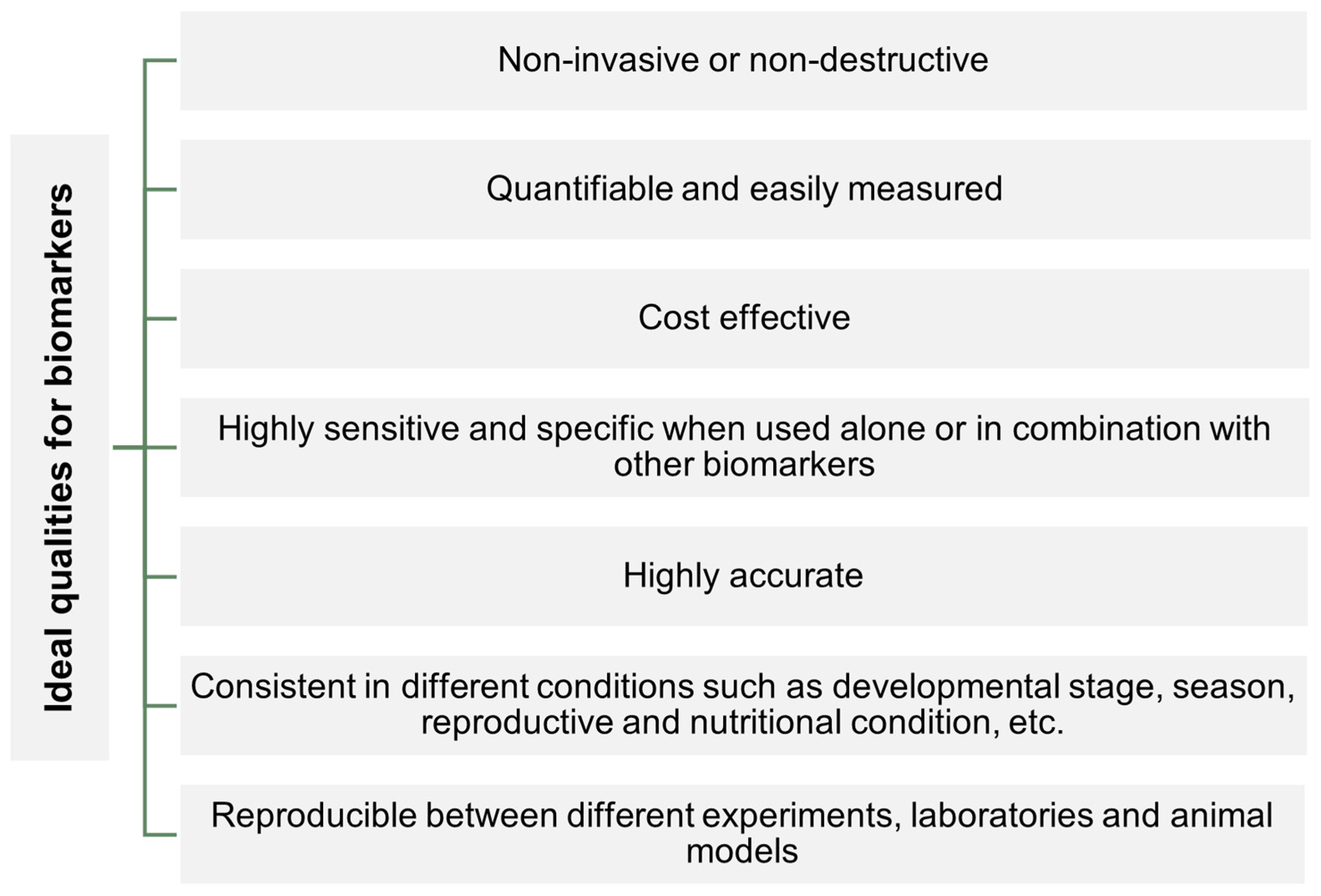
A biomarker is essentially any measurable indicator of a biological state. The World Health Organization [8] has defined biomarkers as “almost any measurement reflecting an interaction between a biological system and an environmental agent, which may be chemical, physical or biological”. Over the years, biomarkers have been successfully applied in different kinds of studies, such as human health or environmental conditions studies [9,10], and have been refined to more accurately respond to research needs. Despite being widely used in different research areas, only in recent years have biomarkers been employed in studies of aquaculture fish health. When applied in fish production, biomarkers allow the obtention of information on physiological and metabolic processes and relate them to fish welfare, health, reproduction, and growth [11]. A biomarker also allows a fast understanding and detection of the effects of changes in production practices on fish stress and health [12], allowing for rapid corrections and the introduction of improvements in production management. Figure 1 presents a summary of the desirable characteristics of a biomarker.
Biomarkers can be evaluated in different types of samples, from organs to tissues and cells. However, with the increase in awareness of animal welfare, the use of non-lethal biomarkers has gained relevance [13]. Non-lethal biomarkers can be obtained from body fluids (such as blood, plasma, mucus, urine, and genital fluids) and feces [14].
The same biomarker may be used in different areas of aquaculture research; therefore, it becomes crucial to understand how biomarkers may interact with each other and how the response of a group of biomarkers can diagnose a particular problem in aquaculture fish.
Figure 1. Ideal qualities of biomarkers. Adapted from [11,15].
Fishes 09 00289 g001

3. Biomarkers Used for Fish Health and Welfare

The following subsections provide a brief description and examples of the biomarkers that are most commonly used to assess the impact of different aquaculture-related challenges to fish health and welfare.

3.1. Metabolism Biomarkers

Metabolism is an essential physiological trait that can influence fish health and welfare. Although more often associated with nutrition, fish metabolism can be affected by other factors such as stress and disease. Thus, metabolism-associated biomarkers are often used in various areas of research.
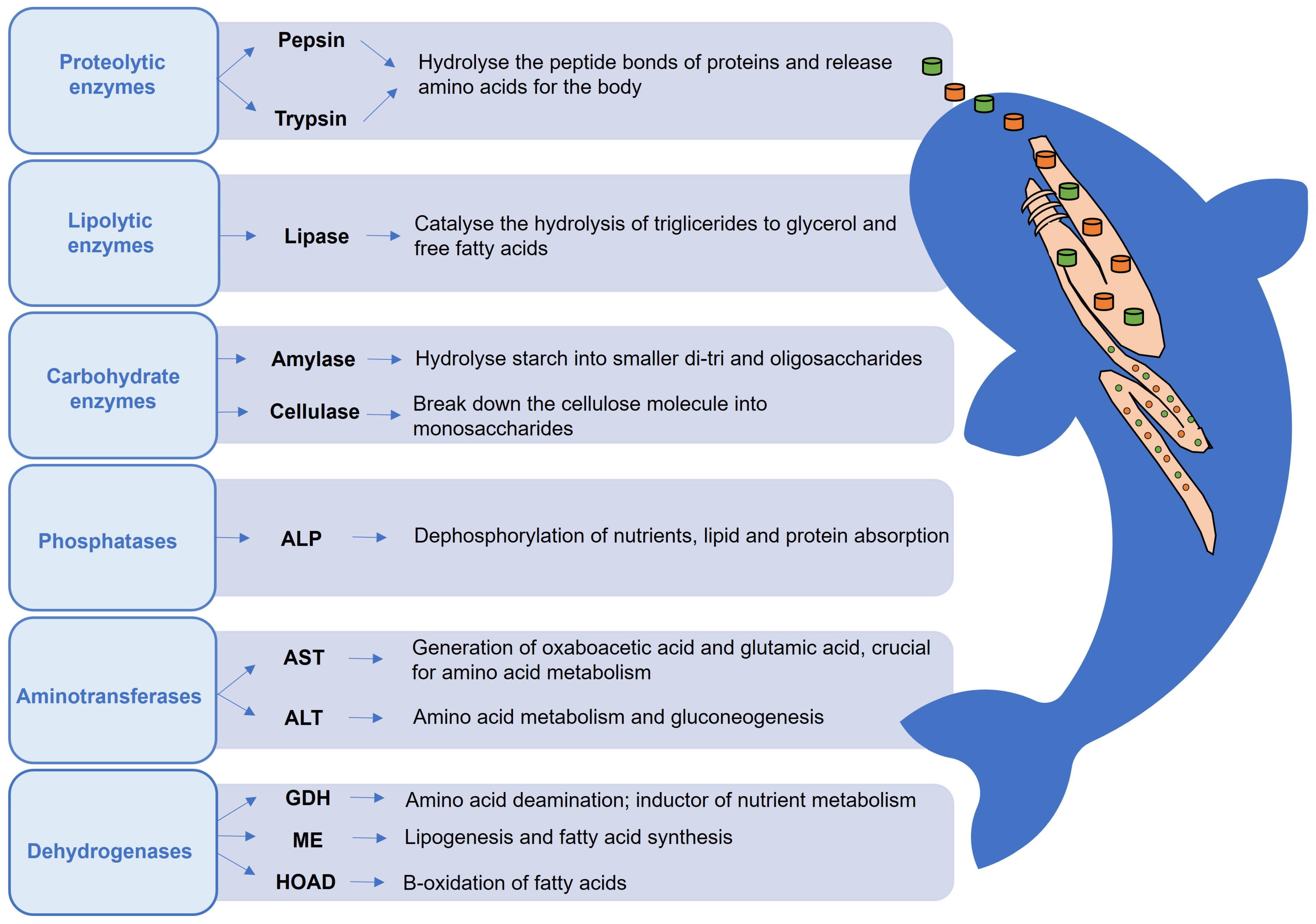
Enzymatic activity is one of the most used biomarkers to assess fish health. The vast variety of enzymes involved in digestive, metabolic, and defense processes makes them a suitable choice to assess physiological effects related to multiple factors, including those related to nutrition. For instance, digestive enzymes are commonly used as biomarkers of dietary nutrient availability, as they act directly on ingested feeds, breaking down nutrients into absorbable molecules [16].
The activity of enzymes can be influenced by several factors, including fish age, species, health status, feeding habits, and external factors such as dietary nutrient composition, water temperature, and fish stocking density [17,18]. Besides being used to evaluate fish digestion and metabolism, digestive and intermediary metabolism-related enzymes can also be valuable biomarkers of diseases and chemical use in aquaculture. A summary of the physiological roles of the digestive and key intermediary metabolism enzymes most used as biomarkers for fish health evaluation is summarized in Figure 2.
Hormones can also be important metabolism biomarkers. For instance, the insulin-like growth factor (Igf1) can be used to evaluate the effects of stress and nutrition on fish growth, as it is involved in physiological processes related to somatic growth and metabolism [19]. Igf1’s production in the liver is triggered by a growth hormone [20], which is regulated by food consumption and nutrient intake [21]. Catecholamines are fast-acting glucoregulatory hormones (such as adrenaline and noradrenaline) that can influence carbohydrate and lipid metabolism [22]. As biomarkers, catecholamines are associated with stress scenarios as they are released immediately after the onset of stress to trigger physiological reactions such as cardiovascular and respiratory functions and the release of energy [20]. Due to their quick release, they can, however, be difficult to quantify without specific equipment [23]
Several metabolites can also be used as biomarkers of metabolism, such as MOPEG (3-methoxy-4-hydroxyphenyl ethylene glycol) sulfate, a metabolite involved in the adaptative response of the hypothalamic-pituitary-interrenal (HPI) axis in the activation of lipolysis, gluconeogenesis, and 3-hydroxy valeric acid, the latter of which a biomarker of biotin deficiency [24].
Figure 2. Overview of the main digestive and intermediary metabolism enzymes in fish commonly used as biomarkers. ALP (Alkaline phosphatase); ALT (Alanine aminotransferase); AST (Aspartate aminotransferase); GDH (Glutamate dehydrogenase); ME (Malic enzyme); HOAD (β-hydroxyacyl-CoA dehydrogenase). It is possible to categorize these enzymes according to the substrate they act on. Adapted from [16,17,18,25,26,27,28,29,30,31].
Fishes 09 00289 g002

3.2. Oxidative Stress Biomarkers

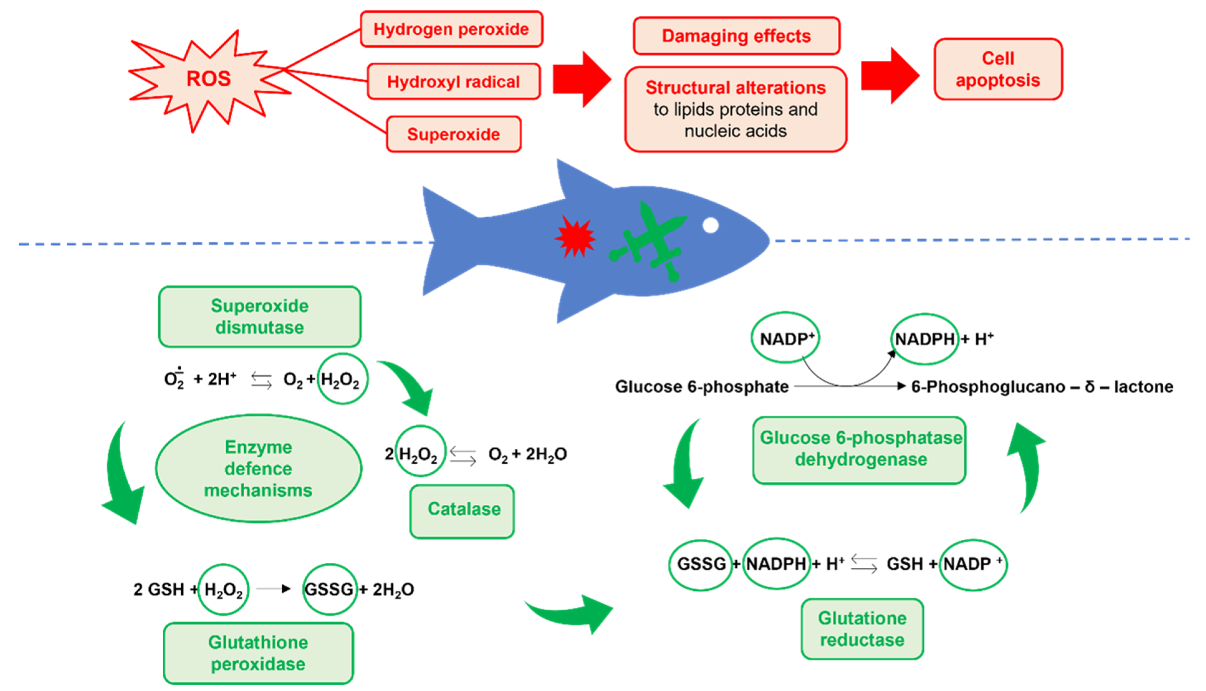
Oxidative stress is due to an imbalance between antioxidants and reactive oxygen species (ROS), caused by either the depletion of the first or excessive accumulation of the latter, or both, which leads to changes in the organism that alter homeostasis [32,33]. Different stress situations (such as environmental conditions and nutrition) can affect ROS balance, and antioxidant enzymes and molecules such as glutathione and vitamin C act as the first line of defense against ROS, making them optimal biomarkers of oxidative stress (Figure 3) [34].
Superoxide dismutase (SOD) is the first enzyme involved in cellular antioxidant protection. By catalyzing the conversion of two superoxide radicals into hydrogen peroxide and molecular oxygen, this enzyme effectively scavenges these radicals. The hydrogen peroxide produced is then reduced to water and oxygen by catalase (CAT) or glutathione peroxidase (GPx). Glutathione (GSH) also has an important role in the antioxidant defense of the cell, as it is involved in the reaction catalyzed by glutathione peroxidase besides acting as an antioxidant by itself [35]. Glutathione reductase (Gr) and glucose 6-phosphatase dehydrogenase (G6PDH) are also involved in oxidative stress defense by regenerating GSH and providing the NADPH essential for GPx activity [36,37,38]. The final products of oxidation damage are also good biomarkers of oxidative stress. For instance, malondialdehyde (MDA) is a final product of lipid peroxidation, namely the ROS degradation of polyunsaturated fatty acids, and is often used as a biomarker of oxidative stress [39].
The concentration of protein thiol groups/protein carbonyl groups (aldehydes and ketones resulting from the interaction of hydroxyl ions with amino acids) and advanced protein oxidation products (protein products containing dityrosine) are important indicators of protein oxidation that can occur for instance during starvation periods. These groups are often associated with the redox state, and their increase also reflects a rise in oxidative stress [40,41]. Additionally, thiobarbituric acid reactive substances (TBARS) are also often used as oxidative stress biomarkers, as they are produced when lipoperoxidation occurs [42]. Since protein carbonylation happens as a result of protein oxidation, and since lipid peroxidation is provoked by free oxygen radicals, these are also two good indicators of oxidative stress [43].
Heat shock proteins (HSPs) are also very well-known stress biomarkers in fish as they are produced in different stressful situations, such as heat exposure, handling, toxic chemicals, or pathogen infections [12,44,45]. HSPs help to stabilize and refold proteins, thus, reducing damage inflicted by protein-denaturing stressors [20]. The response of HSPs, however, can be highly variable as it is influenced by factors such as the intensity and duration of the stressor, as well as the organism’s adaptation to prior stressors [46]. Ethoxy resofurin O-deethylase (EROD) is another biomarker mainly applied to chemical toxicity in fish as its increase usually signals high detoxification activity [47].
Figure 3. Overview of oxidative stress defense mechanisms in fish. Reactive oxygen species (ROS) are a natural product of oxygen metabolism which can have a deleterious effect on the organism, particularly when present in excess. Therefore, most organisms have mechanisms to help break down ROS. In fish, similarly to other animals, various enzymes have a crucial role in the defense against ROS. Adapted from [34,36,37,38].
Fishes 09 00289 g003

3.3. Immunological Biomarkers

In vertebrates, including fish, the immune system is usually separated into two types of immunological response, the innate immune response and the adaptative immune response. Innate immunity is seen as the front and first quick line of defense against threats, relying on physical barriers and unspecific humoral and cellular responses [48]. While the innate immune response is unspecific, acting against different kinds of threats, the adaptative immune response is targeted against a specific antigen, resulting usually in a stronger, faster, and more specific immune response [48,49].
Innate immune enzymes are commonly measured in the plasma and include lysozymes, esterases, phosphatases, proteases, and anti-proteases [50]. Lysozyme is known to have an important role in the lysis of bacteria and is present in the mucus and blood [51,52]. Proteases are known to have various roles, from directly attacking pathogens to wound healing and tissue reorganization, functions shared with the enzyme alkaline phosphatase [50]. Peroxidase is involved in phagocytic activity and has an important function in controlling infections [53,54].
The complement system is composed of several components of proteic and non-proteic nature (C3, C4, C1, and others), and is an important component of both innate and adaptative immunity, with several roles against pathogenic infection, such as pathogen lysis and induction of proinflammatory cytokines [48,50]. These cytokines are responsible for the activation and release of acute-phase proteins, which are essential for regulating inflammatory reactions and restoring organism homeostasis [55,56]. Acute-phase proteins can also be used as biomarkers of fish health, as their level in the serum is maintained for long after the first stimulus of infection [51]. For instance, C-reactive protein (CRP) is an acute phase protein with immune functions contributing to the opsonization of bacteria, fungi, and parasites thus having a high potential for disease diagnosis [51].
The leukocyte respiratory burst activity is also often used as an indicator of innate immunity as it is associated with the release of cytokines and inflammatory response in fish [57,58].

3.4. Biochemical Biomarkers

Biochemical biomarkers are commonly measured in the plasma, making them potential non-lethal biomarkers that are fast and accessible to collect in aquaculture facilities without the need to sacrifice animals. Biochemical biomarkers have long been used in aquaculture studies, and when combined with other biomarkers can help make an accurate evaluation of the fish’s physiological status.
Parameters such as plasmatic cortisol, glucose, and lactate are widely used in fish as biomarkers of stressful conditions such as hypoxia, chasing, and confinement [59]. Cortisol is one of the first hormones released upon fish exposure to an acute stress scenario and affects various physiological traits of the fish with the ultimate goal of relocating the energy driven to growth and other functions to immediate survival [20]. When using cortisol as a biomarker, it is important to highlight that its levels vary often according to species, place, and time sampled [60]. Plasma glucose levels indicate changes in the metabolism and energy demand of the organism [34], and lactate, which is produced during anaerobic metabolism, increases when fish are subjected to exhaustive exercise and hypoxia [20]. Because both glucose and lactate levels are influenced by general metabolic processes unrelated to stress responses, interpreting baseline results can be challenging [23]
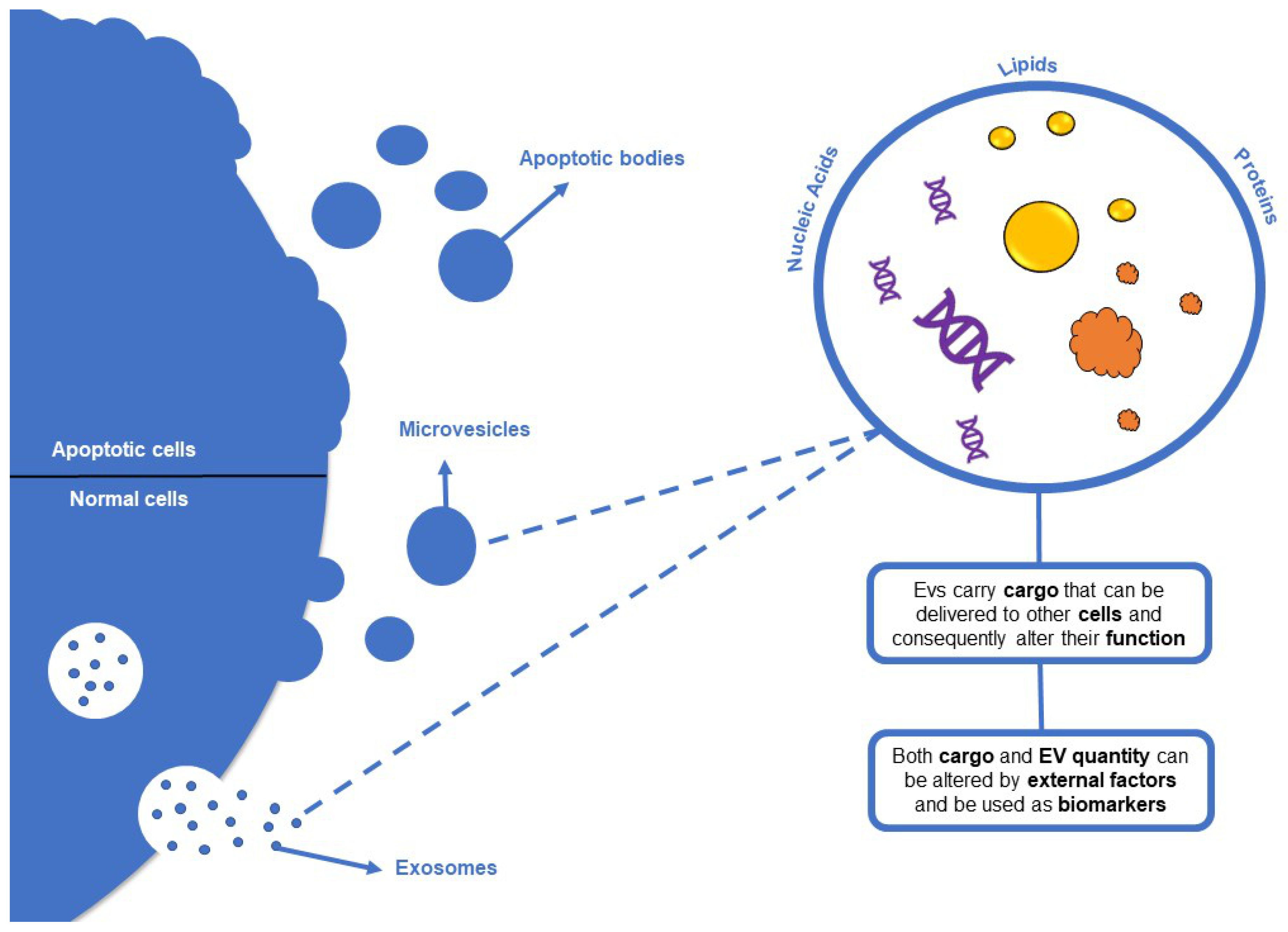
Plasma extracellular vesicles (Evs) are also promising biomarkers (Figure 4). EVs are small structures that are released from parent cells to engage in cell communication by transferring cargo (proteins, enzymes, DNA, lipids, RNA, and micro-RNA) [61,62]. The Evs function is mainly related to cell-to-cell communication as they pass on different cargo from one cell to the other and this can have outcomes on cell function and overall physiology, for example by stimulating the organism’s immunity (Doyle and Wang, 2019). These vesicles and their cargo have been successfully used as biomarkers of health and treatments in mammals such as mice and humans [62]. In fish, Evs have been shown to have biomarker potential since their cargo and their number can be affected by external factors [63,64].
Figure 4. Overview of extracellular vesicles (EV). Extracellular vesicles can be produced by various types of normal cells and are commonly divided into three types. Exosomes are smaller than microvesicles but both vesicles share similar cargo, which can consist of proteins, lipids, and nucleic acids. Apoptotic bodies, on the other hand, are Evs that are produced by dying cells and typically contain intact organelles, chromatin, and glycosylated proteins. Adapted from [63,64].
Fishes 09 00289 g004

3.5. Mucosal and Mucin-Associated Biomarkers

Recently, attention has been turned to the mucosa and mucus in fish, which can be collected from the skin and gills, as well as in the intestine. Most importantly, since they are present in skin and gills mucus, mucins are easily collected without animal sacrifice, and therefore their exploitation as potential non-lethal biomarkers in different areas, from disease to nutrition, is encouraged. Studies in the mucosal tissues of the fish although more recent show promising results in different areas from disease to nutrition [65,66,67].
Mucins have been getting attention for their biomarker potential due to the importance of mucosal tissue in the defense of the organism. Mucins are O-glycosylated glycoproteins present in the epithelia and their expression can be disrupted by injuries and other challenges [68]. Mucins are the main components of the mucus matrix and are present in the skin, gills, and mucosal lining of the alimentary tract, and are important for the protection of such tissues against pathogens and other threats [69]. Mucin dysregulation has been also shown to have an impact on intestinal function and this makes them potential biomarkers for dietary changes [68]. Mucins are also promising biomarkers of handling, crowding stress, and disease in aquaculture fish.
Goblet cells can be found in the skin and other mucosal surfaces of fish and are responsible for mucus production, which has an important role in immunological defense [20]. Furthermore, they are associated with the production of mucins which, as mentioned, are also important biomarkers of fish health [70].
Rodlet cells are only present in fish and they have been described in a few teleost species [50]. These cells can be found in the epithelial tissue, being particularly important in the skin, gills, and intestine, where they are involved in mucus production, being part of the defense mechanisms against pathogens, and their presence is generally altered by stressful conditions [44,71,72].

4. Application of Biomarkers in Aquaculture Studies

This section provides examples of how the biomarkers are used in different areas of study in aquaculture.

4.1. Nutrition

Adequate nutrition and feeding are fundamental for maintaining fish health and well-being, improving performance and disease resistance, and reducing susceptibility to environmental impacts [14,73]. With aquaculture production increasing, studies on how dietary nutrients and ingredients affect such parameters are becoming increasingly important.
As fish meal and fish oil become less available and unsustainable, their use is becoming more limited, and this led to the increased use of alternative ingredients in carnivorous fish aquafeeds [74]. The ingredient and nutritional composition of novel diets can be highly variable, and even though often do not impact growth, their effect on fish health, stress, and disease susceptibility is gaining more attention. The use of functional ingredients in aquafeeds has been explored as a strategy to improve novel aquafeed utilization and improve fish performance, health, and immune and oxidative status [73]. Functional ingredients are characterized by having added value beyond nutritional requirements and can be micronutrients (such as some minerals and vitamins) [75], macronutrients (such as some amino acids) [76,77], antioxidants and immunostimulants (such as algae and plant extracts) [73,78], or supplements used to alleviate the effects of antinutritional factors (ANFs) commonly found in plant feedstuffs [73]. However, novel feeds can have cumulative negative effects and result in less-than-optimal health [79]. Therefore, with the recent increase in new dietary formulations, researchers have turned to testing whether different diets can influence fish health and welfare, using and developing different biomarkers to better understand the connection between feeds, nutrients, and physiological processes.
For instance, Abdel-Tawwab, et al. [80] studied the effect of a diet with a multi-stimulant blend supplement (which included probiotics, sodium butyrate, various digestive enzymes, phytase, and zinc methionine) on common carp (Cyprinus carpio). The supplement increased the activities of intestinal amylase, lipase, and protease, suggesting that digestion efficiency was improved. Further, oxidative stress biomarkers showed that hepatic MDA values significantly decreased and SOD, CAT, and GPx activities were increased, indicating an improvement in the oxidative status of fish fed the supplemented diet [80]. Similarly, in Nile tilapia (Oreochromis niloticus) fed diets supplemented with sodium butyrate and exposed to heat stress, higher SOD, CAT, and GPx activity was observed along with a decrease in MDA and cortisol levels, and an upregulation of the gene expression of liver HSP70 [81]. Additionally, a positive effect on the immune responses of heat-stressed tilapia was also observed with the supplemented diet, as shown by the increased number of goblet cells, increased lysozyme, and phagocytic activities [81].
Goblet cell levels and lysozyme activity were also used as biomarkers in the assessment of the influence of dietary incorporation of vitamin C in Nile tilapia [82]. In this study, higher lysozyme activity and a higher number of goblet cells in fish fed the vitamin C-supplemented diets indicate an improvement in immune defense. Further, an increased serum SOD activity and reduced glutathione (GSH) levels as well as increased liver CAT and SOD activity and GSH levels and decreased serum MDA levels in the supplemented diets indicate the antioxidant potential of vitamin C [82].
Black soldier fly (Hermetia illucens; HM) is an example of a novel feed ingredient that is being much studied as fish meal and fish oil replacement in the diets of various fish species [33,83,84,85,86,87,88]. For instance, to evaluate the dietary inclusion level and the metabolic effects of HM in diets for meagre (Argyrosomus regius), markers such as plasma glucose, the hepatic activity of ALT, AST, GDH, glucose 6-phosphate dehydrogenase (G6PDH), β-hydroxy acyl-CoA dehydrogenase (HOAD), and malic enzyme, as well as the activity of intestine alkaline protease, trypsin, α-amylase, and lipase were used [84]. Of these, alkaline protease activity increased and trypsin activity decreased with higher HM inclusion indicating that the replacement of fish meal with HM should not exceed 17% to avoid adverse effects on digestive enzyme activity. Using similar markers, [89] showed that HM could replace up to 19.5% of fish meal in diets for European seabass (Dicentrarchus labrax), while not affecting digestive enzyme activity.
Biomarkers such as SOD, CAT GPx, MDA values, EROD, GST, GR, and total glutathione were also used to understand the effects of dietary inclusion of HM in the oxidative stress of rainbow trout (Oncorhynchus mykiss) [88]. In this study, although the dietary inclusion of insect meal did not affect most of the oxidative stress biomarkers analyzed, higher levels of EROD in the liver and GST in the kidney were observed with high dietary HM inclusion (40%), leading the authors to conclude that the dietary HM inclusion should be lower than 20%. On the other hand, using similar biomarkers in Siberian sturgeon (Acipenser baerii) juveniles fed diets with increasing levels of HM, a disturbance of several oxidative stress indicators was observed both in the kidney and the liver, with GPx being lower and SOD and GR being higher in fish fed the highest insect meal inclusion (37.5%) [33]. GST and EROD were also increased by said diet but only in the kidney. All these studies are examples of the variability of biomarker responses in different fish species fed similar ingredients, highlighting the relevance of using more than one biomarker to more precisely evaluate the searched response.
Carob seed germ meal (CSGM) is an example of an alternative ingredient evaluated for inclusion in meagre diets within the circular economy approach. In a study, digestive enzymes (alkaline protease, trypsin, lipase, and α-amylase), as well as amino acid catabolism enzymes (ALT, AST, and GDH) were evaluated, and decreased activity of trypsin, total alkaline protease, and lipase was observed, indicating that high levels of CSGM negatively affected the digestive enzyme activity of the fish. [90]. Moreover, the humoral and immune responses of meagre in response to CSGM were also evaluated by using plasma and intestine biomarkers. In the plasma hemolytic activity (ACH50) increased in fish fed all levels of CSGM while lysozyme increased only in fish fed the lowest level of CGSM and esterase decreased in the highest CGSM. Meanwhile, in the intestine, only antiprotease and alkaline phosphatase activity increased in fish fed with CSGM [91].
Mucins are mostly present in the posterior intestine and are also good biomarkers of the effects of alternative ingredients. For instance, in gilthead sea bream it was shown that I-Muc mucin was less expressed when fish were fed a vegetable oil-based diet as opposed to fish oil [68]. On the other hand, in Nile tilapia, mucin-like protein (muc) expression was up-regulated by limonene supplementation in diets [92] and in Atlantic salmon, the expression of muc2 was up-regulated in fish fed a fermented soybean meal diet [93].
Biomarkers can be also used to uncover the effects of fasting or feeding strategies in fish. For example, in gilthead sea bream fasted for ten days biomarkers of malnutrition included increased circulating levels of five sub-products of L-carnitine and catecholamines, increased concentration of urea cycle-related compounds (such as citrulline and arginine), and increased MOPEG sulfate and reduced levels of glutathione [24]. Biomarkers enzymes involved in antioxidant processes such as CAT, SOD, GPx, GR, GST, and G6PDH, as well as the concentration of MDA and other oxidative stress products, including the concentration of protein thiol groups, protein carbonyl groups, and advanced oxidation protein products were also used to evaluate feeding regimes (different periods of starvation and refeeding) in the intestinal oxidative status of stellate sturgeon (Acipenser stellatus) [40].

4.2. Stress

In captivity, particularly under intensive production, fish are often exposed to stressful situations such as crowding, handling, transportation, poor water quality, hypoxia, and others [94], which can lead to disease outbreaks [73]. Under such stressful circumstances, fish responses can vary depending on species, rearing conditions, type, and level of stress [94]. Due to the highly variable responses, it is important to find reliable methods to measure stress levels; this is an instance in which biomarkers can be extremely useful. Stress responses are generally regulated by the neural, endocrine, and immune systems [95], in which stress biomarkers can be searched.
As an example, when Artic charr (Salvelinus alpinus) fish were subjected to chronic heat exposure, the expression of HSP increased, dropping as soon as the heat exposure was over, showing the potential of these proteins as biomarkers of thermic stress in this species [96]. Also, in sea bass (Dicentrarchus labrax), both the gene expression and concentration of HSP (HSP70) were increased in a variety of tissues in response to handling and abrupt temperature changes [44]. In this study, other stress biomarkers were used, such as MAD, nitrotyrosine, 4-hydroxy-2-nonenal, and the presence of rodlet cells in the intestine and the kidney.
Mucins can also be useful biomarkers of stress on fish. For instance, in Atlantic salmon, subjected to short-term (handling) and prolonged exposure (high stocking densities) to stress, mucin transcription varied both with stress conditions and tissue analyzed [69]. During short-term stress, mucin transcription was up-regulated in the gills but down-regulated in the intestine and skin, probably as a result of imbalances in the metabolic costs due to stress [69]. On the other hand, after prolonged stress, mucin transcription was up-regulated in the skin but not in other tissues analyzed. This indicates that mucins may be useful as tissue-specific biomarkers for the evaluation of the effects of stressful situations.
Biomarkers that evaluate stress effects on fish physiology at a molecular level can also be very important in improving production. To study how handling stress and water temperature affected yellow perch (Perca flavescens), [95] evaluated serum and hepatic Igf1 protein level and mRNA expression, the protein level and expression of heat shock protein (hps70), and the expression of several oxidative stress biomarkers (glutathione-peroxidase, gpx3; superoxide dismutase, sod1; and glutathione reductase, gr). The study showed that handling stress and salt treatment heightened these stress biomarkers at a water temperature of 26 °C when compared to 2000 °C and 14 °C conditions.
Serum Evs and specific components of their cargo (peptidyl arginine deiminases, heat-shock protein, complement component C3, myosin heavy chain, and glyceraldehyde-3-phosphatase dehydrogenase) were used as biomarkers of rearing temperature effects on Atlantic cod (Gadus morhua) [61]. In this study, it was found that the number of serum Evs was significantly lower in fish reared at 9 °C when compared to 4 °C, while deiminated protein targets (such as complement component C3) were higher in Evs of fish raised at 4 °C [61].
The use of molecular biomarkers can also be useful to further validate and understand the effect of certain conditions. For instance, in a study aiming to determine suitable fish densities in late larval Delta smelt (Hypomesus transpacificus) [97], whole-body cortisol level was measured as a biomarker associated with transcriptomic biomarkers related to cortisol production, such as glutathione s-transferase, mineralocorticoid receptor 1, glucocorticoid receptor 2, pro-opiomelanocortin, 11-beta-hydroxysteroid-dehydrogenase type 1 and 2, beta-actin, and glyceraldehyde-3-phosphate dehydrogenase. The results of the study showed a direct relationship between cortisol levels and gene expression of mineralocorticoid receptor 1, glucocorticoid receptor 2, and 11-beta-hydroxysteroid-dehydrogenase -2, which were lower in fish reared at high densities.

4.3. Infectious Diseases

Biomarkers can help with the early detection of immune status depression induced by stressors that make fish more susceptible to pathogens [98], and detect the onset of pathogenesis before any symptoms or lesions occur [99], helping to early combat disease, avoid its spread in the facilities, and avoid antibiotic use.
Although blood is the most studied biological fluid for biomarkers of disease in humans and farm animals, only a few studies focused solely on its use to uncover potential biomarkers of disease in fish. For instance, in Atlantic salmon, the serum proteome profile was used for finding biomarkers triggered by pancreatic disease caused by alphavirus subtype 3 (SAV3) [100]. In this study, proteins such as transferrin, albumin, antithrombin, apdiprotein, hemopexin, and complement components were highlighted as having biomarker potential that could be further validated by complementary approaches. In another study, the enzyme enolase was also validated as a non-lethal biomarker for white muscle myopathy triggered by pancreatic disease [101]. Other biomarkers for myopathy are creatine kinase (CK) and lactate dehydrogenase (LDH), whose serum levels were shown to correlate well with heart and skeletal muscle inflammation and cardiomyopathy [102].
Mucins can also be important biomarkers of infection in fish. For instance, gill transcriptome analysis in Atlantic salmon showed an upregulation of the expression of Muc5 upon infection by salmon gill poxvirus (SGPV) [103]. In this study, an alteration of IL-22, a proinflammatory cytokine that can be induced upon epithelial damage, was also detected showing the potential of cytokines as biomarkers in the early detection of disease. In common carp, CRP was used as a biomarker of cyprinid herpesvirus 3 infections, as its levels increased significantly in the serum of infected fish [104]. In Atlantic salmon, lysozyme activity and its mRNA expression were also used as biomarkers of infection with Aeromonas salmonicida, as both increased upon infection [105]. In this study, higher activity and expression of ALP (which is thought to act as an antibacterial agent), the antioxidant enzymes SOD, GPx, and CAT, and the aminotransferases ALT and AST were also observed, showing their potential as biomarkers of pathogen infection, as they respond to the impairment of physiological functions caused by pathogens, namely when there is damage to tissues such as the liver or the heart [105].
Extracellular vesicles (Evs) can also be used as biomarkers of infection. Upon a pathological event, EVs can not only adapt their protein content for the immune response but can also capture and present components of intracellular pathogens to the immune system [106]. For instance, Evs isolated from Atlantic salmon infected with Piscirickettsia salmonis contained 35 unique proteins, most of them involved in immune system response and antigen presentation, showing that EV content was altered in infected fish when compared to healthy individuals [106]. Thus, analysis of EV content could be a helpful biomarker in future diagnosis of this pathogen.
Exosome miRNA analysis also has biomarker potential to detect infection during the early phases of disease. For instance, in the Chinese tongue sole (Cynoglossus semilaeuis), a comparison of the microRNA (miRNA) profiles of epidermal mucous exosomes produced in the mucosa of fish between healthy fish and fish infected with Vibrio harveyi showed that dre-miR-184 and dre-miR-205-5p were less expressed while dre-miR-100-5p was more expressed 7 days after infection [107].
Biomarkers can also be important to understand differences in the immune responses between survivors and victims of disease. For instance, in crucian carps (Carassius auratus) infected with Edwardsiella tarda, survivors presented changes in key metabolites and metabolic pathways that might be related to response to the bacterial infection, such as lower levels of D-mannose and higher levels of palmitic acid, lower fructose and mannose metabolism, and higher unsaturated fatty acid biosynthesis [108]. Unsaturated fatty acids are possible modulators of immune functions, whilst D-mannose could be important for bacterial endocytosis [108] therefore having biomarker potential.
A proper understanding of the immunoregulatory function of the intestinal epithelium could also provide relevant information for the development and improvement of strategies for the prevention and treatment of pathologies and to establish biomarkers of pathogenicity [74]. For instance, in grass carp (Ctenopharyngodon della), besides inducing histological alterations, infection with Aeromonas hydrophila provoked intestinal inflammation as confirmed by the increased expression of pro-inflammatory cytokines (IL-1β, IL-8, and TNF-α) and myeloperoxidase (MPO) activity, which allows the evaluation of neutrophil activation in response to infection [109].

4.4. Chemicals, Antibiotics and Vaccines

To control disease and its spread in aquaculture, most fish producers commonly apply chemotherapeutics or antibiotics to the animals. Despite the benefits and quick defense against pathogens that these chemicals can offer [34], the use of chemotherapeutics or antibiotics might cause long-term impacts on fish growth, health, and tissue contamination that are sometimes not easily detected [12]. In recent years, many researchers have focused on finding, validating, and applying biomarkers that provide an understanding of the physiological changes that might occur due to the application of antibiotics, vaccines, anesthetics, disinfectants, and other chemicals.
Dichlorvos, for example, is a chemical used in aquaculture that is used to act against ectoparasites. Although it has been banned in most European countries, it remains in use in others [110]. By using biomarkers such as cholinesterases (ChE) activity, lipid peroxidation, RNA/DNA ratio, glutathione S-transferases activity, and heat shock proteins, it was observed that treatment of gilthead sea bream fingerlings with this compound caused oxidative stress and, as lipid peroxidation increased, ChE activity was inhibited and RNA/DNA ratio decreased [12].
To study the effects caused by the antibiotic sulfamethazine (currently banned in Europe [111]) in mrigal (Cirrhinus mrigala), oxidative stress biomarkers were used, namely SOD, CAT, lipid peroxides, and GPx, activities together with hematological parameters such as hemoglobin content, glucose levels, hematocrit, and red blood cells (RBC) and white blood cells (WBC) number [34]. In this study, it was observed that the hematological, biochemical, and antioxidant status of the fish was affected depending on the dose and duration of exposure to the antibiotic. In another study, focusing on the use of sulfamethazine in Nile tilapia, it was also observed that this antibiotic affected the antioxidant status of the fish, as it increased hepatic CAT and GST activity, but did not affect hematological parameters [112].
The effects of another commonly used antibiotic, oxytetracycline, were also studied in Nile tilapia using serum biomarkers such as glucose (as a marker of stress), ALT and AST (as markers of liver function), creatine (as a marker of kidney function) and c-reactive protein (as a marker of innate immune response) [113]. In this study, an increase in all biomarkers was observed, indicating a negative effect of the use of this antibiotic in Nile tilapia.
Hematological parameters are usually considered good indicators of fish health status, as they often reflect metabolic changes in tissues and organs, and usually enter rapidly in contact with compounds with toxic effects, and therefore they may be a good source of biomarkers for chemicals and antibiotics [114].
In a recent study in Nile tilapia, several hematological, biochemical, and immunological biomarkers were used to evaluate the effects of antimicrobial treatment with peptide LL-37 against Streptococcus agalactiae [115]. The hematological parameters measured were RBC count and hemoglobin concentration, while biochemical parameters measured were protein and albumin concentration in the blood, serum ALT and AST activity, lysozyme concentration (SLC), complement activity (ACH50), and leukocyte respiratory burst activity. In this study no significant differences between the control and fish medicated with LL-37 were observed, suggesting that this antimicrobial agent has few negative effects in Nile tilapia.
The effects of four anesthetics (tricaine methane sulfonate, clove oil, 2-phenoxyethanol, and propiscin) in rainbow trout were evaluated using several blood biochemical and oxidative stress parameters as biomarkers [43]. The parameters utilized were plasma glucose concentration, ALT, AST, lactate dehydrogenase, and creatine kinase activity, as well as ammonia and lactate concentration, carbonyl proteins (CP), peroxidation of lipids (LPO), SOD, GPx, and GR activity. The results showed that plasma glucose, lactate, and ammonia levels and ALT and AST activities increased in some of the treatments when compared to the control group. The oxidative stress biomarkers also showed differences, with LPO and CP being higher in some treatments and the activities of SOD, GPx, and GR being lower between anesthetic treatments.
Disinfectants are usually used in aquaculture as prophylactic and therapeutic compounds that help treat and prevent diseases, but these chemicals may affect the structure and function of cell membranes as they can lead to lipid and protein oxidation [116]. To study the effects of different disinfectants in the heart of rainbow trout, oxidative stress biomarkers were applied by [116]. In this study, it was observed that CAT activity and TBARS increased whilst SOD activity decreased with the use of formalin and chlorine dioxide [116].
To evaluate the antioxidant defense during vaccination against furunculosis in rainbow trout, TBARS, together with other stress biomarkers, was used in one study [117]. In this study, TBARS levels increased in the gill and liver of vaccinated fish and decreased in the brain tissue. Further, muscle and liver SOD activity was higher in vaccinated fish while CAT, GR, and GPx activities were reduced in the muscle and gill and increased in the liver.
A summary of all the biomarkers explored in the previous sections can be found in the following table (Table 1).
Table 1. Summary table of the main biomarkers used for assessing and protecting fish health in aquaculture.
BiomarkerImpact StudiedOrganism TissueSpeciesNon-Lethal PotentialReferences
3-hydroxysovaleric acidNutritionSerumSparus aurataYes[24]
4- hydroxy-2-nonenalStressGill, Brain, Liver, SpleenDicentrarchus labraxUnknown[44]
Alanine transaminase (ALT)Nutrition, Chemical,
Disease
Serum, Mucus, Skin, LiverOreochromis niloticus, Oncorhynchus mykiss, Salmo salar, Argyrosomus regiusYes[43,83,90,105,113,115]
Alkaline phosphataseNutrition, DiseaseSerum, Mucus, Skin, IntestineSalmo salar, Argyrosomus regiusYes[91,105]
Alkaline proteaseNutritionIntestineArgyrosomus regiusUnknown[84,90]
AmylaseNutritionIntestineCyprinus carpio, Argyrosomus regiusUnknown[80,84,90]
Aspartate transaminase (AST)Nutrition, Chemical,
Disease
Serum, Mucus, Skin, LiverOreochromis niloticus, Oncorhynchus mykiss, Salmo salar, Argyrosomus regiusYes[43,83,90,105,113,115]
Carbonyl proteinsNutrition,
Chemical
Intestine, Gill, Liver, Brain, Muscle, HeartAcipenser stellatus, Oncorhynchus mykissYes[40,43,116]
Catalase (CAT)Nutrition,
Chemical
Liver, Kidney, Intestine, Serum, Mucus, Skin, HeartCyprinus carpio, Oncorhynchus mykiss, Acipenser baerii, Acipenser stellatus, Oreochromis niloticus, Salmo salar, Cirrhinus mrigalaYes[33,34,40,80,81,82,88,105,112,116]
CatecholaminesNutritionSerumSparus aurataYes[24]
Cholinesterases (ChE)ChemicalsBrain, MuscleSparus aurataUnknown[12]
Complement system activity (ACH50)ChemicalSerum, IntestineOreochromis niloticus, Argyrosomus regiusYes[91,115]
CortisolNutrition, StressSerumOreochromis niloticus, Hypomesus transpacificusYes[81,97]
C-reactive protein (CRP)ChemicalSerumOreochromis niloticusYes[113]
Creatine kinaseDisease,
Chemical
SerumSalmo salar, Oncorhynchus mykissYes[43,102]
Cytokine IL-22DiseaseGillSalmo salarUnknown[103]
EnolaseDiseaseSerumSalmo salarYes[101]
Ethoxyresorufin O-deethylase (EROD)NutritionLiver, KidneyOncorhynchus mykissUnknown[33,88]
Extracellular vesicles (EVs) (+ cargo)Stress, DiseaseSerum, Skin (Epidermal mucus)Gadus morhua, Salmo salar, Cynoglossus semilaeuisYes[61,106,107]
GlucoseNutrition, Stress,
Chemical
SerumOreochromis niloticus, Cirrhinus mrigala, Oncorhynchus mykiss, Argyrosomus regiusYes[34,43,81,83,113]
Glucose 6-phosphate dehydrogenase (G6PDH)NutritionIntestine, LiverAcipenser stellatus, Argyrosomus regiusUnknown[40,83]
Glutamate dehydrogenase (GDH)NutritionLiverArgyrosomus regiusUnknown[83,90]
GlutathioneNutritionSerum, LiverOreochromis niloticusYes[82]
Glutathione peroxidase (GPx)Nutrition, Stress,
Chemical
Liver, Kidney, Intestine,
Serum, Brain, Muscle, Gill, Heart
Cyprinus carpio, Oncorhynchus mykiss, Acipenser baerii, Acipenser stellatus, Oreochromis niloticus, Perca flavescens, Cirrhinus mrigalaYes[33,34,40,43,80,81,88,95,116]
Glutathione reductase (GR)Nutrition, StressLiver, Kidney, Intestine, Brain, Gill, Muscle, HeartOncorhynchus mykiss, Acipenser baerii, Acipenser stellatus, Perca flavescensYes[33,40,43,88,95,116]
Glutathione S-transferase (GST)Nutrition, ChemicalLiver, Kidney, Intestine, GillOncorhynchus mykiss, Acipenser baerii, Acipenser stellatus, Sparus aurata, Oreochromis niloticusYes[12,33,40,88,112]
Goblet cellsNutritionIntestineOreochromis niloticusUnknown[81,82]
HematocritChemicalPlasmaOreochromis niloticus, Cirrhinus mrigalaYes[34,112]
Hemoglobin concentrationNutrition, ChemicalPlasmaOreochromis niloticus, Cirrhinus mrigalaYes[34,81,112,115]
Heat shock protein (Hsp)Nutrition, Stress, ChemicalLiver, Gills, Kidney, Serum, MuscleOreochromis niloticus, Dicentrarchus labrax, Salvelinus alpinus, Perca flavescens, Sparus aurataYes[12,44,81,95,96]
Insulin-like growth factor (Igf1)StressSerum, LiverPerca flavescensYes[95]
Intestinal pro-inflammatory cytokines (IL-1β, IL-8 and TNF-α)DiseaseIntestineCtenopharyngodon idellaUnknown[109]
LactateChemicalSerumOncorhynchus mykissYes[43]
Lactate dehydrogenase (LDH)Disease,
Chemical
SerumSalmo salar, Oncorhynchus mykissYes[43,102]
Leukocyte respiratory burst activityChemicalSerumOreochromis niloticusYes[115]
LipaseNutritionIntestineCyprinus carpio, Argyrosomus regiusUnknown[80,84,90]
LysozymeNutrition, Disease,
Chemical
Serum, Mucus, Skin, IntestineOreochromis niloticus, Salmo salar, Argyrosomus regiusYes[81,82,91,105,115]
Malondialdehyde (MDA)NutritionLiver, Kidney, Intestine, SerumOncorhynchus mykiss, Acipenser stellatus, Oreochromis niloticus, Dicentrarchus labraxYes[33,40,44,81,82,88]
MucinsNutrition, Stress, DiseaseIntestine, Gill, SkinSparus aurata, Salmo salar, Oreochromis niloticusYes[68,69,92,103]
Myeloperoxidase (MPO)DiseaseIntestineCtenopharyngodon idellaUnknown[109]
NitrotyrosineStressLiver, Kidney, MuscleDicentrarchus labraxUnknown[44]
PeroxidaseDisease, NutritionPlasma, Mucus, SkinSalmo salar, Argyrosomus regiusYes[91,105]
Peroxidation of lipids (LOP)ChemicalPlasma, Liver, Intestine, Gill, Brain, MuscleCirrhinus mrigala, Oncorhynchus mykissYes[34,43]
ProteaseNutritionIntestineCyprinus carpioUnknown[80]
Protein thiol groupsNutritionIntestineAcipenser stellatusUnknown[40]
Red blood cells (RBCs)Nutrition, ChemicalPlasmaOreochromis niloticus, Cirrhinus mrigala, Argyrosomus regiusYes[34,81,91,112,115]
RNA/DNA ratioChemicalMuscleSparus aurataUnknown[12]
Rodlet cellsStressIntestine, Renal tubes, GillDicentrarchus labraxUnknown[44]
Superoxide dismutase (SOD)Nutrition, Stress, Disease,
Chemical
Liver, Kidney, Intestine, Serum, Mucus, Skin, Brain, Muscle, Gill, HeartCyprinus carpio, Oncorhynchus mykiss, Acipenser baerii, Acipenser stellatus, Oreochromis niloticus, Perca flavescens, Salmo salar, Cirrhinus mrigalaYes[33,34,40,43,80,81,82,88,95,105,116]
Thiobarbituric acid reactive substances (TBARS)ChemicalBrain, HeartSparus aurata, Oncorhynchus mykissUnknown[12,116,117]
TrypsinNutritionIntestineArgyrosomus regiusUnknown[84,90]
White blood cells (WBCs)Nutrition, ChemicalPlasmaOreochromis niloticus, Cirrhinus mrigala, Argyrosomus regiusYes[34,81,91,112]

5. Advancing Biomarkers through Omics Technologies

Omics, such as genomics (gene analysis), transcriptomics (mRNA analysis), proteomics (protein analysis), and metabolomics (metabolite analysis), are used to study structures, functions, interconnections, and dynamics of biological molecules within organisms [14,118]. By providing information on the biological system, from molecules to the whole animal, omics helps to identify key molecules of the complex molecular mechanisms of biological processes and understand their performance under different conditions [99]. Thus, omics are important tools for the discovery and validation of biomarkers for fish health, using a systems biology approach [119,120].
In recent years, omics have gained attention, and their increasing application in aquaculture research has provided important information on fish health and welfare. Different techniques can be used within each omics (Table 2), and recent developments in analytical methods have provided researchers with highly efficient and accurate methods of analysis that provide a huge amount of information. The data obtained within each omics approach can be integrated, using computational biology and bioinformatics tools, to connect the identified molecules with the appropriate biological mechanisms, thus improving and validating various biomarkers.
Genomics focuses on the study of the genome of organisms [13]. The objective of genomics is to sequence, assemble, and analyze the structure and function of the genome [14,121]. The DNA sequence provides information on processes that can occur in the organism when it expresses specific genes [14]. As the availability of genomic information on aquaculture fish increases, it becomes easier for other “omics” technologies to provide further knowledge on various fish species [122]. The application of genomics for the study of fish health in aquaculture is however still limited, due to the lack of information on the genome of most produced species. Therefore, genomics is often used in combination with other omics that can contribute to compensating for the lack of genomic data.
Contrarily to the genome, which remains relatively stable within an organism, the transcriptome changes with the fish’s developmental stage, physiological condition, nutritional status, health, and environment [74]. Transcriptomics analyzes gene expression by quantification of transcripts [99]. This allows for analyzing more holistically the biological processes and biochemical pathways that are occurring at a certain moment in the organism [74]. By providing information on the expression of genes associated with growth, reproduction, developmental, immunological, oxidative and nutritional status, and toxicological effects in fish, the analysis of RNA transcripts can be very useful in aquaculture [119], as it allows for the discovery and quantification of molecular markers related to the above-mentioned traits. However, a transcripted gene is not necessarily translated into a protein and therefore transcriptomics does not account accurately for a functional protein produced [123].
The use of proteomics has been relevant to uncovering important physiological molecules relevant as biomarkers for fish health and welfare, and environmental stress [120]. Proteomic technologies allow for the identification and quantification of translated proteins that are present in the fish at a certain moment and allow for the evaluation of changes in protein activity in response to alterations in fish’s physiological status [123,124]. Proteomics allows the discovery of key proteins that are differently synthesized, degraded, or modified by external factors such as diet, pathogens, and environmental conditions [123]. Studying the association between different proteins makes it easier to uncover their functions and involvement in biological processes and diseases [98]. Thus, analysis of protein-protein interactions (PPI) networks helps the discovery of interconnected proteins with biomarker potential associated with a particular biological status (for example in stress response). Proteomic information can be obtained from body fluids (such as blood plasma and genital fluids), mucus (from the skin, gills, or buccal glands), or internal organs (such as the liver, intestine, reproductive organs, brain, and others) [124].
Metabolomics aims to unveil metabolites and their related chemical processes [14]. Since metabolites are the end products of cellular processes, they are key indicators of how biological systems react to exogenous influences [125]. Both quantitative and qualitative analysis of molecules produced in the metabolic processes can be achieved by metabolomic technology [122,126,127]. In a metabolomic analysis, it is possible to limit the identification and quantification to a pre-defined selection of metabolites from a particular biological sample and focus on their specific chemical properties. However, if a wider metabolomic approach is preferred, identification and quantification are planned to avoid the exclusion of metabolites [125]. Metabolomics techniques can have some advantages over proteomics and transcriptomics because metabolic molecule structures are usually not species-specific and therefore analytical assays don’t need to be constantly readapted to different animal models, making metabolomic assays possible to apply to species that do not have the genome uncovered [14]. Metabolomics can help to obtain more information on the connection between health, welfare, nutritional status, and adaptation to the environment, and thus help the identification of biomarkers for monitoring fish health, stress, and nutritional status [14].
Besides the above-mentioned omics, other omics technologies have been recently developed to target more specific areas. For instance, lipidomics studies the lipidome, which consists of the set of lipids of a cell or organism, and allows for the analysis of the structure, function, and interactions of lipids [99]. Phenomics studies phenomes, the physical and biochemical traits of an organism, which can be altered due to genetic mutations and environmental effects on the genome [99]. Microbiomics comprises the study of the microbiome, which can provide important information, for instance, on how dietary modifications or the environment affect fish health and immunity through microbiota modifications and interactions [73]. Nutrigenomics studies the effects of nutrients on gene expression and the resulting alterations in fish metabolism and health [73,99]. This helps study the fish’s nutritional and metabolic status and the effects of feeds on fish health and growth performance.
Despite its increased application in aquaculture research, there are still obstacles to the wide use of omics technology. To this day, omics remain a high-priced analysis, often not easily accessible in some research and production scenarios. Additionally, the data obtained can be complex, requiring complex analytics programs and expertise for its interpretation. Moreover, the characterization of the genome, transcriptome, proteome, and metabolome is a delicate task, demanding particular care with sampling and experimental design [128].
Table 2. Omics analytical methods used for the discovery and validation of biomarkers.
OmicsAnalytical MethodReference
GenomicsSNP technology[122]
GWAS
TranscriptomicsRT-PCR[44]
cDNA microarray[13]
RNA-seq technology[74]
Proteomics2D-PAGE[124]
LC-MS/MS[120,124]
1D-SDS-PAGE[124]
HPLC-ESI-MS/MS[124]
DIGE[120,124]
MALDI-TOF/TOF[120,124]
Immunohistochemistry[44]
MetabolomicsNMR[129]
Mass spectrometry (MS)-based[129]
SNP—Single nucleotide polymorphisms; GWAS—Genome-wide association studies; RT-PCR—Real-time polymerase chain reaction; 2D-PAGE—Two-dimensional polyacrylamide gel electrophoresis; LC-MS/MS—Liquid chromatography-mass spectrometry; 1D-SDS-PAGE—One dimension Sodium Dodecyl Sulfate polyacrylamide gel electrophoresis; HPLC-ESI-MS/MS—High-performance liquid chromatography/electrospray ionization tandem mass spectrometry; DIGE—2D Fluorescence difference gel electrophoresis; MALDI-TOF/TOF—Matrix-assisted laser desorption/ionization time-of-flight/time-of-flight; NMR—Nuclear Magnetic resonance.

6. Conclusions and Future Remarks

The application of biomarkers in aquaculture is a useful strategy to evaluate the nutritional, immune, oxidative, physiological, and health status of fish, and to foresee, at an early stage, the potential impacts on fish health of dietary manipulation, operational stressors, and diseases, as well as to support producers to act informedly to solve such situations and reduce loss of the final product.
As highlighted in this review, biomarkers response can be quite variable and sometimes apparently contradictory. Suitable biomarkers should be consistent in a complex context, meaning they should be responsive irrespective of factors such as sex, developmental stage, season, or reproductive condition, to name a few factors.
Understanding the biomarker response may not be straightforward. For instance, although an increased level of a biomarker can be a signal of stress, a low level of the same biomarker does not necessarily indicate that there is no stress. Moreover, the same biomarker (e.g., oxidative stress enzymes) can signal nutritional imbalances, environmental or social stress, disease, or exposure to antibiotics, making them of poor diagnosis relevance. Such ambiguity highlights the importance of establishing biomarkers that are accurate, sensitive, and specific. This may include combinations of biomarkers as long as their response is correlated to a particular condition, leaving little room for differences and interpretations that come from the variability of the biological status itself.
Farm animals have long been recognized as individualized species with individualized health care. Biomarker thresholds were established according to the species, while fish are often regarded as a whole group, expected to respond similarly to similar conditions. However, there is a pressing need to recognize that each fish species responds differently to similar stimuli and to establish threshold values for biomarkers of health status for each fish species. Aquaculture fish species being cultivated for many generations typically have reduced variability when compared to wild fish, which alters their response to external factors, but that can be an advantage when searching for biomarkers as responses are expected to be more consistent.
Most of the studies on biomarkers have been limited to previous knowledge and rely mostly on responses observed in different species, which may leave out important information and potential biomarkers (or sets of biomarkers). In this scenario, omics tools may play a pivotal role at an early stage of biomarker discovery by revealing stimuli-response(s) relations otherwise unseen by the use of traditional, long-time used sets of measurements. Thus, potential biomarkers pinpointed by high throughput -omics setups can be later validated by quantification of target responses to known stimuli, and their dose-response relationship and response in multi-stimuli confounding experiments assessed to evaluate their robustness and diagnosis relevance.
Lastly, a special focus needs to be given to the establishment of non-lethal and non-invasive biomarkers to reduce animal sacrifice and reduce losses in aquaculture. Thus, as a starting point to establish biomarkers for assessing fish health in aquaculture, future studies should attempt to connect different biomarker responses to increase diagnosis precision while focusing on non-lethal biomarkers. These non-lethal biomarkers would allow monitoring of fish health without the need to sacrifice animals, contributing to the implementation of health surveillance programs and improvement of health management procedures in aquaculture operations, as well as improving the public image of the aquaculture industry.

Author Contributions

Conceptualization, J.O. and A.C.; writing—original draft preparation, J.O.; writing—review and editing, J.O., A.C. and A.O.-T.; supervision, A.C. and A.O.-T. All authors have read and agreed to the published version of the manuscript.

Funding

The researcher, J. Oliveira, was supported by a grant from the Foundation for Science and Technology (FCT), Portugal (UI/BD/150902/2021).

Conflicts of Interest

The authors declare no conflicts of interest.

References

  1. Standing Commitee on Agricultural Research. Strategic Research and Innovation Agenda: For the European Partnership on Animal Health and Welfare (EUP AH&W SRIA); Standing Commitee on Agricultural Research: Brussels, Belgium, 2023. [Google Scholar]
  2. Katharios, P. Disease Prevention in Farmed Fish: New Developments and Research Needs; Standing Commitee on Agricultural Research: Brussels, Belgium, 2019; p. 48. [Google Scholar]
  3. FAO. Fishery and Aquaculture Statistics—Yearbook 2021. In FAO Yearbook of Fishery and Aquaculture Statistics; Food and Agriculture Organization of United Nations: Rome, Italy, 2024. [Google Scholar]
  4. Manfrin, A.; Messori, S.; Arcangeli, G. Strengthening Fish Welfare Research through a Gap Analysis Study; Standing Committee on Agricultural Research: Brussels, Belgium, 2018. [Google Scholar]
  5. Barreto, M.O.; Planellas, S.R.; Yang, Y.; Phillips, C.; Descovich, K. Emerging indicators of fish welfare in aquaculture. Rev. Aquac. 2022, 14, 343–361. [Google Scholar] [CrossRef]
  6. Witeska, M.; Kondera, E.; Ługowska, K.; Bojarski, B. Hematological methods in fish—Not only for beginners. Aquaculture 2022, 547, 737498. [Google Scholar] [CrossRef]
  7. Kiron, V. Fish immune system and its nutritional modulation for preventive health care. Anim. Feed Sci. Technol. 2012, 173, 111–133. [Google Scholar] [CrossRef]
  8. WHO. Biomarkers and Risk Assessment: Concepts and Principles. Available online: http://www.inchem.org/documents/ehc/ehc/ehc155.htm#SectionNumber:1.2 (accessed on 31 May 2021).
  9. Sim, D.; Brothers, M.C.; Slocik, J.M.; Islam, A.E.; Maruyama, B.; Grigsby, C.C.; Naik, R.R.; Kim, S.S. Biomarkers and Detection Platforms for Human Health and Performance Monitoring: A Review. Adv. Sci. 2022, 9, 2104426. [Google Scholar] [CrossRef] [PubMed]
  10. Lam, P.K.S. Use of biomarkers in environmental monitoring. Ocean Coast. Manag. 2009, 52, 348–354. [Google Scholar] [CrossRef]
  11. Sánchez, J.P.; Calduch-Giner, J.; Sitjà-Bobadilla, A.; Nácher-Mesyre, J.; Waagbo, R.; Berntssen, M.H.G.; Skiba, S.; Sándor, Z.; Montero, D.; Terova, G.; et al. Understanding Biomarkers in Fish Nutrition—Technical Booklet; ARRAINA—Advanced Research Initiatives for Nutrition and Aquaculture: Brussels, Belgium, 2016. [Google Scholar]
  12. Varó, I.; Navarro, J.C.; Nunes, B.; Guilhermino, L. Effects of dichlorvos aquaculture treatments on selected biomarkers of gilthead sea bream (Sparus aurata L.) fingerlings. Aquaculture 2007, 266, 87–96. [Google Scholar] [CrossRef]
  13. Raposo de Magalhães, C.S.F.; Cerqueira, M.A.C.; Schrama, D.; Moreira, M.J.V.; Boonanuntanasarn, S.; Rodrigues, P.M.L. A Proteomics and other Omics approach in the context of farmed fish welfare and biomarker discovery. Rev. Aquac. 2020, 12, 122–144. [Google Scholar] [CrossRef]
  14. Alfaro, A.C.; Young, T. Showcasing metabolomic applications in aquaculture: A review. Rev. Aquac. 2016, 10, 135–152. [Google Scholar] [CrossRef]
  15. Benninghoff, A. Toxicoproteomics—The next step in the evolution of environmental biomarkers. Toxicol. Sci. 2007, 95, 1–4. [Google Scholar] [CrossRef] [PubMed]
  16. Rust, M.B. 7—Nutritional Physiology. In Fish Nutrition, 3rd ed.; Halver, J.E., Hardy, R.W., Eds.; Academic Press: San Diego, CA, USA, 2003; pp. 367–452. [Google Scholar] [CrossRef]
  17. Volkoff, H.; Rønnestad, I. Effects of temperature on feeding and digestive processes in fish. Temperature 2020, 7, 307–320. [Google Scholar] [CrossRef] [PubMed]
  18. Assan, D.; Kuebutornye, F.K.A.; Hlordzi, V.; Chen, H.; Mraz, J.; Mustapha, U.F.; Abarike, E.D. Effects of probiotics on digestive enzymes of fish (finfish and shellfish); status and prospects: A mini review. Comp. Biochem. Physiol. Part B Biochem. Mol. Biol. 2022, 257, 110653. [Google Scholar] [CrossRef] [PubMed]
  19. Picha, M.E.; Turano, M.J.; Beckman, B.R.; Borski, R.J. Endocrine Biomarkers of Growth and Applications to Aquaculture: A Minireview of Growth Hormone, Insulin-Like Growth Factor (IGF)-I, and IGF-Binding Proteins as Potential Growth Indicators in Fish. North Am. J. Aquac. 2008, 70, 196–211. [Google Scholar] [CrossRef]
  20. Farrell, A.P. Encyclopedia of Fish Physiology; Elsevier: Amsterdam, The Netherlands, 2011. [Google Scholar] [CrossRef]
  21. Beckman, B.R. Perspectives on concordant and discordant relations between insulin-like growth factor 1 (IGF1) and growth in fishes. Gen. Comp. Endocrinol. 2011, 170, 233–252. [Google Scholar] [CrossRef] [PubMed]
  22. Fabbri, E.; Capuzzo, A.; Moon, T.W. The role of circulating catecholamines in the regulation of fish metabolism: An overview. Comp. Biochem. Physiol. Part C Pharmacol. Toxicol. Endocrinol. 1998, 120, 177–192. [Google Scholar] [CrossRef] [PubMed]
  23. Sopinka, N.M.; Donaldson, M.R.; O’Connor, C.M.; Suski, C.D.; Cooke, S.J. 11—Stress Indicators in Fish. In Fish Physiology; Schreck, C.B., Tort, L., Farrell, A.P., Brauner, C.J., Eds.; Academic Press: Cambridge, MA, USA, 2016; Volume 35, pp. 405–462. [Google Scholar]
  24. Gil-Solsona, R.; Nácher-Mestre, J.; Lacalle-Bergeron, L.; Sancho, J.V.; Calduch-Giner, J.A.; Hernández, F.; Pérez-Sánchez, J. Untargeted metabolomics approach for unraveling robust biomarkers of nutritional status in fasted gilthead sea bream (Sparus aurata). PeerJ 2017, 5, e2920. [Google Scholar] [CrossRef] [PubMed]
  25. Zambonino Infante, J.L.; Cahu, C.L. Dietary modulation of some digestive enzymes and Metabolic processes in developing marine fish: Applications to diet formulation. Aquaculture 2007, 268, 98–105. [Google Scholar] [CrossRef]
  26. Bakke, A.M.; Glover, C.; Krogdahl, Å. 2—Feeding, digestion and absorption of nutrients. In Fish Physiology; Grosell, M., Farrell, A.P., Brauner, C.J., Eds.; Academic Press: Cambridge, MA, USA, 2010; Volume 30, pp. 57–110. [Google Scholar]
  27. Metón Teijeiro, I.; Salgado Martín, M.d.C.; Anemaet, I.G.; González, J.D.; Fernández González, F.J.; Vázquez Baanante, M.I. Alanine aminotransferase: A target to improve utilisation of dietary nutrients in aquaculture. In Recent Advances in Pharmaceutical Sciences V; Torrero, D.M., Vinardell, M.P., Palazón, J., Eds.; Research Signpost: Thiruvananthapuram, India, 2015; pp. 133–148. [Google Scholar]
  28. Chimela, W.; Mesua, N.; Abdulraheem, B.-A. Aspartate Transaminase (AST) Activity in Selected Tissues & Organs of Clarias gariepinus Exposed to Different Levels of Paraquat. J. Environ. Anal. Toxicol. 2014, 4, 1000214. [Google Scholar] [CrossRef]
  29. Liu, Z.; Zhou, Y.; Liu, S.; Zhong, H.; Zhang, C.; Kang, X.; Liu, Y. Characterization and dietary regulation of glutamate dehydrogenase in different ploidy fishes. Amino Acids 2012, 43, 2339–2348. [Google Scholar] [CrossRef] [PubMed]
  30. Botham, K.M.; Mayes, P.A. Oxidation of Fatty Acids: Ketogenesis. In Harper’s Illustrated Biochemistry, 31st ed.; Rodwell, V.W., Bender, D.A., Botham, K.M., Kennelly, P.J., Weil, P.A., Eds.; McGraw-Hill Education: New York, NY, USA, 2018. [Google Scholar]
  31. Tocher, D.R.; Glencross, B.D. Lipids and Fatty Acids. In Dietary Nutrients, Additives, and Fish Health, 1st ed.; Lee, C.-S., Lim, C., III, D.M.G., Webster, C.D., Eds.; John Wiley & Sons: Hoboken, NJ, USA, 2015. [Google Scholar]
  32. Halliwell, B. Free radicals, antioxidants, and human disease: Curiosity, cause, or consequence? Lancet 1994, 344, 721–724. [Google Scholar] [CrossRef] [PubMed]
  33. Caimi, C.; Gasco, L.; Biasato, I.; Malfatto, V.; Varello, K.; Prearo, M.; Pastorino, P.; Bona, M.C.; Francese, D.R.; Schiavone, A.; et al. Could Dietary Black Soldier Fly Meal Inclusion Affect the Liver and Intestinal Histological Traits and the Oxidative Stress Biomarkers of Siberian Sturgeon (Acipenser baerii) Juveniles? Animals 2020, 10, 155. [Google Scholar] [CrossRef] [PubMed]
  34. Ramesh, M.; Thilagavathi, T.; Rathika, R.; Poopal, R.K. Antioxidant status, biochemical, and hematological responses in a cultivable fish Cirrhinus mrigala exposed to an aquaculture antibiotic Sulfamethazine. Aquaculture 2018, 491, 10–19. [Google Scholar] [CrossRef]
  35. Quintas, A.; Freire, A.P.; Halpern, M.J. Bioquímica. Organização Molecular da Vida; LIDEL: Lisboa, Portugal, 2008. [Google Scholar]
  36. Berg, J.M.; Tymoczko, J.L.; Gregory, J.; Gatto, J.; Stryer, L. Biochemistry, 8th ed.; W. H. Freeman and Company: New York, NY, USA, 2015. [Google Scholar]
  37. Ciftci, M. Effects of some drugs on the activity of glucose 6-phosphate dehydrogenase from rainbow trout (Oncorhynchus mykiss) erythrocytes in vitro. J. Enzym. Inhib. Med. Chem. 2005, 20, 485–489. [Google Scholar] [CrossRef] [PubMed]
  38. Nelson, D.; Cox, M.; Hoskins, A. Lehninger Principles of Biochemistry, 8th ed.; W. H. Freeman and Company: New York, NY, USA; Macmillan Learning: New York, NY, USA, 2021. [Google Scholar]
  39. Dzoyem, J.P.; Kuete, V.; Eloff, J.N. 23—Biochemical Parameters in Toxicological Studies in Africa: Significance, Principle of Methods, Data Interpretation, and Use in Plant Screenings. In Toxicological Survey of African Medicinal Plants; Kuete, V., Ed.; Elsevier: Amsterdam, The Netherlands, 2014; pp. 659–715. [Google Scholar] [CrossRef]
  40. Florescu, I.E.; Georgescu, S.E.; Dudu, A.; Balaș, M.; Voicu, S.; Grecu, I.; Dediu, L.; Dinischiotu, A.; Costache, M. Oxidative Stress and Antioxidant Defense Mechanisms in Response to Starvation and Refeeding in the Intestine of Stellate Sturgeon (Acipenser stellatus) Juveniles from Aquaculture. Animals 2021, 11, 76. [Google Scholar] [CrossRef] [PubMed]
  41. Baba, S.P.; Bhatnagar, A. Role of Thiols in Oxidative Stress. Curr. Opin. Toxicol. 2018, 7, 133–139. [Google Scholar] [CrossRef] [PubMed]
  42. Catalán, V.; Frühbeck, G.; Gómez-Ambrosi, J. Chapter 8—Inflammatory and Oxidative Stress Markers in Skeletal Muscle of Obese Subjects. In Obesity; del Moral, A.M., Aguilera García, C.M., Eds.; Academic Press: Cambridge, MA, USA, 2018; pp. 163–189. [Google Scholar] [CrossRef]
  43. Velisek, J.; Stara, A.; Li, Z.-H.; Silovska, S.; Turek, J. Comparison of the effects of four anaesthetics on blood biochemical profiles and oxidative stress biomarkers in rainbow trout. Aquaculture 2011, 310, 369–375. [Google Scholar] [CrossRef]
  44. Fiocchi, E.; Civettini, M.; Carbonara, P.; Zupa, W.; Lembo, G.; Manfrin, A. Development of molecular and histological methods to evaluate stress oxidative biomarkers in sea bass (Dicentrarchus labrax). Fish Physiol. Biochem. 2020, 46, 1577–1588. [Google Scholar] [CrossRef]
  45. Iwama, G.; Thomas, P.; Forsyth, R.; Vijayan, M. Heat shock expression in fish. Rev. Fish Biol. Fish. 1998, 8, 35–56. [Google Scholar] [CrossRef]
  46. Iwama, G.K.; Afonso, L.O.; Todgham, A.; Ackerman, P.; Nakano, K. Are hsps suitable for indicating stressed states in fish? J. Exp. Biol. 2004, 207, 15–19. [Google Scholar] [CrossRef]
  47. Gagnon, M.M.; Rawson, C.A. Bioindicator species for EROD activity measurements: A review with Australian fish as a case study. Ecol. Indic. 2017, 73, 166–180. [Google Scholar] [CrossRef]
  48. van Muiswinkel, W.B.; Vervoorn-Van der Wal, B. The immune system of fish. In Fish Diseases and Disorders: Protozoan and Metazoan Infections; Woo, P.T.K., Ed.; CAB International: Wallingford, UK, 2006; Volume 1, pp. 678–701. [Google Scholar]
  49. Rauta, P.R.; Nayak, B.; Das, S. Immune system and immune responses in fish and their role in comparative immunity study: A model for higher organisms. Immunol. Lett. 2012, 148, 23–33. [Google Scholar] [CrossRef]
  50. Salinas, I.; Ding, Y.; Fernández-Montero, Á.; Sunyer, J.O. Mucosal Immunity in Fish. In Principles of Fish Immunology: From Cells and Molecules to Host Protection; Buchmann, K., Secombes, C.J., Eds.; Springer International Publishing: Cham, Switzerland, 2022; pp. 387–443. [Google Scholar] [CrossRef]
  51. Roy, S.; Kumar, V.; Kumar, V.; Behera, B.K. Acute Phase Proteins and their Potential Role as an Indicator for Fish Health and in Diagnosis of Fish Diseases. Protein Pept. Lett. 2017, 24, 78–89. [Google Scholar] [CrossRef] [PubMed]
  52. Tort, L.; Balasch, J.; Mackenzie, S. Fish Immune System. A crossroads between innate and adaptive responses. Inmunologia 2003, 22, 277–286. [Google Scholar]
  53. Kreutz, L.C.; Gil Barcellos, L.J.; de Faria Valle, S.; de Oliveira Silva, T.; Anziliero, D.; Davi dos Santos, E.; Pivato, M.; Zanatta, R. Altered hematological and immunological parameters in silver catfish (Rhamdia quelen) following short term exposure to sublethal concentration of glyphosate. Fish Shellfish Immunol. 2011, 30, 51–57. [Google Scholar] [CrossRef] [PubMed]
  54. Ellis, A.E. Immunity to bacteria in fish. Fish Shellfish Immunol. 1999, 9, 291–308. [Google Scholar] [CrossRef]
  55. Gabay, C.; Kushner, I. Acute-phase proteins and other systemic responses to inflammation. N. Engl. J. Med. 1999, 340, 448–454. [Google Scholar] [CrossRef] [PubMed]
  56. Gruys, E.; Toussaint, M.J.; Niewold, T.A.; Koopmans, S.J. Acute phase reaction and acute phase proteins. J. Zhejiang Univ. Sci. B 2005, 6, 1045–1056. [Google Scholar] [CrossRef] [PubMed]
  57. Rieger, A.M.; Hall, B.E.; Barreda, D.R. Macrophage activation differentially modulates particle binding, phagocytosis and downstream antimicrobial mechanisms. Dev. Comp. Immunol. 2010, 34, 1144–1159. [Google Scholar] [CrossRef] [PubMed]
  58. Biller-Takahashi, J.D.; Takahashi, L.S.; Saita, M.V.; Gimbo, R.Y.; Urbinati, E.C. Leukocytes respiratory burst activity as indicator of innate immunity of pacu Piaractus mesopotamicus. Braz. J. Biol. 2013, 73, 425–429. [Google Scholar] [CrossRef] [PubMed]
  59. Lai, F.; Royan, M.R.; Gomes, A.S.; Espe, M.; Aksnes, A.; Norberg, B.; Gelebart, V.; Rønnestad, I. The stress response in Atlantic salmon (Salmo salar L.): Identification and functional characterization of the corticotropin-releasing factor (crf) paralogs. Gen. Comp. Endocrinol. 2021, 313, 113894. [Google Scholar] [CrossRef]
  60. Lemos, L.S.; Angarica, L.M.; Hauser-Davis, R.A.; Quinete, N. Cortisol as a Stress Indicator in Fish: Sampling Methods, Analytical Techniques, and Organic Pollutant Exposure Assessments. Int. J. Environ. Res. Public Health 2023, 20, 6237. [Google Scholar] [CrossRef] [PubMed]
  61. Magnadóttir, B.; Uysal-Onganer, P.; Kraev, I.; Dodds, A.W.; Guðmundsdóttir, S.; Lange, S. Extracellular vesicles, deiminated protein cargo and microRNAs are novel serum biomarkers for environmental rearing temperature in Atlantic cod (Gadus morhua L.). Aquac. Rep. 2020, 16, 100245. [Google Scholar] [CrossRef]
  62. Ramirez, S.H.; Andrews, A.M.; Paul, D.; Pachter, J.S. Extracellular vesicles: Mediators and biomarkers of pathology along CNS barriers. Fluids Barriers CNS 2018, 15, 19. [Google Scholar] [CrossRef] [PubMed]
  63. Doyle, L.M.; Wang, M.Z. Overview of Extracellular Vesicles, Their Origin, Composition, Purpose, and Methods for Exosome Isolation and Analysis. Cells 2019, 8, 727. [Google Scholar] [CrossRef] [PubMed]
  64. Zaborowski, M.; Balaj, L.; Breakefield, X.; Lai, C. Extracellular Vesicles: Composition, Biological Relevance, and Methods of Study. BioScience 2015, 65, 783–797. [Google Scholar] [CrossRef]
  65. Cabillon, N.A.R.; Lazado, C.C. Mucosal Barrier Functions of Fish under Changing Environmental Conditions. Fishes 2019, 4, 2. [Google Scholar] [CrossRef]
  66. Yu, Y.; Wang, Q.; Huang, Z.; Ding, L.; Xu, Z. Immunoglobulins, Mucosal Immunity and Vaccination in Teleost Fish. Front. Immunol. 2020, 11, 567941. [Google Scholar] [CrossRef] [PubMed]
  67. Caipang, C.M.A.; Lazado, C.C. 9—Nutritional impacts on fish mucosa: Immunostimulants, pre- and probiotics. In Mucosal Health in Aquaculture; Beck, B.H., Peatman, E., Eds.; Academic Press: San Diego, CA, USA, 2015; pp. 211–272. [Google Scholar] [CrossRef]
  68. Pérez-Sánchez, J.; Estensoro, I.; Redondo, M.J.; Calduch-Giner, J.A.; Kaushik, S.; Sitjà-Bobadilla, A. Mucins as Diagnostic and Prognostic Biomarkers in a Fish-Parasite Model: Transcriptional and Functional Analysis. PLoS ONE 2013, 8, e65457. [Google Scholar] [CrossRef] [PubMed]
  69. Sveen, L.R.; Grammes, F.T.; Ytteborg, E.; Takle, H.; Jørgensen, S.M. Genome-wide analysis of Atlantic salmon (Salmo salar) mucin genes and their role as biomarkers. PLoS ONE 2017, 12, e0189103. [Google Scholar] [CrossRef] [PubMed]
  70. Salinas, I.; Parra, D. 6—Fish Mucosal Immunity: Intestine. In Mucosal Health in Aquaculture; Academic Press: Cambridge, MA, USA, 2015. [Google Scholar] [CrossRef]
  71. Leino, R.L. Ultrastructure of immature, developing, and secretory rodlet cells in fish. Cell Tissue Res. 1974, 155, 367–381. [Google Scholar] [CrossRef] [PubMed]
  72. Reite, O.B. The rodlet cells of teleostean fish: Their potential role in host defence in relation to the role of mast cells/eosinophilic granule cells. Fish Shellfish Immunol. 2005, 19, 253–267. [Google Scholar] [CrossRef] [PubMed]
  73. Martin, S.A.M.; Król, E. Nutrigenomics and immune function in fish: New insights from omics technologies. Dev. Comp. Immunol. 2017, 75, 86–98. [Google Scholar] [CrossRef] [PubMed]
  74. Martin, S.A.M.; Dehler, C.E.; Król, E. Transcriptomic responses in the fish intestine. Dev. Comp. Immunol. 2016, 64, 103–117. [Google Scholar] [CrossRef] [PubMed]
  75. Herrera, M.; Mancera, J.M.; Costas, B. The Use of Dietary Additives in Fish Stress Mitigation: Comparative Endocrine and Physiological Responses. Front. Endocrinol. 2019, 10, 447. [Google Scholar] [CrossRef] [PubMed]
  76. Herrera, M.; Matias, A.C.; Soares, F.; Ribeiro, L.; Moreira, M.; Salamanca, N.; Jerez-Cepa, I.; Mancera, J.M.; Astola, A. Effect of amino acid supplementation and stress on expression of molecular markers in meagre (Argyrosomus regius). Aquaculture 2021, 534, 736238. [Google Scholar] [CrossRef]
  77. Li, P.; Mai, K.; Trushenski, J.; Wu, G. New developments in fish amino acid nutrition: Towards functional and environmentally oriented aquafeeds. Amino Acids 2009, 37, 43–53. [Google Scholar] [CrossRef] [PubMed]
  78. Monteiro, M.; Sousa, C.; Coutinho, F.; Castro, C.; Fontinha, F.; Guerreiro, I.; Pousão, P.; Matos, E.; Díaz-Rosales, P.; Oliva-Teles, A.; et al. Functional Feeds to Tackle Meagre (Argyrosomus regius) Stress: Physiological Responses under Acute Stressful Handling Conditions. Mar. Drugs 2021, 19, 598. [Google Scholar] [CrossRef] [PubMed]
  79. Krogdahl, Å.; Penn, M.; Thorsen, J.; Refstie, S.; Bakke, A.M. Important antinutrients in plant feedstuffs for aquaculture: An update on recent findings regarding responses in salmonids. Aquac. Res. 2010, 41, 333–344. [Google Scholar] [CrossRef]
  80. Abdel-Tawwab, M.; Monier, M.N.; Abdelrhman, A.M.; Dawood, M.A.O. Effect of dietary multi-stimulants blend supplementation on performance, digestive enzymes, and antioxidants biomarkers of common carp, Cyprinus carpio L. and its resistance to ammonia toxicity. Aquaculture 2020, 528, 735529. [Google Scholar] [CrossRef]
  81. Dawood, M.A.O.; Eweedah, N.M.; Elbialy, Z.I.; Abdelhamid, A.I. Dietary sodium butyrate ameliorated the blood stress biomarkers, heat shock proteins, and immune response of Nile tilapia (Oreochromis niloticus) exposed to heat stress. J. Therm. Biol. 2020, 88, 102500. [Google Scholar] [CrossRef] [PubMed]
  82. Ibrahim, R.E.; Ahmed, S.A.A.; Amer, S.A.; Al-Gabri, N.A.; Ahmed, A.I.; Abdel-Warith, A.-W.A.; Younis, E.-S.M.I.; Metwally, A.E. Influence of vitamin C feed supplementation on the growth, antioxidant activity, immune status, tissue histomorphology, and disease resistance in Nile tilapia, Oreochromis niloticus. Aquac. Rep. 2020, 18, 100545. [Google Scholar] [CrossRef]
  83. Guerreiro, I.; Castro, C.; Antunes, B.; Coutinho, F.; Rangel, F.; Couto, A.; Serra, C.R.; Peres, H.; Pousão-Ferreira, P.; Matos, E.; et al. Catching black soldier fly for meagre: Growth, whole-body fatty acid profile and metabolic responses. Aquaculture 2020, 516, 734613. [Google Scholar] [CrossRef]
  84. Guerreiro, I.; Serra, C.R.; Coutinho, F.; Couto, A.; Castro, C.; Rangel, F.; Peres, H.; Pousão-Ferreira, P.; Matos, E.; Gasco, L.; et al. Digestive enzyme activity and nutrient digestibility in meagre (Argyrosomus regius) fed increasing levels of black soldier fly meal (Hermetia illucens). Aquac. Nutr. 2020, 27, 142–152. [Google Scholar] [CrossRef]
  85. Moutinho, S.; Pedrosa, R.; Magalhães, R.; Oliva-Teles, A.; Parisi, G.; Peres, H. Black soldier fly (Hermetia illucens) pre-pupae larvae meal in diets for European seabass (Dicentrarchus labrax) juveniles: Effects on liver oxidative status and fillet quality traits during shelf-life. Aquaculture 2021, 533, 736080. [Google Scholar] [CrossRef]
  86. Fawole, F.J.; Adeoye, A.A.; Tiamiyu, L.O.; Ajala, K.I.; Obadara, S.O.; Ganiyu, I.O. Substituting fishmeal with Hermetia illucens in the diets of African catfish (Clarias gariepinus): Effects on growth, nutrient utilization, haemato-physiological response, and oxidative stress biomarker. Aquaculture 2020, 518, 734849. [Google Scholar] [CrossRef]
  87. Fawole, F.J.; Labh, S.N.; Hossain, M.S.; Overturf, K.; Small, B.C.; Welker, T.L.; Hardy, R.W.; Kumar, V. Insect (black soldier fly larvae) oil as a potential substitute for fish or soy oil in the fish meal-based diet of juvenile rainbow trout (Oncorhynchus mykiss). Anim. Nutr. 2021, 7, 1360–1370. [Google Scholar] [CrossRef] [PubMed]
  88. Elia, A.C.; Capucchio, M.T.; Caldaroni, B.; Magara, G.; Dörr, A.J.M.; Biasato, I.; Biasibetti, E.; Righetti, M.; Pastorino, P.; Prearo, M.; et al. Influence of Hermetia illucens meal dietary inclusion on the histological traits, gut mucin composition and the oxidative stress biomarkers in rainbow trout (Oncorhynchus mykiss). Aquaculture 2018, 496, 50–57. [Google Scholar] [CrossRef]
  89. Magalhães, R.; Sánchez-López, A.; Leal, R.S.; Martínez-Llorens, S.; Oliva-Teles, A.; Peres, H. Black soldier fly (Hermetia illucens) pre-pupae meal as a fish meal replacement in diets for European seabass (Dicentrarchus labrax). Aquaculture 2017, 476, 79–85. [Google Scholar] [CrossRef]
  90. Couto, A.; Barroso, C.; Guerreiro, I.; Pousão-Ferreira, P.; Matos, E.; Peres, H.; Oliva-Teles, A.; Enes, P. Carob seed germ meal in diets for meagre (Argyrosomus regius) juveniles: Growth, digestive enzymes, intermediary metabolism, liver and gut histology. Aquaculture 2016, 451, 396–404. [Google Scholar] [CrossRef]
  91. Guardiola, F.A.; Barroso, C.; Enes, P.; Couto, A.; Díaz-Rosales, P.; Afonso, A.; Kanashiro, E.; Peres, H.; Matos, E.; Oliva-Teles, A.; et al. Humoral and mucosal immune responses in meagre (Argyrosomus regius) juveniles fed diets with varying inclusion levels of carob seed germ meal. Fish Shellfish Immunol. 2018, 79, 209–217. [Google Scholar] [CrossRef] [PubMed]
  92. Aanyu, M.; Betancor, M.B.; Monroig, O. Effects of dietary limonene and thymol on the growth and nutritional physiology of Nile tilapia (Oreochromis niloticus). Aquaculture 2018, 488, 217–226. [Google Scholar] [CrossRef]
  93. Catalán, N.; Villasante, A.; Wacyk, J.; Ramírez, C.; Romero, J. Fermented Soybean Meal Increases Lactic Acid Bacteria in Gut Microbiota of Atlantic Salmon (Salmo salar). Probiotics Antimicrob. Proteins 2018, 10, 566–576. [Google Scholar] [CrossRef] [PubMed]
  94. Abdel-Tawwab, M.; Monier, M.N.; Hoseinifar, S.H.; Faggio, C. Fish response to hypoxia stress: Growth, physiological, and immunological biomarkers. Fish Physiol. Biochem. 2019, 45, 997–1013. [Google Scholar] [CrossRef] [PubMed]
  95. Eissa, N.; Wang, H.-P.; Yao, H.; Shen, Z.-G.; Shaheen, A.A.; Abou-ElGheit, E.N. Expression of Hsp70, Igf1, and Three Oxidative Stress Biomarkers in Response to Handling and Salt Treatment at Different Water Temperatures in Yellow Perch, Perca flavescens. Front. Physiol. 2017, 8, 683. [Google Scholar] [CrossRef] [PubMed]
  96. Quinn, N.L.; McGowan, C.R.; Cooper, G.A.; Koop, B.F.; Davidson, W.S. Ribosomal genes and heat shock proteins as putative markers for chronic, sublethal heat stress in Arctic charr: Applications for aquaculture and wild fish. Physiol. Genom. 2011, 43, 1056–1064. [Google Scholar] [CrossRef] [PubMed]
  97. Hasenbein, M.; Fangue, N.A.; Geist, J.P.; Komoroske, L.M.; Connon, R.E. Physiological stress biomarkers reveal stocking density effects in late larval Delta Smelt (Hypomesus transpacificus). Aquaculture 2016, 450, 108–115. [Google Scholar] [CrossRef]
  98. Waiho, K.; Afiqah-Aleng, N.; Iryani, M.T.M.; Fazhan, H. Protein–protein interaction network: An emerging tool for understanding fish disease in aquaculture. Rev. Aquac. 2021, 13, 156–177. [Google Scholar] [CrossRef]
  99. Mohanty, B.; Mohanty, S.; Mitra, T.; Mahanty, A.; Ganguly, S.; Singh, S. Omics Technology in Fisheries and Aquaculture. In Advances in Fish Research; Narendra Publishing House: Delhi, India, 2019; pp. 1–30. [Google Scholar]
  100. Braceland, M.; Bickerdike, R.; Tinsley, J.; Cockerill, D.; McLoughlin, M.F.; Graham, D.A.; Burchmore, R.J.; Weir, W.; Wallace, C.; Eckersall, P.D. The serum proteome of Atlantic salmon, Salmo salar, during pancreas disease (PD) following infection with salmonid alphavirus subtype 3 (SAV3). J. Proteom. 2013, 94, 423–436. [Google Scholar] [CrossRef] [PubMed]
  101. Braceland, M.; McLoughlin, M.F.; Tinsley, J.; Wallace, C.; Cockerill, D.; McLaughlin, M.; Eckersall, P.D. Serum enolase: A non-destructive biomarker of white skeletal myopathy during pancreas disease (PD) in Atlantic salmon Salmo salar L. J. Fish Dis. 2015, 38, 821–831. [Google Scholar] [CrossRef] [PubMed]
  102. Yousaf, M.N.; Powell, M.D. The effects of heart and skeletal muscle inflammation and cardiomyopathy syndrome on creatine kinase and lactate dehydrogenase levels in Atlantic salmon (Salmo salar L.). Sci. World J. 2012, 2012, 741302. [Google Scholar] [CrossRef] [PubMed]
  103. Gjessing, M.C.; Krasnov, A.; Timmerhaus, G.; Brun, S.; Afanasyev, S.; Dale, O.B.; Dahle, M.K. The Atlantic Salmon Gill Transcriptome Response in a Natural Outbreak of Salmon Gill Pox Virus Infection Reveals New Biomarkers of Gill Pathology and Suppression of Mucosal Defense. Front. Immunol. 2020, 11, 2154. [Google Scholar] [CrossRef] [PubMed]
  104. Pionnier, N.; Adamek, M.; Miest, J.J.; Harris, S.J.; Matras, M.; Rakus, K.; Irnazarow, I.; Hoole, D. C-reactive protein and complement as acute phase reactants in common carp Cyprinus carpio during CyHV-3 infection. Dis. Aquat. Org. 2014, 109, 187–199. [Google Scholar] [CrossRef] [PubMed]
  105. Du, Y.; Yi, M.; Xiao, P.; Meng, L.; Li, X.; Sun, G.; Liu, Y. The impact of Aeromonas salmonicida infection on innate immune parameters of Atlantic salmon (Salmo salar L). Fish Shellfish Immunol. 2015, 44, 307–315. [Google Scholar] [CrossRef] [PubMed]
  106. Lagos, L.; Tandberg, J.; Kashulin-Bekkelund, A.; Colquhoun, D.J.; Sørum, H.; Winther-Larsen, H.C. Isolation and Characterization of Serum Extracellular Vesicles (EVs) from Atlantic Salmon Infected with Piscirickettsia Salmonis. Proteomes 2017, 5, 34. [Google Scholar] [CrossRef] [PubMed]
  107. Zhao, N.; Zhang, B.; Xu, Z.; Jia, L.; Li, M.; He, X.; Bao, B. Detecting Cynoglossus semilaevis infected with Vibrio harveyi using micro RNAs from mucous exosomes. Mol. Immunol. 2020, 128, 268–276. [Google Scholar] [CrossRef] [PubMed]
  108. Guo, C.; Huang, X.-Y.; Yang, M.-J.; Wang, S.; Ren, S.-T.; Li, H.; Peng, X.-X. GC/MS-based metabolomics approach to identify biomarkers differentiating survivals from death in crucian carps infected by Edwardsiella tarda. Fish Shellfish Immunol. 2014, 39, 215–222. [Google Scholar] [CrossRef]
  109. Song, X.; Zhao, J.; Bo, Y.; Liu, Z.; Wu, K.; Gong, C. Aeromonas hydrophila induces intestinal inflammation in grass carp (Ctenopharyngodon idella): An experimental model. Aquaculture 2014, 434, 171–178. [Google Scholar] [CrossRef]
  110. Okoroiwu, H.U.; Iwara, I.A. Dichlorvos toxicity: A public health perspective. Interdiscip. Toxicol. 2018, 11, 129–137. [Google Scholar] [CrossRef] [PubMed]
  111. Awaisheh, S.S.; Khalifeh, M.S.; Rahahleh, R.J.; Al-Khaza’leh, J.M.; Algroom, R.M. Sulfamethazine contamination level and exposure assessment in domestic and imported poultry meats in Jordan. Vet. World 2019, 12, 1992–1997. [Google Scholar] [CrossRef] [PubMed]
  112. Sampaio, F.G.; Carra, M.L.; Jonsson, C.M.; Gonçalves, V.T.; Dal’Bo, G.; Nunes, K.S.D.; Valim, J.H.; Dallago, B.S.L.; do Nascimento de Queiroz, S.C.; Reyes, F.G.R. Effects of Dietary Exposure to Sulfamethazine on the Hematological Parameters and Hepatic Oxidative Stress Biomarkers in Nile Tilapia (Oreochromis niloticus). Bull. Environ. Contam. Toxicol. 2016, 97, 528–535. [Google Scholar] [CrossRef]
  113. Julinta, R.B.; Abraham, T.J.; Roy, A.; Singha, J.; Boda, S.; Patil, P.K. Dietary influences of oxytetracycline on the growth and serum biomarkers of Oreochromis niloticus (L.). Ecotoxicol. Environ. Saf. 2019, 186, 109752. [Google Scholar] [CrossRef] [PubMed]
  114. Shiogiri, N.S.; Ikefuti, C.V.; Carraschi, S.P.; da Cruz, C.; Fernandes, M.N. Effects of azithromycin on tilapia (Oreochromis niloticus): Health status evaluation using biochemical, physiological and morphological biomarkers. Aquac. Res. 2017, 48, 3669–3683. [Google Scholar] [CrossRef]
  115. de Sousa, E.L.; Assane, I.M.; Santos-Filho, N.A.; Cilli, E.M.; de Jesus, R.B.; Pilarski, F. Haematological, biochemical and immunological biomarkers, antibacterial activity, and survival in Nile tilapia Oreochromis niloticus after treatment using antimicrobial peptide LL-37 against Streptococcus agalactiae. Aquaculture 2021, 533, 736181. [Google Scholar] [CrossRef]
  116. Tkachenko, H.; Kurhaluk, N.; Grudniewska, J. Biomarkers of oxidative stress and antioxidant defences as indicators of different disinfectants exposure in the heart of rainbow trout (Oncorhynchus mykiss Walbaum). Aquac. Res. 2015, 46, 679–689. [Google Scholar] [CrossRef]
  117. Tkachenko, H.; Kurhaluk, N.; Grudniewska, J.; Andriichuk, A. Tissue-specific responses of oxidative stress biomarkers and antioxidant defenses in rainbow trout Oncorhynchus mykiss during a vaccination against furunculosis. Fish Physiol. Biochem. 2014, 40, 1289–1300. [Google Scholar] [CrossRef] [PubMed]
  118. Coughlin, S.S. Toward a road map for global -omics: A primer on -omic technologies. Am. J. Epidemiol. 2014, 180, 1188–1195. [Google Scholar] [CrossRef] [PubMed]
  119. Chandhini, S.; Rejish Kumar, V.J. Transcriptomics in aquaculture: Current status and applications. Rev. Aquac. 2019, 11, 1379–1397. [Google Scholar] [CrossRef]
  120. Forné, I.; Abián, J.; Cerdà, J. Fish proteome analysis: Model organisms and non-sequenced species. Proteomics 2010, 10, 858–872. [Google Scholar] [CrossRef] [PubMed]
  121. Primrose, S.B.; Twyman, R. Principles of Genome Analysis and Genomics; Wiley: Hoboken, NJ, USA, 2002. [Google Scholar]
  122. Huete-Pérez, J.A.; Quezada, F. Genomic approaches in marine biodiversity and aquaculture. Biol. Res. 2013, 46, 353–361. [Google Scholar] [CrossRef] [PubMed]
  123. Rodrigues, P.M.; Silva, T.S.; Dias, J.; Jessen, F. PROTEOMICS in aquaculture: Applications and trends. J. Proteom. 2012, 75, 4325–4345. [Google Scholar] [CrossRef] [PubMed]
  124. Ahmed, F.; Kumar, G.; Soliman, F.M.; Adly, M.A.; Soliman, H.A.M.; El-Matbouli, M.; Saleh, M. Proteomics for understanding pathogenesis, immune modulation and host pathogen interactions in aquaculture. Comp. Biochem. Physiol. Part D Genom. Proteom. 2019, 32, 100625. [Google Scholar] [CrossRef] [PubMed]
  125. Fiehn, O. Metabolomics—The link between genotypes and phenotypes. Plant Mol. Biol. 2002, 48, 155–171. [Google Scholar] [CrossRef] [PubMed]
  126. Dunn, W.B.; Ellis, D.I. Metabolomics: Current analytical platforms and methodologies. TrAC Trends Anal. Chem. 2005, 24, 285–294. [Google Scholar] [CrossRef]
  127. Young, T.; Alfaro, A.C. Metabolomic strategies for aquaculture research: A primer. Rev. Aquac. 2018, 10, 26–56. [Google Scholar] [CrossRef]
  128. Canellas, A.L.B.; Costa, W.F.; Freitas-Silva, J.; Lopes, I.R.; de Oliveira, B.F.R.; Laport, M.S. In sickness and in health: Insights into the application of omics in aquaculture settings under a microbiological perspective. Aquaculture 2022, 554, 738132. [Google Scholar] [CrossRef]
  129. Miller, M.G. Environmental metabolomics: A SWOT analysis (strengths, weaknesses, opportunities, and threats). J. Proteome Res. 2007, 6, 540–545. [Google Scholar] [CrossRef] [PubMed]
Disclaimer/Publisher’s Note: The statements, opinions and data contained in all publications are solely those of the individual author(s) and contributor(s) and not of MDPI and/or the editor(s). MDPI and/or the editor(s) disclaim responsibility for any injury to people or property resulting from any ideas, methods, instructions or products referred to in the content.