Article

Transcriptome Sequencing Analysis of Sex-Related Genes in the Gonads of Mytilus unguiculatus

National Engineering Research Center for Marine Aquaculture, Zhejiang Ocean University, Zhoushan 316022, China
*
Author to whom correspondence should be addressed.
Fishes 2023, 8(9), 456; https://doi.org/10.3390/fishes8090456
Submission received: 1 August 2023 / Revised: 9 September 2023 / Accepted: 9 September 2023 / Published: 11 September 2023
(This article belongs to the Special Issue Omics Analysis in Marine Invertebrate Biology)

Abstract

:
In the cultivation of Mytilus unguiculatus, the broodstock are shade-dried to stimulate sperm and egg production. To identify the functional genes affecting gonad development in M. unguiculatus, the transcriptome of gonads in mussels stimulated by shade-drying and those not stimulated were compared. Differential gene expression analysis revealed that there were 22 differentially expressed genes (DEGs) in the testis and 70 DEGs in the ovary among the experimental groups. KEGG enrichment analysis identified a total of 11 pathways that might be related to environmental stimuli affecting gonadal development. Nicotinic acetylcholine receptors (AChRs), the cholecystokinin A receptor (CCKAR), hypocretin (orexin) receptor 2 (HCRTR2), and gamma-aminobutyric acid type B receptor (GABBR) were highly expressed in the neuroactive ligand-receptor interaction pathway, indicating that these genes might be involved in the transduction of environmental information that stimulates gonadal development. Meanwhile, nuclear receptor co-repressor 2 (NCoR2) was highly expressed in the notch signaling pathway, indicating that NCoR2 might be involved in the regulation of gonad development. To validate the transcriptome data, we selected five DEGs in the KEGG signaling pathway, including AChRs, CCKAR, HCRTR2, GABAB, and NCoR2, for real-time quantitative PCR (RT-qPCR), which produced results consistent with the RNA-Seq data. The transcriptome analysis and gene pathway identification in this study have enhanced our comprehension of the reproductive mechanisms in M. unguiculatus.
Key Contribution: This study explored the regulatory role of AChRs, CCKAR, HCRTR2, GABAB, and NCoR2 genes in the drying-based stimulation of reproductive activities of M. unguiculatus. It laid the foundation for exploring the molecular regulation mechanism of gonad development and reproductive function in M. unguiculatus.

1. Introduction

Mussels (Mytilus) are a dominant marine genus, present in estuaries, as well as subtidal and intertidal zones [1]. The hard-shelled mussel Mytilus unguiculatus Valenciennes, 1858 (also known as Mytilus coruscus Gould, 1861), is an ecologically and economically important mussel with a high nutritional value and commodity price [2]. Hard-shelled mussels are mainly distributed in coastal China [3] and other coastal areas of Asia [4]. To date, wild hard-shelled mussel populations have been greatly reduced due to overfishing and changes in the marine environment, such as ocean acidification [5], warmer water temperatures due to global warming [6], and the salinity effects of changes in climate and river flows on estuaries and intertidal zones [7]. Therefore, research on mussel breeding technology is necessary to enhance the development of the industry.
Research on gonadal development has always been a significant aspect of the reproductive biology of marine bivalves. Currently, many experts categorize the gonadal development of bivalve mollusks into five distinct stages based on characteristics such as the color, size, and external morphology of the gonads. These stages are the proliferation stage, growth stage, maturation stage, spawning stage, and resting stage [8]. Growth and reproductive performance are two main economic traits that directly affect the efficiency and progress of selective breeding of target varieties, thereby affecting the industry’s development. Hence, research related to the gonadal development of M. unguiculatus is valuable. Cheng et al. [9] analyzed the fertility degree and biochemical components of the gonads of M. unguiculatus at different developmental stages, and it was found that gonadal development and biochemical composition varied. Gonadal development reached its highest point during the development of germ cells but rapidly declined after gamete release, reaching its lowest point during the resting period. During the development of germ cells, carbohydrates gradually transformed into yolk-like substances, and the contents of fat and crude protein reached their peak during the mature stage but decreased to their lowest level during the resting period. The total sugar content was highest during the gamete release period and lowest during the mature stage. Wang et al. [10] conducted transcriptome sequencing analysis of sex-related genes in the gonads of M. unguiculatus, and the results showed that dmrt4, foxl2, zp4, and other genes were highly expressed in the ovary as compared to the testis, while sox2 showed a lower expression in the ovary. These analyses provided fundamental knowledge of the gonadal development process and related functional genes in M. unguiculatus.
During the cultivation of hard-shelled mussels, the broodstock are shade-dried for 4–6 h to stimulate sperm and egg production [11]. The fluctuations and changes in the environment can not only exert potential effects on the phenotypic characteristics of mussels but also impose food restriction and other physiological pressures during critical periods such as development and reproduction [12]. Apart from certain hermaphroditic species such as scallops and clownfish [13,14], as well as some species like freshwater eels [15] that undergo gender differentiation later in life, the gender of the majority of animals is established early and remains constant throughout their lifetime, and this is referred to as gonochorism [16]. The gonad, testis or ovary, is regulated by genes that express themselves differently among the sexes [17]. In M. unguiculatus, gonadal development exhibits typical bivalve characteristics, consisting of five periods: proliferation, growth, maturation, discharge, and resting [4]. Recent studies have reported several genes, including Foxl2, Dmrt, SoxE, SoxH, and Wnt4 in shellfish [18,19,20,21], which are associated with gonadal development and sex differentiation.
RNA-seq is a powerful and useful technology that offers a comprehensive view of gene transcription, elucidating the activities of specific biological processes [22,23]. It has been widely adopted to identify crucial genes and pathways governing the response of aquatic animals including fishes, shrimps, mollusks, and some reptiles [24,25,26,27] to the environment, thereby shedding light on the underlying physiological and immune mechanisms. In the context of transcriptomic studies related to gonads, numerous authors have investigated the molecular mechanisms of gonadal development in shellfish via transcriptome sequencing or candidate gene analysis. Additionally, they have identified known functional genes through transcriptome sequencing. For example, Sun et al. [28] discovered a novel HMG-box-containing homolog of the Sox gene in Crassostrea gigas (named CgSox-like), which was found to be involved in the sex determination or gonadal development process in male oysters. Lian [29] selected several genes related to sex determination and differentiation in Tapes conspersus, including Zglp1, Foxl2, Tssk1, and Spag6, and showed the effect of their expression on gonadal development. Liao [30] performed transcriptomic sequencing on testes and ovaries of Cyclina sinensis at the proliferative stage by RNA-seq and identified DEGs associated with sex determination and the differentiation of the Dmrt1, Hpgds, Ctsd, Smad4, Sox30, Cdk2, Foxl2, and Gata4 genes, as well as the gonadal hair of Ccnb3, Cdc20, and Vn by screening fertility-associated DEGs. Furthermore, several researchers conducted comparative transcriptome analyses on gonads across various developmental stages. For instance, Sun et al. [31] investigated the expression of Foxl2 and Dmrt1l in testis and ovary samples collected at different development periods, as well as in annual gonad samples in the Pacific oyster Crassostrea gigas. The study demonstrated that Log10 (Foxl2/Dmrt1l) values were an effective method for performing sex identification in oysters. Zhao [13] analyzed the expression and function of the Foxl2 and Dmrt1 genes in the scallop Patinopecten yessoensis and found significant differences in the expression level of Dmrt between the growth and maturity stages of male gonads. The expression level of Dmrt1 in male individuals was significantly higher than that in females, while the expression level of Foxl2 in the ovary at maturity was the highest, when its expression was significantly higher than that in the testis. Transcriptomic analysis of shellfish gonads at different developmental stages has thus become a significant approach to study their developmental molecules and regulatory mechanisms.
To gain a deeper understanding of the functional genes involved in gonadal development in M. unguiculatus and to examine their expression patterns, we isolated gonadal tissues that were stimulated by drying and performed transcriptome analysis. The study unveils potential pathways and candidate genes involved in the regulation of reproductive processes in the gonadal tissues of M. unguiculatus in response to environmental stimuli. These data provide essential support for elucidating the underlying regulatory mechanisms governing reproductive activities in M. unguiculatus and offer theoretical guidance for the breeding of clam seedlings.

2. Materials and Methods

2.1. Animals and Experimental Conditions

A total of 120 M. unguiculatus were collected from Gouqi Island, Zhoushan City, Zhejiang Province (30°43′12 N, 122°46′48 E). All the mussels were acclimated in a laboratory environment for one week, in 10 L tank at 18 °C and salinity of 30‰ before treatment. Commercial Chlorella powder was supplied daily, and filtered seawater was replaced in half of the tanks every day. Dead individuals were picked out in time to avoid compromising the results, with a total of 11 dead individuals discarded during the week.
Subsequently, four groups were set up: the control male group (CM), the control female group (CF), the shade-dried stimulation treatment male group (EM), and the shade-dried stimulation treatment female group (EF).
Gonad samples of the CM and CF groups were collected by observing whether sperm production and ovulation were present. If sperm production and ovulation were found, the mussels were picked out and not used. In this process, a total of 15 hard-shelled mussels were discarded. If there was no spermatogenesis or ovulation, the gonadal glands were collected directly after dissection. Since the male and female mussels cannot be directly judged from their appearance, we confirmed male and female identification by observing the gonads after dissection; gender was visually ascertained based on gonadal coloration, with males exhibiting a white hue and females displaying an orange hue [32]. A total of 3 female samples and 27 male samples were obtained during the dissection process and stored in RNA Store (Cwbio, Taizhou, China) at −80 °C.
The gonad samples of the EM and EF groups were collected by placing the remaining 60 hard-shelled mussels in a tray for drying stimulation for 6 h, and then placed in seawater with a salinity of 30‰; 30 min later; the hard-shelled mussels with sperm production or ovulation were dissected for observation and the collection of gonad samples. A total of 6 female and 32 male gonad samples were collected and stored in RNA Store (Cwbio, Taizhou, China) at −80 °C.
Finally, the collected samples were labeled and grouped, and 3 samples from each group were selected and sent to BGI Genomics (Shenzhen, China) for transcriptome sequencing.

2.2. RNA Extraction and Library Construction

Total RNA was extracted using Trizol (Solarbio, Beijing, China), and the concentration and integrity of the total RNA were evaluated using NanoDrop (Thermo Fisher, Waltham, MA, USA) and agarose gel electrophoresis, followed by storage at −80 °C for later use. The preparation and sequencing of the transcriptome library for this study were performed by BGI Genomics (Shenzhen, China) on the Illumina HiSeq 4000 platform.

2.3. Data Assembly, Functional Annotation, and Differential Gene Screening and Clustering

Removing PCR duplication, the cleaned reads were de novo assembled into unigenes by Trinity software (version 2.1.1, created by Brian Haas, Waltham, MA, USA) (https://github.com/trinityrnaseq/trinityrnaseq/wiki) (accessed on 10 March 2023) with default parameter settings [33]. Raw reads were obtained from HiSeq sequencing. The sequencing data were filtered with SOAPnuke (version 1.5.2, created by Ruiqiang Li, Beijing, China) (https://github.com/BGI-flexlab/SOAPnuke) (accessed on 10 March 2023) [34] by: (1) removing reads containing the sequencing adapter; (2) removing reads whose low-quality base ratio (base quality less than or equal to 5) was more than 20%; (3) removing reads whose unknown base (‘N’ base) ratio was more than 5%; afterwards, clean reads were obtained and stored in FASTQ format. HISAT2 (version 2.0.4, created by Daehwan Kim, MD, USA) (http://daehwankimlab.github.io/hisat2/manual/) (accessed on 10 March 2023) [35] was used to align the clean reads to the reference genome obtained from NCBI (https://www.ncbi.nlm.nih.gov/genome/?term=Mytilus+coruscus) (accessed on 8 March 2023). Bowtie2 (version 2.2.5, created by Ben Langmead, MD, USA) (Bowtie 2: fast and sensitive read alignment (sourceforge.net)) (accessed on 10 March 2023) [36] was used to align the clean reads to the reference coding gene set, and gene expression levels were calculated using RSEM (version 1.2.12, created by Bo Li, MO, USA) (GitHub-deweylab/RSEM: RSEM: accurate quantification of gene and isoform expression from RNA-Seq data) (accessed on 10 March 2023) [37]. Finally, DESeq2 (version 1.4.5, created by Simon Anders, WA, USA) (Bioconductor - DESeq2) (accessed on 10 March 2023) [38] was used for differential expression analysis to identify differentially expressed genes (DEGs) with a false discovery rate (FDR) < 0.05 and |log2FoldChange| ≥ 1.00. To understand changes in phenotype, Phyper (https://en.wikipedia.org/wiki/Hypergeometric_distribution) (accessed on 10 March 2023) based on the hypergeometric test was used for Gene Ontology (GO; http://www.geneontology.org/) (accessed on 10 March 2023) [39] and the Kyoto Encyclopedia of Genes and Genomes (KEGG; https://www.kegg.jp/) (accessed on 10 March 2023) [40] enrichment analysis of annotated DEGs, with functional enrichment typically considered significant when the Q value <= 0.05 [41]. Based on the results of GO and KEGG enrichment analysis, the pathways and GO classes with high enrichment scores were selected, and the differentially expressed genes related to oviposition and reproduction were screened for cluster analysis.

2.4. RT-qPCR

Five DEGs in the KEGG signaling pathway associated with environmental stimulation and affecting gonadal development were selected for RT-qPCR validation, including the nicotinic acetylcholine receptors, invertebrate (AChRs), cholecystokinin A receptor (CCKAR) from males, and the hypocretin (orexin) receptor 2 (HCRTR2), gamma-aminobutyric acid type B receptor (GABAB), and nuclear receptor co-repressor 2 (NCoR2) from females, were examined using quantitative real-time PCR (qRT-PCR). Table 1 displays information on the gene-specific primers. Reactions were performed in a total volume of 10 μL, containing 5 μL 2 × TB Green Premix E × Taq (Tli RNaseH Plus) (TaKaRa, Dalian, China), 0.4 μL forward and reverse primers, 0.4 μL ROX Reference Dye (50×), 1 uL cDNA, and 3.4 uL H2O. The thermal cycling profile consisted of predenaturation at 95 °C for 30 s, with 40 cycles of denaturation at 95 °C for 5 s and annealing at 58–60 °C for 34 s. The 2ΔΔCt method was used to analyze gene transcription levels [42], which were then normalized to the reference gene (β-actin). Data analysis and graphing were performed using GraphPad Prism 8.0.

3. Results

3.1. Assembly, Annotation, and Bioinformatical Analysis

Transcriptome sequencing was performed on six testis samples from the groups CM and EM, generating a total of 276.14 million raw reads with an average of 46 million raw reads per sample. In addition, six ovary samples from the groups CF and EF were sequenced, resulting in 274.39 million raw reads with an average of 45 million raw reads per sample. After filtering, 258.01 million and 257.87 million clean reads were obtained for males and females, respectively, with a corresponding average of 43 million and 42.9 million reads per sample. The combined clean data from each sample totaled more than 6.30 Gb, with Q20 and Q30 percentages higher than 96% and 90%, respectively. The proportion of clean reads accounted for more than 92% of the raw data (The detailed data is available in Supplementary Tables S1 and S2).

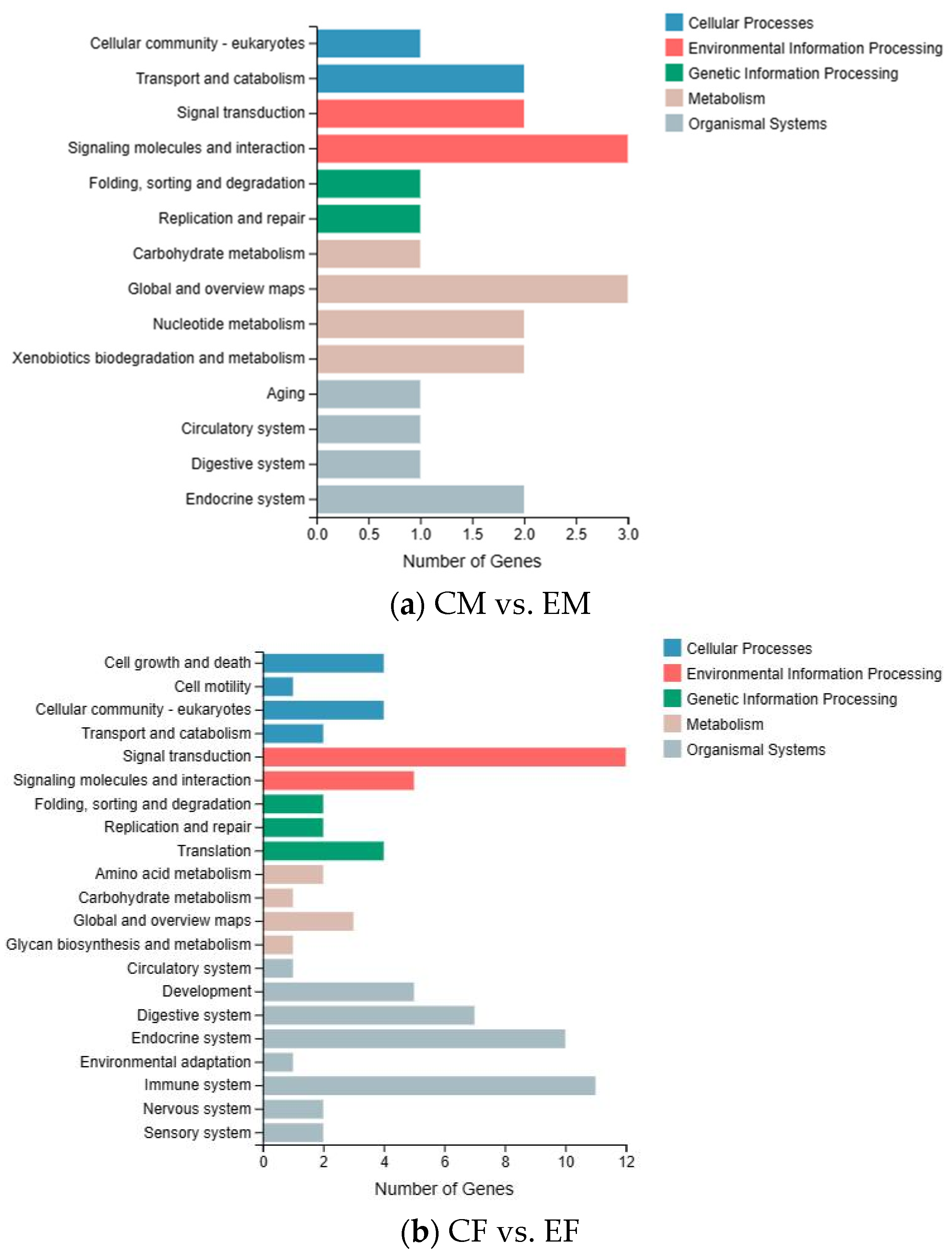
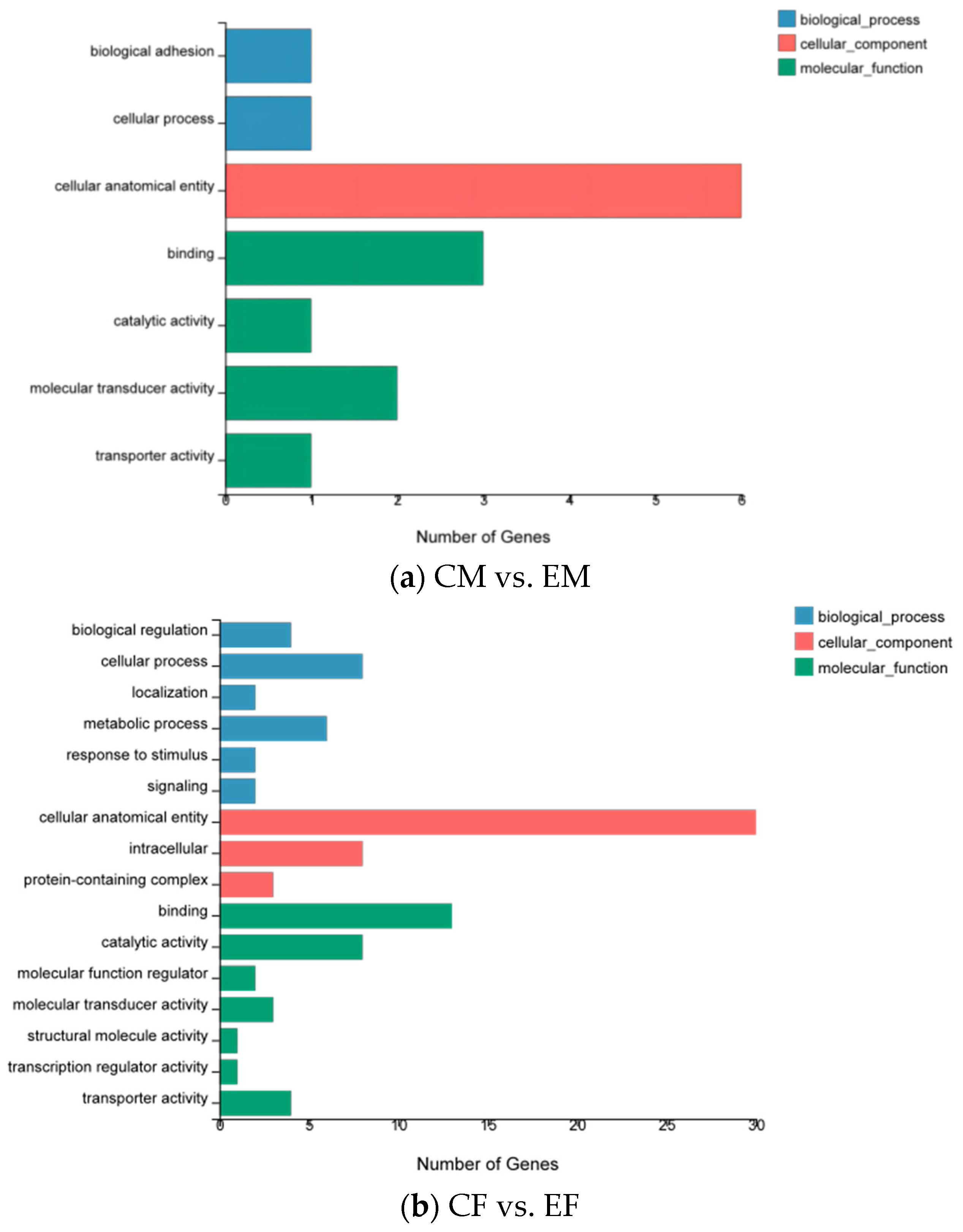
3.2. Functional Annotation and Classification of the Unigenes

Based on the annotated results from the GO database (the detailed data is available in Supplementary Tables S3 and S4), the groups CM and EM were classified into seven subcategories of three GO categories: “Biological process”, “Cellular component”, and “Molecular function”. The largest category was “Cellular component”, in which subcategories, “Cellular anatomical entity”, contained the largest number of unigenes at six, respectively. Additionally, “Biological adhesion”, “Cellular process”, “Catalytic activity”, and “Transporter activity” contained only one gene each. In the groups CF and EF, the results were classified into 16 subcategories of three GO categories, similar to the male groups, with “Cellular component” being the largest category. “Cellular anatomical entity” contained the most unigenes at 30, while “Structural molecule activity” and “Transporter activity” had the lowest number of genes at 1 (Figure 1, and the detailed data is available in Supplementary Table S5).
According to the KEGG database annotation results, eight genes in males were classified into 14 different secondary pathways of five branches. The top two secondary pathways were “Signaling molecules and interaction”, and “Global and overview maps”, each containing three genes. In females, 33 genes were classified into 21 different secondary pathways of five branches, with the top three secondary pathways being “Signal transduction”, “Endocrine system”, and “Immune system”, with 12, 10, and 11 genes, respectively (Figure 2).

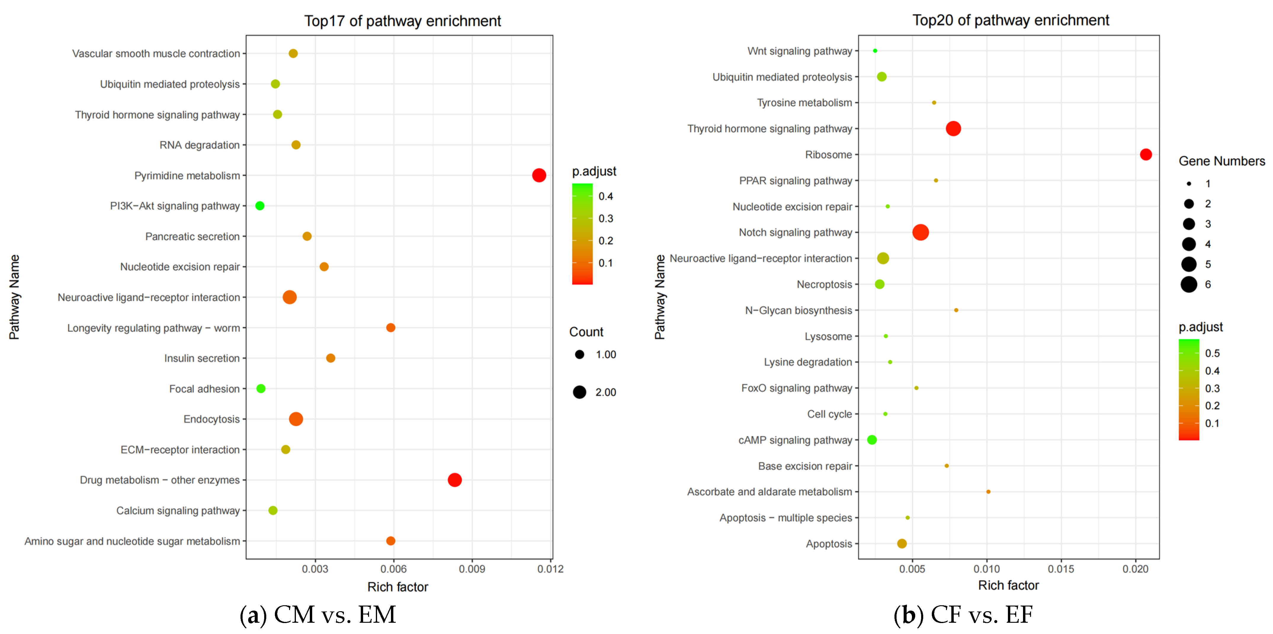
3.3. KEGG Enrichment Analysis

Based on the KEGG enrichment analysis of the DEGs in gonads from the groups CM and EM of males, two KEGG signaling pathways were potentially impacted: “Pyrimidine metabolism” and “Drug metabolism—other enzymes”. Additionally, among the 17 pathways shown in Figure 3a (the detailed data is available in Supplementary Table S6), we identified 7 pathways that might be related to environmental stimuli affecting gonadal development, including “Pyrimidine metabolism”, “Neuroactive ligand–receptor interaction”, “Nucleotide excision repair”, “RNA degradation”, “ECM–receptor interaction”, “Ubiquitin mediated proteolysis”, and “Thyroid hormone signaling”. In the groups CF and EF, we found three potentially impacted KEGG signaling pathways in Figure 3b: “Ribosome”, “Thyroid hormone signaling”, and “Notch signaling pathway”. Furthermore, among the 20 pathways shown in Figure 3b (the detailed data is available in Supplementary Table S7), we identified 12 pathways that might be related to environmental stimuli affecting gonadal development, including “Ribosome”, “Thyroid hormone signaling”, “Notch signaling pathway”, “Nucleotide excision repair”, “FoxO signaling pathway”, “Neuroactive ligand–receptor interaction”, “Ubiquitin mediated proteolysis”, and “Wnt signaling pathway”.
To investigate the relationship between environmental stimulation and gonadal development, we selected 22 DEGs from males and 70 DEGs from females related to gonadal development for DEG clustering analysis to show their expression patterns. The result of cluster analysis showed that some genes were differentially expressed between the control groups and experimental groups (Figure 4). For males, the heat maps for DEGs demonstrated that LG10.g3141 and LG08.g2375 were highly expressed in both the groups CM and EM. Moreover, the invertebrate nicotinic acetylcholine receptors (AChRs) were highly expressed only in the group CM, and LG12.g708 was highly expressed only in the group EM. In contrast, the heat maps for DEGs demonstrated that there were no genes that were simultaneously highly expressed in both female groups. Five genes were highly expressed only in the group (CF), including LG12.g1371, LG09.g2415, LG12.g708, LG08.g1150, and LG08.g1458. Three genes were highly expressed only in the group EF, including Contig02065.g1, LG12.g2275, and LG01.g3352.

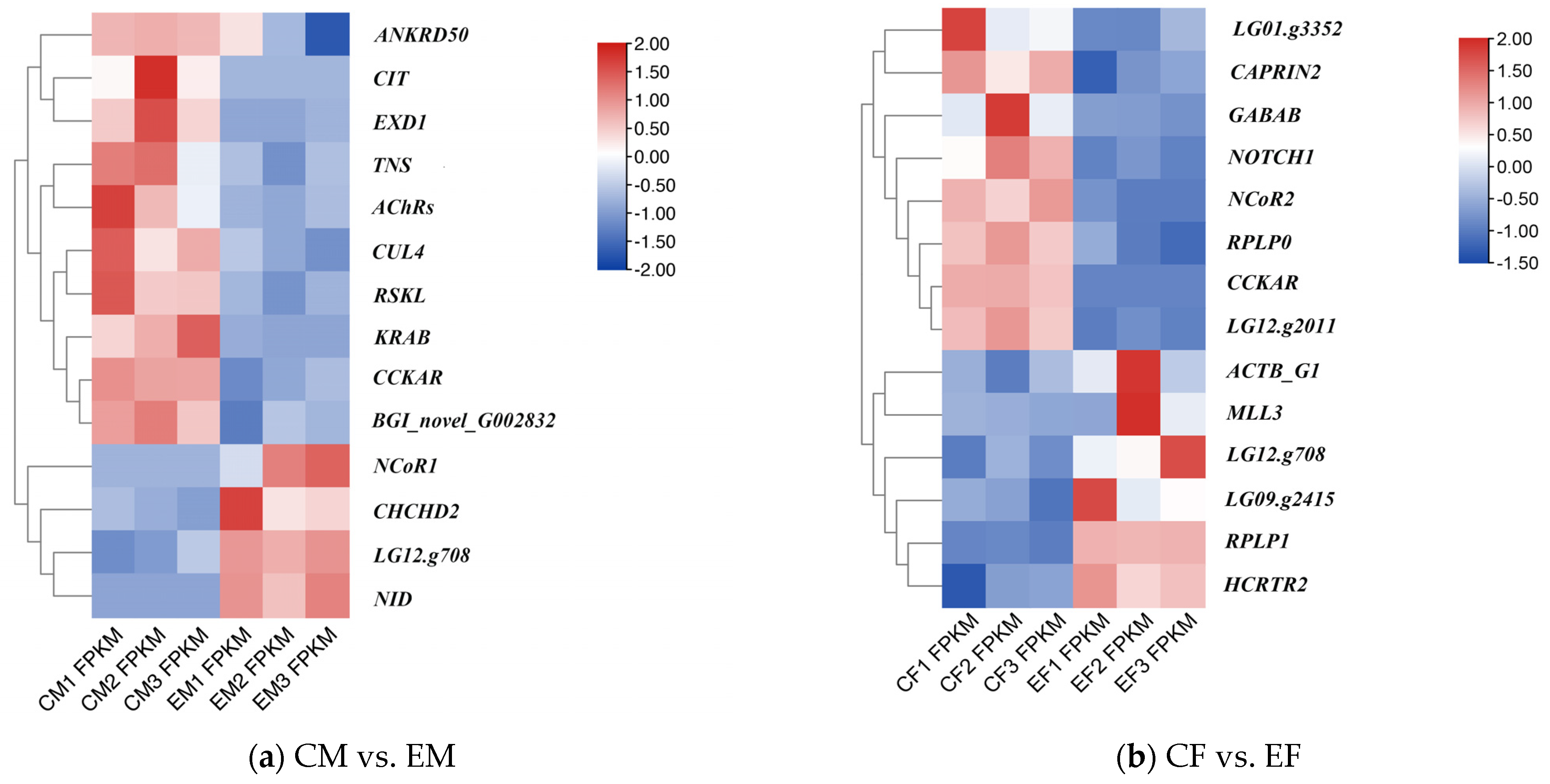
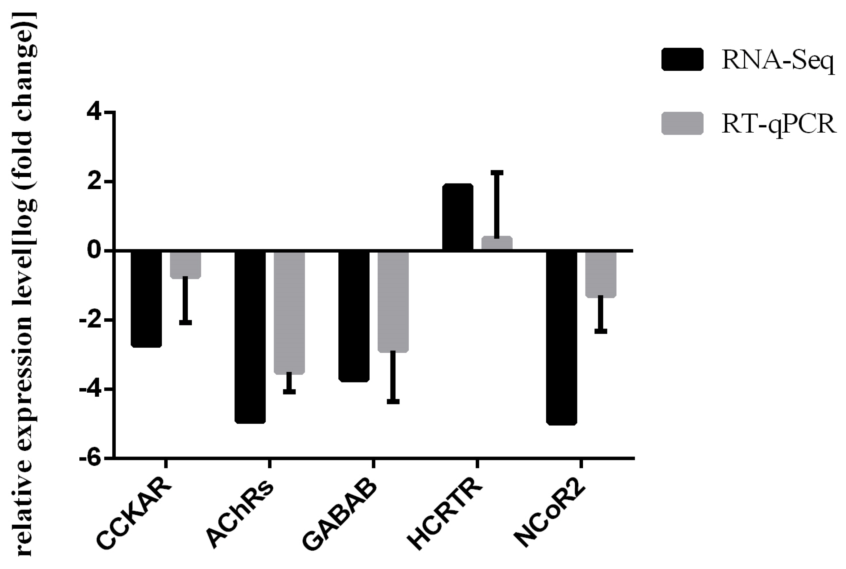
3.4. RT-qPCR Validation

The RT-qPCR data confirmed the expression patterns of the four down-regulated genes, CCKAR, AChRs, GABAB, and NCoR2, as observed in the RNA-seq data (Figure 5). Additionally, the one up-regulated gene in the RNA-seq, HCRTR2, was also up-regulated in the RT-qPCR validation. These results suggest that the transcriptomic data are reliable and that the expression levels of the selected genes are consistent with the findings from the transcriptome analysis.

4. Discussion

4.1. Analysis of KEGG Enrichment Pathways

The KEGG enrichment analysis of metabolic pathways provides insights into the potential mechanisms underlying the effects of environmental stimulation on gonadal development in M. unguiculatus. The enrichment analysis identified a total of 11 pathways that might be related to this process, including those related to environment-stimulated interactions in neural tissue, hormone metabolism, cell physiological activity, and gonadal development. Interestingly, two pathways, “Pyrimidine metabolism” and “Ribosome”, were potentially impacted in this analysis. DNA and RNA comprise the genetic material in living organisms, widely found in all animals, plants, and microorganisms [43]. Ribosomes, in turn, are made of RNA and proteins, so pyrimidines and ribosomes are also present in living organisms. In this transcriptome KEGG enrichment analysis, the “Pyrimidine metabolism” and “Ribosome” pathways were potentially impacted; thus, it is hypothesized that pyrimidine and ribosomes might indirectly affect the development of the gonads in M. unguiculatus.
Following drying-based stimulation, interestingly, four pathways were potentially impacted in both sexes of M. unguiculatus. These pathways are the neuroactive ligand–receptor interaction, nucleotide excision repair, ubiquitin-mediated proteolysis, and thyroid hormone signaling. The neuroactive ligand–receptor interaction pathway is a complex signaling pathway that involves all receptors and ligands associated with intracellular and extracellular signaling pathways on the plasma membrane. Notably, a previous study examining the effects of 17α-methyltestosterone exposure on gonadal histology in Pseudorasbora parva also found enrichment of the neuroactive ligand–receptor interaction pathway in both sexes [44]. The nucleotide excision repair pathway is a pluripotent pathway that recognizes and removes various types of DNA lesions induced by environmental sources to counteract their deleterious effects [45]. A study on triploid oysters revealed that hypermethylated genes involved in nucleotide excision repair may have functional significance in producing gametes during both meiosis divisions in M-3nα [46]. The ubiquitin proteolytic system plays a critical role in several basic cellular processes, such as regulating the cell cycle, modulating immune and inflammatory responses, controlling signal transduction pathways, and facilitating development and differentiation [47]. ECM –receptor interaction and FoxO signaling pathways are also directly or indirectly involved in the physiological activities of cells [48,49]. The energy metabolism of animals undergoes dynamic changes that significantly impact the secretion of reproduction-related hormones, ultimately affecting individual reproductive performance [50]. The thyroid hormone (TH) plays a crucial role in regulating growth, development, differentiation, and metabolism. TH signaling modulates energy expenditure through both central and peripheral pathways, and its effects are executed at the cellular level via TH receptor binding. In previous studies, the thyroid hormones (THs) were shown to play important roles in development, metamorphosis, and metabolism in vertebrates. Zebrafish is the best model system to study the involvement of thyroid hormones in vertebrate development, and can help in more detailed studies of embryonic development outside the ovaries of zebrafish [51]. Several researchers have found that THs may be involved in oyster embryo development and the process of oyster metamorphosis [52]. However, the correlation with the TH signaling pathway in invertebrates is relatively limited, and further exploration is needed.
Gonadal development is a complex process that encompasses somatic cell growth and germ cell growth, involving diverse biological phenomena such as sex determination, oogenesis, and spermatogenesis [53]. Among the key signaling pathways involved in embryogenesis, the highly conserved Wnt family of secreted proteins plays a critical role in mediating cell–cell signaling events [54], and in recent transcriptome studies, the Wnt signaling pathway has been implicated in regulating gonad development in both fish and shellfish species [55,56]. The notch signaling cascade, mediated by the notch gene encoding transmembrane receptors, is another important pathway involved in basic processes such as cell proliferation, stem cell maintenance, and differentiation during embryonic and adult development in animals [57]. The notch signaling pathway has been shown to regulate sexual differentiation in the mollusks Hyriopsis cumingii and Crassostrea hongkongensis [58,59]. In the current transcriptome study of M. unguiculatus, the enrichment of these pathways suggests their potential involvement in the regulation of gonad development in this species. Moreover, environmental stimulation can influence these pathways, leading to changes in cell development and ultimately affecting gonadal development and gametogenesis in M. unguiculatus.

4.2. Key Differentially Expressed Genes

The present study has shown that several genes, including the invertebrate nicotinic acetylcholine receptors (AChRs), and cholecystokinin A receptor (CCKAR) from males, as well as the hypocretin (orexin) receptor 2 (NCRTR2), gamma-aminobutyric acid type B receptor (GABAB), and nuclear receptor co-repressor 2 (NCoR2) from females, may play important roles in regulating gonadal development in M. unguiculatus.
The nicotinic acetylcholine receptors (AChRs) belong to a family of acetylcholine-gated cation channels, which are primarily expressed in muscles and nerves within the peripheral nervous system but are also expressed in lower levels on neurons throughout the central nervous system [60]. Moreover, numerous studies have put forth the proposition that the acetylcholinesterase gene (AChE) regulates a multitude of crucial functions. These encompass the frontal ciliary activity of the gill epithelium, temperature resistance, ciliary activity involved in the transportation of suspended particulate matter, valve opening, and embryo development [61]. In studies of vertebrates, the castration of male zebra finches Taeniopygia guttata leads to a 50% reduction in AChE activity and a 40% decrease in the number of acetylcholine receptors (AChRs) [62,63]. Similarly, decreases in size, AChE activity, and AChR number occur in the dorsal bulbocavernosus muscle after the castration of rats [64,65]. The present results suggest that AChRs are involved in up-regulating and down-regulating the neuroactive ligand–receptor interaction pathway in males, indicating that AChRs may contribute to gonad development in male M. unguiculatus.
The cholecystokinin gene (CCK) encodes a peptide hormone and neurotransmitter that plays a crucial role in the regulation of feeding behavior. Studies using subtype-selective agonists have suggested that the anorectic effect of exogenous cholecystokinin is mediated by the CCKA receptor (CCKAR) [66,67,68]. CCK is widely expressed in important neuronal pathways and co-localizes with classical neurotransmitters such as dopamine in the ventral tegmental area [69], GABA in the hippocampus [70], and CRF in the paraventricular nucleus of the hypothalamus [71]. Investigations in Lophotrochozoa species have demonstrated that the expression analysis of oysters with varying nutritional status or during their reproductive cycle highlights the potential role of Cragi-CCK signaling in regulating feeding behavior and its possible involvement in coordinating nutrition and energy storage in the gonad [72]. The results show that CCKAR is involved in the regulation and down-regulation of the neuroactive ligand–receptor interaction pathway, which might be involved in the coordination of energy storage in the gonads in M. unguiculatus.
The hypocretin (HCRT), also referred to as orexin, system encompasses a set of regulatory peptides that govern diverse neuroendocrine and autonomic functions, such as feeding behavior and sleep–wakefulness regulation [73]. Orexins have been associated with several functional pathways, including the modulation of gonadotropin-releasing hormone (GnRH) neurons and the secretion of pituitary gonadotropins, based on numerous in vivo and in vitro experiments in both rodent [74,75,76,77,78,79,80,81,82] and human models [83]. In addition, studies of the metabolism of the anchovy Coilia nasus in their natural feeding state during migration have shown that females make up a large percentage of the feeding group during migration. When migrating closer, females expend more energy relative to males due to ovarian development, and a larger proportion of feeding individuals are females [84]. The results of this study of hard-shelled mussels showed that the expression of HCRTR2 in the gonads of the female was higher than that of males, which was consistent with the results of the C. nasus study above. This suggests that HCRTR2 might play an important role in regulating gonadal development, particularly in females, in M. unguiculatus. In addition, because vertebrate nervous systems are more complex than those of invertebrates, we must further study the expression of these genes in M. unguiculatus.
The synthesis of gamma-aminobutyric acid (GABA, neurotransmitter) and kisspeptins (neuropeptides) by specialized neurons innervating the hypothalamus can stimulate gonadotropin (FSH and LH) release and regulate reproductive functions [85]. A study on the sex differentiation mechanism of the scallop Patinopecten yessoensis has shown that ovary-biased genes are enriched in neurotransmitter transporters and GABA-ergic synapse genes, suggesting that neurotransmitters such as GABA and glycine may play a role in scallop ovary development [20]. The current findings indicate a higher expression of GABAB in female M. unguiculatus than in males, and down-regulation, suggesting that GABAB may be involved in the regulation of ovarian development.
The duplication and divergence of the nuclear receptor co-repressor (NCoR) gene during vertebrate evolution has resulted in NCoR1 (also known as NCoR) and NCoR2 (also known as SMRT), which have distinct molecular properties and mediate different biological functions [86,87,88,89,90,91,92]. NCoR1 and NCoR2 were initially recognized as transcriptional corepressors that interact with unliganded thyroid and retinoid receptors [93,94]. Their indispensable regulatory roles in normal development are suggested by embryonic lethality in both NCoR and NCoR2 knockouts in mice [95]. In the study of Caenorhabditis elegans, GEI-8 was identified as the closest homolog of NCoR and SMRT in C. elegans, and it was found to regulate gonad development and neuronal functions in C. elegans [96]. Hence, the identification of the NCoR/SMRT homolog in C. elegans allows us to extend the conserved developmental functions of these crucial corepressors to invertebrates. Based on the results of this study and the progress of previous studies, it is speculated that Ncor2 may also regulate gonad development in M. unguiculatus.
In summary, the identified genes in M. unguiculatus, including AChRs, CCKAR, NCRTR2, GABAB, and NCoR2, may play a significant role in gonadal development in response to drying stimulation, providing valuable insights into the reproductive biology of this species. However, further research is needed to investigate the specific mechanisms by which these genes affect gonadal development following drying stimulation and their broader implications for the reproductive biology of the species.

5. Conclusions

In conclusion, transcriptomic analysis of gonads from M. unguiculatus stimulated by drying in the shade and those not to stimulated identified 33,778 unigenes, enriching the database of gonadal development-related genes in M. unguiculatus. Comparative analysis of the transcriptomes revealed 11 key pathways that may be involved in the gonadal development of M. unguiculatus; these pathways are mainly involved in environmental stimulation interactions in neural tissue, hormone metabolism, cell physiological activity, and gonadal development. Importantly, several key genes (AChRs, CCKAR, HCRTR2, GABAB, NCoR2) were identified that may play roles in environmental information processing and gonadal developmental sex differentiation. This study has obtained transcriptomic data from the gonadal tissues in thick-shelled mussels following desiccation stimulation. It has enriched our understanding of gene expression related to gonadal development and reproductive activities in M. unguiculatus. These data provide a foundational basis for further investigating the intrinsic molecular regulatory mechanisms associated with reproductive activities in M. unguiculatus.

Supplementary Materials

The following supporting information can be downloaded at: https://www.mdpi.com/article/10.3390/fishes8090456/s1, Table S1: Detailed information about raw and filtered reads; Table S2 Quality of filtered reads; Table S3: Males GO annotation and classification of unigenes; Table S4: Famels GO annotation and classification of unigenes; Table S5: Figure 1 detailed data; Table S6: Figure 3(a) detailed data; Table S7: Figure 3(b) detailed data.

Author Contributions

Methodology, Y.M.; software, R.Y. and J.L.; data analysis and sample processing, Y.M.; writing—original draft preparation, Y.M.; writing—review and editing, P.Q., J.L. and Y.Y.; funding acquisition, Y.Y. All authors have read and agreed to the published version of the manuscript.

Funding

This research was funded by the National Natural Science Foundation of China (42107301, 41976111, 42176099, 42020104009, and 42076119), NSFC Projects of International Cooperation and Exchanges (42020104009), and the Natural Science Foundation for Distinguished Young Scholars of Zhejiang Province (No. LR22D060002).

Institutional Review Board Statement

The study was conducted in accordance with the Declaration of Helsinki, and approved by the Animal Research and Ethics Committee of Zhejiang Ocean University. Approval code: ZJOU2023025.

Informed Consent Statement

Not applicable.

Data Availability Statement

All sequencing data associated with this project were deposited in the National Center for Biotechnology Information (NCBI) Sequence Read Archive database with the BioProject Accession Number: PRJNA997475 (https://www.ncbi.nlm.nih.gov/sra/PRJNA997475) (accessed on 27 June 2023).

Conflicts of Interest

The authors declare no conflict of interest.

References

  1. Koehn, R.K. The genetics and taxonomy of species in the genus Mytilus. Aquaculture 1991, 94, 125–145. [Google Scholar] [CrossRef]
  2. Zhang, W.; Li, R.; Chen, X.; Wang, C.; Gu, Z.; Mu, C.; Huang, J. Molecular identification reveals hybrids of Mytilus coruscus × Mytilus galloprovincialis in mussel hatcheries of China. Aquacult. Int. 2020, 28, 85–93. [Google Scholar] [CrossRef]
  3. Ye, Y.Y.; Li, J.J.; Wu, C.W.; Xu, M.Y.; Guo, B.Y. Genetic analysis of mussel (Mytilus coruscus) populations on the coast of East China Sea revealed by ISSR-PCR markers. Biochem. Syst. Ecol. 2012, 45, 1–6. [Google Scholar] [CrossRef]
  4. An, H.S.; Lee, J.W. Development of microsatellite markers for the Korean mussel, Mytilus coruscus (Mytilidae) using next-generation sequencing. Int. J. Mol. Sci. 2012, 13, 10583–10593. [Google Scholar] [CrossRef] [PubMed]
  5. Guinotte, J.M.; Fabry, V.J. Ocean acidification and its potential effects on marine ecosystems. Ann. N. Y. Acad. Sci. 2008, 1134, 320–342. [Google Scholar] [CrossRef] [PubMed]
  6. Harley, C.D.; Randall, H.A.; Hultgren, K.M.; Miner, B.G.; Sorte, C.J.; Thornber, C.S.; Rodriguez, L.F.; Tomanek, L.; Williams, S.L. The impacts of climate change in coastal marine systems. Ecol. Lett. 2006, 9, 228–241. [Google Scholar] [CrossRef] [PubMed]
  7. Nigel, W.; Arnell, S.; Gosling, N. The impacts of climate change on river flow regimes at the global scale. J. Hydrol. 2013, 486, 351–364. [Google Scholar]
  8. Huang, C.M. Studies on the Comparative Anatomy of Four Marine Bivalve Molluscs Gastropods and Byssus; Hainan University: Hainan, China, 2011. [Google Scholar]
  9. Cheng, L.; Xu, S.L.; Liu, F.; Qi, F. Study on the meat condition index and biochemical compositions of Mytilus unguiculatus in different gonad development stages. ICES J. Mar. Sci. 2013, 31, 68–73. [Google Scholar]
  10. Wang, M.; Xia, J.; Jawad, M.; Wei, W.; Gui, L.; Liang, X.; Li, M. Transcriptome sequencing analysis of sex-related genes and miRNAs in the gonads of Mytilus coruscus. Front. Mar. Sci. 2022, 9, 1013857. [Google Scholar] [CrossRef]
  11. Chang, K.M.; Wu, J.F. Study on artificial propagation of mussel Mytilus unguiculatus. South. China Fish. Sci. 2007, 3, 26–30. [Google Scholar]
  12. Secor, S.M.; Carey, H.V. Integrative physiology of fasting. Compr. Physiol. 2016, 6, 773–825. [Google Scholar] [PubMed]
  13. Zhao, D. Expression Characteristics and Function of Genes Dmrtl and Foxl2 in the Scallop Patinopecten yessoensis; Shanghai Ocean University: Shanghai, China, 2021. [Google Scholar]
  14. Wang, H.; Qu, M.; Tang, W.; Liu, S.; Ding, S. Transcriptome Profiling and Expression Localization of Key Sex-Related Genes in a Socially-Controlled Hermaphroditic Clownfish, Amphiprion clarkii. Int. J. Mol. Sci. 2022, 23, 9085. [Google Scholar] [CrossRef] [PubMed]
  15. Davey, A.J.H.; Jellyman, D.J. Sex Determination in Freshwater Eels and Management Options for Manipulation of Sex. Rev. Fish. Biol. Fish. 2005, 15, 37–52. [Google Scholar] [CrossRef]
  16. Prevedelli, D.; N’Siala, G.M.; Simonini, R. Gonochorism vs. hermaphroditism: Relationship between life history and fitness in three species of Ophryotrocha (Polychaeta: Dorvilleidae) with different forms of sexuality. J. Anim. Ecol. 2006, 75, 203–212. [Google Scholar] [CrossRef] [PubMed]
  17. Li, X.Y.; Mei, J.; Ge, C.T.; Liu, X.L.; Gui, J.F. Sex determination mechanisms and sex control approaches in aquaculture animals. Sci. China Life Sci. 2022, 65, 1091–1122. [Google Scholar] [CrossRef] [PubMed]
  18. Naimi, A.; Martinez, A.S.; Specq, M.L.; Mrac, A.; Diss, B.; Mathieu, M.; Sourdaine, P. Identification and expression of a factor of the DM family in the oyster Crassostrea gigas. Comp. Biochem. Physiol. A Mol. Integr. Physiol. 2009, 152, 189–196. [Google Scholar] [CrossRef] [PubMed]
  19. Shi, J.; Hong, Y.; Sheng, J.; Peng, K.; Wang, J. De novo transcriptome sequencing to identify the sex-determination genes in Hyriopsis schlegelii. Biosci. Biotechnol. Biochem. 2015, 79, 1257–1265. [Google Scholar] [CrossRef] [PubMed]
  20. Li, Y.; Zhang, L.; Sun, Y.; Ma, X.; Wang, J.; Li, R.; Zhang, M.; Wang, S.; Hu, X.; Bao, Z. Transcriptome sequencing and comparative analysis of ovary and testis identifies potential key sex-related genes and pathways in Scallop Patinopecten yessoensis. Mar. Biotechnol. 2016, 18, 453–465. [Google Scholar] [CrossRef]
  21. Shi, Y.; Liu, W.; He, M. Proteome and transcriptome analysis of ovary, intersex gonads, and testis reveals potential key sex reversal/differentiation genes and mechanism in Scallop Chlamys nobilis. Mar. Biotechnol. 2018, 20, 220–245. [Google Scholar] [CrossRef]
  22. Hrdlickova, R.; Toloue, M.; Tian, B. RNA -Seq methods for transcriptome analysis. Wiley Interdiscip. Rev. RNA 2016, 8, 1364. [Google Scholar] [CrossRef]
  23. Mutz, K.O.; Heilkenbrinker, A.; Lönne, M.; Walter, J.-G.; Stahl, F. Transcriptome analysis using next-generation sequencing. Curr. Opin. Biotechnol. 2013, 24, 22–30. [Google Scholar] [CrossRef] [PubMed]
  24. Wang, Y.; Zhang, H.; Lu, Y.; Wang, F.; Liu, L.; Liu, J.; Liu, X. Comparative transcriptome analysis of zebrafish (Danio rerio) brain and spleen infected with spring viremia of carp virus (SVCV). Fish. Shellfish. Immunol. 2017, 69, 35–45. [Google Scholar] [CrossRef] [PubMed]
  25. Li, S.; Zhang, X.; Sun, Z.; Li, F.; Xiang, J. Transcriptome analysis on Chinese shrimp Fenneropenaeus chinensis during WSSV acute infection. PLoS ONE 2013, 8, e58627. [Google Scholar] [CrossRef] [PubMed]
  26. Wang, Q.; Li, J.; Guo, H. Transcriptome analysis and discovery of genes involved in immune pathways in Solen strictus (Gould, 1861) under Vibrio anguillarum. Fish Shellfish Immunol. 2019, 88, 237–243. [Google Scholar] [CrossRef] [PubMed]
  27. Qi, Z.; Zhang, Q.; Wang, Z.; Ma, T.; Zhou, J.; Holland, J.W.; Gao, Q. Transcriptome analysis of the endangered Chinese giant salamander (Andrias davidianus): Immune modulation in response to Aeromonas hydrophila infection. Vet. Immunol. Immunopathol. 2016, 169, 85–95. [Google Scholar] [CrossRef] [PubMed]
  28. Sun, D.F.; Yu, H.; Li, Q. Examination of the role of CgSox-like in sex determination and gonadal development in the Pacific oyster Crassostrea gigas. Aquaculture 2023, 566, 739234. [Google Scholar] [CrossRef]
  29. Lian, C.P. Gonadal Development and Reproductive Cycle, Transcriptomic Studies and Analysis of the Effects of Morphological Traits on Live Body Weight in Tapes Conspersus; Guangxi University: Guangxi, China, 2023. [Google Scholar]
  30. Liao, X.T. Study on the Gonadal Development and Expression of Sex Related Genes Dmrtl and Foxl2 in Clam Cyclina sinensis; Jiangsu Ocean University: Jiangsu, China, 2022. [Google Scholar]
  31. Sun, D.F.; Yu, H.; Li, Q. Early gonadal differentiation is associated with the antagonistic action of Foxl2 and Dmrt1l in the Pacific oyster. Comp. Biochem. Phys. B 2023, 265, 110831. [Google Scholar] [CrossRef] [PubMed]
  32. Wang, T.; Kong, H.; Shang, Y.; Dupont, S.; Peng, J.; Wang, X.; Deng, Y.; Hu, M.; Wang, Y. Ocean acidification but not hypoxia alters the gonad performance in the thick shell mussel Mytilus coruscus. Mar. Pollut Bull 2021, 167, 112282. [Google Scholar] [CrossRef]
  33. Haas, B.J.; Papanicolaou, A.; Yassour, M.; Grabherr, M.; Blood, P.D.; Bowden, J.; Regev, A. De novo transcript sequence reconstruction from RNA-seq using the trinity platform for reference generation and analysis. Nat. Protoc. 2013, 8, 1494–1512. [Google Scholar] [CrossRef]
  34. Li, R.; Li, Y.; Kristiansen, K. SOAP: Short oligonucleotide alignment program. Bioinformatics 2008, 24, 713–714. [Google Scholar] [CrossRef]
  35. Kim, D.; Langmead, B.; Salzberg, S.L. HISAT: A fast spliced aligner with low memory requirements. Nat. Methods 2015, 12, 357–360. [Google Scholar] [CrossRef] [PubMed]
  36. Langmead, B.; Salzberg, S.L. Fast gapped-read alignment with Bowtie 2. Nat. Methods 2012, 9, 357–359. [Google Scholar] [CrossRef] [PubMed]
  37. Li, B.; Dewey, C.N. RSEM: Accurate transcript quantification from RNA-Seq data with or without a reference genome. BMC Bioinform. 2011, 12, 323. [Google Scholar] [CrossRef] [PubMed]
  38. Love, M.I.; Huber, W.; Anders, S. Moderated estimation of fold change and dispersion for RNA-seq data with DESeq2. Genome Biol. 2014, 15, 550. [Google Scholar] [CrossRef] [PubMed]
  39. Bindea, G.; Mlecnik, B.; Hackl, H.; Charoentong, P.; Tosolini, M.; Kirilovsky, A.; Galon, J. ClueGO: A Cytoscape plug-in to decipher functionally grouped gene ontology and pathway annotation networks. Bioinformatics 2009, 25, 1091–1093. [Google Scholar] [CrossRef] [PubMed]
  40. Kanehisa, M.; Goto, S. KEGG: Kyoto encyclopedia of genes and genomes. Nucleic Acids Res. 2000, 28, 27–30. [Google Scholar] [CrossRef]
  41. Abdi, H. The Bonferonni and Šidák corrections for multiple comparisons. Encycl. Meas. Stat. 2007, 1, 2007. [Google Scholar]
  42. Livak, K.J.; Schmittgen, T.D. Analysis of relative gene expression data using real-time quantitative PCR and the 2(-Delta Delta C(T)) method. Methods 2001, 25, 402–408. [Google Scholar] [CrossRef]
  43. Alberts, B.; Johnson, A.; Lewis, J.; Raff, M.; Roberts, K.; Walter, P. Molecular Biology of the Cell; Garland Science: New York, NY, USA, 2014. [Google Scholar]
  44. Wang, J.; Zhou, J.; Yang, Q.; Wang, W.; Liu, Q.; Liu, W.; Liu, S. Effects of 17 α-methyltestosterone on the transcriptome, gonadal histology and sex steroid hormones in Pseudorasbora parva. Theriogenology 2020, 155, 88–97. [Google Scholar] [CrossRef]
  45. Reardon, J.T.; Sancar, A. Nucleotide excision repair. Prog. Nucleic Acid. Res. Mol. Biol. 2005, 79, 183–235. [Google Scholar]
  46. Sun, D.F.; Yu, H.; Li, Q. Genome-wide differential DNA methylomes provide insights into the infertility of Triploid Oysters. Mar. Biotechnol. 2022, 24, 18–31. [Google Scholar] [CrossRef] [PubMed]
  47. Ciechanover, A.; Orian, A.; Schwartz, A.L. Ubiquitin-mediated proteolysis: Biological regulation via destruction. Bioessays 2000, 22, 442–451. [Google Scholar] [CrossRef]
  48. Lee, H.J.; Jang, M.; Kim, H.; Kwak, W.; Park, W.; Hwang, J.Y.; Lee, C.K.; Jang, G.W.; Park, M.N.; Kim, H.C.; et al. Comparative transcriptome analysis of adipose tissues reveals that ECM-receptor interaction is involved in the depot-specific adipogenesis in cattle. PLoS ONE 2013, 8, e66267. [Google Scholar] [CrossRef] [PubMed]
  49. Farhan, M.; Wang, H.; Gaur, U.; Little, P.J.; Xu, J.; Zheng, W. FOXO signaling pathways as therapeutic targets in cancer. Int. J. Biol. Sci. 2017, 13, 815–827. [Google Scholar] [CrossRef] [PubMed]
  50. Zhong, Z.; Chen, G.; Tu, H.; Yao, X.; Peng, X.; Lan, X.; Tang, Q.; Yi, S.; Xia, Z.; Cai, M.; et al. Transcriptomic analysis and functional gene expression in different stages of gonadal development of Macrobrachium rosenbergii. Fishes 2023, 8, 94. [Google Scholar] [CrossRef]
  51. Huang, W.; Xu, F.; Qu, T.; Zhang, R.; Li, L.; Que, H.; Zhang, G. Identification of thyroid hormones and functional characterization of thyroid hormone receptor in the Pacific oyster Crassostrea gigas provide insight into evolution of the thyroid hormone system. PLoS ONE 2015, 10, e0144991. [Google Scholar] [CrossRef] [PubMed]
  52. Lazcano, I.; Pech-Pool, S.M.; Olvera, A.; García-Martínez, I.; Palacios-Pérez, S.; Orozco, A. The importance of thyroid hormone signaling during early development: Lessons from the zebrafish model. Gen. Comp. Endocr. 2023, 334, 114225. [Google Scholar] [CrossRef] [PubMed]
  53. Yue, C.; Li, Q.; Yu, H. Gonad transcriptome analysis of the Pacific oyster Crassostrea gigas identifies potential genes regulating the sex determination and differentiation process. Mar. Biotechnol. 2018, 20, 206–219. [Google Scholar] [CrossRef]
  54. Logan, C.Y.; Nusse, R. The Wnt signaling pathway in development and disease. Annu. Rev. Cell Dev. Biol. 2004, 20, 781–810. [Google Scholar] [CrossRef]
  55. Wang, Y.; Chen, Y.; Cao, M.; Wang, X.; Wang, G.; Li, J. Identification of wnt2 in the pearl mussel Hyriopsis cumingii and its role in innate immunity and gonadal development. Fish. Shellfish. Immunol. 2021, 118, 85–93. [Google Scholar] [CrossRef]
  56. Sreenivasan, R.; Jiang, J.; Wang, X.; Bártfai, R.; Kwan, H.Y.; Christoffels, A.; Orbán, L. Gonad differentiation in zebrafish is regulated by the canonical Wnt signaling pathway. Biol. Reprod. 2014, 90, 45. [Google Scholar] [CrossRef] [PubMed]
  57. Borggrefe, T.; Oswald, F. The Notch signaling pathway: Transcriptional regulation at Notch target genes. Cell Mol. Life Sci. 2009, 66, 1631–1646. [Google Scholar] [CrossRef] [PubMed]
  58. Wang, Y.Y.; Duan, S.H.; Wang, G.L.; Li, J.L. Integrated mRNA and miRNA expression profile analysis of female and male gonads in Hyriopsis cumingii. Sci. Rep. 2021, 11, 665. [Google Scholar] [CrossRef] [PubMed]
  59. Wei, P.; He, P.; Zhang, X.; Li, W.; Zhang, L.; Guan, J.; Peng, J. Identification and characterization of microRNAs in the gonads of Crassostrea hongkongensis using high-throughput sequencing. Comp. Biochem. 2019, 31, 100606. [Google Scholar] [CrossRef] [PubMed]
  60. Lindstrom, J. Nicotinic acetylcholine receptors in health and disease. Mol. Neurobiol. 1997, 15, 193–222. [Google Scholar] [CrossRef] [PubMed]
  61. Corsi, I.; Pastore, A.M.; Lodde, A.; Palmerini, E.; Castagnolo, L.; Focardi, S. Potential role of cholinesterases in the invasive capacity of the freshwater bivalve, Anodonta woodiana (Bivalvia: Unionacea): A comparative study with the indigenous species of the genus, Anodonta sp. Comp. Biochem. Physiol. C Toxicol. Pharmacol. 2007, 145, 413–419. [Google Scholar] [CrossRef] [PubMed]
  62. Bleisch, W.V.; Luine, V.N. Modulation of acetylcholinesterase activity and acetylcholine receptor number in an androgen sensitive neuromuscular system. Soc. Neurosci. Abstr. 1981, 7, 805. [Google Scholar]
  63. Luine, V.N.; Nottebohm, F.; Harding, C.; McEwen, B.S. Androgen affects cholinergic enzymes in syringeal motor neurons and muscle. Brain Res. 1980, 192, 89–107. [Google Scholar] [CrossRef]
  64. Bleisch, W.V.; Harrelson, A.L.; Luine, V.N. Testosterone increases acetylcholine receptor number in the “levator ani” muscle of the rat. J. Neurobiol. 1982, 13, 153–161. [Google Scholar] [CrossRef]
  65. Gutmann, E.; Tucek, S.; Hanzlíková, V. Changes in the choline acetyltransferase and cholinesterase activities in the levator ani muscle of rats following castration. Physiol. Bohemoslov. 1969, 18, 195–203. [Google Scholar]
  66. Cooper, S.J.; Dourish, C.T. Multiple cholecystokinin (CCK) receptors and CCK-monoamine interactions are instrumental in the control of feeding. Physiol. Behav. 1990, 48, 849–857. [Google Scholar] [CrossRef] [PubMed]
  67. Moran, T.H. Cholecystokinin and satiety: Current perspectives. Nutrition 2000, 16, 858–865. [Google Scholar] [CrossRef] [PubMed]
  68. Crawley, J.N.; Corwin, R.L. Biological actions of cholecystokinin. Peptides 1994, 15, 731–755. [Google Scholar] [CrossRef] [PubMed]
  69. Hökfelt, T.; Skirboll, L.; Rehfeld, J.F.; Goldstein, M.; Markey, K.; Dann, O. A subpopulation of mesencephalic dopamine neurons projecting to limbic areas contains a cholecystokinin-like peptide: Evidence from immunohistochemistry combined with retrograde tracing. Neuroscience 1980, 5, 2093–2124. [Google Scholar] [CrossRef] [PubMed]
  70. Gulyás, A.I.; Görcs, T.J.; Freund, T.F. Innervation of different peptide-containing neurons in the hippocampus by GABAergic septal afferents. Neuroscience 1990, 37, 31–44. [Google Scholar] [CrossRef] [PubMed]
  71. Mezey, E.; Reisine, T.D.; Skirboll, L.; Beinfeld, M.; Kiss, J.Z. Cholecystokinin in the medial parvocellular subdivision of the paraventricular nucleus. Co-existence with corticotropin-releasing hormone. Ann. N. Y. Acad. Sci. 1985, 448, 152–156. [Google Scholar] [CrossRef] [PubMed]
  72. Schwartz, J.; Dubos, M.P.; Pasquier, J.; Zatylny, C.; Favrel, P. Emergence of a cholecystokinin/sulfakinin signalling system in Lophotrochozoa. Sci. Rep. 2018, 8, 16424. [Google Scholar] [CrossRef] [PubMed]
  73. Silveyra, P.; Cataldi, N.I.; Lux-Lantos, V.; Libertun, C. Gonadal steroids modulated hypocretin/orexin type-1 receptor expression in a brain region, sex and daytime specific manner. Regul. Pept. 2009, 158, 121–126. [Google Scholar] [CrossRef]
  74. Campbell, R.E.; Grove, K.L.; Smith, M.S. Gonadotropin-releasing hormone neurons coexpress orexin 1 receptor immunoreactivity and receive direct contacts by orexin fibers. Endocrinology 2003, 144, 1542–1548. [Google Scholar] [CrossRef]
  75. Furuta, M.; Funabashi, T.; Kimura, F. Suppressive action of orexin A on pulsatile luteinizing hormone secretion is potentiated by a low dose of estrogen in ovariectomized rats. Neuroendocrinology 2002, 75, 151–157. [Google Scholar] [CrossRef]
  76. Irahara, M.; Tamura, T.; Matuzaki, T.; Saito, S.; Yasui, T.; Yamano, S.; Kamada, M.; Aono, T. Orexin-A suppresses the pulsatile secretion of luteinizing hormone via beta-endorphin. Biochem. Biophys. Res. Commun. 2001, 281, 232–236. [Google Scholar] [CrossRef] [PubMed]
  77. Kohsaka, A.; Watanobe, H.; Kakizaki, Y.; Suda, T.; Schioth, H.B. A significant participation of orexin-A, a potent orexigenic peptide, in the preovulatory luteinizing hormone and prolactin surges in the rat. Brain Res. 2001, 898, 166–170. [Google Scholar] [CrossRef] [PubMed]
  78. Pu, S.; Jain, M.R.; Kalra, P.S.; Kalra, S.P. Orexins, a novel family of hypothalamic neuropeptides, modulate pituitary luteinizing hormone secretion in an ovarian steroid-dependent manner. Regul. Pept. 1998, 78, 133–136. [Google Scholar] [PubMed]
  79. Russell, S.H.; Small, C.J.; Kennedy, A.R.; Stanley, S.A.; Seth, A.; Murphy, K.G.; Taheri, S.; Ghatei, M.A.; Bloom, S.R. Orexin A interactions in the hypothalamo-pituitary gonadal axis. Endocrinology 2001, 142, 5294–5302. [Google Scholar] [CrossRef]
  80. Silveyra, P.; Catalano, P.N.; Lux-Lantos, V.; Libertun, C. Impact of proestrous milieu on expression of orexin receptors and prepro-orexin in rat hypothalamus and hypophysis: Actions of Cetrorelix and Nembutal. Am. J. Physiol. Endocrinol. Metab. 2007, 292, E820–E828. [Google Scholar] [CrossRef] [PubMed]
  81. Small, C.J.; Goubillon, M.L.; Murray, J.F.; Siddiqui, A.; Grimshaw, S.E.; Young, H.; Sivanesan, V.; Kalamatianos, T.; Kennedy, A.R.; Coen, C.W.; et al. Central orexin A has site-specific effects on luteinizing hormone release in female rats. Endocrinology 2003, 144, 3225–3236. [Google Scholar] [CrossRef] [PubMed]
  82. Tamura, T.; Irahara, M.; Tezuka, M.; Kiyokawa, M.; Aono, T. Orexins, orexigenic hypothalamic neuropeptides, suppress the pulsatile secretion of luteinizing hormone in ovariectomized female rats. Biochem. Biophys. Res. Commun. 1999, 264, 759–762. [Google Scholar] [CrossRef]
  83. Kok, S.W.; Roelfsema, F.; Overeem, S.; Lammers, G.J.; Frolich, M.; Meinders, A.E.; Pijl, H. Pulsatile LH release is diminished, whereas FSH secretion is normal, in hypocretin deficient narcoleptic men. Am. J. Physiol. Endocrinol. Metab. 2004, 287, E630–E636. [Google Scholar] [CrossRef]
  84. Yin, D.; Lin, D.; Ying, C.; Ma, F.; Yang, Y.; Wang, Y.; Tan, J.; Liu, K. Metabolic mechanisms of Coilia nasus in the natural food intake state during migration. Genomics 2020, 112, 3294–3305. [Google Scholar] [CrossRef]
  85. Zohar, Y.; Muñoz-Cueto, J.A.; Elizur, A.; Kah, O. Neuroendocrinology of reproduction in teleost fish. Gen. Comp. Endocrinol. 2010, 165, 438–455. [Google Scholar] [CrossRef]
  86. Ritter, M.J.; Amano, I.; Imai, N.; Soares, D.; Oliveira, L.; Vella, K.R.; Hollenberg, A.N. Nuclear receptor corepressors, NCOR1 and SMRT, are required for maintaining systemic metabolic homeostasis. Mol. Metab. 2021, 53, 101315. [Google Scholar] [CrossRef] [PubMed]
  87. Stanya, K.J.; Kao, H.Y. New insights into the functions and regulation of the transcriptional corepressors SMRT and N-CoR. Cell Div. 2009, 4, 7. [Google Scholar] [CrossRef] [PubMed]
  88. Perissi, V.; Jepsen, K.; Glass, C.K.; Rosenfeld, M.G. Deconstructing repression: Evolving models of co-repressor action. Nat. Rev. Genet. 2010, 11, 109–123. [Google Scholar] [CrossRef] [PubMed]
  89. Oberoi, J.; Fairall, L.; Watson, P.J.; Yang, J.C.; Czimmerer, Z.; Kampmann, T.; Goult, B.T.; Greenwood, J.A.; Gooch, J.T.; Kallenberger, B.C.; et al. Structural basis for the assembly of the SMRT/NCoR core transcriptional repression machinery. Nat. Struct. Mol. Biol. 2011, 18, 177–184. [Google Scholar] [CrossRef] [PubMed]
  90. Watson, P.J.; Fairall, L.; Schwabe, J.W. Nuclear hormone receptor co-repressors: Structure and function. Mol. Cell Endocrinol. 2012, 348, 440–449. [Google Scholar] [CrossRef] [PubMed]
  91. Hudson, G.M.; Watson, P.J.; Fairall, L.; Jamieson, A.G.; Schwabe, J.W.R. Insights into the recruitment of class IIa histone deacetylases (HDACs) to the SMRT/NCoR transcriptional repression complex. J. Biol. Chem. 2015, 290, 18237–18244. [Google Scholar] [CrossRef] [PubMed]
  92. Desravines, D.C.; Serna, M.I.; Schneider, R.; Mas, P.J.; Aleksandrova, N.; Jensen, M.R.; Blackledge, M.; Hart, D.J. Structural characterization of the SMRT corepressor interacting with histone deacetylase 7. Sci. Rep. 2017, 7, 3678. [Google Scholar] [CrossRef]
  93. Hörlein, A.J.; Näär, A.M.; Heinzel, T.; Torchia, J.; Gloss, B.; Kurokawa, R.; Ryan, A.; Kamei, Y.; Söderström, M.; Glass, C.K. Ligand-independent repression by the thyroid hormone receptor mediated by a nuclear receptor co-repressor. Nature 1995, 377, 397–404. [Google Scholar] [CrossRef]
  94. Chen, J.D.; Evans, R.M. A transcriptional co-repressor that interacts with nuclear hormone receptors. Nature 1995, 377, 454–457. [Google Scholar] [CrossRef]
  95. Jepsen, K.; Hermanson, O.; Onami, T.M.; Gleiberman, A.S.; Lunyak, V. Combinatorial roles of the nuclear receptor corepressor in transcription and development. Cell 2000, 102, 753–763. [Google Scholar] [CrossRef]
  96. Mikoláš, P.; Kollárová, J.; Sebková, K.; Saudek, V.; Yilma, P.; Kostrouchová, M.; Krause, M.W.; Kostrouch, Z.; Kostrouchová, M. GEI-8, a homologue of vertebrate nuclear receptor corepressor NCoR/SMRT, regulates gonad development and neuronal functions in Caenorhabditis elegans. PLoS ONE 2013, 8, e58462. [Google Scholar] [CrossRef]
Figure 1. GO functional classification of gonadal transcriptome differentially expressed genes before and after shade-dried stimulation in M. unguiculatus. (a) represents the GO functional classification of differentially expressed genes between the control group and the experimental group for male gonads, while (b) pertains to the GO functional classification of differentially expressed genes between the control group and the experimental group for female gonads.
Fishes 08 00456 g001
Figure 2. Classification map of the KEGG signaling pathway of the gonad transcriptome differentially expressed genes before and after shade-dried stimulation in M. unguiculatus. (a) represents the KEGG signal pathway classification of differentially expressed genes between the control group and the experimental group for male gonads, while (b) pertains to the KEGG signal pathway classification of differentially expressed genes between the control group and the experimental group for female gonads.
Fishes 08 00456 g002
Figure 3. Enrichment analysis of the KEGG signal differentially expressed genes in the gonadal transcriptome of M. unguiculatus before and after shade-dried stimulation. (a) represents the enrichment analysis of KEGG signaling pathways for differentially expressed genes between the control and experimental groups in male gonads, while (b) pertains to the enrichment analysis of KEGG signaling pathways for differentially expressed genes between the control and experimental groups in female gonads.
Fishes 08 00456 g003
Figure 4. Heatmap analysis on the top 14 differentially expressed genes selected based on their expression levels. (a) ANKRD50: ankyrin repeat domain-containing protein 50. CIT: citron Rho-interacting kinase. EXD1: exonuclease 3′−5′ domain-containing protein 1. TNS: tensin. AChRs: nicotinic acetylcholine receptor, invertebrate. CUL4: cullin 4. RSKL: ribosomal protein S6 kinase−like. KRAB: domain−containing zinc finger protein. CCKAR: cholecystokinin A receptor. BGI_novel_G002832: the gene has not yet been named. NCoR1: nuclear receptor co−repressor 1. CHCHD2: coiled−coil−helix−coiled−coil−helix domain-containing protein 2. LG12.g708: the gene has not yet been named. NID: nidogen (entactin). (b) LG01.g3352: the gene has not yet been named. CAPRIN2: caprin-2. GABAB: gamma-aminobutyric acid type B receptor. NOTCH1: notch 1. NCoR2: nuclear receptor co-repressor 2. RPLP0: ribosomal protein lateral stalk subunit P0. CCKAR: cholecystokinin A receptor. LG12.g2011: the gene has not yet been named. ACTB_G1: actin beta. MLL3: histone−lysine N-methyltransferase MLL3. LG12.g708: the gene has not yet been named. LG09.g2415: the gene has not yet been named. RPLP1: ribosomal protein lateral stalk subunit P1. HCRTR2: hypocretin (orexin) receptor 2.
Fishes 08 00456 g004
Figure 5. RT−qPCR validation of gonadal development−related genes under environmental stimulation. Five genes were selected. CCKAR: cholecystokinin A receptor. AChRs: nicotinic acetylcholine receptor, invertebrate. GABAB: gamma-aminobutyric acid type B receptor. HCRTR2: hypocretin (orexin) receptor 2. NCoR2: nuclear receptor co-repressor 2.
Fishes 08 00456 g005
Table 1. Primers used for RT-qPCR analysis.
GenePrimer Sequence (5′-3′)
AChRsF: TGAGGTTTGGTCAGAGCTGAAATGG
R: CCTTGCCTCCCGAACAACACTC
CCKARF: TTATCACGAACGCCTTGGTACTGTC
R: CGTCGATGCTTCAGGCTCAGTG
HCRTR2F: GCAGATAGGGCGTGAGATTAACCG
R: TTTCCGTCTTCGCTTTACTGTACCG
GABABF: AGTTGAGAAAGGACCCGGTCTATGG
R: TTGTCTCCGTCTATGGCCCAGTC
NCoR2F: ACCCGATTTTGATGCCGAGAATGG
R: AGCCTTGCCTGTCTCAACGAATC
Disclaimer/Publisher’s Note: The statements, opinions and data contained in all publications are solely those of the individual author(s) and contributor(s) and not of MDPI and/or the editor(s). MDPI and/or the editor(s) disclaim responsibility for any injury to people or property resulting from any ideas, methods, instructions or products referred to in the content.

