Article

Handling Effects on Histological Identification of Female Reproductive Status: Examples from Tropical Deepwater Snappers

by
Eva Schemmel
1,* and
Nancy J. Brown-Peterson
2
1
NMFS Pacific Islands Fisheries Science Center, Fisheries Research and Monitoring Division, Life History Program, Daniel K. Inouye Regional Center, 1845 Wasp Blvd, Honolulu, HI 96816, USA
2
Center for Fisheries Research and Development, The University of Southern Mississippi, 703 East Beach Dr., Ocean Springs, MS 39564, USA
*
Author to whom correspondence should be addressed.
Fishes 2023, 8(8), 406; https://doi.org/10.3390/fishes8080406
Submission received: 8 July 2023 / Revised: 2 August 2023 / Accepted: 3 August 2023 / Published: 4 August 2023
(This article belongs to the Special Issue Reproductive Biology of Fish: Past, Present and Future)

Abstract

:
Post-mortem ovarian tissue degradation from handling effects, such as delays in preservation, can lead to inaccurate reproductive assessments. Therefore, it is important to understand the differences between handling effects and natural ovarian atresia. Seven treatments of various holding temperatures and times to preservation were applied to whole fish and extracted ovaries of three tropical deepwater snapper species. Ovarian post-mortem degradation was observed at 12 h at ambient temperature and after 48 h iced or refrigerated for both whole fish and extracted ovaries; however, degradation did not adversely affect correct reproductive phase classification in fishes stored in ice from 12 to 48 h post-capture. A clear histological indicator of post-mortem degradation was the breakdown of ooplasm organelles at the oocyte periphery observed in vitellogenic and cortical alveolar oocytes. However, the chorion typically remains intact during post-mortem degradation, in contrast to natural atresia. Effects on post-ovulatory follicles were most discernible after 96 h on ice or refrigeration. Freezing gonadal tissue resulted in drastic changes to oocyte shape and morphology, making reproductive phase classification challenging. We advise that (1) freezing ovaries should be avoided where possible, and (2) gonad tissue should ideally be preserved immediately but no later than 48 h post-capture.
Key Contribution: This study identifies and describes key characteristics of handling effects on fish ovaries, which will serve to reduce confusion during the reproductive staging of histological samples that have been collected from markets, fishery-dependent sampling, or kept on ice for >48 h. A better understanding of post-mortem degradation improves reproductive classifications and consistency among scientists.

Graphical Abstract

1. Introduction

Gonad samples are collected to assess reproductive status, including spawning seasonality and maturity. Standardized reproductive terminology has been developed and is used to determine the female reproductive phase (immature, developing, spawning capable, regressing, regenerating) from histological cross-sections of gonad tissue [1]. The female reproductive phase is used to assess two of the most important reproductive characteristics for fisheries management: size at maturity (L50) and spawning season. These reproductive characteristics are used when estimating population productivity and biomass, are common inputs for stock assessments, and are used to set management actions such as size limits, closed seasons, and annual catch limits (ACLs).
It can be difficult to obtain quality and easy-to-assess gonad samples, particularly when relying on collections from fishery-dependent sampling. This can be due to harvested fish being handled under different conditions and for variable time frames in both catch vessels and markets. Fish can be held in coolers with or without ice, and markets can have variable standards for freshness. Additionally, during fishery-independent sampling, time can be limited, and constraints on processing tissue samples onboard ships are common. For example, it is common to freeze fish on fishing vessels while at sea. There has been some research assessing the validity of using previously frozen samples for fish reproduction assessments [2,3], with one showing observed changes to follicle size and shape [2]. However, more research in this area is needed, and the accuracy of the histological staging of frozen samples likely depends on the reader’s experience.
Post-mortem degeneration may mimic natural atresia. Ovarian atresia is a normal process involving the degeneration of the oocyte and follicular wall. Atresia can be observed during all reproductive phases with the exception of immature but is most pronounced during the regressing phase, particularly in warm-water multiple-spawning species. Atresia processes appear to be similar across species [4,5]. Atresia is a diagnostic characteristic used in determining reproductive phases with the presence or absence and stage of atresia important for identifying mature vs. immature individuals and for determining the spawning season [1]. Atresia of ovarian follicles is described as a process that includes apoptosis (programmed cell death), autophagy (catabolism of proteins and organelles), and heterophagy (phagocytosis by macrophage-like granulosa cells) (reviewed in [4]). Atresia is broken down into four phases [4,5]: alpha (initial), beta (intermediate), gamma (advanced), and delta (final). Initial stages of atresia (alpha) are characterized by lysis of the nuclear envelope, tears in the chorion (vitelline envelope/zona pellicuda), and breakdown of organelles in the ooplasm periphery. Intermediate stages of atresia (beta) are characterized by liquefaction of the yolk globules, fragmentation to disintegration of the chorion, and enlarged phagocytizing follicle cells. Advanced stages of atresia (gamma) are characterized by yolk almost completely phagocytized by follicle cells and theca richly vascularized. In the final stages of atresia (delta) the yolk is completely reabsorbed, and yellow-brownish pigments are present with connective tissue surrounding the remaining follicle. These ovarian atresia processes and stages are thought to be conserved across teleosts with external fertilization [4,6].
It may be difficult to differentiate natural atresia from post-mortem degradation. Ultimately, poor fish and gonad handling practices will lead to oocyte degradation and post-mortem atresia. This can lead to inaccuracies in staging female reproductive phases if differences between post-mortem degradation and natural atresia are not recognized. Identifying handling effects related to time at room temperature, time on ice, and freezing before fixing the gonadal tissue is necessary to determine handling effects on sample quality and interpretation.
The literature reviewing handling effects on gonadal tissue is limited. However, of particular interest is a study by Geroge et al. [7] that compared room temperature, chilled, and freezing on entire fish carcasses and investigated the artifacts and post-mortem degradation in prominent tissues, including gill arches, liver, skin, muscle, kidney, spleen, heart, intestine, brain, and gonad. They saw moderate to severe effects on gonadal tissue after 24 h room temperature, 48 h refrigeration, and 48 h freezing. Unfortunately, this study [7] did not provide histological images of the gonads under treatments or describe the morphological changes that occurred. Furthermore, the treatments were capped at 48 h, which does not span the time range needed to determine potential post-mortem market effects on fish. This limits the use of this study in determining if, when, and how post-mortem degradation of gonadal tissue through different handling effects can impact reproductive assessments.
The objective of this study was to observe the effects of handling and freezing on ovarian tissues. Of particular importance was the determination of characteristics of post-mortem follicular degradation that can be differentiated from normal follicular atresia, and what handling effects make determination of reproductive status more prone to error. To investigate this, we histologically examined ovarian tissues under a variety of pre-preservation treatments to assess the relative time to detectable post-mortem degradation and under what conditions reproductive diagnosis is no longer possible or inadvisable. Although this study was carried out using tropical deepwater snappers, the results can be extrapolated to a variety of fish taxa.

2. Materials and Methods

Female Pristipomoides filamentosus, P. auricilla, and Etelis carbunculus were sampled in April 2022 onboard the NOAA research vessel Oscar Elton Sette in the Main Hawaiian Islands and in August 2022 onboard the NOAA research vessel Rainier in the Mariana Islands. Fishing was done from small boats launched from the main vessels. The NOAA research vessel Rainier did not have ice, so freezer packs and Yeti coolers were used to keep captured fish cool until we returned to the research vessel in the afternoon to offload and process the fish. Fish were captured between 830 h and 1600 h and kept in a cooler on ice or with ice packs in Yeti coolers until processing. All fish were processed within 12 h of being captured. Fish size (fork length [FL], cm), fish weight (0.1 kg), and gonad weight (0.1 g) were taken.
Two separate sample preparations were completed to compare the degradation of gonads extracted versus gonads left intact inside the fish and were termed “extracted” and “intact”. For the extracted sample preparation (n = 14 fish), seven cross-section samples (~1 cm3) of the ovary were taken upon initial processing within 12 h post-capture (reference tissue [R]). Each cross-section of the ovary was put into a histology cassette and placed into one of seven treatments (reference tissue = R; 12 h ambient (held at ambient temperature, ranging from 20.0–29.5 °C); 24 h (1 day; immersed in ice or refrigerated); 48 h (2 days; immersed in ice or refrigerated), 96 h (4 days; immersed in ice or refrigerated); 144 h (6 days; immersed in ice or refrigerated), or frozen for a minimum of 3 days. Times refer to the amount of time tissues were on ice or refrigerated after the reference tissues were preserved. For smaller gonads, when there was not enough gonad tissue for all treatments, only reference, 48 h, 144 h, and frozen treatments were done. For the intact sample preparation (n = 8 fish), the entire fish was placed into one of five treatments (reference, 48 h (refrigerated), 96 h (refrigerated), 144 h (refrigerated), or frozen. Due to a limited number of samples available, the 12 h ambient and 24 h treatments were not completed for intact fish. For intact, whole fish, gonads were extracted once the treatment time was completed. Frozen samples (n = 17 fish) were processed at ambient temperature, and gonadal tissue was sampled quickly and put directly into a fixative so as not to allow too much thawing. All samples from all treatments were fixed in 10% buffered formalin at ambient temperature. Gonads were held in the fixative for a minimum of one month. Refrigeration was used for fish captured on the NOAA research vessel Rainier for the 24 h, 48 h, 96 h, and 144 h treatments, whereas coolers with ice were used on the NOAA research vessel Oscar Elton Sette.
Ovarian samples were embedded in paraffin, sectioned at 5 µm, and stained with hematoxylin and eosin counterstaining (H&E) at the John A. Burns Medical School Histopathology Core Facility at the University of Hawaii. Female reproductive phases were assigned following criteria by Lowerre-Barbieri et al. [8] (Table 1). Regenerating females were differentiated from immature females by having two or more diagnostic criteria of prior spawning activity (e.g., thick ovary walls (tunica), atretic oocytes (alpha, beta, gamma, or delta), post-ovulatory follicles, muscle bundles, melanomacrophage centers (MMC), or enlarged blood vessels) [1,8].
Histological slides were assessed using a compound microscope under 4×, 10×, and 20× magnification. Post-mortem degradation was assessed by comparing histological slide images among treatments to the reference treatment, with characteristic changes in oocyte morphology documented. Oocyte characteristics examined among treatments were oocyte shape and size, expansion of follicular cells, nucleus and chorion appearance, presence or absence of “liquefaction”, and the appearance of vitellogenin (yolk globules). To examine the effects of post-mortem degeneration on determination of reproductive phases, post-mortem effects were ranked (0 (no effect) to 4 (severe degeneration)) based on oocyte characteristics for each reproductive phase at each time point (data available in Supplementary Material). The mean (±SE) rank for each time point was calculated. A Kruskal–Wallis test was done to determine if the rank severity differed over each time point with all reproductive phases combined. The Dunn test was done to determine which time points differed from the reference tissue. Additionally, these tests were done at each time point to determine if there were differences in post-mortem degradation severity between reproductive phases at each time point. All statistical tests were done in R [9] and using the FSA package [10].

3. Results

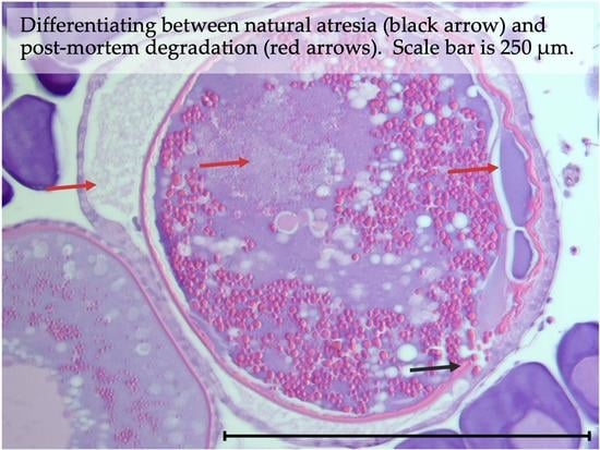
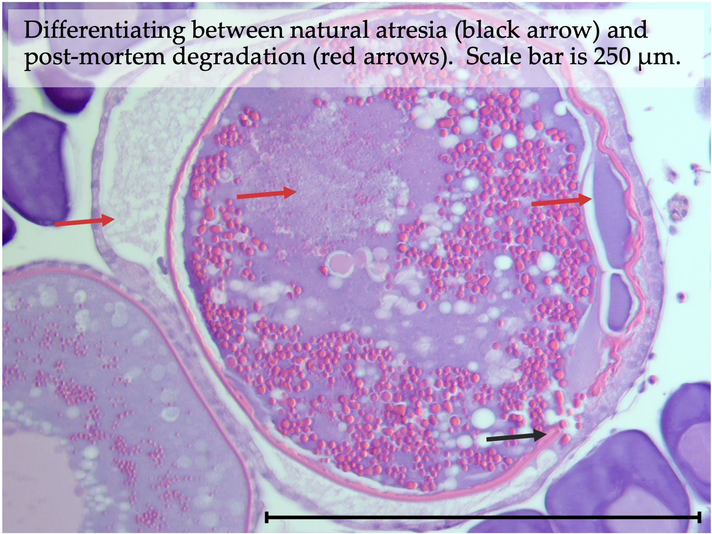
3.1. Natural Ovarian Atresia

Natural ovarian follicular atresia of vitellogenic oocytes in tropical deepwater snapper was separated into four stages (alpha, beta, gamma, and delta). Under alpha atresia, the disintegration of organelles occurs in the periphery of the ooplasm with enlarged granulosa cells, and the chorion loses its structural integrity (i.e., shrinking, breaking into segments; Figure 1A). Beta atresia is characterized by phagocytic vacuoles entering the ooplasm, complete breakdown of the chorion, and liquefaction and breakdown of vitellogenin (Figure 1B). Under gamma atresia, the chorion is no longer visible, the vitellogenin has been reabsorbed, and large vacuoles remain (Figure 1C). Lastly, delta atresia is characterized by remnant connective tissue and accumulation of yellow-brownish pigments (Figure 1D). Delta atresia is often difficult to differentiate from old (36–48 h) post-ovulatory follicles (POF) in warm water fishes. Melano-macrophage centers (MMC) were commonly observed in tropical deepwater snappers (Figure 1E), representing a phagocytic or immune response. Occasionally, during natural ovarian follicular atresia, oocytes can become encysted (Figure 1F).

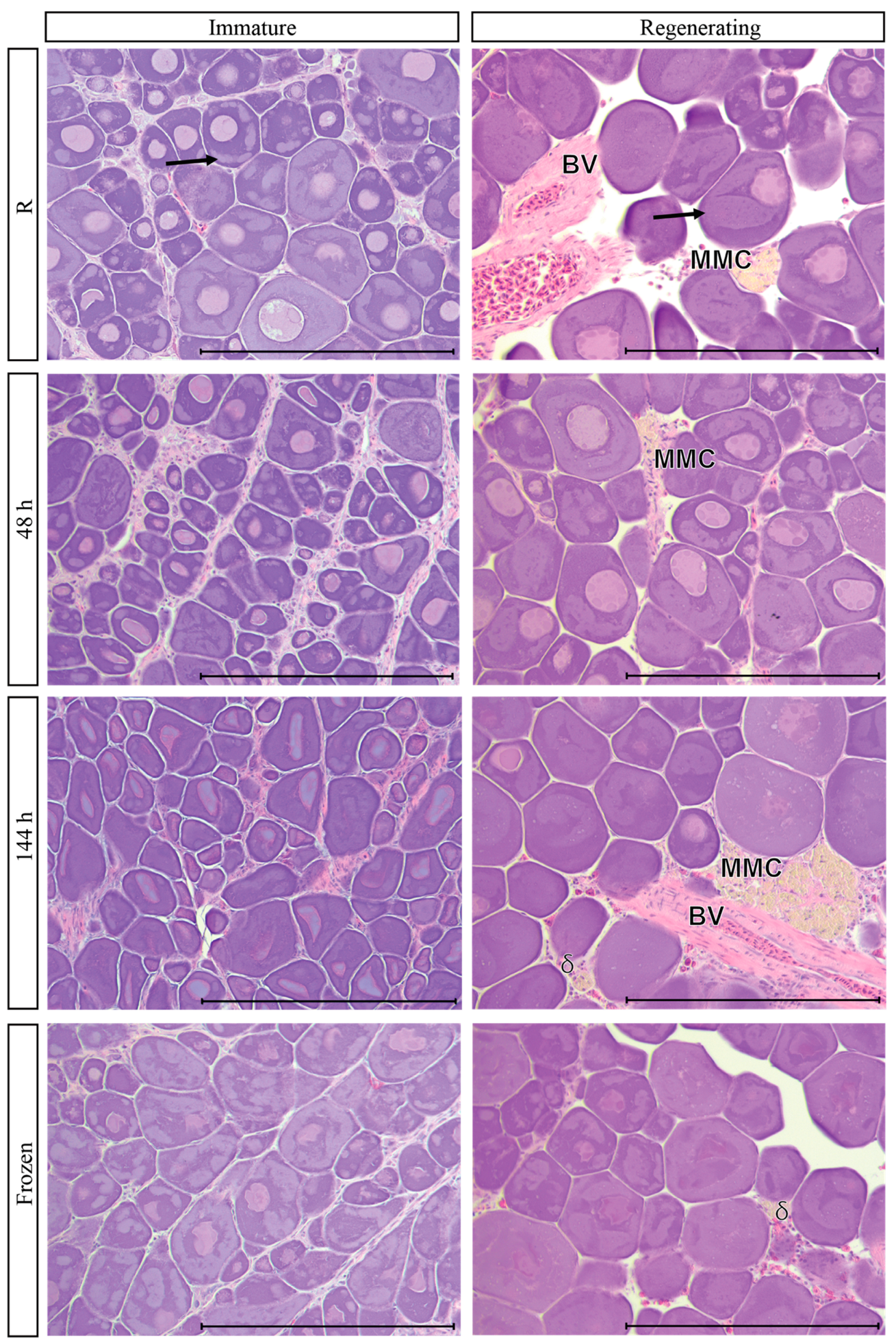
3.2. Reproductive Phases of Females Examined

For each of the three species examined, fish in various reproductive phases were evaluated to observe the effect of each treatment on ovarian follicles compared to the reference tissue. For P. filamentous, there were four immature females, two regenerating females, and two late developing females. Three regressing, one late developing, and one spawning E. carbunculus females and one spawning female P. auricilla were also sampled (Table 2). For all these fish, gonads were extracted before treatments were applied. For immature and regenerating females, there was only enough gonad tissue to do four treatments (reference, 48 h, 144 h, and frozen). All seven treatments were applied to the late developing, spawning, and regressing females. For fish with intact gonads, only four reproductive phases were examined from Etelis carbunculus: late developing, spawning, regressing, and regenerating (Table 2).

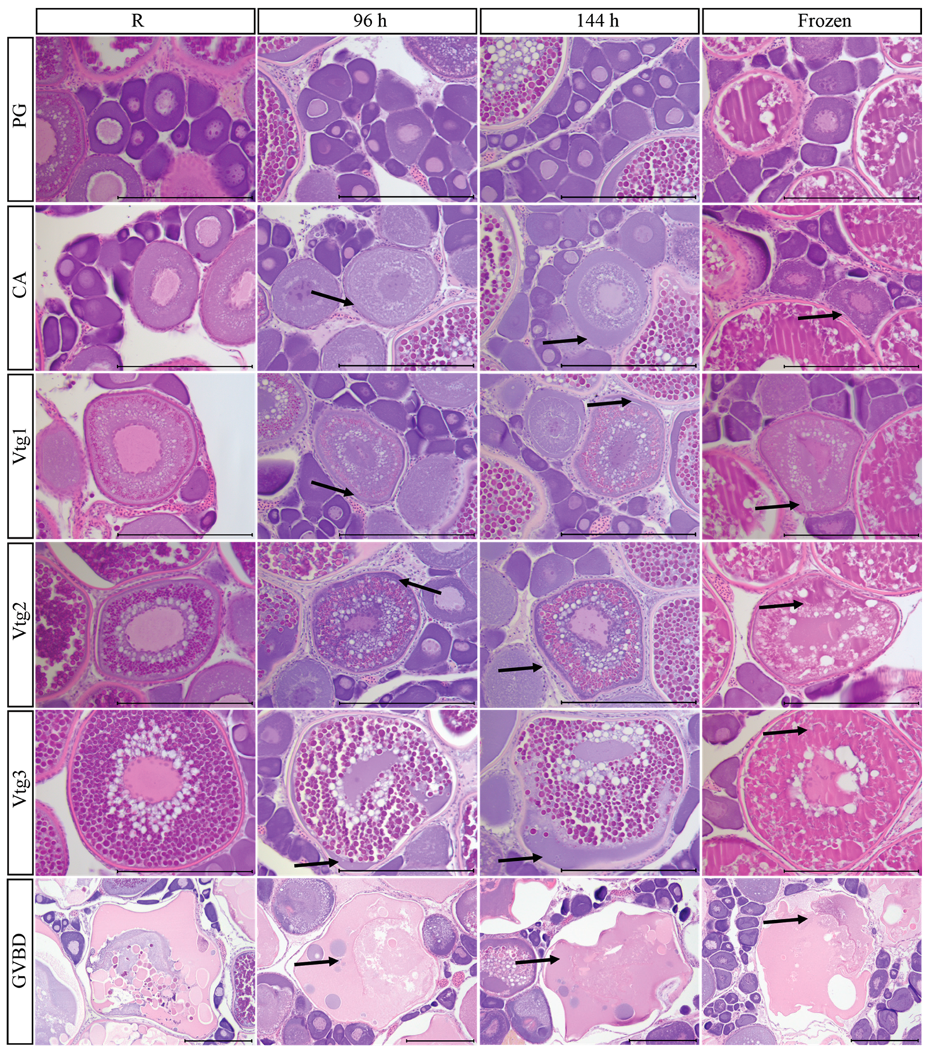
3.3. Primary Growth Oocytes

For E. carbunculus, primary growth (PG) oocytes in the perinucleolar (PN) stage did not appear to change in appearance or shape under the 12 h ambient, 24 h, 48 h, or 96 h treatments compared to the reference treatment (Figure 2). Under the 144 h treatment, the nucleus of PN oocytes appeared to be cloudy and potentially beginning to break down (Figure 2 and Figure 3). Freezing did not have a discernible effect on the identification of PN oocytes (Figure 2 and Figure 3). Primary growth oocytes looked similar in regenerating females compared to immature females under all treatments (Figure 3).
A granular ooplasm was observed in the PG oocytes of P. filamentosus (Figure 3). These structures were observed to shrink but were still visible at 144 h on ice (Figure 3). Freezing did not change the appearance of the granular ooplasm (Figure 2).

3.4. Cortical Alveolar Oocytes

Effects of post-mortem degradation were minor for cortical alveolar (CA) oocytes after 12 h at ambient temperature and 24 and 48 h on ice (Figure 2). However, post-mortem degeneration was clearly evident in CA oocytes at 96 h and 144 h, with the beginning of liquefaction seen at 96 h and degradation of organelles around the periphery of the cytoplasm, early breakdown of the nucleus discernable, and liquefaction obvious at 144 h (Figure 2). The chorion remained intact under all treatments. Freezing had little effect on CA oocytes, and the cortical alveoli were still present, along with an intact nucleus (Figure 2).

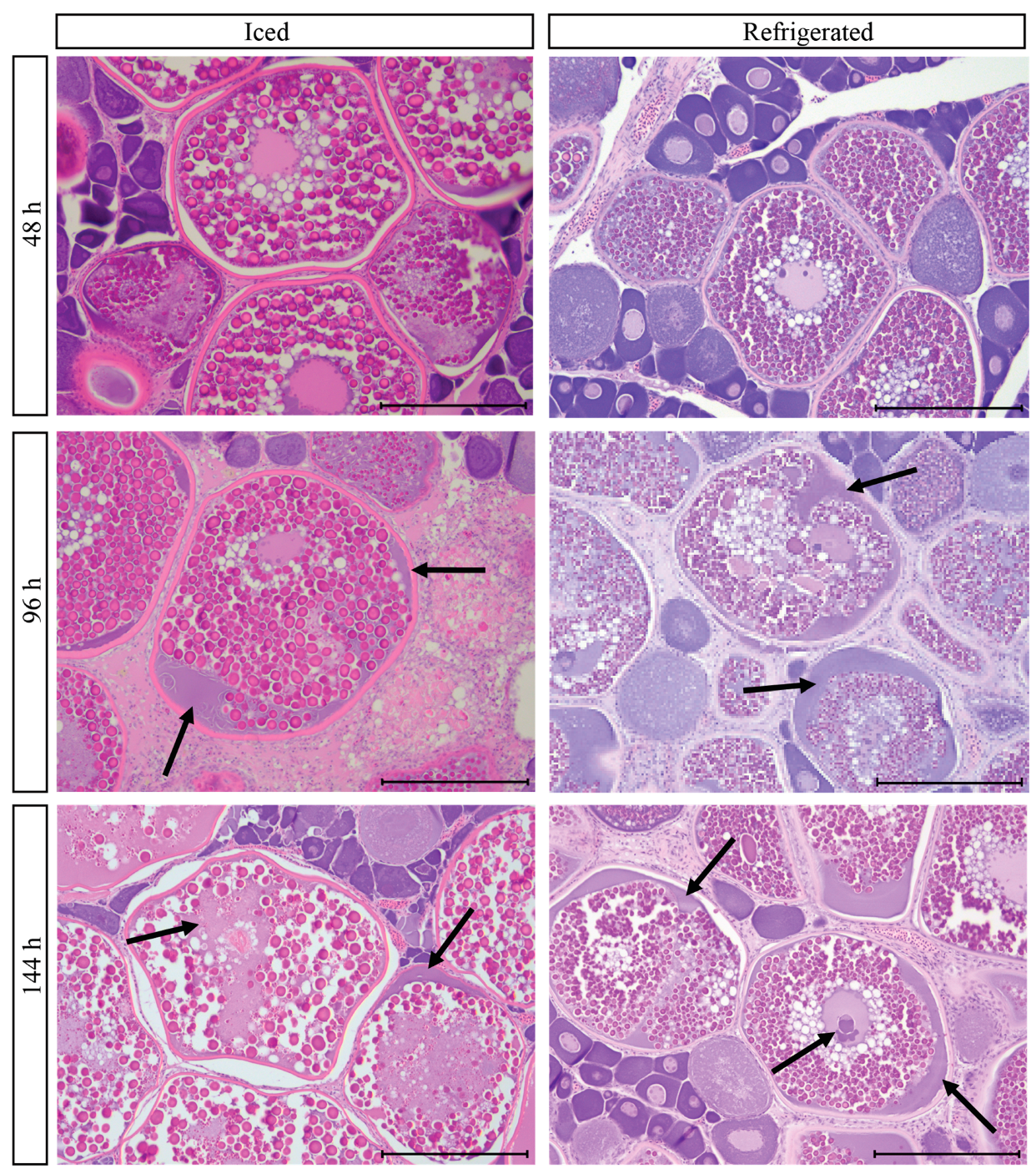
3.5. Vitellogenic Oocytes

Handling effects were observed after 12 h ambient and 48 h on ice or refrigeration for all stages of vitellogenic oocytes. However, post-mortem degradation was more apparent and pervasive in later-stage vitellogenic oocytes. At 12 h ambient room temperature, there was a breakdown of ooplasm organelles at the oocyte periphery of Vtg3 oocytes, staining purple, which was not seen in Vtg1 and Vtg2 (Figure 2). All stages of vitellogenic oocytes had post-mortem degradation after 96 h. The breakdown of ooplasm organelles increased with time on ice or refrigeration, resulting in larger areas of the periphery of the oocyte staining purple, particularly for Vtg3 oocytes (Figure 2 and Figure 4). The chorion remained intact under all treatments. Additionally, Vtg2 and Vtg3 oocytes become hard to differentiate from each other at 96 and 144 h (Figure 2). For all vitellogenic oocytes, with increasing time to fixation, it becomes hard to determine if the oocyte is a vitellogenic oocyte with post-mortem degradation or if it is atretic. Freezing caused the vitellogenic oocytes to change in appearance, with expansion and distortion of the vitellogenin within the developing oocyte; there was more pronounced distortion as oocytes progressed from Vtg1 to Vtg3 due to the increased amount of vitellogenin present.
Differences were observed in post-mortem degradation of vitellogenic oocytes between treatments on ice compared to treatments with refrigeration, with treatments on ice appearing to degrade slightly faster than those refrigerated, particularly for the Vtg3 oocytes (Figure 4). The nuclei of Vtg3 oocytes appear to dissolve at 144 h for fish held on ice but remain intact for most, but not all, of the Vtg3 oocytes after 144 h for fish that were refrigerated (Figure 4).

3.6. Oocyte Maturation

Oocytes undergoing the germinal vesicle breakdown (GVBD) stage of oocyte maturation were found in P. auricilla. At 12 h ambient, 96 h, and 144 h refrigerated, the GVBD oocytes appear to contract, and distortion or dissolution of the remaining vitellogenin and cytoplasm occurs (Figure 2). However, GVBD oocytes were easily recognized under all treatments (Figure 2), which would allow accurate classification of the spawning phase.

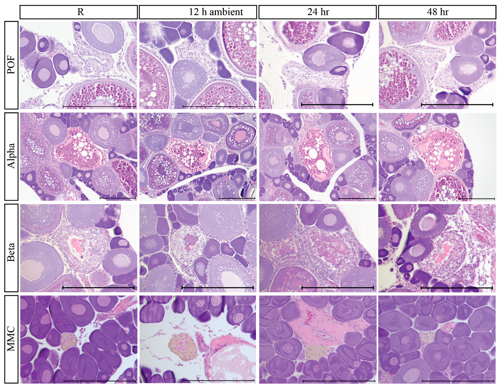
3.7. Post-Ovulatory Follicles

Post-ovulatory follicle complexes (POFs) ~24 h old, recognized by follicular cells that are not completely collapsed and have a small amount of remaining space between follicular layers, were observed in a spawning E. carbunculus female. These POFs were easily identified in reference tissue, 12 h ambient, 24 h, and 48 h refrigeration (Figure 5). However, POFs were more compact and harder to accurately identify at 96 h and 144 h (Figure 5). Indeed, POFs after 96 h refrigeration could be mistaken for an atretic oocyte with a compromised chorion. Post-ovulatory follicles were more compact but still easily identifiable in the frozen treatment (Figure 5), although determining the age of the POF was more challenging when frozen. Older POFs would be even harder to identify with increasing post-mortem degradation.

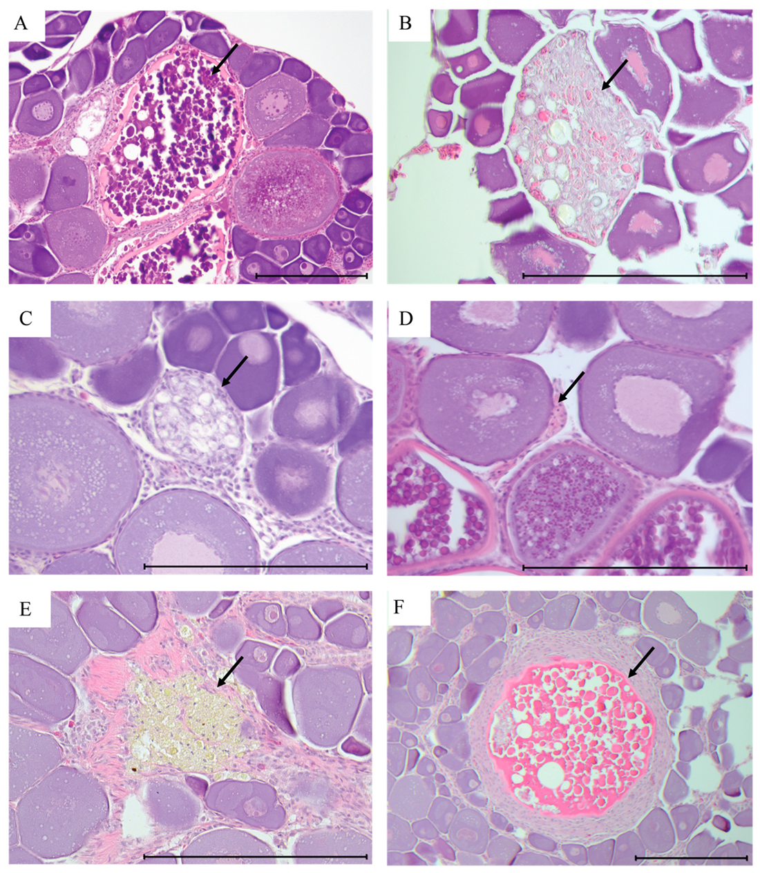
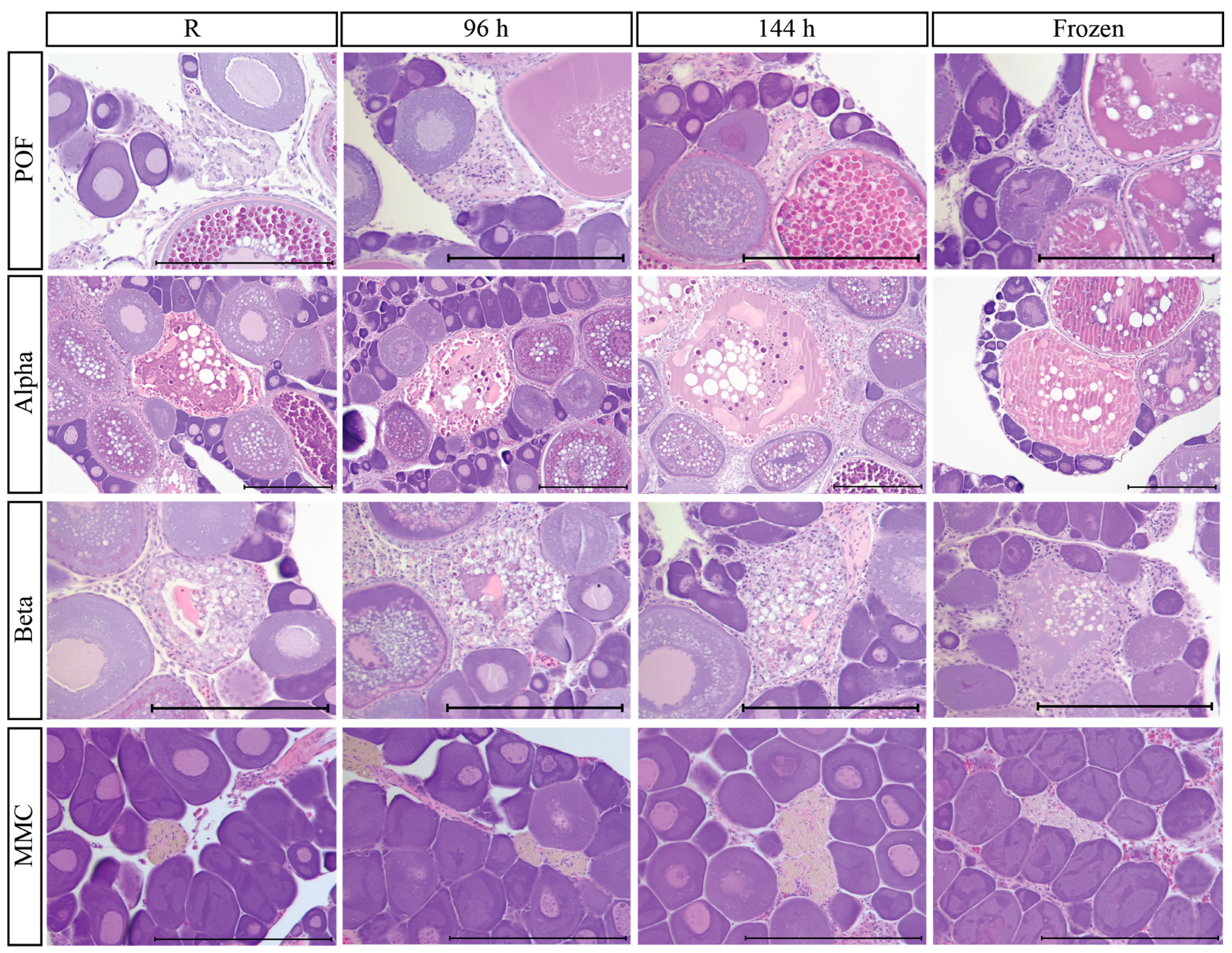
3.8. Atretic Oocytes and Indicators of Prior Spawning

Atretic oocytes remained identifiable under all treatments (Figure 5). Size and appearance remained similar and were generally recognizable as the same stage of atresia, but atresia appears to progress further at later time points. In particular, beta atresia starts to resemble gamma at later time points and after freezing. However, it was hard to compare among time points because there were various substages of atresia present in the same individual. At 96 h and 144 h, some additional breakdown was evident, and at 144 h, additional debris was observed inside the gonad. The MMC remained identifiable in all treatments (Figure 3 and Figure 5), although the distinct brownish coloration was not visible in the frozen specimens, which may result in incorrect classification as beta or gamma atresia. Muscle bundles, delta atresia, and blood vessels were identifiable in all treatments in regenerating females (Figure 3).

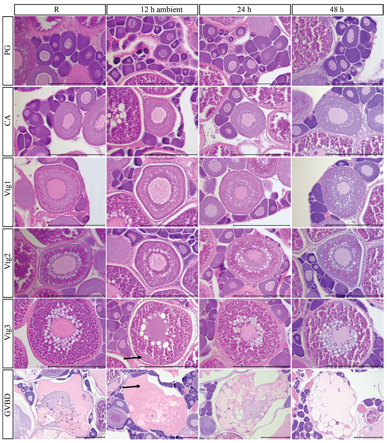
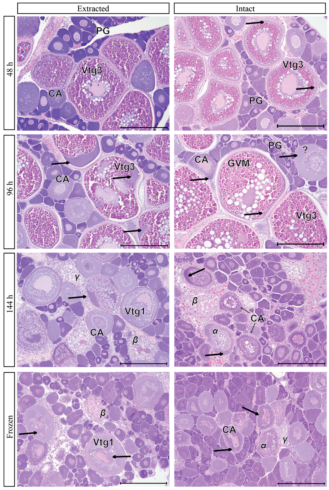
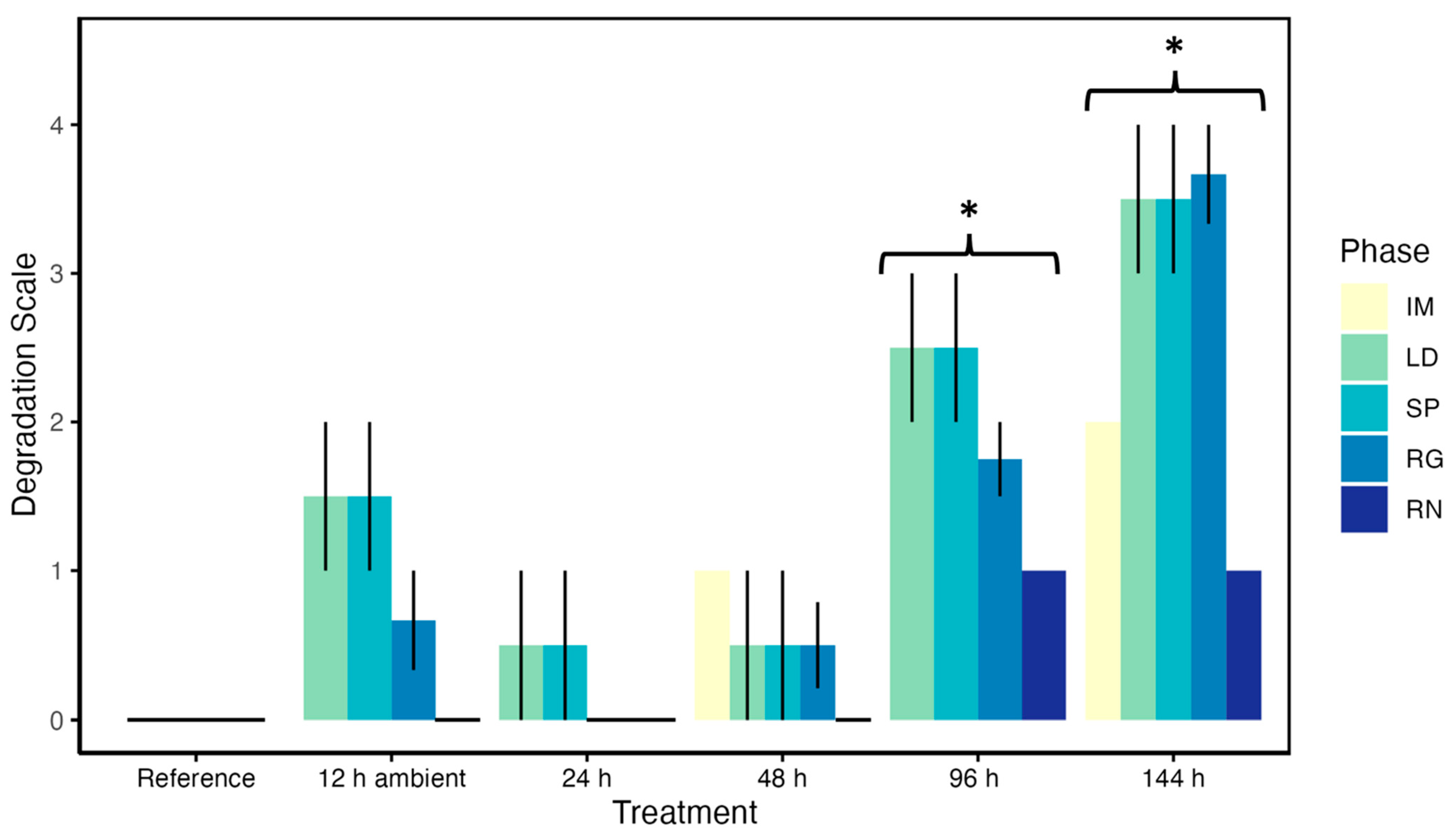
3.9. Post-Mortem Degradation in In Situ Ovaries

Eight E. carbunculus were left intact with one treatment applied to the whole fish in order to determine if histological ovarian morphological changes associated with handling effects were similar in appearance to those where the gonad had been extracted prior to treatment (Table 2). Intact E. carbunculus were sampled at 48 h, 96 h, 144 h, and frozen. While a reference sample was not taken under this scenario, reference tissues are shown in Figure 2 for each oocyte stage. Different sample preparations (gonad intact vs. gonad extracted) showed slight post-mortem degradation characteristics at 48 h (Figure 6). However, there is a clear difference in the post-mortem degradation in Vtg3 oocytes at 96 h between extracted and intact gonads, with increased degradation in extracted gonads compared to intact (Figure 6). Intact ovaries undergoing oocyte maturation (germinal vesicle migration [GVM]) showed minor degradation at 96 h (Figure 6), in comparison to the greater degradation of GVBD oocytes from extracted samples at 96 h (Figure 2), but could still be readily identified. There was substantial and similar post-mortem degradation for extracted and intact regressing individuals at 144 h and frozen, with difficulties distinguishing between atresia stages as well as identifying CA oocytes, particularly in the intact gonads (Figure 6).
Confidence in reproductive staging from the intact gonad method was high for the late developing and spawning individuals with Vtg3 or GVM oocytes at 48 h and 96 h. However, our confidence staging intact regressing individuals was very low at 144 h and when frozen. For example, sample RA-22-01-035 was fixed after 144 h on refrigeration and was classified as regressing; the predominance of atretic oocytes in multiple stages of atresia provides confidence in this classification. However, there are some oocytes stages that we could not identify with confidence in the intact gonad (Figure 6), and the identified alpha stage atresia oocytes may be misclassified as healthy but degraded, vitellogenic oocytes since post-mortem degradation was hard to differentiate from alpha atresia at 144 h.

3.10. Impacts of Post-Mortem Degradation on Reproductive Classification

To evaluate the classification of reproductive phases at varying time points in post-mortem degeneration, we constructed an ordinal scale based on the degeneration of oocyte stages, atresia, and POFs at varying time points based on the descriptions and illustrations above (Table 3). Fish kept on ice and refrigerated were not differentiated for this analysis; thus, there is some variability in the degradation scale from individual to individual.
For fish held on ice for 24 h to 48 h, there was little to no degradation in all observed phases compared to the reference tissue (24 h: Z = 0.64, adjusted p-value = 1.00; 48 h: Z = 1.71, adjusted p-value = 0.71; Figure 7). At 96 h and 144 h, degradation rates increased sharply from the reference tissue (96 h: Z = 4.50, adjusted p-value < 0.01; 144 h: Z = 5.73, adjusted p-value < 0.01) (Figure 7). Regressing fish could be accurately identified at 96 h on ice with only slight degeneration of atretic oocytes, but late developing and spawning-phase fish showed moderate to major degeneration in vitellogenic oocytes at this time point. By 144 h on ice, there was some severe degradation in fish in the late developing, spawning, and regressing phases, with moderate degeneration in immature and slight degeneration in regenerating fish (Figure 7). However, degradation severity was not statistically different among reproductive phases at 96 h (Kruskal–Wallis X2 = 5.88, d.f. = 3, p-value = 0.12) nor at 144 h (Kruskal–Wallis X2 = 9.32, d.f. = 4, p-value = 0.05), likely due to the small sample sizes and individual variation. Importantly, even 144 h on ice did not impair the ability to accurately identify PG oocytes or indicators of prior spawning, such as muscle bundles, delta atresia, or blood vessels, thus allowing differentiation between immature and regenerating fish.
Overall, ovary tissue that has been chilled for 48 h or less has minimal post-mortem degradation and will not influence the histological reproductive classification (Figure 7). Furthermore, while up to 12 h at ambient temperature in the tropics showed slight to moderate degradation in the late developing and spawning phases, with a slight degradation in the regressing phase (Figure 7), histological reproductive classification is not substantially impaired. After 96 h chilled, the most notable effects are the breakdown of organelles in the cytoplasm that results in the periphery of the oocyte staining purple under H&E staining (i.e., moderate to severe degeneration, Table 3); this is most noticeable in vitellogenic oocytes and is characteristic of post-mortem degradation which can be differentiated from natural ovarian atresia. The more troubling effects under longer post-mortem degeneration are the breakdown of the nucleus and progressive breakdown of the chorion, which can mimic natural atresia. These post-mortem effects make staging vitellogenic oocytes challenging and can influence the interpretation of the histological reproductive classification after 96 h chilled in late developing and spawning phases (Figure 7). Furthermore, difficulty distinguishing POFs from late-stage natural atresia after 96 h chilled can influence the classification of both the spawning and regressing phases.

4. Discussion

Our research demonstrates that there are differences between natural ovarian atresia and post-mortem degradation and that handling effects can be identified in histological cross-sections. Under natural ovarian atresia, the chorion shrinks, and granulosa cells enlarge as atresia progresses, with fragmentation of the chorion and granulosa cells entering into the oocyte to phagocytize degenerating material [5,11]. Under post-mortem degradation, the chorion did not break apart nor fragment within 144 h. Post-mortem degradation did cause the breakdown of organelles in the cytoplasm (stained a uniform purple) to become more apparent and started at the edges of the oocyte and worked inward. This is not seen under natural ovarian atresia. Under post-mortem degradation, dissolution of the nuclear envelope was observed at 144 h on ice and, to a lesser extent, in ovaries kept at 144 h in refrigeration. Liquefaction of yolk globules (vitellogenin) was not observed under post-mortem degradation within 144 h refrigeration but was observed at 144 h on ice and is also part of natural ovarian follicular atresia. Finally, ovarian tissue showed slight to moderate degradation when kept at ambient temperatures (no ice) for only 12 h, although it is possible to distinguish reproductive phases.
Overall, keeping gonads in a constant temperature refrigeration unit resulted in slightly less post-mortem degradation than gonads kept directly on ice during the first 96 h. This is likely due to stable temperatures in the refrigerator, whereas ice melts and needs to be replaced multiple times over the course of several days. Thus, differences in stable cooling could explain the observed difference in vitellogenin breakdown between the iced and refrigerated samples. Additionally, gonads that are in direct contact with ice can have some freezing effects, particularly in the periphery, that can exacerbate accurate oocyte stage identification. This inadvertent freezing of ovarian tissue on ice could be ameliorated by placing a portion of the ovarian tissue in a cassette and then on ice, as done in our study, rather than placing gonads directly on ice. Minimally, histological samples from gonads placed directly on ice should be taken from the interior of the gonad rather than the periphery.
We anticipated that handling effects would make it difficult to distinguish immature females from regenerating females. The granular structure seen in PG oocytes of immature ovaries at all stages of post-mortem degradation was not seen in PG oocytes of regenerating females. This structure may be Balbiani bodies, but its function in fish remains unknown [12]. However, despite this difference in PG oocytes, it was not difficult to distinguish between immature and regenerating phase females, regardless of the time tissues were on ice. The identification of mature regenerating females from immature females is made by the presence of multiple diagnostic criteria of prior spawning activity, e.g., thick ovarian walls (tunica), loose spacing among PG oocytes, atretic oocytes (alpha, beta, gamma, and/or delta), POFs, muscle bundles, MMCs, or enlarged blood vesicles [1,13]. Atresia, blood vessels, and muscle bundles remained identifiable under all treatments, and therefore differentiation of immature from regenerating females was possible. The only indicator of prior spawning that may be missed is old POFs since detection became difficult at >96 h on ice or refrigeration and after freezing. However, POFs are rarely observed in regenerating females from warm waters [1,13], although a lack of detectable older POFs in cold water species may be more problematic in correctly assigning reproductive phases in ovaries that have undergone post-mortem degradation.
Large impacts were observed in the post-mortem degradation of vitellogenic oocytes. A common mistake when working with market fish, or fish that have been on ice for many days, may be to misidentify healthy vitellogenic oocytes as early-stage alpha atretic oocytes due to post-mortem degradation. As vitellogenic oocyte development progressed from Vtg1 to Vtg3, post-mortem effects were observed much earlier in the handling process. In particular, the breakdown of organelles and the resulting purple-staining liquefaction were obvious in Vtg3 oocytes after 12 h in ambient temperatures and after 48 h on ice compared to the reference sample. Under natural atresia, the breakdown of the cytoplasm of vitellogenic oocytes stains pink with H&E staining, but under post-mortem atresia, the cytoplasm of vitellogenic oocytes stains purple. There is a decrease in tissue pH in cells after injury or death that should increase the eosin stain affinity [14,15]; however, under post-mortem degeneration, more hematoxylin staining appeared in the periphery of the oocytes. Perhaps there is leakage of basophilic cell materials such as ribosomes, rough endoplasmic reticulum, or DNA into the cytoplasm during post-mortem degradation as the cells lose their structural integrity, which would result in more hematoxylin staining. Although oocytes in post-mortem degradation could still be identified as vitellogenic, it became progressively more difficult to distinguish among the vitellogenic stages as time on ice or refrigeration increased; we have demonstrated that this can affect accurate identification of reproductive phases and, therefore, spawning season duration.
Handling effects can also make the identification of POFs challenging. Although recent POFs remained easily identifiable after 12 h at ambient temperatures or up to 48 h on ice or refrigeration compared to the reference sample, the size of POFs decreased with increasing time on ice or in refrigeration and became harder to differentiate from gamma and delta atresia within the ovary. This could lead to misidentification of the spawning season since POFs are an indication of recent spawning [1,8]. Furthermore, in batch-spawning species, compromised identification of POFs can affect estimates of spawning interval and reproductive output in terms of annual fecundity [16,17].
Fish collected from fishery-dependent sampling are often frozen, which can make histological interpretation of gonadal tissues challenging. It is not uncommon to see the use of frozen gonad tissue in reproductive studies; however, very little is documented on how freezing changes ovarian tissue [2,3,18]. Furthermore, there is a difference in opinion on whether frozen gonads are adequate for histological reproductive assessments [3,19,20]. Overall, our confidence in staging frozen ovaries was low. We show that the freezing of ovarian samples resulted in reduced spacing between oocytes, loss of integrity and shape changes of vitellogenin globules in vitellogenic oocytes, changes in oocyte shape, and reduced ability to detect POFs. We found it difficult to differentiate between atresia and POFs and to determine which stage of vitellogenic oocytes were present in frozen ovaries, whether they were intact or extracted. One of the biggest issues when assessing frozen tissue is the loss of observable structure in the vitellogenic oocytes and, thus, the limited ability to differentiate vitellogenic oocyte stages. Freezing results in irreversible structural alterations in intracellular proteins such as vitellogenin [21], and freezing can result in phospholipid de-esterification [22] and, thus, the loss of structural integrity of the vitellogenin. While immediate freezing allowed the identification of the presence of vitellogenic oocytes, but not necessarily the stage of vitellogenesis, freezing compromised the identification of POFs, and therefore made it more difficult to accurately identify reproductive phase and spawning seasonality. Indeed, Kopf [23] found that POFs or atresia were misclassified in 18% of frozen samples compared to the reference tissue that was fixed in 10% buffered formalin. Ultimately, the use of frozen gonadal tissue is risky for the accurate identification of reproductive phases, but in some circumstances, there may not be an alternative, particularly in pelagic fisheries that spend a long time at sea. Therefore, we recommend that histological documentation be included demonstrating that the frozen tissue can be accurately classified for the species, time period, and region if it is included in the assessment.
Keeping ovaries intact in fish that are kept on ice or refrigerated, rather than removing them, may reduce post-mortem effects to some extent. Although direct comparisons of the same fish among different time points were not possible for fish with intact ovaries in our study, comparison to the reference tissue for fish in the same reproductive phase is appropriate. For instance, the degradation of vitellogenic oocytes was slight but evident at 48 h in both the extracted vs. intact gonads in comparison with reference tissue. As fish were kept on ice longer than 96 h, continued degradation effects were evident in both intact and removed gonad treatments. Post-mortem degradation effects were observed in tertiary vitellogenic oocytes at 96 h but were greater in extracted ovaries compared to intact ovaries. At 144 h, post-mortem degradation was so extensive for both the extracted and intact ovaries that it was not possible to determine if one treatment was worse than the other for fish in the regressing phase. Keeping intact, large-bodied warm-water fishes on ice may exacerbate post-mortem degradation compared to smaller-bodied fishes since it would take longer for the gonads in larger fish to be sufficiently chilled, thus staving off post-mortem degeneration; intact cooling of both large and small-bodied cold-water fishes may result in less post-mortem degradation than observed here for warm-water tropical species.
There are many nuances when it comes to determining handling effects on gonad samples. For instance, post-mortem degradation acted faster on the samples held in a cooler with ice than when samples were held under refrigeration. Additionally, the ambient air temperature in our tropical sampling areas ranged from 21.1 to 30.6 °C; higher temperatures can certainly accelerate the degradation of samples held without ice, while sampling in cooler regions may result in less degradation for fish held without ice. Furthermore, treatment of frozen tissue is also important, as gonadal tissue may be more damaged or affected in the periphery of the sample due to the freeze/thaw cycle [2]. Avoiding a freeze/thaw cycle can make it difficult to excise gonads from a frozen fish. Ultimately, these nuances make it challenging to determine the precise time that post-mortem degradation will result in inaccurate histological classification of ovarian tissue.
We provide a general reference from which to develop best handling practices and help in the identification of handling effects from fishery-dependent sampling. Although our data are based on tropical snapper species, similar post-mortem degradation has been commonly observed by this study’s authors in a wide range of temperate and warm-water oviparous species following delayed preservation. Further research is needed to determine the timing of post-mortem degradation in temperate and cold-water species, as timing of degradation likely varies based on the temperature of the water as well as the size of the species. Furthermore, species with different reproductive strategies and life histories, such as ovoviviparous species and total spawners, may demonstrate variations in the post-mortem degradation described here and need additional study.

5. Conclusions

Based on data from this study, we recommend that iced samples be processed within 48 h of capture, although the reproductive phase can be determined with moderate confidence from samples held for 96 h on ice despite significant degradation of POFs by this time period. Refrigeration appears to preserve the gonads better than a cooler with ice, allowing accurate determination of oocyte stages and, thus, reproductive phases up to 96 h. Furthermore, keeping fish well-iced intact results in less ovarian degradation than removing gonads and storing them on ice. However, when doing fishery-dependent sampling, it is common to sample fish that have been held on ice for days (average of four to five days or 96–120 h). This time frame seems to be the maximum amount of time for intact fish on ice before post-mortem degradation of the ovary becomes a serious issue for assessing reproductive status in tropical deepwater snappers; it was very difficult to accurately determine the reproductive phase of fish held on ice for 6 days (144 h).
This research on handling effects can be used to support the identification of gonadal degradation and to help differentiate between post-mortem effects and natural atresia. Additionally, we have provided information to help fish reproductive biologists identify the characteristics of post-mortem degradation and reduce errors in assessing reproductive phases from gonads collected from fishery-dependent sampling. Finally, this study serves as a guide to support methods and best practices for the collection and handling of fish reproductive samples under less-than-ideal conditions.

Supplementary Materials

The following supporting information can be downloaded at: https://www.mdpi.com/article/10.3390/fishes8080406/s1, Post-mortem degradation severity ranking data.

Author Contributions

E.S. and N.J.B.-P. developed the experimental design. E.S. collected and analyzed the samples. E.S. and N.J.B.-P. wrote the manuscript. All authors have read and agreed to the published version of the manuscript.

Funding

This project was supported by grants from the National Institute on Minority Health and Health Disparities (U54MD007601) National Institutes of Health.

Institutional Review Board Statement

Biological sampling followed Institutional Animal Care and Use Standards under University of Hawaii IACUC (Protocol number 13-1696-8).

Data Availability Statement

All available data are presented in the paper and contained within Supplementary Materials.

Acknowledgments

The Pacific Islands Fish Science Center Life History Program, in particular, Joseph O’Malley, Erin Reed, and Kristen Dahl, for logistic support. The crew of the NOAA research vessel Oscar E. Sette and the NOAA research vessel Rainier. The John Burns School of Medicine Histopathology Core for histological processing of gonad samples, in particular, Miyoko Bellinger. The Maturity Assessment Reproductive Variability and Life Strategies (MARVLS) group for review of the research and methodological advice.

Conflicts of Interest

The authors declare no conflict of interest.

References

  1. Brown-Peterson, N.J.; Wyanski, D.M.; Saborido-Rey, F.; Macewicz, B.J.; Lowerre-Barbieri, S.K. A standardized terminology for describing reproductive development in fishes. Mar. Coast. Fish. 2011, 3, 52–70. [Google Scholar] [CrossRef]
  2. Wuenschel, M.J.; Deroba, J.J. The reproductive biology of female Atlantic herring in US waters: Validating classification schemes for assessing the importance of spring and skipped spawning. Mar. Coast. Fish. 2019, 11, 487–505. [Google Scholar] [CrossRef] [Green Version]
  3. Young, J.; Drake, A.; Brickhill, M.; Farley, J.; Carter, T. Reproductive dynamics of broadbill swordfish, Xiphias gladius, in the domestic longline fishery off eastern Australia. Mar. Freshw. Res. 2003, 54, 315–332. [Google Scholar] [CrossRef]
  4. Corriero, A.; Zupa, R.; Mylonas, C.C.; Passantino, L. Atresia of ovarian follicles in fishes, and implications and uses in aquaculture and fisheries. J. Fish. Dis. 2021, 44, 1271–1291. [Google Scholar] [CrossRef] [PubMed]
  5. Miranda, A.; Bazzoli, N.; Rizzo, E.; Sato, Y. Ovarian follicular atresia in two teleost species: A histological and ultrastructural study. Tissue Cell 1999, 31, 480–488. [Google Scholar] [CrossRef] [PubMed] [Green Version]
  6. Cassel, M.; de Paiva Camargo, M.; Oliveira de Jesus, L.W.; Borella, M.I. Involution processes of follicular atresia and post-ovulatory complex in a characid fish ovary: A study of apoptosis and autophagy pathways. J. Mol. Histol. 2017, 48, 243–257. [Google Scholar] [CrossRef]
  7. George, J.; Van Wettere, A.J.; Michaels, B.B.; Crain, D.; Lewbart, G.A. Histopathologic evaluation of post-mortem autolytic changes in bluegill (Lepomis macrohirus) and crappie (Pomoxis anularis) at varied time intervals and storage temperatures. PeerJ 2016, 4, e1943. [Google Scholar] [CrossRef] [PubMed] [Green Version]
  8. Lowerre-Barbieri, S.; Fries, C.; Brown-Peterson, N.; Moncrief-Cox, H.; Barnett, B. Best Practices for Standardized Reproductive Data and Methodology to Estimate Reproductive Parameters for Red Snapper in the Gulf of Mexico. 2022. 43p. Available online: https://sedarweb.org/documents/sedar-74-dw-36-best-practices-for-standardized-reproductive-data-and-methodology-to-estimate-reproductive-parameters-for-red-snapper-in-the-gulf-of-mexico/ (accessed on 1 July 2023).
  9. R Core Team. R: A Language and Environment for Statistical Computing; R Foundation for Statistical Computing: Vienna, Austria, 2022; Available online: https://www.R-project.org/ (accessed on 28 June 2023).
  10. Ogle, D.H.; Doll, J.C.; Wheeler, A.P.; Dinno, A. FSA: Simple Fisheries Stock Assessment Methods. R Package Version 0.9.4. 2023. Available online: https://CRAN.R-project.org/package=FSA (accessed on 28 June 2023).
  11. Yang, Y.; Wang, G.; Li, Y.; Hu, J.; Wang, Y.; Tao, Z. Oocytes skipped spawning through atresia is regulated by somatic cells revealed by transcriptome analysis in Pampus argenteus. Front. Mar. Sci. 2022, 9, 927548. [Google Scholar] [CrossRef]
  12. Zelazowska, M.; Kilarski, W.; Bilinski, S.M.; Podder, D.D.; Kloc, M. Balbiani cytoplasm in oocytes of a primitive fish, the sturgeon Acipenser gueldenstaedtii, and its potential homology to the Balbiani body (mitochondrial cloud) of Xenopus laevis oocytes. Cell Tissue Res. 2007, 329, 137–145. [Google Scholar] [CrossRef] [PubMed]
  13. Pacicco, A.E.; Brown-Peterson, N.J.; Murie, D.J.; Allman, R.J.; Snodgrass, D.; Franks, J.S. Reproductive biology of yellowfin tuna (Thunnus albacares) in the northcentral US Gulf of Mexico. Fish. Res. 2023, 261, 106620. [Google Scholar] [CrossRef]
  14. Lemasters, J.J.; Nieminen, A.-L.; Qian, T.; Trost, L.C.; Elmore, S.P.; Nishimura, Y.; Crowe, R.A.; Cascio, W.E.; Bradham, C.A.; Brenner, D.A.; et al. The mitochondrial permeability transition in cell death: A common mechanism in necrosis, apoptosis and autophagy. Biochim. Biophys. Acta (BBA)—Bioenerg. 1998, 1366, 177–196. [Google Scholar] [CrossRef] [Green Version]
  15. Von Knebel Doeberitz, M.; Wentzensen, N. CHAPTER 1—The Cell: Basic Structure and Function. In Comprehensive Cytopathology, 3rd ed.; Bibbo, M., Wilbur, D., Eds.; W.B. Saunders: Edinburgh, UK, 2008; pp. 3–22. [Google Scholar]
  16. Lowerre-Barbieri, S.K.; Ganias, K.; Saborido-Rey, F.; Murua, H.; Hunter, J.R. Reproductive timing in marine fishes: Variability, temporal scales, and methods. Mar. Coast. Fish. 2011, 3, 71–91. [Google Scholar] [CrossRef] [Green Version]
  17. Hunter, J.R.; Macewicz, B. Improving the accuracy and precision of reproductive information used in fisheries. In Modern Approaches to Assess Maturity and Fecundity of Warm and Cold Water Fish and Squids; Havforskningsinstituttet: Bergen, Norway, 2003; pp. 57–68. [Google Scholar]
  18. Mackie, M.; Lewis, P. Assessment of Gonad Staging Systems and Other Methods Used in the Study of the Reproductive Biology of Narrow-Barred Spanish Mackerel, Scomberomorus commerson, in Western Australia; Fisheries Research Report; Department of Fisheries: Perth, Australia, 2001; No. 136. [Google Scholar]
  19. Ferreri, R.; Basilone, G.; D’Elia, M.; Traina, A.; Saborido-Rey, F.; Mazzola, S. Validation of macroscopic maturity stages according to microscopic histological examination for European anchovy. Mar. Ecol. 2009, 30, 181–187. [Google Scholar] [CrossRef]
  20. Kopf, R.; Davie, P.; Bromhead, D.; Young, J. Reproductive biology and spatiotemporal patterns of spawning in striped marlin Kajikia audax. J. Fish. Biol. 2012, 81, 1834–1858. [Google Scholar] [CrossRef] [PubMed]
  21. Mazur, P.; Cole, K.W. Roles of unfrozen fraction, salt concentration, and changes in cell volume in the survival of frozen human erythrocytes. Cryobiology 1989, 26, 1–29. [Google Scholar] [CrossRef] [PubMed]
  22. Brouwers, J.F.; Gadella, B.M. In situ detection and localization of lipid peroxidation in individual bovine sperm cells. Free Radic. Bio Med. 2003, 35, 1382–1391. [Google Scholar] [CrossRef] [PubMed]
  23. Kopf, R.K. Age, Growth, and Reproductive Dynamics of Striped Marlin, Kajikia audax in the Southwest Pacific Ocean; Charles Sturt University: Bathurst, Australia, 2010. [Google Scholar]
Figure 1. Natural atresia in tropical deepwater snapper. (A) Alpha, (B) beta, (C) gamma, (D) delta atresia, (E) melano-macrophagic center (MMC), and (F) atretic encysted oocytes. Arrows indicate each stage of atresia. Scale bar is 250 μm.
Fishes 08 00406 g001
Figure 2. Treatment effects (Reference [R], 12 h ambient room temperature, 24 h, 48 h, 96 h, 144 h, and frozen) on primary growth oocytes (PG) in perinucleolar stage, cortical alveolar oocytes (CA), primary vitellogenic oocytes (Vtg1), secondary vitellogenic oocytes (Vtg2), and tertiary vitellogenic oocytes (Vtg3) from Etelis carbunculus (RA-22-01-001), post-ovulatory follicles (POFs) from E. carbunculus (RA-22-01-002), and germinal vesicle breakdown oocytes (GVBD) from Pristipomoides auricilla (RA-22-01-010). Arrows indicate post-mortem degradation. Scale bar is 250 μm. Sample ID and associated data are in Table 2.
Fishes 08 00406 g002aFishes 08 00406 g002b
Figure 3. Handling effects on an immature female Pristipomoides filamentosus (SE22-02-003) with primary growth oocytes and a regenerating female (SE22-02-017) P. filamentosus with primary growth oocytes, melanomacrophage centers (MMC), delta atresia ( δ ) , and large blood vessels (BV). Sampled fresh (Reference [R]), 48 h, 144 h on ice, and after freezing. Arrows point to a cloudy cytoplasm, which may be Balbiani bodies. This can be seen in all treatments but are much harder to detect after 144 h on ice. Scale bar is 250 μm. Sample ID and associated data are in Table 2.
Fishes 08 00406 g003
Figure 4. Post-mortem degradation (arrows) on extracted gonads for late developing female P. filamentosus held on ice (SE-22-02-009) and on late developing E. carbunculus held in a controlled temperature refrigerator (RA-22-01-001) at 48 h, 96 h, and 144 h. Tertiary vitellogenic (Vtg3) oocytes shown. Scale bar is 250 μm. Sample ID and associated data are in Table 2.
Fishes 08 00406 g004
Figure 5. Treatment effects (Reference [R], 12 h ambient, 24 h, 48 h, 96 h, 144 h, and frozen) on post-ovulatory follicles (POF), alpha atresia, beta atresia, and melanomacrophage centers (MMC) in Etelis carbunculus (RA-22-01-002 [POF], RA-22-01-006 [alpha atresia], RA-22-01-003 [beta atresia]) and in Prisitipomoides filamentosus (SE-22-02-017 [MMC atresia]). Scale bar is 250 μm. Sample ID and associated data are in Table 2.
Fishes 08 00406 g005aFishes 08 00406 g005b
Figure 6. Handling effects (black arrows) on extracted gonads and on intact gonads (whole fish). Extracted gonads from late developing female E. carbunculus (48 h and 96 h: RA-22-01-001) and regressing female E. carbunculus (144 h and frozen: RA-22-01-003). Intact gonads from a late developing female E. carbunculus (48 h: RA-22-01-032), a spawning female E. carbunculus (96 h: RA-22-01-037), and regressing females E. carbunculus (144 h: RA-22-01-035, Frozen: RA-22-01-047). The “?” is an unknown oocyte stage due to extensive post-mortem degradation. Scale bar is 250 μm. Sample ID and associated data are in Table 2.
Fishes 08 00406 g006
Figure 7. Mean (± SE) scale of observed gonad tissue degradation from 0 (none) to severe (4) based on reproductive phases (refer to Table 3 for specific scale determination). No 12 h ambient, 24 h, or 96 h treatments were done on immature females due to insufficient amount of gonad tissue. Significant differences between treatments as compared to reference are indicated by *. IM = Immature, LD = Late Developing, SP = Spawning, RG = Regressing, RN = Regenerating.
Fishes 08 00406 g007
Table 1. Reproductive phases following Lowerre-Barbieri et al. [8].
Reproductive PhaseMost Advanced Oocyte StagePhysiologically MatureCharacteristics
ImmatureOogonia, chromatin nucleolar (CN), or perinucleolar (PN)NoSmall ovary with CN and PN primary growth (PG) oocytes. Thin ovary wall, no muscle bundles or large blood vessels.
Early Developing Cortical alveolar (CA)YesOnly PG and CA oocytes, no evidence of POFs, some atresia may be present, muscle bundles, and large blood vessels can be present in non-virgin females.
Late Developing Vitellogenic oocytes (Vtg1, Vtg2, Vtg3)YesCan have vitellogenic oocytes at any stage. No post-ovulatory follicle complexes (POFs) present. Some atresia may be present.
SpawningGerminal vesicle migration (GVM), germinal vesicle breakdown (GVBD), hydrated oocytes (H), ovulated eggs, POFsYesUndergoing oocyte maturation (lipid coalescence, GVM, GVBD, hydrated oocytes), ovulated eggs, or POF of any age. Can contain atretic oocytes.
RegressingPN, CA, and/or vitellogenicYesOvaries dominated by oocytes in any stage of atresia (50% or more). May contain some CA or Vtg oocytes and POF > 24 h.
RegeneratingPrimary growth oocytes (PG)YesOnly PG present (CN and PN). May contain unabsorbed material from past spawning events, gamma or delta atresia, large muscle bundles, large blood vessels, and thick ovarian wall.
Table 2. Pristipomoides filamentosus, P. auricilla, and Etelis carbunculus sampled for handling effect treatments. Fish were captured in April onboard the NOAA ship Oscar Elton Sette (SE) in the Main Hawaiian Islands and in August onboard the NOAA ship Rainier in the Mariana Islands (RA). All bolded fish had gonads extracted after treatments (intact). CA = cortical aleveolar oocytes, GVBD = germinal vesicle breakdown, GVM = germinal vesicle migrations, PG = primary growth oocytes, POF = post-ovulatory follicle complex, Vtg1 = primary vitellogenic oocytes, Vtg2 = secondary vitellogenic oocytes, Vtg3 = tertiary vitellogenic oocytes. Species codes: PRFL = P. filamentosus, ETCA = E. carbunculus, PRAU = P. auricilla.
Sample IDSpeciesFL (cm)Weight (kg)Gonad Weight (g)Reproductive PhaseMost Advanced Oocyte PresentAtresia Present (Alpha, Beta, Gamma, Delta)
SE-22-02-003PRFI30.20.5<0.1ImmaturePGNone
SE-22-02-004PRFI24.20.3<0.1ImmaturePGNone
SE-22-02-005PRFI26.20.3<0.1ImmaturePGNone
SE-22-02-006PRFI24.50.3<0.1ImmaturePGNone
SE-22-02-008PRFI35.00.53.4RegeneratingPGdelta
SE-22-02-009PRFI64.93.856.0Late DevelopingVtg3alpha, beta, and delta
SE-22-02-015PRFI37.50.79.4RegressingVtg2alpha, beta, gamma, and delta
SE-22-02-017PRFI38.00.76.4RegeneratingPGdelta
RA-22-01-001ETCA39.01.135.0Late DevelopingVtg3delta
RA-22-01-002ETCA32.20.615.0SpawningVtg3, POFsgamma and delta
RA-22-01-003ETCA29.00.46.5RegressingVtg1alpha and beta-gamma
RA-22-01-006ETCA24.10.23.0RegressingVtg2alpha, beta, and gamma
RA-22-01-010 PRAU28.50.58.8SpawningGVBDdelta
RA-22-01-032ETCA35.20.916.0Late DevelopingVtg3gamma
RA-22-01-034ETCA40.31.117.0Late DevelopingVtg3beta and gamma
RA-22-01-035ETCA34.40.67.6RegressingVtg2alpha, beta, and gamma
RA-22-01-037ETCA26.60.36.0SpawningGVMalpha, beta, and gamma
RA-22-01-038ETCA27.30.42.9RegressingCAalpha, beta, and gamma
RA-22-01-039ETCA28.50.42.6RegeneratingPGgamma, delta
RA-22-01-040ETCA28.00.42.6RegressingVtg1alpha, beta, and gamma
RA-22-01-043ETCA29.40.51.8RegressingCAalpha, beta, and gamma
RA-22-01-047ETCA24.90.31.3RegressingCAalpha, beta, and gamma
Table 3. Impact on reproductive phase based on ordinal scale of degradation of oocyte stages, atresia, and POFs at the varying time points based on descriptions. No early developing fish were observed. GVBD = germinal vesicle breakdown; OM = oocyte maturation; PG = primary growth oocyte; POF = post-ovulatory follicle complex; Vtg = vitellogenic oocyte.
Phase0 (None)1 (Slight)2 (Moderate)3 (Substantial)4 (Severe)
Immature (IM)No apparent changes from referencePG cytoplasm slightly cloudyPG cytoplasm cloudy and nucleolus no longer visible, PG shape changesNot observedNot observed
Late Developing (LD)No apparent changes from referenceMinor dissolution of cytoplasm in oocyte periphery of VtgIncreased dissolution of cytoplasm in Vtg, ~1/4 of Vtg oocyte periphery undergoing dissolution1/4 or more of cytoplasm of Vtg oocyte dissolution, nucleus of Vtg oocytes begin to rupture and breakdown 1/2 or more of cytoplasm of Vtg oocyte dissolution, nucleus of Vtg oocytes broken down
Spawning (SP)No apparent changes from referenceOM and POFs identifiable, minor dissolution of cytoplasm in oocyte periphery of Vtg~1/4 of Vtg and OM oocyte periphery undergoing dissolution, GVBD oocyte becoming cloudy, POFs identifiable1/4 or more of cytoplasm of Vtg oocyte dissolution, nucleus of Vtg oocytes begin to rupture and breakdown, GVBD oocyte becoming cloudy and shape changes, POFs small and hard to ID1/2 or more of cytoplasm of Vtg oocyte dissolution, nucleus of Vtg oocytes broken down, GVBD oocyte cloudy with increased space between the chorion and oocyte, POFs cannot be differentiated from atresia
Regressing (RG)No apparent changes from referenceSlight reduced ID of atresiaDifferentiation between late-stage atresia difficultAtresia stages hard to determineAtresia stages cannot be identified
Regenerating (RN)No apparent changes from referencePG cytoplasm slightly cloudyNot observedNot observedNot observed
Disclaimer/Publisher’s Note: The statements, opinions and data contained in all publications are solely those of the individual author(s) and contributor(s) and not of MDPI and/or the editor(s). MDPI and/or the editor(s) disclaim responsibility for any injury to people or property resulting from any ideas, methods, instructions or products referred to in the content.

