Article

Effects of LPS, Poly (I:C) and Edwardsiella tarda on the Expression Patterns of IL-17 Family Members and Their Receptors in Spotted Sea Bass (Lateolabrax maculatus)

1
Key Laboratory of Exploration and Utilization of Aquatic Genetic Resources, Ministry of Education, Shanghai Ocean University, Shanghai 201306, China
2
International Research Center for Marine Biosciences, Ministry of Science and Technology, Shanghai Ocean University, Shanghai 201306, China
3
National Pathogen Collection Center for Aquatic Animals, Shanghai Ocean University, Shanghai 201306, China
4
State Key Laboratory of Marine Environmental Science, College of Ocean and Earth Sciences, Xiamen University, Xiamen 361102, China
5
Institute of Fisheries Science, Anhui Academy of Agricultural Sciences, Hefei 230031, China
*
Author to whom correspondence should be addressed.
Fishes 2023, 8(8), 405; https://doi.org/10.3390/fishes8080405
Submission received: 3 July 2023 / Revised: 31 July 2023 / Accepted: 2 August 2023 / Published: 4 August 2023
(This article belongs to the Section Biology and Ecology)

Abstract

:
In fish, the immune system plays a crucial role in defending against pathogen-induced infections. The interleukin 17 (IL-17) family, which is a well-studied class of cytokines, serves as a key component of the immune response against external pathogens. In this research, four IL-17 ligands (IL-17A/F1, IL-17B, IL-17C and IL-17D) and one receptor (IL-17RB) genes were identified from spotted sea bass (Lateolabrax maculatus). Alignment analysis showed that the C-terminal region of IL-17 ligands in spotted sea bass was highly conserved. The expression of the IL-17 ligand and receptor genes differed in unstimulated tissues. To investigate the impact of various factors on the immune response of spotted sea bass, we assessed the effect of lipopolysaccharide (LPS), polyinosinic–polycytidylic acid [poly (I:C)] and Edwardsiella tarda treatment on the IL-17 ligands and receptor responses. Our results reveal that the expression of the IL-17 ligand and receptor transcripts is modulated by LPS, poly (I:C) and E. tarda, indicating their significant role in the immune system.
Key Contribution: IL-17 ligand and receptor transcripts were modulated by the LPS, poly (I:C) and E. tarda in spotted sea bass, indicating their significant role in the immune system.

1. Introduction

Cytokines have a significant impact on shaping the immune response against specific microbial threats [1]. Among cytokine families, the interleukin-17 (IL-17) family plays a crucial role in the regulation of immune responses. Mammals have been found to possess six members of IL-17 family cytokines, namely IL-17A, IL-17B, IL-17C, IL-17D, IL-17E and IL-17F [2]. IL-17A has been identified as a potent synergistic factor with tumor necrosis factor-α (TNF-α), leading to the upregulation of various target genes, particularly IL-6 [3]. In mammals, IL-17B has been known to promote the release of TNF-α and IL-1β in the THP-1 monocytic cell line, as well as exhibit chemotactic properties for neutrophils [4]. Moreover, recombinant IL-17B has been found to suppress the matrix adhesion and cellular migration of endothelial cells [5]. IL-17C has been found to promote microbial defense mechanisms and maintain barrier integrity in the intestine and skin [6,7]. However, IL-17C serves as a prompt local autocrine reaction to epithelial injury. Recent research has unveiled that keratinocytes and cutaneous neurons produce IL-17C, which has been demonstrated to safeguard sensory neurons in the periphery during the reactivation of the Herpes simplex virus. This protection facilitates the survival and proliferation of sensory neurons, enabling the replacement of damaged nerve tissue [8]. IL-17D elicits conventional pro-inflammatory cytokine reactions, including IL-6, IL-8, and GM-CSF [9]. Furthermore, studies on IL-17D-deficient mice propose involvement in this cytokine in the surveillance of tumors and viruses mediated by NK cells [10]. Unlike other IL-17 family members, IL-17E (IL-25) uniquely induces the expression of type 2 immunity-related factors, such as IL-4, IL-5 and IL-13, and this distinct function of IL-25 has led to its separate interleukin designation [11,12]. On the other hand, IL-17A and IL-17F exhibit the highest level of homology among members of the IL-17 family and have the ability to form heterodimers. Consequently, it has been postulated that these cytokines exhibit analogous physiological capabilities [13,14]. Th17 cells are the primary producers of IL-17A and IL-17F ligands. However, IL-17B is widely generated by various cell types, including intestinal epithelial cells, neurons, chondrocytes and breast cancer cells [15]. Epithelial cells, rather than immune cells, are responsible for the production of IL-17C [16]. However, within the immune system, IL-17D is exclusively expressed in naïve CD4+ T and B cells [17,18]. Intrinsic immune and respiratory epithelial cells are sources of IL-17E, which plays a crucial role in allergy [15].
IL-17 ligands play pivotal roles in the inflammatory response and host defense against extracellular pathogens. These IL-17s activate immune signaling pathways by binding to their specific receptors, known as IL-17 receptors (IL-17Rs, IL-17RA–E) [19]. Upon binding to IL-17 ligands, the intracellular SEFIR domain of IL-17R proteins interacts directly with Act1 adaptor/ubiquitin ligase, initiating signaling cascades and stimulating the production of other inflammatory cytokines and chemokines commonly associated with innate immune responses [20,21,22]. Among the IL-17R family, IL-17RA serves as a general signaling subunit for four IL-17 members (IL-17A, C, E and F) [23]. Human IL-17A and IL-17F activate signaling by binding to a heterodimeric receptor complex composed of IL-17RA and IL-17RC, respectively [24]. IL-17RA can also form a receptor complex with IL-17RB or IL-17RE to bind to IL-17E or IL-17C [7,15,25]. Additionally, the homodimer of IL-17RB or IL-17RE can be utilized by IL-17B or IL-17C, receptively [26,27]. Although the ligands for IL-17RD are still unclear, it has been identified as capable of forming a heterodimeric complex with IL-17RA [28]. Meanwhile, IL-17RB has been reported to function as a monomeric receptor for IL-17B signaling [29].
In 2006, the discovery of five IL-17 members (IL-17A/F1-3, IL-17C and IL-17D) in zebrafish marked the first report of the IL-17 family in fish [30]. In addition to the high homology of both isoforms, several IL-17A/F genes have been found in fish, closely located on the same chromosome, which makes it difficult to identify direct homologues [31]. While IL-17E is exclusive to mammals [2], a fish-specific IL-17 ligand, known as IL-17N, has been identified in transcriptomic and genomic investigations [31]. The presence of IL-17B in teleosts has been a topic of debate for a long time. Regarding the IL-17 cytokine family, studies have identified up to seven IL-17 ligands in various fish species, including rainbow trout (Oncorhynchus mykiss) [32,33], Atlantic salmon (Salmo salar) [34], zebrafish (Danio rerio) [30], fugu (Takifugu rubripes) [25], Japanese medaka (Oryzias latipes) [31], channel catfish (Ictalurus punctatus) [35], miiuy croaker (Miichthys miiuy) [2], common carp (Cyprinus carpio) [36,37,38], large yellow croaker (Larimichthys crocacea) [39,40], grass carp (Ctenopharyngodon idella) [41,42], Siberia sturgeon (Acipenser baerii) [43],tonguesole (Cynoglossus semilaevis) [44], turbot (Scophthalmus maximus) [45], European sea bass (Dicentrarchus labrax) [15] and spotted sea bass (Lateolabrax maculatus) [46]. As for the IL-17R cytokine family, studies have identified up to five IL-17 receptors in various fish species, including large yellow croaker [39], Japanese medaka [47], rainbow trout [33], channel catfish [35] and spotted sea bass [46]. Each deduced protein of large yellow croaker IL-17 receptors shows typical IL-17 receptor domain structure, including signal peptide, extracellular FNIII domain (IL-17RA/RB/RD) or IL-17_R_N domain (IL-17RC/RE), transmembrane domain and SEFIR domain in cytoplasmic domain [39]. It is worth noting that the extracellular domain of mammalian IL-17RB is much longer than that of teleost IL-17RB because teleost IL-17RB lacks the FNIII domain (FN2) [39].
On the other hand, the immune functions of IL-17 ligands and receptors have also been reported in different fish species. For instance, the expressions of all identified IL-17 ligands and receptors were significantly changed in various fish species after bacterial infection or PAMP stimulation [35,39,40,41,46,48,49]. After infection with Aeromonas hydrophila, large yellow croaker IL-17RA, IL-17RC, IL-17RD and IL-17RE were significantly upregulated in mucosal tissue (gills) and systemic immune tissue (head kidney and spleen), however large yellow croaker IL-17RB expression was detected in gills, indicating that IL-17 receptors may be involved in host defense against bacterial infection [19]. In addition, not only IL17A/Fs, IL-17C and IL-17D of large yellow croaker [39,40], but also IL-17A/F1 and IL-17D of grass carp [41,42] can induce the expression of pro-inflammatory cytokines via the NF-κB pathway. IL-17B, IL-17C and IL-17D of yellow catfish (Pelteobagrus fulvidraco) can also mediate inflammatory response to eliminate invasive pathogens [50]. In short, there is growing evidence that the IL-17 family plays an important role in fish immunity. Nonetheless further information from diverse teleost fish species is required to elucidate the functions of these genes.
Spotted sea bass (Lateolabrax maculatus) is a highly valuable fish species in the aquaculture industry of southeastern coastal areas of China. However, the outbreak of diseases in spotted sea bass has caused serious economic losses and restricted the development of spotted sea bass aquaculture industry. Further study on the immunity of spotted sea bass will help understand its immune mechanisms. In this study, four IL-17 ligands (IL-17A/F1, IL-17B, IL-17C and IL-17D) and one receptor (IL-17RB) genes were identified from spotted sea bass, and their expressions in normal tissues, as well as in tissues following Edwardsiella tarda infection and PAMP stimulation, were investigated. The results will contribute to our knowledge of the crucial role of IL-17 cytokines in the mucosal-associated immune defense network.

2. Materials and Methods

2.1. Fish

Healthy spotted sea bass (Lateolabrax maculatus) (100 ± 10 g) were purchased from a fish farm near Hangzhou city (Zhejiang Province, China). The fish were subsequently housed in temperature-controlled tanks at 25 ± 2 °C, ensuring proper aeration, for a period of 15 days prior to the commencement of the experiments. Healthy fish without any pathological signs were then selected for the experiments. The fish were anesthetized with 0.05% MS-222 before experiments and dissection. All experimental procedures strictly adhered to the national regulations governing the utilization of laboratory animals, as well as the local guidelines pertaining to animal research.

2.2. Cloning and Identification of IL-17 Ligand and Receptor Genes

Partial sequences of the IL-17 ligand and receptor genes (414 bp of IL-17A/F1, 627 bp of IL-17B, 573 bp of IL-17C, 633 bp of IL-17D and 999 bp of IL-17RB) in spotted sea bass were retrieved from the transcriptomic profiling of spotted sea bass, employing the local blast search method. The local blast was performed using UltraEdit 32 and BioEdit v7.0.9 software. Total RNA extraction from spotted sea bass tissues was carried out using TRIzol reagent (Thermo Fisher Scientific, Waltham, MA, USA). Subsequently, the initial cDNA strand was synthesized in the reverse direction using the First-Strand cDNA Synthesis Kit (YEASEN, Shanghai, China). The amplification of full-length sequences was accomplished through 5′- and 3′-rapid amplification of cDNA end (RACE) polymerase chain reaction (PCR), according to our previous methods [51]. The PCR products were separated on an agarose gel via electrophoresis, purified and ligated into the pMD19-T vector (TaKaRa, Kusatsu, Japan), and pMD19-T was transformed into E. coli DH5α cells (SangonBiotech, Shanghai, China). Finally, positive clones were screened and sent for sequencing on an automatic DNA sequencer (SangonBiotech, China). All primers used for gene cloning are listed in Table 1. The primers were synthesized by GENEWIZ (China), and the annealing/melting temperatures and product sizes were confirmed according to the primer information sheet provided by the company.

2.3. Sequence Analysis

The cDNA and protein sequences of the IL-17 ligand and receptor genes were analyzed in the NCBI database (https://www.ncbi.nlm.nih.gov/, accessed on 31 July 2023) and the ExPASy server (https://www.expasy.org/, accessed on 31 July 2023). The prediction of signal peptides and transmembrane regions was conducted using the SignalP 5.0 software [52]. Sequence alignment was performed using the ClustalW program [53]. For the construction of the phylogenetic tree, sequences were retrieved from Ensembl and the database of NCBI GenBank. The neighbor-joining method was used [54] and enhanced with 10,000 replications using MEGA 7.0 software [55].

2.4. Tissue Expression Analysis of the IL-17 Ligand and Receptor Genes

Tissues including the head kidney, liver, spleen, intestine, muscle, gill, skin and brain were collected from four normal spotted sea bass to investigate expression of the IL-17 ligand and receptor genes. The initial cDNA strand was synthesized utilizing the Hifair II First-Strand cDNA Synthesis SuperMix (gDNA digester plus) (YEASEN, China). The real-time quantitative PCR (qRT-PCR) was performed using the Hieff® qPCR SYBR Green Master Mix (No Rox) (YEASEN, China). Specifically, the qRT-PCR primers were tested using RT-PCR, and the resultant PCR amplicons ligated into the pMD-19T vector (TaKaRa, Japan). The ligation reaction was transformed into E. coli DH5α cells, and positive clones were screened for plasmid preparation using the plasmid Mini Kit I (OMEGA, Georgia, GA, USA). Three clones were sequenced to confirm primer specificity. After sequence verification, plasmid DNA was diluted in 10-fold series. Gene copy numbers were calculated and used for establishing the standard curve for the qPCR quantitation of gene expression. The qRT-PCR reaction was set up as follows: 5 μL SYBR® Green Master Mix, 1 μL cDNA template (20-fold dilution), 0.2 μL forward primer (10 μM), 0.2 μL reverse primer (10 μM) and 3.6 μL H2O. The qRT-PCR reactions were performed using the Light Cycler® 96 system (Roche, Basel, Switzerland), using the following conditions: 1 cycle of 95 °C for 30 s, 40 cycles of 95 °C for 5 s, 62 °C for 30 s, 72 °C for 10 s, followed by 1 cycle of 95 °C for 10 s, 65 °C for 60 s, 97 °C for 1 s. The EF-1α was utilized as an internal reference to determine the relative expression levels of each gene. The primers for qRT-PCR are shown in Table 1.

2.5. Expression Analysis of the IL-17 Ligand and Receptor Genes after LPS/Poly (I:C)/Edwardsiella tarda Infection

Edwardsiella tarda was generously provided by Dr Haixia Xie [56] and was cultured as described previously [57]. To ascertain the transcriptional changes in mRNA levels, a time-course experiment was designed. One-hundred and twenty spotted sea bass were randomly taken and divided into four groups of thirty individuals per group. Two groups were challenged by intraperitoneally injecting 500 μL LPS (5 mg/kg) and poly (I:C) (5 mg/kg), respectively. The third one was intraperitoneally injected 500 μL E. tarda solution (1 × 10⁵ CFU/mL). The last group was treated with PBS as the control. Tissues such as the head kidney, spleen and liver were collected at 6, 12, 24, 48 and 72 h (no E. tarda) after treatment, respectively. The samples were used for RNA extraction, and total RNA was then reversed to cDNA for qRT-PCR. The synthesized cDNA samples were stored at −20 °C until use. Transcript expression analysis of the IL-17 ligand and receptor genes was performed by qRT-PCR, as described above. LPS (L2880, from Escherichia coli O55:B5) and poly(I:C) were purchased from Sigma-Aldrich (St. Louis, MO, USA).

2.6. Statistical Analysis

The data were subjected to analysis using the IBM SPSS package (SPSS 20.0, SPSS Inc., Chicago, IL, USA) via a one-way ANOVA. Statistical significance was determined with the thresholds of “p < 0.05” and “p < 0.01”, which indicate significance and high significance.

3. Results and Discussion

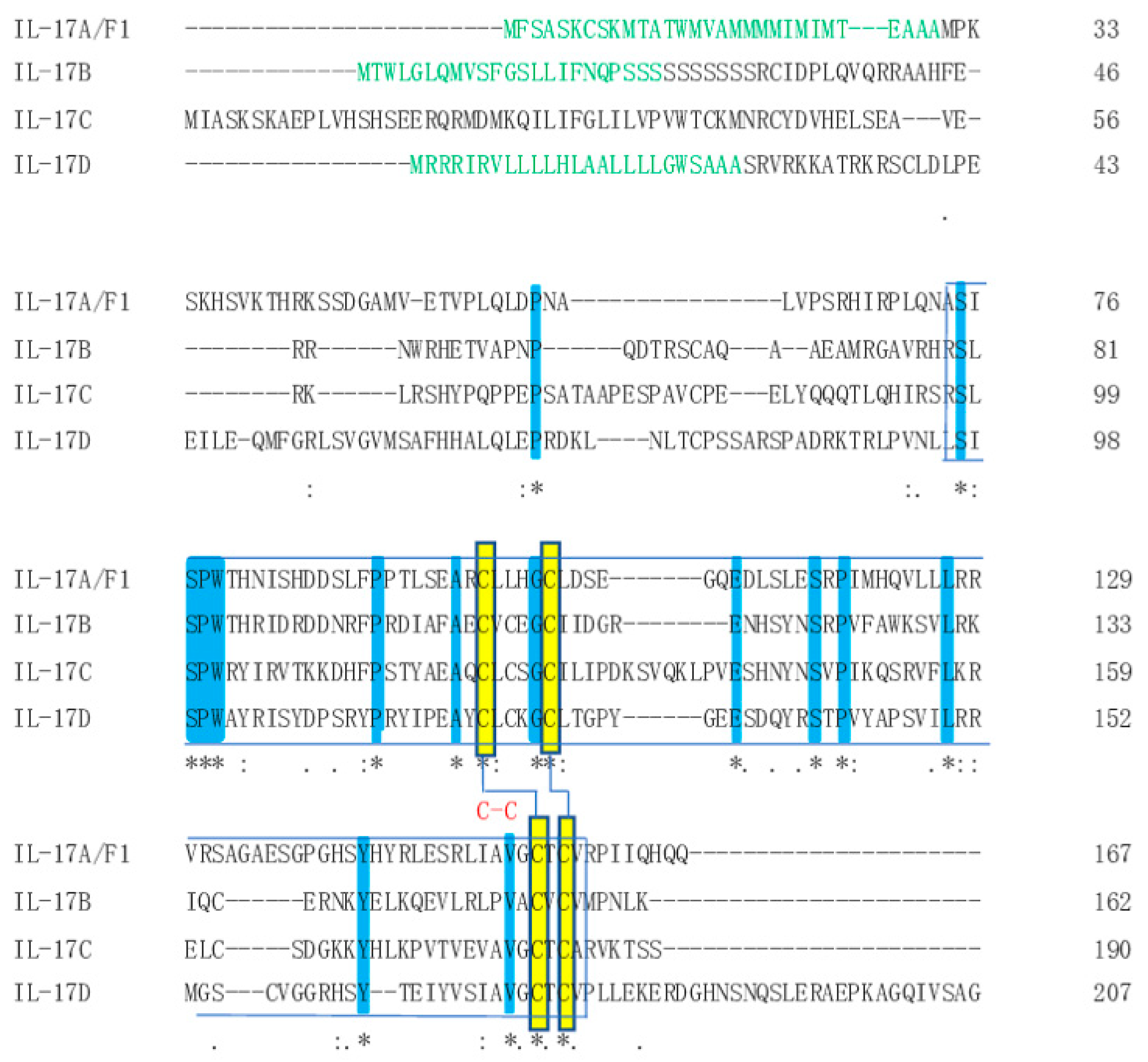
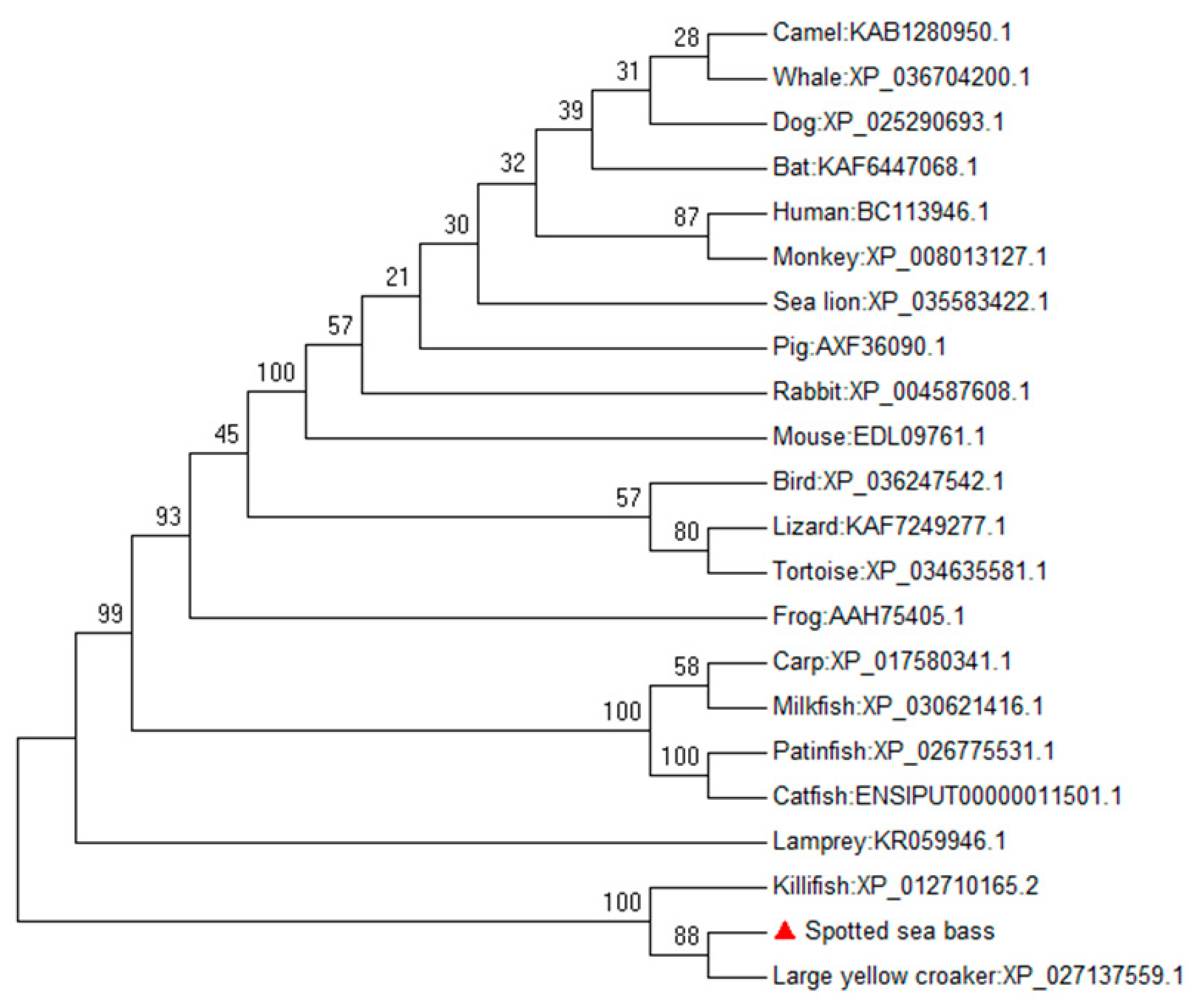
3.1. Identification and Sequence Analysis of the IL-17 Ligand and Receptor Genes

We cloned the complete cDNA sequences of four IL-17 ligands and one IL-17 receptor. Specifically, we isolated the IL-17RB gene, which was identified as the sole receptor for IL-17B. Through sequence analysis, we determined that the spotted sea bass IL-17A/F1 gene comprised a 255 bp 5′-UTR, a 504 bp ORF and a 1192 bp 3′-UTR. The ORF encoded a deduced protein comprising 167 amino acids (aa), which included a signal peptide of 30 aa. The mature peptide of IL-17A/F1 exhibited a theoretical molecular mass of 18.6 kDa and a theoretical pI of 8.7. Similarly, the spotted sea bass IL-17B gene comprised a 40 bp 5′-UTR, a 489 bp ORF and a 667 bp 3′-UTR. The ORF encoded a deduced protein comprising 162 aa, with a signal peptide composed of 23 aa. The mature peptide of IL-17B exhibited a theoretical molecular mass of 18.6 kDa and a theoretical pI of 9.37. Furthermore, we identified that the spotted sea bass IL-17C gene comprised a 42 bp 5′-UTR, a 537 bp ORF (169 aa) and a 743 bp 3′-UTR. The mature peptide of IL-17C exhibited a theoretical molecular mass of 21.6 kDa and a theoretical pI of 9.05. Additionally, the spotted sea bass IL-17D gene comprised a 112 bp 5′-UTR, a 633 bp ORF and a 600 bp 3′-UTR. The ORF encoded a deduced protein comprising 210 aa, of which a signal peptide was composed of 25 aa. The mature peptide of IL-17D exhibited a theoretical molecular mass of 23.5 kDa and a theoretical pI of 9.67. Finally, the spotted sea bass IL-17RB gene comprised a 79 bp 5′-UTR, a 1314 bp ORF and a 215 bp 3′-UTR. The deduced protein encoded by this ORF consisted of 437 aa, including an 18 aa residue signal peptide and a transmembrane domain from 179 to 201 aa. The mature peptide of IL-17RB exhibited a theoretical molecular mass of 48.2 kDa and a theoretical pI of 6.7.
The spotted sea bass IL-17A/F1, IL-17B, IL-17D and IL-17RB proteins severally had a putative signal peptide which was determined to putatively comprise 30, 23, 25 and 18 amino acids at the 5′-end. Notably, deduced protein of the spotted sea bass IL-17C had no signal peptide, which is consistent with European sea bass IL-17C1 [15]. This suggests that it does not release cytokine, and other possible IL-17 members have also been found in frogs and invertebrates [58]. Amino acid sequence analysis of the spotted sea bass IL-17 ligands revealed that all four ligands possess the IL-17 domain. Multiple alignment of IL-17 ligands in spotted sea bass revealed the conservation of four cysteine residues that establish disulfide bridges in their tertiary structures (Figure 1). In addition, for four IL-17 family genes of the spotted sea bass, the C-terminal region showed high similarity with each other, while little conservation was shown in the N-terminal region (Figure 1). Similar results have been observed in the IL-17 family of the miiuy croaker [2]. To explore the relationship between spotted sea bass IL-17B protein and other IL-17B members, a phylogenetic tree was constructed. The analysis revealed a close clustering of spotted sea bass IL-17B ligand sequences with those of other teleost fish species, particularly with a large yellow croaker orthologue (Figure 2).

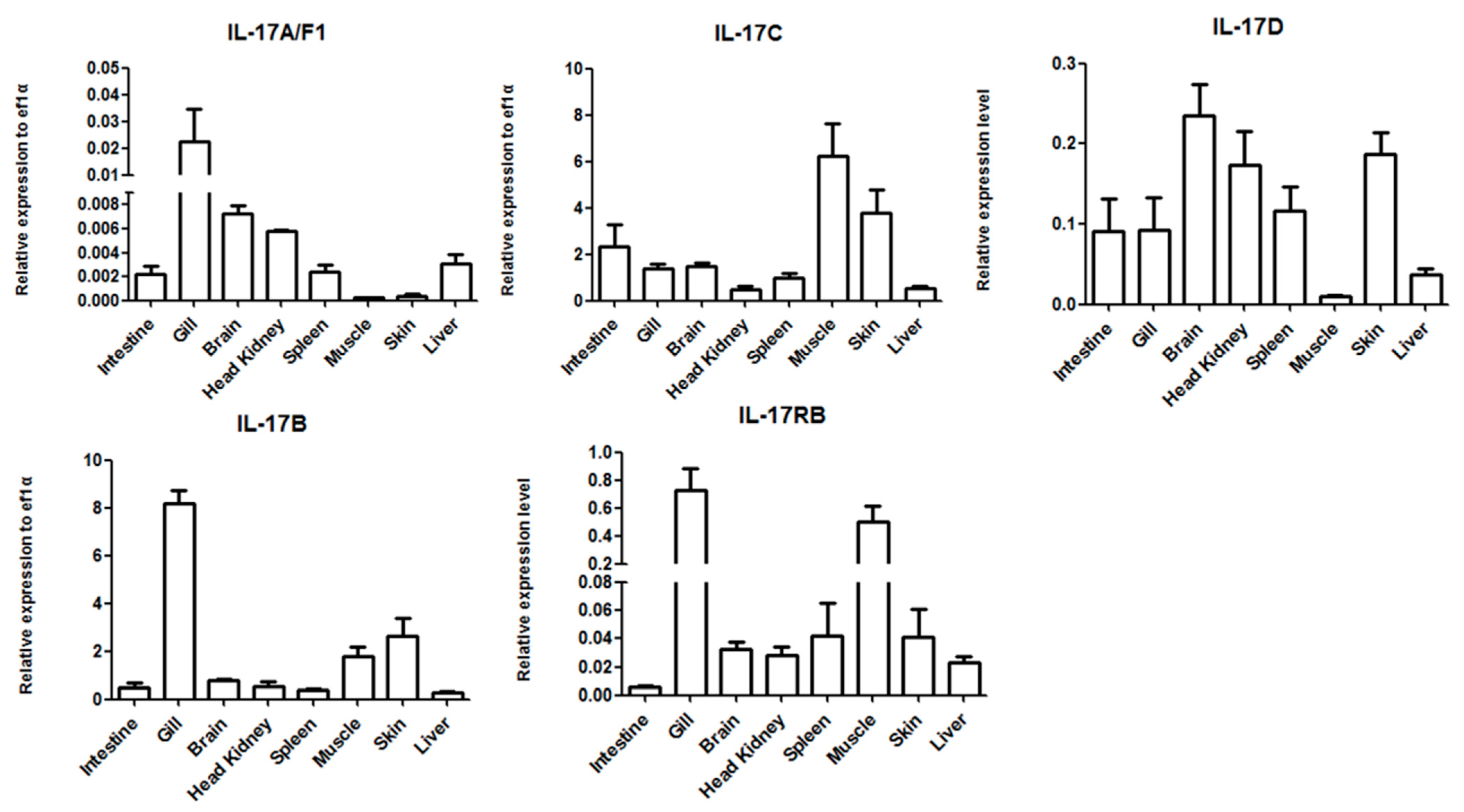
3.2. Tissue Distribution of the IL-17 Ligands and Receptor

Expression analysis of the IL-17 ligand and receptor genes in spotted sea bass tissues showed that IL-17A/F1, IL-17B, IL-17C, IL-17D and IL-17RB transcripts were universally expressed in all the tissues examined. The findings demonstrated that different IL-17 genes have different expression in eight normal tissues mentioned above for detection (Figure 3). Notably, spotted sea bass IL-17A/F1 transcripts showed a highest expression in the gill. IL-17A/F1 expression was found to be high in immune organs such as the gill and head kidney, suggesting that IL-17A/F1 may have the function of defense against pathogens. Given that the gill and skin serve as mucosal tissues, acting as crucial barriers against microorganisms, the substantial expression of IL-17B in these tissues highlights its potential involvement in mucosal immunity against pathogenic invasion. The comparable tissue distribution patterns of IL-17B and IL-17RB in spotted sea bass suggest a potential association with their specific binding properties. Moreover, the abundant expression of IL-17C across all tissues points towards its significant role in tissue homeostasis. Interestingly, IL-17D transcripts exhibited a prominent expression in the brain, as observed in European sea bass, large yellow croaker and sturgeon [15,40,43], indicating a potential function within the nervous system.

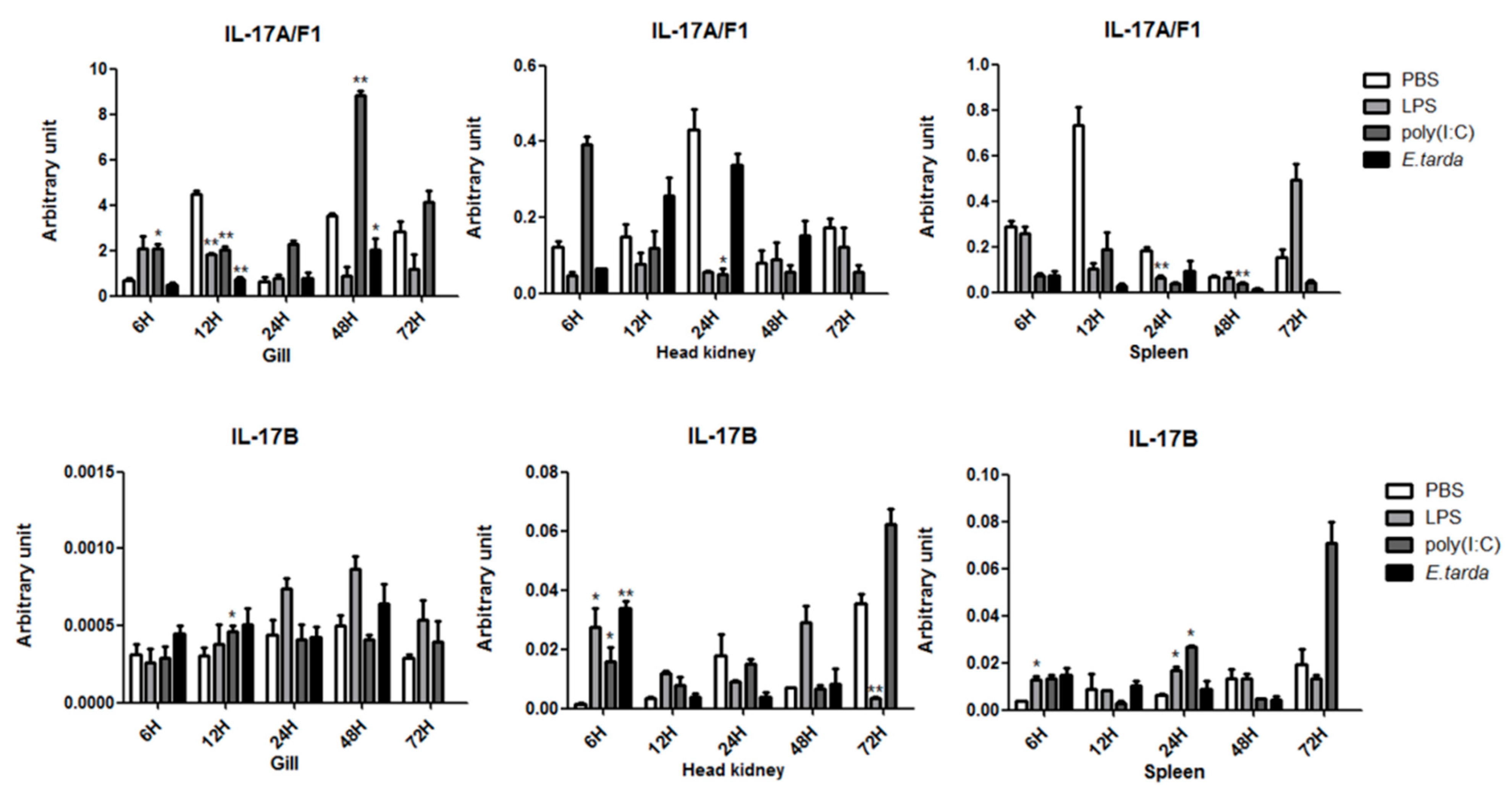
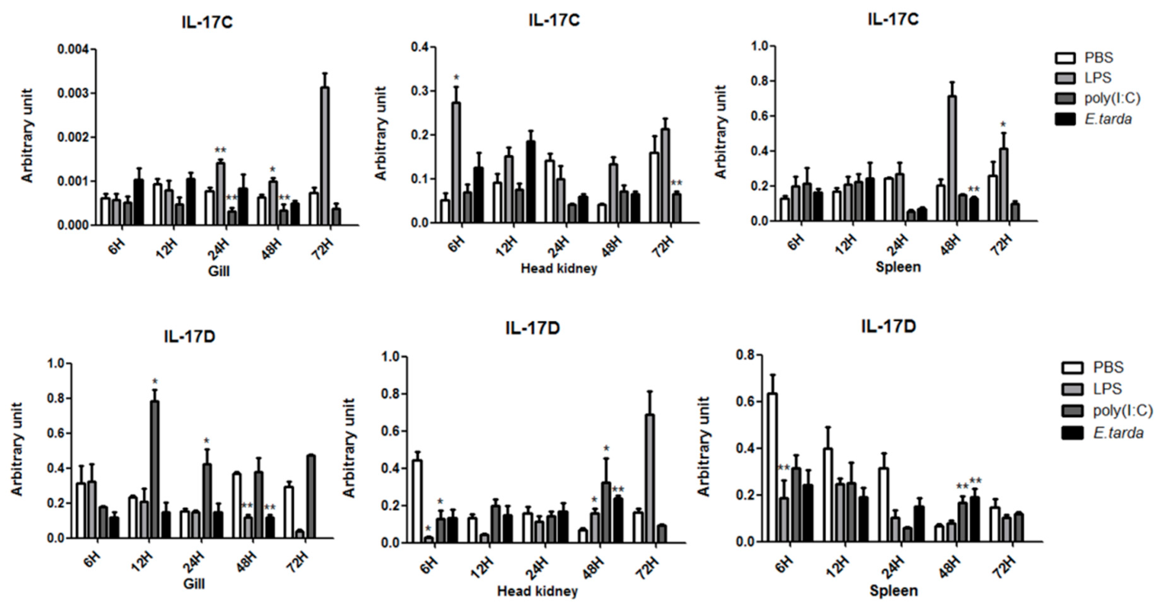
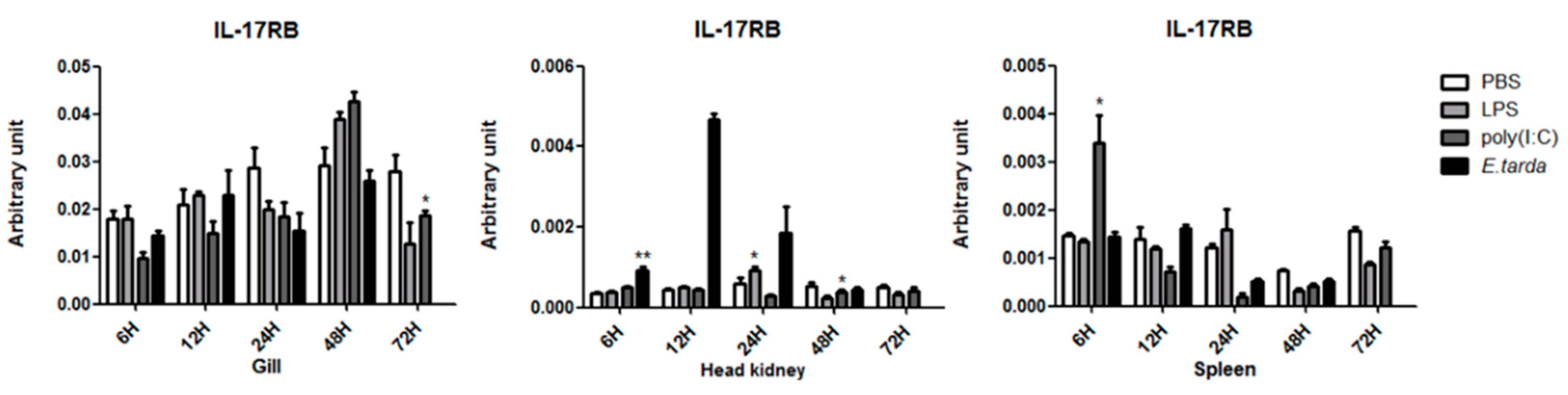
3.3. The mRNA Expression Status of the IL-17 Ligand and Receptor Genes after Injection with LPS/Poly (I:C)/E. tarda

The LPS, a predominant component of Gram-negative bacterial outer membranes, is widely recognized as a virulence factor [59]. The LPS is also an important PAMP that induces inflammation by stimulating host innate immune system responses. In this study, the LPS was employed to assess the immune response of spotted sea bass against bacterial infection. The Gram-negative bacterium E. tarda, known to infect a diverse range of vertebrates from fish to mammals [60], was specifically chosen for this study. Previous investigations [61] have reported severe and fatal cases of E. tarda infection in various fish species such as rockfish [62,63,64], tilapia [65], rainbow trout [66], channel catfish [67]. Poly (I:C), often employed as a viral analog, has been utilized in several studies to evaluate immune responses in fish [61].
In this study, a remarkable and statistically significant down-regulation of IL-17A/F1 expression in the gill was observed at 12 h after injection with the three aforementioned stimuli (Figure 4). Significant variations have been documented among different fish species examined thus far, making it challenging to attribute potential roles to fish IL-17A/F1. IL-17B in head kidney reached maximal expression at 6 h after injection with LPS, so as poly (I:C) and E. tarda (Figure 4), implying its critical involvement in the early response stage upon pathogen adhesion. Furthermore, IL-17C expression in the head kidney was significantly increased 6 h after LPS injection (Figure 5), suggesting its potential unique role during the initial phase of inflammation. However, its expression was subsequently down-regulated in the head kidney at 12–48 h after injection with LPS and poly (I:C) (Figure 5), consistent with findings in miiuy croaker [2]. Additionally, the expression of IL-17D in the immune organ, specifically the head kidney, was significantly up-regulated 48 h after injection with poly (I:C), LPS, and E. tarda (Figure 5), underscoring its involvement in immune response. Notably, the mRNA expression of IL-17RB exhibited a significant up-regulation in the spleen at 6 h after injection with E. tarda, and then down-regulated in 12–48 h (Figure 6). Spotted sea bass IL-17RB in the spleen was also down-regulated at 12–48 h of being injected with Vibrio harveyi [46]. In humans, IL-17A/F is one of the important molecules in the inflammatory response which mainly produced by Th17 cells. Other family members of IL-17 (B, C, and D) may also play a role in inflammation [23]. Notably, the IL-17 family genes are very sensitive to the bacterial component the LPS and quickly respond in fish. Therefore, IL-17 may also play an important role in fish inflammation. The biological activity of the IL-17 family in fish requires further functional research to be understood.

4. Conclusions

Overall, this research successfully cloned and identified five IL-17 genes in spotted sea bass, namely IL-17A/F1, IL-17B, IL-17C, IL-17D and IL-17RB. Alignment and phylogenic analyses revealed the evolutionary conservation of these IL-17 genes. Moreover, an extensive expression of the IL-17 gene has been observed in various tissues. Notably, all five spotted sea bass IL-17 genes exhibited significant responses following injection of the LPS, poly (I:C), and E. tarda. These findings contribute to our knowledge of the crucial role of IL-17 cytokines in the mucosal-associated immune defense network.

Author Contributions

S.W.: investigation, methodology, data curation, and writing-original draft preparation. Z.S.: methodology and writing—review and editing. C.Z. and T.P.: writing—review and editing. S.Y. and Y.C.: investigation and methodology. Q.G.: conceptualization, funding acquisition, project administration, supervision, writing-review and editing. J.Z.: supervision, writing—review and editing. All authors have read and agreed to the published version of the manuscript.

Funding

This research was funded by the National Key Research and Development Program of China (grant number: 2018YFD0900605).

Institutional Review Board Statement

All fish experiments were conducted under the national regulations for laboratory animals in China, and were reviewed and approved by the ethics committee of laboratory animals of the Shanghai Ocean University (SHOU-DW-2019-012).

Data Availability Statement

All the data generated or used during the study appear within the submitted article.

Acknowledgments

We thank Haixia Xie, Institute of Hydrobiology, Chinese Academy of Sciences, for providing Edwardsiella tarda.

Conflicts of Interest

The authors declare no conflict of interest.

References

  1. Holdsworth, S.R.; Gan, P.Y. Cytokines: Names and Numbers You Should Care About. Clin. J. Am. Soc. Nephrol. 2015, 10, 2243–2254. [Google Scholar] [CrossRef] [PubMed] [Green Version]
  2. Yang, Q.; Sun, Y.N.; Su, X.R.; Li, T.W.; Xu, T.J. Characterization of six IL-17 family genes in miiuy croaker and evolution analysis of vertebrate IL-17 family. Fish Shellfish Immun. 2016, 49, 243–251. [Google Scholar] [CrossRef] [PubMed]
  3. Schwarzenberger, P.; Kolls, J.K. Interleukin 17: An example for gene therapy as a tool to study cytokine mediated regulation of hematopoiesis. J. Cell Biochem. 2002, 85, 88–95. [Google Scholar] [CrossRef] [PubMed]
  4. Li, H.Z.; Chen, J.; Huang, A.; Stinson, J.; Heldens, S.; Foster, J.; Dowd, P.; Gurney, A.L.; Wood, W.I. Cloning and characterization of IL-17B and IL-17C, two new members of the IL-17 cytokine family. Proc. Natl. Acad. Sci. USA 2000, 97, 773–778. [Google Scholar] [CrossRef]
  5. Sanders, A.J.; Guo, X.; Mason, M.D.; Jiang, W.G. IL-17B Can Impact on Endothelial Cellular Traits Linked to Tumour Angiogenesis. J. Oncol. 2010, 2010, 817375. [Google Scholar] [CrossRef] [Green Version]
  6. Ramirez-Carrozzi, V.; Sambandam, A.; Luis, E.; Lin, Z.G.; Jeet, S.; Lesch, J.; Hackney, J.; Kim, J.; Zhou, M.J.; Lai, J.; et al. IL-17C regulates the innate immune function of epithelial cells in an autocrine manner. Nat. Immunol. 2011, 12, 1159–1166. [Google Scholar] [CrossRef]
  7. Song, X.Y.; Zhu, S.; Shi, P.Q.; Liu, Y.; Shi, Y.F.; Levin, S.D.; Qian, Y.C. IL-17RE is the functional receptor for IL-17C and mediates mucosal immunity to infection with intestinal pathogens. Nat. Immunol. 2011, 12, 1151–1158. [Google Scholar] [CrossRef]
  8. Peng, T.; Chanthaphavong, R.S.; Sun, S.J.; Trigilio, J.A.; Phasouk, K.; Jin, L.; Layton, E.D.; Li, A.Z.; Correnti, C.E.; De van der Schueren, W.; et al. Keratinocytes produce IL-17c to protect peripheral nervous systems during human HSV-2 reactivation. J. Exp. Med. 2017, 214, 2315–2329. [Google Scholar] [CrossRef]
  9. Starnes, T.; Broxmeyer, H.E.; Robertson, M.J.; Hromas, R. Cutting edge: IL-17D, a novel member of the IL-17 family, stimulates cytokine production and inhibits hemopoiesis. J. Immunol. 2002, 169, 642–646. [Google Scholar] [CrossRef] [Green Version]
  10. Saddawi-Konefka, R.; Seelige, R.; Gross, E.T.E.; Levy, E.; Searles, S.C.; Washington, A.; Santosa, E.K.; Liu, B.C.; O’Sullivan, T.E.; Harismendy, O.; et al. Nrf2 Induces IL-17D to Mediate Tumor and Virus Surveillance. Cell Rep. 2016, 16, 2348–2358. [Google Scholar] [CrossRef] [Green Version]
  11. Fort, M.M.; Cheung, J.; Yen, D.; Li, J.; Zurawski, S.M.; Lo, S.; Menon, S.; Clifford, T.; Hunte, B.; Lesley, R.; et al. IL-25 induces IL-4 IL-5, and IL-13 and Th2-associated pathologies in vivo. Immunity 2001, 15, 985–995. [Google Scholar] [CrossRef] [Green Version]
  12. Hurst, S.D.; Muchamuel, T.; Gorman, D.M.; Gilbert, J.M.; Clifford, T.; Kwan, S.; Menon, S.; Seymour, B.; Jackson, C.; Kung, T.T.; et al. New IL-17 family members promote Th1 or Th2 responses in the lung: In vivo function of the novel cytokine IL-25. J. Immunol. 2002, 169, 443–453. [Google Scholar] [CrossRef] [Green Version]
  13. Wright, J.F.; Guo, Y.; Quazi, A.; Luxenberg, D.P.; Bennett, F.; Ross, J.F.; Qiu, Y.; Whitters, M.J.; Tomkinson, K.N.; Dunussi-Joannopoulos, K.; et al. Identification of an interleukin 17F/17A heterodimer in activated human CD4+ T cells. J. Biol. Chem. 2007, 282, 13447–13455. [Google Scholar] [CrossRef] [Green Version]
  14. Chang, S.H.; Dong, C. A novel heterodimeric cytokine consisting of IL-17 and IL-17F regulates inflammatory responses. Cell Res. 2007, 17, 435–440. [Google Scholar] [CrossRef] [Green Version]
  15. Gonzalez-Fernandez, C.; Chaves-Pozo, E.; Cuesta, A. Identification and Regulation of Interleukin-17 (IL-17) Family Ligands in the Teleost Fish European Sea Bass. Int. J. Mol. Sci. 2020, 21, 2439. [Google Scholar] [CrossRef] [Green Version]
  16. McGeachy, M.J.; Cua, D.J.; Gaffen, S.L. The IL-17 Family of Cytokines in Health and Disease. Immunity 2019, 50, 892–906. [Google Scholar] [CrossRef]
  17. Monin, L.; Gaffen, S.L. Interleukin 17 Family Cytokines: Signaling Mechanisms, Biological Activities, and Therapeutic Implications. Cold Spring Harb. Perspect. Biol. 2018, 10, a028522. [Google Scholar] [CrossRef]
  18. Ashfaq, H.; Soliman, H.; Saleh, M.; El-Matbouli, M. CD4: A vital player in the teleost fish immune system. Vet. Res. 2019, 50, 1. [Google Scholar] [CrossRef] [Green Version]
  19. Ding, Y.; Ai, C.; Mu, Y.; Ao, J.; Chen, X. Molecular characterization and evolution analysis of five interleukin-17 receptor genes in large yellow croaker Larimichthys crocea. Fish Shellfish Immun. 2016, 58, 332–339. [Google Scholar] [CrossRef]
  20. Yao, Z.B.; Fanslow, W.C.; Seldin, M.F.; Rousseau, A.M.; Painter, S.L.; Comeau, M.R.; Cohen, J.I.; Spriggs, M.K. Herpesvirus Saimiri encodes a new cytokine, IL-17, which binds to a novel cytokine receptor. Immunity 1995, 3, 811–821. [Google Scholar] [CrossRef] [Green Version]
  21. Doyle, M.S.; Collins, E.S.; FitzGerald, O.M.; Pennington, S.R. New insight into the functions of the interleukin-17 receptor adaptor protein Act1 in psoriatic arthritis. Arthritis Res. Ther. 2012, 14, 226. [Google Scholar] [CrossRef] [PubMed] [Green Version]
  22. Novatchkova, M.; Leibbrandt, A.; Werzowa, J.; Neubuser, A.; Eisenhaber, F. The STIR-domain superfamily in signal transduction, development and immunity. Trends Biochem. Sci. 2003, 28, 226–229. [Google Scholar] [CrossRef] [PubMed]
  23. Gaffen, S.L. Structure and signalling in the IL-17 receptor family. Nat. Rev. Immunol. 2009, 9, 556–567. [Google Scholar] [CrossRef] [PubMed] [Green Version]
  24. Wright, J.F.; Bennett, F.; Li, B.L.; Brooks, J.; Luxenberg, D.P.; Whitters, M.J.; Tomkinson, K.N.; Fitz, L.J.; Wolfman, N.M.; Collins, M.; et al. The human IL-17F/IL-17A heterodimeric cytokine signals through the IL-17RA/IL-17RC receptor complex. J. Immunol. 2008, 181, 2799–2805. [Google Scholar] [CrossRef] [Green Version]
  25. Korenaga, H.; Kono, T.; Sakai, M. Isolation of seven IL-17 family genes from the Japanese pufferfish Takifugu rubripes. Fish Shellfish Immun. 2010, 28, 809–818. [Google Scholar] [CrossRef]
  26. Chang, S.H.; Reynolds, J.M.; Pappu, B.P.; Chen, G.; Martinez, G.J.; Dong, C. Interleukin-17C Promotes Th17 Cell Responses and Autoimmune Disease via Interleukin-17 Receptor E. Immunity 2011, 35, 611–621. [Google Scholar] [CrossRef] [Green Version]
  27. Lee, J.; Ho, W.H.; Maruoka, M.; Corpuz, R.T.; Baldwin, D.T.; Foster, J.S.; Goddard, A.D.; Yansura, D.G.; Vandlen, R.L.; Wood, W.I.; et al. IL-17E, a novel proinflammatory ligand for the IL-17 receptor homolog IL-17Rh1. J. Biol. Chem. 2001, 276, 1660–1664. [Google Scholar] [CrossRef] [Green Version]
  28. Rong, Z.L.; Wang, A.A.; Li, Z.Y.; Ren, Y.M.; Cheng, L.; Li, Y.H.; Wang, Y.Y.; Ren, F.L.; Zhang, X.N.; Hu, J.; et al. IL-17RD (Sef or IL-17RLM) interacts with IL-17 receptor and mediates IL-17 signaling. Cell Res. 2009, 19, 208–215. [Google Scholar] [CrossRef] [Green Version]
  29. Song, X.Y.; Qian, Y.C. The activation and regulation of IL-17 receptor mediated signaling. Cytokine 2013, 62, 175–182. [Google Scholar] [CrossRef]
  30. Gunimaladevi, I.; Savan, R.; Sakai, M. Identification, cloning and characterization of interleukin-17 and its family from zebrafish. Fish Shellfish Immun. 2006, 21, 393–403. [Google Scholar] [CrossRef]
  31. Kono, T.; Korenaga, H.; Sakai, M. Genomics of fish IL-17 ligand and receptors: A review. Fish Shellfish Immun. 2011, 31, 635–643. [Google Scholar] [CrossRef]
  32. Wang, T.H.; Martin, S.A.M.; Secombes, C.J. Two interleukin-17C-like genes exist in rainbow trout Oncorhynchus mykiss that are differentially expressed and modulated. Dev. Comp. Immunol. 2010, 34, 491–500. [Google Scholar] [CrossRef] [PubMed]
  33. Monte, M.M.; Wang, T.H.; Holland, J.W.; Zou, J.; Secombes, C.J. Cloning and Characterization of Rainbow Trout Interleukin-17A/F2 (IL-17A/F2) and IL-17 Receptor A: Expression during Infection and Bioactivity of Recombinant IL-17A/F2. Infect. Immun. 2013, 81, 340–353. [Google Scholar] [CrossRef] [Green Version]
  34. Wang, T.H.; Jiang, Y.S.; Wang, A.; Husain, M.; Xu, Q.Q.; Secombes, C.J. Identification of the salmonid IL-17A/F1a/b, IL-17A/F2b, IL-17A/F3 and IL-17N genes and analysis of their expression following in vitro stimulation and infection. Immunogenetics 2015, 67, 395–412. [Google Scholar] [CrossRef] [Green Version]
  35. Wang, X.Q.; Li, C.; Thongda, W.; Luo, Y.P.; Beck, B.; Peatman, E. Characterization and mucosal responses of interleukin 17 family ligand and receptor genes in channel catfish Ictalurus punctatus. Fish Shellfish Immun. 2014, 38, 47–55. [Google Scholar] [CrossRef]
  36. Dong, C.J.; Kong, S.N.; Zheng, X.H.; Zhang, J.F.; Nie, G.X.; Li, X.J.; Xu, P. Genome-wide identification of interleukin-17 (IL17) in common carp (Cyprinus carpio) and its expression following Aeromonas hydrophila infection. Gene 2019, 686, 68–75. [Google Scholar] [CrossRef]
  37. Li, H.X.; Yu, J.H.; Li, J.L.; Tang, Y.K.; Yu, F.; Zhou, J.; Yu, W.J. Cloning and characterization of two duplicated interleukin-17A/F2 genes in common carp (Cyprinus carpio L.): Transcripts expression and bioactivity of recombinant IL-17A/F2. Fish Shellfish Immun. 2016, 51, 303–312. [Google Scholar] [CrossRef]
  38. Li, H.X.; Zhang, L.; Li, J.L.; Yu, F.; Wang, M.Y.; Wang, Q.; Wu, Y.S.; Zhang, Q.Y.; Tang, Y.K.; Yu, J.H. Identification, expression and pro-inflammatory effect of interleukin-17 N in common carp (Cyprinus carpio L.). Fish Shellfish Immun. 2021, 111, 6–15. [Google Scholar] [CrossRef]
  39. Ding, Y.; Ao, J.Q.; Ai, C.X.; Chen, X.H. Molecular and functional identification of three interleukin-17A/F (IL-17A/F) homologues in large yellow croaker (Larimichthys crocea). Dev. Comp. Immunol. 2016, 55, 221–232. [Google Scholar] [CrossRef]
  40. Ding, Y.; Ao, J.Q.; Chen, X.H. Comparative study of interleukin-17C (IL-17C) and IL-17D in large yellow croaker Larimichthys crocea reveals their similar but differential functional activity. Dev. Comp. Immunol. 2017, 76, 34–44. [Google Scholar] [CrossRef]
  41. Du, L.Y.; Qin, L.; Wang, X.Y.; Zhang, A.Y.; Wei, H.; Zhou, H. Characterization of grass carp (Ctenopharyngodon idella) IL-17D: Molecular cloning, functional implication and signal transduction. Dev. Comp. Immunol. 2014, 42, 220–228. [Google Scholar] [CrossRef] [PubMed]
  42. Du, L.Y.; Feng, S.Y.; Yin, L.C.; Wang, X.Y.; Zhang, A.Y.; Yang, K.; Zhou, H. Identification and functional characterization of grass carp IL-17A/F1: An evaluation of the immunoregulatory role of teleost IL-17A/F1. Dev. Comp. Immunol. 2015, 51, 202–211. [Google Scholar] [CrossRef] [PubMed]
  43. Zhu, H.; Song, R.X.; Wang, X.W.; Hu, H.X.; Zhang, Z.B. Peritoneal bacterial infection repressed the expression of IL17D in Siberia sturgeon a chondrostean fish in the early immune response. Fish Shellfish Immun. 2017, 64, 39–48. [Google Scholar] [CrossRef]
  44. Chi, H.; Sun, L. Comparative study of four interleukin 17 cytokines of tongue sole Cynoglossus semilaevis: Genomic structure, expression pattern, and promoter activity. Fish Shellfish Immun. 2015, 47, 321–330. [Google Scholar] [CrossRef]
  45. Costa, M.M.; Pereiro, P.; Wang, T.; Secombes, C.J.; Figueras, A.; Novoa, B. Characterization and gene expression analysis of the two main Th17 cytokines (IL-17A/F and IL-22) in turbot, Scophthalmus maximus. Dev. Comp. Immunol. 2012, 38, 505–516. [Google Scholar] [CrossRef] [Green Version]
  46. Mao, X.B.; Tian, Y.; Wen, H.S.; Liu, Y.; Sun, Y.L.; Yanglang, A.; Li, Y. Effects of Vibrio harveyi infection on serum biochemical parameters and expression profiles of interleukin-17 (IL-17)/interleukin-17 receptor (IL-17R) genes in spotted sea bass. Dev. Comp. Immunol. 2020, 110, 103731. [Google Scholar] [CrossRef]
  47. Okamura, Y.; Morimoto, N.; Sawada, S.; Kono, T.; Hikima, J.; Sakai, M. Molecular characterization and expression of two interleukin-17 receptor A genes on different chromosomes in Japanese medaka, Oryzias latipes. Comp. Biochem. Phys. B 2020, 240, 110386. [Google Scholar] [CrossRef]
  48. Harada, N.; Okamura, Y.; Kono, T.; Sakai, M.; Hikima, J. Identification of two interleukin 17 receptor C (IL-17RC) genes and their binding activities to three IL-17A/F ligands in the Japanese medaka, Oryzias latipes. Dev. Comp. Immunol. 2021, 124, 104179. [Google Scholar] [CrossRef]
  49. Zhang, X.Y.; Cao, M.; Xue, T.; Yu, H.H.; Yang, T.Z.; Yan, X.; Li, C. Characterization of IL-17/IL-17R gene family in Sebastes schlegelii and their expression profiles under Aeromonas salmonicida and Edwardsiella piscicida infections. Aquaculture 2022, 551, 737901. [Google Scholar] [CrossRef]
  50. Zhou, X.; Jiang, X.X.; Zhang, G.R.; Ji, W.; Shi, Z.C.; Ma, X.F.; Wei, K.J. Molecular characterization, expression and function analysis of interleukin-17B, C and D genes in yellow catfish (Pelteobagrus fulvidraco). Aquaculture 2022, 552, 737962. [Google Scholar] [CrossRef]
  51. Sun, Z.S.; Qin, Y.T.; Liu, D.J.; Wang, B.J.; Jia, Z.; Wang, J.Y.; Gao, Q.; Zou, J.; Pang, Y. The evolution and functional characterization of CXC chemokines and receptors in lamprey. Dev. Comp. Immunol. 2021, 116, 103905. [Google Scholar] [CrossRef]
  52. Emanuelsson, O.; Brunak, S.; von Heijne, G.; Nielsen, H. Locating proteins in the cell using TargetP, SignalP and related tools. Nat. Protoc. 2007, 2, 953–971. [Google Scholar] [CrossRef]
  53. Oliver, T.; Schmidt, B.; Nathan, D.; Clemens, R.; Maskell, D. Using reconfigurable hardware to accelerate multiple sequence alignment with ClustalW. Bioinformatics 2005, 21, 3431–3432. [Google Scholar] [CrossRef] [PubMed]
  54. Atteson, K. The performance of neighbor-joining methods of phylogenetic reconstruction. Algorithmica 1999, 25, 251–278. [Google Scholar] [CrossRef]
  55. Kumar, S.; Stecher, G.; Tamura, K. MEGA7: Molecular Evolutionary Genetics Analysis Version 7.0 for Bigger Datasets. Mol. Biol. Evol. 2016, 33, 1870–1874. [Google Scholar] [CrossRef] [Green Version]
  56. Yi, L.; Nie, P.; Yu, H.B.; Xie, H.X. Regulation of Type III Secretion of Translocon and Effector Proteins by the EsaB/EsaL/EsaM Complex in Edwardsiella tarda. Infect. Immun. 2017, 85, e00322-1. [Google Scholar] [CrossRef] [Green Version]
  57. Pressley, M.E.; Phelan, P.E.; Witten, P.E.; Mellon, M.T.; Kim, C.H. Pathogenesis and inflammatory response to Edwardsiella tarda infection in the zebrafish. Dev. Comp. Immunol. 2005, 29, 501–513. [Google Scholar] [CrossRef]
  58. Huang, X.D.; Zhang, H.; He, M.X. Comparative and Evolutionary Analysis of the Interleukin 17 Gene Family in Invertebrates. PLoS ONE 2015, 10, e0132802. [Google Scholar] [CrossRef] [PubMed]
  59. Swain, P.; Nayak, S.K.; Nanda, P.K.; Dash, S. Biological effects of bacterial lipopolysaccharide (endotoxin) in fish: A review. Fish Shellfish Immun. 2008, 25, 191–201. [Google Scholar] [CrossRef]
  60. Okuda, J.; Arikawa, Y.; Takeuchi, Y.; Mahmoud, M.M.; Suzaki, E.; Kataoka, K.; Suzuki, T.; Okinaka, Y.; Nakai, T. Intracellular replication of Edwardsiella tarda in murine macrophage is dependent on the type III secretion system and induces an up-regulation of anti-apoptotic NF-kappa B target genes protecting the macrophage from staurosporine-induced apoptosis. Microb. Pathog. 2006, 41, 226–240. [Google Scholar] [CrossRef] [Green Version]
  61. Li, X.; Yuan, S.Y.; Sun, Z.S.; Lei, L.N.; Wan, S.; Wang, J.Y.; Zou, J.; Gao, Q. Gene identification and functional analysis of peptidoglycan recognition protein from the spotted sea bass (Lateolabrax maculatus). Fish Shellfish Immun. 2020, 106, 1014–1024. [Google Scholar] [CrossRef] [PubMed]
  62. Kim, H.S.; Lee, K.W.; Jung, H.S.; Kim, J.; Yun, A.; Cho, S.H.; Kwon, M. Effects of dietary inclusion of yacon, ginger and blueberry on growth, body composition and challenge test of juvenile rockfish (Sebastes schlegeli) against Edwardsiella tarda. Aquacult. Nutr. 2018, 24, 1048–1055. [Google Scholar] [CrossRef]
  63. Ling, S.H.M.; Wang, X.H.; Xie, L.; Lim, T.M.; Leung, K.Y. Use of green fluorescent protein (GFP) to study the invasion pathways of Edwardsiella tarda in in vivo and in vitro fish models. Microbiology 2000, 146, 7–19. [Google Scholar] [CrossRef] [Green Version]
  64. Mohanty, B.R.; Sahoo, P.K. Edwardsiellosis in fish: A brief review. J. Biosci. 2007, 32, 1331–1344. [Google Scholar] [CrossRef]
  65. Wu, J.Y.; Liu, G.B.; Sun, Y.C.; Wang, X.P.; Fang, H.; Jiang, H.; Guo, Z.M.; Dong, J.G. The role of regulator FucP in Edwardsiella tarda pathogenesis and the inflammatory cytokine response in tilapia. Fish Shellfish Immun. 2018, 80, 624–630. [Google Scholar] [CrossRef]
  66. Reddacliff, G.L.; Hornitzky, M.; Whittington, R.J. Edwardsiella tarda septicaemia in rainbow trout (Oncorhynchus mykiss). Aust. Vet. J. 1996, 73, 30. [Google Scholar] [CrossRef]
  67. Li, M.; Wang, Q.L.; Lu, Y.; Chen, S.L.; Li, Q.; Sha, Z.X. Expression profiles of NODs in channel catfish (Ictalurus punctatus) after infection with Edwardsiella tarda, Aeromonas hydrophila, Streptococcus iniae and channel catfish hemorrhage reovirus. Fish Shellfish Immun. 2012, 33, 1033–1041. [Google Scholar] [CrossRef]
Figure 1. Amino acid sequence alignment of the IL-17 in spotted sea bass. The predicted signal peptide and conserved region are marked as the highlighted words in green and blue, respectively. Cysteine residues in forming disulfide bonds are denoted by boxed representation, highlighted and connected with the line. Identical residues are denoted by “*”, and similar residues are represented by “:” or “.”.
Fishes 08 00405 g001
Figure 2. Phylogenetic tree of IL-17B. The bootstrap values for branches are presented as percentages and the symbol () denotes spotted sea bass IL-17B.
Fishes 08 00405 g002
Figure 3. Tissue expression of IL-17 genes in spotted sea bass. Levels of IL-17 genes expression were normalized to that of EF-1α. Data are presented as the mean ± SEM (n = 3).
Fishes 08 00405 g003
Figure 4. Expression analysis of spotted sea bass IL-17A/F1 and IL-17B in three tissues after infection by the LPS, poly (I:C), and E. tarda. The expression levels of target genes were normalized relative to EF1α (mean ± SEM, n = 3). *: p < 0.05 and **: p < 0.01.
Fishes 08 00405 g004
Figure 5. Expression analysis of IL-17C and IL-17D in spotted sea bass was conducted across three tissues after infection by the LPS, poly (I:C), and E. tarda. The expression levels of target genes were normalized relative to EF1α (mean ± SEM, n = 3). *: p < 0.05 and **: p < 0.01.
Fishes 08 00405 g005
Figure 6. Expression analysis of spotted sea bass IL-17RB in three tissues after infection by the LPS, poly (I:C), and E. tarda. The expression levels of target genes were normalized relative to EF1α (mean ± SEM, n = 3). *: p < 0.05 and **: p < 0.01.
Fishes 08 00405 g006
Table 1. Sequence of primers.
GenePrimersSequence (5′ to 3′)Application
IL-17A/F1F-longGGTGACATCATCAGAGGAAGTCTATGCPCR cloning
R-longTGCTGGTGTTGGATGATGGGTCPCR cloning
F-shortTGACGGCGATCAGGATAAATGTPCR cloning
R-shortACAGCGATGAGGCGGGACTPCR cloning
5′R-longAAAGCGTTGGGGTCAAGCTGCAG5′-RACE
5′R-shortGAGTGTCCTCGTCCTCCTTCTTGGTTC5′-RACE
3′F-longCAGCATCAAAGTGCTCCAAATGACGGC3′-RACE
3′F-shortGTTCCTAGCAGACACATCAGGCCACT3′-RACE
FGGACACACAACATCAGCCATGATGACqRT-PCR
RAGCAGCACCTGGTGCATGATqRT-PCR
IL-17BF-PCRATGGAGCCGCGGGTCACACTCAPCR cloning
R-PCRAGGCCACTGGGAGCATAAGAACTTPCR cloning
5′R-longCCCGTCGATGATGCAGCCCTCAC5′-RACE
5′R-shortCGACGGTCTCGTGCCTCCAGTTG5′-RACE
3′F-longCGTGATATCGCTTTTGCCGAGTGTGT3′-RACE
3′F-shortCAGGAAGTTCTTATGCTCCCAGTGGC3′-RACE
FTGGACACACAGAATAGACCGAGACGqRT-PCR
RCAGGCCACTGGGAGCATAAGAACTTqRT-PCR
IL-17CF-PCRATGATAGCAAGTAAAAGCAAAGCCGAACCPCR cloning
R-PCRGTACAGCCCACAGCTACTTCCPCR cloning
5′R-longCCGGTTCATCTTGCACGTCCACACG5′-RACE
5′R-shortTGTGAACGAGGGGTTCGGCTTTGCT5′-RACE
3′F-longCAGGATGCATCCTAATTCCGGACAAGTC3′-RACE
3′F-shortAAGAGGGAGCTCTGCAGTGATGGAAAG3′-RACE
FCTCATCTTTGGTCTTATCCTCGqRT-PCR
RGCTCCTCAGCTTCCTCTCTACTqRT-PCR
IL-17DF-longTCCTGCTGCTGCTGCACCTPCR cloning
R-longCCTTTCTTGCCCGCAGAGACGAPCR cloning
F-shortTGCTGCTGGGCTGGTCGGPCR cloning
R-shortCGGCTTTGGGCTCTGCTCTPCR cloning
5′R-longCAGGCTCTGGTTGCTGTTGTGGC5′-RACE
5′R-shortCTTCCTGGTGGCCTTCTTCCGCA5′-RACE
3′F-longCAGAGGCGTACTGCCTGTGT3′-RACE
3′F-shortCTGAGGAGGATGGGCTCCTGTGT3′-RACE
FCTACGCTCCGTCCGTCATCqRT-PCR
RCGGCTTTGGGCTCTGCTCTqRT-PCR
IL-17RBF-PCRCCCTCCTATCAGTTCTCACCCTPCR cloning
R-PCRAGCTTCCCTTAACTGCGCTGCPCR cloning
5′R-longCCGCTGATGCTGTAGATGCCAGTGCTT5′-RACE
5′R-shortCACCAGCATCACTTTTAAGTCGGCCAGG5′-RACE
3′F-longATCCCAGCTGCAGCTCATGACCTTTACC3′-RACE
3′F-shortGCAGGACAAGAGGCCTCATGACCTGG3′-RACE
FGCCTGCCAGTCTTTTTGTCTqRT-PCR
RTGCTCTGCTGATGTCCTTCTqRT-PCR
EF-1αEF-1α FATCTCTGGATGGCACGGAGAqRT-PCR
EF-1α RCAGTGTGGTTCCGCTAGCATCqRT-PCR
Disclaimer/Publisher’s Note: The statements, opinions and data contained in all publications are solely those of the individual author(s) and contributor(s) and not of MDPI and/or the editor(s). MDPI and/or the editor(s) disclaim responsibility for any injury to people or property resulting from any ideas, methods, instructions or products referred to in the content.

