Article

Acute Effects of Fluoxetine on Stress Responses and Feeding Motivation in Nile Tilapia

by
Julia P. G. A. Miranda
1,
Ana Beatriz J. Isaac
1,
Rebeca B. Silva
1,
Leandro C. S. Toledo
1,
Leonardo J. G. Barcellos
2,3,4,
Helton Carlos Delicio
1 and
Rodrigo Egydio Barreto
1,*
1
Department of Structural and Functional Biology (Physiology), Institute of Biosciences of Botucatu, Aquaculture Center of São Paulo State University—CAUNESP, UNESP, R. Prof. Dr. Antônio Celso Wagner Zanin, 250—Distrito de Rubião Junior, Botucatu 18618-689, SP, Brazil
2
Programa de Pós-Graduação em Bioexperimentação, Universidade de Passo Fundo, BR 285, São José, Passo Fundo 99052-900, RS, Brazil
3
Curso de Medicina Veterinária, Universidade de Passo Fundo, Passo Fundo 99052-900, RS, Brazil
4
Programa de Pós-Graduação em Farmacologia, Universidade Federal de Santa Maria, Santa Maria 97105-900, RS, Brazil
*
Author to whom correspondence should be addressed.
Fishes 2023, 8(7), 348; https://doi.org/10.3390/fishes8070348
Submission received: 21 June 2023 / Accepted: 29 June 2023 / Published: 4 July 2023
(This article belongs to the Special Issue Impacts of Anthropogenic Stressors on Fish Physiology)

Abstract

:
The selective serotonin reuptake inhibitor fluoxetine is one of the most commonly administered psychotropic medications; however, it has been recognized as toxic to aquatic life. In this study, we showed that stress responses and feeding motivation in Nile tilapia were affected by acute exposure to fluoxetine. To reach that conclusion, we exposed Nile tilapia to 0, 1 or 10 µg/L (environmentally/biologically relevant doses) of fluoxetine over a 24 h period and then exposed them to a handling stressor. We found that the 10 µg/L dose enhanced cortisol response to stress but caused an earlier decrease in the ventilation boost induced by that stressor. An immediate ventilation boost after stressful stimuli indicates sympathetic activation. Thus, this suggests that fluoxetine decreased sympathetic nervous system activity but augmented hypothalamus–pituitary–interrenal axis activity in the fish. Both feeding latency and ingestion were similar among the tested conditions; however, a multiple logistic regression model revealed that in the presence of a stressor or fluoxetine, the Nile tilapia tended to ingest less food but there was a higher probability of this decrease to be associated with fluoxetine. We concluded that acute exposure to environmentally/biologically relevant fluoxetine concentrations over 24 h acted as a modifying factor for Nile tilapia stress physiology and tended to interfere with feeding motivation. An acute stress response is an emergency reaction that contributes to the recovery of homeostasis. In the presence of fluoxetine, modifications of acute stress responses and the tendency to reduce food intake, which restricts the ability to replace the energy spent on stress responses, could compromise the resumption of homeostasis and an animal’s adjustment to different environmental contexts, such as those associated with aquaculture, in which anthropogenic stressors inevitably occur.
Key Contribution: Fluoxetine modulated ongoing acute stress responses in Nile tilapia since this drug increased the cortisol response to handling, while attenuating ventilation rate. Fluoxetine and stress also tended to decrease food intake in this species.

1. Introduction

Stress is the state in which the homeostasis of an organism is threatened or disturbed by internal and/or external stimuli to the body (stressors). Stressor-induced responses involve behavioral and physiological reactions (stress responses) that contribute to the resumption of homeostasis. In fish, stress responses initially involve the activation of the sympathetic autonomic nervous system, resulting in, for example, an increase in blood adrenaline levels [1,2] and cardiorespiratory adjustments, such as increased heart rate [3] and respiratory activity [4,5,6,7]. In parallel to these responses, the hypothalamus–pituitary–interrenal (HPI) neuroendocrine axis is also activated, culminating in an increase in plasma cortisol levels after the onset of previous responses [8,9,10]. Both secreted hormones (adrenaline and cortisol) can increase glucose levels, providing energy to the body to deal with and overcome stressors [11].
At least in mammals, serotonin (5-hydroxytryptamine) is one of the main neurotransmitters of the central nervous system. Serotoninergic fibers innervate brain structures, such as the hippocampus, prefrontal cortex, amygdala and hypothalamus, and modulate both sympathetic and hypothalamus–pituitary–adrenal axis activity, which are the main stress response systems [12,13,14]. Regarding fish, despite their neuroanatomical and genetic differences from mammals, the serotoninergic system (SS) influence in their stress response systems seems to be qualitatively similar to a certain degree [15,16]. In fact, stress and behavioral responses in fish, especially in relation to HPI axis activity, is affected by SS activity [17]. For example, increased serotonergic activity due to the administration of the 5-hydroxytryptamine–1A serotonin receptor agonist 8-hydroxy-2-(di-n-propylamine)-tetralin causes an increase in plasma cortisol levels in fish in a dose-dependent manner [18].
Pharmaceutical drugs are being increasingly detected in aquatic environments, including the antidepressant fluoxetine (FLX), which is a selective serotonin reuptake inhibitor (SSRI) [19,20,21,22,23,24,25,26]. The concentration of FLX found in aquatic environments typically varies in the range of ng/L to µg/L [27]. FLX prevents the cellular reuptake of serotonin by 5-HT transporters, resulting in an increase in extracellular levels of serotonin [28,29]. The presence of FLX in aquatic environments is a relevant concern in terms of its potential adverse effects on non-target organisms [30]. Along this line, since FLX results in an increase in serotonin concentrations, any physiological processes regulated by serotonin may be susceptible to environmental exposure to FLX, such as stress.
The effects of FLX on cortisol levels in fish have been studied but there are still many questions to be answered, especially due to the occurrence of divergent evidence [31,32]. Regarding HPI axis activity, the exposure of fish to a stressor typically increases cortisol levels [8,9,10] but acute exposure to fluoxetine has been observed to attenuate cortisol increases in fish [32,33,34]. However, no acute effects of FLX on cortisol increases during stress responses have been observed (e.g., social stress [29] and skin and blood chemical alarm cues [32]). Moreover, acute or chronic exposure to FLX can induce an increase in cortisol levels [28,35,36]. Considering responses associated with sympathetic system activity, to the best of our knowledge, there is no direct evidence of the effects of fluoxetine in fish. In mammals, however, the acute administration of fluoxetine has been shown to attenuate sympathetic activity [14]. This evidence has clearly shown the neuroendocrine-modulating (‘disrupting’) effects of fluoxetine on physiological variables linked to stress responses [31]. Given the consequences in relation to these variables, behaviors associated with physiological stress systems may be also affected.
Feeding behavior is influenced by several different variables, including abiotic factors, food cues, circulating levels of nutrients, hormones and neuropeptides [37,38,39]. Along this line, in fact, feeding performance is affected by both fluoxetine and stress. Fluoxetine is considered to be anorexigenic in fish [40,41] and mammals [42] and the administration of serotonin causes a decrease in food intake in fish [43]. Stress can affect brain pathways associated with appetite, thereby dysregulating food intake mechanisms in fish [44]. Thus, stress and FLX could interact and affect feeding motivation in fish.
Based on this evidence, we evaluated whether acute exposure to environmentally relevant doses of fluoxetine (1 and 10 µg/L) over 24 h modulate stress responses and feeding motivation in Nile tilapia (Oreochromis niloticus) that were subjected to a handling stressor. This species has been recognized as a suitable animal model for studying the biological effects of FLX [45], but few studies have been conducted using Nile tilapia as the experimental model. This species is of great economic relevance as it is the most cultivated freshwater fish in the world [46,47] and our theoretical understanding of the phenomenon of stress contributes toward improving tilapia husbandry (e.g., stress reduction) [48]. As stress indicators, we evaluated ventilatory rate [4], fluctuations in this variable indirectly indicate changes in sympathetic system activity [2], as well as plasma cortisol levels [8,9,10]. We also evaluated whether feeding behavior was affected by these factors to infer changes in behavioral motivation [49,50].

2. Materials and Methods

2.1. Ethical Approval

This study agreed with Brazilian legislation regulated by the National Council for the Control of Animal Experimentation (CONCEA) and the Ethical Principles in Animal Research formulated by the Brazilian Society of Science in Laboratory Animals and was approved by the Bioscience Institute (UNESP) Ethics Committee on Use of Animals (CEUA) (5589170521).

2.2. Fish and Holding Conditions

2.2.1. Stock Population

In this study, we used Nile tilapia (Oreochromis niloticus L.; GIFT strain; ~4-month-old fish; ~7–8 cm; all males) that were randomly caught from our stock population, which is kept in a 1000-L indoor masonry tank (100 individuals) with constant aeration, a continuous flow of dechlorinated water for uninterrupted water renewal and mechanical–biological filtration. The water temperature was ~26 °C, maintained using room air conditioners. The photoperiod was 12 h of light and 12 h of dark, from 6 a.m. to 6 p.m., with an abrupt transition from light to dark, controlled by an electronic timer. The fish were fed with commercial pelleted chow for tilapias (36% protein; Supra Tilapia®; Alisul Alimentos S/A—SUPRA; Rio Claro, Brazil) once a day until satiety.

2.2.2. Experimental Aquaria

The water used in the experimental aquaria came from the public water treatment company of the State of São Paulo, Brazil (SABESP; Companhia de Saneamento Básico do Estado de São Paulo), which provides high quality water to the population. The presence of FLX is considered very rare in untreated water here and absent in treated water [51]. Nevertheless, the water used for our experiments was initially treated with sodium hypochlorite to remove any existing organic molecules, including any potential traces of FLX [52]. Afterward, sodium thiosulfate was used to eliminate any chlorine/chloramine residue. The presence of chlorine/chloramine in the aquarium water was not observed after the sodium thiosulfate application.
The aquaria water received a constant supply of air from an airstone, which was connected to air pumps via non-toxic silicon tubing. The pH of the water was around 7.0–7.2 and the levels of ammonia and nitrite were <0.002 ppm and <0.25 ppm, respectively. The photoperiod and aquaria temperature were maintained similarly as described above.

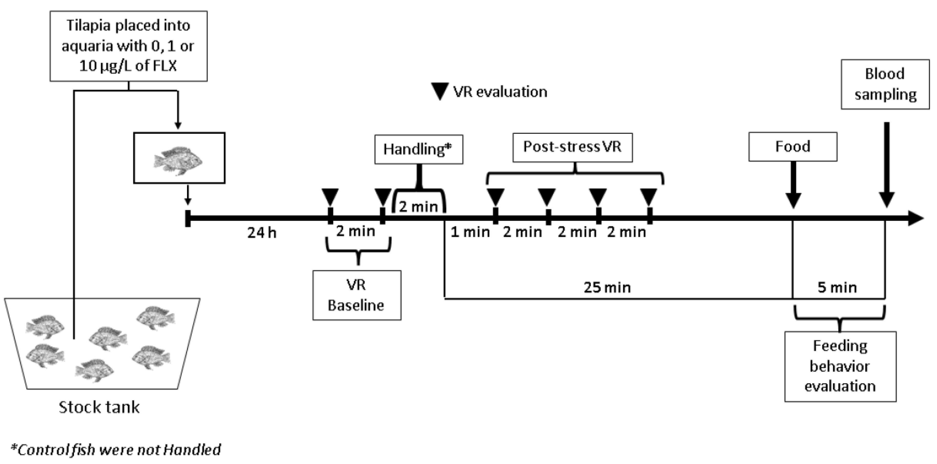
2.3. Experimental Design

As a general strategy, we evaluated the effects of fluoxetine (FLX; CAS number 54910-89-3; obtained from Tocris Bioscience (Ellisville, MO, USA)) on ventilatory rate (VR), plasma cortisol levels and feeding behavior in Nile tilapia that were either exposed or not to a handling stressor. The stock solutions of FLX were made by diluting pure fluoxetine hydrochloride in distilled water (solution vehicle) and fractions of this solution were then added directly into the water in the experimental aquaria to reach the desired FLX concentrations (see below).
To perform this experiment, tilapias from our stock population were introduced singly (1 fish/aquarium) into experimental aquaria (40 cm × 25 cm × 23 cm, with 20 L of dechlorinated tap water) containing 0, 1 or 10 µg/L of FLX for 24 h. The concentration of 0 µg/L was a vehicle control (distilled water), while the concentrations of 1 µg/L [19,22] and 10 µg/L [27,53] were environmentally/biologically relevant doses. After the 24-h period of exposure to the FLX concentrations, one batch of fish from each concentration was subjected to a handling stressor, while another batch remained unhandled (‘non-stress’ control). The handling was conducted by chasing the animal with a net for 1 min, capturing it, exposing it to air (hypoxia) for another 1 min and then reintroducing it into its respective aquarium. Thus, the animals were divided into six experimental conditions (3 (doses) × 2 (stressor conditions) design); n = 10 fish/condition).
Our response variables were quantified according to the following schedule (see Figure 1 for details). Immediately before the animals were exposed to handling, their VR was measured twice, with a gap of 2 min between measurements. The mean value of these two measures was considered as the VR baseline. Then, 1 min after the handling, their VR was measured four more times every 2 min (at minutes 1, 3, 5 and 7 after the end of the handling). Each fish acted as its own control in the case of VR measurements [4]. Next, 25 min after the stressor, we offered food to the fish and their latency to take the pellets and food intake were quantified for 5 min following the introduction of food into the aquarium water. If within this 5 min period the fish did not eat, we considered this maximum time as the latency value (300 s). After this period of 5 min (30 min after the end of the stressor), the fish were captured, anesthetized (described below), measured for mass and length and blood was collected via cardiac puncture to quantify plasma cortisol levels. The animals that were not exposed to the stressor (‘unhandled’) had their feeding behavior quantified in the same way and went through the same process for blood collection as described above.

2.4. Measured Variables

2.4.1. Ventilation Rate (VR)

VR is an easy, inexpensive, sensible and meaningful tool for the evaluation of the effects of acute stressors in fishes [4,54,55,56]. In this study, VR was measured by quantifying the time required for 10 opercular beats or mouth movements to occur. This measurement was performed visually through a small hole in an opaque curtain to decrease the visual perception of the investigator by the fish [57]. The investigator could see the entire aquarium [57]. From these values, we estimated VR per minute [58].

2.4.2. Feeding Behavior

The feeding behavior was measured in terms of feeding latency and food intake. Feeding latency was defined as the time elapsed from the introduction of pellets into the aquarium (pellets float and stay on the surface—extruded pellets) to the first pellet being bitten (swallowed or not). The latency to bite the food was inferred as the motivation to eat [38]. The consumed food mass was inferred from the total number of pellets ingested. The pellets were introduced onto the water surface of the experimental aquaria through a 70-cm tube, thereby keeping the fish away from the investigator. Each fish received six pellets (36% protein; Supra Tilapia®).

2.4.3. Blood Collection, Sample Processing and Plasma Cortisol Analysis

For blood collection, each fish was captured using an aquarium net and placed in a compartment containing an alcoholic solution of clove oil (stock solution of 100 mg/mL of clove oil obtained from WNF (World’s Natural Fragrances; São Paulo, Brazil)) diluted in water (100 mg/L) [49]. The animals were considered anesthetized when they reached stage II of anesthesia (the loss of equilibrium and reflex and no muscle movement but the maintenance of opercular beats), which is considered a suitable handleable stage [59,60]. After anesthesia, blood was collected via cardiac puncture using heparinized syringes. Any possible handling-induced changes in stress responses were prevented by taking less than 2 min between capture and blood sampling. Blood was centrifuged (10,000 rpm for 5 min) and plasma was collected, transferred to capped tubes and frozen (~−20 °C) until required for analysis. Plasma cortisol levels were determined from the plasma samples using a commercial ELISA kit (DRG® Cortisol ELISA, DRG International Inc., Springfield, NJ, USA), as validated for fish [61].

2.5. Data Analyses

Our statistical analysis was conducted using Statistica v10. (Stat Soft). VR was analyzed via a mixed ANOVA (one between-subject categorical predictor and one within-subject repeated measures ANOVA), where the independent variable was fluoxetine concentration and the comparisons over time were the repeated measures, complemented by Duncan’s tests. Plasma cortisol levels were compared using a 2-way ANOVA, with fluoxetine concentration and the stressor as independent variables, complemented by Newman–Keuls tests. No outlier values were found for either VR or cortisol, based on the common criterion of the mean ± 3 × standard deviation. Due to their highly heteroscedastic characters, feeding latency and ingestion were examined using Scheirer–Ray–Hare tests, with fluoxetine concentration and the stressor as independent variables. Moreover, multiple logistic regression was also performed, considering fish food intake with fluoxetine concentration and the stressor as independent variables. For this analysis, the stressor and food ingestion were transformed in binary variables (handled fish = 1; unhandled fish = 0; food ingestion = 1; no food ingestion = 0), whilst fluoxetine doses were considered as nominally stated. It has also been recommended to deal with outliers prior to MLR analysis [62]. Therefore, because of the food ingestion data heterogeneity, we used the Winsor approach to detect outliers (lower–higher fences = Quartile 1 − 1.5 × Q1 − Q3 Interquartile range and Quartile 3 + 1.5 × Q1 − Q3 IQR) and the data were winsorized [63] prior to transformation in a binary variable. Differences were considered significant when p < 0.05.

3. Results

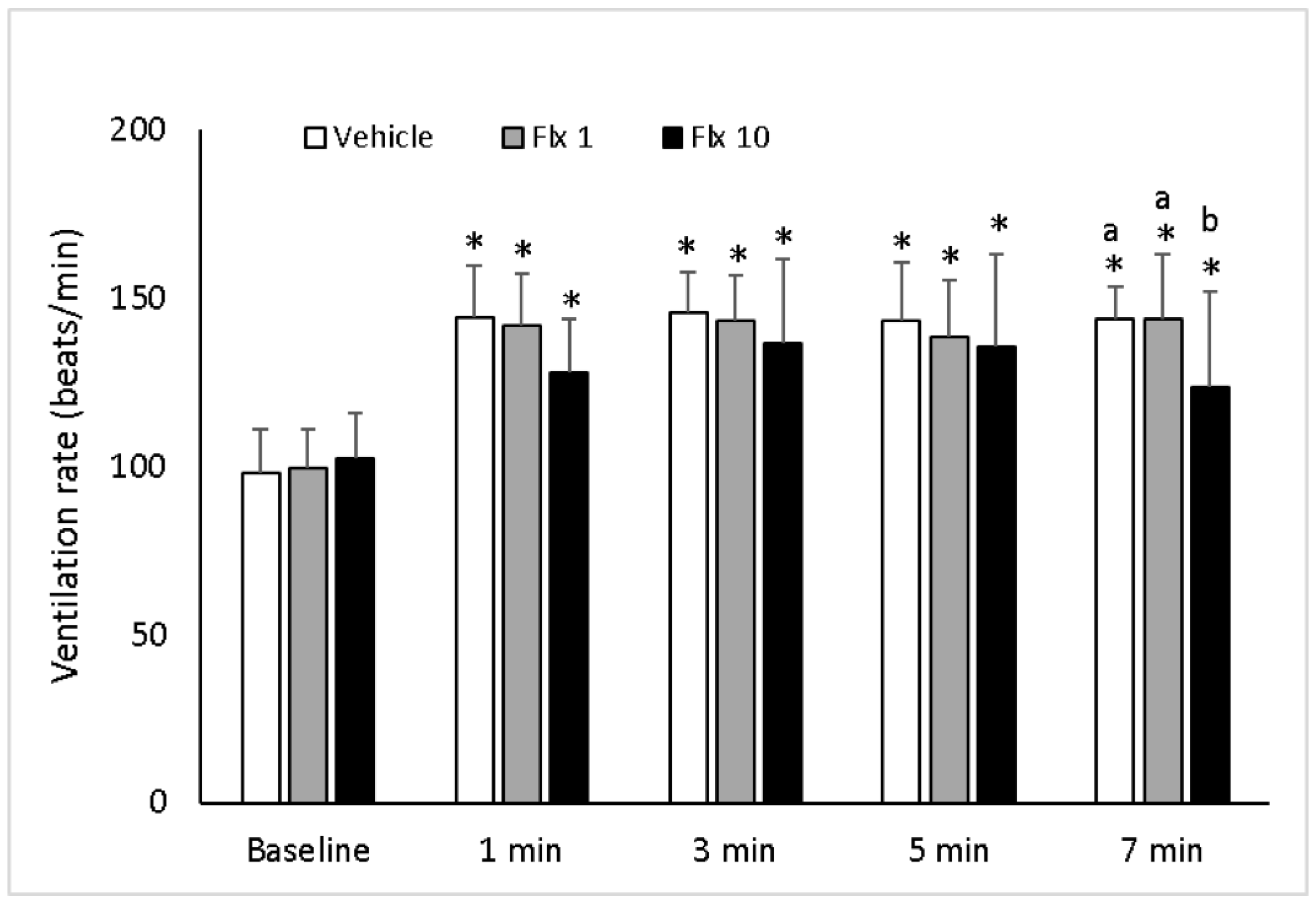
3.1. Ventilation Rate (VR)

Firstly, there were no differences between the baseline values among the experimental groups (vehicle control, ‘handling’ + fluoxetine 1 µg/L (H-FLX1) and ‘handling’ + fluoxetine 10 µg/L (H-FLX10)), indicating that all animals started from similar resting VR conditions (Figure 2). This statistical similarity indicated that fluoxetine did not affect the baseline VR levels. However, we found significant interactions between FLX, handling and time, as revealed by the ANOVA. After the handling stressor, VR increased significantly in relation to the baseline VR, but there was a similar magnitude of VR elevation among the groups, except for the last VR measurement (7 min) (Figure 2). In this last measurement, the VR values were lower in the H-FLX10 group compared to the vehicle and H-FLX1 groups, although still higher than the baseline, indicating that fluoxetine interfered with this variable (Figure 2).

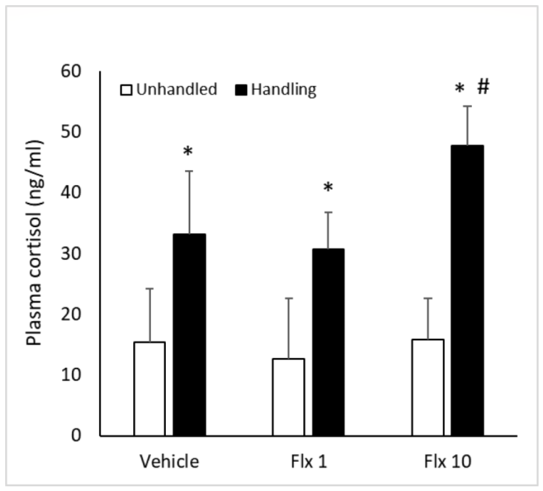
3.2. Plasma Cortisol

We lost one quantification in the experimental condition of 10 µg/L of fluoxetine without stress (‘unhandled’), because we obtained a very low value (an outside of cortisol standard curve value (‘off-curve’)); so, in this condition and for this variable only, we ended up with n = 9. A 2-way ANOVA revealed that the interaction between the stressor and fluoxetine had a significant influence on plasma cortisol levels (Figure 3). Unhandled animals had similar cortisol levels, indicating that fluoxetine did not affect the baseline values of this hormone. The animals that were exposed to the stressor had increased cortisol levels in relation to their respective controls (non-stressed fish). Furthermore, we found that the handled fish in the H-FLX10 condition had higher cortisol levels than the fish in the vehicle control and H-FLX1 conditions, which were similar to each other. This result indicated that the highest tested concentration of fluoxetine enhanced cortisol levels during response to stress in Nile tilapia.

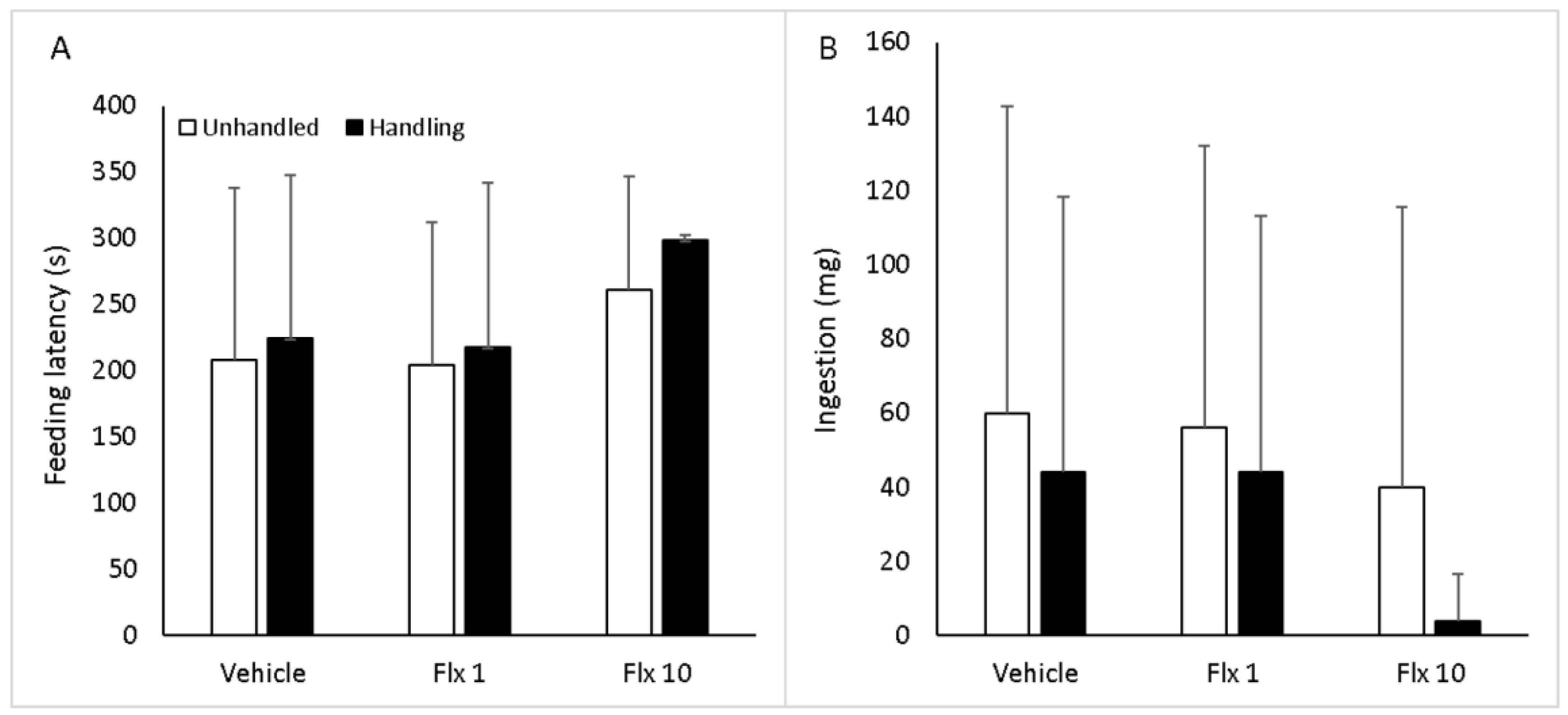
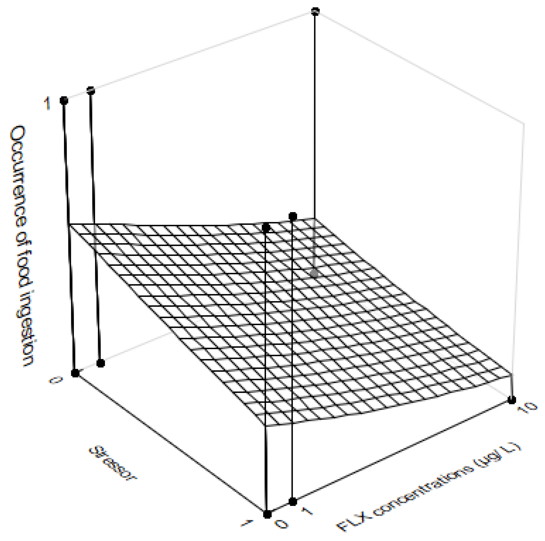
3.3. Feeding Behavior

We did not observe any significant effects of the interaction between fluoxetine and the stressor on either feeding latency (Figure 4A) or food intake (Figure 4B). In addition, neither of these factors (fluoxetine and the stressor) affected feeding latency or food intake in isolation. On the other hand, a multiple logistic regression analysis revealed that there was a significant association between handling, fluoxetine and food ingestion (Figure 5). In this case, the presence of the stressor or the higher dose of FLX tended to reduce food ingestion, but there was a higher odds ratio (unit changes) for FLX (ORFLX = 0.86 × ORSTRESSOR = 0.37).

4. Discussion

In this study, we showed that fluoxetine (FLX) modulated stress responses and feeding motivation in Nile tilapia. We observed that Nile tilapia increased plasma cortisol levels in response to a handling stressor and this response was enhanced by acute exposure (24 h) to 10 µg/L of FLX. This observation suggested that FLX had a possible additive effect on cortisol levels that were already raised by exposure to stress. The VR of the tilapia increased as a function of exposure to handling but decreased significantly at the end of our VR quantification interval in tilapia exposed to 10 µg/L of FLX. Regarding feeding behavior, both handling and FLX seemed to be linked to the tendency to decrease food ingestion but there was a higher probability of this effect with FLX.
The increase in plasma cortisol levels during stress responses in fish is a typical occurrence and this glucocorticoid is one of the main indicators of stress in fish [8,9,10]. In this study, in addition to handling, tilapias that were exposed to the highest tested concentration of FLX (10 µg/L) had further increases in plasma cortisol levels. However, some studies have shown that FLX (with acute or chronic administration) attenuates increases in cortisol levels during stress responses [32,33,34,64,65,66], while others have indicated that FLX can induce elevations in plasma cortisol levels in fish [28,34,36]. This phenomenon is even more complex because there is evidence that FLX can attenuate increases in cortisol levels in fish exposed to physical stressors (e.g., confinement or net pursuit) but does not have the same effect when fish are exposed to chemical cues that indicate predation risk [32]. In vertebrates, the activation of central 5-HT receptors can induce the release of glucocorticoids, such as cortisol, either directly by action on the adrenal gland or indirectly by activating the hypothalamus–pituitary–adrenal (HPA) axis (in mammals [12]) and a similar relationship has been suggested in fish (reviewed in [16]). Along this line, a single injection of the drug citalopram, another selective serotonin reuptake inhibitor, has been shown to increase ACTH and corticosteroid levels in male rats [67]. Based on the above evidence, it is plausible to assume that the increases in cortisol concentrations observed in this study depended on the intensity of the effects that FLX exerted at any possible sites along the HPI axis (directly on the interrenal gland and/or by stimulating the superior parts of the HPI axis) during the ongoing stress responses in the tilapia. Thus, we suggest that FLX and handling interacted and induced a possible additive effect, resulting in a stronger effect than that observed in response to exposure to handling alone. In future studies, it is necessary to investigate the action of FLX in inducing responses at other sites along the HPI axis to clarify this possible additive effect.
VR is another stress indicator in fish that is sensitive to a wide range of stimuli of different intensities and it is typically a variable that increases during stress responses [4,50,54,55,56,68,69]. The tilapia in the present study started with similar baseline VR levels and, after handling, the animals in all conditions (vehicle, FLX1 and FLX10 groups) showed hyperventilation. Thus, this stimulus was effective in inducing increases in VR. However, at the final time interval of the VR evaluation, the group exposed to 10 µg/L presented lower VR values than the vehicle controls and the animals exposed to 1 µg/L, although they were still higher than the baseline levels. Immediate changes (as was the case in this study) in VR during stress indicate a possible increase in sympathetic nervous system activity [2]. Thus, we suggest that FLX at the highest concentration tested (10 µg/L) attenuated sympathetic activity during stress, resulting in earlier decreases in VR than in the other conditions. This explanation is plausible because a decrease in sympathetic activity in mammals with a reduction in heart rate has already been reported as an effect of FLX [14]. Furthermore, in toadfish, FLX has been found to decrease cardiorespiratory activity to hypoxia [70], a stressor that usually rapidly increases sympathetic activity and VR [71], responses that allow fish to uptake more oxygen to deal with stress [72].
Stress [44] and chronic exposure to FLX [40,41] can reduce feeding performance. In this study, feeding latency, which is indicative of feeding motivation [38], was not affected by stress, FLX or the interaction between these two variables. Considering the Scheirer–Ray–Hare test only, food ingestion was also not affected by the evaluated factors. However, a logistic regression model indicated that exposure to the stressor and FLX seemed to be associated with the tendency of the tilapia to decrease their ingestion of food, although exposure to FLX had a higher probability of reducing food ingestion than exposure to the stressor. Taking the results of these analyses together, we assumed that the observed effects of the stressor and FLX were small, especially because the variability in the tilapia’s food ingestion responses was high. However, these effects, especially the effects of FLX, seemed to be realistic and were in line with previous reports on tilapia and other fish species that underwent protocols of FLX or serotonin administration. Serotonin controls appetite in fish and there is evidence that it has a stronger effect when administered intracerebroventricularly than when administered intraperitoneally [66]. An inhibitory action of serotonin on food intake has also been observed in rainbow trout (Onchorynchus mykiss) when its levels were increased in some brain regions [73,74]. In experiments that have studied chronic exposure to equal/higher doses of FLX in fish, decreases in food intake and growth have also been documented (for instance, 100 μg/L in zebrafish [27] and 10 or 100 μg/L in fathead minnow (Pimephales promelas) [53]). The acute exposure (96 h) of Nile tilapia to a high concentration of FLX (1 mg/L, a simulation of excessive environmental contamination) induced a long-term reduction in growth, a variable that is dependent on food ingestion [75]. This evidence corroborates our finding that there was a higher probability of tilapia not ingesting food as an effect of exposure to high concentrations of FLX than exposure to the stressor.

5. Conclusions

We concluded that fluoxetine modulated ongoing stress responses in Nile tilapia since this drug increased cortisol response to handling, while attenuating ventilation rate. Fluoxetine and stress also tended to interfere with feeding intake in this species. This suggests that fluoxetine is a relevant variable that affects adaptive biological processes in Nile tilapia.

Author Contributions

R.E.B. and J.P.G.A.M. wrote the main manuscript (first draft). All authors reviewed the manuscript to reach the final version. R.E.B., J.P.G.A.M., A.B.J.I., R.B.S. and L.C.S.T. conceptualized the methodology and collected the data. R.E.B., L.J.G.B. and H.C.D. analyzed the data. All authors have read and agreed to the published version of the manuscript.

Funding

A.B.J.I. (process number 2021/05735-5), R.B.S. (process number 2021/05772-8) and L.C.S.T. (process number 2021/05768-0) were recipients of scientific scholarships from FAPESP (Fundação de Amparo à Pesquisa do Estado de São Paulo). R.E.B. (process number 304288/2021-7) and L.J.G.B. (process number 302167/2022-6) were the recipients of a research productivity grant from CNPq (Conselho Nacional de Desenvolvimento Científico e Tecnológico).

Institutional Review Board Statement

This study agreed with Brazilian legislation regulated by the National Council for the Control of Animal Experimentation (CONCEA) and the Ethical Principles in Animal Research formulated by the Brazilian Society of Science in Laboratory Animals and was approved by the Bioscience Institute (UNESP) Ethics Committee on Use of Animals (CEUA) (5589170521).

Data Availability Statement

The data will be made available upon request.

Conflicts of Interest

The authors declare no conflict of interest.

References

  1. Reid, S.G.; Bernier, N.J.; Perry, S.F. The adrenergic stress response in fish: Control of catecholamine storage and release. Comp. Biochem. Physiol. C—Pharmacol. Toxicol. Endocrinol. 1998, 120, 1–27. [Google Scholar] [CrossRef] [PubMed] [Green Version]
  2. Perry, S.F.; Bernier, N.J. The acute humoral adrenergic stress response in fish: Facts and fiction. Aquaculture 1999, 177, 285–295. [Google Scholar] [CrossRef]
  3. Altimiras, J.; Larsen, E. Non-invasive recording of heart rate and ventilation rate in rainbow trout during rest and swimming. Fish go wireless! J. Fish Biol. 2000, 57, 197–209. [Google Scholar] [CrossRef]
  4. Barreto, R.E.; Volpato, G.L. Caution for using ventilatory frequency as an indicator of stress in fish. Behav. Proc. 2004, 66, 43–51. [Google Scholar] [CrossRef] [PubMed]
  5. Barreto, R.E.; Volpato, G.L. Ventilatory frequency of Nile tilapia subjected to different stressors. J. Exp. Anim. Sci. 2006, 43, 189–196. [Google Scholar] [CrossRef]
  6. Barreto, R.E.; Volpato, G.L. Ventilation rates indicate stress-coping styles in Nile tilapia. J. Biosci. 2011, 36, 851–855. [Google Scholar] [CrossRef]
  7. Maia, C.M.; Volpato, G.L. Environmental light color affects the stress response of Nile tilapia. Zoology 2013, 116, 64–66. [Google Scholar] [CrossRef]
  8. Barton, B.A.; Iwama, G.K. Physiological changes in fish from stress in aquaculture with emphasis on the response and effects of corticosteroids. Ann. Rev. Fish Dis. 1991, 1, 3–26. [Google Scholar] [CrossRef]
  9. Wendelaar-Bonga, S.E. The stress response in fish. Physiol. Rev. 1997, 77, 591–625. [Google Scholar] [CrossRef]
  10. Barton, B.A. Stress in fishes: A diversity of responses with particular reference to changes in circulating corticosteroids. Integrat. Comp. Biol. 2002, 42, 517–525. [Google Scholar] [CrossRef]
  11. Mommsen, T.P.; Vijayan, M.M.; Moon, T.W. Cortisol in teleosts: Dynamics, mechanisms of action, and metabolic regulation. Rev. Fish Biol. Fish. 1999, 9, 211–268. [Google Scholar] [CrossRef]
  12. Calogero, A.E.; Bagdy, G.; Szemeredi, K.; Tartaglia, M.E.; Gold, P.W.; Chrousos, G.P. Mechanisms of serotonin receptor agonist-induced activation of the hypothalamic-pituitary- adrenal axis in the rat. Endocrinology 1990, 126, 1888–1894. [Google Scholar] [CrossRef]
  13. Lowry, C.A. Functional subsets of serotonergic neurones: Implications for control of the hypothalamic–pituitary–adrenal axis. J. Neuroendocrinol. 2002, 14, 911–923. [Google Scholar] [CrossRef]
  14. Tiradentes, R.V.; Pires, J.G.P.; Silva, N.F.; Ramage, A.G.; Santuzzi, C.H.; Futuro Neto, H.A. Effects of acute administration of selective serotonin reuptake inhibitors on sympathetic nerve activity. Braz. J. Med. Biol. Res. 2014, 47, 554–559. [Google Scholar] [CrossRef] [PubMed] [Green Version]
  15. Herculano, A.M.; Maximino, C. Serotonergic modulation of zebrafish behavior: Towards a paradox. Prog. Neuro-Psychopharmacol. Biol. Psych. 2014, 55, 50–66. [Google Scholar] [CrossRef] [Green Version]
  16. Salahinejad, A.; Attaran, A.; Meuthen, D.; Chivers, D.P.; Niyogi, S. Proximate causes and ultimate effects of common antidepressants, fluoxetine and venlafaxine, on fish behavior. Sci. Total Environ. 2022, 807, 150846. [Google Scholar] [CrossRef] [PubMed]
  17. Griffiths, B.B.; Schoonheim, P.J.; Ziv, L.; Voelker, L.; Baier, H.; Gahtan, E. A zebrafish model of glucocorticoid resistance shows serotonergic modulation of the stress response. Front. Behav. Neurosci. 2012, 6, 68. [Google Scholar] [CrossRef] [PubMed] [Green Version]
  18. Winberg, S.; Nilsson, A.; Hylland, P.; Soderstom, V.; Nilsson, G.E. Serotonin as a regulator of hypothalamic-pituitary-interrenal activity in teleost fish. Neurosci. Lett. 1997, 230, 113–116. [Google Scholar] [CrossRef] [PubMed]
  19. Kolpin, D.W.; Furlong, E.T.; Meyer, M.T.; Thurman, E.M.; Zaugg, S.D.; Barber, L.B.; Buxton, H.T. Pharmaceuticals, hormones, and other organic wastewater contaminants in streams, 1999–2000: A national reconnaissance. Environ. Sci. Technol. 2002, 36, 1202–1211. [Google Scholar] [CrossRef] [Green Version]
  20. Metcalfe, C.D.; Miao, X.S.; Koenig, B.G.; Struger, J. Distribution of acidic and neutral drugs in surface waters near sewage treatment plants in the lower Great Lakes, Canada. Environ. Toxicol. Chem. 2003, 22, 2881–2889. [Google Scholar] [CrossRef]
  21. Metcalfe, C.D.; Chu, S.; Judt, C.; Li, H.; Oakes, K.D.; Servos, M.R.; Andrews, D.M. Antidepressants and their metabolites in municipal wastewater, and downstream exposure in an urban watershed. Environ. Sci. Chem. 2010, 29, 79–89. [Google Scholar] [CrossRef]
  22. Christensen, A.M.; Markussen, B.; Baun, A.; Halling-Sørensen, B. Probabilistic environmental risk characterization of pharmaceuticals in sewage treatment plant discharges. Chemosphere 2009, 77, 351–358. [Google Scholar] [CrossRef] [PubMed]
  23. Farré, M.; Pérez, S.; Kantiani, L.; Barceló, D. Fate and toxicity of emerging pollutants, their metabolites and transformation products in the aquatic environment. TrAC—Trends Analyt. Chem. 2008, 27, 991–1007. [Google Scholar] [CrossRef]
  24. Schultz, M.M.; Furlong, E.T.; Kolpin, D.W.; Werner, S.L.; Schoenfuss, H.L.; Barber, L.B.; Blazer, V.S.; Norris, D.O.; Vajda, A.M. Antidepressant pharmaceuticals in two effluent-impacted U.S. streams: Occurrence and fate in water and sediment and selective uptake in fish neural tissue. Environ. Sci. Technol. 2010, 44, 1918–1925. [Google Scholar] [CrossRef] [PubMed]
  25. Brooks, B.W. Fish on Prozac (and Zoloft): Ten years later. Aquat. Toxciol. 2014, 151, 61–67. [Google Scholar] [CrossRef] [PubMed]
  26. Rodrigues, P.; Cunha, V.; Ferreira, M.; Reis-Henriques, M.A.; Oliva-Teles, L.; Guimarães, L.; Carvalho, A.P. Differential Molecular Responses of Zebrafish Larvae to Fluoxetine and Norfluoxetine. Water 2022, 14, 417. [Google Scholar] [CrossRef]
  27. Farias, N.O.; Oliveira, R.; Moretti, P.N.S.; Mona e Pinto, J.; Oliveira, A.C.; Santos, V.L.; Rocha, P.S.; Andrade, T.S.; Grisolia, C.K. Fluoxetine chronic exposure affects growth, behavior and tissue structure of zebrafish. Comp. Biochem. Physiol. C Toxicol. Pharmacol. 2020, 237, 108836. [Google Scholar] [CrossRef]
  28. Morando, M.B.; Medeiros, L.R.; McDonald, M.D. Fluoxetine treatment affects nitrogen waste excretion and osmoregulation in a marine teleost fish. Aquat. Toxicol. 2009, 95, 164–171. [Google Scholar] [CrossRef]
  29. McDonald, M.D.; Gonzalez, A.; Sloman, K.A. Higher levels of aggression are observed in socially dominant toadfish treated with the selective serotonin reuptake inhibitor, fluoxetine. Comp. Biochem. Physiol. 2011, 153C, 107–112. [Google Scholar] [CrossRef]
  30. Correia, D.; Domingues, I.; Faria, M.; Oliveira, M. Effects of fluoxetine on fish: What do we know and where should we focus our efforts in the future? Sci. Total Environ. 2023, 857 Pt 2, 159486. [Google Scholar] [CrossRef]
  31. Mennigen, J.A.; Stroud, P.; Zamora, J.M.; Moon, T.W.; Trudeau, V.L. Pharmaceuticals as Neuroendocrine Disruptors: Lessons Learned from Fish on Prozac. J. Toxicol. Environ. Health—Part B 2011, 14, 387–412. [Google Scholar] [CrossRef] [PubMed]
  32. Abreu, M.S.D.; Giacomini, A.C.V.; Koakoski, G.; Piato, A.L.; Barcellos, L.J.G. Divergent effect of fluoxetine on the response to physical or chemical stressors in zebrafish. PeerJ 2017, 5, e3330. [Google Scholar] [CrossRef] [PubMed] [Green Version]
  33. Abreu, M.S.D.; Koakoski, G.; Ferreira, D.; Oliveira, T.A.; Rosa, J.G.S.D.; Gusso, D.; Giacomini, A.C.V.; Piato, A.L.; Barcellos, L.J.G. Diazepam and fluoxetine decrease the stress response in zebrafish. PLoS ONE 2014, 9, e103232. [Google Scholar] [CrossRef] [PubMed]
  34. Vera-Chang, M.N.; St-Jacques, A.D.; Lu, C.; Moon, T.W.; Trudeau, V.L. Fluoxetine exposure during sexual development disrupts the stress axis and results in sex-and time-dependent effects on the exploratory behavior in adult zebrafish Danio rerio. Front. Neurosci. 2019, 13, 1015. [Google Scholar] [CrossRef] [PubMed]
  35. Sebire, M.; Davis, J.E.; Hatfield, R.; Winberg, S.; Katsiadaki, I. Prozac affects stickleback nest quality without altering androgen, spiggin or aggression levels during a 21-day breeding test. Aquat. Toxicol. 2015, 168, 78–89. [Google Scholar] [CrossRef]
  36. Theodoridi, A.; Tsalafouta, A.; Pavlidis, M. Acute Exposure to Fluoxetine Alters Aggressive Behavior of Zebrafish and Expression of Genes Involved in Serotonergic System Regulation. Front. Neurosci. 2017, 11, 223. [Google Scholar] [CrossRef]
  37. Valassi, E.; Scacchi, M.; Cavagnini, F. Neuroendocrine control of food intake. Nutr. Metab. Cardiovasc. Dis. 2008, 18, 158–168. [Google Scholar] [CrossRef]
  38. Volpato, G.L.; Bovi, T.S.; de Freitas, R.H.A.; da Silva, D.F.; Delicio, H.C.; Giaquinto, P.C.; Barreto, R.E. Red Light Stimulates Feeding Motivation in Fish but Does Not Improve Growth. PLoS ONE 2013, 8, e59134. [Google Scholar] [CrossRef] [Green Version]
  39. Delgado, M.J.; Cerdá-Reverter, J.M.; Soengas, J.L. Hypothalamic integration of metabolic, endocrine and circadian signals in fish: Involvement in cortisol of food intake. Front. Neurosci. 2017, 11, 354. [Google Scholar] [CrossRef] [Green Version]
  40. Mennigen, J.A.; Harris, E.A.; Chang, J.P.; Moon, T.W.; Trudeau, V.L. Fluoxetine affects weight gain and expression of feeding peptides in the female goldfish brain. Regul. Pept. 2009, 155, 99–104. [Google Scholar] [CrossRef]
  41. Mennigen, J.A.; Sassine, J.; Trudeau, V.L.; Moon, T.W. Waterborne fluoxetine disrupts feeding and energy metabolism in the goldfish, Carassius auratus. Aquat. Toxicol. 2010, 100, 128–137. [Google Scholar] [CrossRef] [PubMed]
  42. Carlini, V.P.; Gaydou, R.C.; Schiöth, H.B.; de Barioglio, S.R. Selective serotonin reuptake inhibitor (fluoxetine) decreases the effects of ghrelin on memory retention and food intake. Regul. Pept. 2007, 140, 65–73. [Google Scholar] [CrossRef] [PubMed]
  43. De Pedro, N.; Pinillos, M.L.; Valenciano, A.I.; Alonso-Bedate, M.; Delgado, M.J. Inhibitory effect of serotonin on feeding behavior in goldfish: Involvement of CRH. Peptides 1998, 19, 505–511. [Google Scholar] [CrossRef] [PubMed]
  44. Conde-Sieira, M.; Chivite, M.; Míguez, J.M.; Soengas, J.L. Stress effects on the mechanisms regulating appetite in teleost fish. Front. Endocrinol. 2018, 9, 631. [Google Scholar] [CrossRef] [Green Version]
  45. Vijitkul, P.; Kongsema, M.; Toommakorn, T.; Bullangpoti, V. Investigation of genotoxicity, mutagenicity, and cytotoxicity in erythrocytes of Nile tilapia (Oreochromis niloticus) after fluoxetine exposure. Toxicol. Rep. 2022, 9, 588–596. [Google Scholar] [CrossRef]
  46. Valenti, W.C.; Barros, H.P.; Moraes-Valenti, P.; Bueno, G.W.; Cavalli, R.O. Aquaculture in Brazil: Past, present and future. Aquac. Rep. 2021, 19, 100611. [Google Scholar] [CrossRef]
  47. Naylor, R.L.; Hardy, R.W.; Buschmann, A.H.; Bush, S.R.; Cao, L.; Klinger, D.H.; Little, D.C.; Lubchenco, J.; Shumway, S.E.; Troell, M. A 20-year retrospective review of global aquaculture. Nature 2021, 591, 551–563. [Google Scholar] [CrossRef]
  48. Gonçalves-de-Freitas, E.; Bolognesi, M.C.; Gauy, A.C.d.S.; Brandão, M.L.; Giaquinto, P.C.; Fernandes-Castilho, M. Social Behavior and Welfare in Nile Tilapia. Fishes 2019, 4, 23. [Google Scholar] [CrossRef] [Green Version]
  49. Silva, D.R.; Arvigo, A.L.; Giaquinto, P.C.; Delicio, H.C.; Barcellos, L.J.G.; Barreto, R.E. Effects of clove oil on behavioral reactivity and motivation in Nile tilapia. Aquaculture 2021, 532, 736045. [Google Scholar] [CrossRef]
  50. Carneiro, V.C.L.; Delicio, H.C.; Barreto, R.E. Effects of stress-associated odor on ventilation rate and feeding performance in Nile tilapia. J. Appl. Anim. Welf. Sci. 2022, 1–11. [Google Scholar] [CrossRef]
  51. Carvalho, A.C.C. A Presença de Fármacos e Cafeína em Água Superficial e Destinada ao Consumo Humano (The Presence of Pharmaceuticals and Caffeine in Surface and Treated Water). Ph.D. Thesis, University of São Paulo (USP), São Paulo, SP, Brazil, 2021. [Google Scholar] [CrossRef]
  52. Bedner, M.; MacCrehan, W.A. Reactions of the amine-containing drugs fluoxetine and metoprolol during chlorination and dechlorination processes used in wastewater treatment. Chemosphere 2006, 65, 2130–2137. [Google Scholar] [CrossRef] [PubMed]
  53. Weinberger, J.; Klaper, R. Environmental concentrations of the selective serotonin reuptake inhibitor fluoxetine impact specific behaviors involved in reproduction, feeding and predator avoidance in the fish Pimephales promelas (fathead minnow). Aquat. Toxicol. 2014, 151, 77–83. [Google Scholar] [CrossRef] [PubMed] [Green Version]
  54. Gibson, A.K.; Mathis, A. Opercular beat rate for rainbow darters Etheostoma caeruleam exposed to chemical stimuli from conspecific and heterospecific. J. Fish Biol. 2006, 69, 224–232. [Google Scholar] [CrossRef]
  55. Samson, E.; Brownscombe, J.W.; Cooke, S.J. Behavioural and reflex responses of mottled mojarra Eucinostomus lefroyi (Gerreidae) to cold shock exposure. Aquat. Biol. 2014, 23, 101–108. [Google Scholar] [CrossRef] [Green Version]
  56. Freret-Meurer, N.V.; Carmo, T.F.; Cabiró, G. Opercular beat: A non-invasive and rapid method to detect stress in seahorses. J. Appl. Aquac. 2021, 33, 291–299. [Google Scholar] [CrossRef]
  57. Roza-e-Silva, M.L.; Pereira, R.T.; Arvigo, A.L.; Zanuzzo, F.S.; Barreto, R.E. Effects of water flow on ventilation rate and plasma cortisol in Nile tilapia introduced into novel environment. Aquac. Rep. 2020, 18, 100531. [Google Scholar] [CrossRef]
  58. Alvarenga, C.M.D.; Volpato, G.L. Agonistic profile and metabolism in alevins of the Nile tilapia. Physiol. Behav. 1995, 57, 75–80. [Google Scholar] [CrossRef]
  59. Gilderhus, P.A.; Marking, L.L. Comparative efficacy of 16 anaesthetic chemicals on rainbow trout. N. Am. J. Fish. Manag. 1987, 7, 288–292. [Google Scholar] [CrossRef]
  60. Stoskopf, M. Anaesthesia. In Aquaculture for Veterinarians; Brown, L., Ed.; Pergamon Press: Oxford, UK, 1993; pp. 161–167. [Google Scholar]
  61. Sink, T.D.; Lochmann, R.T.; Fecteau, K.A. Validation, use, and disadvantages of enzyme-linked immunosorbent assay kits for detection of cortisol in channel catfish, largemouth bass, red pacu and golden shiners. Fish Physiol. Biochem. 2007, 75, 165–171. [Google Scholar] [CrossRef]
  62. Zar, J.H. Biostatistical Analysis, 5th ed.; Prentice-Hall/Pearson: Upper Saddle River, NJ, USA, 2010. [Google Scholar]
  63. Kwak, S.K.; Kim, J.H. Statistical data preparation: Management of missing values and outliers. Korean J. Anesthesiol. 2017, 70, 407–411. [Google Scholar] [CrossRef]
  64. Egan, R.J.; Bergner, C.L.; Hart, P.C.; Cachat, J.M.; Canavello, P.R.; Elegante, M.F.; Elkhayat, S.I.; Bartels, B.K.; Tien, A.K.; Tien, D.H.; et al. Understanding behavioural and physiological phenotypes of stress and anxiety in zebrafish. Behav. Brain Res. 2009, 205, 38–44. [Google Scholar] [CrossRef] [Green Version]
  65. Giacomini, A.C.V.V.; Abreu, M.S.; Giacomini, L.V.; Siebel, A.M.; Zimerman, F.F.; Rambo, C.L.; Mocelin, R.; Bonan, C.D.; Piato, A.L.; Barcellos, L.J.G. Fluoxetine and diazepam acutely modulate stress induced-behavior. Behav. Brain Res. 2016, 296, 301–310. [Google Scholar] [CrossRef]
  66. Abreu, M.S.D.; Giacomini, A.C.V.V.; Kalueff, A.V.; Barcellos, L.J.G. The smell of “anxiety”: Behavioral modulation by experimental anosmia in zebrafish. Physiol. Behav. 2016, 157, 67–71. [Google Scholar] [CrossRef]
  67. Jensen, J.B.; Jessop, D.S.; Harbuz, M.S.; Mørk, A.; Sánchez, C.; Mikkelsen, J.D. Acute and long-term treatments with the selective serotonin reuptake inhibitor citalopram modulate the HPA axis activity at different levels in male rats. J. Neuroendocrinol. 1999, 11, 465–471. [Google Scholar] [CrossRef] [PubMed]
  68. Pereira, R.T.; Leutz, J.A.C.M.; Valença-Silva, G.; Barcellos, L.J.G.; Barreto, R.E. Ventilation responses to predator odors and conspecific chemical alarm cues in the frillfin goby. Physiol. Behav. 2017, 179, 319–323. [Google Scholar] [CrossRef] [PubMed] [Green Version]
  69. Zanuzzo, F.S.; Bovolato, A.L.C.; Pereira, R.T.; Valença-Silva, G.; Barcellos, L.J.G.; Barreto, R.E. Innate response based on visual cues of sympatric and allopatric predators in Nile tilapia. Behav. Proc. 2019, 164, 109–114. [Google Scholar] [CrossRef] [PubMed]
  70. Panlilio, J.; Marin, S.; Lobl, M.M.; McDonald, D. Treatment with the selective serotonin reuptake inhibitor, fluoxetine, attenuates the fish hypoxia response. Sci. Rep. 2016, 6, 31148. [Google Scholar] [CrossRef] [Green Version]
  71. Perry, S.F.; Reid, S.G.; Gilmour, K.M.; Boijink, C.L.; Lopes, J.M.; Milsom, W.K.; Rantin, F.T. A comparison of adrenergic stress responses in three tropical teleosts exposed to acute hypoxia Amer. J. Physiol.—Regul. Integrat. Comp. Physiol. 2004, 287, R188–R197. [Google Scholar] [CrossRef]
  72. Fernandes, M.N.; Rantin, F.T. Relationships between oxygen availability and metabolic cost of breathing in Nile tilapia (Oreochromis niloticus): Aquacultural consequences. Aquaculture 1994, 127, 339–346. [Google Scholar] [CrossRef]
  73. Ruibal, C.; Soengas, J.; Aldegunde, M. Brain serotonina and the control of food intake in rainbow trout (Onchorhynchus mykiss): Effects of changes in plasma glucose levels. J. Comp. Physiol. 2002, 188, 479–484. [Google Scholar]
  74. Pérez, J.J.M.; Mancebo, M.J.; Aldegunde, M. The involvement of HT-like receptors in the regulation of food intake in rainbow trout (Onchorhynchus mykiss). Comp. Biochem. Physiol. C 2014, 161, 1–6. [Google Scholar]
  75. Varela, A.C.C.; Soares, S.M.; Fortuna, M.; Costa, V.C.; Piasson, Í.B.; Mozatto, M.T.; Siqueira, L.; Barcellos, H.H.A.; Barreto, R.E.; Barcellos, L.J.G. A single exposure to sub-lethal concentrations of a glyphosate-based herbicide or fluoxetine-based agent on growth performance in Nile tilapia. J. Toxicol. Environ. Health A 2023, 13, 1–9. [Google Scholar] [CrossRef]
Figure 1. A scheme of the experimental schedule. VR = ventilation rate.
Fishes 08 00348 g001
Figure 2. The mean (±SD) of the ventilation rates (VRs) of Nile tilapia exposed to handling and fluoxetine. A mixed ANOVA revealed the significant effects of fluoxetine and handling on the VR of Nile tilapia (F(8;108) = 2.07; p = 0.046). The post-stressor VR values were higher than the baseline VR values (*). For the last time measurement, the letters indicate the comparison among the conditions and different letters denote statistical differences.
Fishes 08 00348 g002
Figure 3. The mean (±SD) of the plasma cortisol levels of handled and unhandled Nile tilapia that were either exposed or not to fluoxetine. A 2-way ANOVA showed the significant interaction between the effects of the stressor and fluoxetine (F(2;53) = 4.57; p = 0.015). The cortisol levels of the fish in all groups that were exposed to handling (black bars) were higher than those of their respective controls (white bars; ‘unhandled’ fish (*)). Note: # compares the black bars and denotes that the cortisol levels of handled fish in the 10-µg/L FLX group were higher than those in groups that were exposed to the handling stressor and a lower concentration of FLX (the vehicle or 1 µg/L groups).
Fishes 08 00348 g003
Figure 4. The mean (±SD) of the feeding latency (A) and ingestion (B) of handled and unhandled Nile tilapia that were either exposed or not to fluoxetine. There were no observed influences of the interaction between the effects of handling and fluoxetine (Scheirer–Ray–Hare test) on latency (H(2;54) = 0.11; p = 0.90) or ingestion (H(2;54) = 0.10; p = 0.91). Moreover, no isolated effects of the factors (Scheirer–Ray–Hare test) were observed for latency (stressor effect: H(1;54) = 1.28; p = 0.26; fluoxetine effect: H(2;54) = 2.01; p = 0.14) or ingestion (stressor effect: H(1;54) = 1.79; p = 0.19; fluoxetine effect,: H(2;54) = 1.49; p = 0.24).
Fishes 08 00348 g004
Figure 5. A multiple logistic regression analysis of the food ingestion of handled and unhandled Nile tilapia that were either exposed or not to fluoxetine. This regression revealed a significant association between the effects of the stressor and fluoxetine on food ingestion (p = 0.022; odds ratio for FLX = 0.86; odds ratio for stressor = 0.37). This association suggests a tendency to observe reductions in food ingestion as an effect of the stressor and fluoxetine. The surface function is Z = exp(0.24410011384368 + (−0.99810948382)X + (−0.1530157885811)Y)/(1 + exp(0.24410011384368 + (−0.99810948382)X + (−0.1530157885811)Y)).
Fishes 08 00348 g005
Disclaimer/Publisher’s Note: The statements, opinions and data contained in all publications are solely those of the individual author(s) and contributor(s) and not of MDPI and/or the editor(s). MDPI and/or the editor(s) disclaim responsibility for any injury to people or property resulting from any ideas, methods, instructions or products referred to in the content.

