Article

DUSP2 Deletion Inhibits Macrophage Migration by Inhibiting ERK Activation in Zebrafish

1
Center for Advanced Interdisciplinary Science and Biomedicine of IHM, Division of Life Sciences and Medicine, University of Science and Technology of China, Hefei 230026, China
2
Sanford Burnham Prebys Medical Discovery Institute, La Jolla, CA 92037, USA
*
Authors to whom correspondence should be addressed.
These authors contributed equally to this work and are co-first authors.
Fishes 2023, 8(6), 310; https://doi.org/10.3390/fishes8060310
Submission received: 1 May 2023 / Revised: 7 June 2023 / Accepted: 9 June 2023 / Published: 12 June 2023

Abstract

:
Dual-specificity phosphatase 2 (DUSP2) regulates the activation of members of the mitogen-activated protein kinase (MAPK) family, which is involved in a variety of cellular processes including cell proliferation, differentiation, apoptosis, and migration. DUSP2 also regulates the expression of inflammatory mediators in macrophages; however, it remains unknown whether DUSP2 participates in macrophage migration. Here, using the tail fin injury model in zebrafish larvae, we found that the deletion of DUSP2 inhibited the expression of pro-inflammatory cytokines and macrophage chemokines. Moreover, live imaging results showed that the migration of macrophages to the injury site was inhibited after DUSP2 deletion. This inhibitory effect was mediated through the reduced activation of extracellular regulated protein kinases (ERK) in DUSP2 knockout zebrafish.
Key Contribution: Using zebrafish as a model organism; we studied the effect of DUSP2 on macrophage migration in vivo; we found that DUSP2 deletion inhibited macrophage migration by inhibiting the activation of ERK.

1. Introduction

The migration of immune cells is a key step in and major feature of the inflammatory response [1]. Specifically, macrophage migration is a vital process in host defense and homeostasis maintenance [2,3]. Abnormal macrophage migration leads to cytokine accumulation, tissue destruction, and tumor formation, and it is a key factor in the development and progression of many autoimmune diseases, inflammatory diseases, and cancers [4,5]. Therefore, macrophage migration is a potential target for therapeutic strategies for these diseases.
The DUSP2 protein regulates the activation of members of the MAPK family [6,7]. The MAPK family mainly comprises extracellular signal-regulated kinase (ERK), c-Jun amino-terminal kinase (JNK), and p38, and it mediates the transduction pathway of inflammation induction [8,9,10,11]. Previous research has demonstrated that DUSP2-deficient macrophages exhibit increased activation of JNK, decreased activation of p38 and ERK, and reduced expression of inflammatory mediators, suggesting the positive role of DUSP2 in regulating innate immune function through the MAPK [12]. However, DUSP2 adversely affects the differentiation of TH17 cells and the anti-tumor effect of T cells [13,14], suggesting its negative involvement in adaptive immunity. Thus, DUSP2 is an important part of the immune function of an organism.
The deletion of DUSP2 inhibits the expression of inflammatory mediators in macrophages, but it remains to be clarified whether DUSP2 affects macrophage migration and, if it does, how [12]. This study aimed to answer this question by using the macrophage reporter line Tg(mpeg1:mCherry) and the dusp2−/− mutant previously generated in our lab [15,16].
There is a clear temporal separation between innate and adaptive immunity in zebrafish [17,18]. Furthermore, transparency in the early developmental stages of zebrafish allows for visual manipulation and observations, and transgenic zebrafish lines that specifically label immune cells facilitate the analysis of discrete immune events [15,19]. Based on these advantages, we investigated the effect of DUSP2 on macrophage migration and its potential mechanisms in zebrafish larvae with the tail fin injury model. We found that, through the reduced activation of extracellular regulated protein kinases (ERK), dusp2 knockout inhibited the migration of macrophages to the injury site.

2. Materials and Methods

2.1. Zebrafish Lines

The zebrafish lines used in this experiment included wild-type (WT) zebrafish, dusp2−/− mutant zebrafish, and transgenic zebrafish Tg(mpeg1:mCherry) and Tg(mpeg1:mCherry); dusp2−/−. The dusp2−/− mutant zebrafish was previously obtained by knocking out the dusp2 gene with CRISPR/Cas9 [16]. The dusp2−/− mutant zebrafish was crossed with Tg(mpeg1:mCherry) to obtain the F1 generation, and the Tg(mpeg1:mCherry); dusp2−/− was identified after incross among F1 littermates (Figure 1). All animal experimental protocols were approved by the University of Science and Technology of China (USTC) Animal Resources Center and the University Animal Care and Use Committee and the Committee on the Ethics of Animal Experiments of the USTC (Permit Number: USTCACUC1103013).

2.2. Tail Fin Injury

The experimental model used in this study was a tail fin injury model. The slide and a sterile scalpel were sterilized with 75% ethanol prior to tail fin injury. At 4 days post-fertilization (dpf), the zebrafish larvae were then anesthetized with MS-222 (Sigma, Saint Louis, USA, E10505) and placed on the slide, and the tail fins were cut from the end of the spinal cord with a sterile scalpel under a stereomicroscope (Olympus SZX-16, Tokyo, Japan).

2.3. In Vivo Imaging

In the absence of tail fin injury, macrophages in the 800 μm region from the end of the spinal cord forward were imaged at 10× using a laser confocal microscope (Olympus FV1000, Tokyo, Japan), and macrophages in this area were enumerated, giving the total number of macrophages in the sample. At different time points after the tail fin injury, macrophages in the 250 μm region from the end of the spinal cord forward were imaged at 10× using a fluorescence microscope (Olympus BX60, Tokyo, Japan); similarly, macrophages in this region were counted, giving the number of macrophages that migrated to the injury site. For the analysis of macrophage motility velocity, a laser confocal microscope (Olympus FV1000, Tokyo, Japan) was used to image continuously for 30 min at 10×, scanning 1 z-stack every 1 min. The imaging time points were before the tail fin injury and 2.5 h post-injury (hpi)–3 h post-injury (hpi) for each group of 8 zebrafish larvae. The data obtained were analyzed for macrophage motility using the software ImageJ (National Institutes of Health, Bethesda, MD, USA), and only those macrophages that migrated towards the injury site were counted.

2.4. Drug Treatment

To investigate the effect of the ERK protein on macrophage migration, we used an ERK inhibitor, PD0325901 (MCE, State of New Jersey, USA, HY-10254), to inhibit the phosphorylation of the ERK protein [20]. The working liquid concentration of PD0325901 was 20 μM, and the control group was treated with 0.1% DMSO. Zebrafish larvae were treated with PD0325901 or DMSO three hours before tail fin injury. Zebrafish larvae continued to be treated with either the inhibitor or DMSO after tail fin injury.

2.5. Quantitative Real-Time PCR

Total RNA was extracted using a TRIZOL reagent (Takara, Beijing, China) without tail fin injury or 1 h after tail fin injury. The expression levels of dusp2, inflammatory cytokines, and chemokines were evaluated using qPCR and the SYBR Green kit (Vazyme, Nanjing, China). The samples were obtained from 3 independent experiments, each with 3 replicates, and were subjected to a standard protocol (pre-denaturation at 95 °C for 5 min, followed by 44 cycles of 95 °C for 15 s and 60 °C for 30 s, and, finally, 95° C for 15 s, 60 °C for 60 s, and 95 °C for 15 s). Relative mRNA expression levels were obtained by standardizing to the β-actin mRNA level using the 2−ΔΔCt method. Table 1 showed the specific primers used in the PCR.

2.6. Western Blot

The zebrafish larvae were lysed with RIPA (Sangon, Shanghai, China) without tail fin injury or 1 h after tail fin injury. The primary antibody of p-ERK (CST, 4370T,1:1000) from rabbits was incubated overnight at 4 °C. The primary antibody of ERK (CST, 4695T, 1:1000) from rabbits and the primary antibody of β-actin (HuaBio, Hangzhou, China, ET1701-80, 1:1000) from rabbits were incubated for 3 h at 37 °C. The HRP-conjugated goat anti-rabbit antibody (Proteintech, SA00001-2, Chicago, IL, USA, 1:5000) was incubated for 1 h at 37 °C. We used ImageJ (National Institutes of Health, Bethesda, USA) software to calculate the gray value of the strip.

2.7. Statistical Analysis

Data were analyzed using the Student’s t-test with GraphPad Prism 8.0 (Santiago, Chile, USA). One-way analyses of variance (ANOVA) were used as indicated in the results. All data are presented as the mean ± SEM. p < 0.05 was assumed to be a significant difference.

3. Results

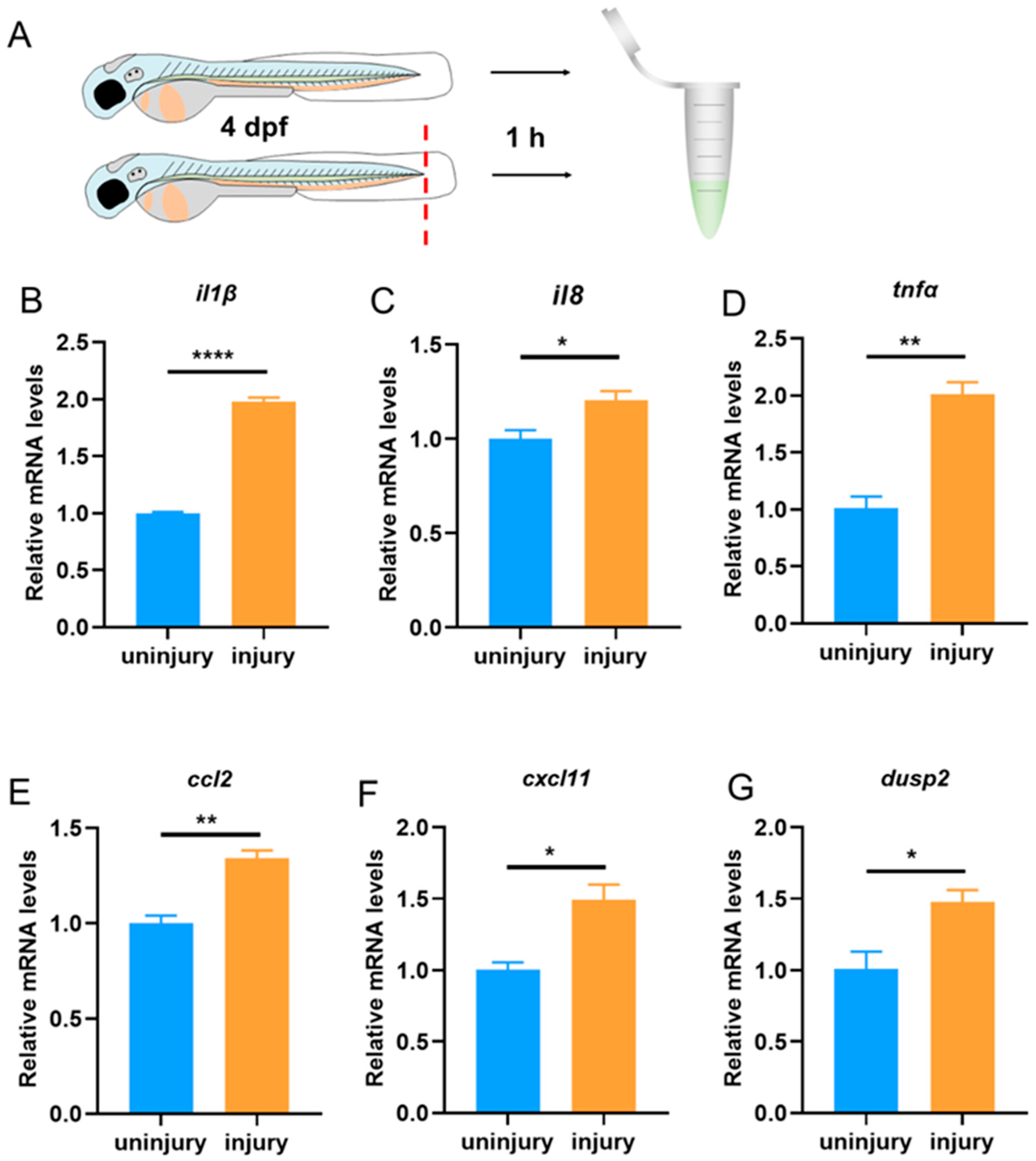
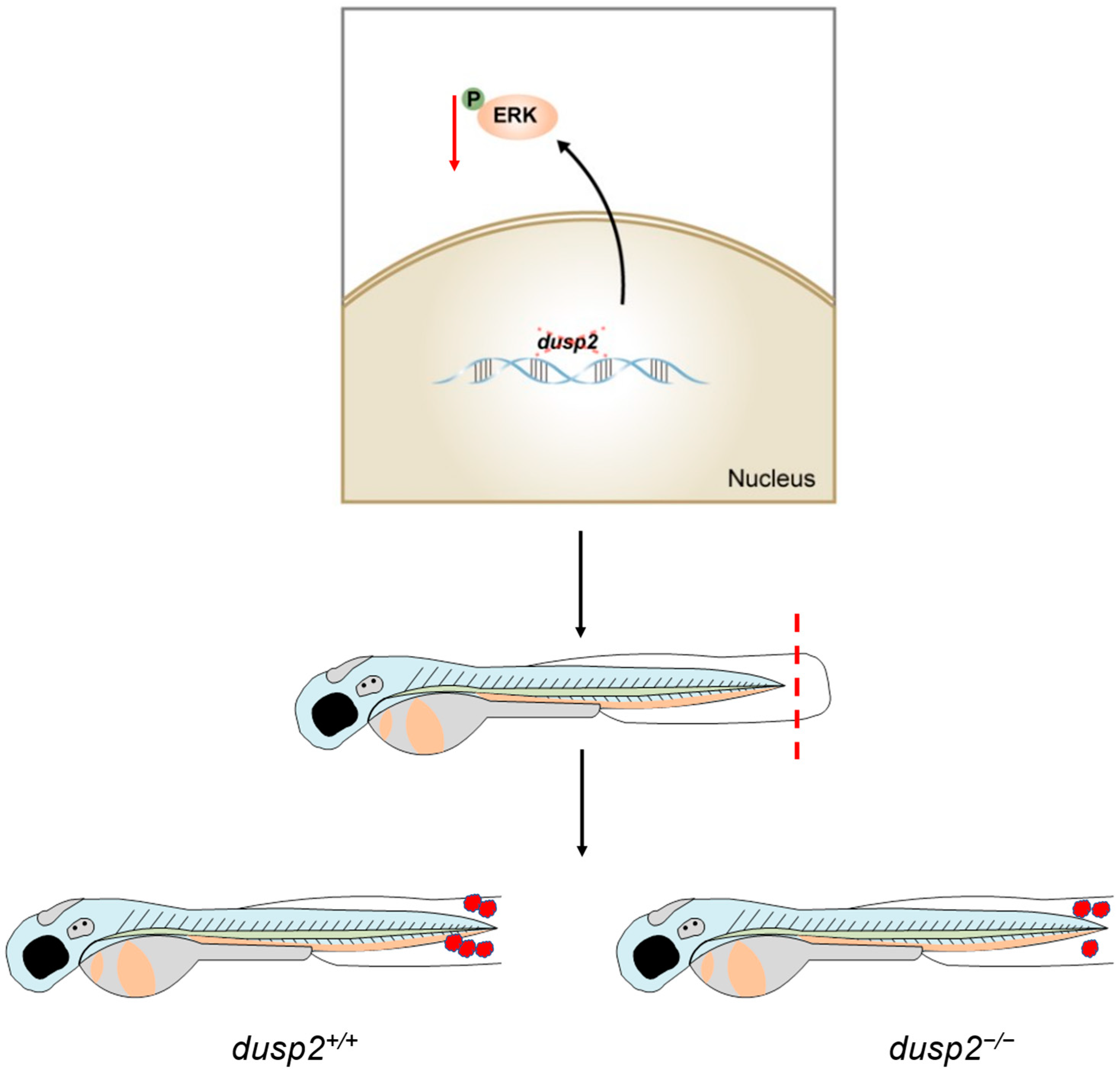
3.1. The Expression of dusp2 Was Significantly Up-Regulated in Acute Inflammation Induced by Tail Fin Injury

First, we used a tail fin injury model to investigate whether DUSP2 is involved in acute inflammation induced by tail fin injuries in zebrafish larvae (Figure 2A). The expression of pro-inflammatory cytokines was up-regulated and neutrophils and macrophages were recruited to the injury site after the tail fin injury [21]. One hour after the tail fin injury, the qPCR results showed that the expression of the pro-inflammatory cytokines IL-1β, IL-8, and TNF-α was significantly increased (Figure 2B–D). Meanwhile, the expression of the chemokines CCL2 and CXCL11 was also significantly up-regulated (Figure 2E,F). These results suggest that the tail fin injury model successfully induced acute inflammation in zebrafish larvae. Furthermore, we found that the expression of dusp2 was significantly up-regulated (Figure 2G), indicating that DUSP2 might be involved in acute inflammation induced by tail fin injuries in zebrafish larvae.

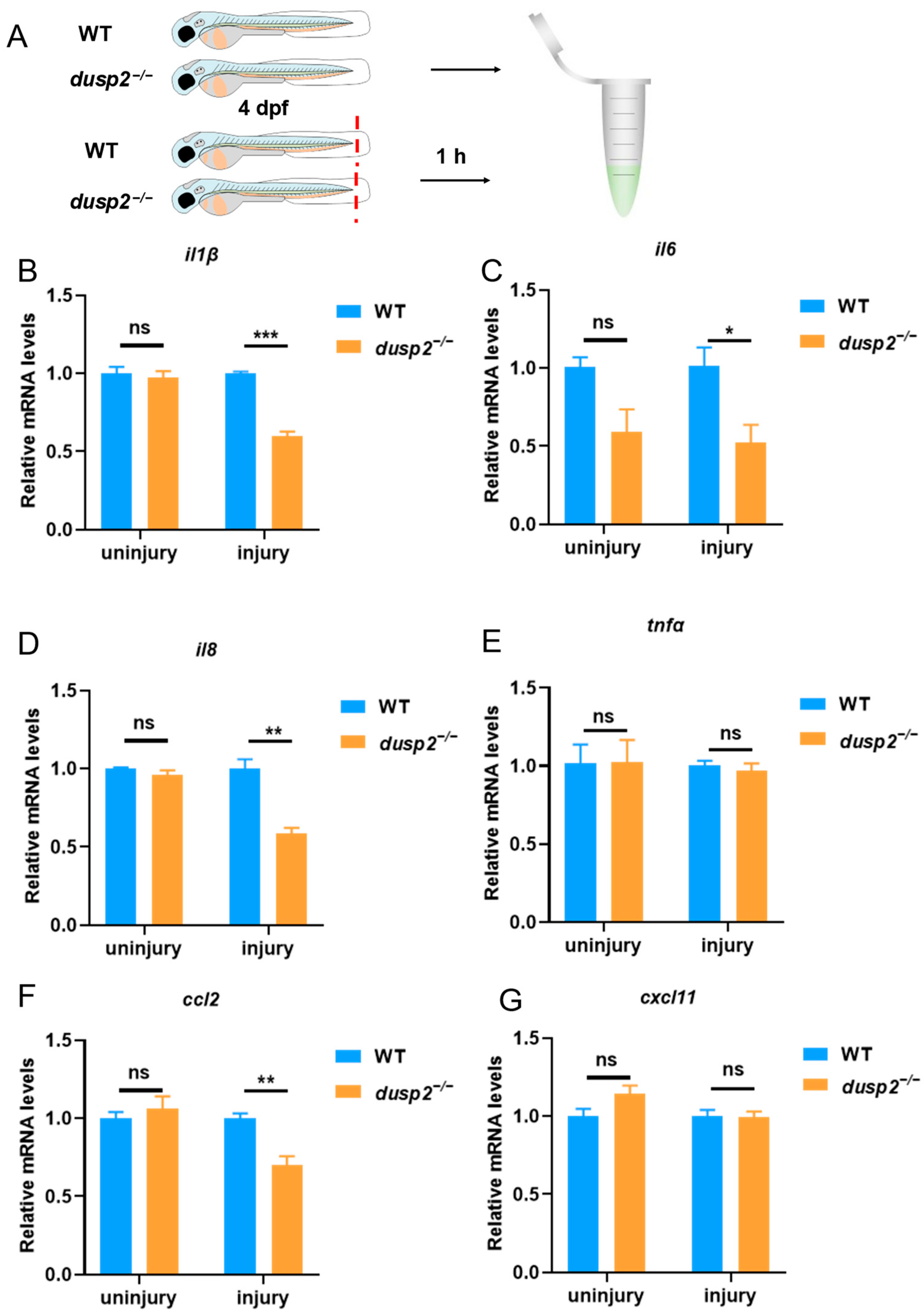
3.2. DUSP2 Deletion Significantly Decreased the Expression of Pro-Inflammatory Cytokines and Chemokines after Tail Fin Injury

Next, we aimed to explore whether the deletion of DUSP2 affects the expression of pro-inflammatory cytokines and chemokines in zebrafish larvae. CCL2 and CXCL11 are key chemokines involved in macrophage migration and infiltration [22] Here, we focused on the expression of four pro-inflammatory cytokines (IL-1β, IL-6, IL-8, TNF-α) and two macrophage chemokines (CCL2, CXCL11) in WT and dusp2−/− zebrafish larvae before and after tail fin injury. The results of the qPCR showed that the deletion of DUSP2 did not affect the expression of the pro-inflammatory cytokines IL-1β, IL-6, IL-8, and TNF-α and the macrophage chemokines CCL2 and CXCL11 in the absence of a tail fin injury (Figure 3B–G). However, the deletion of DUSP2 suppressed the expression of the pro-inflammatory cytokines IL-1β, IL-6, and IL-8 and the macrophage chemokine CCL2 after tail fin injury (Figure 3B–D,F); it did not affect the expression of TNF-α and CXCL11 (Figure 3E,G). These results suggest that DUSP2 is involved in the acute inflammation induced by tail fin injuries in zebrafish larvae and might be involved in the migration of macrophages.

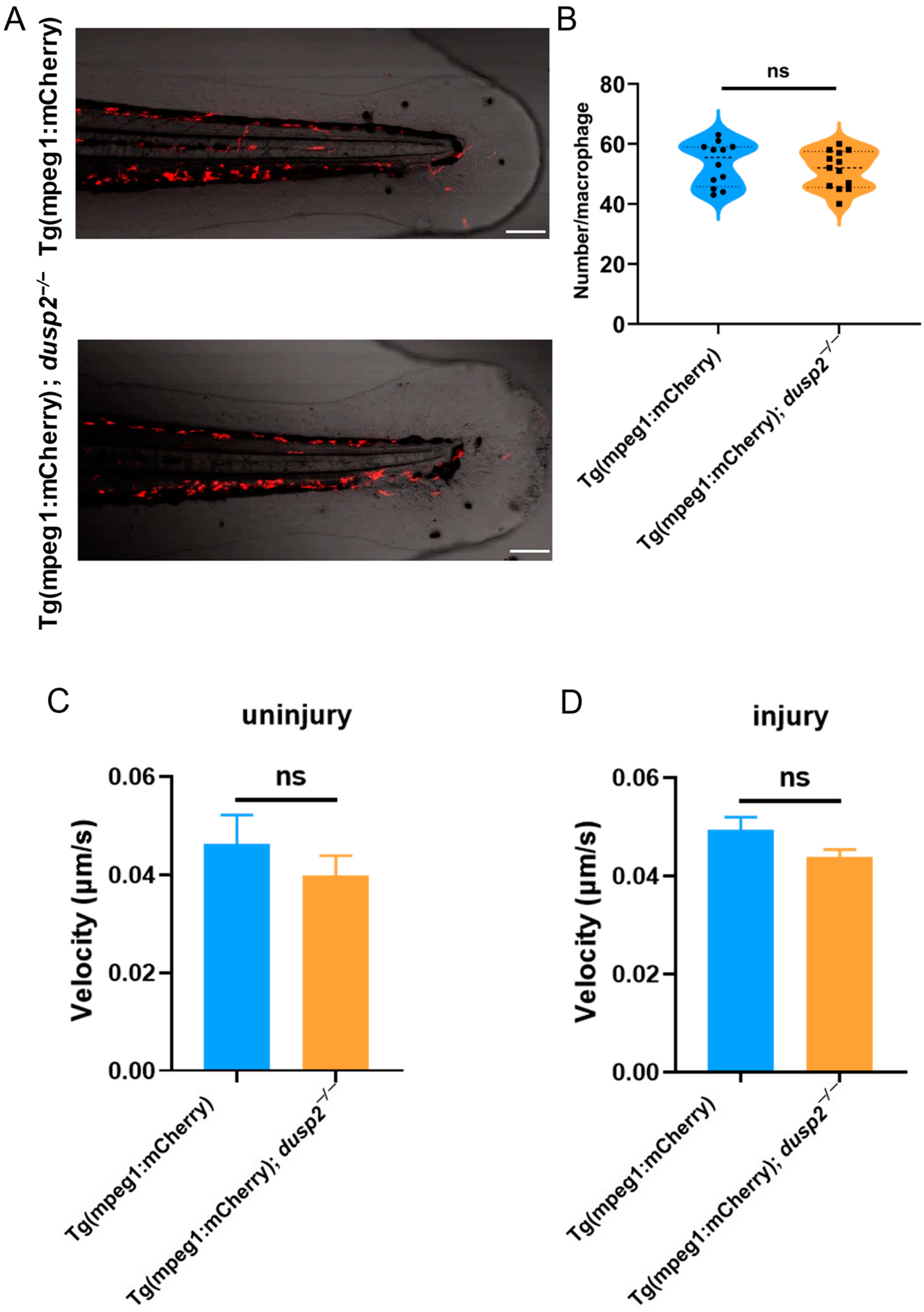
3.3. DUSP2 Deletion Inhibited the Migration of Macrophages to the Injury Site

To investigate whether the deletion of DUSP2 affects the migration of macrophages, we used a fluorescent microscope to image and enumerated the macrophages at the injury site at 1, 3, and 6 h after tail fin injury. The results showed that the deletion of DUSP2 inhibited the migration of macrophages to the injury site at 1 and 3 h, but not at 6 h after tail fin injury (Figure 4). Therefore, DUSP2 seems to influence only the early recruitment of macrophages.
Next, we investigated why DUSP2 knockout (KO) decreased the number of migrating macrophages at 1 and 3 h after injury. First, we checked whether DUSP2 KO reduced the total number of macrophages in zebrafish larvae. Based on an approach established in our lab, the number of macrophages within 800 μm of the end of the spinal cord forward was used to characterize the total number of macrophages in the zebrafish larvae [23]. The results showed that there was no significant difference in the total number of macrophages between the mutant and control (Figure 5A,B), indicating that the total number of macrophages was not the reason why fewer macrophages migrated after injury.
The second possible explanation that came to our attention was the motility rate of the macrophages. After tail fin injury, neutrophils and macrophages migrated to the injury site at different velocities [24]. We assessed the effect of DUSP2 on the motility velocity of the macrophages by real-time analysis. The results showed that DUSP2 KO had no significant effect on the motility of the macrophages either in uninjured fish (Figure 5C) or 2.5–3 h post-injury (Figure 5D). Hence, the decreased number of migrating macrophages in the DUSP2 KO larvae was not due to a decreased migration rate.

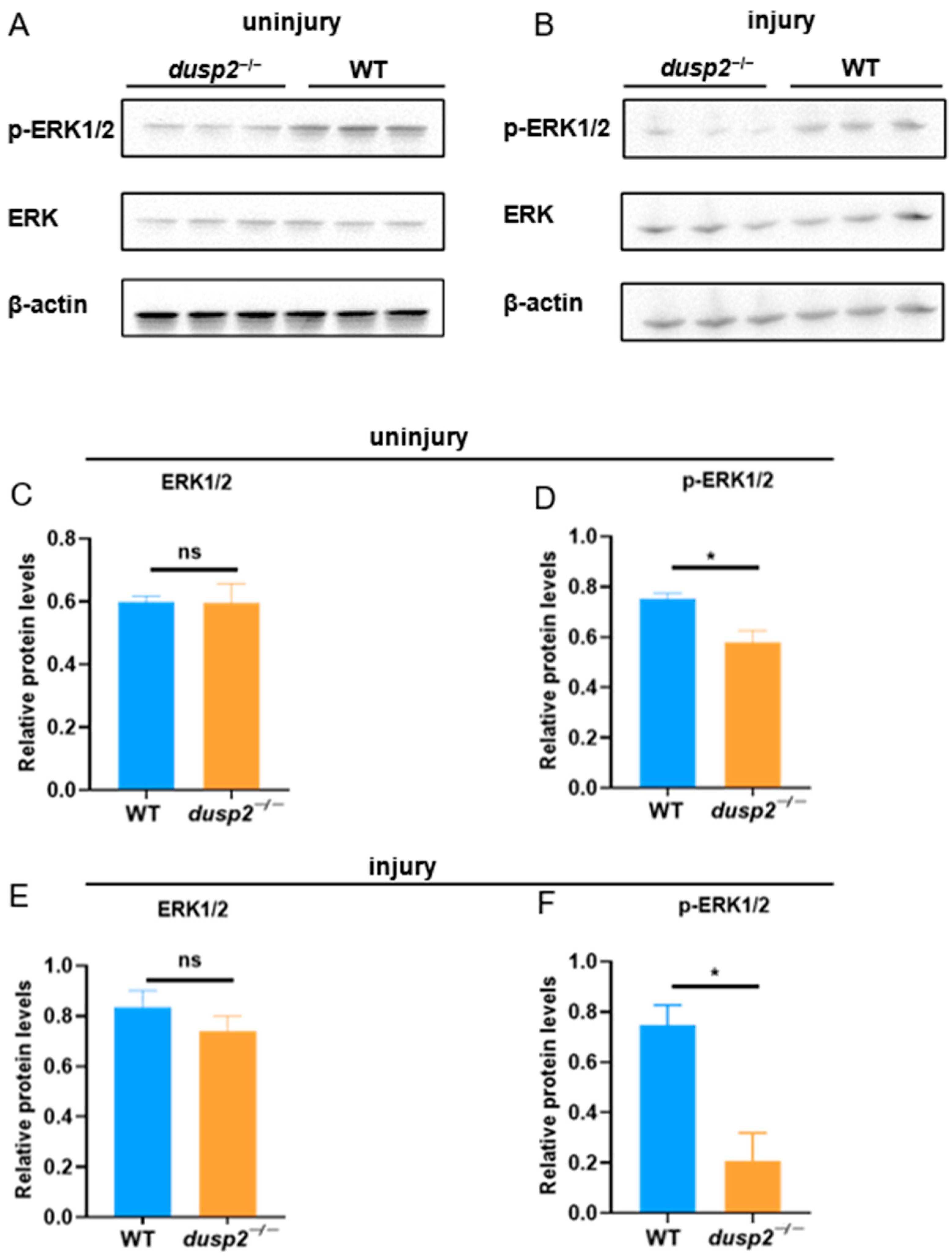
3.4. DUSP2 Deletion Inhibited ERK Protein Phosphorylation

After excluding the possible contributions of the macrophage pool and motility, we hypothesized that DUSP2 KO may downregulate some signaling pathways that promote macrophage migration and/or upregulate some that inhibit it. Based on a previous finding that the activation of ERK was decreased in DUSP2-deficient immune cells [12], we speculated that the deletion of DUSP2 might reduce macrophage migration via ERK in zebrafish larvae. Western blotting results showed that, even without injury, the phosphorylation level of ERK was significantly lower in DUSP2 KO than in the control (Figure 6A,D), while the protein level of ERK was not significantly different (Figure 6A,C). The same observations were made after the tail fin injury (Figure 6B,E,F). These results suggest that the deletion of DUSP2 inhibited the activation of ERK. Returning to our hypothesis, we have identified a factor, ERK, the activation of which was inhibited by DUSP2 KO.

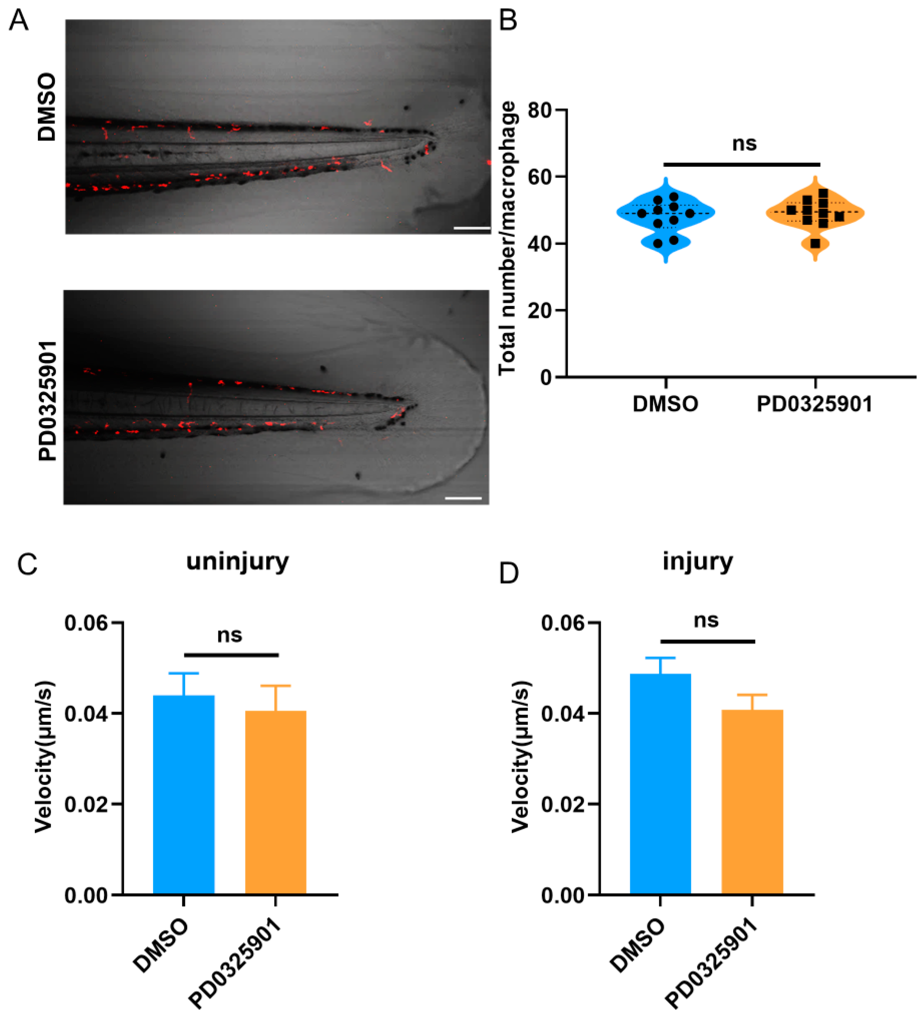
3.5. ERK Inhibitor Inhibited the Migration of Macrophages

To find the final missing piece of the puzzle, it was necessary to confirm whether the reduced activation of ERK leads to fewer migrating macrophages. To this end, we used an inhibitor, PD0325901, to reduce the level of ERK activation [20,25]. As expected, the migration of macrophages to the injury site was significantly reduced in the inhibitor group compared to the control (Figure 7). Taking these findings together, the puzzle is completed. The reduced migration of macrophages after DUSP2 KO is due to the reduced activation of ERK.
To consolidate our conclusions, we explored whether the total number or motility of macrophages were influenced by the inhibitor treatment. The results showed no significant difference in either case (Figure 8). Thus, the ERK inhibitor had no effect on the total number or motility of macrophages in zebrafish larvae. We have further verified our hypothesis that DUSP2 KO reduced macrophage migration via the inhibition of ERK activation.

4. Discussion

An acute inflammatory response was induced by a tail fin injury. Neutrophils were recruited first at the site of the injury, followed by macrophages, and the recruitment of TNF-α-positive pro-inflammatory macrophages peaked at six hpa [26,27]. In our study, we found that the deletion of DUSP2 reduced the migration of macrophages to the injury site at one hour and three hours, but not at six hours after tail fin injury. We speculated that the deletion of DUSP2 might mainly affect the early macrophage recruitment. However, the use of the ERK inhibitor inhibited the migration of macrophages to the injury site at one, three, and six hours after tail fin injury. This suggested that the effect of ERK on the macrophages was stronger than DUSP2, possibly because ERK was downstream of DUSP2.
The DUSP2 protein regulated the activation of the MAPK family, which is involved in a series of cellular processes such as cell proliferation, differentiation, apoptosis, and migration [7,28,29,30,31,32]. MAPK family members include ERK, JNK, and p38 [33,34]. ERK is involved in regulating the migration of a variety of cells, including macrophages [35,36,37,38]. Previous research indicated that the deletion of DUPS2 would lead to increased levels of phosphorylation of JNK but decreased levels of phosphorylation of ERK and p38 in immune cells [12]. Our group also demonstrated that the deletion of DUSP2 increased the level of phosphorylation of JNK [16]. In this study, the deletion of DUSP2 led to a decreased level of phosphorylation of ERK, and the use of the ERK inhibitor inhibited macrophage migration, suggesting that the effect of DUSP2 on macrophage migration was mediated by ERK activation.
Extensive research has focused on the function of DUSP2 in immunology and tumors [12,13,14,39,40,41]. Recent studies have found that DUS2P also plays a role in the nervous system, such as by participating in axon regeneration [16,42,43]. Our research results indicate that DUSP2 is involved in the migration of macrophages in zebrafish larvae, which is consistent with previous studies on DUSP2’s involvement in immune responses [12].
CCL2 is one of the key chemokines of macrophages and is involved in macrophage migrations after tail fin injuries in zebrafish [22,44]. In our study, the deletion of DUSP2 consistently suppressed the expression of CCL2 after tail fin injury. This also suggests that DUSP2 was involved in the migration of macrophages.
The relative mRNA levels of TNF-α and CXCL11 were not significantly different between the WT fish and the DUSP2 mutant group. This suggested that, in our experimental model, DUSP2 might not regulate immunity through TNF-α and CXCL11.
The immune system of zebrafish is highly conserved with that of humans, so zebrafish constitute a good model for studying inflammation [17,18]. The tail fin structure of zebrafish is simple and the tissue is transparent. Therefore, tail fin injury is the most commonly used model to induce inflammation in zebrafish [26,45]. When combined with transgenic zebrafish specifically labeling immune cells, it allows researchers to track the behavior of immune cells and identify relevant regulatory molecules. Previous studies on macrophage migration mostly focused on in vitro experiments [46,47,48,49,50]. Here, we performed live imaging in zebrafish and found that the knockout of DUSP2 led to a decrease in the number of macrophages migrating to the injury site, which suggested that DUSP2 might be an internal factor regulating macrophage cell migration.
The accumulation of macrophages in the inflammatory site exacerbates the inflammatory response and is a key factor in the occurrence and development of many inflammatory diseases and cancers. Migration is a key step whereby macrophages enter the inflammatory site and participate in the pathological process [51]. Therefore, inhibiting the migration of macrophages is a potential anti-inflammatory strategy. This study identified an endogenous factor, DUSP2, that regulated ERK, a factor that promotes macrophage migration, providing a target for developing treatments for macrophage-related diseases.
As well as macrophages, neutrophils are another type of phagocyte recruited to the injury site in zebrafish. However, in our preliminary experiments, neutrophil behavior did not display obvious differences between the control and DUSP2 KO. Previous research has noted that DUSP2 mainly affects the function of macrophages [12]. Therefore, in this research, we chose macrophages as our object and explored their behavior in depth after DUSP2 KO.

5. Conclusions

In this study, we found that the expression of dusp2 was significantly up-regulated in acute inflammation induced by tail fin injuries in zebrafish larvae. It was preliminarily speculated that DUSP2 might participate in the acute inflammation induced by tail fin injury in zebrafish larvae. Subsequently, it was found that the deletion of DUSP2 inhibited the expression of proinflammatory cytokines and macrophage chemokines after tail fin injury. By using live imaging and conducting tail fin injury in Tg(mpeg1:mCherry); dusp2−/−, it was found that the deletion of DUSP2 inhibited the migration of macrophages to the injury site. We verified that DUSP2 deletion might inhibit the migration of macrophages by inhibiting the activation of ERK, using the methods of Western blotting and the ERK inhibitor. To sum up, DUSP2 was involved in the regulation of the migration of macrophages in zebrafish larvae through the ERK signal pathway (Figure 9).

Author Contributions

Y.-J.L., X.-L.W. and B.H. designed the experiments. Y.-J.L., X.-L.W., L.-Y.S. and Z.-A.Z. carried out the experiments. Y.-J.L. analyzed the data and wrote the manuscript. X.-L.W., Z.-Y.W., S.-C.G. and B.H. revised the manuscript. Y.-J.L., X.-L.W., L.-Y.S., Z.-Y.W., Z.-A.Z., S.-C.G. and B.H. All authors have read and agreed to the published version of the manuscript.

Funding

This work was funded by the “Research Funds of the Center for Advanced Interdisciplinary Science and Biomedicine of IHM” (QYZD20220002), the National Natural Science Foundation of China, No. 82071357, and the Ministry of Science and Technology of China, No. 2019YFA0405600 (to B.H.).

Institutional Review Board Statement

All animal experimental protocols were approved by the University of Science and Technology of China (USTC) Animal Resources Center and University Animal Care and Use Committee and the Committee on the Ethics of Animal Experiments of the USTC (Permit Number: USTCACUC1103013).

Informed Consent Statement

Not applicable.

Data Availability Statement

The data that support the findings of this study are available from the corresponding author upon reasonable request.

Acknowledgments

This work was supported by the Core Facility Center for Life Sciences, University of Science and Technology of China.

Conflicts of Interest

The authors declare no conflict of interest.

References

  1. Trepat, X.; Chen, Z.; Jacobson, K. Cell migration. Compr. Physiol. 2012, 2, 2369–2392. [Google Scholar] [CrossRef] [Green Version]
  2. Sheu, K.M.; Hoffmann, A. Functional Hallmarks of Healthy Macrophage Responses: Their Regulatory Basis and Disease Relevance. Annu. Rev. Immunol. 2022, 40, 295–321. [Google Scholar] [CrossRef] [PubMed]
  3. Navegantes, K.C.; de Souza Gomes, R.; Pereira, P.A.T.; Czaikoski, P.G.; Azevedo, C.H.M.; Monteiro, M.C. Immune modulation of some autoimmune diseases: The critical role of macrophages and neutrophils in the innate and adaptive immunity. J. Transl. Med. 2017, 15, 36. [Google Scholar] [CrossRef] [PubMed] [Green Version]
  4. Shapouri-Moghaddam, A.; Mohammadian, S.; Vazini, H.; Taghadosi, M.; Esmaeili, S.A.; Mardani, F.; Seifi, B.; Mohammadi, A.; Afshari, J.T.; Sahebkar, A. Macrophage plasticity, polarization, and function in health and disease. J. Cell. Physiol. 2018, 233, 6425–6440. [Google Scholar] [CrossRef] [PubMed]
  5. Gao, W.J.; Liu, J.X.; Liu, M.N.; Yao, Y.D.; Liu, Z.Q.; Liu, L.; He, H.H.; Zhou, H. Macrophage 3D migration: A potential therapeutic target for inflammation and deleterious progression in diseases. Pharmacol. Res. 2021, 167, 105563. [Google Scholar] [CrossRef]
  6. Lang, R.; Hammer, M.; Mages, J. DUSP meet immunology: Dual specificity MAPK phosphatases in control of the inflammatory response. J. Immunol. 2006, 177, 7497–7504. [Google Scholar] [CrossRef] [Green Version]
  7. Wu, M.H.; Lin, S.C.; Hsiao, K.Y.; Tsai, S.J. Hypoxia-inhibited dual-specificity phosphatase-2 expression in endometriotic cells regulates cyclooxygenase-2 expression. J. Pathol. 2011, 225, 390–400. [Google Scholar] [CrossRef]
  8. Loesch, M.; Chen, G. The p38 MAPK stress pathway as a tumor suppressor or more? Front. Biosci. 2008, 13, 3581–3593. [Google Scholar] [CrossRef] [Green Version]
  9. Ji, R.R.; Gereau, R.W.t.; Malcangio, M.; Strichartz, G.R. MAP kinase and pain. Brain Res. Rev. 2009, 60, 135–148. [Google Scholar] [CrossRef]
  10. Jin, Z.; Sheng, H.; Wang, S.; Wang, Y.; Cheng, Y. Network pharmacology study to reveal active compounds of Qinggan Yin formula against pulmonary inflammation by inhibiting MAPK activation. J. Ethnopharmacol. 2022, 296, 115513. [Google Scholar] [CrossRef]
  11. Kciuk, M.; Gielecińska, A.; Budzinska, A.; Mojzych, M.; Kontek, R. Metastasis and MAPK Pathways. Int. J. Mol. Sci. 2022, 23, 3847. [Google Scholar] [CrossRef] [PubMed]
  12. Jeffrey, K.L.; Brummer, T.; Rolph, M.S.; Liu, S.M.; Callejas, N.A.; Grumont, R.J.; Gillieron, C.; Mackay, F.; Grey, S.; Camps, M.; et al. Positive regulation of immune cell function and inflammatory responses by phosphatase PAC-1. Nat. Immunol. 2006, 7, 274–283. [Google Scholar] [CrossRef]
  13. Dan, L.; Liu, L.; Sun, Y.; Song, J.; Yin, Q.; Zhang, G.; Qi, F.; Hu, Z.; Yang, Z.; Zhou, Z.; et al. The phosphatase PAC1 acts as a T cell suppressor and attenuates host antitumor immunity. Nat. Immunol. 2020, 21, 287–297. [Google Scholar] [CrossRef]
  14. Lu, D.; Liu, L.; Ji, X.; Gao, Y.; Chen, X.; Liu, Y.; Liu, Y.; Zhao, X.; Li, Y.; Li, Y.; et al. The phosphatase DUSP2 controls the activity of the transcription activator STAT3 and regulates TH17 differentiation. Nat. Immunol. 2015, 16, 1263–1273. [Google Scholar] [CrossRef] [PubMed]
  15. Ellett, F.; Pase, L.; Hayman, J.W.; Andrianopoulos, A.; Lieschke, G.J. mpeg1 promoter transgenes direct macrophage-lineage expression in zebrafish. Blood 2011, 117, e49–e56. [Google Scholar] [CrossRef] [PubMed] [Green Version]
  16. Shao, G.J.; Wang, X.L.; Wei, M.L.; Ren, D.L.; Hu, B. DUSP2 deletion with CRISPR/Cas9 promotes Mauthner cell axonal regeneration at the early stage of zebrafish. Neural Regen. Res. 2023, 18, 577–581. [Google Scholar] [CrossRef] [PubMed]
  17. Stockhammer, O.W.; Zakrzewska, A.; Hegedûs, Z.; Spaink, H.P.; Meijer, A.H. Transcriptome profiling and functional analyses of the zebrafish embryonic innate immune response to Salmonella infection. J. Immunol. 2009, 182, 5641–5653. [Google Scholar] [CrossRef] [Green Version]
  18. Brugman, S. The zebrafish as a model to study intestinal inflammation. Dev. Comp. Immunol. 2016, 64, 82–92. [Google Scholar] [CrossRef] [Green Version]
  19. Novoa, B.; Figueras, A. Zebrafish: Model for the study of inflammation and the innate immune response to infectious diseases. Adv. Exp. Med. Biol. 2012, 946, 253–275. [Google Scholar] [CrossRef] [Green Version]
  20. Jiang, T.; Gong, Y.; Zhang, W.; Qiu, J.; Zheng, X.; Li, Z.; Yang, G.; Hong, Z. PD0325901, an ERK inhibitor, attenuates RANKL-induced osteoclast formation and mitigates cartilage inflammation by inhibiting the NF-κB and MAPK pathways. Bioorg. Chem. 2022, 132, 106321. [Google Scholar] [CrossRef]
  21. Nguyen-Chi, M.; Laplace-Builhé, B.; Travnickova, J.; Luz-Crawford, P.; Tejedor, G.; Lutfalla, G.; Kissa, K.; Jorgensen, C.; Djouad, F. TNF signaling and macrophages govern fin regeneration in zebrafish larvae. Cell. Death Dis. 2017, 8, e2979. [Google Scholar] [CrossRef] [PubMed] [Green Version]
  22. Xie, Y.; Tolmeijer, S.; Oskam, J.M.; Tonkens, T.; Meijer, A.H.; Schaaf, M.J.M. Glucocorticoids inhibit macrophage differentiation towards a pro-inflammatory phenotype upon wounding without affecting their migration. Dis. Model. Mech. 2019, 12, dmm037887. [Google Scholar] [CrossRef] [PubMed] [Green Version]
  23. Ren, D.L.; Li, Y.J.; Hu, B.B.; Wang, H.; Hu, B. Melatonin regulates the rhythmic migration of neutrophils in live zebrafish. J. Pineal Res. 2015, 58, 452–460. [Google Scholar] [CrossRef]
  24. Li, L.; Yan, B.; Shi, Y.Q.; Zhang, W.Q.; Wen, Z.L. Live imaging reveals differing roles of macrophages and neutrophils during zebrafish tail fin regeneration. J. Biol. Chem. 2012, 287, 25353–25360. [Google Scholar] [CrossRef] [PubMed] [Green Version]
  25. Liu, W.; Zhang, L.; Sun, S.; Tang, L.S.; He, S.M.; Chen, A.Q.; Yao, L.N.; Ren, D.L. Cordycepin inhibits inflammatory responses through suppression of ERK activation in zebrafish. Dev. Comp. Immunol. 2021, 124, 104178. [Google Scholar] [CrossRef]
  26. van den Bos, R.; Cromwijk, S.; Tschigg, K.; Althuizen, J.; Zethof, J.; Whelan, R.; Flik, G.; Schaaf, M. Early Life Glucocorticoid Exposure Modulates Immune Function in Zebrafish (Danio rerio) Larvae. Front. Immunol. 2020, 11, 727. [Google Scholar] [CrossRef]
  27. Bohaud, C.; Johansen, M.D.; Jorgensen, C.; Ipseiz, N.; Kremer, L.; Djouad, F. The Role of Macrophages During Zebrafish Injury and Tissue Regeneration Under Infectious and Non-Infectious Conditions. Front. Immunol. 2021, 12, 707824. [Google Scholar] [CrossRef] [PubMed]
  28. Yang, F.; Whelan, E.C.; Guan, X.; Deng, B.; Wang, S.; Sun, J.; Avarbock, M.R.; Wu, X.; Brinster, R.L. FGF9 promotes mouse spermatogonial stem cell proliferation mediated by p38 MAPK signalling. Cell. Prolif. 2021, 54, e12933. [Google Scholar] [CrossRef] [PubMed]
  29. Zhang, C.; Zhao, S.; Tan, Y.; Pan, S.; An, W.; Chen, Q.; Wang, X.; Xu, H. The SKA3-DUSP2 Axis Promotes Gastric Cancer Tumorigenesis and Epithelial-Mesenchymal Transition by Activating the MAPK/ERK Pathway. Front. Pharmacol. 2022, 13, 777612. [Google Scholar] [CrossRef] [PubMed]
  30. Dong, N.; Li, X.; Xue, C.; Zhang, L.; Wang, C.; Xu, X.; Shan, A. Astragalus polysaccharides alleviates LPS-induced inflammation via the NF-κB/MAPK signaling pathway. J. Cell. Physiol. 2020, 235, 5525–5540. [Google Scholar] [CrossRef]
  31. Lancaster, G.I.; Kraakman, M.J.; Kammoun, H.L.; Langley, K.G.; Estevez, E.; Banerjee, A.; Grumont, R.J.; Febbraio, M.A.; Gerondakis, S. The dual-specificity phosphatase 2 (DUSP2) does not regulate obesity-associated inflammation or insulin resistance in mice. PLoS ONE 2014, 9, e111524. [Google Scholar] [CrossRef]
  32. Sun, Y.; Liu, W.Z.; Liu, T.; Feng, X.; Yang, N.; Zhou, H.F. Signaling pathway of MAPK/ERK in cell proliferation, differentiation, migration, senescence and apoptosis. J. Recept. Signal Transduct. Res. 2015, 35, 600–604. [Google Scholar] [CrossRef]
  33. Guo, Y.J.; Pan, W.W.; Liu, S.B.; Shen, Z.F.; Xu, Y.; Hu, L.L. ERK/MAPK signalling pathway and tumorigenesis. Exp. Ther. Med. 2020, 19, 1997–2007. [Google Scholar] [CrossRef] [PubMed] [Green Version]
  34. Decean, H.P.; Brie, I.C.; Tatomir, C.B.; Perde-Schrepler, M.; Fischer-Fodor, E.; Virag, P. Targeting MAPK (p38, ERK, JNK) and inflammatory CK (GDF-15, GM-CSF) in UVB-Activated Human Skin Cells with Vitis vinifera Seed Extract. J. Environ. Pathol. Toxicol. Oncol. 2018, 37, 261–272. [Google Scholar] [CrossRef]
  35. Carmo, A.A.; Costa, B.R.; Vago, J.P.; de Oliveira, L.C.; Tavares, L.P.; Nogueira, C.R.; Ribeiro, A.L.; Garcia, C.C.; Barbosa, A.S.; Brasil, B.S.; et al. Plasmin induces in vivo monocyte recruitment through protease-activated receptor-1-, MEK/ERK-, and CCR2-mediated signaling. J. Immunol. 2014, 193, 3654–3663. [Google Scholar] [CrossRef] [PubMed] [Green Version]
  36. Bose, S.; Kim, S.; Oh, Y.; Moniruzzaman, M.; Lee, G.; Cho, J. Effect of CCL2 on BV2 microglial cell migration: Involvement of probable signaling pathways. Cytokine 2016, 81, 39–49. [Google Scholar] [CrossRef] [PubMed]
  37. Samson, S.C.; Khan, A.M.; Mendoza, M.C. ERK signaling for cell migration and invasion. Front. Mol. Biosci. 2022, 9, 998475. [Google Scholar] [CrossRef]
  38. Zhou, S.L.; Hu, Z.Q.; Zhou, Z.J.; Dai, Z.; Wang, Z.; Cao, Y.; Fan, J.; Huang, X.W.; Zhou, J. miR-28-5p-IL-34-macrophage feedback loop modulates hepatocellular carcinoma metastasis. Hepatology 2016, 63, 1560–1575. [Google Scholar] [CrossRef] [Green Version]
  39. Hamamura, K.; Nishimura, A.; Chen, A.; Takigawa, S.; Sudo, A.; Yokota, H. Salubrinal acts as a Dusp2 inhibitor and suppresses inflammation in anti-collagen antibody-induced arthritis. Cell. Signal. 2015, 27, 828–835. [Google Scholar] [CrossRef] [Green Version]
  40. Dong, W.; Li, N.; Pei, X.; Wu, X. Differential expression of DUSP2 in left- and right-sided colon cancer is associated with poor prognosis in colorectal cancer. Oncol. Lett. 2018, 15, 4207–4214. [Google Scholar] [CrossRef] [Green Version]
  41. Lo Nigro, C.; Vivenza, D.; Denaro, N.; Lattanzio, L.; Fortunato, M.; Crook, T.; Merlano, M.C. DUSP2 methylation is a candidate biomarker of outcome in head and neck cancer. Ann. Transl. Med. 2018, 6, 271. [Google Scholar] [CrossRef] [PubMed]
  42. Morente, V.; Pérez-Sen, R.; Ortega, F.; Huerta-Cepas, J.; Delicado, E.G.; Miras-Portugal, M.T. Neuroprotection elicited by P2Y13 receptors against genotoxic stress by inducing DUSP2 expression and MAPK signaling recovery. Biochim. Biophys. Acta 2014, 1843, 1886–1898. [Google Scholar] [CrossRef] [PubMed] [Green Version]
  43. Wiessner, C. The dual specificity phosphatase PAC-1 is transcriptionally induced in the rat brain following transient forebrain ischemia. Brain Res. Mol. Brain Res. 1995, 28, 353–356. [Google Scholar] [CrossRef] [PubMed]
  44. Talsma, A.D.; Niemi, J.P.; Pachter, J.S.; Zigmond, R.E. The primary macrophage chemokine, CCL2, is not necessary after a peripheral nerve injury for macrophage recruitment and activation or for conditioning lesion enhanced peripheral regeneration. J. Neuroinflamm. 2022, 19, 179. [Google Scholar] [CrossRef]
  45. Xie, Y.; Meijer, A.H.; Schaaf, M.J.M. Modeling Inflammation in Zebrafish for the Development of Anti-inflammatory Drugs. Front. Cell Dev. Biol. 2020, 8, 620984. [Google Scholar] [CrossRef]
  46. Pérez-Rodríguez, S.; Borau, C.; García-Aznar, J.M.; Gonzalo-Asensio, J. A microfluidic-based analysis of 3D macrophage migration after stimulation by Mycobacterium, Salmonella and Escherichia. BMC Microbiol. 2022, 22, 211. [Google Scholar] [CrossRef]
  47. Shiraishi, A.; Uruno, T.; Sanematsu, F.; Ushijima, M.; Sakata, D.; Hara, T.; Fukui, Y. DOCK8 Protein Regulates Macrophage Migration through Cdc42 Protein Activation and LRAP35a Protein Interaction. J. Biol. Chem. 2017, 292, 2191–2202. [Google Scholar] [CrossRef] [Green Version]
  48. Wang, Y.; Su, J.; Yuan, B.; Fu, D.; Niu, Y.; Yue, D. The role of C1QBP in CSF-1-dependent PKCζ activation and macrophage migration. Exp. Cell. Res. 2018, 362, 11–16. [Google Scholar] [CrossRef]
  49. Gui, P.; Labrousse, A.; Van Goethem, E.; Besson, A.; Maridonneau-Parini, I.; Le Cabec, V. Rho/ROCK pathway inhibition by the CDK inhibitor p27(kip1) participates in the onset of macrophage 3D-mesenchymal migration. J. Cell Sci. 2014, 127 Pt 18, 4009–4023. [Google Scholar] [CrossRef] [Green Version]
  50. Zhang, L.; Xia, X.; Zhang, M.; Wang, Y.; Xing, G.; Yin, X.; Song, L.; He, F.; Zhang, L. Integrated analysis of genomics and proteomics reveals that CKIP-1 is a novel macrophage migration regulator. Biochem. Biophys. Res. Commun. 2013, 436, 382–387. [Google Scholar] [CrossRef]
  51. Gao, W.J.; Liu, J.X.; Xie, Y.; Luo, P.; Liu, Z.Q.; Liu, L.; Zhou, H. Suppression of macrophage migration by down-regulating Src/FAK/P130Cas activation contributed to the anti-inflammatory activity of sinomenine. Pharmacol. Res. 2021, 167, 105513. [Google Scholar] [CrossRef] [PubMed]
Figure 1. (A) Zebrafish strain with dusp2 mutation previously constructed by CRISPR/Cas9. (B) Generation of Tg(mpeg1:mCherry); dusp2−/− zebrafish.
Fishes 08 00310 g001
Figure 2. (A) Collection of WT zebrafish larvae without tail fin injuries and 1 h after tail fin injury for total RNA extraction. (BF) The mRNA expression levels of the pro-inflammatory cytokines IL-1β (B), IL-8 (C) and TNF-α (D) and the macrophage chemokines CCL2 (E) and CXCL11 (F) were significantly higher in the tail fin injury samples than in the control. (G) The mRNA expression levels of dusp2 were significantly up-regulated in larvae with tail fin injuries. Data were obtained from 3 independent experiments, each of which used samples from 30 zebrafish larvae; * p < 0.05, ** p< 0.01, **** p < 0.0001. Error bars represent the SEM.
Fishes 08 00310 g002
Figure 3. (A) Collection of WT and dusp2−/− zebrafish larvae without tail fin injuries and 1 h after tail fin injuries for total RNA extraction. (BG) The mRNA expression levels of the pro-inflammatory cytokines IL-1β (B), IL-6 (C), IL-8 (D) and TNF-α (E) and the macrophage chemokines CCL2 (F) and CXCL11 (G) were not significantly different between the dusp2 mutant and WT groups in the absence of tail fin injuries. The mRNA expression levels of the pro-inflammatory cytokines IL-1β (B), IL-6 (C), and IL-8 (D) and the macrophage chemokine CCL2 (F) were significantly lower in the dusp2 mutants than the WT group after tail fin injury. The mRNA expression levels of TNF-α (E) and CXCL11 (G) were not significantly different between the dusp2 mutant and WT groups after tail fin injury. Data were obtained from 3 independent experiments, each of which used samples from 30 zebrafish larvae; * p < 0.05, ** p < 0.01, *** p < 0.001, ns: no significance; error bars represent the SEM.
Fishes 08 00310 g003
Figure 4. (A) Image of macrophages migrating to the injury site at various time points after tail fin injury. (B) The number of macrophages migrating to the injury site was significantly reduced in the dusp2 mutant groups than in the control group at 1 (n = 30, n = 30) and 3 hpi (n = 33, n = 34). The number of macrophages migrating to the injury site was not significantly different between the dusp2 mutant (n = 30) and control groups (n = 32) at 6 hpi. The results of this experiment were obtained from 3 independent experiments. scale bar: 100 μm; hpi: hours post-injury. ** p < 0.01, ns: no significance; error bars represent SEM.
Fishes 08 00310 g004
Figure 5. (A) Image of macrophages within 800 μm of the end of the spinal cord forward. (B) The total number of macrophages in the zebrafish larvae were not significantly different between the dusp2 mutant (n = 13) and control groups (n = 12). (C) The movement velocity of the macrophages was not significantly different between the dusp2 mutant (n = 8) and control groups (n = 8) in the absence of a tail fin injury. (D) The movement velocity of macrophages migrating to the injury site was not significantly different between the dusp2 mutant (n = 8) and control groups (n = 8) at 2.5 hpi–3 hpi. Scale bar: 100 μm; hpi: hours post-injury; ns: no significance; error bars represent SEM.
Fishes 08 00310 g005
Figure 6. (A) Representative image of the results of the Western Blot in the absence of injury, with data from three sets of samples. (B) Representative image of the results of the Western Blot after tail fin injury; data from three sets of samples. (C) Statistical plot of ERK1/2 protein levels in the absence of injury: there was no significant difference in the background levels of ERK1/2 between the dusp2 mutant and control groups. (D) Statistical plot of p-ERK protein levels in the absence of injury: the levels of p-ERK were significantly decreased in the dusp2 mutant group. (E) Statistical plot of ERK1/2 protein levels after tail fin injury: there was no significant difference in the background levels of ERK1/2 between the dusp2 mutant and control groups. (F) Statistical plot of p-ERK protein levels after tail fin injury: the levels of p-ERK were significantly decreased in the dusp2 mutant group. Data were obtained from 3 independent experiments, each of which used samples from 30 zebrafish larvae; * p < 0.05; ns: no significance; error bars represent SEM.
Fishes 08 00310 g006
Figure 7. (A) Zebrafish larvae at 4 dpf were pretreated with PD0325901; after tail fin injury, treatment with PD0325901 was continued, followed by observation of the macrophages migrating to the injury site under a fluorescence microscope (Olympus BX60, Tokyo, Japan). (B) Image representation of macrophages migrating to the injury site at various time points after tail fin injury. (C) The number of macrophages migrating to the injury site was significantly reduced in the PD0325901 group compared to the control at 1 (n = 31, n = 32), 3 (n = 34, n = 34), and 6 hpi (n = 32, n = 33). The results of this experiment were obtained from three independent experiments. The red dashed line indicates the site of the injury; hpi: hours post-injury; scale bar: 100 μm.**** p < 0.0001; error bars represent SEM.
Fishes 08 00310 g007
Figure 8. (A) Image of macrophages within 800 μm of the end of the spinal cord forward. (B) The total number of macrophages in the zebrafish larvae were not significantly different between the PD0325901 group (n = 10) and the control (n = 10). (C) The movement velocity of the macrophages was not significantly different between the PD0325901 group (n = 8) and the control (n = 8) in the absence of a tail fin injury. (D) The movement velocity of the macrophages migrating to the injury site was not significantly different between the PD0325901 (n = 8) group and the control (n = 8) at 2.5 hpi–3 hpi. Scale bar: 100 μm; hpi: hours post-injury; ns: no significance; error bars represent SEM.
Fishes 08 00310 g008
Figure 9. Working mechanism diagram of DUSP2.
Fishes 08 00310 g009
Table 1. Primers used in this study.
Primer NamePrimer Sequence (5′–3′)
il1β-qRT-PCRForward: GTACTCAAGGAGATCAGCGG
Reverse: CTCGGTGTCTTTCCTGTCCA
il6-qRT-PCRForward: TTTGAAGGGGTCAGGATCAG
Reverse: TCATCACGCTGGAGAAGTTG
il8-qRT-PCRForward: CACTTAGGCAAAATGACCAGCA
Reverse: AGACCTCTCAAGCTCATTCCTTC
tnfα-qRT-PCRForward: GCGCTTTTCTGAATCCTACG
Reverse: TGCCCAGTCTGTCTCCTTC
dusp2-qRT-PCRForward: ATCGGCGACCCTCTCGAGATCTC
Reverse: GACACCACGGAGCTCTTGGACCT
cxcl11-qRT-PCRForward: GGCACAGTGAAGAGCTCCAT
Reverse: TGAGCTTGTTTGGGCAGTGT
ccl2-qRT-PCRForward: TCTGCACTAACCCGACTGAGA
Reverse: CATCTTAGGCGCTGTCACCAG
β-actin-qRT-PCRForward: TCCGGTATGTGCAAAGCCGG
Reverse: CCACATCTGCTGGAAGGTGG
Disclaimer/Publisher’s Note: The statements, opinions and data contained in all publications are solely those of the individual author(s) and contributor(s) and not of MDPI and/or the editor(s). MDPI and/or the editor(s) disclaim responsibility for any injury to people or property resulting from any ideas, methods, instructions or products referred to in the content.

