Article

Dietary Selenium-Rich Lactobacillus plantarum Alleviates Cadmium-Induced Oxidative Stress and Inflammation in Bulatmai barbel Luciobarbus capito

1
Key Open Laboratory of Cold Water Fish Germplasm Resources and Breeding of Heilongjiang Province, Heilongjiang River Fisheries Research Institute, Chinese Academy of Fishery Sciences, Harbin 150070, China
2
Engineering Technology Research Center of Saline-Alkaline Water Fisheries (Harbin), Chinese Academy of Fishery Sciences, Harbin 150070, China
*
Author to whom correspondence should be addressed.
These authors contributed equally to this work.
Fishes 2023, 8(3), 136; https://doi.org/10.3390/fishes8030136
Submission received: 12 January 2023 / Revised: 12 February 2023 / Accepted: 24 February 2023 / Published: 27 February 2023

Abstract

:
Cadmium (Cd) poses a great threat to the breeding of aquatic economic animals. The present study aimed to explain the antagonistic effects of selenium-enriched Lactobacillus plantarum (SL) on Cd toxicity through the expression of oxidative and inflammatory factors. A total of 225 Bulatmai barbel Luciobarbus capito (L. capito) were divided into 3 groups, namely, the control group, the Cd group (Cd, 0.05 mg·L−1), and the SL + Cd group (Cd, 0.05 mg·L−1; Nano Se, 5 mg·kg−1; L. plantarum, 105 CFU·g−1). The experiment lasted for 28 d, Sampling at 14 and 28 d, respectively. The results showed that Cd exposure caused obvious pathological damage to the liver and kidney, and the serum parameter ALT increased significantly (p < 0.05). In the Cd group, the concentration of Cd in the kidney was significantly increased (p < 0.05), and the activities of catalase (CAT), superoxide dismutase (SOD), and glutathione peroxidase (GSH-Px) in the kidney and gill were significantly decreased; malonaldehyde (MDA) increased significantly (p < 0.05) Then the mRNA expression levels in the kidney and liver were measured. Cd exposure significantly decreased the mRNA expressions of SOD, CAT, GSH-Px, HO-1, I-κBα, and Nrf2 (p < 0.05). The Cd-treated group showed significantly increased NF-kBp65, TGF-β, IL-8, IL-1, Keap1, and TNF-α expression levels. SL significantly alleviated the changes in the above indicators. The results of this study suggest that SL can trigger the Nrf2 signalling pathway and NF-kB signalling pathway to alleviate Cd toxicity. SL might be a potential drug for the treatment of Cd poisoning.

Graphical Abstract

1. Introduction

Heavy metal contamination is a severe challenge facing humans and aquatic animals because it has an unknown biological role and exerts sustained toxicity at low concentrations [1,2]. Cadmium (Cd) is widely used in agriculture and industry and is inevitably released into natural waters, causing water pollution [3]. The major sources of Cd contamination in water are batteries, electroplating, pigments, plastics, industrial effluents, phosphate fertilizers, and cigarette smoke [4,5].
Cd enters the fish body via gill, skin penetration, and the food chain [6,7,8]. Cd is a nonbiodegradable inorganic chemical that mostly accumulates in the kidney, gill, and liver [9]. The main target organs of Cd are the liver and kidney; Cd entering the body is first stored in the liver and then reaches the kidney through blood circulation, causing continuous damage to the liver and kidney [10]. It was found that the damage of Cd to kidney tissue mainly manifested in glomerular atrophy and space expansion, and the damage to liver tissue mainly manifested in cell degeneration, intracellular vacuolization, and pyknosis [11,12]. As a nonessential biological element, Cd at low levels can cause widespread harm to humans and animals, resulting in oxidative stress, DNA damage, and hepatonephrotoxicity [13,14]. Therefore, it is necessary to develop a safe and efficient antidote.
The nanoselenium synthesized by probiotics has the characteristics of easy absorption and high safety. Selenium (Se) exists in the active centre of the antioxidant enzymes glutathione peroxidase (GSH-Px) and thioredoxin reductase (TrxR) in the form of selenocysteine [15]. A se-enriched diet enhances the antioxidant capacity of the liver and kidney in mice to resist the toxicity of low concentrations of Cd [16]. Se increases the expression of selenoprotein genes and reduces Cd toxicity by inhibiting the expression of inflammatory factors in the chicken kidney [17]. In vitro experiments have shown that L. plantarum has the ability to remove Cd [18]. In vivo experiments also showed that it promoted the excretion of heavy metals in the body. Oral administration of Luciobarbus capito (L. plantarum) CCFM8661 promotes the excretion of Cd by regulating the enterohepatic circulation of bile acids (BAs), reducing intestinal motility disorders, and protecting the intestinal barrier in mice [19,20,21]. Recently, we characterized the morphology of nano selenium particles and clarified their influence on the intestinal microbiome [22]. However, it cannot be determined whether intestinal microorganisms indirectly alleviate Cd toxicity, directly play a role at the genetic level, or both, which needs further verification. Therefore, this study started with inflammation- and oxidation-related genes to explore the protective effect of SL.

2. Materials and Methods

2.1. Preparation of Selenium-Enriched L. plantarum and Feed

The production of selenium-enriched L. plantarum and feed was performed according to our previous research [22].

2.2. Experimental Feed and Conditions

L. capito (75.40 ± 0.88 g) were obtained from Hulan Test Station, Heilongjiang River Fisheries Research Institute, Harbin, Heilongjiang Province, China. The commercial feed of Tianjin Aonong Biotechnology Co., Ltd. was used as the basal diet, and the composition of the feed was 35% crude protein, 4.0% crude fat, and 15% ash. Before the exposure trial, in order to adapt to the laboratory conditions, the fish were kept under laboratory conditions for 14 d. The fish were fed a basal diet twice daily (09:00 and 17:00) at a rate of 2% of body weight. During culture, the dissolved oxygen was 6.32 ± 0.12 mg·L−1, and the water temperature was 23 ± 2 °C, with light: dark = 13:11 h.

2.3. Experimental Design and Experimental Diets

A total of 225 fish were randomly divided into 3 groups with 3 replicates each (25 fish per tank) and kept in 200-L tempered glass tanks with Cd (0.05 mg·L−1) and/or dietary SL (including Se 5 mg·kg−1 and L. plantarum 105 CFU·g−1). The groups received treatments as follows: control group, Cd, (Cd, 0.05 mg·L−1), and SL+Cd, (Cd, 0.05 mg·L−1, Nano Se, 5 mg·kg−1 and L. plantarum 105 CFU·g−1). The settings for Se and Cd concentrations refer to previous studies [23,24]. The experiment lasted for 28 d. The control group received the same amount of sterile water, and all food was stored at 4 °C for subsequent experiments. To maintain the activity of L. plantarum, the food was fed within 2 d. In addition, the water was replaced once a day with half a tank of water, and Cd was added according to the experimental concentration to maintain the concentration of Cd in the water.

2.4. Sample Collection

The fish were fasted for a day before sample collection. At 14 and 28 d, 9 fish had tissue samples collected and were randomly selected from each tank. The fish were euthanized with sodium mesylate 222 (300 mg·L−1; Sigma, MO, USA). The kidney, gill, and liver were collected, frozen in liquid nitrogen, and stored at −80 °C.

2.5. Histopathological Studies

Liver and kidney were fixed with tissue fixative (Meilun Biotechnology Co., Ltd., Dalian, China), and then a histopathological examination was performed. The fixed tissue samples were embedded in paraffin, and paraffin sections were made with a continuous microtome and stained with HE. Finally, histopathological changes were observed under an optical microscope (Olympus BX53, Tokyo, Japan).

2.6. Antioxidant Response Analysis

Superoxide dismutase (SOD), catalase (CAT), glutathione peroxidase (GSH-Px), and malondialdehyde (MDA) were measured in tissues. Dipalanine aminotransferase (ALT) was measured in serum. The above indicators are measured using the commercially available ELISA kit (Nanjing Jiancheng Bioengineering Research Institute, Jiangsu, China) and according to the recommendations of the manufacturer.

2.7. Cd and Se Accumulation

The kidneys were sampled from each group to observe the Cd and Se concentrations at 14 d. The measurement method refers to that of Qin et al. [25]. The sample (0.5 g) was transferred to a PTFE (polytetrafluoroethylene) vessel, HNO3 and ultrapure water were added, and it was digested with a MarXpress microwave closure system (CEM, North Carolina, NC, USA). Then, the concentrations of Se and Cd in the kidney were determined with an Agilent 7500cx ICP–MS (Agilent Technologies, Santa Clara, CA, USA).

2.8. Reverse-Transcriptase Real-Time PCR (RT–PCR)

Total RNA was extracted from the liver and kidney using the Simply P Total RNA Extraction Kit ( Bioer Technology Co., Ltd., Hangzhou, China) according to the instructions, followed by agarose gel electrophoresis. Then, the quality of the RNA was identified by an 8-channel micro-UV–vis spectrophotometer (Gene Company Limited, Hong Kong, China), and the OD260/280 was in the range of 1.8–2.1. One microlitre of total RNA was used for reverse transcription of cDNA using the PrimeScript RT reagent Kit with gDNA Eraser (Takara Biomedical Technology Co., Ltd., Kyoto, Japan) for quantitative real-time PCR (qRT-PCR) according to the instructions. qRT-PCRs were performed on a QuantStudio 6 Real-Time PCR System. The primer sequences used for gene expression analysis are shown in Table 1. The relative expression was analysed according to the 2–ΔΔCT method.

2.9. Statistical Analysis

Data are expressed as the mean ± standard deviation (SD) for each group. The differences in the means among the groups were analysed using one-way analysis of variance (ANOVA), and multiple comparisons were performed using Duncan’s method with the data analysis software SPSS 20 (SPSS, Chicago, IL, USA). p < 0.05 indicated statistical significance.

3. Results

3.1. Histopathological Study

Under a light microscope, the control group showed normal renal structures (Figure 1a). In the Cd group, the glomeruli were atrophied, and large gaps were observed; the epithelium of the renal tubules was thin, exfoliated, and dissolved, and the nucleus disappeared (Figure 1c). The above histopathological changes were alleviated in the SL+Cd treatment group (Figure 1b). The hepatocytes in the control group were closely arranged, the cell membrane was clearly visible, and the nucleus was clearly visible in the middle (Figure 1d). In the Cd group, some liver nuclei disintegrated and disappeared, and pyknosis occurred. Hepatocyte necrosis was observed. The boundaries of the hepatocytes were vague. Bleeding was observed, and a large number of cells were vacuolated (Figure 1f). After feeding SL, the above pathological changes were alleviated, some cells exhibited vacuolization, the nucleus was clearly visible, and the boundary of the cell membrane was slightly clear (Figure 1e).

3.2. Hepatic Markers

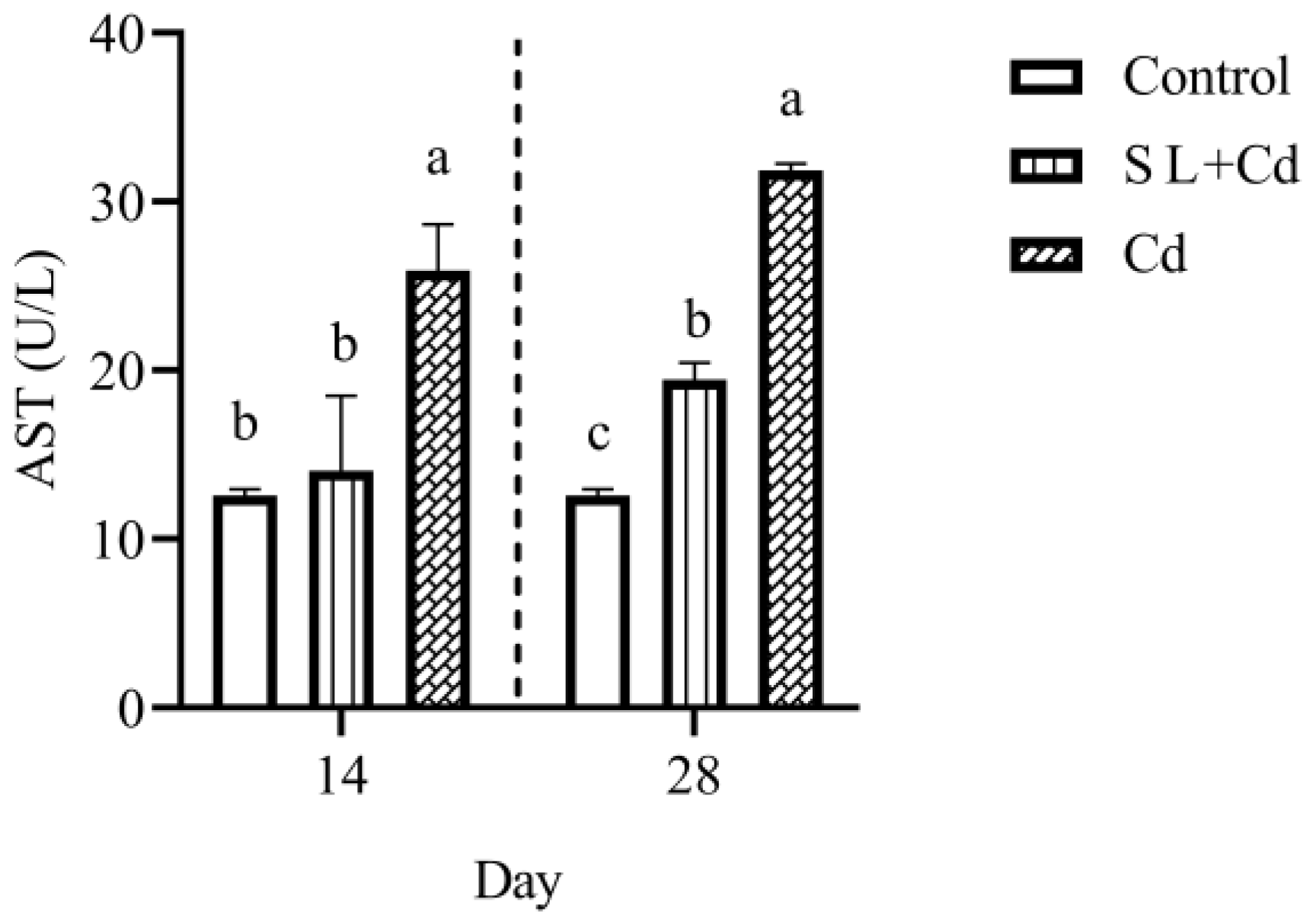
The serum biochemical parameter results are shown in Figure 2. At 14 and 28 d, Cd stress caused a significant increase in AST (p < 0.05). Compared with the Cd group, SL significantly reduced the elevation of AST (p < 0.05).

3.3. Cd and Se Concentrations

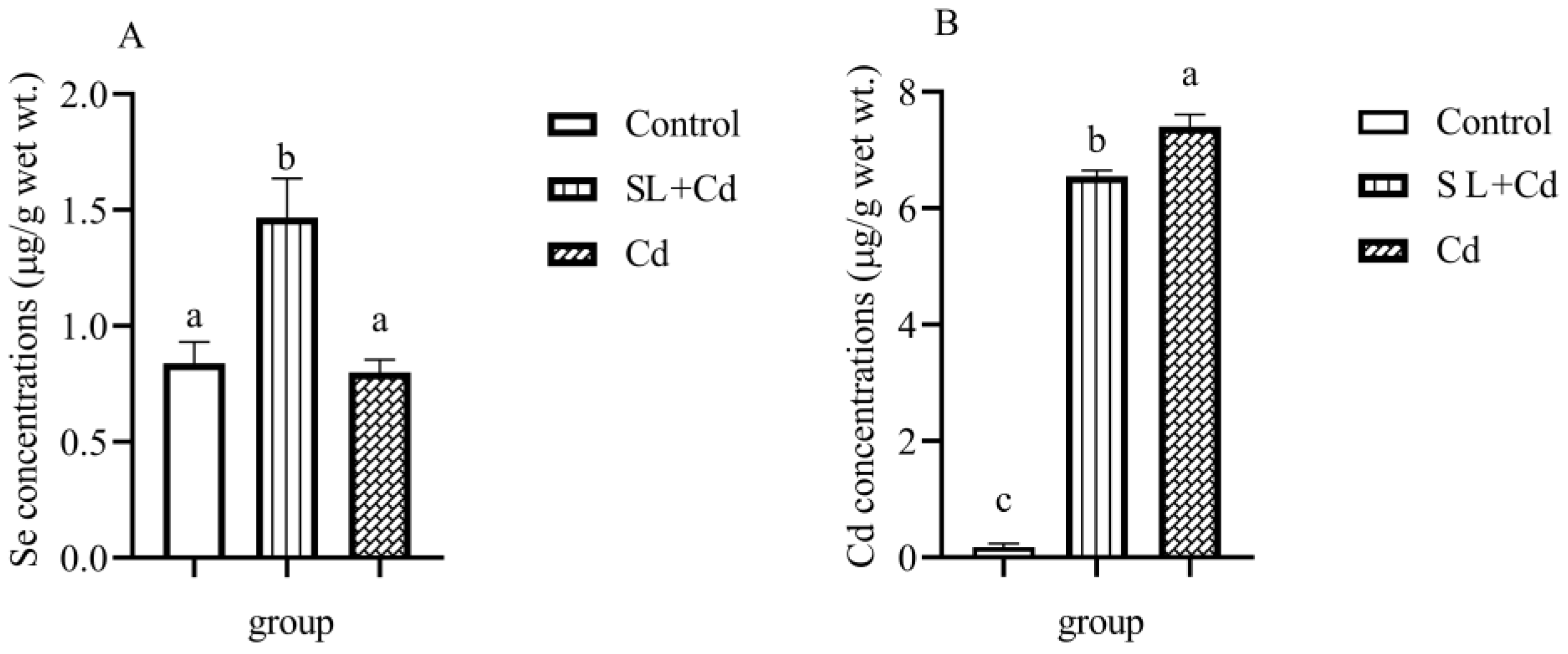
The Se concentration in the kidney was significantly increased in the SL group in comparison with the control and Cd groups (p < 0.05), and there was no significant difference between the control group and the Cd group (p > 0.05). The Cd concentration in the kidney was significantly increased in the Cd group compared with the control group and SL group (p < 0.05), but the Cd concentration was significantly decreased in the SL group compared with the Cd group (p < 0.05) (Figure 3).

3.4. Antioxidant Responses

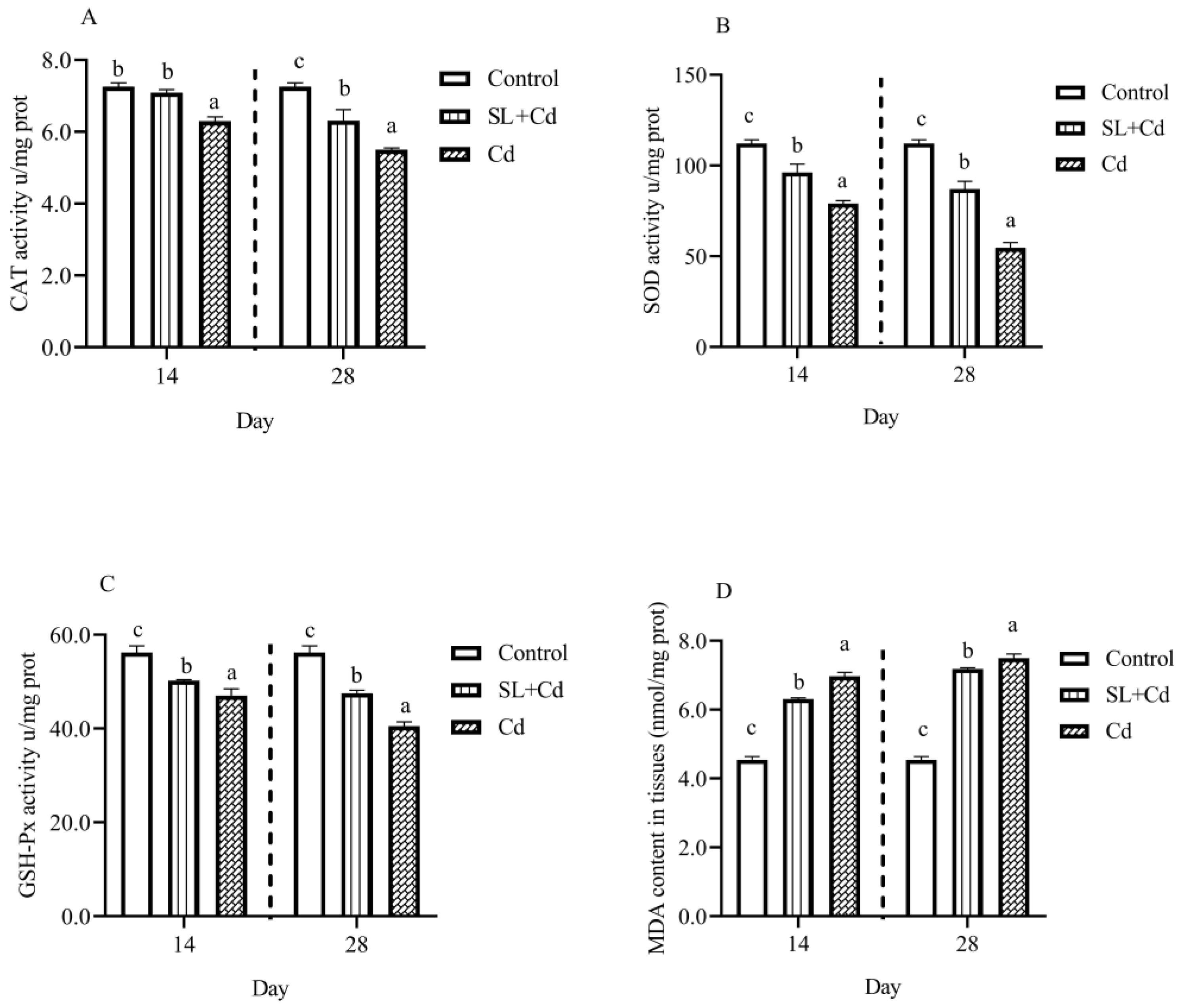
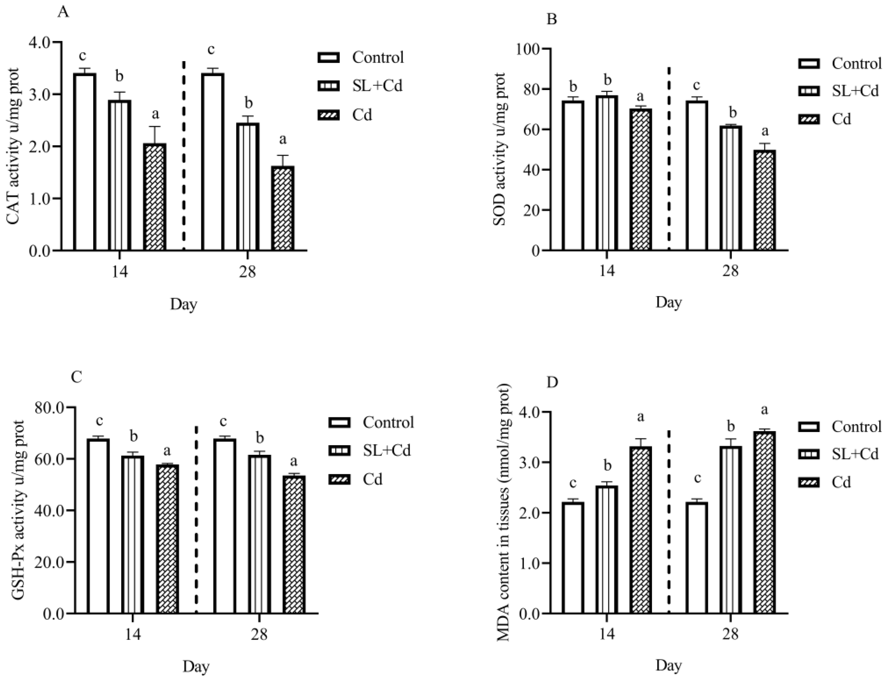
The results obtained showed that, compared to the control group, the activities of CAT, SOD, and GSH-Px were significantly decreased in the Cd-treated group (p < 0.05). However, the MDA content was significantly increased at 14 d and 28 d in the kidney and gill (p < 0.05). The activities of CAT, SOD, and GSH-Px in the SL + Cd group were significantly increased, and there was a significant reduction in MDA levels compared with the Cd-treated group in the kidney and gill (p < 0.05) (Figure 4 and Figure 5).

3.5. Antioxidant-Related Gene Expression

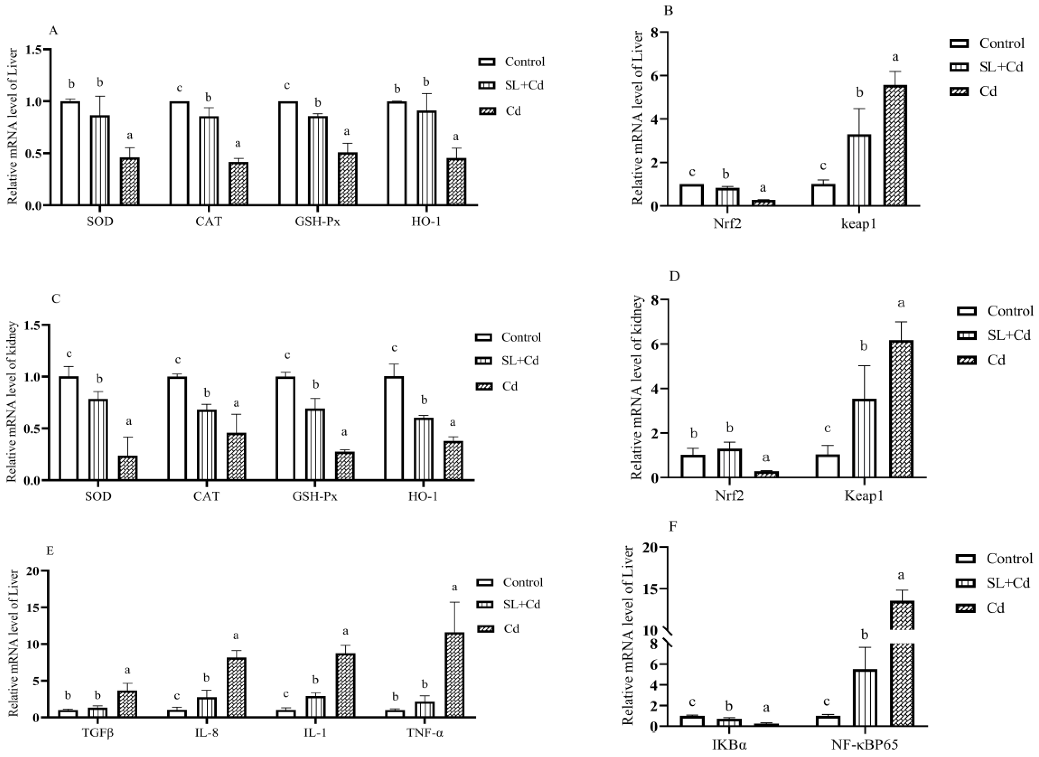
On the 28th day, the mRNA expression levels of SOD, CAT, GSH-Px, HO-1, Keap1, and Nrf2 in the kidney and liver tissues of L. capito are shown in Figure 6. Compared with the control group, Cd exposure significantly downregulated the mRNA expressions of SOD, CAT, GSH-PX, HO-1, and Nrf2 but upregulated the mRNA expression of Keap1 (p < 0.05). Compared with the Cd group, feeding SL significantly increased the mRNA expression levels of SOD, CAT, GSH-Px, HO-1, and Nrf2 but decreased the mRNA expression level of Keap1 under Cd stress (p < 0.05).
The mRNA expressions of TGF-β, IL-8, IL-1, TNF-α, NF-kBP65, and IkBa in the liver tissues of L. capito are shown in Figure 6. Compared with the control group, Cd exposure significantly upregulated the mRNA expressions of TGF-β, IL-8, IL-1, TNF-α, and NF-kBP65 but downregulated the mRNA expression of IkBα (p < 0.05). Compared with the Cd group, feeding selenium-enriched L. plantarum significantly decreased the mRNA expression levels of TGF-β, IL-8, IL-1, TNF-α, and NF-kBP65 but increased the mRNA expression level of IkBα under Cd stress (p < 0.05).

4. Discussion

Since many chemical factories are built near the water, aquaculture water is inevitably polluted by various harmful heavy metals, and high levels of heavy metal accumulation have been found in aquatic animals due to bioaccumulation [26]. In this study, the Cd concentration in the kidney was significantly increased in the Cd group. The kidney is one of the main organs for metallothionein (MT) and metal retention, and Cd tends to accumulate in the kidney in the form of Cd metallothionein complexes filtered by the glomerulus, reabsorbed by proximal tubular cells via endocytosis, and degraded by proteases to release Cd, which may lead to Cd accumulation in the kidney [26,27]. After feeding with the SL diet, the accumulation of Cd in the SL + Cd group was significantly decreased compared with that in the Cd group. Se reduced the accumulation of Cd in the kidney and liver by increasing the affinity of Cd for high-molecular-weight proteins (such as MT), and when the affinity gradually weakens, it may reduce the accumulation of Cd in tissues [24]. Se also forms complexes with Cd, inhibiting the accumulation of Cd [28]. L. plantarum CCFM8610 promotes bile acid synthesis and bile glutathione excretion in the liver of mice, and Cd is excreted into the intestine with bile. L. plantarum CCFM8610 entering the gut binds to Cd and is excreted in the faeces, thereby reducing the accumulation of Cd in the kidney and liver [19]. SL reduced Cd accumulation mainly in the following ways: sequestration of Cd into biologically inert complexes, Cd adsorption by L. plantarum, or via the action of Se-dependent antioxidant enzymes.
Oxidative stress occurs when the antioxidant system and reactive oxygen species are out of balance [29]. The enzymatic antioxidant defence system consisting of CAT, SOD, and GSH-Px plays an indispensable role in scavenging ROS and preventing lipid peroxidation (LPO). SOD is an important defence enzyme that catalyses the conversion of superoxide anion radicals to H2O2 and molecular oxygen, thereby preventing superoxide-induced damage, and CAT and GSH-Px metabolize H2O2 into water [1,13]. Cd causes the production of reactive oxygen species (ROS), which in turn damages cells and results in apoptosis [30]. Cd decreased the levels of SOD, GSH-Px, and T-AOC and increased the content of MDA in the liver and kidney of mice [16]. Our results showed that, compared to the control group, the activities of CAT, SOD, and GSH-Px were significantly decreased in the Cd-treated group of kidney and gill. Due to the strong interaction of Cd with the metal-binding moieties (Cu, Zn, and Mn) of SOD, it may be responsible for the decrease in SOD activity [31]. Cd binds to the cysteine of glutathione (GSH), leading to the inactivation of GSH-Px and therefore failing to metabolize H2O2 to water [1]. The reduction of CAT, SOD, and GSH-Px means that the fish are subjected to severe oxidative stress, which in turn leads to a large accumulation of H2O2. SOD, CAT, GSH-Px, and GSH were significantly decreased by Cd, but Se treatment significantly alleviated the changes in antioxidant levels [32]. Oral administration of L. plantarum CCFM8610 alleviated the Cd-induced reduction in the activities of CAT, SOD, GSH, and GSH-Px and decreased the content of MDA [10]. The results from our study showed that supplementation with SL in feed can significantly increase the activities of CAT, SOD, and GSH-Px. Treatment with Se alleviates oxidative stress in rat kidney tissues by Cd, mainly by reducing MDA levels and increasing the activities of GSH-Px and TrxR [33]. Se is a form of selenocysteine in the active centres of antioxidant proteins (GSH-Px and TrxR) and is involved in regulating the redox state of cells [34]. Se or L. plantarum diets had the ability to enhance the activity of the body’s antioxidant enzymes [10,35]. Compared with the cadmium group, the level of antioxidant enzymes in the SL + Cd group was significantly increased. MDA is the product of LPO in organisms. When animal cells undergo oxidative stress, LPO occurs, and the determination of MDA is widely used as an indicator of lipid peroxidation. Our finding was consistent with previous research results showing that Cd exposure resulted in increased MDA content in the kidney and gill [32]. Increased MDA content means high levels of LPO in the fish, and high levels of LPO may lead to tissue oxidative damage. However, the MDA content was significantly decreased after feeding with SL. In conclusion, SL alleviated the oxidative damage of Cd stress by increasing the level of antioxidant enzymes and reducing the content of MDA.
Prolonged and persistent absorption of heavy metals leads to inflammation and oxidative stress. NF-kB is an important regulator of inflammation and is involved in the expression of a large number of genes related to inflammation, immune response, and so on. Tissues are induced to produce inflammation and pathological damage under the stress of chemical sources. In the present study, Cd stress caused the glomerular atrophy space to become larger; for renal tubular epithelial cells, it resulted in sloughing, lysis, nuclear disappearance, hepatocyte nuclear disintegration, disappearance, nuclear pyknosis, hepatocellular necrosis, blurred boundaries of hepatocytes, inflammatory cell infiltration, and massive cellular vacuolization. This significantly increased renal NF-kBp65, TGF-β, IL-8, IL-1, and TNF-α relative expression, and SL supplementation significantly alleviated the increase in the above indicators. Studies have shown that Cd causes hepatic and renal oxidative stress and inflammation in mice and leads to pathological damage in tissues [11,23]. Elboshy M E et al. found that Cd significantly increased the relative expressions of IL-1β, TNFα, IL-6, and IL-10 in mouse liver [32]. TNFα is an activator of the NF-kB signalling pathway, and Cd may activate TNF α and further the inflammatory response [36]. NF-kB controls the expression of proinflammatory cytokines and chemokines (IL-1α, IL-1β, IL-2, IL-6, IL-8, and TNF-α) in this signalling pathway and plays an important role in regulating inflammation [36]. Therefore, we speculated that inhibiting the expression of TNF α may alleviate the NF-κB-induced inflammatory response [37]. Aluminium (Al) has been reported to potentially cause inflammatory bowel disease (IBD), and some therapeutic approaches to IBD have also focused on inhibiting TNF α functionally [38,39]. In addition, Se antagonizes heavy metals, alleviating inflammation and oxidative stress. Studies have shown that selenium alleviates the expression of the inflammatory factors NF-kBp65, TNF-α, TGF-β, IL-8, and IL-1 induced by mercury (Hg) in the carp spleen and kidney [40]. In addition, the serum AST in the SL+CD group decreased significantly compared with that in the Cd group, further indicating that Se decreased liver damage. The results of the present study illustrate that Se has a protective effect on the inflammatory response induced by Cd in L. capito.
Nrf2 is a major regulator of the oxidative stress response, inhibiting ROS production and regulating the transcription of downstream target genes [41]. Keap1 is a homodimer protein that binds to Cul3/Rbx1, leading to ubiquitination and degradation of Nrf2 [42]. HO-1 is an antioxidant gene present in the kidney that decomposes the prooxidant heme, thereby producing antioxidant metabolites [43]. When oxidative stress exists for a long time, the redox balance is broken, exceeding the body’s tolerance capacity, resulting in tissue damage and apoptosis, and Nrf2 is inhibited [44,45]. It has been reported that the expression of Nrf2 and HO-1 was inhibited, and the expression of Keap1 was upregulated in the liver of mice caused by Cd [23]. Zhao et al. found that Cd inhibits the Nrf2 signalling pathway in pig myocardium and reduces the expression of Nrf2 and its downstream target genes NQO1, SOD1, SOD2, CAT, and GSH-Px1 [41]. In this study, Cd stress reduced the expression of Nrf2 and its downstream genes SOD, CAT, GSH Px, and HO-1 in the liver and kidney, and SL alleviated the decline in these indicators. Se is a strong antioxidant. L. plantarum has also been reported to improve the antioxidant level of the body. Some studies have shown that Se and L. plantarum can reduce Cd toxicity [10,32]. Su et al. (2021) reported that Cd inhibited the expression of the antioxidant genes Nrf2 and GSH-Px in the liver of mice, and this inhibitory effect disappeared after feeding Se-enriched rice [16]. Zhang et al. (2017) reported that Se alleviated Cd-induced inhibition of the Nrf2-HO-1 system in chicken hepatocytes [46]. Se alleviated the pulmonary toxicity of tacrolimus by upregulating the expression of HO-1 [47]. Oral and intraperitoneal administration of Cd both caused oxidative stress in the liver and kidney of mice, leading to a decline in GSH, GSH-Px, SOD, and CAT activities, while L. plantarum CCFM8610 significantly alleviated oxidative stress in the body [10]. Se protects kidney tissues from Cd toxicity by upregulating the expression of most selenoprotein genes (such as GPx1, GPx2, GPx3, and GPx4) [17]. TGF-β1 is an oxidant, and TGF-β1 inhibits mouse hepatocytes and pancreas β expression levels of SOD, GSH-Px and CAT mRNA in cells [48]. Meanwhile, TGF-β1 can promote the expression of NF-kB [49]. This is similar to our results in that TGF-β decreased the expression levels of SOD, CAT, and GSH-Px and finally resulted in the activities of the corresponding antioxidant enzymes. It also promoted the expression of NF-kB and its downstream genes. Taken together, our results suggest that SL can alleviate the Cd toxicity of L. capito from both antioxidative and anti-inflammatory aspects. On the one hand, triggering the Nrf2-HO-1 system promoted the expression of antioxidant genes and selenoprotein genes and supplemented antioxidant enzymes with Cd consumption. On the other hand, it alleviates inflammation by inhibiting the activation of the NF-kB signalling pathway.

5. Conclusions

The results of this study confirm that SL has a protective effect against long-term Cd poisoning of L. capito. SL can reduce Cd accumulation in the kidney, alleviate oxidative stress and inflammatory reactions in the liver, kidney, and gill, and reduce liver and kidney tissue damage. We demonstrated that SL can protect against Cd-induced oxidative stress and inflammation. The next research will elaborate on the mechanism of SL against Cd toxicity at the protein level, with a view to making it a food additive and applying it in aquaculture in the future.

Author Contributions

Q.Z. and X.S. writing—review and editing, conceptualization, software, data curation; W.X. writing—review and editing, methodology, data curation; L.G. and S.T. data curation, investigation; H.W. and X.C. investigation. All authors have read and agreed to the published version of the manuscript.

Funding

The work was supported by National Key Research and Development Project (2020YFD0900402, 2019YFD0900405); Central Public-interest Scientific Institution Basal Research Fund (2020ZX0103, 2020TD56); The Heilongjiang Province Natural Science Foundation of China (LH2019C088); Central Public-interest Scientific Institution Basal Research Fund, CAFS (NO. 2020TD31).

Institutional Review Board Statement

The study was approved by the Heilongjiang River Fisheries Research Institute Application for Laboratory Animal Welfare and Ethical review (protocol code 20210815-001, approved on 15 August 2021).

Data Availability Statement

The data presented in this study are available on request from the corresponding author.

Acknowledgments

We would like to thank all of the students in our team for their help with the collection of samples.

Conflicts of Interest

The authors declare no conflict of interest.

References

  1. Ognjanovic, B.I.; Markovic, S.D.; Ethordevic, N.Z.; Trbojevic, I.S.; Stajn, A.S.; Saicic, Z.S. Cadmium-induced lipid peroxidation and changes in antioxidant defense system in the rat testes: Protective role of coenzyme Q(10) and vitamin E. Reprod. Toxicol. 2010, 29, 191–197. [Google Scholar] [CrossRef]
  2. Zhao, L.; Zhao, J.L.; Bai, Z.; Du, J.; Shi, Y.; Wang, Y.; Wang, Y.; Liu, Y.; Yu, Z.; Li, M.Y. Polysaccharide from dandelion enriched nutritional composition, antioxidant capacity, and inhibited bioaccumulation and inflammation in Channa asiatica under hexavalent chromium exposure. Int. J. Biol. Macromol. 2022, 201, 557–568. [Google Scholar] [CrossRef]
  3. Kumar, N.; Kumar, V.; Panwar, R.; Ram, C. Efficacy of indigenous probiotic Lactobacillus strains to reduce cadmium bioaccessibility—An in vitro digestion model. Environ. Sci. Pollut. Res. 2017, 24, 1241–1250. [Google Scholar] [CrossRef]
  4. Wuana, R.A.; Okieimen, F.E. Heavy metals in contaminated soils: A review of sources, chemistry, risks and best available strategies for remediation. Int. Sch. Res. Not. 2011, 2011, 402647. [Google Scholar] [CrossRef] [Green Version]
  5. Stohs, S.J.; Bagchi, D. Oxidative mechanisms in the toxicity of metal ions. Free Radic. Biol. Med. 1995, 18, 321–336. [Google Scholar] [CrossRef] [Green Version]
  6. Pourang, N.; Richardson, C.A.; Mortazavi, M.S. Heavy metal concentrations in the soft tissues of swan mussel (Anodonta cygnea) and surficial sediments from Anzali wetland, Iran. Environ. Monit. Assess. 2010, 163, 195–213. [Google Scholar] [CrossRef] [PubMed]
  7. Shirriff, C.S.; Heikkila, J.J. Characterization of cadmium chloride-induced BiP accumulation in Xenopus laevis A6 kidney epithelial cells. Comp. Biochem. Physiol. C Toxicol. Pharmacol. 2017, 191, 117–128. [Google Scholar] [CrossRef] [PubMed]
  8. Ordiano-Flores, A.; Galvan-Magana, F.; Sanchez-Gonzalez, A.; Paez-Osuna, F. Evidence for Interrupted Biomagnification of Cadmium in Billfish Food Chain Based on Stable Carbon and Nitrogen Isotopes from Southwestern of Gulf of California. Biol. Trace Elem. Res. 2020, 195, 215–225. [Google Scholar] [CrossRef]
  9. Yeo, W.J.; Ahn, H.J.; Hwang, I.U.; Lee, K.H.; Han, K.N. Cadmium Accumulation and mRNA Expression Associated with Detoxification in Various Organs of Black Seabream (Acanthopagrus schlegelii) Exposed to Cadmium-contaminated Diet. Ocean Sci. J. 2020, 55, 373–382. [Google Scholar] [CrossRef]
  10. Zhai, Q.; Wang, G.; Zhao, J.; Liu, X.; Narbad, A.; Chen, Y.Q.; Zhang, H.; Tian, F.; Chen, W. Protective effects of Lactobacillus plantarum CCFM8610 against chronic cadmium toxicity in mice indicate routes of protection besides intestinal sequestration. Appl. Environ. Microbiol. 2014, 80, 4063–4071. [Google Scholar] [CrossRef] [Green Version]
  11. Jihen, E.H.; Imed, M.; Fatima, H.; Abdelhamid, K. Protective effects of selenium (Se) and zinc (Zn) on cadmium (Cd) toxicity in the liver and kidney of the rat: Histology and Cd accumulation. Food Chem. Toxicol. 2008, 46, 3522–3527. [Google Scholar] [CrossRef]
  12. Pari, L.; Shagirtha, K. Hesperetin protects against oxidative stress related hepatic dysfunction by cadmium in rats. Exp. Toxicol. Pathol. 2012, 64, 513–520. [Google Scholar] [CrossRef] [PubMed]
  13. Waisberg, M.; Joseph, P.; Hale, B.; Beyersmann, D. Molecular and cellular mechanisms of cadmium carcinogenesis. Toxicology 2003, 192, 95–117. [Google Scholar] [CrossRef]
  14. Seif, M.M.; Madboli, A.N.; Marrez, D.A.; Aboulthana, W.M.K. Hepato-Renal protective Effects of Egyptian Purslane Extract against Experimental Cadmium Toxicity in Rats with Special Emphasis on the Functional and Histopathological Changes. Toxicol. Rep. 2019, 6, 625–631. [Google Scholar] [CrossRef] [PubMed]
  15. Kieliszek, M.; Blazejak, S. Selenium: Significance, and outlook for supplementation. Nutrition 2013, 29, 713–718. [Google Scholar] [CrossRef] [PubMed]
  16. Su, Y.; Li, L.; Farooq, M.U.; Huang, X.; Zheng, T.D.; Zhang, Y.J.; Ei, H.H.; Panhwar, F.H.; Tang, Z.C.; Zeng, R.; et al. Rescue effects of Se-enriched rice on physiological and biochemical characteristics in cadmium poisoning mice. Environ. Sci. Pollut. Res. 2021, 28, 20023–20033. [Google Scholar] [CrossRef] [PubMed]
  17. Wang, X.; Bao, R.; Fu, J. The Antagonistic Effect of Selenium on Cadmium-Induced Damage and mRNA Levels of Selenoprotein Genes and Inflammatory Factors in Chicken Kidney Tissue. Biol. Trace Elem. Res. 2018, 181, 331–339. [Google Scholar] [CrossRef] [PubMed]
  18. Kirillova, A.V.; Danilushkina, A.A.; Irisov, D.S.; Bruslik, N.L.; Fakhrullin, R.F.; Zakharov, Y.A.; Bukhmin, V.S.; Yarullina, D.R. Assessment of Resistance and Bioremediation Ability of Lactobacillus Strains to Lead and Cadmium. Int. J. Microbiol. 2017, 2017, 9869145. [Google Scholar] [CrossRef] [Green Version]
  19. Zhai, Q.X.; Liu, Y.; Wang, C.; Zhao, J.X.; Zhang, H.; Tian, F.W.; Lee, Y.K.; Chen, W. Increased Cadmium Excretion due to Oral Administration of Lactobacillus plantarum Strains by Regulating Enterohepatic Circulation in Mice. J. Agric. Food Chem. 2019, 67, 3956–3965. [Google Scholar] [CrossRef]
  20. Liu, Y.; Wu, J.P.; Xiao, Y.; Liu, Q.; Yu, L.L.; Tian, F.W.; Zhao, J.X.; Zhang, H.; Chen, W.; Zhai, Q.X. Relief of Cadmium-Induced Intestinal Motility Disorder in Mice by Lactobacillus plantarum CCFM8610. Front. Immunol. 2020, 11, 619574. [Google Scholar] [CrossRef] [PubMed]
  21. Zhai, Q.X.; Tian, F.W.; Zhao, J.X.; Zhang, H.; Narbad, A.; Chen, W. Oral Administration of Probiotics Inhibits Absorption of the Heavy Metal Cadmium by Protecting the Intestinal Barrier. Appl. Environ. Microb. 2016, 82, 4429–4440. [Google Scholar] [CrossRef] [Green Version]
  22. Shang, X.; Xu, W.; Zhao, Z.; Luo, L.; Zhang, Q.; Li, M.; Sun, Q.; Geng, L. Effects of exposure to cadmium (Cd) and selenium-enriched Lactobacillus plantarum in Luciobarbus capito: Bioaccumulation, antioxidant responses and intestinal microflora. Comp. Biochem. Physiol. C Toxicol. Pharmacol. 2022, 257, 109352. [Google Scholar] [CrossRef] [PubMed]
  23. Liu, C.; Zhu, Y.; Lu, Z.; Guo, W.; Tumen, B.; He, Y.; Chen, C.; Hu, S.; Xu, K.; Wang, Y.; et al. Cadmium Induces Acute Liver Injury by Inhibiting Nrf2 and the Role of NF-kappaB, NLRP3, and MAPKs Signaling Pathway. Int. J. Environ. Res. Public Health 2019, 17, 138. [Google Scholar] [CrossRef] [Green Version]
  24. Jamwal, A.; Lemire, D.; Driessnack, M.; Naderi, M.; Niyogi, S. Interactive effects of chronic dietary selenomethionine and cadmium exposure in rainbow trout (Oncorhynchus mykiss): A preliminary study. Chemosphere 2018, 197, 550–559. [Google Scholar] [CrossRef] [PubMed]
  25. Qin, D.L.; Jiang, H.F.; Bai, S.Y.; Tang, S.Z.; Mou, Z.B. Determination of 28 trace elements in three farmed cyprinid fish species from Northeast China. Food Control 2015, 50, 1–8. [Google Scholar] [CrossRef]
  26. Wang, N.; Guo, Z.; Zhang, Y.; Zhang, P.; Liu, J.; Cheng, Y.; Zhang, L.; Li, Y. Effect on intestinal microbiota, bioaccumulation, and oxidative stress of Carassius auratus gibelio under waterborne cadmium exposure. Fish Physiol. Biochem. 2020, 46, 2299–2309. [Google Scholar] [CrossRef]
  27. Wimmer, U.; Wang, Y.; Georgiev, O.; Schaffner, W. Two major branches of anti-cadmium defense in the mouse: MTF-1/metallothioneins and glutathione. Nucleic Acids Res. 2005, 33, 5715–5727. [Google Scholar] [CrossRef]
  28. Zwolak, I. The Role of Selenium in Arsenic and Cadmium Toxicity: An Updated Review of Scientific Literature. Biol. Trace Elem. Res. 2020, 193, 44–63. [Google Scholar] [CrossRef] [Green Version]
  29. Unsal, V.; Dalkiran, T.; Cicek, M.; Kolukcu, E. The Role of Natural Antioxidants against Reactive Oxygen Species Produced by Cadmium Toxicity: A Review. Adv. Pharm. Bull. 2020, 10, 184–202. [Google Scholar] [CrossRef] [Green Version]
  30. Zhao, R.; Yu, Q.; Hou, L.; Dong, X.; Zhang, H.; Chen, X.; Zhou, Z.; Ma, J.; Huang, S.; Chen, L. Cadmium induces mitochondrial ROS inactivation of XIAP pathway leading to apoptosis in neuronal cells. Int. J. Biochem. Cell Biol. 2020, 121, 105715. [Google Scholar] [CrossRef]
  31. Acharya, U.R.; Mishra, M.; Patro, J.; Panda, M.K. Effect of vitamins C and E on spermatogenesis in mice exposed to cadmium. Reprod. Toxicol. 2008, 25, 84–88. [Google Scholar] [CrossRef] [PubMed]
  32. El-Boshy, M.E.; Risha, E.F.; Abdelhamid, F.M.; Mubarak, M.S.; Hadda, T.B. Protective effects of selenium against cadmium induced hematological disturbances, immunosuppressive, oxidative stress and hepatorenal damage in rats. J. Trace Elem. Med. Biol. 2015, 29, 104–110. [Google Scholar] [CrossRef]
  33. El-Sharaky, A.S.; Newairy, A.A.; Badreldeen, M.M.; Eweda, S.M.; Sheweita, S.A. Protective role of selenium against renal toxicity induced by cadmium in rats. Toxicology 2007, 235, 185–193. [Google Scholar] [CrossRef]
  34. Lee, K.H.; Jeong, D. Bimodal actions of selenium essential for antioxidant and toxic pro-oxidant activities: The selenium paradox (Review). Mol. Med. Rep. 2012, 5, 299–304. [Google Scholar] [CrossRef]
  35. Saffari, S.; Keyvanshokooh, S.; Zakeri, M.; Johari, S.A.; Pasha-Zanoosi, H.; Mozanzadeh, M.T. Effects of dietary organic, inorganic, and nanoparticulate selenium sources on growth, hemato-immunological, and serum biochemical parameters of common carp (Cyprinus carpio). Fish Physiol. Biochem. 2018, 44, 1087–1097. [Google Scholar] [CrossRef]
  36. Freitas, M.; Fernandes, E. Zinc, cadmium and nickel increase the activation of NF-kappaB and the release of cytokines from THP-1 monocytic cells. Metallomics 2011, 3, 1238–1243. [Google Scholar] [CrossRef]
  37. Ge, J.; Guo, K.; Zhang, C.; Talukder, M.; Lv, M.W.; Li, J.Y.; Li, J.L. Comparison of nanoparticle-selenium, selenium-enriched yeast and sodium selenite on the alleviation of cadmium-induced inflammation via NF-kB/IkappaB pathway in heart. Sci. Total Environ. 2021, 773, 145442. [Google Scholar] [CrossRef]
  38. Vaghari-Tabari, M.; Jafari-Gharabaghlou, D.; Sadeghsoltani, F.; Hassanpour, P.; Qujeq, D.; Rashtchizadeh, N.; Ghorbanihaghjo, A. Zinc and Selenium in Inflammatory Bowel Disease: Trace Elements with Key Roles? Biol. Trace Elem. Res. 2021, 199, 3190–3204. [Google Scholar] [CrossRef]
  39. Pineton de Chambrun, G.; Body-Malapel, M.; Frey-Wagner, I.; Djouina, M.; Deknuydt, F.; Atrott, K.; Esquerre, N.; Altare, F.; Neut, C.; Arrieta, M.C.; et al. Aluminum enhances inflammation and decreases mucosal healing in experimental colitis in mice. Mucosal Immunol. 2014, 7, 589–601. [Google Scholar] [CrossRef] [PubMed] [Green Version]
  40. Shang, X.C.; Wang, B.; Sun, Q.S.; Zhang, Y.; Lu, Y.T.; Liu, S.J.; Li, Y.H. Selenium-enriched Bacillus subtilis reduces the effects of mercury-induced on inflammation and intestinal microbes in carp (Cyprinus carpio var. specularis). Fish Physiol. Biochem. 2022, 48, 215–226. [Google Scholar] [CrossRef] [PubMed]
  41. Zhao, X.; Wang, S.; Li, X.; Liu, H.; Xu, S. Cadmium exposure induces TNF-alpha-mediated necroptosis via FPR2/TGF-beta/NF-kappaB pathway in swine myocardium. Toxicology 2021, 453, 152733. [Google Scholar] [CrossRef] [PubMed]
  42. Niture, S.K.; Khatri, R.; Jaiswal, A.K. Regulation of Nrf2-an update. Free Radic. Biol. Med. 2014, 66, 36–44. [Google Scholar] [CrossRef] [PubMed] [Green Version]
  43. Shin, D.H.; Park, H.M.; Jung, K.A.; Choi, H.G.; Kim, J.A.; Kim, D.D.; Kim, S.G.; Kang, K.W.; Ku, S.K.; Kensler, T.W.; et al. The NRF2-heme oxygenase-1 system modulates cyclosporin A-induced epithelial-mesenchymal transition and renal fibrosis. Free Radic. Biol. Med. 2010, 48, 1051–1063. [Google Scholar] [CrossRef] [PubMed] [Green Version]
  44. Du, J.H.; Xu, M.Y.; Wang, Y.; Lei, Z.; Yu, Z.; Li, M.Y. Evaluation of Taraxacum mongolicum flavonoids in diets for Channa argus based on growth performance, immune responses, apoptosis and antioxidant defense system under lipopolysaccharide stress. Fish Shellfish Immunol. 2022, 131, 1224–1233. [Google Scholar] [CrossRef]
  45. Yu, Z.; Zhao, L.; Zhao, J.L.; Xu, W.; Guo, Z.; Zhang, A.Z.; Li, M.Y. Dietary Taraxacum mongolicum polysaccharide ameliorates the growth, immune response, and antioxidant status in association with NF-κB, Nrf2 and TOR in Jian carp (Cyprinus carpio var. Jian). Aquaculture 2022, 547, 737522. [Google Scholar] [CrossRef]
  46. Zhang, C.; Lin, J.; Ge, J.; Wang, L.L.; Li, N.; Sun, X.T.; Cao, H.B.; Li, J.L. Selenium triggers Nrf2-mediated protection against cadmium-induced chicken hepatocyte autophagy and apoptosis. Toxicol. In Vitro 2017, 44, 349–356. [Google Scholar] [CrossRef]
  47. Ibrahim, S.A.; Eltahawy, N.F.; Abdalla, A.M.; Khalaf, H.M. Protective effects of selenium in tacrolimus-induced lung toxicity: Potential role of heme oxygenase 1. Can. J. Physiol. Pharmacol. 2021, 99, 1069–1078. [Google Scholar] [CrossRef]
  48. Reddi, A.S.; Bollineni, J.S. Selenium-deficient diet induces renal oxidative stress and injury via TGF-beta1 in normal and diabetic rats. Kidney Int. 2001, 59, 1342–1353. [Google Scholar] [CrossRef] [Green Version]
  49. Fernandez-Gonzalez, A. TGF-beta and NF-kappaB Cross-Talk: Unexpected Encounters in the Developing Lung. Am. J. Respir. Cell Mol. Biol. 2021, 64, 275–276. [Google Scholar] [CrossRef]
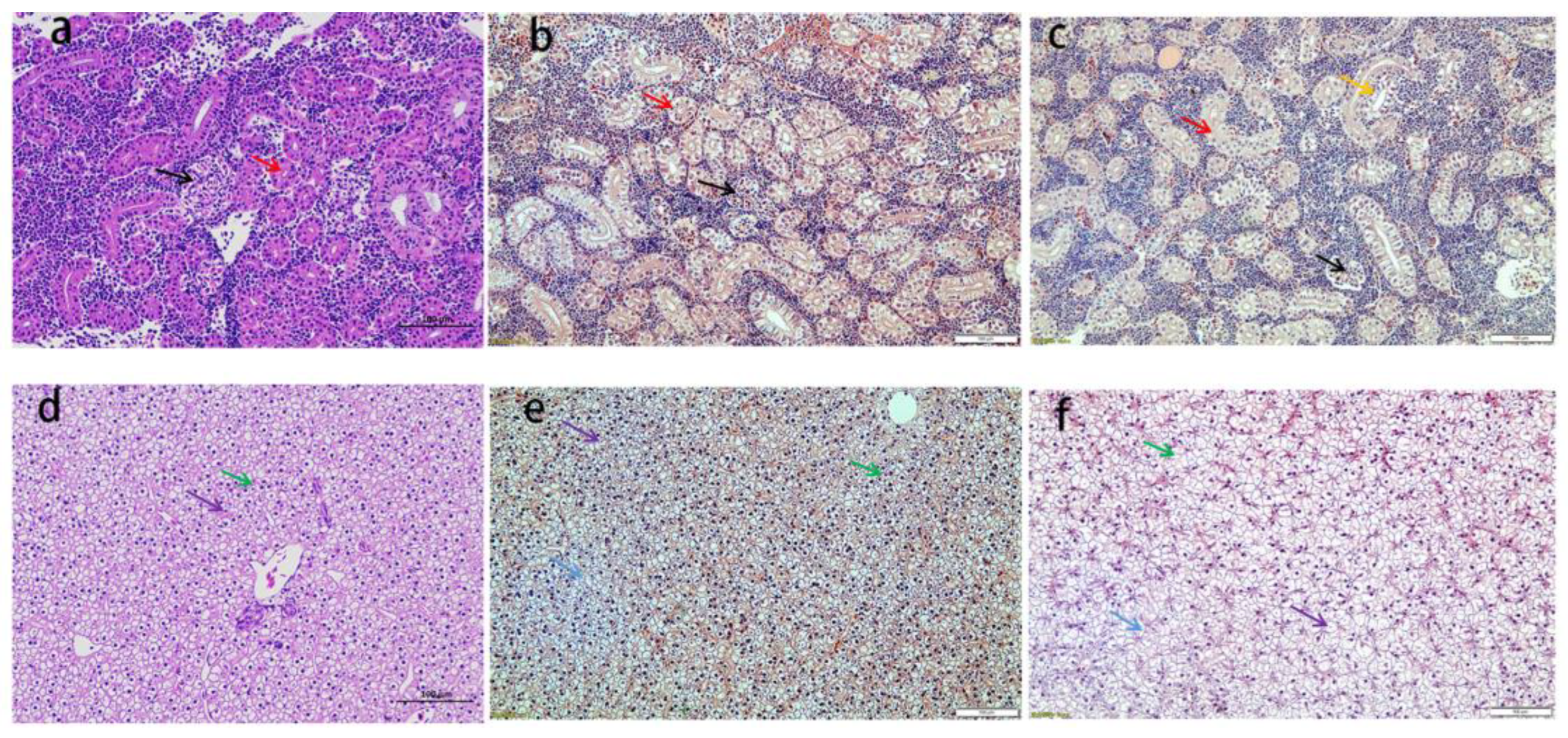
Figure 1. The histopathological analysis in kidney and liver. Note: kidney, control group (a); SL + Cd group (b); Cd group (c); liver, control group (d); SL + Cd group (e); Cd group (f). The red and yellow arrows point to the proximal convoluted tubules, and the black arrows point to the glomeruli. The green arrow points to the border of hepatocytes, the blue arrow points to the vacuolization of hepatocytes, and the purple arrow points to the hepatocyte nuclear pyknosis (HE staining, magnification 200×).
Fishes 08 00136 g001
Figure 2. Effects of SL and/or Cd on serum parameters of L. capito. The data were expressed as mean ± SD. Different letters represented significant differences (p < 0.05), and the same letter represented non-significant difference (p > 0.05).
Fishes 08 00136 g002
Figure 3. The concentrations of Se (A) and Cd (B) in the kidney of L. capito treated with Cd and/or SL. The data were expressed as mean ± SD. Different letters represented significant differences (p < 0.05), and the same letter represented non-significant difference (p > 0.05).
Fishes 08 00136 g003
Figure 4. Effects of Cd and/or SL on CAT (A), SOD (B), GSH-Px (C), and MDA (D) activities in the kidney of L. capito. The data were expressed as mean ± SD. Different letters represented significant differences (p < 0.05), and the same letter represented non-significant difference (p > 0.05).
Fishes 08 00136 g004
Figure 5. Effects of Cd and/or SL on CAT (A), SOD (B), GSH-Px (C), and MDA (D) activities in the gill of L. capito. The data were expressed as mean ± SD. Different letters represented significant differences (p < 0.05), and the same letter represented non-significant difference (p > 0.05).
Fishes 08 00136 g005
Figure 6. Effects of SL and/or Cd on the mRNA level of SOD, CAT, GSH-Px, HO-1, Nrf2, and Keap1 in the kidney and liver of L. capito (AF). The data were expressed as mean ± SD. Different letters represented significant differences (p < 0.05), and the same letter represented non-significant difference (p > 0.05).
Fishes 08 00136 g006
Table 1. Primers used for qPCR.
ForwardReverse
SODCGCACTTCAACCCTTACAACTTTCCTCATTGCCTCC
CATGAAGTTCTACACCGATGAGGCCAGAAATCCCAAACCAT
GSH-PxGAGGCACAACAGTCAGGGATTAGTTCACTTCCAGCTTCTCCAGA
Nrf2TTCCCGCTGGTTTACCTTACCGTTTCTTCTGCTTGTCTTT
Keap-1GCTCTTCGGAAACCCCTGCCCCAAGCCCACTACA
HO-1TCAGCCCATCTACTTCCCTCAGGCAGGCACTGTTACTCTCT
TGF-βTTGGGACTTGTGCTCTATAGTTCTGCTGGGATGTTT
IL-8ATGAGTCTTAGAGGTCTGGGTACAGTGAGGGCTAGGAGGG
IL-1ACCAGCTGGATTTGTCAGAAGACATACTGAATTGAACTTTG
TNFαGGTGATGGTGTCGAGGAGGAATGTCATCCTTTCTGCTCTGGTT
NF-kBP65GGCAGGTGGCGATAGTGTTCATTCCTTCAGTTCTCTTGCG
IkBaTCTTGCCATTATTCACGAGGTGTTACCACAGTCATCCACCA
b-actinTGAAGATCCTGACCGAGCGTGGAAGAAGAGGCAGCGGTTC
Disclaimer/Publisher’s Note: The statements, opinions and data contained in all publications are solely those of the individual author(s) and contributor(s) and not of MDPI and/or the editor(s). MDPI and/or the editor(s) disclaim responsibility for any injury to people or property resulting from any ideas, methods, instructions or products referred to in the content.