Article

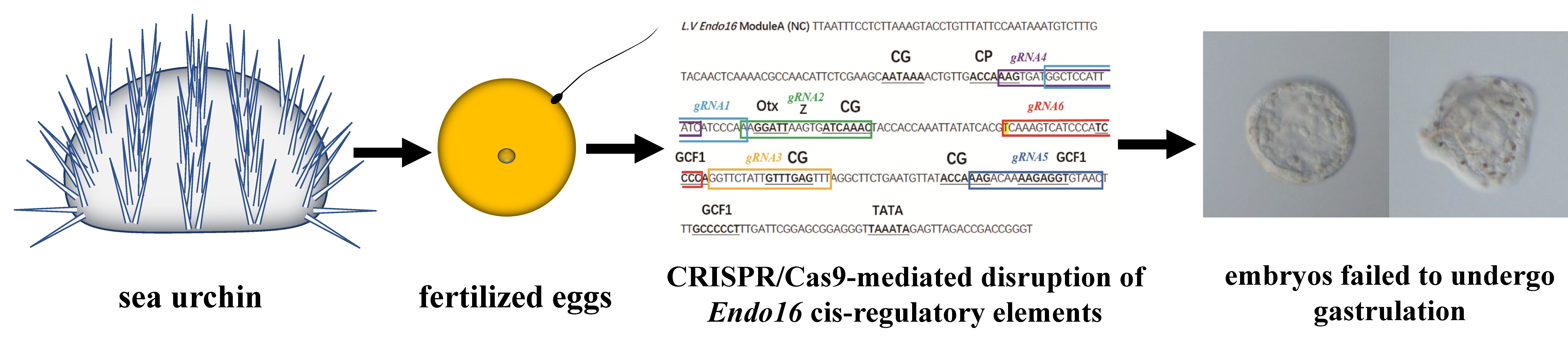
CRISPR/Cas9-Mediated Disruption of Endo16 Cis-Regulatory Elements in Sea Urchin Embryos

1
Department of Biology, Duke University, Durham, NC 27708, USA
2
CAS Key Laboratory of Marine Ecology and Environmental Sciences, Institute of Oceanology, Chinese Academy of Sciences, Qingdao 266071, China
3
Laboratory for Marine Ecology and Environmental Science, Qingdao National Laboratory for Marine Science and Technology, Qingdao 266237, China
4
University of Chinese Academy of Sciences, Beijing 100049, China
5
School of Applied Biosciences and Chemistry, HAN University of Applied-Sciences, 6500 Jk Nijmegen, The Netherlands
*
Author to whom correspondence should be addressed.
Fishes 2023, 8(2), 118; https://doi.org/10.3390/fishes8020118
Submission received: 10 January 2023 / Revised: 16 February 2023 / Accepted: 17 February 2023 / Published: 20 February 2023
(This article belongs to the Special Issue The Applications of Genome Editing and Genomics in Aquaculture)

Abstract

:
Sea urchins have become significant mariculture species globally, and also serve as invertebrate model organisms in developmental biology. Cis-regulatory elements (enhancers) control development and physiology by regulating gene expression. Mutations that affect the function of these sequences may contribute to phenotypic diversity. Cis-regulatory targets offer new breeding potential for the future. Here, we use the CRISPR/Cas9 system to disrupt an enhancer of Endo16 in developing Lytechinus variegatus embryos, in consideration of the thorough research on Endo16’s regulatory region. We designed six gRNAs against Endo16 Module A (the most proximal region of regulatory sequences, which activates transcription in the vegetal plate and archenteron, specifically) and discovered that Endo16 Module A-disrupted embryos failed to undergo gastrulation at 20 h post fertilization. This result partly phenocopies morpholino knockdowns of Endo16. Moreover, we conducted qPCR and clone sequencing experiments to verify these results. Although mutations were not found regularly from sequencing affected individuals, we discuss some potential causes. In conclusion, our study provides a feasible and informative method for studying the function of cis-regulatory elements in sea urchins, and contributes to echinoderm precision breeding technology innovation and aquaculture industry development.

Graphical Abstract

1. Introduction

With the increasing demand for food structure optimization, aquatic food has become an important source of quality food and nutrition for human beings [1,2]. It can supply critical nutrients [3] and benefit human health [4], reduce meat intake [5], fill the nutrient gap [6], and support vulnerable people [7]. Natural fisheries are frequently overfished and unable to meet the rising demand. Many countries encourage and support significant efforts to expand marine aquaculture, particularly for high value products. One such group is the sea urchin, which can be found from shallow shores to deep waters [8,9]. Sea urchins are economically important echinoderms with high edible and medicinal values, and their gonads are rich in carotenoids, polyunsaturated fatty acids, phospholipids and sulphated fucans [10]. As a result, sea urchins have become significant mariculture species globally [11,12,13]. However, the sea urchin breeding industry is also facing serious challenges because of limited breeding technology, lack of good varieties, and low larvae production efficiency.
Gene editing is a new genetic engineering technology that can modify specific target genes in the genome of an organism accurately. In the breeding process, gene editing technology can realize the modification and alteration of the genetic loci of target traits in varieties so as to accelerate the improvement of varieties. With the improvement and development of the CRISPR technology, gene editing has become more and more widely used in aquatic animals. The application of gene editing technology in aquatic animals enables us to obtain more fishery resources with high quality, and contributes to the healthy and sustainable development of fisheries. CRISPR/Cas9 genome editing has been successfully used in fishes and molluscs, including Danio rerio [14,15], Petromyzon marinus [16], Crepidula fornicate [17], and Lymnaea stagnalis [18]. As a representative species of echinoderms, gene editing work has also been reported for sea urchins [19,20,21,22,23,24]. Initially, the application of CRISPR/Cas9 for genome editing was focused on the study of protein-coding genes. However, several CRISPR/Cas9-based tools have recently been applied to studying non-coding cis-regulatory elements (CRE).
CRE, such as enhancers and promoters, control development and physiology by regulating gene expression. Mutations that affect the function of these sequences contribute to phenotypic diversity within and between species [25]. Compared to promoters, enhancers tend to be more variable between species; they are the type of CRE that is most often thought to be responsible for cis-regulatory divergence [26]. It has been shown that even single-nucleotide alterations in these regulatory sequences can have substantial effects on gene expression and cause pathological conditions [27,28,29,30,31]. In gene editing for breeding, editing cis-regulatory elements instead of coding regions reduces the impact on other normal life activities of the organisms and may be more secure for the survival of the edited individual [32]. Cis-regulatory targets offer new breeding potential for the future. However, few predicted enhancer elements have been shown to affect the transcription of their putatively-regulated genes or to alter developmental phenotypes when perturbed in situ.
In this study an enhancer of Endo16 was selected for editing, mainly in consideration of the thorough research on its regulatory region, which can help establish a technical means for breeding application by editing cis-regulatory elements. Endo16 encodes a secreted glycoprotein in the embryo and larval midgut and is required for sea urchin gastrulation [33]. Transient expression assays using reporter constructs have demonstrated that 2.2 kb of sequence immediately upstream of the transcriptional start site is sufficient to drive Endo16 expression in Strongylocentrotus purpuratus [34] and in Lytechinus variegatus [35]. The most proximal region of this regulatory region, module A, activates transcription in the vegetal plate and archenteron, specifically [34]. In addition, the alignment of the Endo16 regulatory sequences shows that module A is well conserved between S. purpuratus and L. variegatus [35]. Deletions or disruptions of Module A within its native chromosomal context have not been performed. Based on results from reporter constructs and knock-down experiments, such manipulations are predicted to decrease the transcription of Endo16 and disrupt the morphogenesis of the archenteron during embryonic development.
Here, we applied the CRISPR/Cas9 system to examine the role of disruption of the Endo16 Module A enhancer in sea urchins. Our results show that Endo16 Module A-disrupted embryos fail to undergo gastrulation at 20 h post fertilization. This result partly phenocopies morpholino knockdowns of Endo16. In addition, we conducted qPCR and clone sequencing experiments to verify these results. Our study indicates that the CRISPR/Cas9 system can effectively be used in sea urchin embryos for cis-regulatory element editing, confirming results from a recent study [23]. In the future, manipulating gene functions by editing cis-regulatory sequences through CRISPR-Cas9 can be used to quickly identify the key regulatory sites necessary for gene expression, and the molecular as well as morphological phenotypes that result from perturbing transcription. Meanwhile, it will provide a construction method for a gene editing breeding system of sea urchins, so as to obtain new varieties of sea urchin with excellent target traits, thus supporting the healthy development of the sea urchin industry.

2. Materials and Methods

2.1. Experimental Sea Urchins and Embryos

Adult L. variegatus were obtained from the Duke University Marine Laboratory (Beaufort, NC, USA) or collected commercially by KP Aquatics LLC (Tavernier, FL, USA) or Reeftopia (Florida Keys, FL, USA). A total of about 100 sea urchins were used for this experiment, and sampling was carried out mainly in summer and autumn. Gametes were obtained by 0.5 M KCl injection. The fertilization process was as previously described [36]. Embryos were cultured in artificial sea water at 22 °C.

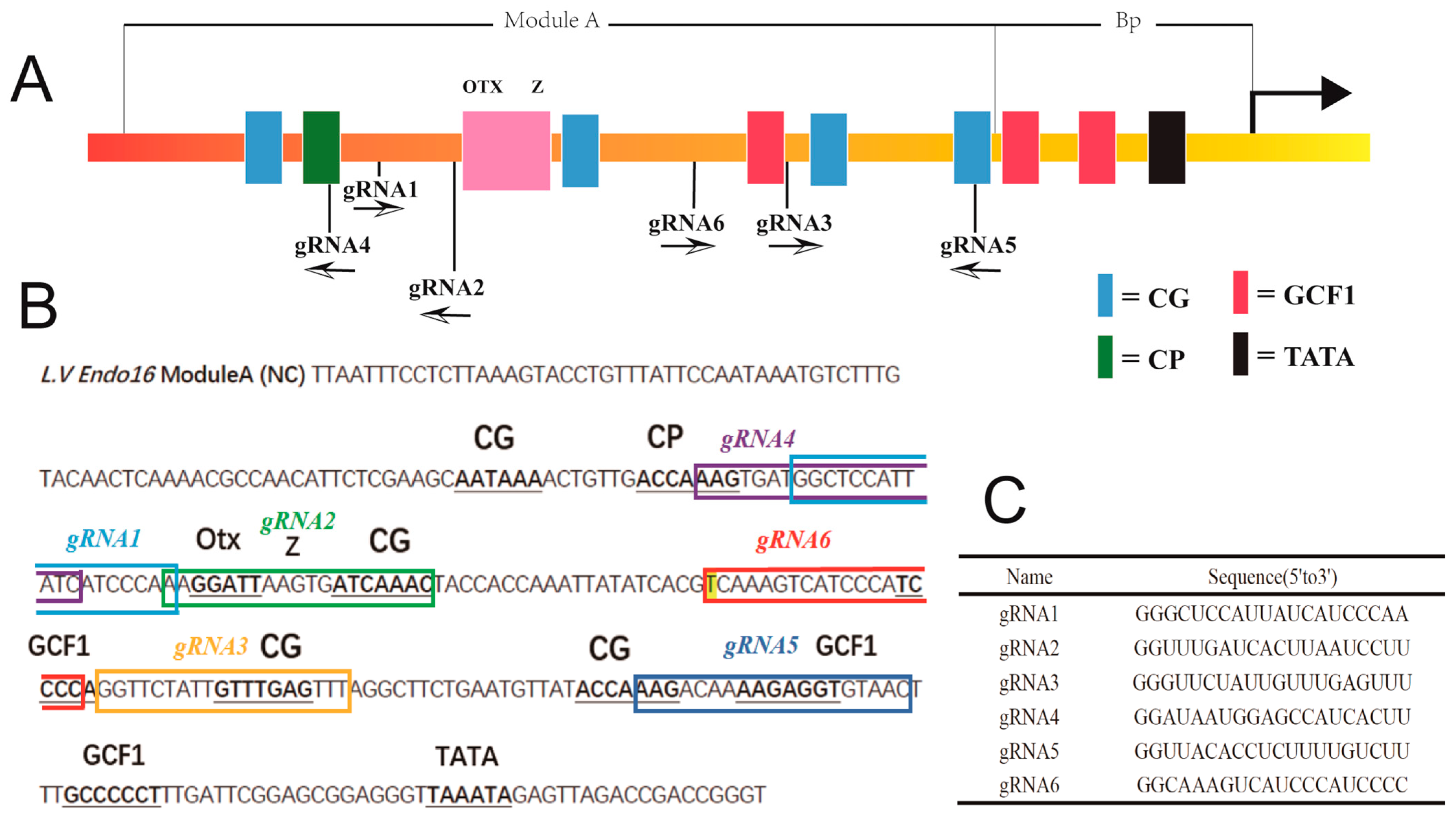
2.2. gRNAs Preparation

We designed six gRNAs using CRISPRscan (http://www.crisprscan.org/?page=sequence; 31 May 2018) (Figure 1). The target sites contain two guanine nucleotides at the 5′ end for the initial transcription of gRNAs using the T7 RNA polymerase, while the 3′ end is adjacent to an NGG motif (PAM) in Endo16 Module A. For initial assessment, the gRNAs labeled in bright green were considered first due to their high estimated cleavage efficiencies and lack of potential off-target sites. Then, we blasted these candidate target sequences against the sea urchin genome at EchinoBase (http://www.echinobase.org/Echinobase/; 31 May 2018) [37]. We selected six gRNAs based on the CRISPRscan score and the uniqueness of the target sequence. CRISPRscan provided the sequence of the guide, with the T7 sequence and tail sequence attached. We ordered the selected gRNA sequences that CRISPRscan provided and the 80 nucleotides tail primer sequence from Eton Bioscience (https://www.etonbio.com/; 31 May 2018). We annealed and extended the gRNAs and the tail primer via PCR machine using Phusion Master mix (Phusion High-Fidelity PCR Master Mix with HF Buffer) (F531S, ThermoFisher, Waltham, MA, USA). Then, we purified the gRNAs by QIAquick PCR Purification Kit (28104, Qiagen, Valencia, CA, USA). In vitro transcription was conducted using a MEGAshortscript™ T7 Transcription Kit (AM1354, ThermoFisher, Waltham, MA, USA) and purified by alcohol precipitation.

2.3. Microinjection, Drug Treatment and Imaging

We made a microinjection mixture containing Cas9 mRNA, gRNA, 20% glycerol (G5516, Merck, Kenilworth, NJ, USA), fluorescein isothiocyanate (FITC) dye (F10240, ThermoFisher, Waltham, MA, USA) and nuclease-free water (AM9935, ThermoFisher, Waltham, MA, USA) to a final volume of 5 μL. The concentration range of Cas9 mRNA was 250–750 ng/μL. The concentration range of gRNA was 100–400 ng/μL. The addition of glycerol to the microinjection system serves as an indicator. One can be confident that the egg was injected if the glycerol diffuses throughout the cytoplasm. Use the size of the bolus of glycerol solution in the egg cytoplasm as a rough indication of the volume injected. We kept the mixture on ice before microinjection. Then, we injected the solution into fertilized sea urchin eggs. The diameter of the injected solution was about one third to one fourth of the egg (<25% of the egg volume). Controls for this experiment included injecting Cas9 mRNA and other microinjection mixtures without the gRNAs, to evaluate the effect of injection and the effectiveness of gRNA. We incubated the injected embryos and control embryos at 22 °C. After the embryos reached the desired stage, they were subjected to genomic DNA isolation for genotyping, RNA isolation for gene expression evaluation, and imaging.
We selected several control embryos and embryos with expected phenotype and transferred them to concavity slides (Electron Microscopy Sciences; 100491-022). Embryos were imaged with a Zeiss Axioplan microscope at 20×. Images were postprocessed with Adobe Photoshop and ImageJ.

2.4. Isolation of Genomic DNA from Single Embryo and Clone Sequencing

We washed the embryos with filtered seawater and transferred individual embryos in a volume of 0.5 μL of sea water to 0.2 mL PCR tubes containing 1 μL of 1 × NEBufferTM2 (B7002S, New England Biolabs, Ipswich, MA, USA). The samples were incubated at 94 °C for 10 min, and then cooled down to 4 °C for 10 min. A total of 0.5 μL of proteinase K (5 mg/mL) (25530049, ThermoFisher, Waltham, MA, USA) was added and then the samples were incubated at 55 °C for 2 h. The samples were then boiled at 94 °C for 10 min and the solution was diluted two- to fivefold for PCR.
We designed primer pairs encompassing the gRNA target region using the Primer-BLAST online tool (www.ncbi.nlm.nih.gov/tools/primer-blast; accessed on 20 June 2018). We checked the free energy (ΔG) of the selected primer pairs using the OligoAnalyzer 3.1 online tool (sg.idtdna.com/calc/analyzer; accessed on 20 June 2018). The recommended parameters were: ΔG of 3′ end hairpin >−2 kcal/mol; ΔG of 3′ end self/cross dimer >−5 kcal/mol. The primers for clone sequencing are shown in Table 1.
Conventional PCR was conducted using the DreamTaq Hot Start PCR Master Mix (K9011, ThermoFisher, Waltham, MA, USA). We performed PCR using the following thermal cycling conditions: 95 °C, 2 min; 95 °C, 30 s, 69 °C, 30 s, 72 °C, 1 min, repeated 37 rounds; 72 °C, 10 min. The PCR products were purified using a QIAquick PCR Purification Kit (28104, Qiagen, Valencia, CA, USA). DNA fragments from embryos were cloned individually into the pGEM®-T Easy Vector (Promega, Madison, WI, USA). Thirty-two bacterial colonies were randomly selected for plasmid DNA extraction and sequencing.

2.5. Isolation of RNA from Mixed Embryos and Real-Time PCR Validation

Thirty embryos of each group (three biological replicates × 10 embryos per biological replicate) were used for real-time PCR experimental sampling. The embryos were added to 300 μL of TRI Reagent® (93289, Merck, Kenilworth, NJ, USA), and mixed thoroughly. The RNA was extracted and purified by using a Direct-zol™ RNA MicroPrep Kit (R2060, EAD Scientific, Miami, FL, USA). The First-Strand cDNA was synthesized by using the SuperScript™ II Reverse Transcriptase Kit (18064-022, ThermoFisher, Waltham, MA, USA). According to the sequence information (https://www.ncbi.nlm.nih.gov/gene/446165; accessed on 20 June 2018; https://www.echinobase.org/entry/gene/showgene.do?method=display&geneId=23195218; accessed on 20 June 2018), primers were designed for optimal performance using the primer3 (v0.4.0; http://bioinfo.ut.ee/primer3-0.4.0/primer3/; accessed on 20 June 2018) (Table 1).
Gene-expression levels were determined using the KAPA SYBR® FAST qPCR Master Mix (2X) Kit (KR0389, Kapa Biosystems, Wilmington, MA, USA). The conditions of qPCR were as follows: enzyme activation, 95 °C, 3 min; denaturation, 95 °C, 3 s, annealing/extension/data acquisition, 60 °C, 20 s, repeated 40 rounds; dissociation, 72 °C, 40 s. Ubiquitin was used as a reference gene for internal standardization. The 2−ΔΔCT method was used to calculate the expression level [38]. The data of the mRNA expression level were presented as the mean ± standard deviation (n = 3), and they were statistically analyzed by t-test. p values < 0.05 were considered statistically significant. Statistical analysis was performed using SPSS 18 software.

3. Results

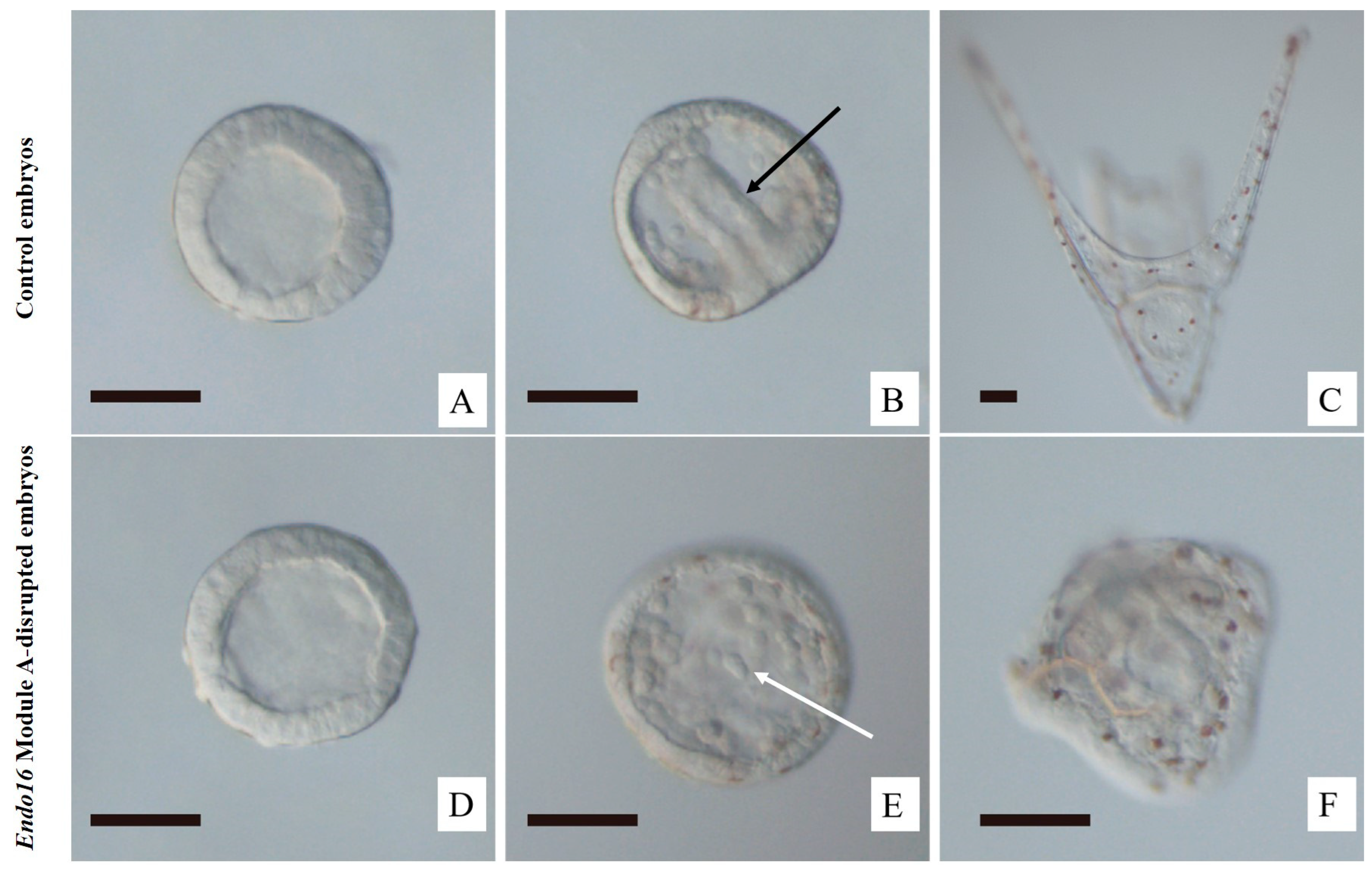
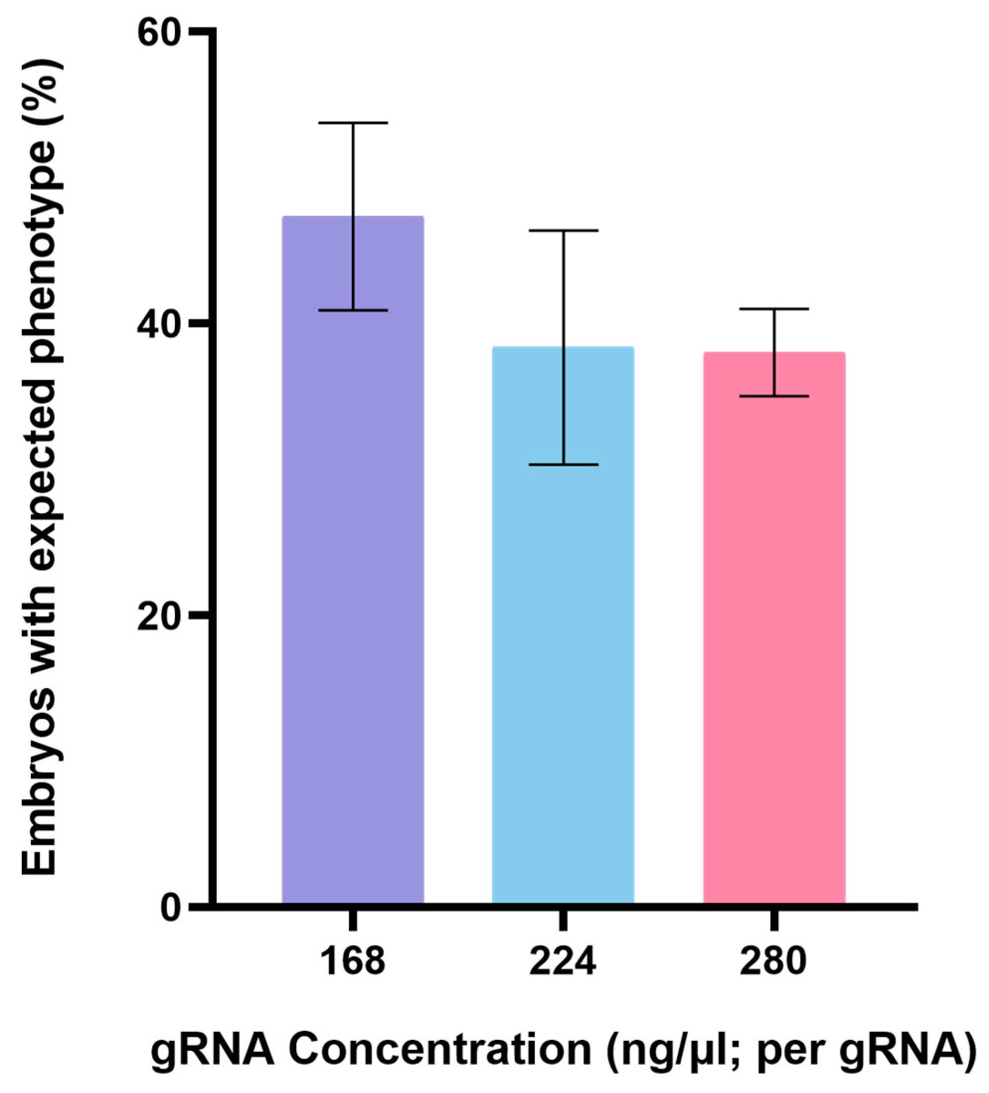
3.1. CRISPR/Cas9-Mediated Genome Editing of Endo16 Module A Produced Mutated Phenotype

Endo16 Module A is responsible for initiating expression in the vegetal plate in the early embryo [39]. Module A interacts with all of the other Endo16 cis-regulatory modules, and is either absolutely required for their operation or synergistically enhances their output. Module A functions are mediated through interactions at eight different target sites for DNA binding proteins (Figure 1A). Consideration of cleavage efficiencies and potential off-target sites led us to design six gRNAs. With the exception of gRNA1, the gRNAs all overlap known transcription factor-DNA binding sites (Figure 1B). Figure 1C shows the specific sequences of the six gRNAs.
We began our Endo 16 Module A disruption experiment with different combinations of three gRNAs (112 ng/μL per gRNA or168 ng/μL per gRNA) and Cas9 mRNA (258.72 ng/μL), and screened for embryos showing developmental abnormalities. At the blastula stage the gRNAs and Cas9 mRNA did not appear to have any effect on development, although some of the embryos injected with gRNAs and Cas9 mRNA (Figure 2D) were not as spherical as the control embryos (Figure 2A). At 20 h post fertilization, only control embryos injected with Cas9 mRNA had formed an archenteron (Figure 2B). However, the embryos injected with gRNAs and Cas9 mRNA failed to undergo gastrulation (Figure 2E). At the pluteus stage, control embryos developed into four-arm larvae with a gut (Figure 2C). On the other hand, Endo16 ModuleA-disrupted embryos developed abnormally without arms or gut (Figure 2F), and most did not survive. The percentage of mutated and dead embryos injected with gRNA and Cas9 mRNA at different combinations and concentrations was examined at the gastrula stage and is summarized in Table 2. The combination of three gRNAs was chosen at the beginning of the experiment so that more regions were edited, with the aim of increasing the success rate of gene editing. After successful editing, a combination of two gRNAs was used in order to see if a long sequence deletion between the two gRNAs could be detected. However, no long sequence deletion was detected in this study. In the end, experiments with a single gRNA were carried out.
As Table 2 shows, we conducted the injection experiments with different combinations of two gRNAs (168 ng/μL per gRNA, 224 ng/μL per gRNA, and 280 ng/μL per gRNA) and Cas9 mRNA (258.72 ng/μL). The results show that the ratio of mutated embryos did not increase as the gRNA concentration increased (p > 0.05, Figure 3). We then tried Endo16 Module A disruption by injecting one gRNA and Cas9 mRNA. In embryos injected with gRNA6 and Cas9 mRNA, gastrulation and the gut were not affected and the embryos developed into normal pluteus larvae. The other five gRNAs induced a mutated phenotype; gRNA1, gRNA2 and gRNA5 worked best. gRNA6 may not have worked due to a missing “T” (Figure 1).

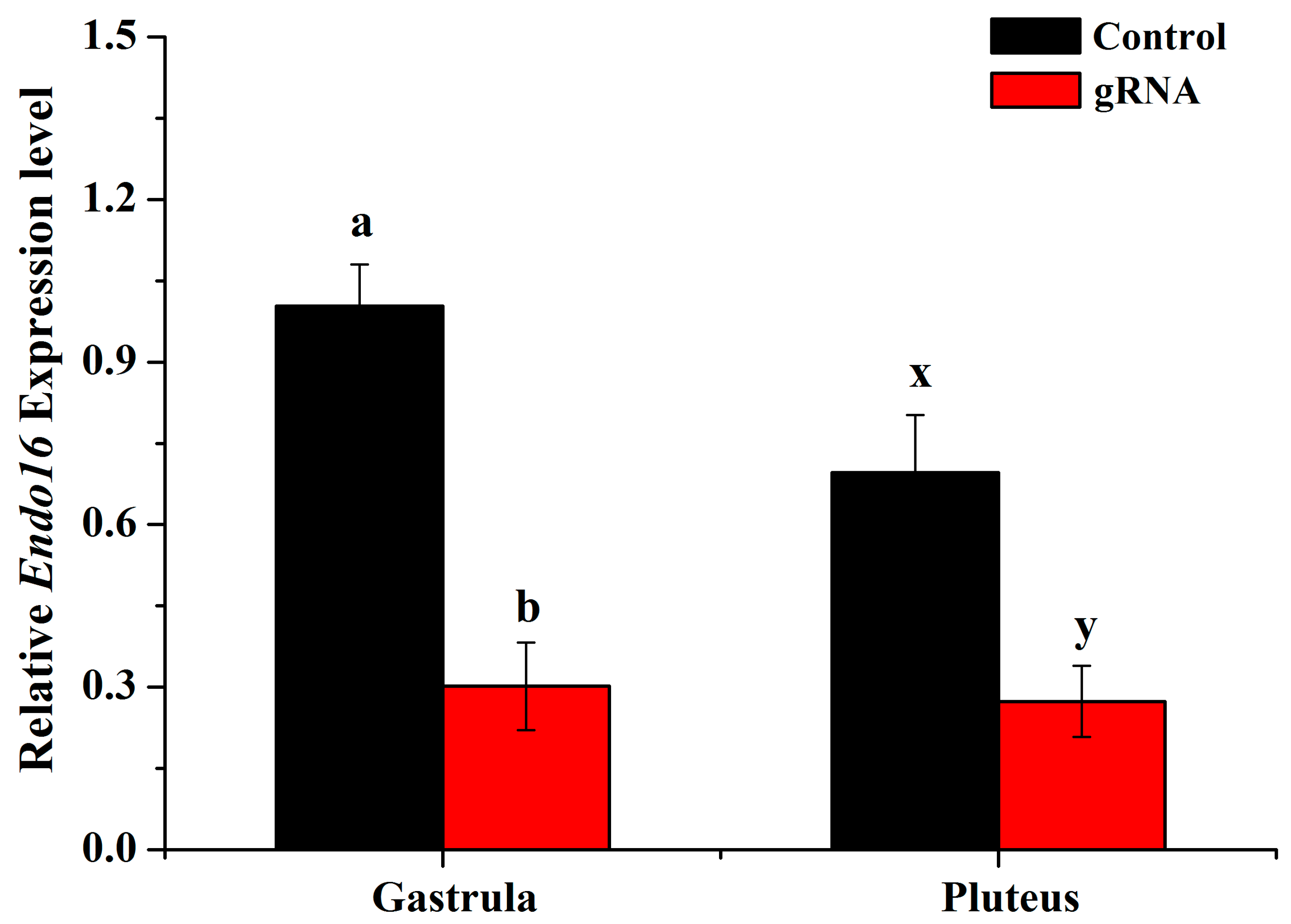
3.2. Disruption of Endo16 Module A Using Cas9 and gRNAs Caused a Downregulation of Endo16 Expression

Based on the mRNA sequence of Lytechinus variegatus Endo16, we designed primers for real-time PCR. cDNA was prepared from mutants and wild-type embryos in gastrula and pluteus stages. The results show that Endo16 mRNA expression level in abnormal embryos was lower than in control embryos in both gastrula and pluteus stages (Figure 4, Table S1). The low expression level of Endo16 in abnormal embryos may be caused by the disruption of the Endo16 enhancer. The real-time PCR results may demonstrate that the level of Endo16 mRNA in embryos which had been injected with gRNAs and the Cas9 mRNA was less than half that of controls at both gastrula and pluteus stages. In addition, our qPCR results showed that in the case of disruption of the Endo16 cis-regulatory element, the gene expression level of Endo16, although significantly lower than that of the control embryos, was still detectable. Together with a potential highly effective NHEJ repair system in sea urchins [40,41,42] and other reasons mentioned in our discussion section, all may lead to the recovery of a small archenteron-like structure (Figure 2F). However, this structure rarely fused with the ectoderm to form a complete gut.

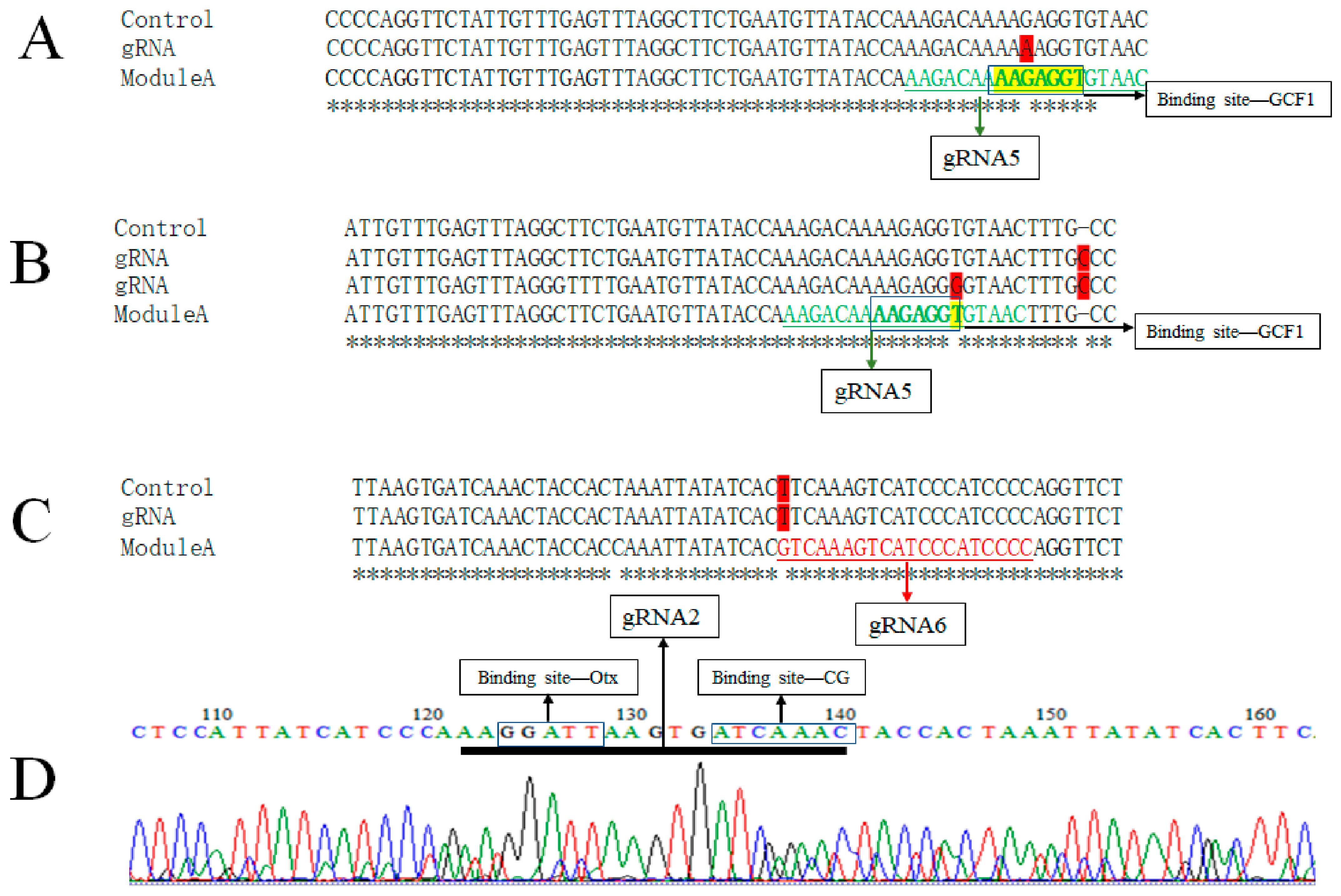
3.3. Genotype of Embryos Injected with gRNA and Cas9 mRNA

To precisely link the observed morphological and molecular phenotypes with the genotype of module A, we conducted targeted DNA sequencing of individual embryos that had been injected with gRNA and Cas9 mRNA at the gastrula stage. We cloned the PCR amplicons from individual control embryos and from individual experimental embryos that failed to undergo gastrulation into the pGEM-T easy vector for sequencing. From the clone sequencing results, we did not find long deletions. However, we did find a point mutation in the gRNA5 binding site (GCF1) region (Figure 5A,B) and an insertion near the binding sites (Figure 5B). In addition, there was a mismatched base in gRNA6 when we designed the gRNA6 using the Module A template sequence (Figure 5C), which is likely the result of natural genetic variation. This may be the reason that gRNA6 did not produce a reliable phenotype.

4. Discussion

Gene editing is an emerging genetic engineering technique that can modify specific target genes in an organism’s genome with relative precision. Gene editing technologies have been widely applied in various fields, including disease control, trait improvement, drug development, and gene therapy, and have greatly promoted the study of biological gene functions [43,44]. Among them, the CRISPR-Cas systems with diversity, modularity, and efficacy are driving a biotechnological revolution [45] and providing new methods and research ideas for the study of gene function, the analysis of economic traits and the genetic improvement of aquatic animals. Currently, the successful application of CRISPR/Cas9 in aquatic animals is expected to usher in an era of “precision breeding” in aquatic animal breeding. However, aquatic animal gene editing research is still in its infancy and faces many problems and challenges. One of the most problematic issues is that direct knockout of the major gene affects normal life activities, making it difficult for edited individuals to survive, stunting growth and resulting in poor practical application. The editing of target cis-regulatory elements using CRISPR/Cas9 technology to achieve genetic regulation of target traits will greatly improve the efficiency of breeding [46].
Here, we use the CRISPR/Cas9 system to disrupt an enhancer of Endo16 in developing L. variegatus embryos. The results showed that Endo16 Module A-disrupted embryos failed to undergo gastrulation at 20 h post fertilization. Compared with control embryos, the disruption of Endo16 enhancers using Cas9 and gRNAs caused a downregulation of Endo16 expression. However, we did not find mutations regularly from the clone sequencing results. This may be due in part to the time required to translate Cas9 from the injected mRNA, which likely results in mosaic genome editing and the majority of cells in the embryo having either no edits or different edits. Consistent with this possibility, we found multi-peaks throughout the clone sequencing results (Figure 5D). The binding of Cas9/gRNA to the target sequence may prevent the binding of transcription factors to cis-regulatory elements, thus causing gene silencing. It affects gene transcription, although its DNA sequence mutations are not detected on a large scale. This is relatively similar to how CRISPRi (CRISPR interference or inhibition) works. It is also possible that sea urchins simply have a highly effective NHEJ repair system. Although the sample size of this sequencing was small, we did find multi-peaks in some cases, which does suggest that mutations were induced. More verification experiments need to be conducted to link the phenotype with the genotype. While additional optimization will be needed to improve efficiency, our results and those of Pipelow et al. (2021) [23] collectively indicate that targeting mutations using gRNAs and Cas9 is a feasible and informative method for studying the function of cis-regulatory elements in sea urchin embryos. In addition, applying this method to echinoderm breeding is more conducive to reducing the interference with other normal life activities of the edited individuals, improving the survival rate of the edited individuals, and thus improving the breeding efficiency.
However, the application of the CRISPR/Cas9 system in aquaculture breeding is still facing several technical challenges. First, genome annotation and gene regulatory network (GRN) studies need to be strengthened. Thanks to the well-established GRN of sea urchins, the cis-regulatory elements were successfully edited and the expected phenotypes were obtained in this study. In aquaculture species, genes and regulatory elements which are associated with important traits, such as growth, nutrition and disease resistance, are still limited. Second, efficient delivery methods of CRISPR systems into fertilized eggs at the one-cell stage need to be developed. Aquatic breeding requires editing a large number of fertilized eggs in a short period of time, which is difficult to achieve with the microinjection method used in this study. With the improvement and innovation of gene editing technology, some more efficient and accurate gene editing systems continue to emerge. It is believed that gene editing technology will have a broader application prospect in the field of aquaculture.

5. Conclusions

Aquatic products are the third largest source of animal protein in the world, and aquatic animals provide economical and high-quality animal protein. In recent years, efficient, accurate and low-cost CRISPR technology has become an important tool for exploring gene functions, resolving life phenomena and germplasm creation, and it is increasingly used in aquatic biology. In this study, we used an important mariculture species, the sea urchin, as a research object to focus on cis-regulatory elements, and applied the CRISPR/Cas9 gene editing system to achieve changes in sea urchin embryonic traits. Our results indicate that targeting mutations using gRNAs and Cas9 is a feasible and informative method for studying the function of cis-regulatory elements in sea urchin embryos. Manipulating gene functions by editing cis-regulatory sequences through CRISPR-Cas9, instead of the more typical mutation of coding regions, will minimize secondary effects of cellular responses to nonsense mediated decay pathways or to mutant protein products by premature stop codons.

Supplementary Materials

The following supporting information can be downloaded at: https://www.mdpi.com/article/10.3390/fishes8020118/s1, Table S1: The raw data of mRNA expression level analysis.

Author Contributions

Conceptualization, L.X. and G.A.W.; methodology, L.X. and G.A.W.; software, L.X.; validation, L.X., M.L. and F.R.; formal analysis, L.X. and L.W.; investigation, L.X.; resources, L.W.; data curation, L.X.; writing—original draft preparation, L.X.; writing—review and editing, M.L. and G.A.W.; visualization, L.X.; supervision, G.A.W.; project administration, G.A.W.; funding acquisition, G.A.W. and L.X. All authors have read and agreed to the published version of the manuscript.

Funding

This research was funded by a National Science Foundation Grant IOS-1929934 to GAW, and National Natural Science Foundation of China (No. 42106109) and Natural Science Foundation of Shandong Province Youth Project ZR2020QD100 grants to LLX.

Institutional Review Board Statement

Not applicable for studies involving invertebrates.

Data Availability Statement

Not applicable.

Acknowledgments

We would like to thank all the members of Wray Lab for their support and help with planning experiments.

Conflicts of Interest

The authors declare no conflict of interest. The funders had no role in the design of the study; in the collection, analyses, or interpretation of data; in the writing of the manuscript; or in the decision to publish the results.

References

  1. Golden, C.D.; Koehn, J.Z.; Shepon, A.; Passarelli, S.; Free, C.M.; Viana, D.F.; Matthey, H.; Eurich, J.G.; Gephart, J.A.; Fluet-Chouinard, E.; et al. Aquatic foods to nourish nations. Nature 2021, 598, 315–320. [Google Scholar] [CrossRef] [PubMed]
  2. Naylor, R.L.; Kishore, A.; Sumaila, U.R.; Issifu, I.; Hunter, B.P.; Belton, B.; Bush, S.R.; Cao, L.; Gelcich, S.; Gephart, J.A.; et al. Blue food demand across geographic and temporal scales. Nat. Commun. 2021, 12, 5413. [Google Scholar] [CrossRef] [PubMed]
  3. Golden, C.D.; Allison, E.H.; Cheung, W.W.L.; Dey, M.M.; Halpern, B.S.; McCauley, D.J.; Smith, M.; Vaitla, B.; Zeller, D.; Myers, S.S. Nutrition: Fall in fish catch threatens human health. Nature 2016, 534, 317–320. [Google Scholar] [CrossRef] [PubMed] [Green Version]
  4. Rimm, E.B.; Appel, L.J.; Chiuve, S.E.; Djoussé, L.; Engler, M.B.; Kris-Etherton, P.M.; Mozaffarian, D.; Siscovick, D.S.; Lichtenstein, A.H. Seafood long-chain n-3 polyunsaturated fatty acids and cardiovascular disease: A science advisory from the American Heart Association. Circulation 2018, 138, e35–e47. [Google Scholar] [CrossRef]
  5. Gallet, C.A. The demand for fish: A meta-analysis of the own-price elasticity. Aquac. Econ. Manag. 2009, 13, 235–245. [Google Scholar] [CrossRef]
  6. Vermeulen, S.J.; Park, T.; Khoury, C.K.; Béné, C. Changing diets and the transformation of the global food system. Ann. N. Y. Acad. Sci. 2020, 1478, 3–17. [Google Scholar] [CrossRef] [PubMed]
  7. Bernstein, A.S.; Oken, E.; de Ferranti, S.; Lowry, J.A.; Ahdoot, S.; Baum, C.R.; Bole, A.; Byron, L.G.; Landrigan, P.J.; Marcus, S.M.; et al. Fish, shellfish, and children’s health: An assessment of benefits, risks, and sustainability. Pediatrics 2019, 143, e20190999. [Google Scholar] [CrossRef] [Green Version]
  8. Mcbride, S.C. Sea urchin aquaculture. Am. Fish. Soc. Symp. 2005, 46, 179–208. [Google Scholar]
  9. Paredes, E. Biobanking of a Marine Invertebrate Model Organism: The Sea Urchin. J. Mar. Sci. Eng. 2016, 4, 7. [Google Scholar] [CrossRef] [Green Version]
  10. Dvoretsky, A.G.; Dvoretsky, V.G. Aquaculture of green sea urchin in the Barents Sea: A brief review of Russian studies. Rev. Aquac. 2020, 12, 2080–2090. [Google Scholar] [CrossRef]
  11. James, P.J. A comparison of roe enhancement of the sea urchin Evechinus chloroticus in sea-based and land-based cages. Aquaculture 2006, 253, 290–300. [Google Scholar] [CrossRef]
  12. George, S.B.; Lawrence, J.M.; Lawrence, A.L. Complete larval development of the sea urchin Lytechinus variegatus fed an artificial feed. Aquaculture 2004, 242, 217–228. [Google Scholar] [CrossRef]
  13. Christiansen, J.S.; Silkavuoplo, S.I. The relationship between feed intake and gonad growth. of single and stocked green sea urchin (Strongylocentrotus droebachiensis) in a raceway culture. Aquaculture 2007, 262, 163–167. [Google Scholar] [CrossRef]
  14. Hwang, W.Y.; Fu, Y.; Reyon, D.; Maeder, M.L.; Tsai, S.Q.; Sander, J.D.; Peterson, R.T.; Yeh, J.-R.J.; Joung, J.K. Efficient genome editing in zebrafish using a CRISPR-Cas system. Nat. Biotechnol. 2013, 31, 227–229. [Google Scholar] [CrossRef] [PubMed] [Green Version]
  15. Lu, J.; Fang, W.; Huang, J.; Li, S. The application of genome editing technology in fish. Mar. Life Sci. Technol. 2021, 3, 326–346. [Google Scholar] [CrossRef]
  16. Square, T.; Romášek, M.; Jandzik, D.; Cattell, M.V.; Klymkowsky, M.; Medeiros, D.M. CRISPR/Cas9-mediated mutagenesis in the sea lamprey, Petromyzon marinus: A powerful tool for understanding ancestral gene functions in vertebrates. Development 2015, 142, 4180–4187. [Google Scholar] [CrossRef] [Green Version]
  17. Perry, K.J.; Henry, J.Q. CRISPR/Cas9-mediated genome modification in the mollusc, Crepidula fornicata. Genes 2015, 53, 237–244. [Google Scholar] [CrossRef]
  18. Abe, M.; Kuroda, R. The development of CRISPR for a mollusc establishes the formin Lsdia1 as the long-sought gene for snail dextral/sinistral coiling. Development 2020, 146, dev175976. [Google Scholar] [CrossRef] [Green Version]
  19. Lin, C.-Y.; Su, Y.-H. Genome editing in sea urchin embryos by using a CRISPR/Cas9 system. Dev. Biol. 2016, 409, 420–428. [Google Scholar] [CrossRef] [Green Version]
  20. Oulhen, N.; Wessel, G.M. Albinism as a visual, in vivo guide for CRISPR/Cas9 functionality in the sea urchin embryo. Mol. Reprod. Dev. 2016, 83, 1046–1047. [Google Scholar] [CrossRef]
  21. Wessel, G.M.; Kiyomoto, M.; Shen, T.-L.; Yajima, M. Genetic manipulation of the pigment pathway in a sea urchin reveals distinct lineage commitment prior to metamorphosis in the bilateral to radial body plan transition. Sci. Rep. 2020, 10, 1973. [Google Scholar] [CrossRef] [PubMed] [Green Version]
  22. Wessel, G.M.; Wada, Y.; Yajima, M.; Kiyomoto, M. Sperm lacking Bindin are infertile but are otherwise indistinguishable from wildtype sperm. Sci. Rep. 2021, 11, 21583. [Google Scholar] [CrossRef] [PubMed]
  23. Pieplow, A.; Dastaw, M.; Sakuma, T.; Sakamoto, N.; Yamamoto, T.; Yajima, M.; Oulhen, N.; Wessel, G.M. CRISPR-Cas9 editing of non-coding genomic loci as a means of controlling gene expression in the sea urchin. Dev. Biol. 2021, 472, 85–97. [Google Scholar] [CrossRef] [PubMed]
  24. Shevidi, S.; Uchida, A.; Schudrowitz, N.; Wessel, G.M.; Yajima, M. Single nucleotide editing without DNA cleavage using CRISPR/Cas9-deaminase in the sea urchin embryo. Dev. Dyn. 2017, 246, 1036–1046. [Google Scholar] [CrossRef] [Green Version]
  25. Wittkopp, P.J.; Kalay, G. Cis-regulatory elements: Molecular mechanisms and evolutionary processes underlying divergence. Nat. Rev. Genet. 2012, 13, 59–69. [Google Scholar] [CrossRef]
  26. Wray, G.A. The evolutionary significance of cis-regulatory mutations. Nat. Rev. Genet. 2007, 8, 206–216. [Google Scholar] [CrossRef]
  27. Bauer, D.E.; Kamran, S.C.; Lessard, S.; Xu, J.; Fujiwara, Y.; Lin, C.; Shao, Z.; Canver, M.C.; Smith, E.C.; Pinello, L.; et al. An Erythroid Enhancer of BCL11A Subject to Genetic Variation Determines Fetal Hemoglobin Level. Science 2013, 342, 253–257. [Google Scholar] [CrossRef] [Green Version]
  28. Harismendy, O.; Notani, D.; Song, X.; Rahim, N.G.; Tanasa, B.; Heintzman, N.; Ren, B.; Fu, X.D.; Topol, E.J.; Rosenfeld, M.G.; et al. 9p21 DNA variants associated with coronary artery disease impair interferon-gamma signalling response. Nature 2011, 470, 264. [Google Scholar] [CrossRef] [Green Version]
  29. Maurano, M.T.; Humbert, R.; Rynes, E.; Thurman, R.E.; Haugen, E.; Wang, H.; Reynolds, A.P.; Sandstrom, R.; Qu, H.; Brody, J.; et al. Systematic Localization of Common Disease-Associated Variation in Regulatory DNA. Science 2012, 337, 1190–1195. [Google Scholar] [CrossRef] [Green Version]
  30. Musunuru, K.; Strong, A.; Frank-Kamenetsky, M.; Lee, N.E.; Ahfeldt, T.; Sachs, K.V.; Li, X.; Li, H.; Kuperwasser, N.; Ruda, V.M.; et al. From noncoding variant to phenotype via SORT1 at the 1p13 cholesterol locus. Nature 2010, 466, 714–719. [Google Scholar] [CrossRef] [Green Version]
  31. Oldridge, D.A.; Wood, A.C.; Weichert-Leahey, N.; Crimmins, I.; Sussman, R.; Winter, C.; McDaniel, L.D.; Diamond, M.; Hart, L.S.; Zhu, S.; et al. Genetic predisposition to neuroblastoma mediated by a LMO1 super-enhancer polymorphism. Nature 2015, 528, 418–421. [Google Scholar] [CrossRef] [Green Version]
  32. Crisp, P.A.; Bhatnagar-Mathur, P.; Hundleby, P.; Godwin, I.D.; Waterhouse, P.M.; Hickey, L.T. Beyond the gene: Epigenetic and cis-regulatory targets offer new breeding potential for the future. Curr. Opin. Biotechnol. 2021, 73, 88–94. [Google Scholar] [CrossRef]
  33. Romano, L.A.; Wray, G.A. Endo16 is required for gastrulation in the sea urchin Lytechinus variegatus. Dev. Growth Differ. 2006, 48, 487–497. [Google Scholar] [CrossRef]
  34. Yuh, C.-H.; Ransick, A.; Martinez, P.; Britten, R.J.; Davidson, E.H. Complexity and organization of DNA-protein interactions in the 5′-regulatory region of an endoderm-specific marker gene in the sea urchin embryo. Mech. Dev. 1994, 47, 165–186. [Google Scholar] [CrossRef] [PubMed]
  35. Romano, L.A.; Wray, G.A. Conservation of Endo16 expression in sea urchins despite evolutionary divergence in both cis and trans-acting components of transcriptional regulation. Development 2003, 130, 4187–4199. [Google Scholar] [CrossRef] [PubMed] [Green Version]
  36. Cheers, M.S.; Ettensohn, C.A. Rapid Microinjection of Fertilized Eggs. Methods Cell Biol. 2004, 74, 287–310. [Google Scholar] [CrossRef] [PubMed]
  37. Cameron, R.A.; Samanta, M.; Yuan, A.; He, D.; Davidson, E. SpBase: The sea urchin genome database and web site. Nucleic Acids Res. 2009, 37, 750. [Google Scholar] [CrossRef] [Green Version]
  38. Schmittgen, T.D.; Livak, K.J. Analyzing real-time PCR data by the comparative CT method. Nat. Protoc. 2008, 3, 1101–1108. [Google Scholar] [CrossRef]
  39. Yuh, C.-H.; Bolouri, H.; Davidson, E.H. Genomic Cis-Regulatory Logic: Experimental and Computational Analysis of a Sea Urchin Gene. Science 1998, 279, 1896–1902. [Google Scholar] [CrossRef] [Green Version]
  40. Cui, M.; Lin, C.-Y.; Su, Y.-H. Recent advances in functional perturbation and genome editing techniques in studying sea urchin development. Briefings Funct. Genom. 2017, 16, 309–318. [Google Scholar] [CrossRef] [Green Version]
  41. Reinardy, H.C.; Bodnar, A.G. Profiling DNA damage and repair capacity in sea urchin larvae and coelomocytes exposed to genotoxicants. Mutagenesis 2015, 30, 829–839. [Google Scholar] [CrossRef] [PubMed] [Green Version]
  42. Le Bouffant, R.; Cormier, P.; Cueff, A.; Bellé, R.; Mulner-Lorillon, O. Sea urchin embryo as a model for analysis of the signaling pathways linking DNA damage checkpoint, DNA repair and apoptosis. Cell. Mol. Life Sci. 2007, 64, 1723–1734. [Google Scholar] [CrossRef] [PubMed]
  43. Hsu, P.D.; Lander, E.S.; Zhang, F. Development and Applications of CRISPR-Cas9 for Genome Engineering. Cell 2014, 157, 1262–1278. [Google Scholar] [CrossRef] [PubMed] [Green Version]
  44. Yang, Z.; Yu, Y.; Tay, Y.X.; Yue, G.H. Genome editing and its applications in genetic improvement in aquaculture. Rev. Aquac. 2021, 14, 178–191. [Google Scholar] [CrossRef]
  45. Knott, G.J.; Doudna, J.A. CRISPR-Cas guides the future of genetic engineering. Science 2018, 361, 866–869. [Google Scholar] [CrossRef] [Green Version]
  46. Lopes, R.; Korkmaz, G.; Agami, R. Applying CRISPR–Cas9 tools to identify and characterize transcriptional enhancers. Nat. Rev. Mol. Cell Biol. 2016, 17, 597–604. [Google Scholar] [CrossRef]
Figure 1. Positions and sequences of gRNAs targeting the Endo16 Module A. (A) A schematic representation of the Endo16 Module A locus and the positions of the six gRNAs. The arrows indicate the orientation of the gRNAs. The colored boxes indicate the binding sites with transcription factor. (B) The specific sequence of Endo16 Module A and binding sites. (C) The specific sequence of the six gRNAs.
Fishes 08 00118 g001
Figure 2. Effect of disruption of Endo16 cis-regulatory elements on the development of L. variegatus. In the blastula stage, there is no difference between embryos injected with gRNAs (A) and control embryos (D). In the gastrula stage, embryos injected with gRNAs failed to undergo gastrulation (E) compared to control embryos (B). In the pluteus stage, embryos injected with gRNAs displayed morphological abnormalities and failed to develop into four-arm pluteus larvae with a functional gut (F) compared with control embryos (C). Black arrow indicates the normally developing gastrum and white arrow indicates disorganized cells in the blastocoel. Scale bar (black line) = 100 μm.
Fishes 08 00118 g002
Figure 3. Ratios of mutated embryos in different gRNA concentration of two gRNA combinations (p > 0.05). The data of ratios were presented as the mean ± standard deviation (n = 3), and they were statistically analyzed by one-way ANOVA with a Tukey test.
Fishes 08 00118 g003
Figure 4. mRNA expression level of Endo16 detected by real-time PCR. Different letters a, b indicate significant difference for the gastrula stage (p < 0.05), and different letters x, y indicate significant difference for the pluteus stage (p < 0.05). The error bar indicates standard deviation (n = 3).
Fishes 08 00118 g004
Figure 5. Point mutations, insertion and mosaicism of embryos revealed by sequencing. (A) Sanger sequencing from one embryo reveals a point mutation in the gRNA5 target sequence. (B) Sanger sequencing from two embryos reveals a point mutation in the gRNA5 target sequence and an insertion near the transcription factor binding sites. (C) A mismatched base is found in gRNA6. (D) TIDE analysis of PCR products from one embryo reveals mosaicism. Green text indicates the target sequence of gRNA. The boxed region indicates the transcription factor biding site. Text highlighted in red indicates the point mutation. “Control” indicates the sequence from control embryos. “gRNA” indicates the sequence from embryos injected with gRNA. “Module A” indicates the sequence used for gRNA design. Asterisk indicates the same nucleotide site. Sequences underlined in black in (D) indicate the target sequence of gRNA2.
Fishes 08 00118 g005
Table 1. Primers used for verification.
PrimerSequence (5’ to 3’)Annealing TemperatureProduct SizeAim
1-FGACAGAGACCGTATCGAATTAACATGCG69 °C406 bpEndo16 Module A cloning
1-RTTCGACCACGCCACGGCCAGCACGG
2-FGACCTGTAGCGAACACACAAAGCCG60 °C441 bpEndo16 mRNA expression level analysis
2-RTCACGGCAGTGCAGATGGCCTCG
3-FCACAGGCAAGACCATCACA147 bpHousekeeping gene—Ubiquitin
3-RGAGAGAGTGCGACCATCCTC
Table 2. Phenotypic ratios of embryos injected with Cas9 mRNA (258.72 ng/μL) and gRNAs scored at the gastrula stage.
CombinationgRNA Concentration (ng/μL; per gRNA)Embryos with MicroinjectionDead Embryos % (n)Alive Normal Embryos % (n)Embryos with Expected Phenotype % (n)
gRNA 1 and 2 and 311218715 (28)75 (119)25 (40)
gRNA 4 and 5 and 61123340 (13)60 (12)40 (8)
gRNA 1 and 2 and 316810037 (37)52 (33)48 (30)
gRNA 4 and 5 and 61686632 (21)56 (25)44 (20)
gRNA 2 and 4 and 516819515 (30)65 (108)35 (57)
gRNA 1 and 3 and 616817926 (46)70 (93)30 (40)
gRNA 4 and 516819019 (37)48 (73)52 (80)
gRNA 1 and 316817522 (38)50 (68)50 (69)
gRNA 2 and 616816113 (21)60 (84)40 (56)
gRNA 4 and 52248919 (17)54 (39)46 (33)
gRNA 1 and 32247020 (14)61 (34)39 (22)
gRNA 2 and 62246310 (6)70 (40)30 (17)
gRNA 4 and 528020216 (32)59 (100)41 (70)
gRNA 1 and 328019812 (24)62 (107)38 (67)
gRNA 2 and 628018720 (38)65 (97)35 (52)
gRNA116811323 (27)35 (30)65 (56)
gRNA216814520 (29)39 (45)61 (71)
gRNA316812424 (30)74 (70)26 (24)
gRNA41684353 (23)70 (14)30 (6)
gRNA51684161 (25)38 (6)62 (10)
gRNA61684050 (20)100 (20)0 (0)
Disclaimer/Publisher’s Note: The statements, opinions and data contained in all publications are solely those of the individual author(s) and contributor(s) and not of MDPI and/or the editor(s). MDPI and/or the editor(s) disclaim responsibility for any injury to people or property resulting from any ideas, methods, instructions or products referred to in the content.

