Review

Cadmium as an Endocrine Disruptor That Hinders the Reproductive and Developmental Pathways in Freshwater Fish: A Review

by
Kaakarlu Shivakumar Vinanthi Rajalakshmi
1,†,
Wen-Chao Liu
2,†,
Balasubramanian Balamuralikrishnan
3,†,
Arun Meyyazhagan
1,
Govindharajan Sattanathan
4,
Manikantan Pappuswamy
1,
Kadanthottu Sebastian Joseph
1,
Kuppusamy Alagesan Paari
1,* and
Jang-Won Lee
5,*
1
Department of Life Sciences, School of Sciences, Christ University, Bangalore 560029, Karnataka, India
2
Department of Animal Science, College of Coastal Agricultural Sciences, Guangdong Ocean University, Zhanjiang 524088, China
3
Department of Food Science and Biotechnology, College of Life Science, Sejong University, Seoul 05006, Republic of Korea
4
Department of Zoology, St. Joseph University, Chumukedima 797115, Nagaland, India
5
Department of Integrative Biological Sciences and Industry, Sejong University, Seoul 05006, Republic of Korea
*
Authors to whom correspondence should be addressed.
These authors contributed equally to this work.
Fishes 2023, 8(12), 589; https://doi.org/10.3390/fishes8120589
Submission received: 29 August 2023 / Revised: 20 November 2023 / Accepted: 28 November 2023 / Published: 30 November 2023
(This article belongs to the Special Issue Effects of Trace Elements on Aquatic Animals)

Abstract

:
Cadmium (Cd) is a non-essential element with sub-lethal effects even at low concentrations. The persistent nature of Cd and its tendency to bioaccumulate eventually create harmful effects on water biota, including fish. Cd affects various aspects of hormonal action in fish since it bioaccumulates in the endocrine system and hinders the synthesis, secretion, and metabolic activity of hormones, causing severe damage along the hypothalamus–pituitary–gonadal axis. Linking reproductive and developmental impairments in fish with ecologically relevant concentrations of individual metals can be challenging due to the complexity of aquatic ecosystems. This review deliberated the significant and novel trends of toxicological difficulties and approaches, including elucidating environmental sources’ bioavailability and Cd-induced toxic effects in freshwater fish. Both acute and chronic exposure to Cd can cause a range of adverse effects, such as growth inhibition, impaired reproductive capacity, endocrine disruption, and developmental abnormalities in freshwater fish, as evidenced by the present review. These investigations support the concept of Cd as a naturally available pollutant that causes irreversible damage in fish. These findings will help to understand the etiology of environmental circumstances that pose substantial dangers to fish health and are also crucial for preventing and treating exposure-related reproductive disturbances in freshwater fish due to environmental pollution.
Key Contribution: Cadmium has been found to cause cellular-to-organ-level damage in fish. This review examines the toxicity of cadmium exposure along the hypothalamus–pituitary–gonadal–liver axis, focusing on the reproductive and developmental alterations in freshwater fish.

Graphical Abstract

1. Introduction

The contamination of the freshwater environment with trace elements discharged from anthropogenic activities and natural resources has become an eco-environmental concern over the past few decades [1]. Trace elements refer to any metallic elements that occur in nature, possess a relatively high density (3.5 to 7 g cm−3), and are toxic even at ppb levels [2,3]. Some metals, Cu, Fe, Mn, Co, Zn, and Ni, are essential micro-nutrients required for various biochemical and physiological functions in biological systems [4,5]. In comparison, non-essential metals such as cadmium (Cd), Pb, and Hg with no established biological processes are known to induce multiple organ damages even at trace concentrations and are hence considered a burden when they enter the host [5,6]. However, each metal is recognized to have distinct physicochemical characteristics that bestow toxicological modes of action [7].
Among the trace elements, Cd is considered one of the most ubiquitous and toxic chemicals that adversely affect the permanence of freshwater biota [8]. Cadmium occurs in water or environmental matrices (such as sediment, aerosols, etc.) and accumulates in aquatic organisms such as mollusks, crustaceans, and fish [9,10]. Fish are the central community that reflects the quality of aquatic systems by accumulating large amounts of toxic metals, as they are one of the topmost consumers in the aquatic food chain [11]. Freshwater fish have also become the primary route for transporting Cd along the food chain due to their significant contribution to the diet of their higher trophic levels and rural communities [12,13]. The bioaccumulation of freshwater fish organs with Cd led to an impairment in their feral populations by altering their reproductive and physiological functions [12]. Once absorbed, the Cd is moved by circulation to either a storage location (bones, liver) or carried further to other organs such as the kidneys, gills, muscles, and gonads [14]. The chronic exposure of fish to Cd can disrupt the natural system and may endanger the affected fish species. Therefore, hazardous metals in freshwater aquatic systems are currently a major issue for the sustainability of the ecosystem and living things. Since Cd is known to have many harmful impacts, such as neurotoxic, genotoxic, and endocrinological effects on fish, it is essential to monitor its bioaccumulation patterns [15,16].
The exposure of freshwater biota to endocrine-disrupting metals like Cd can have significant implications for the population dynamics of affected fish species. Reduced fertility, skewed sex ratios, and altered reproductive behavior can lead to decreased population size and genetic diversity, which can have long-term effects on the survival and viability of a species [17]. Due to the crucial involvement of the endocrine system in regulating homeostasis and other physiological processes, the study of the effects of such a disruption may be significant. The stimulation of the endocrine system via the hypothalamicpituitary–gonadal (HPG) axis has been the center of outstanding research on how animals react to stressors [18]. In response to a wide range of stressors, including endocrine-disrupting metals, the hypothalamus pumps releasing factors along this axis, stimulating the production of hormones. Emerging research suggests that fish can be a potent biological model for assessing the hormonal status and viability of aquatic environments. They can be a powerful species model for examining endocrine signaling pathways related to testicular function, spermatogenesis, and their interaction with reproductive behavior.
The impact of Cd on the endocrine disruption of freshwater fish is a multifaceted study dealing with the abnormalities caused by inducing alterations in the normal functioning of the endocrine system. However, decades-long research dealing with fish health related to environmental conditions impacted by Cd has revealed an increasing testimonial of altered developmental effects on the reproductive health of fish along the HPG axis. Surprisingly, only a few works from the literature address the action and biological consequences of Cd as an endocrine disruptor in freshwater fish. Hence, this review aimed to accumulate the significant hormonal changes caused by the bioaccumulation of Cd in the bodies of freshwater fish from different perspectives. It also elucidates the mechanism of action by which Cd affects the circuits of the HPG axis in controlling reproductive and developmental pathways in freshwater fish.

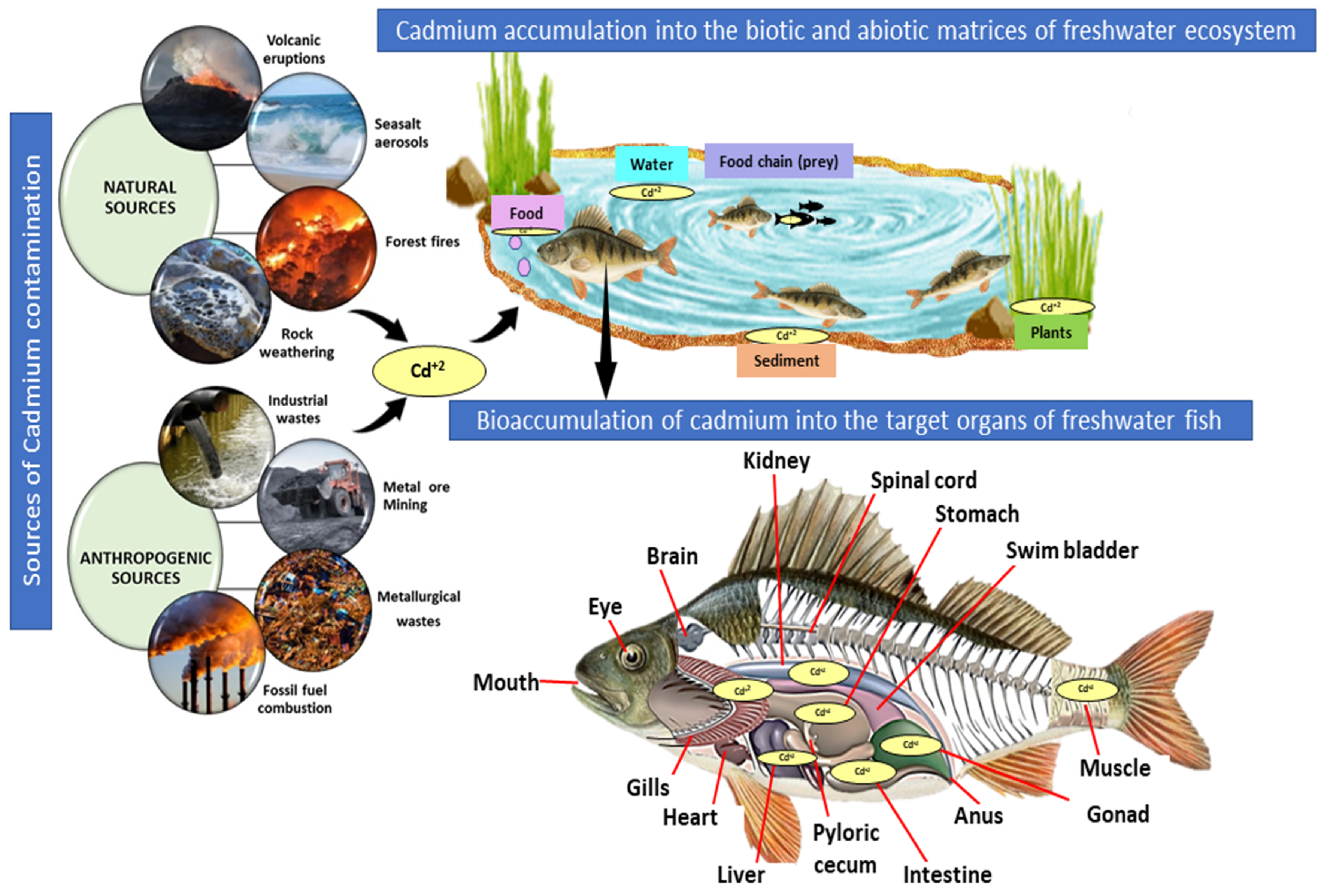
2. Sources of Cadmium Contamination in Aquatic Systems

Cadmium is relatively minor in the Earth’s crust, with concentrations ranging from 0.1 to 0.5 ppm, but it can be found in small amounts in zinc, lead, and copper ores [19,20]. Natural Cd emissions to the aquatic biota can result from volcanic eruptions (60%), forest fires, sea salt aerosol formation, the weathering of rocks, or other biological factors (Figure 1). Refined Cd is being used in the manufacturing of electrodes in batteries (83%), the production of Cd-containing pigments (8%), electroplating and coatings (7%), plastic stabilizers (1.2%), nonferrous alloys, solar devices, and other applications (0.8%) [21,22]. With historical and current activities, water systems near these Cd-emitting industries have revealed a significant increase in Cd levels in the water (<1.0 to 77 μg/L), sediment, and aquatic organisms [19]. The aquaculture project for fish farming installed in part of a disused mining pool showed the accumulation of Cd in the tissues of the freshwater species Barbonymus schwanenfeldii, Cyclocheilicthys apogon, Hampala microlepidota, Hemibagrus nemurus, Mystacoleucus marginatus, and Oreochromis niloticus [23,24].
In freshwater ecosystems, Cd can exist as a hydrated ion or as ionic compounds with other inorganic or organic substances [25]; while soluble forms (sulfate and chloride salts of Cd) may migrate in water [26], Cd is relatively immobile in insoluble complexes [27]. Cd is a serious global environmental pollutant and has the potential to alter higher trophic levels for centuries. Freshwater fish could be susceptible to Cd exposure and subsequent toxicity through dietary and waterborne exposure [28,29,30]. The negative impact of chronic Cd exposure (3 to 6 months) was reported in channel catfish, Ictalurus punctatus, with a reduced survival rate and altered growth patterns [30]. Similarly, another study using rainbow trout, Oncorhynchus mykiss, showed that prolonged exposure to Cd reduced the accumulation rate of Ca in gills, which is possibly a result of a blockage of Ca channels by cytosolic Cd [31]. The WHO recommended levels of Cd in environmental matrixes range from 0.01 to 1 mg/kg in soil and 3 μg/L in water, so partitioning the accumulation of Cd between the sediment and water is a key element in determining its availability to aquatic organisms. The pathways of Cd uptake and accumulation can vary depending on the species present and their feeding habits. For example, predatory fish feed on smaller fish that have accumulated higher levels of Cd in their tissues than herbivorous fish that do not consume other fish [32]. Cadmium concentrations in benthic macroinvertebrates and fish tissues were highly linked, indicating metal transfer via a feeding mechanism (Figure 1). As a result, fish that live in contaminated waters collect Cd in their tissues [33].

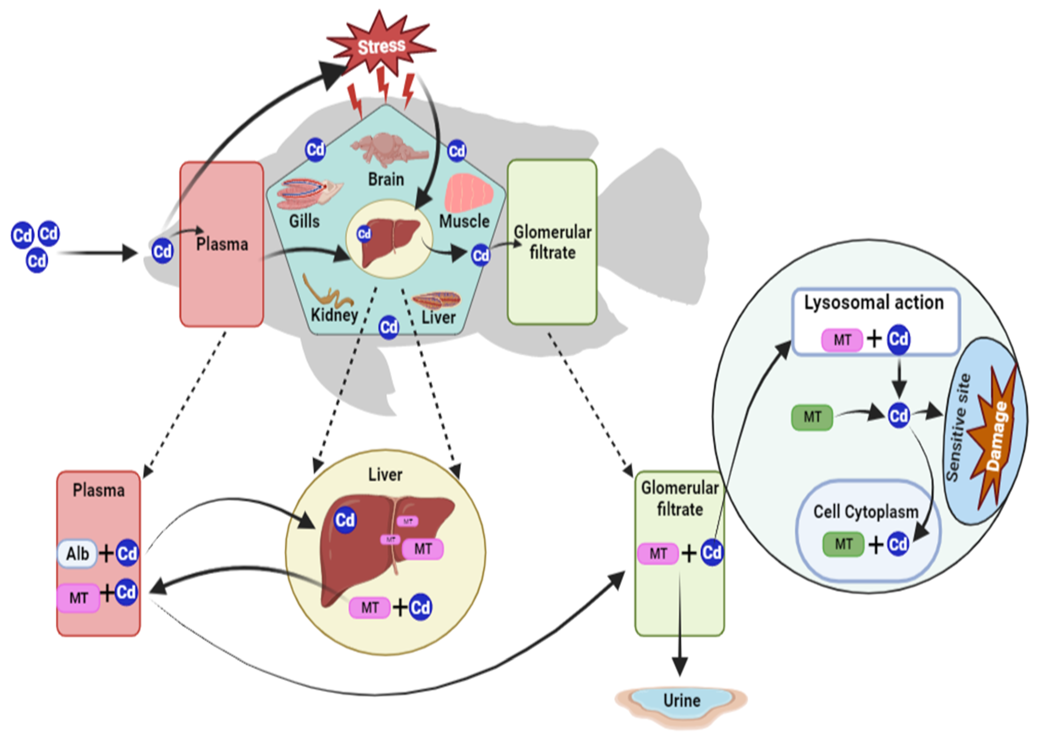
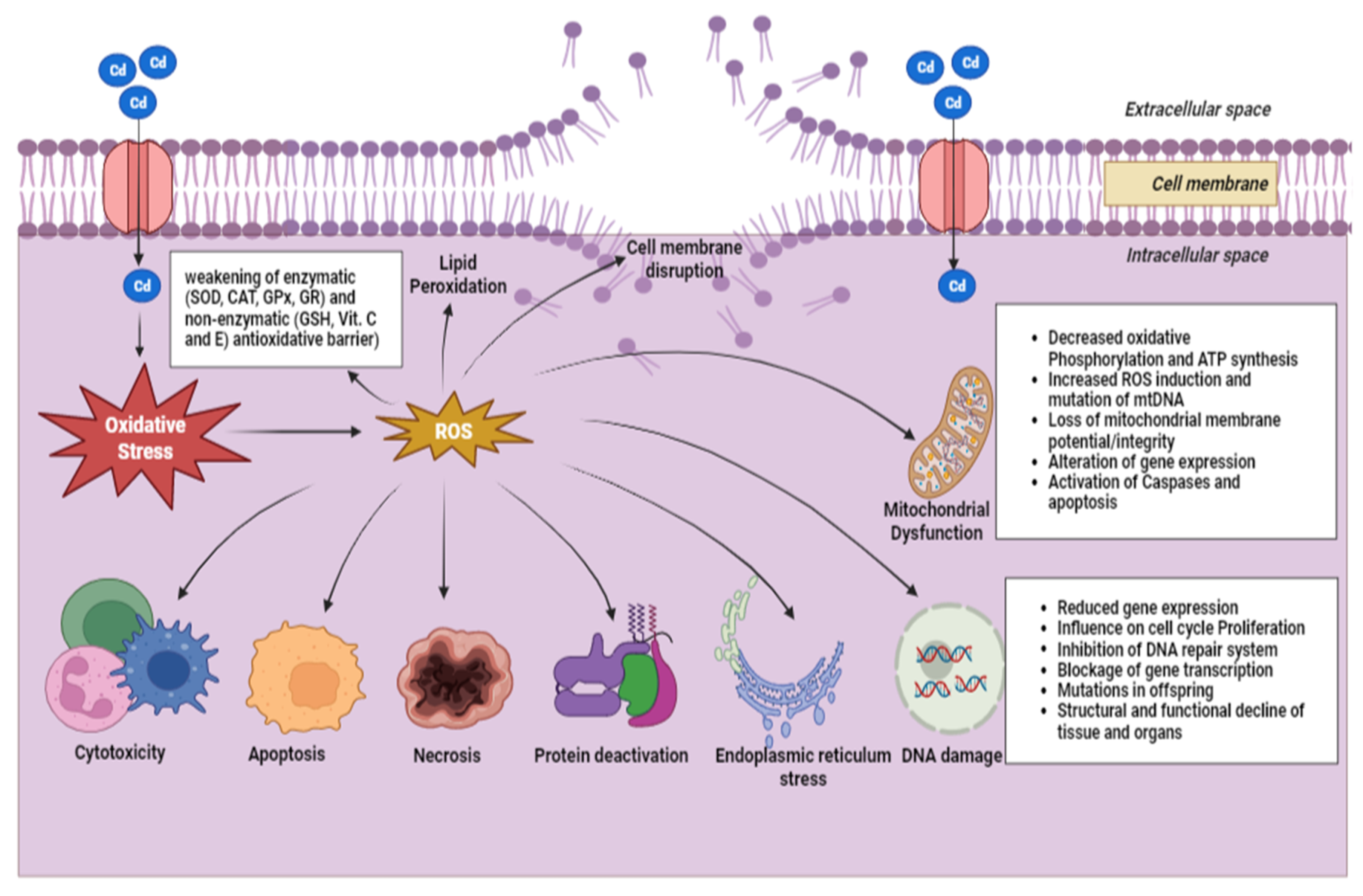
3. Bioaccumulation of Cadmium in Freshwater Fish

The tendency of trace elements to enter and alter a biological system is referred to as bioavailability [34]. Even though bioaccumulation and bioavailability are distinctly discussed in direct and indirect exposures, both processes are interdependent in natural systems [35]. Fish bioaccumulate essential nutrients regularly, including proteins, lipids, fats, amino acids, trace vitamins, and minerals [36]. Many harmful trace elements accumulate in freshwater biota through bioconcentration, which can eventually threaten freshwater fish and those who consume them [37].
As a hazardous metal, Cd may serve as a stress inducer in fish, which may impart toxicity to the aquatic ecosystem even at a very low concentration [38]. Fish absorb Cd through direct contact with a polluted medium or through the intake of food contaminated with Cd (Figure 1). Different fish species living in the same habitat may acquire varying quantities of Cd in their tissues due to differences in their living and dietary patterns. According to studies, Serranochromis thumbergi, a predatory fish, has shown the least accumulation of Cd in their tissues, which can be due to the inverse correlation of metal concentration with the trophic level. Since fish gills are constantly exposed to Cd ions in their aquatic environment, their metal intake significantly differs from that of terrestrial species [39]. Gills are considered the primary entrance site for the penetration of dissolved Cd metals into the body of freshwater fish. Generally, Cd uptake could occur through passive transport, the process by which metal ions enter a fish’s body through diffusion. Cd can also enter through carrier-mediated transport, which involves the binding of metal ions to specific proteins on the surface of chloride cells in the gills.
In contrast, the Cd ions linked to any chemical compounds are devoured by intestinal endocytosis [40]. Cadmium that is absorbed across the gills or intestinal walls is circulated, attached to the transport proteins, and transported to various tissues throughout the body [41]. A few immunochemical studies revealed that the dietary uptake of Cd by rainbow trout via the gut could accumulate in the chloride cells of the gills via calcium channels, thereby stimulating metallothionein (MT) expression [42]. The gastrointestinal tract facilitates Cd absorption in fish, and a modest proportion of Cd available in a dissolved form may be assimilated directly through contact with the skin [43]. Dietary Cd intake had a considerable influence on gastrointestinal Cd absorption and disposal in the gut (particularly pyloric caeca) and non-gut tissues (liver, gills, and kidney) of fish [44].
Fish adsorb dissolved Cd in its free ionic form, which is indirectly attained via diet when Cd is ingested into organisms of lower trophic positions [20,45]. Distinct Cd forms influence the effect of Cd on aquatic organisms, each with a toxicity range and bio-concentration factors. The free divalent ion (Cd2+) will be the dominant and most bio-available toxic form in well-aerated freshwater bodies containing low carbon contents (Figure 2) [12]. Exposure to this form stimulates the production of a small, cysteine-rich, low-molecular-weight metal-binding protein MT in the liver that binds to metals such as Cd [46]. It reduces toxicity by limiting its availability to cells and tissues [47,48]. Long-term exposure to Cd causes chronic toxicity and may result in larval mortality and pathophysiological effects in fish such as Heteropneustes fossilis [49], such as stunted and decreased developmental rates in juvenile channel catfish [30]. Temporary growth reduction, reproductive endocrine disruption, and incomplete embryo development have been observed in juvenile Colossoma macropomum and Oreochromis mossambicus [50,51].

4. Metallothionein as a Cadmium Detoxifier

The production of MT is a well-known metabolic detoxification mechanism for trace elements, and it has been well documented that exposure to Cd can induce the expression of MT [52]. A study demonstrated a positive correlation between Cd accumulation and MT induction in the tissues (liver > kidney > gills > brain) of freshwater catfish, Clarias gariepinus, exposed to waterborne Cd [53]. The toxic effects are partially a function of the amount of free Cd2+ relative to MT-bound Cd (MT-Cd), which is available to interact with cellular components such as enzymes. Because Cd exposure causes MT induction, MT levels can be employed as a biomarker for acute waterborne Cd contamination. MT induction was reported in various tissues of juvenile rainbow trout, with the liver showing the highest levels, followed by the kidney and gills. Negligible amounts were found in the brain and muscles (Figure 2) [54]. Another study showed higher concentrations of MT in the posterior kidney of Oncorhynchus kisutch, suggesting its association with excretory functions [55].
MT plays a crucial role in the liver’s dealing with elevated levels of Cd. Following absorption, Cd detoxification occurs so that Cd is bound to albumin and transported to the liver, where it binds to MT [56]. The MT-Cd complex is then transported to lysosomes, where Cd is released, and free ions are excreted into the tubular fluid before eventually being eliminated in the urine (Figure 2). This process is essential for protecting the liver from Cd’s toxic effects and preventing Cd accumulation in the body [57]. Cadmium binds to MT, which serves as a protective mechanism against the harmful effects of Cd by sequestering the metal and preventing it from interacting with other proteins like enzymes, resulting in cellular protection. Metallothionein is also important in regulating the levels of zinc and copper, which are essential for normal cellular function. A few studies indicated that Cd may compete with other metals (Zn and Cu) for binding sites on MT and non-MT proteins in the tissues of rainbow trout that were exposed to Cd and a mixture (Cd + Zn + Cu) through water and food sources. Despite Cd levels remaining lower than the MT binding capacity in both exposure groups, the metallic mixture exceeded the MT binding capacity in all tissues of Cd-exposed fish [54,58].

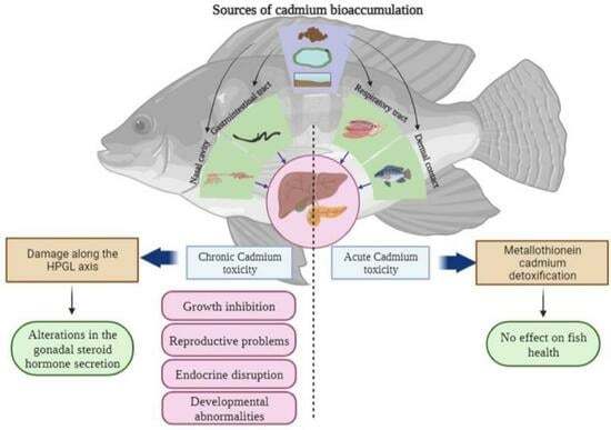
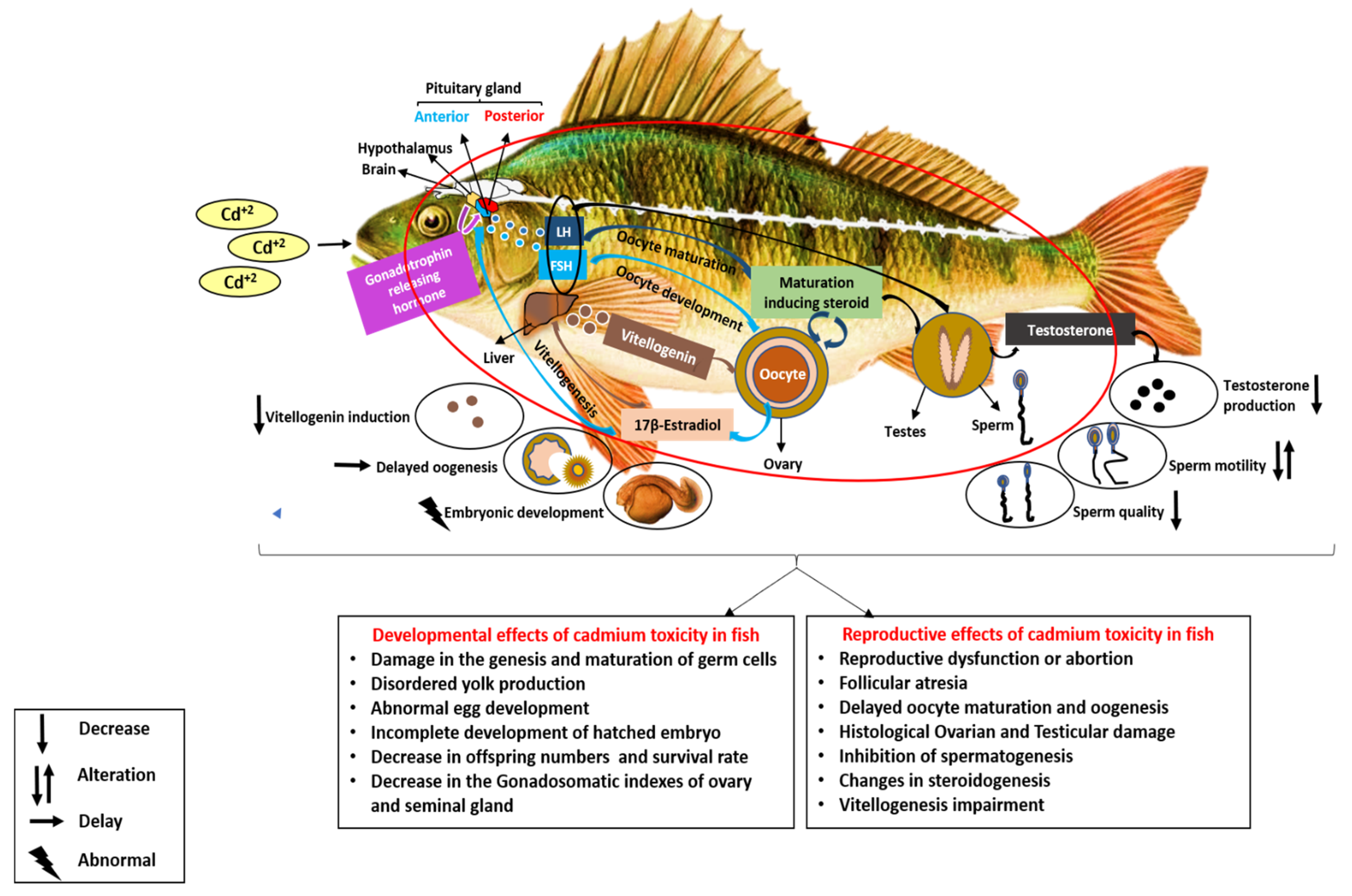
5. Cadmium as an Endocrine Disruptor in Freshwater Fish

Fish with a high concentration of Cd in their tissues may exhibit modest endocrine changes that impede reproduction [51]. In toxicity studies, several reproductive and developmental biomarkers have been employed, including:
  • Enzymatic activity: Changes in the activity of certain enzymes can indicate the presence of toxic substances in the fish body. For example, Cd burden in fish can affect the activity of enzymes involved in steroid hormone synthesis or detoxification.
  • Pituitary gonadotropins: alterations in gonadotropin levels can indicate a disruption of the HPG axis, leading to downstream effects on reproductive development and function.
  • Steroid hormones: changes in hormone levels can lead to developmental abnormalities, reproductive dysfunction, or other adverse effects.
  • Growth abnormalities: exposure to toxic substances can impair normal growth and development in fish, causing deformities or other abnormalities that can have adverse effects on survival and reproductive success (Table 1) [59].
  • Vitellogenin: This yolk precursor protein, produced by the liver in response to estrogen stimulation, shows elevated levels of vitellogenin in male fish exposed to estrogenic compounds. This can indicate feminizing effects and a disruption in reproductive function (Figure 3) (Table 2).
  • Gonadal structures: an examination of gonadal tissues can reveal abnormalities such as atrophy, hypertrophy, or intersex characteristics, indicating possible exposure to endocrine-disrupting compounds.
Cadmium can cause premature and delayed hatching, deformed larval development, and mortality in newly hatched larvae (Figure 3). Indeed, these developmental alterations during the embryonic stage decrease the number and quality of progeny [60,61]. However, these factors, evaluated over a specific period, provide insights into the reproductive damage that can be affected by environmentally relevant concentrations during reproductive phases. So, the analysis of biomarkers must be assessed to determine the potential impacts of environmental contaminants on fish populations and ecosystems.
The involvement of various endocrine pathways in reproduction and development during Cd exposure in fish has been studied [62,63]. However, the mechanisms underlying these effects are not fully understood. Previous studies indicate that Cd can impact gene transcription and interfere with the normal functioning of fish hormones, such as estrogen, testosterone, and thyroid hormones [64]. The exposure of fish to Cd triggered biological responses involving oxidative damage in intracellular molecules by generating ROS in cells (Figure 4) [65,66,67]. Cadmium has been shown to interfere with gonadal development and steroidogenesis. This interference can be caused by the release of cell membrane receptors, triggered by the activation of the protein kinase C signal pathway. In vitro experimental studies on Cd toxicity show that it impedes LH and FSH binding to receptors and alters steroid production in ovarian cells [68]. The regulation of steroidogenic acute regulatory protein production through activating the xenoestrogen receptor in the ovary of the largemouth bass, Micropterus salmoides, was also reported [69]. However, further studies are needed to comprehend how transcriptional factors interact with Cd2+ to regulate the activity of steroidogenic genes. The regulatory action of FSH on gonadal steroid hormone synthesis has been altered when the Cd interferes with calcium ions (Ca+2). Cadmium can directly influence gene transcription by substituting zinc (Zn2+) in the DNA-binding zinc-finger domain. It particularly changes the P450 side-chain cleavage (P450 scc) gene activity by turning on a cis-acting element in the promoter (regulatory) region [70]. The receptor of estrogen, which serves as a transcription factor and is ligand-dependent, typically mediates the impact of estrogens. The expression of the estrogen receptor gene in the liver of rainbow trout can be increased by estrogens [71]. It has been reported that Cd reduces the transcription factor’s binding with DNA, suppressing the estrogen receptor’s biological activity in rainbow trout [72].
The combined effect of Cd coupled with estradiol (E2) on the expression of multiple genes involved in juvenile rainbow trout fish reproduction was assessed using a relative reverse transcription PCR methodology. Cadmium decreased vitellogenin and both rtERα isoforms’ (rtERαS, rtERαL) mRNA levels in the liver in response to E2 in a dose-dependent manner. However, in fish brain tissue, the treatments with Cd alone reduced the expression of rtERαL and showed a significant enhancement in the expression of salmon GnRH genes [62]. These outcomes support the notion that Cd acts as an endocrine disruptor by inhibiting estrogen-stimulated genes in the liver and having a central impact on GnRH in the brain. Cadmium has been found to suppress gonadal development during the pre-reproductive season and impair protein synthesis in adult fish ovaries [73]. In vitro and in vivo experiments linked to Cd2+-induced developmental defects, early hatching, growth retardation, increased mortality, and testicular hormone imbalance in the embryos of various fish species, including Rhamdia quelen [74], Ide, Leuciscus idus [75], and rainbow trout [76,77]. During the exposure of the eggs and larvae of Medaka, Oryzias latipes, to 0.1–1 ppm of Cd, the eggs showed more accumulation than the larvae [78].
Cadmium in larvae is readily transported into different organs or tissues. In contrast, most of the Cd in eggs is found in the chorion and is rarely carried into the embryos due to its resistance to Cd. However, their hatchability and survival rate decreased, and noticeable changes in their behavior, such as erratic swimming and a loss of equilibrium, were observed. However, the Japanese medaka raised from eggs exposed to 0–10 mg/L of Cd for two weeks and re-exposed to Cd at the adult stage did not exhibit significant changes in their reproductive capacity, except for some alterations in male steroid and vitellogenin levels [78]. Similarly, there were no changes in gonadosomatic indexes, estrogen receptors, or vitellogenin. At the same time, steroid hormone secretion was considerably decreased in both sexes of Japanese medaka subjected to Cd (0–10 ppb) for an exposure period of 7 weeks [79]. With findings from multiple experimental examinations, it can be postulated that the impacts of Cd toxicity on reproductive and developmental outcomes (Table 1 and Table 2) are more pronounced in the hypothalamic–hypophyseal–gonadal axis.

5.1. Reproductive Effects of Cadmium in Freshwater Fish

The reproductive toxicity of trace elements frequently depends on the degree, duration, and mode of exposure, the distribution and accumulation patterns in tissues, and the rate of depuration. The toxicokinetics of Cd in fish are additionally influenced by biological parameters, such as nutritional conditions, age, species, and sex [80]. Cadmium is known as an endocrine disruptor and is regarded as a metallo-estrogen due to its various effects on the hormones and enzymes involved in reproduction. This endocrine disruptor activity can be studied through the HPG axis due to its importance and control over the reproduction of fish. The bioaccumulation of Cd in fish disrupts endocrine mechanisms in producing hormones’ active synthesis, discharge, and metabolism. Acute exposure to sublethal Cd concentrations influences alterations in endocrine status and carbohydrate metabolism in O. niloticus [81]. Research on in vitro and in vivo studies investigating the impact of Cd toxicity on freshwater fish has observed that salmonids are more sensitive to Cd [82]. Several studies suggest that even modest levels of Cd exposure disrupt the action of sex hormones in both the male and female reproductive systems of fish. Cd interferes with the biogenesis of androgens, estrogens, and progesterone in both in vivo and in vitro tests, which is associated with impaired sex differentiation and gametogenesis [83].
On the other hand, it can potentially bind both the estrogen and androgen receptors. Different frameworks of the HPG axis can be used as biomarkers to evaluate the level and operation of the disruption caused [84]. The survival efficacy of the offspring produced is also affected due to the consequences of Cd toxicity on sex hormones. A study on rainbow trout suggested that Cd acts as an endocrine disruptor, directly impacting estrogen receptor activity and further affecting several estrogens signaling pathways, causing deformities in egg development in gonads and disordered yolk formation [85]. In O. niloticus, exposure to 143.78 and 161.15 g/L Cd concentrations for 30 and 60 days reportedly deteriorated the amount of spermatocyte, thus forming immature spermatids [86]. A study evaluated 1 mg/g of Cd with several testicular irregularities in the Sertoli and Leydig cells of red tilapia, eliciting abnormal offspring [73].
The administration of Cd in the male Juvenile tilapia’s diet led to the percolation of blood, decreased sperm motility, and fibrosis in the lobular walls [73]. Chronic Cd exposure through dissolved Cd and food intake induced testicular damage in O. niloticus [86]. An experiment conducted on pejerrey fish, Odontesthes bonariensis, by exposing them to Cd for about 14 days resulted in morphological damage to the gonadal organs, i.e., a truncation of spermatic lobules, fibrotic testis–ovarian structures, and pyknotic cells [87]. The probability of Cd exposure can cause toxic effects by intensifying sperm quality due to decreased cell motility and larval mortality in Prochilodus magdalenae [63]. It can also affect sperm maturation in the common snook, Centropomus undecimalis [88]. An in vitro study conducted with sperm cells of zebrafish, Danio rerio, showed substantial alterations in the motility rate, the curvilinear motion of sperm, and DNA integrity (Figure 4) when exposed to different Cd concentrations [89]. A similar disruption resulted from pejerrey fish being exposed to 0.25 µg/L of CdCl2, adversely affecting sperm quality, fertilization rate, hatching probability, and mortality percentage [90].
The fish common carp, Cyprinus carpio, collected from Cd-polluted zones showed significantly lower testosterone production than in unpolluted riverine zones [91]. In vitro studies on rainbow trout exposed to CdCl2 toxicity showed that it induced a testicular steroidogenic imbalance [92]. In contrast, sub-chronic Cd exposure showed decreased gonadal hormonal secretion in male Medaka [79]. A Cd concentration of even 5 and 10 µg/L was proven to be harmful to zebrafish sperm cells. Lower spermatozoa movement affects these species’ fertility rate, lowering their longevity. The mentioned concentrations of Cd, 5 and 10 µg/L, led to a decrease in the plasma membrane integrity. This is essential in maintaining the membrane’s selective permeability, thereby affecting the metabolism of the spermatozoa [89]. Increased DNA damage may also lead to an increased chance of mutation among offspring (Figure 4). The accumulation and influence of Cd also affect the gonadal structure and, thereby, the reproductive function [93]. A 1 mg/g Cd dosage fed to red tilapia caused structural degradation of the spermatogenic elements and decreased sperm number and motility [73]. An earlier study showed that Puntius sarana was susceptible to a 20 mg/L Cd dose. This experiment was conducted over 30 days, and the after-effects concluded testicular injury [94]. Males of Pimephales promelas exposed to a concentration of 50 mg/L showed a decreased rate of spawning and fertility [95]. Variations in the male secondary sexual characteristics, including fewer nuptial tubercles, were also detected in the study at a concentration of 25 mg/L. Research also observed testicular changes in Gymnotus carapo, with the appearance of necrotic areas and germ cell reduction at a dosage of 5 M. A dosage of 20 M induced a further decrease in the germ cell number and was followed by the complete absence of the cells at a dosage of 40 M. This dosage of 40 M might also be capable of causing irreparable damage to the testes due to the cellular changes brought about by the Cd toxicity. Furthermore, germ cell genesis and maturation are affected by Cd vulnerability. Spermatocyte alteration at dosages of 20 and 30 M was also detected in the study [96].
Cadmium may collectively have disruptive effects on the production of gonadal steroids by modulating either the hypothalamus–pituitary axis or the gonadal cells’ capacity to stimulate androgen production [64]. A higher Cd exposure induces dysfunctional ovarian growth, spawning errors, and the detachment of the follicular membrane from the underlying ooplasm. In comparison, exposure to a low dosage of Cd minimizes the breeding egg count and hatchability percentages and increases fry abnormalities. Juveniles of red tilapia fed with different doses of Cd were found to have altered gonadal structure, ovarian maturation, reproduction, and developmental parameters (Figure 3). A Cd dosage of 1 mg/g changed the assembly of the ovaries by bringing about the deformity in the ova. The follicle walls were dissociated from the ova along with a greater vacuole count, eventually risking the offspring growth rate of red Tilapia [71]. A study analyzed the destructive effects of minimal concentrations of Cd exposure, specifically 0.1, 1, 10, and 100 μg/L, on the HPG axis in R. quelen females. It found no considerable changes in the plasma levels of testosterone, estradiol, hepatic, and gonadosomatic indexes after exposure, as reported in reference [84]. The accumulation of Cd in the ovaries induces the stimulation of estrogen receptors. However, the expression of vitellogenin was markedly reduced in O. niloticus when exposed to Cd at concentrations of 70.32 and 143.78 µg/L for 30 days and at 143.78 µg/L for 60 days, respectively [86]. Varying effects on sex-related gene expression in gonads were observed, along with ovarian injuries and down-regulated vitellogenin expression in the ovaries. On further examination of the maternal transfer of the Cd metal, the results revealed the passing of the accumulated metal onto the eggs but not the fry.
Cadmium toxicity could also alter different parameters related to the reproductive axis. Tilapia zillii exhibited deterioration in the oocyte’s granulosa, restricting oocyte maturation and enhancing ovarian atresia due to Cd toxicity [97]. In goldfish, a Cd dose of 10 mg/g ceased ovarian development at the vacuolization stage, resulting in the termination of the ovulation process in the females [29]. Similar results were observed in an experiment executed for a female Prussian carp, Carassius gibelio, with a 4.0 mg/L dose of Cd [98]. Another study reported an alteration in the ovaries when subjected to concentrations of 50, 100, and 200 μg/L for 60 days (the dosages were higher than the environmentally relevant concentration of 5 μg/L of Cd) [84]. Several experimental data indicated that Cd’s negative impact on gonadal development might be due to fluctuations in steroid hormonal production and expressions of sex-related genes (Table 1 and Table 2).
Table 1. Reported reproductive effects of cadmium in freshwater fish.
Fish SpeciesFeatures of FishCadmium ConcentrationTime of ExposureEffectsReferences
StageLengthWeight
Carassius auratusSperm and eggNANA10 mg/gNSDiscontinuation of ovulation[29]
Danio rerioReproductive phaseNSNS 5 10   µ g / L 10 minSperm motility affected and disintegration of the plasma membrane[89]
Gymnotus carapoSexually maturedNSNS5–40 M24–96Reduced germ cells[96]
Oreochromis niloticus2-month-old12.38 ± 0.92 cm58.80 ± 13.84 g70.32–143.78 µg/L30 and 60 daysAltered effects on sex-related gene expression and deformed follicle[86]
Pimephales promelasSexually maturedNSNS50 mg/L21 daysReduced fertility and spawning[95]
Oreochromis sp. (Red tilapia)Sexually matured8.2 ± 2.5 cm38.3 ± 2.5 g1 mg/gNSDecreased sperm motility and deformed ova[73]
Rhamdia quelenAdult20.0 ± 4.4 cm70.5 ± 38.0 g>100 µg/LNSIncreased hepatic vitellogenin expression[84]
Carassius auratusfingerlingsNS6 ± 1.2 g0.5 g14 daysPronounced release of AST, ALT, Cortisol, and glucose concentrations[17]
Danio rerio7 weeks oldNSNS20 μg/L48 daysReduced gonadosomatic index and 17 β-estradiol and vitellogenin concentrations in plasma[18]
Siganus rivulatus8–14 months19.8 ± 1.9 cmNSNSNSAlterations in the levels of testosterone, β-estradiol, and progesterone hormones[83]
Colossoma macropomumNSNSNS0.6, 1.2 and 1.8 mg/LNSIncreased SOD enzyme activity and LPO levels in sperm cells. Impairment of fertilization and hatching rate of the oocytes.[50]
Odontesthes bonariensisAdult14.82 ± 0.56 cm41.17 ± 6.51 g0.25 μg/L14 daysDecreased follicle-stimulating hormone transcript levels and showed structural damages in spermatic lobules, fibrosis, testis, and ova[87]
Tilapia zilliAdult11.5–22.8 cm32–235.4 g0.57 ± 0.11 µg/g wet wt.AnnualIncreased atresia, degenerative and necrotic changes in the oocytes, and conversion of ovarian to testicular cells.[97]
Carassius gibelio B.Adult23.25 ± 0.49 cm204.65 ± 12.58 g0.4 or 4.0 mg/L3–5 monthsDecreased GSI, impaired LH secretion during exposure, and stimulation of spawning[98]
Danio rerioEmbryoNSNSNS72 hInhibition of Estradiol induction of Aromatase-B in radial glial cells[99]
Pimephales promelas12 months oldNSNS5 μg/L21 daysImpaired gametogenesis, reduced steroid levels and vitellogenesis, and delayed oogenesis.[100]
Note: NA—not applicable; NS—not specified.
Table 2. Reported developmental effects of cadmium in freshwater fish.
Fish SpeciesFeatures of FishCadmium
Concentration
Time of ExposureEffectsReferences
StageLengthWeight
Danio rerio6 days post-fertilization larvaeNSNS10 µg/L3 daysDeformed larvae[101]
Danio rerioEmbryoNANAEnvironmentally relevant concentration24 to 72 h
post fertilization
Alterations in optomotor responses in the treated larvae of all ages[8]
Gymnotus carapoSexually matured36.8 ± 6.0 cm205.8 ± 59.9 g5–40 M24–96 hDamaged germ cell genesis[96]
Odontesthes bonariensisSexually maturedNSNS0.25–2.5 µg/LNSReduced hatching, embryo, and larvae survival[90]
Oreochromis niloticus2-month-old12.38 ± 0.92 cm58.80 ± 13.84 g143.78–161.15 g/L30 and 60 daysPremature spermatid[86]
Prochilodus magdalenaeTwo-year-oldNS282.3 ± 40.8 g0.0025–2.5 ppmNSLow fertility and decreased egg production[63]
Oplegnathus fasciatusFingerlingsNS5.5 ± 0.06 g162 mg/kgNSReduced growth rate[14]
Carassius auratusFingerlingsNS6 ± 1.2 g0.5 g14 daysDilation in sinusoids and unusual Kupffer cells occurs[17]
Ictalurus punctatusFertilization to 6-month-old fingerlingsNSNSNS3 monthsIncreased metallothionein gene expression in heat-shock proteins in the liver[30]
Trematomus hansoniAdult21.2–24.6 cm130–159 g0.89 µM5 daysIncreased metallothionein gene transcription[52]
Ide Leuciscus idus21 days post-hatchingNSNS100 μg/LNSReduced embryonic survival[75]
Oncorhynchus mykissEmbryos and larvaeNANS0.5–8.0 μg/L96 hInduced geno–cytotoxicity disturbs cardio-respiratory system activity and negatively impacts fish development at early life stages.[77]
Danio rerioEmbryoNANA30 µM1–3 days post fertilizationIncreased mortality rate and delayed hatching[102]
Gasterosteus aculeatusAdult40.40 ± 0.49 mm955.80 ± 55.13 mg1 ppb15, 60, and 120 daysReduced germ cell quality, decreased hatching rate, and increased mortality rate.[103]
Oryzias javanicusNewly spawned eggsNSNS0.01–0.10 ppmNSFailure in embryo development[104]
Cyprinus carpio L.AdultNS768.50 g0.5 mg/LNSReduced body weight gain[64]
Danio rerioAdult
(120 days post fertilization)
NSNS1 µmol/LNSMalformations in the produced offspring[105]
Channa marulius and Hepteroneustes fossillisJuveniles and smaller adultsNSNS0–10 mg/L96 hRespiratory strategy and adaptation to low-ionic-strength environments[82]
Note: NA—not applicable; NS—not specified.

5.2. Developmental Effects of Cadmium on Freshwater Fish

Freshwater fish exposed to Cd have exhibited significant impairment in reproductive capacity as well as retardation in overall growth and development. The fundamental consequence is due to the structural and functional failure of the fish gonads, in addition to the annihilation of the reproductive development system [51,61,86]. According to reports, Cd negatively affects fish in two ways: either it directly inhibits the gene expression and secretion of the endocrine hormone concerning the HPG axis, or it may affect sex hormone synthesis, thereby reducing the survival efficacy of the offspring produced [99,100]. The exposure of Silurus soldatovi to a dosage of 10 μg/L of Cd reportedly reduced the larval survival rate, thereby supporting the concept of Cd as an environmental pollutant that causes embryotoxicity in fish [106]. A study suggested a disordered yolk production abnormality in egg development in rainbow trout gonads because Cd exposure results in the suppression of estrogen receptor gene expression [85]. Cadmium exposure into the reproductive tissue interferes with gonadal differentiation [107].
A study on their growth rate, survival rate, locomotor activity, and sensory activities indicated that zebrafish embryos exposed to Cd exhibited delayed hatching and a higher mortality rate [102]. Another toxicity assessment analysis of zebrafish showed abnormalities in embryonic development, producing offspring with varying extents of deformity (Table 1) [18,105,108]. Embryonic abnormalities account for impairments in the olfactory placode and defects in the head where the subdivisions of the brain are not well defined, therefore ultimately targeting neurogenesis [109,110]. Further, in the case of Gasterosteus aculeatus, exposure to 1 ppb of Cd for about 15, 60, and 120 days reduced both the egg and the sperm quality [103]. A review of G. carapo revealed varying outcomes concerning varying administered Cd dosages. A treatment with a Cd concentration of 20 M for 24 h led to a reduction in male germ cells. A concentration of 40 M of Cd treatment leads to a complete lack of germ cells. After 96 h of exposure at all Cd concentrations, the germ cells had a critical impairment [96]. Advanced-stage embryos of O. bonariensis were studied, where an analysis with 0.25 and 2.5 µg/L of Cd showed a decrease in the hatching rate by 20%, and the mortality rate in both the embryonic and larval stages was increased (Table 1 and Table 2) [90]. Under 4–10 mg/L Cd conditions, the observed germ cells were disrupted at a post-meiotic stage in Labeo bata [111].
Cadmium has been proven to be a potent agent responsible for reproductive and developmental abnormalities in fish. However, the effects may vary from species to species regarding their susceptibility and acquired Cd dosage. There are impacts on both females and their offspring. For females, a reduced fecundity rate has been reported, whereas offspring have shown a decreased rate of gamete development [112]. A study on P. magdalenae eggs resulted in a lower fertility rate and deceased eggs with 0.0025 and 2.5 ppm of CdCl2, respectively [63]. The authors studied multiple parameters of Oryzias javanicus, such as premature hatching, life span of the juvenile, and embryonic development [104], where the tropical fish were exposed to Cd. At a 0.01–0.10 ppm exposure, embryonic development was restrained with a 10–37.5% mortality rate. However, at a 0.01–0.05 ppm exposure, premature hatching decreased by up to 20%. Another study on red sea bream, Pagrus major, again observed a lower hatchability rate at more than a 0.8 mg/L concentration. The mortality rate was greater, and unusual morphological changes were observed at more than a 0.4 mg/L concentration [113]. Several in vitro and in vivo studies have shown Cd-induced alterations in the steroidogenesis, oocyte maturation, growth retardation, increased mortality rate, premature hatching, and abnormal embryonic development in fish like common carp. An incubation with Cd minimized the swellings of common carp eggs at a 5–50 mg/dm3 concentration [114], and in some cases, craniofacial anomalies and yolk sac deformities were observed [60]. Ide embryos were considered and subjected to Cd, which gave numerous results. The hatching process was retarded compared to the control groups, wherein the larvae body size significantly dropped and induced a mortality rate of up to 50% [75]. In zebrafish, defective embryos were reported at increasing concentrations, i.e., 3.3, 6.7, and 13.3 µM. The same experiment also calculated the hatching period, which showed no or delayed hatching [115]. Similarly, 6 days post fertilization, zebrafish exposed to 25 and 125 µg/L of Cd showed detrimental effects, with reduced opercular bone growth and skeletal malformation during larval development [101].

6. Research Gaps and Future Research Directions

The complexity of aquatic ecosystems can make our understanding of the biogeochemistry of pollutants such as Cd more challenging [116]. Furthermore, due to the fluctuation in the levels and types of pollutants over time, the consequences of Cd accumulation and its biomagnification on each trophic level have become more diverse and complex [117]. Although numerous studies have focused on understanding the bioaccumulation patterns of Cd and its toxicity as an oxidative stressor in various tissues of freshwater fish species [118,119,120,121,122], there is limited information available on its impacts on endocrine disruption. As Cd is highly bioavailable to aquatic animals in freshwater, including fish, it readily accumulates in various tissues, including those related to reproduction and endocrine functions [123]. To substantiate the hypothesis that Cd in aquatic habitats harms fish’s reproductive and developmental health, the current review has compiled data from recent research investigations. Most of the literature has focused on various networks of endocrine disruption caused by Cd at the gonad level, as well as the assessment of chromosomal structural abnormalities and modifications in the gonadsomatic index (GSI), gametogenesis, steroidogenesis, and vitellogenesis (Table 2) [68,69,86].
A few investigations have strongly evidenced the role of the HPG axis in the reproductive impairments in fish brought on by Cd exposure. The release of pituitary LH or hypothalamic GnRH may be affected by Cd through altering brain neurotransmission [64]. Trace elements found in the aquatic biota can synergistically or antagonistically impact fish reproduction and development [124]. The studies involving the wild fish population attempt to distinguish the interactive roles of multiple trace elements and the effect of different concentrations of an individual trace element on the endocrine system [10,59,105]. Therefore, future environmental toxicology studies should employ holistic in vitro and in vivo analyses of fish health related to multiple trace elements. Studies evidencing Cd exposure at low-level and environmentally relevant concentrations are limited. Thus, the toxic effects of ecologically relevant doses of Cd on fish reproductive-endocrine systems and their mechanism of action on multiple sites along the reproductive axis should be the primary concern of researchers. Emerging studies will thoroughly explore the control of neuroendocrine function during metal-induced disruption of reproductive activities in fish. Therefore, broad research is needed to understand the mechanisms behind Cd’s actions as an endocrine disruptor.
However, the correlation of reproductive abnormalities in wild fish with an ecologically pertinent amount of Cd is challenging to show. Some trace elements, like Cd, are found to be available in natural water systems, which in turn cause a noxious impact on wild fish populations [125]. The concentration of Cd absorption and its accumulation into tissues/organs depend on the fish’s physiological, ecological, chemical, and physical conditions [126]. So, thorough investigations are needed, particularly on commercially important and threatened freshwater fish under various contaminated circumstances. Merely comprehending the interdependence between water quality parameters and Cd concentration in fish, along with the presence of other metals and their synergistic effects with Cd, is inadequate to bridge the knowledge gaps between Cd accumulation in fish and its potential ecological consequences. Likewise, an in-depth comprehension of the species-level seasonal shifts of Cd absorption by fish living in their native habitats aids in safeguarding them from impending threats. The present review contains strong evidence that demonstrates the serious implications caused by the chronic exposure of Cd on the development and reproduction of fish.

7. Conclusions

The present review examined reports concerned with the endocrine-disrupting activity of Cd, which impacts various signaling pathways crucial to the development and reproduction of freshwater fish. This comprehensive analysis of Cd exposure’s endocrine effects has revealed a greater research gap to fill. Since Cd has evolved to be a vital part of our ecosystem and livelihood, stopping its production or utilization in the decades ahead is impossible. Due to increased industrialization, the widespread dispersal of Cd and its destructive effects pose serious issues for higher tropic levels, as there will be more possibilities for accidental or incidental exposure of Cd to freshwater living beings. So, there is a need for more research investigations that characterize the mechanisms of reproductive endocrine disruption, its mode of action, and alterations caused at different levels of Cd exposure in fish to interpret toxicological solutions referring to fish health and diseases.

Author Contributions

This review article was produced through collaboration between the authors. Conceptualization, K.A.P., J.-W.L., B.B. and W.-C.L.; writing—original draft preparation, K.S.V.R., W.-C.L. and B.B.; selecting bibliographic sources, A.M., M.P., K.S.J., G.S. and B.B.; M.P. and B.B. coordinated the working group; writing—review and editing, K.A.P., M.P., A.M., K.S.J., G.S., B.B., W.-C.L. and J.-W.L. All authors have read and agreed to the published version of the manuscript.

Funding

This work was carried out with the support of the National Research Foundation of Korea (NRF-2021R1I1A1A01058128). The authors are grateful and extend their appreciation to the authorities for their support.

Data Availability Statement

The data presented in this study are available on request from the corresponding authors.

Conflicts of Interest

The authors hereby declare that they have no conflict of interest and have no known competing financial interests or personal relationships that could have appeared to influence the work reported in this paper.

References

  1. Masindi, V.; Muedi, K.L. Environmental contamination by heavy metals. Heavy Met. 2018, 10, 115–132. [Google Scholar] [CrossRef]
  2. Gautam, R.K.; Sharma, S.K.; Mahiya, S.; Chattopadhyaya, M.C. Contamination of Heavy Metals in Aquatic Media: Transport, Toxicity and Technologies for Remediation. In Heavy Metals in Water: Presence, Removal and Safety; Royal Society of Chemistry: Cambridge, UK, 2014; pp. 1–24. [Google Scholar] [CrossRef]
  3. Duruibe, J.O.; Ogwuegbu, M.O.C.; Egwurugwu, J.N. Heavy metal pollution and human biotoxic effects. Int. J. Phys. Sci. 2007, 2, 112–118. [Google Scholar]
  4. LeFauve, M.K.; Connaughton, V.P. Developmental exposure to heavy metals alters visually-guided behaviors in zebrafish. Curr. Zool. 2017, 63, 221–227. [Google Scholar] [CrossRef] [PubMed]
  5. Hejna, M.; Gottardo, D.; Baldi, A.; Dell’Orto, V.; Cheli, F.; Zaninelli, M.; Rossi, L. Review: Nutritional ecology of heavy metals. Animal 2018, 12, 2156–2170. [Google Scholar] [CrossRef] [PubMed]
  6. Waalkes, M.P.; Misra, R.R.; Chang, L.W. Toxicology of Metals; CRC: Boca Raton, FL, USA, 1996; pp. 1003–1026. [Google Scholar]
  7. Tchounwou, P.B.; Yedjou, C.G.; Patlolla, A.K.; Sutton, D.J. Heavy metal toxicity and the environment. Exp. Suppl. 2012, 101, 133–164. [Google Scholar] [PubMed]
  8. Garai, P.; Banerjee, P.; Mondal, P.; Saha, N.C. Effect of heavy metals on fishes: Toxicity and bioaccumulation. J. Clin. Toxicol. 2021, 11, 1–10. Available online: https://www.researchgate.net/profile/Pradip-Mondal/publication/353848075 (accessed on 23 January 2023).
  9. Zhang, Y.; Lu, X.; Wang, N.; Xin, M.; Geng, S.; Jia, J.; Meng, Q. Heavy metals in aquatic organisms of different trophic levels and their potential human health risk in Bohai Bay, China. Environ. Sci. Pollut. Res. 2016, 23, 17801–17810. [Google Scholar] [CrossRef]
  10. Georgescu, B.; Georgescu, C.; Dărăban, S.; Bouaru, A.; Paşcalău, S. Heavy metals acting as endocrine disrupters. Sci. Pap. Anim. Sci. Biotechnol. 2011, 44, 89–93. [Google Scholar]
  11. Zhao, S.; Feng, C.; Quan, W.; Chen, X.; Niu, J.; Shen, Z. Role of living environments in the accumulation characteristics of heavy metals in fishes and crabs in the Yangtze River Estuary, China. Mar. Pollut. Bull. 2012, 64, 1163–1171. [Google Scholar] [CrossRef]
  12. Perera, P.A.C.T.; Kodithuwakku, S.P.; Sundarabarathy, T.V.; Edirisinghe, U. Bioaccumulation of Cadmium in freshwater fish. An environmental perspective. Insight Ecol. 2015, 4, 1–12. [Google Scholar] [CrossRef]
  13. Akter, R.; Thilsted, S.H.; Hossain, N.; Ishihara, H.; Yagi, N. Fish is the preferred animal-source food in the rural community of Southern Bangladesh. Sustainability 2019, 11, 5764. [Google Scholar] [CrossRef]
  14. Okorie, O.E.; Bae, J.Y.; Lee, J.-H.; Lee, S.; Park, G.-H.; Mohseni, M.; Bai, S.C. Effects of Different Dietary Cadmium Levels on Growth and Tissue Cadmium Content in Juvenile Parrotfish, Oplegnathus fasciatus. Asian-Australas. J. Anim. Sci. 2014, 27, 62–68. [Google Scholar] [CrossRef] [PubMed]
  15. Baatrup, E. Structural and functional effects of heavy metals on the nervous system, including sense organs, of fish. Comp. Biochem. Physiol. Part C Comp. Pharmacol. 1991, 100, 253–257. [Google Scholar] [CrossRef] [PubMed]
  16. Mielcarek, K.; Nowakowski, P.; Puścion-Jakubik, A.; Gromkowska-Kępka, K.J.; Soroczyńska, J.; Markiewicz-Żukowska, R.; Naliwajko, S.K.; Grabia, M.; Bielecka, J.; Żmudzińska, A.; et al. Arsenic, cadmium, lead and mercury content and health risk assessment of consuming freshwater fish with elements of chemometric analysis. Food Chem. 2022, 379, 132167. [Google Scholar] [CrossRef]
  17. Noor, Z.; Khan, S.A.; Noor, M. Assessment of cadmium toxicity and its possible effects on goldfish (Carassius auratus), employing microscopy and biochemical techniques. Microsc. Res. Tech. 2020, 83, 1441–1449. [Google Scholar] [CrossRef]
  18. Zheng, J.; Peng, L.; Xia, L.; Li, J.; Zhu, Q. Effects of continuous and intermittent cadmium exposure on HPGL axis, GH/IGF axis and circadian rhythm signaling and their consequences on reproduction in female zebrafish: Biomarkers independent of exposure regimes. Chemosphere 2021, 282, 130879. [Google Scholar] [CrossRef]
  19. Faroon, O.; Ashizawa, A.; Wright, S.; Tucker, P.; Jenkins, K.; Ingerman, L.; Rudisill, C. Toxicological Profile for Cadmium; Agency for Toxic Substances and Disease Registry (US): Atlanta, GA, USA, 2012; pp. 11–15.
  20. Morrow, H. Cadmium and cadmium alloys. In Kirk-Othmer Encyclopedia of Chemical Technology, 5th ed.; Wiley: New York, NY, USA, 2000; pp. 1–36. [Google Scholar]
  21. Mondal, K.; Ghosh, S.; Haque, S. A review on contamination, bioaccumulation and toxic effect of cadmium, mercury and lead on freshwater fishes. Int. J. Zool. Stud. 2018, 3, 153–159. [Google Scholar]
  22. Muntau, H.; Baudo, R. Sources of cadmium, its distribution and turnover in the freshwater environment. IARC Sci. Publ. 1992, 118, 133–148. [Google Scholar]
  23. Baharom, Z.S.; Ishak, M.Y. Determination of heavy metal accumulation in fish species in Galas River, Kelantan and Beranang mining pool, Selangor. Procedia Environ. Sci. 2015, 30, 320–325. [Google Scholar] [CrossRef]
  24. Kamari, A.; Yusof, N.; Abdullah, H.; Haraguchi, A.; Abas, M.F. Assessment of heavy metals in water, sediment, Anabas testudineus and Eichhornia crassipes in a former mining pond in Perak, Malaysia. Chem. Ecol. 2017, 33, 637–651. [Google Scholar] [CrossRef]
  25. Namieśnik, J.; Rabajczyk, A. The speciation and physicochemical forms of metals in surface waters and sediments. Chem. Speciat. Bioavailab. 2010, 22, 1–24. [Google Scholar] [CrossRef]
  26. Kubier, A.; Wilkin, R.T.; Pichler, T. Cadmium in soils and groundwater: A review. Appl. Geochem. 2019, 108, 104388. [Google Scholar] [CrossRef] [PubMed]
  27. Rahimzadeh, M.R.; Rahimzadeh, M.R.; Kazemi, S.; Moghadamnia, A.A. Cadmium toxicity and treatment: An update. Casp. J. Intern. Med. 2017, 8, 135–145. [Google Scholar]
  28. Jaiswal, A.; Verma, A.; Jaiswal, P. Detrimental effects of heavy metals in soil, plants, and aquatic ecosystems and in humans. J. Environ. Pathol. Toxicol. Oncol. 2018, 37, 183–197. [Google Scholar] [CrossRef] [PubMed]
  29. Szczerbik, P.; Mikołajczyk, T.; Sokołowska-Mikołajczyk, M.; Socha, M.; Chyb, J.; Epler, P. Influence of long-term exposure to dietary cadmium on growth, maturation and reproduction of goldfish (subspecies: Prussian carp Carassius auratus gibelio B.). Aquat. Toxicol. 2006, 77, 126–135. [Google Scholar] [CrossRef] [PubMed]
  30. Paul, J.S.; Small, B.C. Chronic exposure to environmental cadmium affects growth and survival, cellular stress, and glucose metabolism in juvenile channel catfish (Ictalurus punctatus). Aquat. Toxicol. 2021, 230, 105705. [Google Scholar] [CrossRef]
  31. Verbost, P.M.; Van Rooij, J.; Flik, G.; Lock, R.A.C.; Bonga, S.W. The movement of cadmium through freshwater trout branchial epithelium and its interference with calcium transport. J. Exp. Biol. 1989, 145, 185–197. Available online: https://repository.ubn.ru.nl/bitstream/handle/2066/16645/10833, (accessed on 15 December 2022). [CrossRef]
  32. Irwin, R.; Van Mouwerik, M.; Stevens, L.; Seese, M.D.; Basham, W. Environmental Contaminants Encyclopedia, Copper Entry; Water Resources Division: Fort Collins, CO, USA, 1997.
  33. Hossain, M.B.; Tanjin, F.; Rahman, M.S.; Yu, J.; Akhter, S.; Noman, M.A.; Sun, J. Metals Bioaccumulation in 15 commonly consumed fishes from the lower Meghna River and adjacent areas of Bangladesh and associated human health hazards. Toxics 2022, 10, 139. [Google Scholar] [CrossRef]
  34. Price, G.; Patel, D.A. Drug bioavailability. In StatPearls [Internet]; Statpearls Publishing: Treasure Island, FL, USA, 2021. [Google Scholar]
  35. Drexler, J.; Fisher, N.; Henningsen, G.; Lanno, R.; McGeer, J.; Sappington, K.; Beringer, M. Issue Paper on the Bioavailability and Bioaccumulation of Metals; US Environmental Protection Agency Risk Assessment Forum: Washington, DC, USA, 2003.
  36. Lall, S.P.; Kaushik, S.J. Nutrition and metabolism of minerals in fish. Animals 2021, 11, 2711. [Google Scholar] [CrossRef]
  37. Ali, H.; Khan, E.; Ilahi, I. Environmental chemistry and ecotoxicology of hazardous heavy metals: Environmental persistence, toxicity, and bioaccumulation. J. Chem. 2019, 2019, 6730305. [Google Scholar] [CrossRef]
  38. Wood, C.M.; Farrell, A.P.; Brauner, C.J. (Eds.) Homeostasis and Toxicology of Essential Metals; Academic Press: Cambridge, MA, USA, 2012; Volume 1. [Google Scholar]
  39. Nakayama, S.M.; Ikenaka, Y.; Muzandu, K.; Choongo, K.; Oroszlany, B.; Teraoka, H.; Mizuno, N.; Ishizuka, M. Heavymetal accumulation in lake sediments, fish (Oreochromis niloticus and Serranochromis thumbergi) and crayfish (Cherax quadricarinatus) in LakeItezhi-tezhi and Lake Kariba, Zambia. Arch. Environ. Contam. Toxicol 2010, 59, 291–300. [Google Scholar] [CrossRef] [PubMed]
  40. Wood, C.M. Toxic responses of the gill. In Target Organ Toxicity in Marine and Freshwater Teleosts; CRC Press: Boca Raton, FL, USA, 2001; pp. 1–89. [Google Scholar]
  41. Smet, H.D.; Blust, R. Stress responses and changes in protein metabolism in carp Cyprinus carpio during cadmium exposure. Ecotoxicol. Environm. Saf. 2001, 48, 255–262. [Google Scholar] [CrossRef] [PubMed]
  42. Olsson, P.; Kling, P.; Hogstrand, C. Mechanism of Heavy Metal Accumulation and Toxicity in Fish. In Metal Metabolism in Aquatic Environments; Langston, W.J., Bebianno, M.J., Eds.; Chapman and Hall: London, UK, 1998; pp. 321–341. [Google Scholar] [CrossRef]
  43. Li, H.; Mai, K.; Ai, Q.; Zhang, C.; Zhang, L. Effects of dietary squid viscera meal on growth and cadmium accumulation in tissues of large yellow croaker, Pseudosciaena crocea R. Front. Agric. China 2009, 3, 78–83. [Google Scholar] [CrossRef]
  44. Branca, J.J.V.; Pacini, A.; Gulisano, M.; Taddei, N.; Fiorillo, C.; Becatti, M. Cadmium-induced cytotoxicity: Effects on mitochondrial electron transport chain. Front. Cell Dev. Biol. 2020, 8, 604377. [Google Scholar] [CrossRef] [PubMed]
  45. Adedeji, O.B.; Okocha, R.C. Constraint to aquaculture development in Nigeria and way forward. J. Appl. Sci. Res. 2012, 7, 1133–1140. [Google Scholar]
  46. Nordberg, M.; Nordberg, G.F. Metallothionein and Cadmium Toxicology-Historical Review and Commentary. Biomolecules 2022, 12, 360. [Google Scholar] [CrossRef] [PubMed]
  47. Bradl, H. Heavy Metals in the Environment: Origin, Interaction and Remediation, 1st ed.; Academic Press: New York, NY, USA, 2005. [Google Scholar]
  48. Wright, D.A.; Welbourn, P. Environmental Toxicology; Cambridge University Press: Cambridge, UK, 2002; Volume 11. [Google Scholar]
  49. Puvaneswari, S.; Karuppasamy, R. Accumulation of cadmium and its effects on the survival and growth of larvae of Heteropneustes fossilis (Bloch, 1794). J. Fish. Aquat. Sci. 2007, 2, 27–37. [Google Scholar] [CrossRef]
  50. Pinto, G.L.; da Silva Castro, J.; Val, A.L. Copper and cadmium impair sperm performance, fertilization and hatching of oocytes from Amazonian fish Colossoma macropomum. Chemosphere 2021, 266, 128957. [Google Scholar] [CrossRef]
  51. Amutha, C.; Subramanian, P. Cadmium alters the reproductive endocrine disruption and enhancement of growth in the early and adult stages of Oreochromis mossambicus. Fish Physiol. Biochem. 2013, 39, 351–361. [Google Scholar] [CrossRef]
  52. Bakiu, R.; Pacchini, S.; Piva, E.; Schumann, S.; Tolomeo, A.M.; Ferro, D.; Irato, P.; Santovito, G. Metallothionein expression as a physiological response against metal toxicity in the striped rockcod Trematomus hansoni. Int. J. Mol. Sci. 2022, 23, 12799. [Google Scholar] [CrossRef]
  53. Rose, S.U.M.I.T.; Vincent, S.; Meena, B.; Suresh, A.; Mani, R. Metallothionein induction in fresh water catfish Clarias gariepinus on exposure to cadmium. Int. J. Pharm. Pharm. Sci. 2014, 6, 377–383. Available online: https://www.researchgate.net/publication/284819201 (accessed on 23 January 2023).
  54. Chowdhury, M.J.; Baldisserotto, B.; Wood, C.M. Tissue-specific cadmium and metallothionein levels in rainbow trout chronically acclimated to waterborne or dietary cadmium. Arch. Environ. Contam. Toxicol. 2005, 48, 381–390. [Google Scholar] [CrossRef] [PubMed]
  55. Reichert, W.L.; Federighi, D.A.; Malins, D.C. Uptake and metabolism of lead and cadmium in coho salmon (Oncorhynchus kisutch). Comp. Biochem. Physiol. Part C Comp. Pharmacol. 1979, 63, 229–234. [Google Scholar] [CrossRef] [PubMed]
  56. Nordberg, M.; Nordberg, G.F. Toxicological aspects of metallothionein. Cell. Mol. Biol. 2000, 46, 451–463. [Google Scholar] [PubMed]
  57. Annabi, A.; Messaoudi, I.; Kerkeni, A.; Said, K. Cadmium accumulation and histological lesion in mosquito fish (Gambusia affinis) tissues following acute and chronic exposure. Int. J. Environ. Res. 2011, 5, 745–756. [Google Scholar] [CrossRef]
  58. Hollis, L.; Hogstrand, C.; Wood, C.M. Tissue-specific cadmium accumulation, metallothionein induction, and tissue zinc and copper levels during chronic sublethal cadmium exposure in juvenile rainbow trout. Arch. Environ. Contam. Toxicol. 2001, 41, 468–474. [Google Scholar] [CrossRef] [PubMed]
  59. Kar, S.; Sangem, P.; Anusha, N.; Senthilkumaran, B. Endocrine disruptors in teleosts: Evaluating environmental risks and biomarkers. Aquac. Fish. 2021, 6, 1–26. [Google Scholar] [CrossRef]
  60. Jezierska, B.; Ługowska, K.; Witeska, M. The effects of heavy metals on embryonic development of fish (a review). Fish Physiol. Biochem. 2009, 35, 625–640. [Google Scholar] [CrossRef]
  61. Sfakianakis, D.G.; Renieri, E.; Kentouri, M.; Tsatsakis, A.M. Effect of heavy metals on fish larvae deformities: A review. Environ. Res. 2015, 137, 246–255. [Google Scholar] [CrossRef]
  62. Vetillard, A.; Bailhache, T. Cadmium: An endocrine disrupter that affects gene expression in the liver and brain of juvenile rainbow trout. Biol. Reprod. 2005, 72, 119–126. [Google Scholar] [CrossRef]
  63. Sierra-Marquez, L.; Espinosa-Araujo, J.; Atencio-Garcia, V.; Olivero-Verbel, J. Effects of cadmium exposure on sperm and larvae of the neotropical fish Prochilodus magdalenae. Comp. Biochem. Physiol. Toxicol. Pharmacol. CBP 2019, 225, 108577. [Google Scholar] [CrossRef] [PubMed]
  64. Chakraborty, S.B. Non-Essential Heavy Metals as Endocrine Disruptors: Evaluating Impact on Reproduction in Teleosts. In Proceedings of the Zoological Society; Springer: New Delhi, India, 2021; pp. 1–15. [Google Scholar] [CrossRef]
  65. Chang, X.; Chen, Y.; Feng, J.; Huang, M.; Zhang, J. Amelioration of Cd-induced bioaccumulation, oxidative stress and immune damage by probiotic Bacillus coagulans in common carp (Cyprinus carpio L.). Aquac. Rep. 2021, 20, 100678. [Google Scholar] [CrossRef]
  66. Sevcikova, M.; Modra, H.; Slaninova, A.; Svobodova, Z. Metals as a cause of oxidative stress in fish: A review. Vet. Med. 2011, 56, 537–546. [Google Scholar] [CrossRef]
  67. Nemmiche, S. Oxidative signaling response to cadmium exposure. Toxicol. Sci. 2017, 156, 4–10. [Google Scholar] [CrossRef] [PubMed]
  68. Priya, P.N.; Pillai, A.; Gupta, S. Effect of Simultaneous Exposure to Lead and Cadmium on Gonadotropin Binding and Steroidogenesis on Granulosa Cells: An In Vitro Study; NISCAIR-CSIR: New Delhi, India, 2014.
  69. Prucha, M.S.; Martyniuk, C.J.; Doperalski, N.J.; Kroll, K.J.; Barber, D.S.; Denslow, N.D. Steroidogenic acute regulatory protein transcription is regulated by estrogen receptor signaling in largemouth bass ovary. Gen. Comp. Endocrinol. 2020, 286, 113300. [Google Scholar] [CrossRef] [PubMed]
  70. Qu, J.; Wang, Q.; Sun, X.; Li, Y. The environment and female reproduction: Potential mechanism of cadmium poisoning to the growth and development of ovarian follicle. Ecotoxicol. Environ. Saf. 2022, 244, 114029. [Google Scholar] [CrossRef] [PubMed]
  71. Benninghoff, A.D.; Williams, D.E. Identification of a transcriptional fingerprint of estrogen exposure in rainbow trout liver. Toxicol. Sci. 2008, 101, 65–80. [Google Scholar] [CrossRef]
  72. Navas, J.M.; Segner, H. Estrogen-mediated suppression of cytochrome P4501A (CYP1A) expression in rainbow trout hepatocytes: Role of estrogen receptor. Chem.-Biol. Interact. 2001, 138, 285–298. [Google Scholar] [CrossRef]
  73. El-Ebiary, E.; Wahbi, O.; El-Greisy, Z. Influence of dietary Cadmium on sexual maturity and reproduction of Red Tilapia. Egypt. J. Aquat. Res. 2013, 39, 313–317. [Google Scholar] [CrossRef]
  74. Benaduce, A.P.S.; Kochhann, D.; Flores, E.M.; Dressler, V.L.; Baldisserotto, B. Toxicity of cadmium for silver catfish Rhamdia quelen (Heptapteridae) embryos and larvae at different alkalinities. Arch. Environ. Contam. Toxicol. 2008, 54, 274–282. [Google Scholar] [CrossRef]
  75. Witeska, M.; Sarnowski, P.; Ługowska, K.; Kowal, E. The effects of cadmium and copper on embryonic and larval development of ide Leuciscus idus L. Fish Physiol. Biochem. 2014, 40, 151–163. [Google Scholar] [CrossRef] [PubMed]
  76. Lizardo-Daudt, H.M.; Kennedy, C. Effects of cadmium chloride on the development of rainbow trout Oncorhynchus mykiss early life stages. J. Fish Biol. 2008, 73, 702–718. [Google Scholar] [CrossRef]
  77. Jurgelėnė, Ž.; Stankevičiūtė, M.; Kazlauskienė, N.; Baršienė, J.; Jokšas, K.; Markuckas, A. Toxicological potential of cadmium impact on rainbow trout (Oncorhynchus mykiss) in early development. Bull. Environ. Contam. Toxicol. 2019, 103, 544–550. [Google Scholar] [CrossRef] [PubMed]
  78. Foran, C.M.; Peterson, B.N.; Benson, W.H. Influence of parental and developmental cadmium exposure on endocrine and reproductive function in Japanese medaka (Oryzias latipes). Comp. Biochem. Physiol. Part C Toxicol. Pharmacol. 2002, 133, 345–354. [Google Scholar] [CrossRef] [PubMed]
  79. Tilton, S.C.; Foran, C.M.; Benson, W.H. Effects of cadmium on the reproductive axis of Japanese medaka (Oryzias latipes). Comp. Biochem. Physiol. Part C Toxicol. Pharmacol. 2003, 136, 265–276. [Google Scholar] [CrossRef] [PubMed]
  80. Hattink, J.; De Boeck, G.; Blust, R. The toxicokinetics of cadmium in carp under normoxic and hypoxic conditions. Aquat. Toxicol. 2005, 75, 1–15. [Google Scholar] [CrossRef] [PubMed]
  81. Al-Attar, A.M. Biochemical effects of short-term cadmium exposure on the freshwater fish, Oreochromis niloticus. J. Biol. Sci. 2005, 5, 260–265. [Google Scholar] [CrossRef]
  82. Ferro, J.P.; Ferrari, L.; Eissa, B.L. Acute toxicity of cadmium to freshwater fishes and its relationship with body size and respiratory strategy. Comp. Biochem. Physiol. Part C Toxicol. Pharmacol. 2021, 248, 109109. [Google Scholar] [CrossRef]
  83. Al-Hasawi, Z.M. Adverse Impacts of Toxic Metal Pollutants on Sex Sterosid Hormones of Siganus rivulatus (Teleostei: Siganidae) from the Red Sea. Fishes 2022, 7, 367. [Google Scholar] [CrossRef]
  84. Vicentini, M.; Fernandes, L.D.; Marques, A.E.; Osório, F.H.; Baika, L.M.; Risso, W.E.; dos Reis Martinez, C.B.; Grassi, M.T.; Fávaro, L.F.; Mela, M.; et al. Effects of cadmium on the female reproductive axis of a Neotropical fish. Chemosphere 2022, 286, 131639. [Google Scholar] [CrossRef]
  85. Guevel, R. Inhibition of Rainbow Trout (Oncorhynchus mykiss) Estrogen Receptor Activity by Cadmium. Biol. Reprod. 2000, 63, 259–266. [Google Scholar] [CrossRef] [PubMed]
  86. Luo, Y.; Shan, D.; Zhong, H.; Zhou, Y.; Chen, W.; Cao, J.; Guo, Z.; Xiao, J.; He, F.; Huang, Y.; et al. Subchronic effects of cadmium on the gonads, expressions of steroid hormones and sex-related genes in tilapia Oreochromis niloticus. Ecotoxicology 2015, 24, 2213–2223. [Google Scholar] [CrossRef] [PubMed]
  87. Gárriz, Á.; Del Fresno, P.S.; Carriquiriborde, P.; Miranda, L.A. Effects of heavy metals identified in Chascomu´s shallow lake on the endocrine-reproductive axis of pejerrey fish (Odontesthes bonariensis). Gen. Comp. Endocrinol. 2018, 273, 152–162. [Google Scholar] [CrossRef] [PubMed]
  88. Abascal, F.J.; Cosson, J.; Fauvel, C. Characterization of sperm motility in sea bass: The effect of heavy metals and physicochemical variables on sperm motility. J. Fish. Biol 2007, 70, 509–522. [Google Scholar] [CrossRef]
  89. Acosta, I.B.; Junior, A.S.V.; e Silva, E.F.; Cardoso, T.F.; Caldas, J.S.; Jardim, R.D.; Corcini, C.D. Effects of exposure to cadmium in sperm cells of zebrafish, Danio rerio. Toxicol. Rep. 2016, 3, 696–700. [Google Scholar] [CrossRef]
  90. Gárriz, A.; Miranda, L. Effects of metals on sperm quality, fertilization and hatching rates, and embryo and larval survival of pejerrey fish (Odontesthes bonariensis). Ecotoxicology 2020, 29, 1072–1082. [Google Scholar] [CrossRef]
  91. Ebrahimi, M.; Taherianfard, M. The effects of heavy metals exposure on reproductive systems of cyprinid fish from Kor River. Iran. J. Fish. Sci. 2011, 10, 13–24. Available online: http://hdl.handle.net/1834/11305 (accessed on 23 January 2023).
  92. Lizardo-Daudt, H.M.; Bains, O.S.; Singh, C.R.; Kennedy, C.J. Cadmium chloride-induced disruption of testicular steroidogenesis in rainbow trout, Oncorhynchus mykiss. Arch. Environ. Contam. Toxicol. 2008, 55, 103–110. [Google Scholar] [CrossRef]
  93. Lafuente, A. The hypothalamic–pituitary–gonadal axis is target of cadmium toxicity. An update of recent studies and potential therapeutic approaches. Food Chem. Toxicol. 2013, 59, 395–404. [Google Scholar] [CrossRef]
  94. Kumari, M.; Dutt, N. Cadmium-induced histomorphological changes in the testis and pituitary gonadotrophic hormone secreting cells of the cyprinid Puntius sarana. Ital. J. Zool. 1991, 58, 71–76. [Google Scholar] [CrossRef]
  95. Sellin, M.; Kolok, A. Cd exposures in fathead minnows: Effects on adult spawning success and reproductive physiology. Arch. Environ. Contam. Toxicol. 2006, 51, 594–599. [Google Scholar] [CrossRef] [PubMed]
  96. Vergilio, C.; Moreira, R.; Carvalho, C.; Melo, E. Evolution of cadmium effects in the testis and sperm of the tropical fish Gymnotus carapo. Tissue Cell 2015, 47, 132–139. [Google Scholar] [CrossRef] [PubMed]
  97. Azab, A.; Aly-Eldeen, M.; Khalaf Allah, H.; El-Battal, M. Effect of heavy metals on the ovary of Tilapia zillii in some canals of Nile Delta area, Egypt. Egypt. J. Aquat. Biol. Fish. 2019, 23, 329–345. [Google Scholar] [CrossRef]
  98. Drąg-Kozak, E.; Socha, M.; Gosiewski, G.; Łuszczek-Trojnar, E.; Chyb, J.; Popek, W. Protective effect of melatonin on cadmium-induced changes in some maturation and reproductive parameters of female Prussian carp (Carassius gibelio B.). Environ. Sci. Pollut. Res. Int. 2018, 25, 9915–9927. [Google Scholar] [CrossRef]
  99. Chouchene, L.; Pellegrini, E.; Gueguen, M.M.; Hinfray, N.; Brion, F.; Piccini, B.; Kah, O.; Saïd, K.; Messaoudi, I.; Pakdel, F. Inhibitory effect of cadmium on estrogen signaling in zebrafish brain and protection by zinc. J. Appl. Toxicol. 2016, 36, 863–871. [Google Scholar] [CrossRef] [PubMed]
  100. Driessnack, M.; Matthews, A.; Raine, J.; Niyogi, S. Interactive effects of chronic waterborne copper and cadmium exposure on tissue-specific metal accumulation and reproduction in fathead minnow (Pimephales promelas). Comp. Biochem. Physiol. Toxicol. Pharmacol. CBP 2016, 179, 165–173. [Google Scholar] [CrossRef] [PubMed]
  101. Tarasco, M.; Cardeira, J.; Viegas, M.N.; Caria, J.; Martins, G.; Gavaia, P.J.; Cancela, M.L.; Laizé, V. Anti-Osteogenic Activity of Cadmium in Zebrafish. Fishes 2019, 4, 11. [Google Scholar] [CrossRef]
  102. Risnawati; Diestika, Y.; Khotimah, H.; Ali, M.M.; Widodo, M.A.; Nurdiana; Kalsum, U. Development of cadmium-induced zebrafish larvae improved by Centella asiatica. In AIP Conference Proceedings; AIP Publishing LLC: New York, NY, USA, 2019; Volume 2108, p. 020032. [Google Scholar] [CrossRef]
  103. Hani, Y.M.; Turies, C.; Palluel, O.; Delahaut, L.; Bado-Nilles, A.; Geffard, A.; Dedourge-Geffard, O.; Porcher, J.M. Effects of a chronic exposure to different water temperatures and/or to an environmental cadmium concentration on the reproduction of the threespine stickleback (Gasterosteus aculeatus). Ecotoxicol. Environ. Saf. 2019, 174, 48–57. [Google Scholar] [CrossRef]
  104. Ismail, A.; Yusof, S. Effect of mercury and cadmium on early life stages of Java medaka (Oryzias javanicus): A potential tropical test fish. Mar. Pollut. Bull. 2011, 63, 347–349. [Google Scholar] [CrossRef]
  105. Tian, J.; Hu, J.; He, W.; Zhou, L.; Huang, Y. Parental exposure to cadmium chloride causes developmental toxicity and thyroid endocrine disruption in zebrafish offspring. Comp. Biochem. Physiol. Part C 2020, 234, 108782. [Google Scholar] [CrossRef]
  106. Zhang, H.; Cao, H.; Meng, Y.; Jin, G.; Zhu, M. The toxicity of cadmium (Cd2+) towards embryos and pro-larva of soldatov’s catfish (Silurus soldatovi). Ecotoxicol. Environ. Saf. 2012, 80, 258–265. [Google Scholar] [CrossRef] [PubMed]
  107. Flament, S.; Kuntz, S.; Chesnel, A.; Grillier-Vuissor, I.; Tankozic, C.; Penrad-Mobayed, M.; Chardard, D. Effect of cadmium on gonadogenesis and metamorphosis in Pleurodeles waltl (urodele amphibian). Aquat. Toxicol. 2003, 64, 143–153. [Google Scholar] [CrossRef] [PubMed]
  108. Bera, T.; Kumar, S.; Devi, M.; Kumar, V.; Behera, B.; Das, B. Effect of heavy metals in fish reproduction: A review. J. Environ. Biol. 2022, 43, 631–642. [Google Scholar] [CrossRef]
  109. Blechinger, S.; Kusch, R.; Haugo, K.; Matz, C.; Chivers, D.; Krone, P. Brief embryonic cadmium exposure induces a stress response and cell death in the developing olfactory system followed by long-term olfactory deficits in juvenile zebrafish. Toxicol. Appl. Pharmacol. 2007, 224, 72–80. [Google Scholar] [CrossRef]
  110. Chow, E.; Hui, M.; Lin, C.; Cheng, S. Cadmium inhibits neurogenesis in zebrafish embryonic brain development. Aquat. Toxicol. 2008, 87, 157–169. [Google Scholar] [CrossRef] [PubMed]
  111. Das, R.C. Cadmium toxicity to gonads in a freshwater fish, Labeo bata (Hamilton). Arch. Für Hydrobiol. 1988, 112, 467–474. Available online: https://www.osti.gov/etdeweb/biblio/6704198 (accessed on 23 January 2023). [CrossRef]
  112. Wu, S.; Tsai, P.; Chou, M.; Wang, W. Effects of maternal cadmium exposure on female reproductive functions, gamete quality, and offspring development in zebrafish (Danio rerio). Arch. Environ. Contam. Toxicol. 2013, 65, 521–536. [Google Scholar] [CrossRef]
  113. Cao, L.; Huang, W.; Shan, X.; Xiao, Z.; Wang, Q.; Dou, S. Cadmium toxicity to embryonic-larval development and survival in red sea bream Pagrus major. Ecotoxicol. Environ. Saf. 2009, 72, 1966–1974. [Google Scholar] [CrossRef]
  114. Metin, C. Effects of aqueous cadmium on embryos and larvae of mirror carp. Indian J. Anim. Sci. 2001, 71, 885–888. Available online: https://epubs.icar.org.in/index.php/IJAnS/article/view/37010 (accessed on 23 January 2023).
  115. Fraysse, B.; Mons, R.; Garric, J. Development of a zebrafish 4-day embryo-larval bioassay to assess toxicity of chemicals. Ecotoxicol. Environ. Saf. 2006, 63, 253–267. [Google Scholar] [CrossRef]
  116. Fausch, K.D.; Torgersen, C.E.; Baxter, C.V.; Li, H.W. Landscapes to riverscapes: Bridging the gap between research and conservation of stream fishes: A continuous view of the river is needed to understand how processes interacting among scales set the context for stream fishes and their habitat. BioScience 2002, 52, 483–498. [Google Scholar] [CrossRef]
  117. Thanigaivel, S.; Vickram, S.; Dey, N.; Jeyanthi, P.; Subbaiya, R.; Kim, W.; Govarthanan, M.; Karmegam, N. Ecological disturbances and abundance of anthropogenic pollutants in the aquatic ecosystem: Critical review of impact assessment on the aquatic animals. Chemosphere 2022, 313, 137475. [Google Scholar] [CrossRef] [PubMed]
  118. Asagba, S.O.; Eriyamremu, G.E.; Igberaese, M.E. Bioaccumulation of cadmium and its biochemical effect on selected tissues of the catfish (Clarias gariepinus). Fish Physiol. Biochem. 2008, 34, 61–69. [Google Scholar] [CrossRef] [PubMed]
  119. Qu, R.; Wang, X.; Wang, Z.; Wei, Z.; Wang, L. Metal accumulation and antioxidant defenses in the freshwater fish Carassius auratus in response to single and combined exposure to cadmium and hydroxylated multi-walled carbon nanotubes. J. Hazard. Mater. 2014, 275, 89–98. [Google Scholar] [CrossRef] [PubMed]
  120. Kumari, K.; Khare, A.; Dange, S. The applicability of oxidative stress biomarkers in assessing chromium induced toxicity in the fish Labeo rohita. BioMed Res. Int. 2014, 2014, 782493. [Google Scholar] [CrossRef]
  121. Fatima, M.; Usmani, N.; Firdaus, F.; Zafeer, M.F.; Ahmad, S.; Akhtar, K.; Hossain, M.M. In vivo induction of antioxidant response and oxidative stress associated with genotoxicity and histopathological alteration in two commercial fish species due to heavy metals exposure in northern India (Kali) river. Comp. Biochem. Physiol. Part C Toxicol. Pharmacol. 2015, 176, 17–30. [Google Scholar] [CrossRef]
  122. Wang, N.; Jiang, M.; Zhang, P.; Shu, H.; Li, Y.; Guo, Z.; Li, Y. Amelioration of Cd-induced bioaccumulation, oxidative stress and intestinal microbiota by Bacillus cereus in Carassius auratus gibelio. Chemosphere 2020, 245, 125613. [Google Scholar] [CrossRef]
  123. Jayakumar, P.; Vattapparumbil, I.P. Patterns of cadmium accumulation in selected tissues of the catfish Clarias batrachus (Linn.) exposed to sublethal concentration of cadmium chloride. Vet. Arh. 2006, 76, 167–177. Available online: https://hrcak.srce.hr/5077 (accessed on 23 January 2023).
  124. Wang, C.; Yang, Y.; Wu, N.; Gao, M.; Tan, Y. Combined toxicity of pyrethroid insecticides and heavy metals: A review. Environ. Chem. Lett. 2019, 17, 1693–1706. [Google Scholar] [CrossRef]
  125. Fatima, S.; Muzammal, M.; Rehman, A.; Rustam, S.A.; Shehzadi, Z.; Mehmood, A.; Waqar, M. Water pollution on heavy metals and its effects on fishes. Int. J. Fish. Aquat. Stud. 2020, 8, 6–14. [Google Scholar]
  126. Moiseenko, T.I.; Gashkina, N.A. Distribution and bioaccumulation of heavy metals (Hg, Cd and Pb) in fish: Influence of the aquatic environment and climate. Environ. Res. Lett. 2020, 15, 115013. [Google Scholar] [CrossRef]
Figure 1. Sources of Cd contamination and accumulation in the freshwater ecosystem.
Fishes 08 00589 g001
Figure 2. Mechanism of cadmium uptake, transportation, detoxification, and glomerular filtration in freshwater fish.
Fishes 08 00589 g002
Figure 3. Representation of cadmium effects on development and reproduction in freshwater fish.
Fishes 08 00589 g003
Figure 4. Cadmium-induced oxidative stress damages inside the cell.
Fishes 08 00589 g004
Disclaimer/Publisher’s Note: The statements, opinions and data contained in all publications are solely those of the individual author(s) and contributor(s) and not of MDPI and/or the editor(s). MDPI and/or the editor(s) disclaim responsibility for any injury to people or property resulting from any ideas, methods, instructions or products referred to in the content.

