Article

Molecular Characterization and Antibacterial Potential of Goose-Type Lysozyme from Japanese Pufferfish (Takifugu rubripes)

1
Key Laboratory of Mariculture & Stock Enhancement in North China’s Sea, Ministry of Agriculture, Dalian Ocean University, Dalian 116023, China
2
Key Laboratory of Pufferfish Breeding and Culture in Liaoning Province, Dalian Ocean University, Dalian 116023, China
3
Yantai Marine Economic Research Institute, Yantai 264003, China
*
Authors to whom correspondence should be addressed.
Fishes 2023, 8(12), 577; https://doi.org/10.3390/fishes8120577
Submission received: 18 October 2023 / Revised: 21 November 2023 / Accepted: 24 November 2023 / Published: 26 November 2023
(This article belongs to the Special Issue Fish Diseases Diagnostics and Prevention in Aquaculture)

Abstract

:
Lysozyme plays a crucial role in the innate immune response against bacterial phagocytosis by hydrolyzing the peptidoglycan layer of the bacterial cell wall. In this study, we characterized a goose-type lysozyme gene (TrLysG) in Japanese pufferfish. It is made up of an ORF of 573 bp that encodes a polypeptide of 190 amino acids. TrLysG includes a characteristic bacterial soluble lytic transglycosylase (SLT) domain, which contains three catalytic residues (Glu71, Asp84 and Asp95) and a highly conserved GLMQ motif (Gly90, Leu91, Met92 and Gln93). Phylogenetic analyses revealed that TrLysG is clustered together with its counterparts from other teleost fishes. Furthermore, mRNA expression analyses showed that TrLysG was highly expressed in healthy mucosal tissues (intestines and gills), and considerably up-regulated in response to Vibrio harveyi infection in the intestines, gills, and liver. At pH 6 and 55 °C, the pure recombinant TrLysG (rTrLysG) exhibits optimum activity. It also displayed antimicrobial activity against three Gram-positive bacteria (Streptococcus parauberis, Staphylococcus pasteuri and Staphylococcus epidermidis) as well as five Gram-negative bacteria (Shewanella, Aeromonas hydrophila, Escherichia coli, Vibrio parahaemolyticus and V. harveyi). Our results highlighted the significant role of TrLysG in immune defense against invading pathogens, thereby contributing to the prevention and alleviation of disease spread in aquaculture.
Key Contribution: Lysozymes are crucial enzymes in the innate immune system of fish, and understanding their functional roles is important in advancing our knowledge of host defense mechanisms in aquatic organisms. The present study characterized a goose-type lysozyme in Japanese pufferfish for the first time, and our results provided evidence suggesting the utilization of aquatic lysozyme as a replacement for antibiotics in disease prevention.

Graphical Abstract

1. Introduction

The innate immune defense system of aquatic organisms is the first line of defense against invading pathogens [1,2], where lysozymes are key components due to their antibacterial activity. They catalyze the hydrolysis of the β-(1,4)-glycosidic linkage between the N-acetylmuramic acid (NAM) and N-acetyl-glucosamine (NAG) in bacterial peptidoglycan, leading to bacterial cell lysis [3,4]. Aside from their antibacterial properties, lysozymes also play significant roles in digestion, growth stimulation, anti-inflammatory responses, anti-tumor mechanisms, and even antiviral activities [5,6]. Numerous studies have demonstrated that lysozyme is a ubiquitous enzyme that is widely available in diverse organisms. It has been divided into six categories based on biochemical and structural characteristics [7]. Goose-type lysozyme (LysG) was initially identified in the egg whites of Embden goose [8], and subsequent studies found it in mammals [9], amphibians [10], fish [4,11], and more recently in invertebrates such as abalone [12] and scallop [1]. Among fish species, the first LysG was found in Paralichthys olivaceus [13], and it has since been isolated from a variety of fish species, such as Chinese black sleeper (Bostrychus sinensis) [14] and roughskin sculpin (Trachidermus fasciatus) [15]. These fish lysozymes were known to be induced by bacteria, viruses, and viral mimic poly (I:C) [16,17,18,19], while recombinant proteins showed antimicrobial activity against both Gram-positive and Gram-negative bacteria [13,17,20,21,22,23]. These findings suggested that LysG plays a key role in teleostean fish innate immunity.
Japanese pufferfish (Takifugu rubripes), a highly valued fish in marine aquaculture, is renowned for its delicate meat quality, delicious taste, and rich nutritional content [24]. Production of Japanese pufferfish has increased rapidly with the gradual expansion of its aquaculture. Meanwhile, as a teleost model species, studies have been carried out on Japanese pufferfish in various aspects, including nutrition [25], aquaculture [26,27,28], reproductive biology [29,30], responses to stress [31,32,33], tetrodotoxin [34], intestinal flora [35], growth trait [36], genome assembly [37], breeding [38], innate immunity [39,40,41], and so on. However, as the scale and density of Japanese pufferfish farming continue to expand, the prevalence of fish illnesses grows, leading to significant economic losses [42]. Previous studies have explored immune-related genes in Japanese pufferfish, such as neuromedin U [43], caspases [40], galectins [41], interleukin [44], and T-cell receptor [45]. Nevertheless, no investigation has been conducted on TrLysG and its corresponding protein product.
In this study, the physicochemical properties, phylogenetic analysis, and molecular characteristics of TrLysG were investigated. Additionally, mRNA expression patterns were examined in healthy tissues as well as in the intestines, gills, and liver following V. harveyi infection. Furthermore, we evaluated the antibacterial activity of rTrLysG and explored its optimal pH and temperature conditions. The findings add to our understanding of the antibacterial function of rTrLysG in the innate immune system of Japanese pufferfish. Furthermore, in aquaculture, the use of fish lysozyme could reduce the need for antibiotics, lower the risks of environmental pollution and promote sustainable aquaculture.

2. Materials and Methods

2.1. Sequence Analysis

The TrLysG sequence was taken from the NCBI database (Accession number: NM_001032592) [46]. The molecular weight (MW) and isoelectric point (pI) were predicted using ExPASy [47]. To predict protein signal peptides, the online SignalP 4.0 program was applied to evaluate the inferred amino acid sequence [48]. SMART 6.0 was used to predict functional domains [49]. The determined amino acid sequence of TrLysG was uploaded to the SWISS-MODEL protein fold server for protein structure homology modeling [50]. To align multiple sequences, ESPript 3.0 software was utilized [51].

2.2. Phylogenetic Analysis

The NCBI database was consulted to obtain LysG amino acid sequences from various creatures in order to examine the evolutionary connection of TrLysG, including Mus musculus, Gallus gallus, Sceloporus undulates, Ictalurus furcatus, Takifugu flavidus, Tetraodon nigroviriais, Homo sapiens, Danio rerio, Salmo salar, Acanthochromis polyacanthus, Kryptolebias marmoratus, Astyanax mexicanus, Oncorhynchus mykiss, Bos taurus, Sinocyclocheilus grahami and Pygocentrus nattereri. Phylogenetic analysis was conducted using the maximum likelihood method with 1000 bootstraps in MEGA X (version 10.1.6) software [52].

2.3. Healthy Tissue Collection

The T. rubripes fingerlings, weighing in 190.34 ± 2.13 g and measuring 15.19 ± 0.32 cm in length, were acquired from the Tianzheng Corp (Dalian, China). The fish were kept in good condition and free of illness. Before the testing, the fish were allowed to acclimatize to the lab environment for two weeks. Water temperature and salinity were maintained at 16 °C and 29 ppt, respectively. Before sampling, fish were sedated with MS-222-dissolved seawater. The expression profiles were detected in nine tissues, including the skin, brain, kidney, muscles, intestines, heart, gills, liver, and spleen. The samples were flash-frozen in liquid nitrogen and stored at −80 °C.

2.4. Bacterial Infection and Sample Collection

V. harveyi challenge was performed to assess TrLysG response to bacterial infection of the host. The Dalian Key Laboratory of Marine Animal Disease Control and Prevention provided V. harveyi strain. The bacterial growth and challenge tests were performed as previously described [41,53]. Briefly, following thawing at room temperature, 1 mL of the bacterial solution was inoculated into a conical flask with 50 mL of 2216E liquid medium and incubated at 30 °C. After 24 h incubation (the concentration of OD600nm = 1.1), V. harveyi was resuspended in PBS buffer. The resulting bacterial suspension was diluted to approximately 1 × 10⁷ CFU/mL. Each experimental fish received 0.1 mL of V. harveyi via intraperitoneal injection as the challenge experiment. The control group was administered an equivalent volume of phosphate-buffered saline. At 12, 24, and 48 h following the challenge, tissues from the intestine, gills, and liver were obtained in three biological duplicates for the treatment and control groups. The samples were flash-frozen in liquid nitrogen and then stored at −80 °C until extraction of RNA.

2.5. RNA Extraction and cDNA Synthesis

Following the manufacturer’s instructions, total RNA was obtained using an RNAprep Pure Tissue Kit (TIANGEN, Beijing, China). RNA was quantified for purity and concentration using NV3000 spectrophotometer (Thermo Scientific, Waltham, MA, USA). The A260/280 ratio of every extracted RNA sample was between 1.8 and 2.1, and the concentration was more than 150 g/mL. First-strand cDNA was synthesized using a FastKing gDNA Dispelling RT SuperMix reagent Kit (TIANGEN, Beijing, China) following instructions (1 μg of RNA per 20 μL reaction).

2.6. Expression Analysis via qRT-PCR

qRT-PCR was performed using Talent qPCR PreMix (SYBR Green) reagent Kit (TIANGEN, Beijing, China) on a LightCycler®96 Real-Time PCR System (Roche, Bâle, Swiss). Particular primers were created using the Primer Premier 5 (Table 1). Sangon Biotech (Shanghai, China) produced all primers. Three biological duplicate cDNA samples from control and treatment tissues were examined using Ct values obtained via qRT-PCR. The Ct value of TrLysG in muscle was utilized as a control group in healthy tissues, and β-actin was employed as a reference for normalization of the relative expression. For the examination of gene expression after bacterial infection, distinct time-point treatment groups were compared to their respective control groups. The 2−ΔΔCt technique was utilized to calculate the relative expression fold change after the Ct values were generated based on qRT-PCR. A one-way ANOVA with Duncan test was conducted for statistical analysis, and p < 0.05 values were regarded as statistically significant.

2.7. Expression and Purification of rTrLysG

Mingyan Biological Technology Company (Nanjing, China) synthesized the TrLysG sequence, which was codon-optimized for E. coli. The generated DNA fragment was digested using the restriction enzymes NdeI and HindIII (TaKaRa, Beijing, China ), ligated into the pET-30a (+) expression vector (Novagen, Beijing, China), linearized with the same enzymes, and transformed into competent E. coli DH5 cells (TaKaRa). Sequencing validated the recombinant pET-30a (+)-TrLysG plasmids, which were then transformed into E. coli BL21 (DE3). Single colonies were inoculated into LB medium with 50 g/mL of kanamycin sulfate and incubated at 37 °C until the OD600nm reached 0.6–0.8. IPTG at a final concentration of 0.2 mM was used to stimulate E. coli cells for 16 h at 15 °C. The cells were harvested via centrifugation at 13,400× g for 5 min at 4 °C, and resuspended in BugBuster® lysis buffer (Novagen). After sonicating and centrifuging the suspended cells, the amount of rTrLysG expression in the supernatant was determined using sodium dodecyl sulfate-polyacrylamide gel electrophoresis (SDS-PAGE, Nanjing, China) stained with Coomassie brilliant blue. The rTrLysG protein was purified using a Novagen Ni-IDA column. SDS-PAGE and the Bradford technique were used to determine the purity and concentration of the fusion protein.

2.8. Effect of pH and Temperature on Dissociate Activity of rTrLysG

The optimal pH and temperature for the solubility of rTrLysG were evaluated via the turbidimetric method as reported previously [54]. Streptococcus pasteuri was used as a substrate for rTrLysG. S. pasteuri was cultured overnight at 30 °C in 2216E liquid medium, centrifuged and resuspended in 0.05 M phosphate buffer with pHs ranging from 4.5 to 8.5 (at intervals of 0.5) to an optical density of OD600nm = 0.2. A matrix suspension (100 μL) with different pH was mixed with 50 μL of rTrLysG (400 μg/mL), and the spectrophotometer was used to measure the OD600nm value (A0). The compound was incubated at 37 °C for 2 h, and then transferred to an ice water bath to stop the reaction immediately. The OD600nm value (A) was tested again. The combination with a fixed pH of 7 was incubated in a water bath at temperatures ranging from 15 to 65 °C (at intervals of 10 °C) for 2 h to determine the optimal temperature. The OD600nm was measured before and after incubation (A0 and A, respectively). The enzyme activity (UL) was determined using the following formula: UL = (A0 − A)/A. Measurements were taken in triplicate.

2.9. Antibacterial Activity of rTrLysG by Minimal Inhibitory Concentration

The antibacterial activity of rTrLysG protein was exhibited using a minimal inhibitory concentration (MIC) test against three strains of Gram-positive and five strains of Gram-negative bacteria obtained from Yantai Marine Economic Research Institute and the Dalian Key Laboratory of Marine Animal Disease Control and Prevention. The MIC test procedure was carried out following previous research [55]. Briefly, Gram-negative bacteria including Shewanella, A. hydrophila, E. coli, V. parahaemolyticus and V. harveyi and Gram-positive bacteria including S. parauberis, S. pasteuri and S. epidermidis were cultured in medium to the exponential growth phase, and then adjusted to a final concentration of OD600nm = 0.3. In sterile 96-well plates (Corning, USA), 40µL bacteria and a 40 µL rTrLysG mixture were suspended. The final concentration of recombinant protein in the medium ranged from 0.2 mg/mL to 6.25 × 10⁻³ mg/mL (the difference between each dilution is two times). As a negative control, the same medium concentrations were applied. The mixes were then cultured for 6 to 8 h at the strain’s ideal temperature before being quantified at absorbance 600 nm with a precision micro-plate reader. All tests were carried out in triplicate.

3. Results

3.1. Sequence Analysis of TrLysG

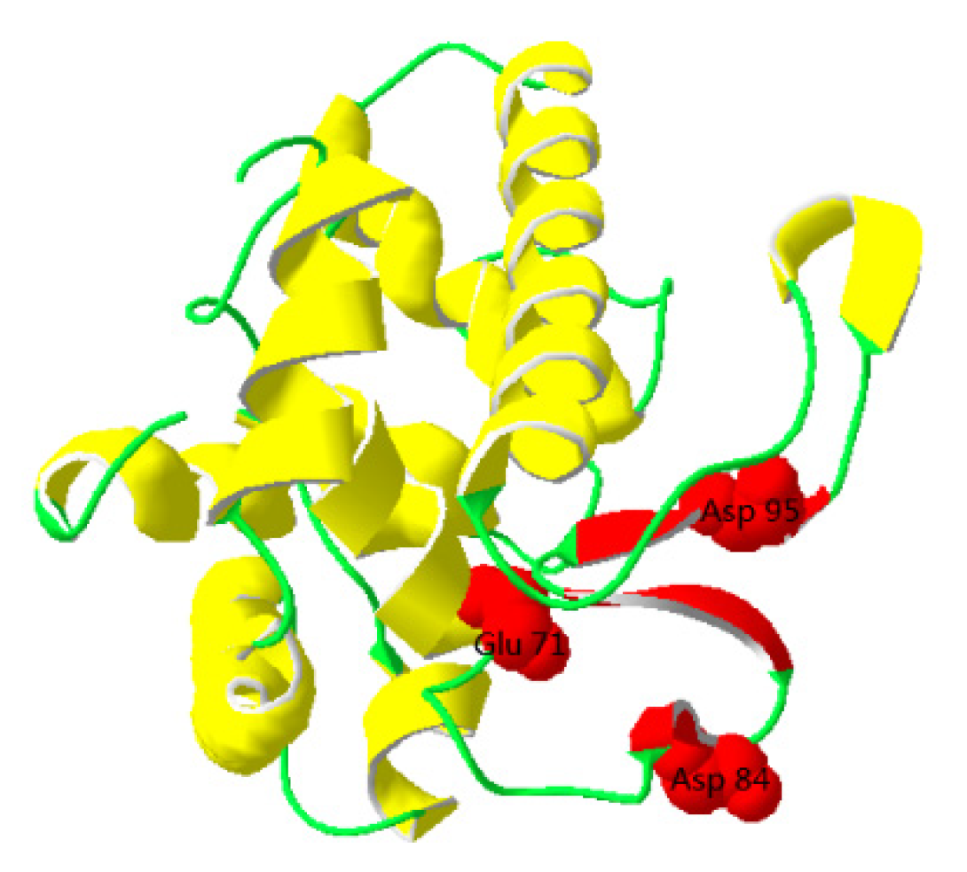
TrLysG is composed of a 573 bp ORF encoding 190 amino acids; its molecular mass is predicted to be 20.87 kDa, with an isoelectric point of 5.96. Analysis using SMART 6.0 software revealed that TrLysG contains a transglycosylase SLT domain spanning from Thr48 to Ser174. Multiple-sequence alignment displayed three catalytic residues (Glu71, Asp84, and Asp95), along with a highly conserved GLMQ motif (Gly90, Leu91, Met92, and Gln93) (Figure 1). This conserved GLMQ motif lies between the catalytic residues in the active site cleft. Furthermore, the predicted 3D structure of TrLysG features five α-helices depicted in yellow and three β-sheets shown in red, connected by coils (Figure 2). The three catalytic residues are located on the α-helix (Glu71) and the coiled coil (Asp84 and Asp95), respectively.

3.2. Phylogenetic Analysis

To understand the relationship between TrLysG and other species in terms of phylogeny, a phylogenetic tree was constructed. The tree is well clustered into four distinct groups, which consist of teleostean, reptilian, avian, and mammalian branches (Figure 3). The teleostean clade of the amino acid tree contains all fish species. TrLysG is classified into the teleost group and has the closest relationship with the T. flavidus counterpart.

3.3. Expression Analysis of TrLysG in Healthy Tissues

TrLysG expression was measured via qRT-PCR in nine healthy tissues. The results revealed that TrLysG exhibited widespread expression across all examined tissues. Specifically, as shown in Figure 4, the intestines and gills demonstrated significantly higher expression of TrLysG compared to other tissues (p < 0.05). On the contrary, the expression levels in remaining six tissues were relatively low, with no significant differences observed among them.

3.4. Temporal Expression Profiles of TrLysG post V. harveyi Injection

The expression pattern of TrLysG was characterized in specific tissues following bacterial infection to clarify its roles in immunity. The relative expression levels of TrLysG were examined in the gills, intestines, and liver after V. harveyi infection at 12, 24, and 48 h post infection (Figure 5). After intraperitoneal injection of V. harveyi, pathological changes were observed mainly at the site of bacterial injection, liquefaction and lesions were seen in the liver and injection region, and the liquefaction became serious over time. No significant morphological changes were seen in the gills or intestines within 48 h. In the intestine, the fold change of TrLysG reached a maximum of 6.6-fold at 24 h, and then declined to a relatively higher level (4-fold) compared to the control group. Furthermore, TrLysG expression in the gill was significantly up-regulated after infection, and reached its peak at 48 h. These results indicated that TrLysG exhibits a similar expression pattern in the intestine and gill. While in the liver, significant up-regulation was observed at all three-time points after V. harveyi infection, with the peak value appearing at 48 h by 5.8-fold compared to the control group.

3.5. Expression and Purification of rTrLysG

The recombinant protein of rTrLysG was successfully expressed in E. coli BL21 (DE3) after induction with 0.2 mM of IPTG at 15 °C for 16 h. SDS-PAGE with Coomassie brilliant blue staining revealed an approximately 21 kDa anticipated protein band of rTrLysG in protein extract supernatant. (Figure S2). After extracting soluble fusion protein from lysate supernatant, SDS-PAGE (Figure S3A) and Western blotting with anti-His tag antibody (Figure S3B) revealed a single band of pure rTrLysG protein. Finally, a total of 7.38 mg rTrLysG protein was obtained.

3.6. Lytic Activity of rTrLysG at Different pH and Temperature

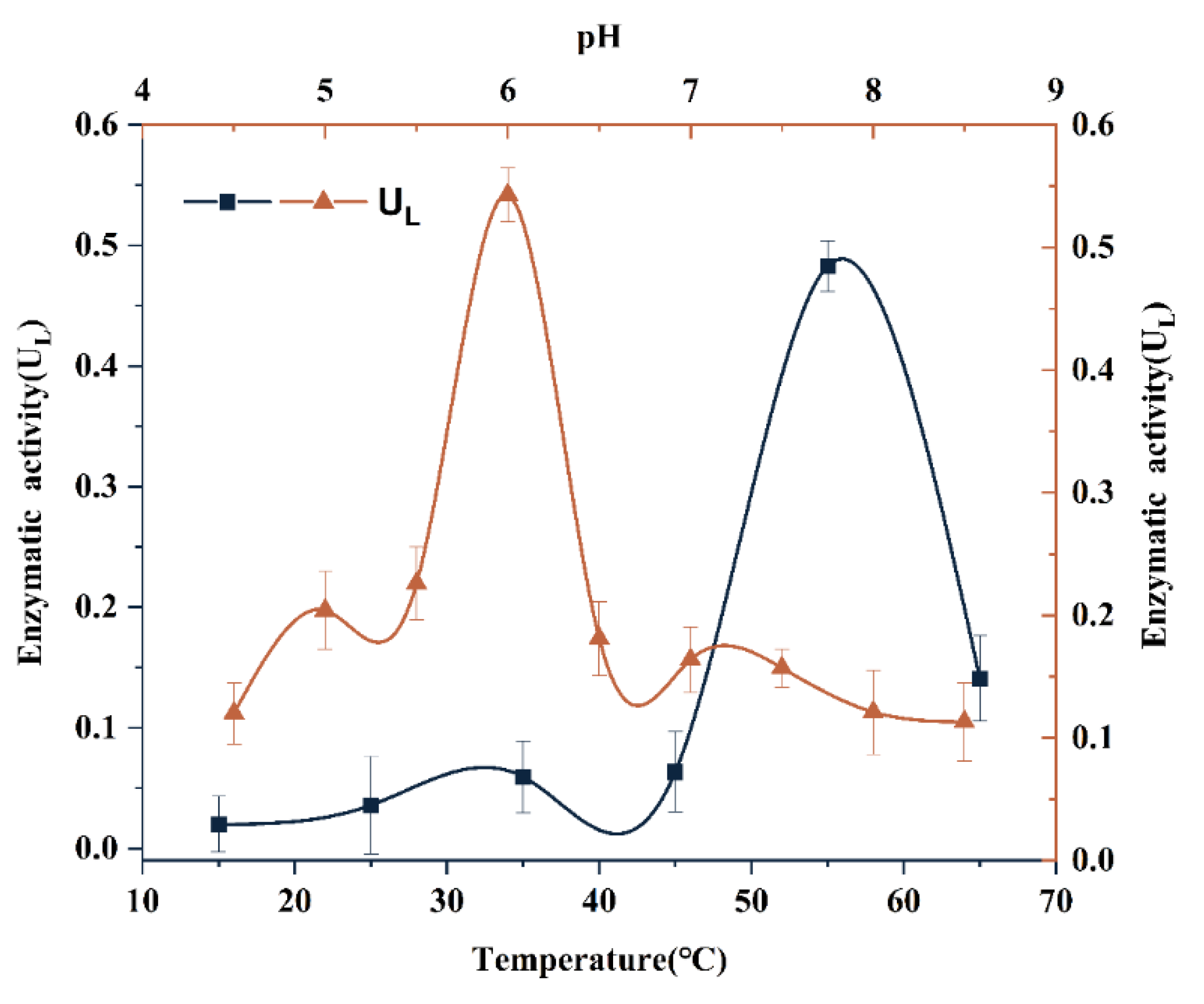
Each fish species lysozyme has a distinct optimal activity at varying pH and temperature. To determine the lytic activity of rTrLysG, a turbidimetric assay was performed using S. pasteuri as the substrate, with temperature ranging from 15 to 65 °C (in intervals of 10 °C) and pH ranging from 4.5 to 8.5 (in intervals of 0.5). The results indicated that the highest lytic activity of rTrLysG occurred within the temperature range of 50–60 °C, with an optimum temperature of 55 °C (UL = 0.48). The pH range suitable for rTrLysG activity was found to be between 5.5 and 6.5, with an optimum pH of 6 (UL = 0.54) (Figure 6).

3.7. Antibacterial Activity of Recombinant Protein

The minimum inhibitory concentration (MIC) refers to the lowest concentration of an antimicrobial that visibly inhibits the growth of the bacteria [56]. It is a critical factor in assessing the antimicrobial activity of lysozyme against bacterial strains. In this study, the MICs of rTrLysG against Gram-negative and Gram-positive bacteria are presented in Table 2. The results showed that these ranged from 50 to 200 μg/mL. The MIC of rTrLysG against S. parauberis was observed to be 100 μg/mL, while the MIC of rTrLysG against S. pasteuri and V. harveyi was 50 μg/mL. These findings confirm that this synthetic protein inhibited the growth of both Gram-negative and Gram-positive bacteria.

4. Discussion

Lysozymes are found ubiquitously across various organisms such as animals, plants, fungi, bacteria, and bacteriophages [3]. They are an opsonin that stimulates the complement system and phagocytes. Furthermore, they are prevalent in the mucus, lymphoid tissue, plasma, and various body fluids across different fish species [2]. Our current study reported the identification of a goose-type lysozyme in Japanese pufferfish. It consists of 190 amino acids, with a calculated molecular mass of 20.87 kDa and a theoretical isoelectric point (pI) of 5.96. Most fish LysG have a similar molecular mass, like sea bass (Dicentrarchus labrax L.), grass carp (Ctenopharyngodon idellus), and rohu (Labeo rohita), ranging from 20.19 to 20.27 kDa [19,20,57]. The TrLysG was found to lack a signal peptide, suggesting its potential as an intracellular protein primarily engaged in defense mechanisms following pathogen invasion of the host cell [58]. Multiple-sequence alignment analysis of TrLysG showed that many amino acids are conserved from fish to mammals, and that it contains three important catalytic residues (Glu71, Asp84 and Asp95). The Glu71 and Asp84 residues are conserved in all fish species and mammals except for Homo sapiens. The conserved GLMQ motif has been reported to enhance bacterial binding [59], and play a role in enzymatic reactions or the regulation of specialized lytic transglycosylase [60]. These residues are essential to the lysozyme’s three-dimensional structure and biological activity [14]. Furthermore, the number of cysteine residues in LysG exhibits significant variability. Research has shown that the formation of disulfide bonds by cysteine residues is essential for maintaining the structural stability of the LysG [61]. Avian and mammalian lysozyme sequences typically possess four cysteine residues, resulting in the formation of two disulfide bridges [62,63]. However, the situation in fish is more complex, with common carp and Atlantic cod having only one cysteine residue, while other species do not have any cysteine residues [19], such as orange-spotted grouper (Epinephelus coioides), Japanese flounder, and Japanese pufferfish in the current study. The lack of cysteine residues may have some effect on the structural stability of the protein. Phylogenetic analysis grouped all fish species into a single cluster, clearly distinguished from that in avian and mammalian studies, with TrLysG exhibiting the highest similarity to T. flavidus. Previous research has revealed that the LysG in fish forms a distinct group due to the absence of the signal peptide sequence [59].
In fish, LysG has been found to be expressed in various tissues. It is mainly produced in the head kidney and spleen, which are important tissues involved in immune responses [23,64,65]. Furthermore, LysG can be also abundantly found in mucosal tissues, such as intestines, skin and gills, which play significant roles in mucosal immunity [66]. In addition to nutrient digestion and absorption, the intestines are also a vital component of animals’ immune defense against harmful substances. Fish gills are exposed to numerous waterborne pathogens, with some pathogens utilizing the gills as a portal of entry into the host [67]. Hence, the substantial presence of LysG could be crucial. TrLysG expression levels were also found to vary among specific tissues, with intestines exhibiting the highest expression, followed by the gills. Similarly, Lates calcarifer and E. coioides exhibited the highest expression levels in the intestines [17,68], while gills showed the highest level of expression in sea bass [57], yellow catfish (Pelteobagrus fulvidraco) [69], and crucian carp (Carassius auratus) [64]. These findings suggested that the significant expression levels of LysG in mucosal tissues may be associated with their crucial functions in mucosal immune responses.
V. harveyi is a Gram-negative bacterium and belongs to the Vibrionaceae family. It is a well-known and dangerous bacterial disease of fish and invertebrates. Naturally diseased fish may exhibit a range of lesions, including liver congestion, muscle necrosis, skin ulcers, and tail rot [70]. In the present study, the intestines and gills were selected for the challenge experiment due to their high expression in normal healthy conditions; moreover, V. harveyi would cause serious granulomatous lesions in the liver of Japanese pufferfish. Therefore, the liver was also selected for gene expression detection. In this study, fish were intraperitoneally injected with V. harveyi for infection challenge; therefore, besides bacterial infection, the upregulation of TrLysG could also be caused by injection. The expression levels of control in different time-points and different tissues were also evaluated. The results showed that TrLysG expression were indeed altered by the injection (Figure S1). TrLysG showed reduced expression compared to 12 h in each tissue injected after 24 h in the liver, indicating that either the effect of injection only lasted for a short period of time (12 h), or the injection only downregulated TrLysG expression. In order to eliminate the effects of the injection, fold changes were calculated in comparison to the corresponding control at each time-point in each tissue, so that the increase in the TrLysG was only caused by bacterial infection. Our findings revealed that TrLysG expression levels were significantly up-regulated in all tested tissues. Similarly, in vivo challenge experiments in other teleost species also demonstrated the up-regulation of LysG in multiple tissues following bacterial infection, such as in the liver of rohu [19], Chinese rare minnow [18], Dabry’s sturgeon [71] and crucian carp [64] after A. hydrophila infection; in the gills of seahorse (Hippocampus abdominalis) [72] after being challenged by Edwardsiella tarda and Streptococcus iniae; and in the intestine of half-smooth tongue sole (Cynoglossus semilaevis) [73] and turbot (Scophthalmus maximus) [22] after Vibrio anguillarum and S. iniae infection, respectively. Notably, TrLysG was significantly up-regulated for three-time points in the liver, indicating that the liver plays an important role at every stage of bacterial infection. These results indicated the significance of this enzyme in immune defense against invading pathogens.
Lysozymes are commonly recognized as muramidase and have the ability to produce bacterial cell lysis, which is a fundamental characteristic shared by various lysozymes [74]. However, the enzymatic activity is not always the same in different conditions; it is usually maximal at a specific temperature and pH, which ensures proper substrate binding and ultimately enhances the antimicrobial efficacy. Therefore, optimum temperature and pH are key factors for optimal enzyme action [75]. Optimal pH values have been found to range from 5.0 to 7.5 in previous research in fish [76]. In our study, we tested the lytic activity of rTrLysG at various temperatures and pH levels using S. cereus as the substrate. The rTrLysG optimal condition was observed at pH 6, consistent with rohu [19]. Meanwhile, different fish species exhibited a broad range of optimal temperatures, spanning from 22 to 60 °C in various studies [13,20,22,57,77], and the rTrLysG demonstrated an optimal temperature of 55 °C. Overall, our findings indicated that the optimum pH and temperature of rTrLysG were both within the range previously described for fish LysG proteins.
The antibacterial activity of rTrLysG was evaluated using MIC tests against three Gram-positive and five Gram-negative bacteria. In our current investigation, rTrLysG exhibited antibacterial effects against multiple Gram-positive bacteria, which has been confirmed in numerous species [19]. However, since Gram-negative bacteria’s peptidoglycan is encased in an outer membrane that contains lipopolysaccharides, lysozymes cannot reach it directly [78]. As reported in chickens, the hen egg white lysozyme (HEWL) displayed limited or negligible activity against Gram-negative bacteria [21,79]. Surprisingly, similar to TrLysG, many teleost lysozymes also showed notable inhibitory effects against Gram-negative bacteria [13,19,57,76]. Studies have found that besides their fundamental role as muramidases, lysozymes may exhibit non-enzymatic bactericidal activities that involve activating bacterial autolysis through the interaction between cationic lysozyme molecules and the cell wall, as well as perturbing, disintegrating, and causing membrane leakage without peptidoglycan hydrolysis [15,18]. Notably, our findings indicated that the lytic action of rTrLysG is quite efficient for both S. pasteuri and V. harveyi with MIC of 50 μg/mL. In aquaculture, the accumulation of antibiotics has resulted in the development of resistance among bacterial pathogens, and resistance developed in the aquatic animals could be transmitted to humans, to the detriment of our health. Therefore, identification of safer alternatives is essential for the healthy and sustainable development of aquaculture. Studies have revealed that the chances of the emergence of resistance to lysozymes is slim [80], making it a promising antibacterial agent for disease control. In the course of aquaculture, lysozymes could be used as feed additives; previous studies have reported that complexes of natural actives with lysozymes shield fish from harmful microbes and increase feed conversion [81]. Furthermore, lysozymes could be added directly to culture water to enhance water quality [82] and maintain microbial balance, thereby strengthening aquatic animals’ resistance to bacterial diseases.

5. Conclusions

We have successfully characterized the molecular features, physicochemical properties, and evolutionary relationships of TrLysG. Transcriptional analyses revealed widespread expression of TrLysG in nine healthy tissues, with the intestines and gills showing the highest abundance. Furthermore, TrLysG exhibited a significant up-regulation following V. harveyi challenge. The purified recombinant protein rTrLysG exhibited bactericidal activity against both Gram-positive and Gram-negative bacteria. Our results demonstrated the crucial role of TrLysG in the immunological response to bacterial infection in T. rubripes, which could further help in harnessing rTrLysG for the development of novel biotherapeutics.

Supplementary Materials

The following supporting information can be downloaded at: https://www.mdpi.com/article/10.3390/fishes8120577/s1, Figure S1: Expression profiles of TrLysG after injection of saline in the intestine, gill and liver.; Figure S2: Expression of recombinant TrLysG protein; Figure S3: Purification of recombinant TrLysG protein.

Author Contributions

Conceptualization, X.C., M.G. and C.J.; methodology, X.C., Z.Y. and H.W; validation, X.C. and Z.Y.; investigation, X.C., Z.Y. and M.G.; writing—original draft preparation, X.C.; visualization, X.C. and R.Z.; resources, X.Y., S.W., L.C. and H.W.; writing—review and editing, C.J. and H.W.; project administration, C.J.; funding acquisition, C.J. All authors have read and agreed to the published version of the manuscript.

Funding

This project was supported by the National Natural Science Foundation of China (32273101), the Central Government Subsidy Project for Liaoning Fisheries (2023), the Dalian High Level Talent Innovation Support Program (2019RQ102), and the China Agriculture Research System (CARS-47).

Institutional Review Board Statement

Our research involving animals was conducted under the guidelines and regulations of the Animal Research and Ethics Committee of Dalian Ocean University and complied with China's existing laws and regulations on biological research (approval code: DLOU2023005; date: 3 March 2023). Every effort has been made to design our research in a way that enhances the positive impact while minimizing any potential risks or negative consequences for animals. This study did not involve endangered or protected species. Fish were humanely sacrificed after being anesthetized with MS-222.

Data Availability Statement

The raw data that support this study are available from the corresponding author upon reasonable request.

Conflicts of Interest

The authors declare no conflict of interest.

References

  1. He, C.; Yu, H.; Liu, W.; Su, H.; Shan, Z.; Bao, X.; Li, Y.; Fu, L.; Gao, X. A goose-type lysozyme gene in Japanese scallop (Mizuhopecten yessoensis): cDNA cloning, mRNA expression and promoter sequence analysis. Comp. Biochem. Physiol. Part B Biochem. Mol. Biol. 2012, 162, 34–43. [Google Scholar] [CrossRef] [PubMed]
  2. Magnadóttir, B. Innate immunity of fish (overview). Fish Shellfish. Immunol. 2006, 20, 137–151. [Google Scholar] [CrossRef] [PubMed]
  3. Jollès, P.; Jollès, J. What’s new in lysozyme research? Always a model system, today as yesterday. Mol. Cell. Biochem. 1984, 63, 165–189. [Google Scholar] [CrossRef] [PubMed]
  4. Saurabh, S.; Sahoo, P.K. Lysozyme: An important defence molecule of fish innate immune system. Aquac. Res. 2008, 39, 223–239. [Google Scholar] [CrossRef]
  5. Lee, W.; Ku, S.-K.; Na, D.H.; Bae, J.-S. Anti-Inflammatory Effects of Lysozyme Against HMGB1 in Human Endothelial Cells and in Mice. Inflammation 2015, 38, 1911–1924. [Google Scholar] [CrossRef] [PubMed]
  6. Xin, X.-D.; He, J.-G.; Qiu, W.; Tang, J.; Liu, T.-T. Microbial community related to lysozyme digestion process for boosting waste activated sludge biodegradability. Bioresour. Technol. 2015, 175, 112–119. [Google Scholar] [CrossRef] [PubMed]
  7. Callewaert, L.; Michiels, C.W. Lysozymes in the animal kingdom. J. Biosci. 2010, 35, 127–160. [Google Scholar] [CrossRef] [PubMed]
  8. Canfield, R.E.; McMurry, S. Purification and characterization of a lysozyme from goose egg white. Biochem. Biophys. Res. Commun. 1967, 26, 38–42. [Google Scholar] [CrossRef]
  9. Irwin, D.M. Evolution of the vertebrate goose-type lysozyme gene family. BMC Evol. Biol. 2014, 14, 188. [Google Scholar] [CrossRef]
  10. Yu, H.; Gao, J.; Lu, Y.; Guang, H.; Cai, S.; Zhang, S.; Wang, Y. Molecular cloning, sequence analysis and phylogeny of first caudata g-type lysozyme in axolotl (Ambystoma mexicanum). Zool. Sci. 2013, 30, 938–943. [Google Scholar] [CrossRef]
  11. Savan, R.; Aman, A.; Sakai, M. Molecular cloning of G type lysozyme cDNA in common carp (Cyprinus carpio L.). Fish Shellfish. Immunol. 2003, 15, 263–268. [Google Scholar] [CrossRef] [PubMed]
  12. Bathige, S.D.N.K.; Umasuthan, N.; Whang, I.; Lim, B.-S.; Jung, H.-B.; Lee, J. Evidences for the involvement of an invertebrate goose-type lysozyme in disk abalone immunity: Cloning, expression analysis and antimicrobial activity. Fish Shellfish. Immunol. 2013, 35, 1369–1379. [Google Scholar] [CrossRef] [PubMed]
  13. Hikima, J.-i.; Minagawa, S.; Hirono, I.; Aoki, T. Molecular cloning, expression and evolution of the Japanese flounder goose-type lysozyme gene, and the lytic activity of its recombinant protein. Biochim. Biophys. Acta (BBA)—Gene Struct. Expr. 2001, 1520, 35–44. [Google Scholar] [CrossRef]
  14. Wei, K.; Ding, Y.; Yin, X.; Zhang, J.; Shen, B. Molecular cloning, expression analyses and functional characterization of a goose-type lysozyme gene from Bostrychus sinensis (family: Eleotridae). Fish Shellfish. Immunol. 2020, 96, 41–52. [Google Scholar] [CrossRef] [PubMed]
  15. Liu, Y.; Zha, H.; Yu, S.; Zhong, J.; Liu, X.; Yang, H.; Zhu, Q. Molecular characterization and antibacterial activities of a goose-type lysozyme gene from roughskin sculpin (Trachidermus fasciatus). Fish Shellfish. Immunol. 2022, 127, 1079–1087. [Google Scholar] [CrossRef] [PubMed]
  16. Zhang, C.; Zhang, J.; Liu, M.; Huang, M. Molecular cloning, expression and antibacterial activity of goose-type lysozyme gene in Microptenus salmoides. Fish Shellfish. Immunol. 2018, 82, 9–16. [Google Scholar] [CrossRef] [PubMed]
  17. Wei, S.; Huang, Y.; Huang, X.; Cai, J.; Wei, J.; Li, P.; Ouyang, Z.; Qin, Q. Molecular cloning and characterization of a new G-type lysozyme gene (Ec-lysG) in orange-spotted grouper, Epinephelus coioides. Dev. Comp. Immunol. 2014, 46, 401–412. [Google Scholar] [CrossRef]
  18. Zhang, Y.; Yang, H.; Song, W.; Cui, D.; Wang, L. Identification and characterization of a novel goose-type and chicken-type lysozyme genes in Chinese rare minnow (Gobiocypris rarus) with potent antimicrobial activity. Genes Genom. 2018, 40, 569–577. [Google Scholar] [CrossRef]
  19. Mohapatra, A.; Parida, S.; Mohanty, J.; Sahoo, P.K. Identification and functional characterization of a g-type lysozyme gene of Labeo rohita, an Indian major carp species. Dev. Comp. Immunol. 2019, 92, 87–98. [Google Scholar] [CrossRef]
  20. Ye, X.; Zhang, L.; Tian, Y.; Tan, A.; Bai, J.; Li, S. Identification and expression analysis of the g-type and c-type lysozymes in grass carp Ctenopharyngodon idellus. Dev. Comp. Immunol. 2010, 34, 501–509. [Google Scholar] [CrossRef]
  21. Whang, I.; Lee, Y.; Lee, S.; Oh, M.-J.; Jung, S.-J.; Choi, C.Y.; Lee, W.S.; Kim, H.S.; Kim, S.-J.; Lee, J. Characterization and expression analysis of a goose-type lysozyme from the rock bream Oplegnathus fasciatus, and antimicrobial activity of its recombinant protein. Fish Shellfish. Immunol. 2011, 30, 532–542. [Google Scholar] [CrossRef] [PubMed]
  22. Zhao, L.; Sun, J.-s.; Sun, L. The g-type lysozyme of Scophthalmus maximus has a broad substrate spectrum and is involved in the immune response against bacterial infection. Fish Shellfish. Immunol. 2011, 30, 630–637. [Google Scholar] [CrossRef] [PubMed]
  23. Wang, R.; Feng, J.; Li, C.; Liu, S.; Zhang, Y.; Liu, Z. Four lysozymes (one c-type and three g-type) in catfish are drastically but differentially induced after bacterial infection. Fish Shellfish. Immunol. 2013, 35, 136–145. [Google Scholar] [CrossRef] [PubMed]
  24. Ogino, Y.; Yamaguchi, A. Reduced lifetime fitness (growth, body condition and survivability) of hatchery-reared tiger pufferfish Takifugu rubripes compared to wild counterparts. J. Fish Biol. 2022, 101, 1270–1284. [Google Scholar] [CrossRef] [PubMed]
  25. Li, L.; Zhang, F.; Meng, X.; Cui, X.; Ma, Q.; Wei, Y.; Liang, M.; Xu, H. Recovery of Fatty Acid and Volatile Flavor Compound Composition in Farmed Tiger Puffer (Takifugu rubripes) with a Fish Oil-Finishing Strategy. Mar. Drugs 2023, 21, 122. [Google Scholar] [CrossRef] [PubMed]
  26. Hou, H.; Zhang, Y.; Ma, Z.; Wang, X.; Su, P.; Wang, H.; Liu, Y. Life cycle assessment of tiger puffer (Takifugu rubripes) farming: A case study in Dalian, China. Sci. Total Environ. 2022, 823, 153522. [Google Scholar] [CrossRef] [PubMed]
  27. Zhang, Q.; Wu, Y.; Li, W.; Wang, J.; Zhou, H.; Zhang, L.; Liu, Q.; Ying, L.; Yan, H. Retinal development and the expression profiles of opsin genes during larval development in Takifugu rubripes. J. Fish Biol. 2023, 102, 380–394. [Google Scholar] [CrossRef]
  28. Liu, Q.; Yan, H.; Hu, P.; Liu, W.; Shen, X.; Cui, X.; Wu, Y.; Yuan, Z.; Zhang, L.; Zhang, Y.; et al. Growth and survival of Takifugu rubripes larvae cultured under different light conditions. Fish Physiol. Biochem. 2019, 45, 1533–1549. [Google Scholar] [CrossRef]
  29. Yan, H.; Shen, X.; Jiang, J.; Zhang, L.; Yuan, Z.; Wu, Y.; Liu, Q.; Liu, Y. Gene Expression of Takifugu rubripes Gonads During AI- or MT-induced Masculinization and E2-induced Feminization. Endocrinology 2021, 162, bqab068. [Google Scholar] [CrossRef]
  30. Shen, X.; Yan, H.; Jiang, J.; Li, W.; Xiong, Y.; Liu, Q.; Liu, Y. Profile of gene expression changes during estrodiol-17β-induced feminization in the Takifugu rubripes brain. BMC Genom. 2021, 22, 851. [Google Scholar] [CrossRef]
  31. Bao, M.; Shang, F.; Liu, F.; Hu, Z.; Wang, S.; Yang, X.; Yu, Y.; Zhang, H.; Jiang, C.; Jiang, J.; et al. Comparative transcriptomic analysis of the brain in Takifugu rubripes shows its tolerance to acute hypoxia. Fish Physiol. Biochem. 2021, 47, 1669–1685. [Google Scholar] [CrossRef] [PubMed]
  32. Shang, F.; Lu, Y.; Li, Y.; Han, B.; Wei, R.; Liu, S.; Liu, Y.; Liu, Y.; Wang, X. Transcriptome Analysis Identifies Key Metabolic Changes in the Brain of Takifugu rubripes in Response to Chronic Hypoxia. Genes 2022, 13, 1347. [Google Scholar] [CrossRef] [PubMed]
  33. Gao, X.-Q.; Fei, F.; Huang, B.; Meng, X.S.; Zhang, T.; Zhao, K.-F.; Chen, H.-B.; Xing, R.; Liu, B.-L. Alterations in hematological and biochemical parameters, oxidative stress, and immune response in Takifugu rubripes under acute ammonia exposure. Comp. Biochem. Physiol. Part C Toxicol. Pharmacol. 2021, 243, 108978. [Google Scholar] [CrossRef] [PubMed]
  34. Zhang, H.; Li, P.; Wu, B.; Hou, J.; Ren, J.; Zhu, Y.; Xu, J.; Si, F.; Sun, Z.; Liu, X. Transcriptomic analysis reveals the genes involved in tetrodotoxin (TTX) accumulation, translocation, and detoxification in the pufferfish Takifugu rubripes. Chemosphere 2022, 303, 134962. [Google Scholar] [CrossRef] [PubMed]
  35. Gao, L.; Zhang, Z.; Xing, Z.; Li, Q.; Kong, N.; Wang, L.; Song, L. The variation of intestinal autochthonous bacteria in cultured tiger pufferfish Takifugu rubripes. Front. Cell. Infect. Microbiol. 2022, 12, 1062512. [Google Scholar] [CrossRef] [PubMed]
  36. Gao, F.-X.; Lu, W.-J.; Shi, Y.; Zhu, H.-y.; Wang, Y.-h.; Tu, H.-q.; Gao, Y.; Zhou, L.; Gui, J.-F.; Zhao, Z. Transcriptome profiling revealed the growth superiority of hybrid pufferfish derived from Takifugu obscurus ♀ × Takifugu rubripes ♂. Comp. Biochem. Physiol. Part D Genom. Proteom. 2021, 40, 100912. [Google Scholar] [CrossRef] [PubMed]
  37. Aparicio, S.; Chapman, J.; Stupka, E.; Putnam, N.; Chia, J.-m.; Dehal, P.; Christoffels, A.; Rash, S.; Hoon, S.; Smit, A.; et al. Whole-Genome Shotgun Assembly and Analysis of the Genome of Fugu rubripes. Science 2002, 297, 1301–1310. [Google Scholar] [CrossRef]
  38. Kuroyanagi, M.; Katayama, T.; Imai, T.; Yamamoto, Y.; Chisada, S.-i.; Yoshiura, Y.; Ushijima, T.; Matsushita, T.; Fujita, M.; Nozawa, A.; et al. New approach for fish breeding by chemical mutagenesis: Establishment of TILLING method in fugu (Takifugu rubripes) with ENU mutagenesis. BMC Genom. 2013, 14, 786. [Google Scholar] [CrossRef]
  39. Fu, X.; Zhang, H.; Tan, E.; Watabe, S.; Asakawa, S. Characterization of the torafugu (Takifugu rubripes) immunoglobulin heavy chain gene locus. Immunogenetics 2015, 67, 179–193. [Google Scholar] [CrossRef]
  40. Yang, X.; Liu, X.; Yang, Z.; Guan, Y.; Zhao, R.; Peng, H.; Cao, X.; Gao, M.; Wang, S.; Jiang, C. Genome-wide characterization of caspase genes in Japanese pufferfish, Takifugu rubripes, and expression profiles in response to Vibrio harveyi infection. J. World Aquac. Soc. 2022, 53, 910–923. [Google Scholar] [CrossRef]
  41. Chen, M.; Liu, X.; Zhou, J.; Wang, X.; Liu, R.; Peng, H.; Li, B.; Cai, Z.; Jiang, C. Molecular characterization and expression analysis of galectins in Japanese pufferfish (Takifugu rubripes) in response to Vibrio harveyi infection. Fish Shellfish. Immunol. 2019, 86, 347–354. [Google Scholar] [CrossRef] [PubMed]
  42. Liu, X.; Jiao, C.; Ma, Y.; Wang, Q.; Zhang, Y. A live attenuated Vibrio anguillarum vaccine induces efficient immunoprotection in Tiger puffer (Takifugu rubripes). Vaccine 2018, 36, 1460–1466. [Google Scholar] [CrossRef] [PubMed]
  43. Kono, T.; Ida, T.; Kawahara, N.; Watanabe, F.; Biswas, G.; Sato, T.; Mori, K.; Miyazato, M. Identification and immunoregulatory function of neuromedin U (Nmu) in the Japanese pufferfish Takifugu rubripes. Dev. Comp. Immunol. 2017, 73, 246–256. [Google Scholar] [CrossRef] [PubMed]
  44. Bird, S.; Zou, J.; Kono, T.; Sakai, M.; Dijkstra, J.M.; Secombes, C. Characterisation and expression analysis of interleukin 2 (IL-2) and IL-21 homologues in the Japanese pufferfish, Fugu rubripes, following their discovery by synteny. Immunogenetics 2005, 56, 909–923. [Google Scholar] [CrossRef] [PubMed]
  45. Wang, K.; Gan, L.; Kunisada, T.; Lee, I.; Yamagishi, H.; Hood, L. Characterization of the Japanese pufferfish (Takifugu rubripes) T-cell receptor α locus reveals a unique genomic organization. Immunogenetics 2001, 53, 31–42. [Google Scholar] [CrossRef] [PubMed]
  46. Rombel, I.T.; Sykes, K.F.; Rayner, S.; Johnston, S.A. ORF-FINDER: A vector for high-throughput gene identification. Gene 2002, 282, 33–41. [Google Scholar] [CrossRef] [PubMed]
  47. Artimo, P.; Jonnalagedda, M.; Arnold, K.; Baratin, D.; Csardi, G.; de Castro, E.; Duvaud, S.; Flegel, V.; Fortier, A.; Gasteiger, E.; et al. ExPASy: SIB bioinformatics resource portal. Nucleic Acids Res. 2012, 40, W597–W603. [Google Scholar] [CrossRef] [PubMed]
  48. Petersen, T.N.; Brunak, S.; von Heijne, G.; Nielsen, H. SignalP 4.0: Discriminating signal peptides from transmembrane regions. Nat. Methods 2011, 8, 785–786. [Google Scholar] [CrossRef]
  49. Letunic, I.; Doerks, T.; Bork, P. SMART 6: Recent updates and new developments. Nucleic Acids Res. 2009, 37, D229–D232. [Google Scholar] [CrossRef]
  50. Waterhouse, A.; Bertoni, M.; Bienert, S.; Studer, G.; Tauriello, G.; Gumienny, R.; Heer, F.T.; de Beer, T.A.P.; Rempfer, C.; Bordoli, L.; et al. SWISS-MODEL: Homology modelling of protein structures and complexes. Nucleic Acids Res. 2018, 46, W296–W303. [Google Scholar] [CrossRef]
  51. Robert, X.; Gouet, P. Deciphering key features in protein structures with the new ENDscript server. Nucleic Acids Res. 2014, 42, W320–W324. [Google Scholar] [CrossRef] [PubMed]
  52. Kumar, S.; Stecher, G.; Li, M.; Knyaz, C.; Tamura, K. MEGA X: Molecular Evolutionary Genetics Analysis across Computing Platforms. Mol. Biol. Evol. 2018, 35, 1547–1549. [Google Scholar] [CrossRef] [PubMed]
  53. Zhang, M.; Geng, H.; Tariq Javed, M.; Xu, L.; Li, X.; Wang, L.; Li, S.; Xu, Y. Passive protection of Japanese pufferfish (Takifugu rubripes) against Vibrio harveyi infection using chicken egg yolk immunoglobulins (IgY). Aquaculture 2021, 532, 736009. [Google Scholar] [CrossRef]
  54. Hultmark, D. Insect lysozymes. Exs 1996, 75, 87–102. [Google Scholar] [CrossRef] [PubMed]
  55. Yang, H.; Li, S.; Li, F.; Yu, K.; Yang, F.; Xiang, J. Recombinant Expression of a Modified Shrimp Anti-Lipopolysaccharide Factor Gene in Pichia pastoris GS115 and Its Characteristic Analysis. Mar. Drugs 2016, 14, 152. [Google Scholar] [CrossRef] [PubMed]
  56. Andrews, J.M. Determination of minimum inhibitory concentrations. J. Antimicrob. Chemother. 2001, 48 (Suppl. S1), 5–16. [Google Scholar] [CrossRef] [PubMed]
  57. Buonocore, F.; Randelli, E.; Trisolino, P.; Facchiano, A.; de Pascale, D.; Scapigliati, G. Molecular characterization, gene structure and antibacterial activity of a g-type lysozyme from the European sea bass (Dicentrarchus labrax L.). Mol. Immunol. 2014, 62, 10–18. [Google Scholar] [CrossRef] [PubMed]
  58. Nilojan, J.; Bathige, S.D.N.K.; Kugapreethan, R.; Thulasitha, W.S.; Nam, B.-H.; Lee, J. Molecular, transcriptional and functional insights into duplicated goose-type lysozymes from Sebastes schlegelii and their potential immunological role. Fish Shellfish. Immunol. 2017, 67, 66–77. [Google Scholar] [CrossRef]
  59. Kumaresan, V.; Bhatt, P.; Ganesh, M.-R.; Harikrishnan, R.; Arasu, M.; Al-Dhabi, N.A.; Pasupuleti, M.; Marimuthu, K.; Arockiaraj, J. A novel antimicrobial peptide derived from fish goose type lysozyme disrupts the membrane of Salmonella enterica. Mol. Immunol. 2015, 68, 421–433. [Google Scholar] [CrossRef]
  60. Höppner, C.; Carle, A.; Sivanesan, D.; Hoeppner, S.; Baron, C. The putative lytic transglycosylase VirB1 from Brucella suis interacts with the type IV secretion system core components VirB8, VirB9 and VirB11. Microbiology 2005, 151, 3469–3482. [Google Scholar] [CrossRef]
  61. Irwin, D.M.; Gong, Z.M. Molecular Evolution of Vertebrate Goose-Type Lysozyme Genes. J. Mol. Evol. 2003, 56, 234–242. [Google Scholar] [CrossRef] [PubMed]
  62. Bulaj, G. Formation of disulfide bonds in proteins and peptides. Biotechnol. Adv. 2005, 23, 87–92. [Google Scholar] [CrossRef] [PubMed]
  63. Kawamura, S.; Ohkuma, M.; Chijiiwa, Y.; Kohno, D.; Nakagawa, H.; Hirakawa, H.; Kuhara, S.; Torikata, T. Role of disulfide bonds in goose-type lysozyme. FEBS J. 2008, 275, 2818–2830. [Google Scholar] [CrossRef] [PubMed]
  64. Wang, M.; Zhao, X.; Kong, X.; Wang, L.; Jiao, D.; Zhang, H. Molecular characterization and expressing analysis of the c-type and g-type lysozymes in Qihe crucian carp Carassius auratus. Fish Shellfish. Immunol. 2016, 52, 210–220. [Google Scholar] [CrossRef] [PubMed]
  65. Myrnes, B.; Seppola, M.; Johansen, A.; Øverbø, K.; Callewaert, L.; Vanderkelen, L.; Michiels, C.W.; Nilsen, I.W. Enzyme characterisation and gene expression profiling of Atlantic salmon chicken- and goose-type lysozymes. Dev. Comp. Immunol. 2013, 40, 11–19. [Google Scholar] [CrossRef] [PubMed]
  66. Uribe, C.; Folch, H.; Enriquez, R.; Moran, G. Innate and adaptive immunity in teleost fish: A review. Vet. Med. 2011, 56, 486–503. [Google Scholar] [CrossRef]
  67. Lieschke, G.J.; Trede, N.S. Fish immunology. Curr. Biol. 2009, 19, 678–682. [Google Scholar] [CrossRef]
  68. Fu, G.H.; Bai, Z.Y.; Xia, J.H.; Liu, F.; Liu, P.; Yue, G.H. Analysis of two lysozyme genes and antimicrobial functions of their recombinant proteins in Asian seabass. PLoS ONE 2013, 8, e79743. [Google Scholar] [CrossRef]
  69. Liu, Q.-N.; Xin, Z.-Z.; Zhang, D.-Z.; Jiang, S.-H.; Chai, X.-Y.; Li, C.-F.; Zhou, C.-L.; Tang, B.-P. Molecular identification and expression analysis of a goose-type lysozyme (LysG) gene in yellow catfish Pelteobagrus fulvidraco. Fish Shellfish. Immunol. 2016, 58, 423–428. [Google Scholar] [CrossRef]
  70. Zhang, X.H.; He, X.; Austin, B. Vibrio harveyi: A serious pathogen of fish and invertebrates in mariculture. Mar. Life Sci. Technol. 2020, 2, 231–245. [Google Scholar] [CrossRef]
  71. Zhang, S.; Xu, Q.; Boscari, E.; Du, H.; Qi, Z.; Li, Y.; Huang, J.; Di, J.; Yue, H.; Li, C.; et al. Characterization and expression analysis of g- and c-type lysozymes in Dabry’s sturgeon (Acipenser dabryanus). Fish Shellfish. Immunol. 2018, 76, 260–265. [Google Scholar] [CrossRef] [PubMed]
  72. Ko, J.; Wan, Q.; Bathige, S.D.N.K.; Lee, J. Molecular characterization, transcriptional profiling, and antibacterial potential of G-type lysozyme from seahorse (Hippocampus abdominalis). Fish Shellfish. Immunol. 2016, 58, 622–630. [Google Scholar] [CrossRef] [PubMed]
  73. Sha, Z.-X.; Wang, Q.-L.; Liu, Y.; Chen, S.-L. Identification and expression analysis of goose-type lysozyme in half-smooth tongue sole (Cynoglossus semilaevis). Fish Shellfish. Immunol. 2012, 32, 914–921. [Google Scholar] [CrossRef] [PubMed]
  74. Chipman, D.M.; Sharon, N. Mechanism of lysozyme action. Science 1969, 165, 454–465. [Google Scholar] [CrossRef] [PubMed]
  75. Mai, W.-j.; Wang, W.-n. Protection of blue shrimp (Litopenaeus stylirostris) against the White Spot Syndrome Virus (WSSV) when injected with shrimp lysozyme. Fish Shellfish. Immunol. 2010, 28, 727–733. [Google Scholar] [CrossRef] [PubMed]
  76. Mai, W.-j.; Hu, C.-q. Molecular cloning, characterization, expression and antibacterial analysis of a lysozyme homologue from Fenneropenaeus merguiensis. Mol. Biol. Rep. 2009, 36, 1587–1595. [Google Scholar] [CrossRef] [PubMed]
  77. Kyomuhendo, P.; Myrnes, B.; Nilsen, I.W. A cold-active salmon goose-type lysozyme with high heat tolerance. Cell. Mol. Life Sci. 2007, 64, 2841–2847. [Google Scholar] [CrossRef]
  78. Liu, J.; Wang, N.; Liu, Y.; Jin, Y.; Ma, M. The antimicrobial spectrum of lysozyme broadened by reductive modification. Poult. Sci. 2018, 97, 3992–3999. [Google Scholar] [CrossRef]
  79. Mine, Y.; Ma, F.; Lauriau, S. Antimicrobial peptides released by enzymatic hydrolysis of hen egg white lysozyme. J. Agric. Food Chem. 2004, 52, 1088–1094. [Google Scholar] [CrossRef]
  80. Grishin, A.V.; Karyagina, A.S.; Vasina, D.V.; Vasina, I.V.; Gushchin, V.A.; Lunin, V.G. Resistance to peptidoglycan-degrading enzymes. Crit. Rev. Microbiol. 2020, 46, 703–726. [Google Scholar] [CrossRef]
  81. Ferraboschi, P.; Ciceri, S.; Grisenti, P. Applications of Lysozyme, an Innate Immune Defense Factor, as an Alternative Antibiotic. Antibiotics 2021, 10, 1534. [Google Scholar] [CrossRef]
  82. El-Kady, A.A.; Magouz, F.I.; Mahmoud, S.A.; Abdel-Rahim, M.M. The effects of some commercial probiotics as water additive on water quality, fish performance, blood biochemical parameters, expression of growth and immune-related genes, and histology of Nile tilapia (Oreochromis niloticus). Aquaculture 2022, 546, 737249. [Google Scholar] [CrossRef]
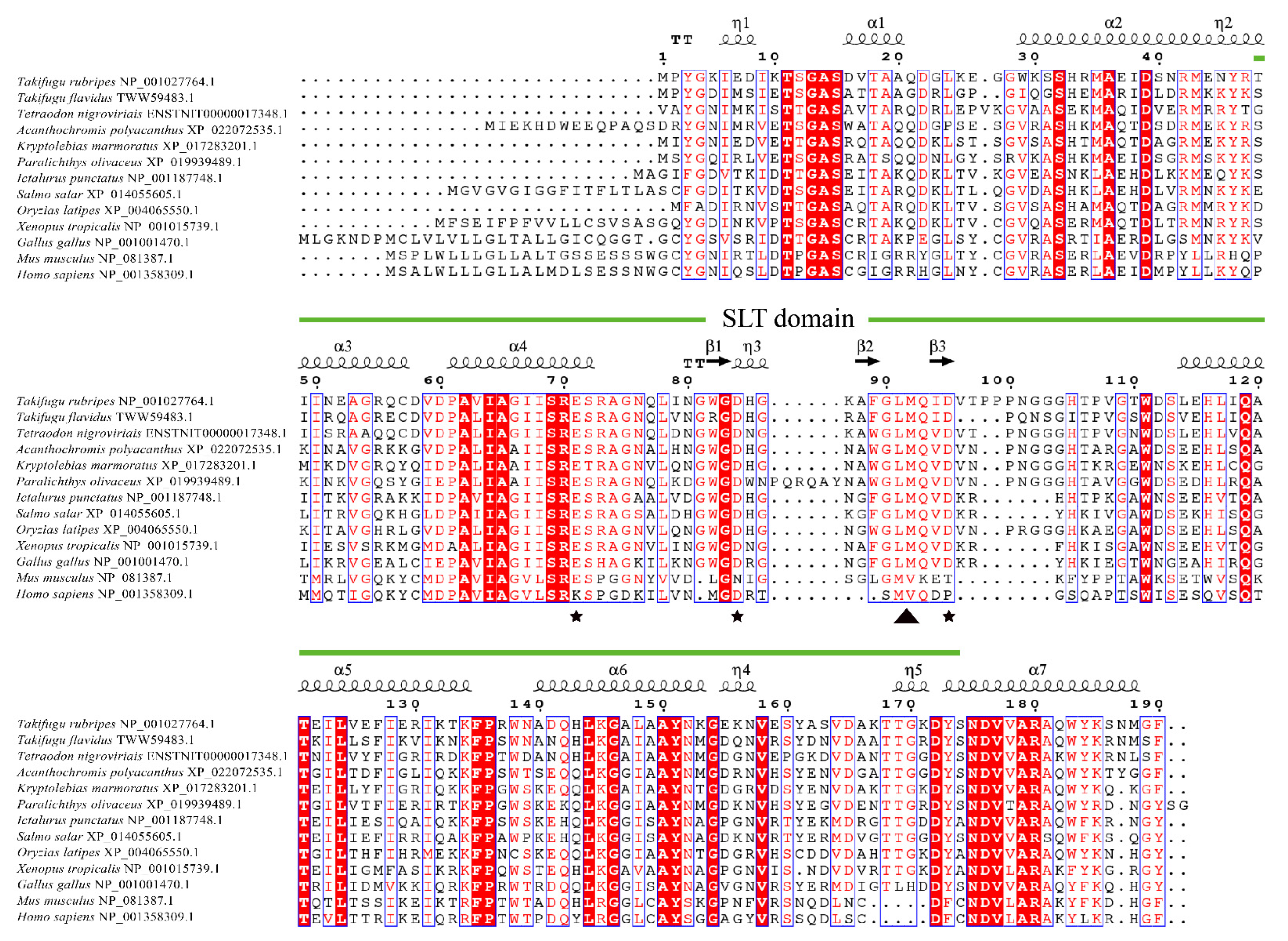
Figure 1. Alignment of the full amino acid sequences of TrLysG with other homologues. Sequences with identical residues are highlighted in red, while those with 75% to 100% similarity are highlighted in red text with blue boxes. The predicted secondary structure of TrLysG is displayed above the alignment, with curves representing alpha helices and arrows representing beta folding. Three catalytic residues (Glu71, Asp84 and Asp95) and the GLMQ motif (Gly90, Leu91, Met92 and Gln93) are highlighted in star and triangle, respectively. The SLT domain is highlighted in green line.
Fishes 08 00577 g001
Figure 2. Predicted tertiary structure of TrLysG. The α-helices are highlighted in yellow. Three catalytic residues (Glu71, Asp84 and Asp95) are labelled in red.
Fishes 08 00577 g002
Figure 3. Phylogenetic analysis of TrLysG with known orthologs from vertebrate phylum. The percentage of trees in which the associated taxa clustered together is shown above the branches. TrLysG is highlighted with a diamond.
Fishes 08 00577 g003
Figure 4. Basal gene expression of TrLysG in nine healthy tissues. Expression profiles were calibrated against those in the muscle. A black dot on the bar represents each piece of data. The different lowercase letters marked at different tissues indicated a significant difference (p < 0.05).
Fishes 08 00577 g004
Figure 5. Expression of TrLysG following infection with V. harveyi in the intestines, gills, and liver. TrLysG expression level was normalized with β-actin and shown as mean ± SE of fold change from the control. A black dot on the bar represents each piece of data. The asterisk (*) indicates a significant difference (p < 0.05).
Fishes 08 00577 g005
Figure 6. Enzymatic assays of rTrLysG. Optimal temperature and pH of rTrLysG, as determined by turbidimetric assay.
Fishes 08 00577 g006
Table 1. Sequences for primers used in the present study.
PrimersSequence (5′-3′)
TrLysG-FATCCTGGTTGAGTTTATC
TrLysG-RAGTAGTCCTTCCCTGTTG
β-actin-FATCCGTAAGGACCTGTATGC
β-actin-RAGTATTTACGCTCAGGTGGG
Table 2. The minimum inhibitory concentration (MIC) of rTrLysG.
BacteriaTypesMIC (μg/mL)
Streptococcus parauberisGram-positive100
Staphylococcus pasteuriGram-positive50
Staphylococcus epidermidisGram-positive200
ShewanellaGram-negative200
Aeromonas hydrophilaGram-negative200
Escherichia coliGram-negative200
Vibrio ParahaemolyticusGram-negative200
Vibrio harveyiGram-negative50
Disclaimer/Publisher’s Note: The statements, opinions and data contained in all publications are solely those of the individual author(s) and contributor(s) and not of MDPI and/or the editor(s). MDPI and/or the editor(s) disclaim responsibility for any injury to people or property resulting from any ideas, methods, instructions or products referred to in the content.