Perspective

Comprehensive Genome-Wide Analysis of Wnt Gene Family and Expression Profiling during Limb Regeneration in Portunus trituberculatus

1
Zhejiang Marine Fisheries Research Institute, Key Laboratory of Mariculturre & Enhancement of Zhejiang Province, Zhoushan 316021, China
2
Ningbo Institute of Oceanography, Ningbo 315800, China
3
School of Marine Science, Ningbo University, Ningbo 315800, China
*
Authors to whom correspondence should be addressed.
Fishes 2022, 7(5), 258; https://doi.org/10.3390/fishes7050258
Submission received: 4 August 2022 / Revised: 15 September 2022 / Accepted: 20 September 2022 / Published: 26 September 2022
(This article belongs to the Special Issue Recent Advances in Crab Aquaculture)

Abstract

:
Wnt genes encode a family of secretory glycoproteins that are involved in various stages of organ development through regulation of cell proliferation, differentiation, and growth. Recently, Wnt genes have been shown to play an important role in regeneration processes. However, there have been no previous genome-wide analyses of the Wnt gene family members in crab species. In this study, a total of 13 Wnt family genes were identified from the genome of the swimming crab Portunus trituberculatus and classified into three main groups based on the conserved domain, protein sequence, and motifs. Chromosome location analysis showed that tandem duplication may have resulted in the expansion of the PtWnt gene family. RNA-seq results indicated that most PtWnt genes had a higher Fragments Per Kilobase of transcript sequences per Million base pairs (FPKM) value in the regenerating limb bud muscle than in the normal limb muscle, and the genes were enriched in a number of pathways that had biological functions underlying limb regeneration such as the cell surface receptor signaling pathway, the hippo signaling pathway, receptor binding, and basal cell carcinoma. Moreover, quantitative real-time PCR (RT-qPCR) analysis confirmed that the expression levels of nine PtWnts except for PtWnt1, PtWnt2, and PtWnt6 had a consistently increasing trend during limb regeneration. PtWnt1, PtWnt2, and PtWnt6 exhibited significantly up- or down-regulated expression at different limb regeneration stages. These results provide valuable information for further evolutionary and functional characterization of PtWnt genes and indicate the complexity and specialized mechanism of the Wnt signaling pathway regulating regeneration in crustaceans.

1. Introduction

Regeneration is a complex biological process by which animals repair damaged or lost body parts [1]. In this regard, limb regeneration in crustaceans has been one of the most extensively studied model systems, as crustaceans, from larvae to adults, have the ability to regenerate an entire injured limb. Previous studies in crustacean limb regeneration have focused on the mechanism of regenerative wound repair [2], descriptions of morphological changes [3,4], neuroendocrine regulation [5,6,7], and expression and functional analysis of development-related genes [8,9]. However, the molecular signaling pathways that coordinate the morphogenesis and cellular events of regenerated limbs have not been reported in crustaceans.
The Wnt signaling pathway is a highly conserved signaling pathway that plays an important role in regulating organ morphogenesis during animal development and regeneration [10,11]. A total of 19 different Wnt gene subfamilies have been identified in humans, and the Wnt ligands encode a set of conserved glycoproteins regulating early development, cell differentiation and proliferation, and organ regeneration [12,13]. Previous studies in arthropods indicated that different species have different numbers of Wnt family members. For example, 12 subfamilies have been identified in Daphnia pulex, while there are only seven Wnt family members in Apis mellifera and Drosophila melanogaster and nine in Tribolium castaneum [14,15]. Moreover, different Wnt ligands have different signal transduction properties in animal regeneration [12,16]. It has been reported that Wnt1, Wnt3a, and Wnt8 play roles in the transformation and axial growth of mouse and Xenopus embryos through the classic Wnt/β-Catenin pathway [17,18], while Wnt5A, Wnt4, and Wnt11 can activate the non-canonical β-catenin-independent Wnt pathway and affect morphogenesis by inducing the extension of the epithelial layer [19,20]. At present, there is limited understanding of the Wnt gene family in crustaceans due to a long-standing paucity of genomic and transcriptomic resources. The latest research has initially revealed the role of six Wnt family members in the growth and development of Litopenaeus vannamei as ecdysone-responsive genes [21]. However, Wnt information concerning gene family identification and the function in regeneration is still limited in other crustaceans, especially in crab species.
Portunus trituberculatus is one of the most economically important aquaculture species in China due to its high yield, high nutritive value, and pleasant taste. The recent accomplishment of genome sequencing in P. trituberculatus provides valuable genomic resources to enable systematic characterization of important development-related gene families in this species [22,23]. Moreover, our previous transcriptome and gene function exploratory studies have indicated that the Wnt4 gene and Wnt signaling play critical roles in limb regeneration of P. trituberculatus [9,24,25]. However, there has been no investigation of genome-wide identification and characterization of the Wnt family in P. trituberculatus. Herein, we carried out a genome-wide identification and evolutionary analysis of the Wnt gene family in P. trituberculatus and further analyzed the expression profiles of the Wnt genes during limb regeneration stages based on transcriptome and quantitative real-time PCR (qRT-PCR) analyses. Additionally, the characterization of genomic structures and conserved motifs, phylogenetic analysis, and chromosomal location analyses were also performed in this study.

2. Materials and Methods

2.1. Identification of the P. trituberculatus and L. vannamei Wnt Families

We collected the whole-genome data of P. trituberculatus with National Center for Biotechnology Information (NCBI) (https://www.ncbi.nlm.nih.gov/, accessed on 13 January 2021) accession number of PRJNA555262 and L. vannamei with NCBI accession number of PRJNA508983 from the NCBI database. The transcriptome data for P. trituberculatus with NCBI accession number PRJNA422610 were collected and used for the expression annotation as described in our previous study [24]. The Hidden Markov Model (HMM) domain profile (PF00110) was obtained from the Pfam database (http://pfam.janelia.org, accessed on 15 January 2021) and was employed to match each potential protein encoded in the genome using HMMER 3.0 [26] with E-value ≤ 10−5. All candidate Wnt protein sequences were further analyzed on the Conserved Domain Database (CDD) (https://www.ncbi.nlm.nih.gov/cdd, accessed on 20 January 2021) and Pfam (https://pfam.xfam.org/, accessed on 20 January 2021) websites. The ExPASy Proteomics Server (https://web.expasy.org/protparam/, accessed on 20 January 2021) was used to analyze the molecular weights, amino acid sequence lengths, and isoelectric points of Wnt proteins of P. trituberculatus.

2.2. Phylogenetic Analysis, Gene Structure, and Motif Identification Analysis of the Wnt Family in P. trituberculatus

Multiple alignment of full-length amino acid sequences of Wnt family proteins of P. trituberculatus and L. vannamei was performed using the Clustal W [27] program. The neighbor-joining phylogenic trees were constructed based on the amino acid sequences using the MEGA 7.0 [28] software, applying the amino acid substitution type and Poisson model and boot strapping procedure with a minimum of 1000 bootstraps. The exon structures of PtWnt genes were determined through the comparison of the coding sequence of each gene with its genomic sequence using the Gene Structure Display Server [29]. Conserved motifs in the Wnt proteins were analyzed using MEME [30], and the parameters were optimized as follows: Any number of repetitions, maximum number of motifs = 10, and the optimum width of each motif set to between 6 and 100 residues.

2.3. Chromosome Localization and Gene Duplication

The chromosomal locations of the PtWnt genes were determined using TBtools software [31], and chromosomal localization mapping was performed with MapChart version 2.32 (https://www.wur.nl/en/show/Mapchart.htm, accessed on 10 February 2021) [32]. The parameters were optimized as follows: Users were required to prepare the input data according to an excel template provided on the website. The input data were divided into two parts. One was gene location (input1), and the other was chromosome length (input2). The former contained 5 fields: gene_ID, gene_start, gene_end, chr_ID, and gene_color. Gene_color isan optional field used to customize color of the gene ID, with the default in black. The latter contains two fields: chr_ID and chr_length. The delimiter between the fields is “TAB”. Users copy and paste the input data into the corresponding textbox, left-click the “DRAW” button and a genetic map is generated. The synonymous substitution rate (Ks), the non-synonymous substitution rate (Ka), and the Ka/Ks ratio were calculated by codeml in the PAML package [33].

2.4. Transcriptome-Based Expression Profiling and Gene Ontology (GO) and Kyoto Encyclopedia of Genes and Genomes (KEGG) Enrichment Analysis of PtWnt Genes

To analyze the expression profiles of PtWnt genes, the NCBI protein NR annotated information (Table S1) and Fragments Per Kilobase of transcript sequences per Million base pairs (FPKM) values (Table S2) of PtWnt were obtained from our published RNA-Seq datasets [24] of regenerative limb bud muscle (BM) and normal muscle (NM) of P. trituberculatus with NCBI accession number PRJNA422610. The expressional heatmaps of PtWnt genes were constructed using the edgeR R package (3.18.1). Unless specifically stated, default parameters were adopted for the tools used in this study.
GO enrichment analysis of PtWnt genes was performed using GOseq R packages based on the Wallenius non-central hypergeometric distribution. KEGG pathway enrichment analysis of PtWnt genes was performed using KOBAS software [34].

2.5. Expression Patterns of the Wnt Family by qRT-PCR during Limb Regeneration in P. trituberculatus

For analyzing the expression patterns of the Wnt gene family during different limb regeneration stages, P. trituberculatus individuals (49.1 ± 3.4 g) were collected from a commercial farm in Fenghua, Zhejiang Province, China (121°44′55″ E, 29°36′35″ N). All crabs were fed for a week and acclimated to the laboratory conditions (23 ppt, 28 °C) before the experimental treatment. Then, the autotomy treatments for removal of the first claw were carried out according to the method used in our previous studies [9,24,25] by gently applying pressure to the limbs. As shown in Figure 1, four landmark developmental stages for the regeneration of the limb were selected for analyzing the expression patterns of Wnt gene family members based on growth of the limb bud in P. trituberculatus. We collected limb bud tissue from the first claw during these four developmental stages from six crabs and combined them to generate a single sample. In total, 36 crabs were randomly sampled, with three biological replicates and three crabs per biological replicate for each developmental stage. All procedures involving animals throughout the experiments were conducted in strict accordance with the National Institute of Health guidelines for the care and use of laboratory animals.
Total RNAs were isolated using Trizol reagent (TaKaRa, Dalian, China) according to the manufacturer’s instructions. Gene-specific primers for the qRT-PCR were designed using Primer Premier 5.0 software [35] (Table S3). The β-actin gene was used as the internal control. The qRT-PCR was performed using a reaction procedure as follows: 3 min at 95 °C for pre-denaturation, 40 cycles of 95 °C for 5 s, 58 °C for 30 s, 72 °C for 30 s. Then, a melting curve analysis was added to check the specificity of the PCR products. All PCR amplifications were repeated with three biological replicates. The relative expression of PtWnt genes was calculated using the 2−ΔΔCt method [36].

2.6. Statistical Analysis

All data are presented as the mean values ± SEM. Significant differences in the qRT-PCR were examined using one-way ANOVA, followed by a Tukey test in SPSS software version 20 (Portsmouth, Hampshire, UK). Values were considered statistically significant if p < 0.05.

3. Results

3.1. Identification of Wnt Genes in P. trituberculatus and L. vannamei

The detailed information on PtWnt genes is presented in Table 1, including the genomic position, chromosome location, gene strand, protein length (aa), theoretical pI, and molecular weight. In total, we identified 13 Wnt genes in P. trituberculatus (Table 1). PtWnt4 was isolated and characterized concerning its function during limb regeneration in our previous study [25]. The remaining 12 genes, to our knowledge, were identified for the first time in crabs in this study, and we named these genes PtWnt1, PtWnt2, PtWnt3, PtWnt5, PtWnt6, PtWnt7, PtWnt8, PtWnt9, PtWnt10, PtWnt11, PtWnt12, and PtWnt13 depending on their position on the chromosome. All 13 PtWnt genes had a complete open reading frame (ORF) (Table S4). The length of the PtWnt proteins varied extensively, ranging from 72 to 666 amino acids, and the average length was 334 amino acids. However, the genomic position, chromosome location, and gene strand information were absent for PtWnt10, PtWnt11, PtWnt12, and PtWnt13. The predicted molecular weights of the 13 PtWnt proteins varied from 8.03 to 73.68 kDa. The results revealed that the pI of PtWnt proteins ranged from 5.39 to 10.54. In L. vannamei, 17 Wnt genes were identified, 12 of which had previously been reported [21,37], while LvWnt3, LvWnt12, LvWnt13, LvWnt14, and LvWnt15 were newly identified.

3.2. Phylogenetic Analysis

To analyze the evolutionary relationships of Wnt proteins in crustaceans, 11 PtWnts and 16 LvWnts were used to construct the phylogenetic tree (Figure 2). The results show that the Wnt proteins were split into eight clusters, which are significantly different on the phylogenetic tree (Figure 2). Phylogenetic tree display that PtWnt1, PtWnt2, PtWnt3, LvWnt2, LvWnt3, LvWnt17, and LvWnt10 were clustered into a clade. PtWnt13, PtWnt12, LvWnt2, LvWnt7, and LvWnt14 were clustered into a clade. PtWnt2, PtWnt3, and LvWnt16 were clustered into a clade. PtWnt3, PtWnt11, LvWnt4, and LvWnt6 were clustered into a clade. PtWnt9, LvWnt5, and LvWnt9 were clustered into a clade. PtWnt8, LvWnt8, and LvWnt1 were clustered into a clade. PtWnt10, LvWnt13, and LvWnt15 were clustered into a clade.

3.3. Characteristics of PtWnt Domains

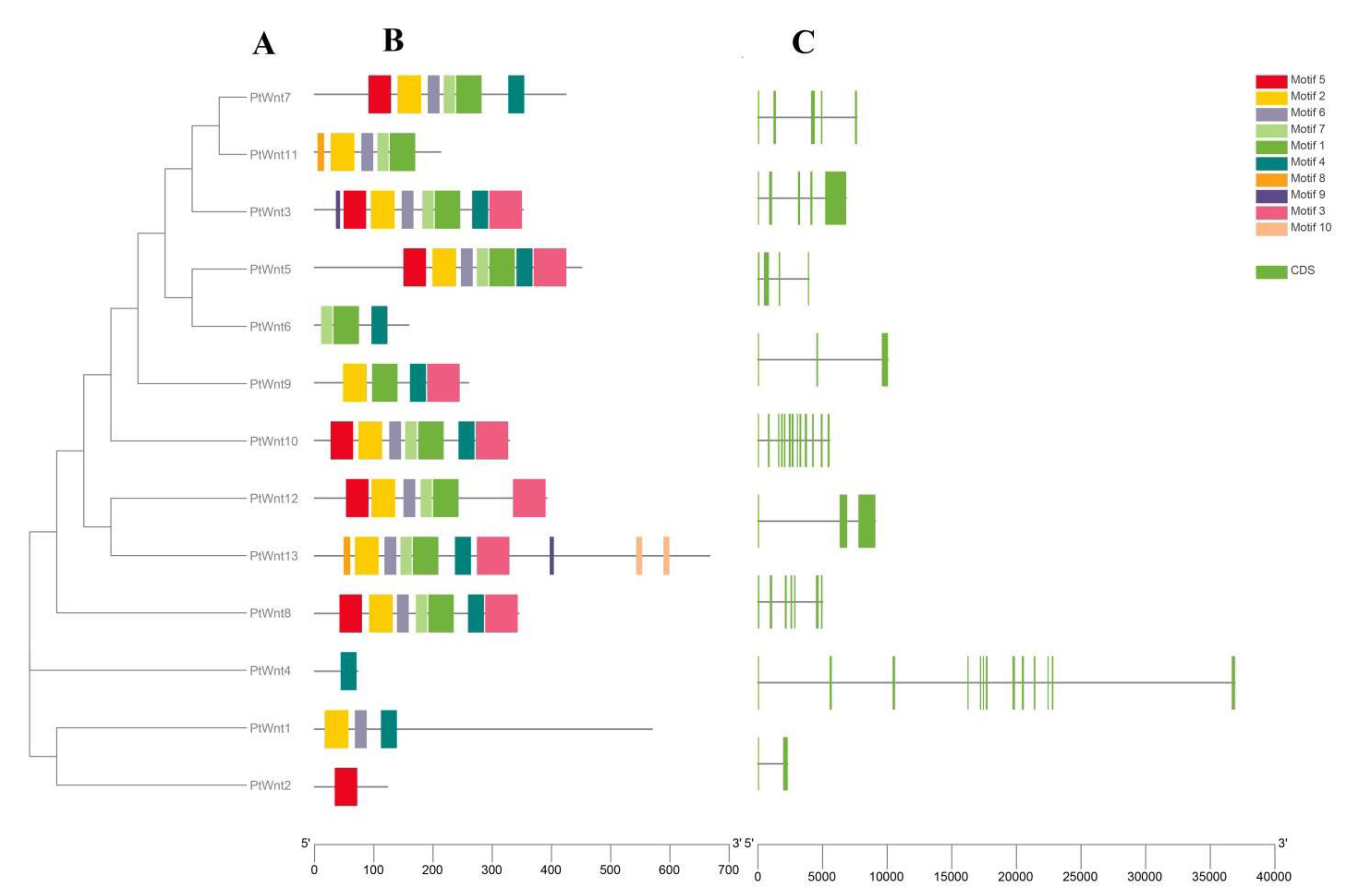
The phylogenetic tree result revealed that the members of the Wnt family in P. trituberculatus were classified into three main groups and nine subgroups (Figure 3A). Meanwhile, the members of subgroup 3 (PtWnt1, PtWnt2, PtWnt4, and PtWnt8) were clustered in a clade. The Multiple Expectation maximization for Motif Elicitation (MEME) online software was used for conserved motif analysis among different PtWnt proteins, and a total of 10 conserved motifs were predicted (Table S5, Figure 3B). The types and numbers of motifs in different PtWnt groups had several differences. Among these, motif 1 was conserved in all 10 PtWnts of group 1, while motif 7 was observed in nine PtWnts of group 1. Motifs 1, 2, 3, 4, 5, 6, and 7 were shared in four PtWnts. Motifs 9 and 10 were only found in two PtWnts. Motif 4 was present in all three groups. Moreover, we observed that the proteins of group I contained more than three motifs. As an exception, PtWnt6 had only three motifs. In addition, PtWnt4 in group II and PtWnt2 in group III only contained one motif (motif 4 or motif 5). The Coding sequence (CDS) structure of PtWnt genes was further characterized. The results showed that the numbers of CDSs in each PtWnt gene varied from 3 to 13 (Figure 3C). The CDSs of PtWnts were disrupted by introns. However, the exact exon-intron structure of PtWnt genes was not further characterized.
The results of the analysis of the structures of PtWnt proteins indicated that all the deduced PtWnt proteins contained a conserved cysteine-rich Wnt1 domain. The signal peptides of 20–31 amino acid residues were only identified in PtWnt2, PtWnt10, and PtWnt13 (Figure 4).

3.4. Chromosomal Distribution

P. trituberculatus have 53 pairs of chromosomes, the chromosomal distribution showed that nine PtWnts were located in five chromosomes of P. trituberculatus (Figure 5). The distribution of genes among different chromosomes was uneven, ranging from 1 to 3 genes per chromosome. In detail, Chromosome 28 and Chromosome 40 harbored only one PtWnt, whereas Chromosome 3 and Chromosome 24 harbored two PtWnts. A maximum of three genes were clustered on chromosome 30. To better understand the evolutionary constraints acting on Wnt gene family, the Ka/Ks ratios of the Wnt gene pairs were calculated. The ratio of Ka/Ks of tandem duplication gene pairs of PtWnt6 and PtWnt7 was 0.3606.

3.5. Regeneration Limb Transcriptome-Based Expression Profiling and GO and KEGG Enrichment Analysis of PtWnt Genes

In order to discover genes underlying the molecular mechanisms governing regenerated limb development in P. trituberculatus, we applied next-generation sequencing technology to identify functional genes potentially associated with limb regeneration in our previous study [24] and found differentially expressed PtWnt genes in the comparison of regenerating limb BM and NM. In this study, the available mRNA-seq data of relative expression in regenerating limb BM and NM for the Wnt gene family were obtained and analyzed for gene expression pattern, followed by GO and KEGG enrichment analyses.
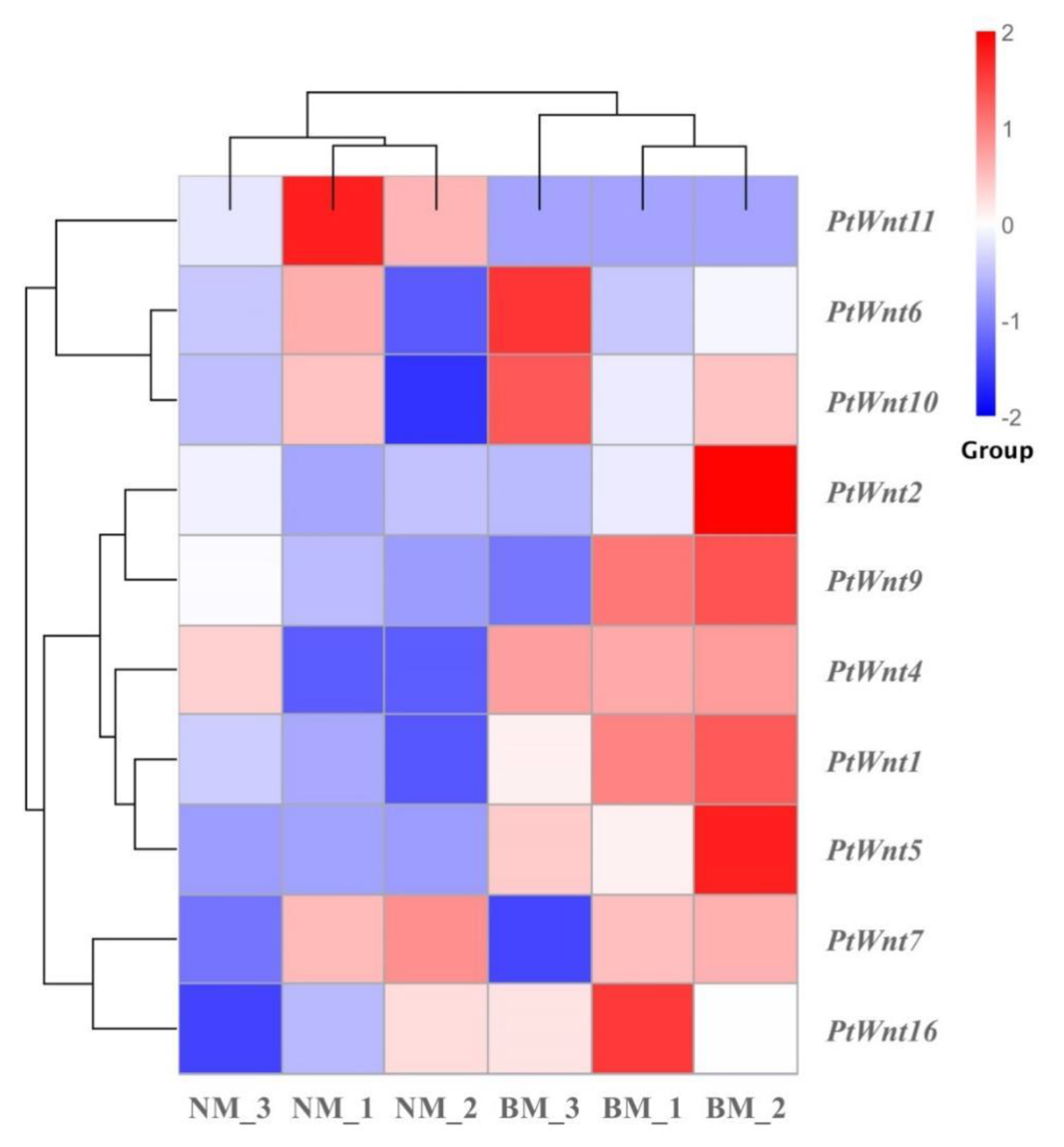
Ten of 13 PtWnt genes (PtWnt1, PtWnt2, PtWnt4, PtWnt5, PtWnt6, PtWnt7, PtWnt9, PtWnt10, PtWnt11, and PtWnt16) were differentially expressed in the comparison of regenerating limb BM and NM (Figure 6 and Table S6). According to the expression patterns in regenerating limb BM and NM, the 10 PtWnt genes were clustered into three main groups, and the genes clustered in the same group shared a similar expression pattern. As shown in Figure 6, PtWnt6, PtWnt10, and PtWnt11 clustered together into the first group, as they appeared to have higher expression levels in NM than in BM. In the second group, PtWnt2, PtWnt9, PtWnt4, PtWnt1, and PtWnt5 were clustered together as they showed higher expression levels in BM than in NM. Finally, PtWnt7 and PtWnt16 were clustered into a single branch, as their expression levels in the two compared groups were randomly distributed.
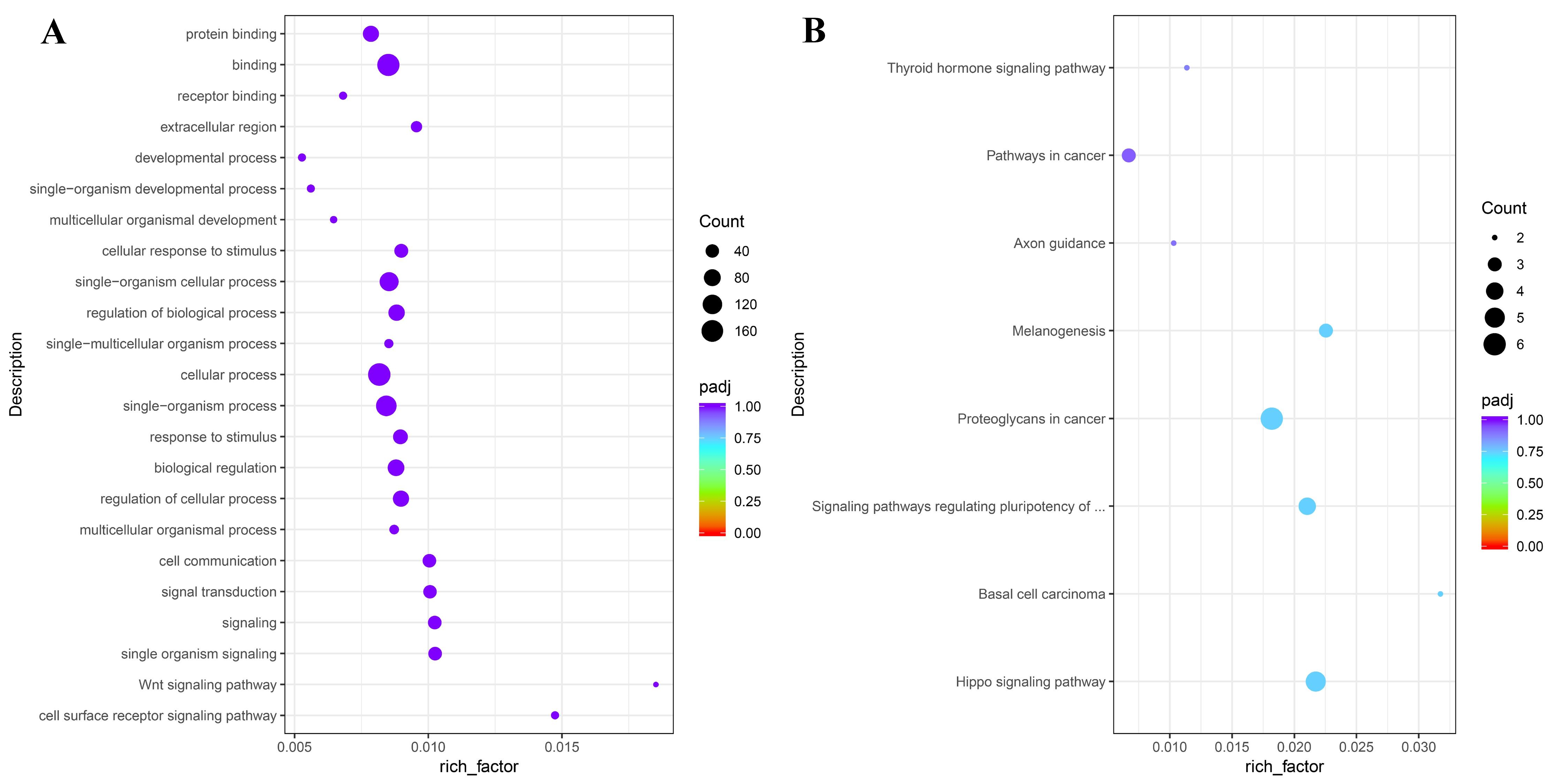
To associate PtWnt genes exhibiting different expression patterns with physiological and morphological changes during limb regeneration, we performed GO enrichment analysis using the GOseq R package. The results suggested that all the PtWnts were sorted into 22 functional groups that belonged to three main GO categories: Biological processes, cellular components, and molecular functions (Figure 7A and Table S7). In the biological process category, the cell surface receptor signaling pathway (GO: 0007166), Wnt signaling pathway (GO: 0016055), single organism signaling (GO: 0044700), signaling (GO: 0023052), and signal transduction (GO: 0007165) were prominently represented. Receptor binding (GO: 0005102), binding (GO: 0005488), and protein binding (GO: 0005515) were included in the molecular function category, while extracellular region (GO: 0005576) was only included in the cellular component category.
To understand potential gene interactions and the biological functions underlying limb regeneration, all PtWnts were searched against the KEGG database and subsequently assigned to eight pathways (Figure 7B and Table S8). The most dramatically enriched pathway was the hippo signaling pathway (ko04390), followed by basal cell carcinoma (ko05217), signaling pathways regulating pluripotency of stem cells (ko04550), proteoglycans in cancer (ko05205), melanogenesis (ko04916), axon guidance (ko04360), pathways in cancer (ko05200), and thyroid hormone signaling pathway (ko04919).

3.6. Expression Profiling of PtWnt Genes at Different Developmental Stages of Limb Regeneration

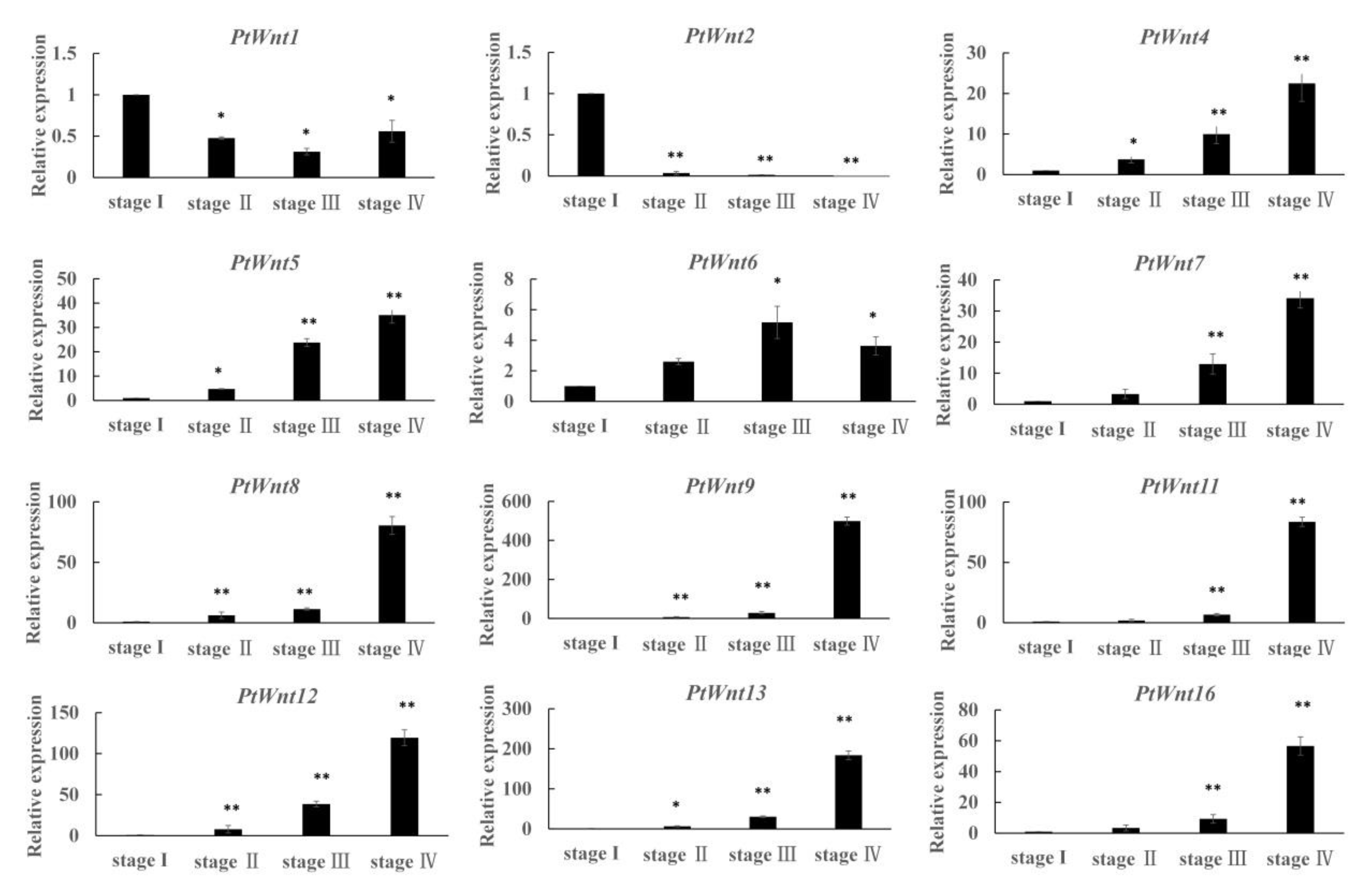
To study the expression patterns of PtWnts during limb regeneration, 12 PtWnts were selected for qRT-PCR. The results showed that the expression levels of nine PtWnts (except PtWnt1, PtWnt2, and PtWnt6) had a similar trend during limb regeneration. Among these, PtWnt4, PtWnt5, and PtWnt13 began their expression at stage I, increased significantly (p < 0.05) at stage II, and increased most significantly (p < 0.01) at stage III and stage IV (Figure 8). In addition, the expression levels of PtWnt7, PtWnt11, and PtWnt16 had a similar trend of increasing slightly at stage I and then clearly increasing (p < 0.01) in stage III and stage IV, throughout the process of limb regeneration. Moreover, PtWnt8, PtWnt9, and PtWnt12 were most significantly up-regulated (p < 0.01) from stage II to stage IV compared to the expression level at stage I. In contrast, PtWnt2 had the highest expression level at stage I and showed a downward trend from stage II to stage IV. Interestingly, the expression of PtWnt1 declined significantly (p < 0.05) at stage II and stage III compared to stage I and increased from stage III to stage IV but was still significantly lower (p < 0.05) than at stage I. Conversely, PtWnt6 continued to increase from stage I to stage III, with no significant difference between stage I and stage II, and then declined in stage IV (p < 0.05).

4. Discussion

Regulation of Wnt signaling and Wnt ligands during regeneration has been widely studied during various regeneration processes of vertebrate and invertebrate animals such as Xenopus limb regeneration [10,38], zebrafish retina regeneration [39,40], and insect limb regeneration [41,42]. Different species of arthropods have different numbers of Wnt family members. In this study, we identified 13 Wnt genes from the newly assembled genome of P. trituberculatus [22], a number that was greater than that in L. vannamei [21] and other arthropods. The higher number of Wnt family members in P. trituberculatus and L. vannamei than in other arthropods is the ultimate result of their higher gene abundance and repetitive sequences of their genome [22]. In contrast, our results suggest that the number of Wnt subfamilies in crustaceans is evolutionarily conserved.
To delineate the evolutionary history and more accurately predict the function of PtWnts, a phylogenetic tree was constructed using the amino acid sequences of P. trituberculatus and L. vannamei. As shown in Figure 2, PtWnt genes were clustered with respective counterparts as expected, and 8 clades were generated. Most clades of orthologues (except for the Wnt4 and Wnt6 clades) were divided into two branches, a P. trituberculatus branch and a L. vannamei branch, suggesting the distant evolutionary relationships among Wnts between P. trituberculatus and L. vannamei. The results were identical to those in previous reports concerning Wnt gene family function and evolution in mollusks [43]. Notably, the gene number of P. trituberculatus was not consistent with L. vannamei in the same clade, which could be attributed to their name annotation depending on their position on the chromosome. In general, Wnt genes have been relatively conserved during evolution.
As shown in Figure 3, only the CDS structure was present, while the main introns and exons of PtWnts were not obtained, which may have contributed to the genome quality. However, the PtWnt gene structure was conserved, as all the deduced PtWnt proteins had a conserved cysteine-rich Wnt1 domain, similar to other crustacean species [21]. The motif composition showed that the protein structures of the PtWnt family were broadly conserved, and some motifs were prevalent in the 13 PtWnt proteins, such as motif 1 and motif 4 in 10 PtWnt proteins and motif 7 in nine proteins. These results demonstrated that the protein domain and motifs of PtWnts were highly conservative. Moreover, the conservative motif of PtWnt proteins also highlights their evolutionary proximity [44]. As a result, the motif analysis further confirmed the annotation of PtWnt genes.
Compared with other previous Wnt gene family studies in crustaceans, this is the first report to map their chromosomal locations and to indicate the occurrence of chromosome duplication events during evolution. Chromosome localization of genes is the result of both fragment replication and tandem duplication and plays an important role in promoting gene family expansion [45,46]. In this study, PtWnt6 and PtWnt7 evolved from tandem duplication, indicating that segment duplication may be a major driving force for PtWnt gene evolution [47].
GO analysis was performed to characterize the functions of PtWnt proteins in limb regeneration of P. trituberculatus. A number of Wnt proteins have been characterized in crustaceans, and a functional analysis of their roles in the defense against pathogenic virus infection has been performed concerning the innate immunity of L. vannamei [21,37]. However, there are limited reports on crab species with which to understand the biological processes associated with Wnt genes during limb regeneration. In this study, cell surface receptor signaling pathway (GO:0007166), single organism signaling (GO:0044700), signaling (GO:0023052), and extracellular region (GO:0005576) were the most enriched processes, besides the Wnt signaling pathway (GO:0016055) that has been well described in previous regeneration studies of other animals [41,48,49,50]. These processes are responsible for their functions during limb regeneration and development. For example, Wnt proteins play roles as secreted glycoproteins by binding to their respective receptors on the cell surface, a process that is pivotal in the signaling pathways. Moreover, the effects of Wnt signaling include a wide range of cellular responses, including cell fate determination and cytoskeletal remodeling, that belong to extracellular region processes [51]. The exact function of the PtWnt gene family involved in a complex network of signaling events of these enriched processes during limb regeneration needs further investigation. Pathway-based KEGG enriched results are helpful for understanding the biological functions of PtWnt genes during limb regeneration, which our previous transcriptome-based analysis [24].
At present, expression and functional analyses of Wnt genes have been reported in several Arthropoda species, including Tribolium castaneum [15] and L. vannamei [21,37]. However, most knowledge concerning the expression of Wnt genes comes from the analysis of tissue-specific expression, and little is known about their expression patterns during regeneration. Recent reports have provided valuable insights into Wnt function and evolution during organ regeneration in other invertebrates such as Hydra [12] and Ruditapes philippinarum [43]. To date, there are no reports on the characterization of the Wnt gene family or their functions in crustaceans. According to our published transcriptome data on regenerating limbs in P. trituberculatus, most PtWnt genes had a higher FPKM value in the regenerating limb BM than in NM (Figure 6) [24]. In that study, the expression levels of Wnt genes in P. trituberculatus were measured during different developmental stages of limb regeneration using qPCR.
An increasing number of gene family functional studies have shown that genes belonging to the same group seem to have similar expression patterns [46]. Moreover, different Wnt ligands have different signal transduction properties during animal regeneration process [12,16]. It has been reported that Wnt1, Wnt3a, and Wnt8 play roles in the transformation and axial growth of mouse and Xenopus embryos through the classic Wnt/β-Catenin pathway [17,18], while Wnt4, Wnt5A, and Wnt11 can activate the non-canonical β-catenin-independent Wnt pathway and affect morphogenesis by inducing the extension of the epithelial layer [19,20]. In our study, five β-catenin-independent Wnt signaling members (Wnt4, Wnt5, Wnt7, Wnt11, and Wnt16) had a similar expression pattern, beginning their expression at stage I, increasing the level at stage II, and increasing most significantly from stage III to stage IV (Figure 8). However, the β-catenin-dependent Wnt signaling members showed different expression trends at different stages. For example, PtWnt2 had the highest expression level at stage I and showed a downward trend from stage II to stage IV. Another β-catenin-dependent signaling gene, PtWnt1, showed the highest expression level at stage I, which declined significantly at stage II and stage III and increased again from stage III to stage IV. Our results showed that there were similar expression patterns not only in the same subfamily but also in some family members. Moreover, it can be predicted that the canonical Wnt/β-catenin signaling pathway and the non-canonical Wnt pathway mediated by different Wnt ligands may cooperate to regulate the preciseness of unknown target genes through cascaded signaling molecules and transcription factors in the process of limb regeneration of P. trituberculatus [17,18,38,50]. The above results indicate the complexity and special mechanism of the Wnt signaling pathway regulating regeneration in crustaceans. In the near future, RNAi and CRISPR/Cas9 techniques will be utilized to uncover the functions of these Wnt genes.

5. Conclusions

Our study represents a comprehensive overview of the Wnt gene family in P. trituberculatus. To summarize, a total of 13 Wnt genes were identified from P. trituberculatus and classified into 11 groups according to the conserved protein domains and phylogenetic analysis. Then, the chromosome locations, gene structures, and motif composition were characterized. Published transcription data and qRT-PCR for different stages of limb regeneration were analyzed to gain further understanding regarding the putative functions of PtWnt genes during limb regeneration. These findings will facilitate future functional characterization of PtWnt genes and provide a foundation for exploring the molecular mechanism during limb regeneration in P. trituberculatus and other crustaceans.

Supplementary Materials

The following supporting information can be downloaded at: https://www.mdpi.com/article/10.3390/fishes7050258/s1, Table S1: Details of Wnt unigenes annotated in NR database from limb regeneration transcriptome of P. trituberculatus; Table S2: Fragments Per Kilobase of transcript sequences per Million base pairs (FPKM) values of Wnt genes from limb regeneration transcriptome of P. trituberculatus; Table S3: Combination of primers used in quantitative real-time PCR (qRT-PCR) assays; Table S4: The KaKs ratios results for the replicated gene pairs of Ptwnt6 and Ptwnt7; Table S5: Multiple Expectation maximization for Motif Elicitation (MEME) motif analysis of Wnt unigenes of P. trituberculatus; Table S6: List of the selected Wnt genes for differential expression analysis; Table S7: Go enrichment of all the annotated PtWnt genes in the comparison regenerative limb bud muscle (BM) and normal muscle (NM); Table S8: Kyoto Encyclopedia of Genes and Genomes (KEGG) enrichment of all the annotated PtWnt genes in the comparison regenerative limb bud muscle (BM) and normal muscle (NM).

Author Contributions

Conceptualization, Y.F., L.L. and C.W.; methodology, Y.F. and L.L.; validation, Y.F.; formal analysis, Y.X.; resources, X.H. and J.H.; data curation, Y.F.; writing—original draft preparation, Y.F.; writing—review and editing, L.L.; supervision, L.L.; funding acquisition, L.L. All authors have read and agreed to the published version of the manuscript.

Funding

This research was funded by the National Natural Science Foundation of China, grant number 32172993; the Ningbo Natural Science Foundation, grant number 202003N4181; Zhejiang Marine Fisheries Research Institute, Key Laboratory of Mariculturre & Enhancement of Zhejiang Province open topic, grant number 2020KF015.

Institutional Review Board Statement

The studies in Portunus trituberculatus were reviewed and approved by the Committee for the Welfare and Ethics of Laboratory Animals of Ningbo University (Approval numbers: 20190410-042, approved on 10 April 2019).

Data Availability Statement

All data generated or analyzed during this study are included in this published article and its supplementary information files.

Conflicts of Interest

The authors declare no conflict of interest.

References

  1. Yokoyama, H.; Ogino, H.; Stoick-Cooper, C.L.; Grainger, R.M.; Moon, R.T. Wnt/beta-catenin signaling has an essential role in the initiation of limb regeneration. Dev. Biol. 2007, 306, 170–178. [Google Scholar] [CrossRef]
  2. Vafopoulou, X. Mechanisms of wound repair in crayfish. ISJ 2009, 6, 125–137. [Google Scholar]
  3. Hopkins, P.M. Regeneration of walking legs in the fiddler crab Uca pugilator. Am. Zool. 1993, 33, 348–356. [Google Scholar] [CrossRef]
  4. Hopkins, P.M. Limb regeneration in the fiddler crab, Uca pugilator: Hormonal and growth factor control. Am. Zool. 2001, 41, 389–398. [Google Scholar] [CrossRef]
  5. Cooper, R.L. Development of sensory processes during limb regeneration in adult crayfish. J. Exp. Biol. 1998, 201, 1745–1752. [Google Scholar] [CrossRef]
  6. Das, S.; Durica, D.S. Ecdysteroid receptor signaling disruption obstructs blastemal cell proliferation during limb regeneration in the fiddler crab, Uca pugilator. Mol. Cell. Endocrinol. 2013, 365, 249–259. [Google Scholar] [CrossRef]
  7. Zhang, C.; Zhang, Q.; Song, X.Z.; Pang, Y.Y.; Song, Y.M.; Wang, Y.Y.; He, L.; Lv, J.H.; Cheng, Y.X.; Yang, X.Z. L-tryptophan promotes the cheliped regeneration of Chinese mitten crab (Eriocheir sinensis) through melatonin, serotonin and dopamine involvement. Aquaculture 2019, 511, 734205. [Google Scholar] [CrossRef]
  8. Ventura, T.; Stewart, M.J.; Chandler, J.C.; Rotgans, B.; Elizur, A.; Hewitt, A.W. Molecular aspects of eye development and regeneration in the Australian redclaw crayfish, Cherax quadricarinatus. Aquac. Fish. 2019, 4, 27–36. [Google Scholar] [CrossRef]
  9. Ren, Z.M.; Fu, Y.Y.; Liu, L.; Liu, X.; Wang, C.L. Expression and function of WNT4 involved in larvae development and limb regeneration in Portunus trituberculatus. J. Oceanol. Limnol. 2021, 39, 306–316. [Google Scholar] [CrossRef]
  10. Kawakami, Y.; Esteban, C.R.; Raya, M.; Kawakami, H.; Martí, M.; Dubova, I.; Izpisúa Belmonte, J.C. Wnt/beta-catenin signaling regulates vertebrate limb regeneration. Genes Dev. 2006, 20, 3232–3237. [Google Scholar] [CrossRef]
  11. De Robertis, E.M. Wnt signaling in axial patterning and regeneration: Lessons from planaria. Sci. Signal. 2010, 6, pe21. [Google Scholar] [CrossRef] [PubMed] [Green Version]
  12. Lengfeld, T.; Watanabe, H.; Simakov, O.; Lindgens, D.; Gee, L.; Law, L.; Schmidt, H.A.; Özbek, S.; Bode, H.; Holstein, T.W. Multiple Wnts are involved in Hydra organizer formation and regeneration. Dev. Biol. 2009, 330, 186–199. [Google Scholar] [CrossRef] [PubMed]
  13. Cho, S.J.; Vallès, Y.; Giani, V.C., Jr.; Seaver, E.C.; Weisblat, D.A. Evolutionary dynamics of the Wnt gene family: A lophotrochozoan perspective. Mol. Biol. Evol. 2010, 27, 1645–1658. [Google Scholar] [CrossRef] [PubMed]
  14. Nusse, R. An ancient cluster of Wnt paralogues. Trends Genet. 2001, 17, 443. [Google Scholar] [CrossRef]
  15. Bolognesi, R.; Farzana, L.; Fischer, T.D.; Brown, S.J. Multiple Wnt genes are required for segmentation in the short-germ embryo of Tribolium castaneum. Curr. Biol. 2008, 18, 1624–1629. [Google Scholar] [CrossRef] [PubMed]
  16. Nusse, R. Wnt signaling and stem cell control. Cell Res. 2008, 18, 523–527. [Google Scholar] [CrossRef]
  17. Shimizu, H.; Julius, M.A.; Giarré, M.; Zheng, Z.; Brown, A.M.; Kitajewski, J. Transformation by Wnt family proteins correlates with regulation of beta-catenin. Cell Growth Differ. 1997, 8, 1349–1358. [Google Scholar]
  18. Miller, J.R. The Wnts. Genome Biol. 2001, 3, REVIEWS3001. [Google Scholar] [CrossRef]
  19. Tada, M.; Concha, M.L.; Heisenberg, C.-P. Non-canonical Wnt signalling and regulation of gastrulation movements. Semin. Cell Dev. Biol. 2002, 13, 251–260. [Google Scholar] [CrossRef]
  20. Tada, M.; Smith, J.C. Xwnt11 is a target of Xenopus Brachyury: Regulation of gastrulation movements via Dishevelled, but not through the canonical Wnt pathway. Development 2000, 127, 2227–2238. [Google Scholar] [CrossRef]
  21. Du, J.L.; Zhang, X.J.; Yuan, J.B.; Zhang, X.X.; Li, F.H.; Xiang, J.H. Wnt gene family members and their expression profiling in Litopenaeus vannamei. Fish Shellfish Immunol. 2018, 77, 233–243. [Google Scholar] [CrossRef] [PubMed]
  22. Tang, B.P.; Zhang, D.Z.; Li, H.R.; Jiang, S.H.; Zhang, H.B.; Xuan, F.J.; Ge, B.M.; Wang, Z.F.; Liu, Y.; Sha, Z.L.; et al. Chromosome-level genome assembly reveals the unique genome evolution of the swimming crab (Portunus trituberculatus). Gigascience 2020, 9, giz161. [Google Scholar] [CrossRef]
  23. Lv, J.J.; Li, R.H.; Su, Z.C.; Gao, B.Q.; Ti, X.B.; Yan, D.P.; Liu, G.J.; Liu, P.; Wang, C.L.; Li, J. A chromosome-level genome of Portunus trituberculatus provides insights into its evolution, salinity adaptation and sex determination. Mol. Ecol. Resour. 2021, 22, 1606–1625. [Google Scholar] [CrossRef] [PubMed]
  24. Liu, L.; Fu, Y.Y.; Zhu, F.; Mu, C.K.; Li, R.H.; Song, W.W.; Shi, C.; Ye, Y.F.; Wang, C.L. Transcriptomic analysis of Portunus trituberculatus reveals a critical role for WNT4 and WNT signalling in limb regeneration. Gene 2018, 658, 113–122. [Google Scholar] [CrossRef] [PubMed]
  25. Fu, Y.Y.; Liu, L.; Wang, C.L.; Zhu, F.; Liu, X. Suppression of limb regeneration by RNA interference of WNT4 in the swimming crab Portunus trituberculatus. Comp. Biochem. Physiol. Part B Biochem. Mol. Biol. 2019, 234, 41–49. [Google Scholar] [CrossRef]
  26. Finn, R.D.; Clements, J.; Eddy, S.R. HMMER web server: Interactive sequence similarity searching. Nucleic Acids Res. 2011, 39, W29–W37. [Google Scholar] [CrossRef]
  27. Thompson, J.D.; Gibson, T.J.; Higgins, D.G. Multiple Sequence Alignment Using ClustalW and ClustalX. Curr. Protoc. Bioinform. 2003, 2.3.1–2.3.22. [Google Scholar] [CrossRef]
  28. Kumar, S.; Stecher, G.; Tamura, K. MEGA7: Molecular evolutionary genetics analysis version 7.0 for bigger datasets. Mol. Biol. Evol. 2016, 33, 1870–1874. [Google Scholar] [CrossRef]
  29. Hu, B.; Jin, J.P.; Guo, A.Y.; Zhang, H.; Luo, J.C.; Gao, G. GSDS 2.0: An upgraded gene feature visualization server. Bioinformatics 2015, 31, 1296–1297. [Google Scholar] [CrossRef]
  30. Bailey, T.L.; Williams, N.; Misleh, C.; Li, W.W. MEME: Discovering and analyzing DNA and protein sequence motifs. Nucleic Acids Res. 2006, 34, W369–W373. [Google Scholar] [CrossRef]
  31. Chen, C.J.; Chen, H.; He, Y.H.; Xia, R. TBtools, a Toolkit for Biologists integrating various biological data handling tools with a user-friendly interface. bioRxiv 2019, 289660. [Google Scholar] [CrossRef]
  32. Voorrips, R.E. MapChart: Software for the graphical presentation of linkage maps and QTLs. J. Hered. 2002, 93, 77–78. [Google Scholar] [CrossRef] [PubMed] [Green Version]
  33. Yang, Z. PAML 4: Phylogenetic analysis by maximum likelihood. Mol. Biol. Evol. 2007, 24, 1586–1591. [Google Scholar] [CrossRef] [PubMed]
  34. Mao, X.; Cai, T.; Olyarchuk, J.G.; Wei, L. Automated genome annotation and pathway identification using the KEGG Orthology (KO) as a controlled vocabulary. Bioinformatics 2005, 21, 3787–3793. [Google Scholar] [CrossRef] [PubMed]
  35. Lalitha, S. Primer premier 5. Biotech Softw. Internet Rep. 2000, 1, 270–272. [Google Scholar] [CrossRef]
  36. Livak, K.J.; Schmittgen, T.D. Analysis of relative gene expression data using real-time quantitative PCR and the 2−ΔΔCT Method. Methods 2001, 25, 402–408. [Google Scholar] [CrossRef]
  37. Zhang, S.; Li, C.Z.; Yang, Q.H.; Dong, X.H.; Chi, S.Y.; Liu, H.Y.; Shi, L.L.; Tan, B.P. Molecular cloning, characterization and expression analysis of Wnt4, Wnt5, Wnt6, Wnt7, Wnt10 and Wnt16 from Litopenaeus vannamei. Fish Shellfish Immunol. 2016, 54, 445–455. [Google Scholar] [CrossRef]
  38. Yokoyama, H.; Maruoka, T.; Ochi, H.; Aruga, A.; Ohgo, S.; Ogino, H.; Tamura, K. Different requirement for Wnt/β-catenin signaling in limb regeneration of larval and adult Xenopus. PLoS ONE 2011, 6, e21721. [Google Scholar] [CrossRef]
  39. Ramachandran, R.; Zhao, X.F.; Goldman, D. Ascl1a/Dkk/beta-catenin signaling pathway is necessary and glycogen synthase kinase-3beta inhibition is sufficient for zebrafish retina regeneration. Proc. Natl. Acad. Sci. USA 2011, 108, 15858–15863. [Google Scholar] [CrossRef]
  40. Meyers, J.R.; Hu, L.; Moses, A.; Kaboli, K.; Papandrea, A.; Raymond, P.A. β-catenin/Wnt signaling controls progenitor fate in the developing and regenerating zebrafish retina. Neural Dev. 2012, 7, 30. [Google Scholar] [CrossRef]
  41. Smith-Bolton, R.K.; Worley, M.I.; Kanda, H.; Hariharan, I.K. Regenerative growth in Drosophila imaginal discs is regulated by Wingless and Myc. Dev. Cell 2009, 16, 797–809. [Google Scholar] [CrossRef]
  42. Shah, M.V.; Namigai, E.K.O.; Suzuki, Y. The role of canonical Wnt signaling in leg regeneration and metamorphosis in the red flour beetle Tribolium castaneum. Mech. Dev. 2011, 128, 342–358. [Google Scholar] [CrossRef] [PubMed]
  43. Bai, Y.T.; Nie, H.T.; Wang, Z.X.; Yan, X.W. Genome-wide identification and transcriptome-based expression profiling of Wnt gene family in Ruditapes philippinarum. Comp. Biochem. Physiol. Part D Genom. Proteom. 2020, 35, 100709. [Google Scholar] [CrossRef] [PubMed]
  44. Islam, M.D.S.; Choudhury, M.; Majlish, A.-N.K.; Islam, T.; Ghosh, A. Comprehensive genome-wide analysis of Glutathione S-transferase gene family in potato (Solanum tuberosum L.) and their expression profiling in various anatomical tissues and perturbation conditions. Gene 2018, 639, 149–162. [Google Scholar] [CrossRef]
  45. Zhou, Z.; Zhang, C.X.; Zhang, Y.L.; Wang, Q.; Li, W.X.; Tian, Y.; Cong, P.H. Bioinformatics and expression analysis of the LysM gene family in apple. Sci. Agric. Sin. 2014, 47, 2602–2612. [Google Scholar] [CrossRef]
  46. Yuan, G.P.; He, S.S.; Bian, S.X.; Han, X.L.; Liu, K.; Cong, P.H.; Zhang, C.X. Genome-wide identification and expression analysis of major latex protein (MLP) family genes in the apple (Malus domestica Borkh.) genome. Gene 2020, 733, 144275. [Google Scholar] [CrossRef] [PubMed]
  47. Cheng, C.; Wang, Y.; Chai, F.M.; Li, S.H.; Xin, H.P.; Liang, Z.C. Genome-wide identification and characterization of the 14-3-3 family in Vitis vinifera L. during berry development and cold- and heat-stress response. BMC Genom. 2018, 19, 579. [Google Scholar] [CrossRef] [PubMed]
  48. Takata, H.; Terada, K.; Oka, H.; Sunada, Y.; Moriguchi, T.; Nohno, T. Involvement of Wnt4 signaling during myogenic proliferation and differentiation of skeletal muscle. Dev. Dyn. 2007, 236, 2800–2807. [Google Scholar] [CrossRef] [PubMed]
  49. von Maltzahn, J.; Chang, N.C.; Bentzinger, C.F.; Rudnicki, M.A. Wnt signaling in myogenesis. Trends Cell Biol. 2012, 22, 602–609. [Google Scholar] [CrossRef]
  50. Ozhan, G.; Weidinger, G. Wnt/β-catenin signaling in heart regeneration. Cell Regen. 2015, 4, 3. [Google Scholar] [CrossRef]
  51. Tanaka, E.M.; Reddien, P.W. The cellular basis for animal regeneration. Dev. Cell 2011, 21, 172–185. [Google Scholar] [CrossRef] [PubMed] [Green Version]
Figure 1. Four landmark developmental stages for the regeneration of the limb in P. trituberculatus. (A) Stage I of regenerative limb with wound healing; (B) stage II of regenerative limb with regeneration limb bud appear; (C) stage III of regenerative limb with segmentation of regeneration limb bud; (D) stage IV of regenerative limb with mature regeneration limb bud.
Fishes 07 00258 g001
Figure 2. Phylogenetic analysis of PtWnt and LvWnt proteins. The ● represent P. trituberculatus, and the ○ represent L. vannamei. LvWnt1, L. vannamei Wnt1 (Accession No. AXN72346.1); LvWnt2, L. vannamei Wnt2 (Accession No. AXN72347.1); LvWnt3, L. vannamei Wnt3 (Accession No. ROT82629.1); LvWnt4, L. vannamei Wnt4 (Accession No. AXN72348.1); LvWnt5, L. vannamei Wnt5 (Accession No. AXN72349.1); LvWnt6, L. vannamei Wnt6 (Accession No. AXN72350.1); LvWnt7, L. vannamei Wnt7 (Accession No. AXN72351.1); LvWnt8, L. vannamei Wnt8 (Accession No. AXN72352.1); LvWnt9, L. vannamei Wnt9 (Accession No. AXN72353.1); LvWnt10, L. vannamei Wnt10 (Accession No. AXN72354.1); LvWnt11, L. vannamei Wnt11 (Accession No. AXN72355.1); LvWnt12, L. vannamei Wnt12 (Accession No. ROT67990.1); LvWnt14, L. vannamei Wnt14 (Accession No. ROT65815.1); LvWnt15, L. vannamei Wnt15 (Accession No. ROT61142.1); LvWnt16, L. vannamei Wnt16 (Accession No. AXN72356.1); LvWnt17, L. vannamei Wnt17 (Accession No. ROT60890.1); PtWnt4, P. trituberculatus Wnt4 (Accession No. MPC90022.1); PtWnt5, P. trituberculatus Wnt5 (Accession No. XP_045122243.1); Pt Wnt6, P. trituberculatus Wnt6 (Accession No. XP_045122246.1); PtWnt7, P. trituberculatus Wnt7 (Accession No. XP_045122227.1); PtWnt8, P. trituberculatus Wnt8 (Accession No. XP_045117623.1); PtWnt9, P. trituberculatus Wnt9 (Accession No. XP_045118050.1); PtWnt10, P. trituberculatus Wnt10 (Accession No. XP_045111453.1); PtWnt11, P. trituberculatus Wnt11 (Accession No. XP_045125872.1); PtWnt12, P. trituberculatus Wnt12 (Accession No. XP_045122415.1); PtWnt13, P. trituberculatus Wnt13 (Accession No. XP_045133204.1); Homo sapiens Wnt4 (Accession No. BAC23080); Mus musculus Wnt4 (Accession No. EDL29922); Danio rerio Wnt4 (Accession No. AAI63457); Xenopus laevis Wnt4 (Accession No. P49338); Gallus gallus Wnt4 (Accession No. NP_990114); H. sapiens Wnt5 (Accession No. BAB62039); M. musculus Wnt5 (Accession No. AAH51406); D. rerio Wnt5 (Accession No. AAI62981); X. laevis Wnt5 (Accession No. P33945); H. sapiens Wnt6 (Accession No. BAB55603); M. musculus Wnt6 (Accession No. AAH48700); Takifugu rubripes Wnt6 (Accession No. XP_011609805.1); X. laevis Wnt6 (Accession No. ABY53107); G. gallus Wnt6 (Accession No. NP_001007595); H. sapiens Wnt7 (Accession No. BAA82509); M. musculus Wnt7 (Accession No. EDK99298); G. gallus Wnt7 (Accession No. NP_989623); D. rerio Wnt7 (Accession No. NP_001020711); X. laevis Wnt7 (Accession No. AAI69513); H. sapiens Wnt10 (Accession No. AAG45153); M. musculus Wnt10 (Accession No. AAB08085); D. rerio Wnt10 (Accession No. AAA03431); G. gallus Wnt10 (Accession No. BAD61009); X. laevis Wnt10 (Accession No. ABG49498); H. sapiens Wnt16 (Accession No. AAD38052); M. musculus Wnt16 (Accession No. AAD49352); D. rerio Wnt16 (Accession No. NP_001093516).
Fishes 07 00258 g002
Figure 3. Unrooted neighbor-joining phylogenetic tree, architecture of conserved motifs, and gene structure of the PtWnt gene family. (A) The phylogenetic tree was generated based on the sequences of PtWnt proteins; (B) conserved motif analysis of PtWnt proteins. Different colored boxes represent different types of motifs; (C) coding sequence (CDS) structure of PtWnt gene family.
Fishes 07 00258 g003
Figure 4. Structure of PtWnt proteins. The yellow boxes indicate the conserved Wnt1 domains. The red boxes show signal peptides. The green boxes indicate non-specific domains.
Fishes 07 00258 g004
Figure 5. Chromosomal location and duplication events of nine Wnt genes on five different chromosomes of P. trituberculatus. Each chromosome number is indicated on the top of the chromosome bar. The scale of the chromosome is in millions of bases (Mb) (The length of green represents the number of bases). Each PtWnt gene is indicated on the exact position of a particular chromosome that can be calculated by using the scale provided on the left. Tandem duplicated genes are indicated by red boxes.
Fishes 07 00258 g005
Figure 6. Heatmap of PtWnt gene expression profiles in regenerating limb bud muscle (BM) and normal limb muscle (NM).
Fishes 07 00258 g006
Figure 7. Gene Ontology (GO) and Kyoto Encyclopedia of Genes and Genomes (KEGG) enrichment analyses of PtWnt gene family members between comparisons of regenerating limb bud muscle versus normal limb muscle. (A) GO enrichment results; (B) statistics of KEGG pathway enrichment.
Fishes 07 00258 g007
Figure 8. Expression patterns of 12 PtWnts at four developmental stages of the regenerating limb. The expression of target genes was detected by qRT-PCR and normalized to β-actin gene as the internal reference. The results were based on three independent biological replications and show mean values ± SD. (* p < 0.05, ** p < 0.01).
Fishes 07 00258 g008
Table 1. The identification of Wnt gene family members in P. trituberculatus.
Gene NameGenomic PositionChrChromosome LengthORFGene StandProtein Length (aa)Theoretical pIMolecular Weight (kDa)
PtWnt15,505,983–5,542,897310,158,9511710reverse5695.3958.91
PtWnt25,544,234–5,546,536310,158,951369reverse12210.0413.65
PtWnt328,210,259–28,217,0844029,382,9461056reverse3519.0939.62
PtWnt46,906,935–6,911,9302812,784,577219forward7210.548.03
PtWnt54,822,813–4,826,7643012,291,4751353reverse4509.3450.85
PtWnt64,925,695–4,935,7533012,291,475477forward1589.6717.95
PtWnt74,941,929–4,949,5903012,291,4751272forward4239.8747.80
PtWnt818,612,647–18,621,7342420,371,4381035forward3449.3738.59
PtWnt918,843,485–18,849,0222420,371,438780reverse2598.9728.59
PtWnt10 987 3289.8636.29
PtWnt11 639 2129.6824.57
PtWnt12 1176 3919.8543.25
PtWnt13 2001 6669.4973.68
ORF, open reading frame.
Publisher’s Note: MDPI stays neutral with regard to jurisdictional claims in published maps and institutional affiliations.