Article

Earlier Activation of Interferon and Pro-Inflammatory Response Is Beneficial to Largemouth Bass (Micropterus salmoides) against Rhabdovirus Infection

Zhejiang Fisheries Technical Extension Center, Hangzhou 310023, China
*
Author to whom correspondence should be addressed.
Fishes 2022, 7(2), 90; https://doi.org/10.3390/fishes7020090
Submission received: 7 March 2022 / Revised: 7 April 2022 / Accepted: 10 April 2022 / Published: 14 April 2022

Abstract

:
In order to understand the immune response of largemouth bass against Micropterus salmoides Rhabdovirus (MSRV), assisting disease resistance breeding, three largemouth bass breeding varieties Micropterus salmoides “Youlu No 3” (U3), “Youlu No 1” (U1) and “Zhelu No 1” (P1) were challenged intraperitoneally with MSRV. Serum and tissues were sampled to study the changes in non-specific immune parameters, viral loads, and transcript levels of immune-related genes, and the cumulative mortality rate was recorded daily for 14 days. The results showed that the cumulative mortality rates in the U1, P1, and U3 groups were 6.66% ± 2.89%, 3.33% ± 2.89%, and 0, respectively. The higher mortality may attribute to the increased viral loads after infection in the liver (2.79 × 10⁵ and 2.38 × 10⁵ vs. 1.3 × 10⁴ copies/mg), spleen (2.14 × 10⁵ and 9.40 × 10⁴ vs. 4.21 × 10³ copies/mg), and kidney (3.59 × 10⁴ and 8.40 × 10³ vs. 2.42 × 10³ copies/mg) in the U1 and P1 groups compared to the U3 group. The serum non-specific immune parameters (lysozyme, catalase, and acid phosphatase) were found to be increased significantly in the U3 group. In addition, the transcripts of interferon-related genes (IFN-γ, IRF3, and IRF7) and pro-inflammatory-related genes (TNF-α and IL-1β) exhibited up-regulation and peaked at 6 h post infection in the U3 group, which also exhibited up-regulation but peaked at 12–24 h post infection in the U1 and P1 groups. In conclusion, these findings indicate that earlier activation of interferon and pro-inflammatory response is beneficial to largemouth bass against MSRV infection. This experiment may provide an insight into understanding the immune mechanism of largemouth bass against MSRV infection and contributes to molecular-assisted selection.

1. Introduction

Largemouth bass (Micropterus salmoides) is a kind of freshwater fish with the advantages of fast growth, slender body, delicious meat, and without inter-muscular spines. Largemouth bass has become an important freshwater aquaculture variety in China which was introduced from the United States in 1983. In 2020, the production of largemouth bass reached 450,000 tons in China, of which Zhejiang Province produced 85,000 tons, accounting for 18.89% of the total production [1]. In recent years, due to the expansion of the intensive breeding scale and the significant increase in breeding density, diseases of largemouth bass have occurred frequently whether in the traditional breeding mode or the recirculating water track breeding mode [2,3,4]. In 2011, a new disease was reported in a largemouth bass farm in Guangdong Province, China, which led to about 200,000 dead fish with a body length of 2–5 cm, and typical clinical symptoms included irregular swimming and body bending [4,5]. Later, rhabdovirus was found to be the cause of the disease [3].
Rhabdoviruses are enveloped single-stranded RNA viruses which can infect a variety of organisms, including mammals, birds, reptiles, fish, insects, and plants [6]. Micropterus salmoides Rhabdovirus (MSRV) was mainly prevalent in the largemouth bass fry culture stage which led to the death of a large number of fry, and no death was found in adult fish [7]. In an epidemiological investigation at Jiaxing and Huzhou City in China, we also found that MSRV is prevalent at water temperatures between 20 °C and 25 °C, and the typical clinical symptoms of moribund fish include twirling at the water surface, abdominal distension and ascites, hyperemia, and hemorrhaging of internal organs. According to our investigation, the mortality could be as high as 90% mortality, which brought farmers huge economic losses. There is currently no suitable drug for the treatment of MSRV. Some studies have shown that subunit vaccines and live attenuated vaccines have good immune protection effects against MSRV of largemouth bass by injection or immersion [8,9]. In addition, Yang et al. [10] detected the anti-MSRV activity of 10 commonly used antiviral drugs by qPCR and found that ribavirin showed a good therapeutic effect on MSRV infection, which improved the survival rate of largemouth bass by injection or oral administration after MSRV infection. However, it is difficult to make suitable bait for feeding due to the small size of the fry, and there is also the risk of drug residue. It takes a long time for vaccines to be developed from laboratory research to industrial application, and further research is needed on the effect on production.
In addition to drug treatment and vaccination, selecting resistant varieties is another effective way to control diseases. Some research progress has been made in the breeding of new resistant fish and crustaceans, such as rainbow trout resistant to Flavobacterium psychrophilum [11], Litopenaeus vannamei resistant to white spot syndrome [12], and tilapia resistant to streptococcosis [13]. The M. salmoides “youlu 1” (U1) and “youlu 3” (U3) are two strains with the largest cultivating area in China at present [14,15], and the M. salmoides “Zhelu 1” (P1) is a new strain bred from this institution. In this study, the resistance of the three largemouth bass strains to MSRV infection was studied by comparing the viral load, non-specific immune response, transcript levels of immune-related genes, and cumulative mortality rates, so as to understand the immune response of largemouth bass against MSRV and provide some insights for selecting suitable culture species and disease-resistant breeding.

2. Materials and Methods

2.1. Fish

M. salmoides “Youlu 1” (U1) and “Youlu 3” (U3) with a body weight of (9.45 ± 1.34) g were purchased from Huzhou Huwang Aquatic Seed Co., Ltd. (China), and M. salmoides “Zhelu 1” (P1) was from our institution. Fish were temporarily kept in an aquarium with a circulating aquaculture system at a water temperature of 25 °C ± 0.5 °C, dissolved oxygen 5–7 mg/L, NH3/NH4+ <0.1 mg/L, and NO2 <0.1 mg/L. Fish were fed with commercial feed twice a day in the morning and evening. After the fish were temporarily raised for 14 days, 10 of each strain were randomly selected for bacteria and virus detection to ensure their health and disease-free status. The animal study protocol was approved by the Institutional Review Board of the Science Technology Department of Zhejiang Province for Zhejiang Fisheries Test and Aquatic Disease Prevention Center [SYXK (Zhe) 2020-0009, 26 May 2020].

2.2. Preparation of Crude Virus Extract

Samples of largemouth bass fry (body length 2.3–3.6 cm) with an outbreak of rhabdovirus disease were collected from a farm in Huzhou, Zhejiang Province. Total RNA was extracted from visceral tissues of ten fry using the SteadyPure Universal RNA Extraction Kit (Accurate Biology, Changsha, China), and RT-PCR detection was performed according to Lei et al. [16]. The amplified products were sequenced by Sangon Biotech (Shanghai, China) Co., Ltd. After PCR detection, fry infected with MSRV were ground with an appropriate amount of zirconium beads according to the ratio of 1 g of tissue to 10 mL of sterile normal saline. The ground tissue suspension was centrifuged at 4 °C (400× g, 15 min; then 800× g, 15 min) and the supernatant was filtered by a 0.22 μm sterile filter (Millipore, Burlington, MA, USA) to prepare crude virus extract, and stored at −80 °C for later use. Meanwhile, primers (Table 1) were designed according to the MSRV qPCR method (see Section 2.4) to detect the copy number of crude virus extract.

2.3. Experimental Design and Sampling

The experiment was divided into six groups with three replicates (30 fish per replicate). Fish in the U1, P1, and U3 groups from Micropterus salmoides “Youlu No 1” (U1), “Zhelu No 1” (P1), and “Youlu No 3” (U3), respectively, were challenged with MSRV, and fish in the U1-C, P1-C, and U3-C groups from Micropterus salmoides “Youlu No 1” (U1), “Zhelu No 1” (P1) and “Youlu No 3” (U3), respectively, were challenged with sterile PBS as a control. Fish were fasted 1 d before the experiment. Fish were anesthetized with MS-222 and injected intraperitoneally with the crude virus extract at a dose of 0.1 mL per tail. At 0, 6, 12, 24, 48, and 72 h post infection (hpi), fish were euthanized with the administration of MS-222, and the blood samples (n = 3) were collected from the caudal vein without anticoagulant. The blood was kept statically at room temperature for 2 h and centrifuged at 12,000× g for 15 min at 4 °C to obtain the serum for enzymatic analysis which was stored at −20 °C until further assayed. In addition, the spleen was sampled and stored in a sample protector for RNA/DNA (Takara, Kyoto, Japan) at −80 °C until RNA extraction to determine the immune-related gene expressions. Liver, spleen, and kidney were sampled under sterile operation and immediately used for the viral loads at 0, 6, 12, 24, 48, 72, 120, 168, 240, and 336 hpi.

2.4. Detecting Viral Loads in the Liver, Spleen and Kidney

The tissues were sampled aseptically, weighed, and then ground with 1 mL normal saline in tissue mortar. The homogenized tissues were used for RNA extraction. Total RNA was extracted using the SteadyPure Universal RNA Extraction Kit (Accurate Biology Inc., Changsha, China), and the concentration and purity of RNA were determined by ScanDrop2 (Analytikjena, Jena, Germany). cDNA was synthesized using the PrimeScript™ RT Reagent Kit with gDNA Eraser (Perfect Real Time) (Takara, Kyoto, Japan) according to the manufacturer’s instructions. Additionally, the viral loads in the liver, spleen, and kidney were detected with qPCR assay by using TB Green® Fast qPCR Mix (Takara, Kyoto, Japan). The primers were designed using the Primer Premier 5 software, and their sequences are listed in Table 1. The qPCR reaction consisted of 1 μL of cDNA template, 0.8 μL of upstream and downstream primers (10 μM), 10 μL of 2 × TB Green® Fast qPCR Mix, 0.4 μL of ROX, and 7 μL of ddH2O, and the reaction conditions were as follows: 94 °C for 2 min; 94 °C for 30 s, 60 °C for 30 s, 40 cycles; dissolution curve. Each sample was run in triplicate. The CT value obtained by qPCR was substituted into the standard curve (unpublished) to calculate the virus copy number, which was divided by the tissue weight to obtain the viral load (copies/mg).

2.5. Enzymatic Assays

The activities of lysozyme (LZM), catalase (CAT), superoxide dismutase (SOD), acid phosphatase (ACP), and alkaline phosphatase (AKP) were measured using assay kits (Nanjing Jiancheng Ins., Nanjing, China), and total complement CH50 was measured using a fish serum CH50 ELISA kit (Shanghai Zhen Ke Co., Ltd., Shanghai, China) in accordance with the manufacturer’s instructions.

2.6. Expression of Immune-Related Genes

Total RNA from the spleen was extracted as described in Section 2.4. The relative expression levels of interferon-gamma (IFN-γ), tumor necrosis factor-alpha (TNF-α), interferon regulatory factor 3 (IRF3), interferon regulatory factor 7 (IRF7), Akt, mammalian target of rapamycin (mTOR), and interleukin-1 beta (IL-1β) [17] were examined through qPCR, as described in Section 2.4. The primers were designed using the Primer Premier 5 software, and their sequences are listed in Table 1. Each sample was run in triplicate. The relative expression was analyzed using the 2∆∆Ct method according to Livak and Schmittgen [18].

2.7. Statistical Analysis

A one-way analysis of variance (ANOVA) using the SPSS 25.0 package (IBM Inc., Armonk, NY, USA) was used for data analysis. Values of p < 0.05 were considered statistically significant.

3. Results

3.1. Viral Loads in Liver, Spleen and Kidney

qPCR assay was employed to measure the amount of viral load in the liver (Figure 1A), spleen (Figure 1B), and kidney (Figure 1C). In the liver (Figure 1A), the viral load increased rapidly and peaked at 12–24 hpi. The highest viral load was significantly lower in the U3 group (1.3 × 10⁴ copies/mg) compared to the P1 (2.38 × 10⁵ copies/mg) and U1 groups (2.79 × 10⁵ copies/mg). In the spleen (Figure 1B), the viral load increased rapidly and peaked at 24 hpi, which was significantly lower in the U3 group compared to the P1 and U1 groups between 6 hpi and 48 hpi. In the kidney (Figure 1C), the viral load was significantly lower in the U3 group compared to the P1 and U1 groups between 6 hpi and 240 hpi. After 336 hpi, the viral loads in the liver, spleen, and kidney in the three groups were all lower than the detection threshold. The results demonstrated that the viral loads in the U3 group were lower in the liver, spleen, and kidney compared to the U1 and P1 groups. No virus was detected in the liver, spleen, and kidney in the P1-C, U1-C, and U3-C groups.

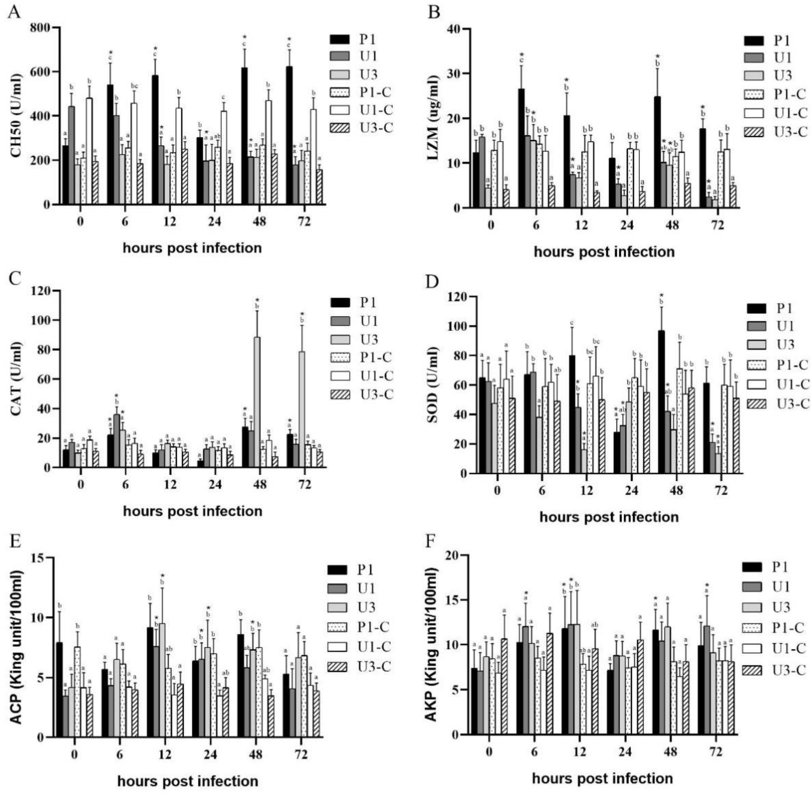
3.2. Enzymatic Assays

In the U3 group, the activities of LZM (Figure 2B), CAT (Figure 2C), and ACP (Figure 2E) increased significantly (p < 0.05) after infection, while the activity of SOD (Figure 2D) decreased significantly (p < 0.05) after infection. In the P1 group, the activities of CH50 (Figure 2A), LZM (Figure 2B), CAT (Figure 2C), SOD (Figure 2D), and AKP (Figure 2F) increased significantly (p < 0.05) after infection. In the U1 group, the activities of CAT (Figure 2C), ACP (Figure 2E), and AKP (Figure 2F) increased significantly (p < 0.05) after infection, while the activities of CH50 (Figure 2A), LZM (Figure 2B), and SOD (Figure 2D) decreased significantly (p < 0.05) after infection.

3.3. Transcripts of Some Immune-Related Genes

The transcript level of pro-inflammatory genes TNF-α (Figure 3B) and IL-1β (Figure 3G) and interferon-related genes IFN-γ (Figure 3A), IRF3 (Figure 3C), and IRF7 (Figure 3D) increased significantly (p < 0.05) after infection and peaked at 6 hpi in the U3 group. The transcript level of pro-inflammatory genes TNF-α (Figure 3B) and IL-1β (Figure 3G) and interferon-related genes IFN-γ (Figure 3A), IRF3 (Figure 3C), and IRF7 (Figure 3D) increased significantly (p < 0.05) after infection and peaked at 12–24 hpi in the P1 group. The transcript level of pro-inflammatory genes IL-1β (Figure 3G) and interferon-related genes IFN-γ (Figure 3A), IRF3 (Figure 3C), and IRF7 (Figure 3D) increased significantly (p < 0.05) after infection and peaked at 12–24 hpi in the U1 group. The transcript level of Akt (Figure 3E) and mTOR (Figure 3F) genes was not significantly changed at 6–72 hpi in all three groups. The change in gene expression levels of IFN-γ (Figure 3A) and IL-1β (Figure 3G) genes was greatest in the three groups.

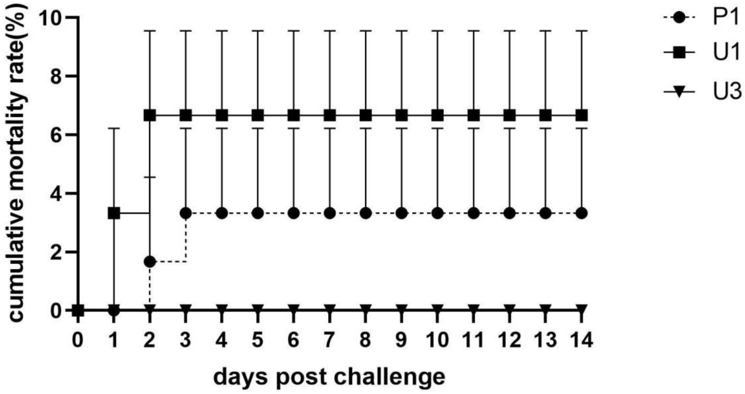
3.4. Cumulative Mortality Rate

Fish were observed daily, and dead fish were removed and sampled to confirm the infection of MSRV. The cumulative mortality was found to be 0, 3.33 ± 2.89%, and 6.67 ± 2.89% in the U3, P1, and U1 groups, respectively (Figure 4). The fish that died and survived had no obvious clinical symptoms. No fish died and no clinical symptoms were found in the P1-C, U1-C, and U3-C groups.

4. Discussion

Largemouth bass (Micropterus salmoides), as an economic fish, has been introduced to many countries for breeding. However, the expansion of breeding scale and the increase in breeding density have come along with a variety of diseases. In addition to the bacterial and parasitic diseases, largemouth bass is susceptible to a variety of DNA and RNA viruses, including iridovirus, reovirus, and rhabdovirus [2,4,6]. Different from most seawater or freshwater rhabdoviruses, Micropterus salmoides rhabdovirus (MSRV) showed significant age specificity and mainly infected fry, which has resulted in increased morbidity and mortality in recent years [3].
The outbreak of disease is related to the invasion and proliferation of pathogens in the host. Gao et al. [7] infected the skin cells of largemouth bass with MSRV and found that the virus replicated rapidly at 6–24 h, which resulted in significant pathological cell changes, and caspase-3 activity also increased significantly at 6–24 h. In this experiment, virus proliferation was detected at 6 hpi, and the viral loads in the liver, spleen, and kidney tissues in the P1 and U1 groups peaked at 12–24 hpi, which confirmed that MSRV can rapidly invade tissue for massive proliferation. Yuan et al. [19] infected grass carp ovary cells with MSRV and found that the relative copy number of virus reached 108 copies/mL at 24 h after infection by qPCR. In this study, the peak viral loads of the three groups were U1 > P1 > U3, which were 2.38 × 10⁵ copies/mg, 2.79 × 10⁵ copies/mg, and 1.3 × 10⁴ copies/mg, respectively. Correspondingly, the cumulative mortality rate was 6.66% ± 2.89%, 3.33% ± 2.89%, and 0, respectively. On the one hand, low virus copy numbers may be related to low mortality, as the viral load was lower in the liver, spleen, and kidney in the U3 group compared to the U1 and P1 groups. On the other hand, MSRV has obvious selectivity to fish age specificity, which was found to cause substantial death in 2–5 cm fry [4]. The fish used in this experiment were 9.45 ± 1.34 g, which may lead to low mortality in the three strains after infection. Large fish of largemouth bass were unsusceptible to MSRV.
Natural immunity plays an important role in fish resistance to pathogen infection. A transcriptomic study found that the RIG-I receptor signaling pathway, MAPK signaling pathway, and PI3K-Akt signaling pathway were related to the resistance of striped snakehead fish to infection of red-spotted grouper nervous necrosis virus (RGNNV) [20]. In this study, the transcript level of PI3K-Akt signaling pathway-related genes (Akt and mTOR) in three strains did not change significantly after infection, which suggested that they may not play an important role in resistance to MSRV infection in largemouth bass. Pro-inflammatory cytokines can regulate the expression of other cytokines to induce an inflammatory response, which plays an important role in antiviral infection [21]. TNF-α and IL-1β are major pro-inflammatory factors in fish, which can induce the production of interferon and activate the immune response [22]. The transcript levels of TNF-α and IL-1β in the U1, P1, and U3 groups increased significantly at 6–24 hpi (p < 0.05) and also promoted the expression of IFN-γ. Rapid expression of pro-inflammatory factors at 6 hpi in the U3 group may induce more antiviral genes, thus inhibiting viral replication.
Gao et al. [7] found that the transcript levels of type I interferon signaling pathway-related genes IRF3, IRF1, and IFN-1 were up-regulated significantly in largemouth bass skin cells infected with MSRV. In this experiment, the expression levels of type Ⅱ interferon-related genes IFN-γ, IRF3, and IRF7 in the U1, P1, and U3 groups were significantly up-regulated after MSRV infection (p < 0.05), which was similar to the results of a previous study. IRF1 was found to induce the expression of type I interferon in zebrafish, which contributed to the resistance of the host to spring viremia of carp virus (SVCV) [23]. In addition, heterologous expression of the IFR3 protein could protect orange-spotted grouper (Epinephelus coioides) against infection of iridovirus and Nodavirus [24]. These results indicate that interferon plays an important role in antiviral infection in fish, which is similar in vertebrates [25]. In this study, the expression levels of type Ⅱ interferon-related genes (IFN-γ, IRF3, and IRF7) in the U3 group were up-regulated significantly and peaked at 6 hpi, while those in the P1 and U1 groups peaked at 12–24 hpi. Especially, the expression levels of IFN-γ in the U3 group increased 275 times at 6 hpi, significantly higher than those in the P1 and U1 groups. Earlier activation of interferon may better inhibit the replication of the virus [26]. However, a study found that recombinant IFN-γ could inhibit viral replication and activate IFN-γ-induced gene expression but could not reduce the mortality of zebrafish infected with SVCV [27]. Interferon plays an important role in the immune response of largemouth bass against MSRV, and its rapid response may inhibit the replication of MSRV in tissues, help fish resist viral infection, and reduce mortality.
MSRV can invade and proliferate in the liver, spleen, and kidney of three largemouth bass strains, activating the expression of interferon-related genes (IFN-γ, IRF3, and IRF7) and pro-inflammatory genes (TNF-α and IL-1β). According to the results of viral load and mortality, the resistance to MSRV in the three groups was U3 > P1 > U1. Earlier activation of interferon and pro-inflammatory genes could inhibit virus replication and increase the resistance of largemouth bass to MSRV. This experiment provided some results for understanding the immune mechanism of largemouth bass against MSRV infection and contributed to molecular-assisted selection.

5. Conclusions

Viral load evidence indicated that MSRV could rapidly invade the visceral tissues of largemouth bass. Combining the results of mortality and transcripts of immune-related genes in the three strains, we speculate that earlier activation of interferon and pro-inflammatory responses could inhibit MSRV replication and reduce mortality, which helps us to understand the antiviral immune response and molecular-assisted selection.

Author Contributions

Formal analysis, X.Z.; funding acquisition, N.Z., F.Z. and X.D.; methodology, Q.L.; resources, X.C.; writing—original draft, R.H. All authors have read and agreed to the published version of the manuscript.

Funding

This research was funded by Public Welfare Technology Research Program of Zhejiang Science and Technology Department (LGN20C190010); China Agriculture Research System of National Characteristic Freshwater Fish Industry Technical System (CARS-46); Major science and technology project for breeding new aquatic variety in the 14th Five-Year Plan (2021C02069-2).

Institutional Review Board Statement

The animal study protocol was approved by the Institutional Review Board of the Science Technology Department of Zhejiang Province for Zhejiang Fisheries Test and Aquatic Disease Prevention Center [SYXK (Zhe) 2020-0009, 26 May 2020].

Acknowledgments

This work was supported by Public Welfare Technology Research Program of Zhejiang Science and Technology Department (LGN20C190010); China Agriculture Research System of National Characteristic Freshwater Fish Industry Technical System (CARS-46); Major science and technology project for breeding new aquatic variety in the 14th Five-Year Plan (2021C02069-2).

Conflicts of Interest

The authors declare no conflict of interest.

References

  1. Fishery Bureau; Ministry of Agriculture. China Fishery Statistical Yearbook; Agriculture Press: Beijing, China, 2021. (In Chinese)
  2. Sibley, S.D.; Finley, M.A.; Baker, B.B.; Puzach, C.; Armién, A.G.; Giehtbrock, D.; Goldberg, T.L. Novel reovirus associated with epidemic mortality in wild largemouth bass (Micropterus salmoides). J. Gen. Virol. 2016, 97, 2482–2487. [Google Scholar] [CrossRef]
  3. Ma, D.M.; Deng, G.C.; Bai, J.J.; Li, S.; Yu, L.; Quan, Y.; Yang, X.; Jiang, X.; Zhu, Z.; Ye, X. A strain of Siniperca chuatsi rhabdovirus causes high mortality among cultured largemouth bass in south China. J. Aquat. Anim. Health 2013, 25, 197–204. [Google Scholar] [CrossRef]
  4. Deng, G.C.; Li, S.J.; Xie, J.; Bai, J.J.; Chen, K.C.; Ma, D.M.; Jiang, X.Y.; Lao, H.H.; Yu, L.Y. Characterization of a ranavirus isolated from cultured largemouth bass (Micropterus salmoides) in China. Aquaculture 2011, 312, 198–204. [Google Scholar] [CrossRef]
  5. Ma, D.M.; Deng, G.C.; Bai, J.J.; Li, S.J.; Jiang, X.Y.; Yang, X.J. Pathogeny of disease characterized by swollen liver and spleen in largemouth bass (Micropterus salmoides). J. Fish. Sci. China 2011, 18, 654–659. [Google Scholar] [CrossRef]
  6. Chen, Z.Y.; Gao, X.C.; Zhang, Q.Y. Whole-genome analysis of a novel fish reovirus (MsReV) discloses aquareovirus genomic structure relationship with host in saline environments. Viruses 2015, 7, 4282–4302. [Google Scholar] [CrossRef] [Green Version]
  7. Gao, E.B.; Chen, G.F. Micropterus salmoides rhabdovirus (MSRV) infection induced apoptosis and activated interferon signaling pathway in largemouth bass skin cells. Fish Shellfish. Immunol. 2018, 76, 161–166. [Google Scholar] [CrossRef]
  8. Zhang, L.J.; Li, N.Q.; Lin, Q.; Liu, L.H.; Liang, H.R.; Huang, Z.B.; Fu, X.Z. An avirulent Micropterus salmoides rhabdovirus vaccine candidate protects Chinese perch against rhabdovirus infection. Fish Shellfish. Immunol. 2018, 77, 474–480. [Google Scholar]
  9. Guo, Z.R.; Zhao, Z.; Zhang, C.; Jia, Y.J.; Qiu, D.K.; Zhu, B.; Wang, G.X. Carbon nanotubes-loaded subunit vaccine can increase protective immunity against rhabdovirus infections of largemouth bass (Micropterus Salmoides). Fish Shellfish. Immunol. 2020, 99, 548–554. [Google Scholar] [CrossRef]
  10. Yang, F.; Song, K.; Zhang, Z.; Chen, C.; Wang, G.; Yao, J.; Ling, F. Evaluation on the antiviral activity of ribavirin against Micropterus salmoides rhabdovirus (MSRV) in vitro and in vivo. Aquaculture 2021, 543, 736975. [Google Scholar] [CrossRef]
  11. Kutyrev, I.; Cleveland, B.; Leeds, T.; Wiens, G.D. Proinflammatory cytokine and cytokine receptor gene expression kinetics following challenge with Flavobacterium psychrophilum in resistant and susceptible lines of rainbow trout (Oncorhynchus mykiss). Fish Shellfish Immunol. 2016, 58, 542–553. [Google Scholar] [CrossRef] [Green Version]
  12. Huang, Y.; Yin, Z.; Weng, S.; He, J.; Li, S. Selective breeding and preliminary commercial performance of Penaeus vannamei for resistance to white spot syndrome virus (WSSV). Aquaculture 2012, 364–365, 111–117. [Google Scholar] [CrossRef]
  13. Zhu, J.L.; Zou, Z.Y.; Li, D.Y.; Xiao, W.; Xu, P.; Yang, H.; Xue, L.Y. Study on resistant difference among four bleeding species of tilapia Oreochromis spp. following Streptococcus agalactiae challenge. Acta Hydrobiol. Sin. 2017, 41, 1232–1241. (In Chinese) [Google Scholar]
  14. Bai, J.J.; Sheng, J. Current status and development trend on China largemouth bass industry. Chin. Fish. Econ. 2013, 31, 104–108. (In Chinese) [Google Scholar]
  15. Zhao, L.; Li, S.J.; Bai, J.J.; Liang, J.; Han, L.; Liang, X. Transfer Food from Zooplankt onto Formulated Feed in Juvenile Selectively Bred Largemouth Bass Micropterus salmoides. Fish. Sci. 2019, 38, 846–850. (In Chinese) [Google Scholar]
  16. Lei, Y.; Qi, R.R.; Cui, L.B.; Xiao, Y.; Zhang, W.W.; Ma, J.H.; Wang, X.P. Diagnosis of rhabdovirus disease in juvenile largemouth bass Micropterus salmonides. J. Dalian Ocean. Univ. 2015, 30, 305–308. (In Chinese) [Google Scholar]
  17. Ping-Yueh, H.; Omkar, B.; Wang, P.C.; Ming-An, T.; Li-Ling, L.; Chen, S.C. Identification, Molecular Cloning of IL-1β and Its Expression Profile during Nocardia seriolae Infection in Largemouth Bass, Micropterus salmoides. Int. J. Mol. Sci. 2016, 17, 1670. [Google Scholar]
  18. Livak, K.J.; Schmittgen, T.D. Analysis of relative gene expression data using real-time quantitative PCR and the 2 (delta delta C (T)) method. Methods 2001, 25, 402–408. [Google Scholar] [CrossRef]
  19. Yuan, X.M.; Lv, S.J.; Shi, W.D.; Hang, X.Y.; Liu, L.; Wu, Y.L. Isolation and Egg-Yolk Antibody Preparation of Micropterus salmonides Rhabdovirus. Prog. Fish. Sci. 2020, 41, 151–157. (In Chinese) [Google Scholar]
  20. Chen, W.; Yi, L.; Feng, S.; Liu, X.; Asim, M.; Zhou, Y.; Lan, J.; Jiang, S.; Tu, J.; Lin, L. Transcriptomic profiles of striped snakehead fish cells (SSN-1) infected with red-spotted grouper nervous necrosis virus (RGNNV) with an emphasis on apoptosis pathway. Fish Shellfish Immunol. 2017, 60, 346–354. [Google Scholar] [CrossRef]
  21. Pleic, I.L.; Buselic, I.; Trumbic, Z.; Bočina, I.; Šprung, M.; Mladineo, I. Expression analysis of the Atlantic bluefin tuna (Thunnus thynnus) pro-inflammatory cytokines, IL-1b, TNFa1 and TNFa2 in response to parasites Pseudocycnus appendiculatus (Copepoda) and Didymosulcus katsuwonicola (Digenea). Fish Shellfish. Immunol. 2015, 45, 946–954. [Google Scholar] [CrossRef]
  22. Watkins, L.R.; Nguyen, K.T.; Lee, J.E.; Maier, S.F. Dynamic regulation of proinflammatory cytokines. Adv. Exp. Med. Biol. 1999, 461, 153–178. [Google Scholar]
  23. Feng, H.; Zhang, Y.B.; Zhang, Q.M.; Li, Z.; Zhang, Q.Y.; Gui, J.F. Zebrafish IRF1 regulates IFN antiviral response through binding to IFNϕ1 and IFNϕ3 promoters downstream of MyD88 signaling. J. Immunol. 2015, 194, 1225–1238. [Google Scholar] [CrossRef] [PubMed] [Green Version]
  24. Huang, Y.; Huang, X.; Cai, J.; OuYang, Z.; Wei, S.; Wei, J.; Qin, Q. Identification of orange-spotted grouper (Epinephelus coioides) interferon regulatory factor 3 involved in antiviral immune response against fish RNA virus. Fish Shellfish Immunol. 2015, 42, 345–352. [Google Scholar] [CrossRef] [PubMed]
  25. Ju, Y.B.; Ohira, T.; Hirono, I.; Aoki, T. Use of a cDNA microarray to study immunity against viral hemorrhagic septicemia (VHS) in Japanese flounder (Paralichthys olivaceus) following DNA vaccination. Fish Shellfish. Immunol. 2005, 18, 135–147. [Google Scholar]
  26. Kinkelin, P.D.; Dorson, M.; Hattenberger-Baudouy, A.M. Interferon synthesis in trout and carp after viral infection. Dev. Comp. Immunol. 1982, 2, 167–174. [Google Scholar]
  27. Lopez, M.A.; Roca, F.J.; Meseguer, J.; Mulero, V. New insights into the evolution of IFNs: Zebrafish group II IFNs induce a rapid and transient expression of IFN-dependent genes and display powerful antiviral activities. J. Immunol. 2009, 182, 3440–3449. [Google Scholar] [CrossRef] [Green Version]
Figure 1. Viral loads in liver (A), spleen (B), and kidney (C) of largemouth bass in the P1, U1, and U3 groups infected with MSRV.
Fishes 07 00090 g001
Figure 2. Activities of total complement CH50 (A), lysozyme (B), catalase (C), superoxide dismutase (D), acid phosphatase (E), and alkaline phosphatase (F) in the serum of largemouth bass in the P1, U1, and U3 groups infected with MSRV, and P1-C, U1-C, and U3-C groups injected with PBS. Different letters indicate significantly different between different groups at the same sampling time (p < 0.05). * Indicate significantly different between 0 h and other sampling time of the same group (p < 0.05).
Fishes 07 00090 g002
Figure 3. The gene expression level of IFN-γ (A), TNF-α (B), IRF3 (C), IRF7 (D), Akt (E), mTOR (F), and IL-1β (G) in the spleen of largemouth bass in the P1, U1, and U3 groups infected with MSRV, and P1-C, U1-C, and U3-C groups injected with PBS. Different letters indicate significantly different between different groups at the same sampling time (p < 0.05). * Indicate significantly different between 0 h and other sampling time of the same group (p < 0.05).
Fishes 07 00090 g003
Figure 4. Cumulative mortality rate of largemouth bass in the P1, U1, and U3 groups infected with MSRV.
Fishes 07 00090 g004
Table 1. Primers used in this study.
Primer NameSequence (5′-3′)GenBank ID
MSRV-qFCACCAGCCACATCAATCCCMK397811.2
MSRV-qRCCCGTCCGTCGCTTGA
actin-FAAAGGGAAATCGTGCGTGACXM_038695351.1
actin-RAAGGAAGGCTGGAAGAGGG
TNF-α-FCTTCGTCTACAGCCAGGCATCGXM_038710731.1
TNF-α-RTTTGGCACACCGACCTCACC
IL-1β-FTGGACTTGGAGATTGCCCA[17]
IL-1β-RAAACCGCACCATGTCGCTG
Akt-FGACAACGAGGAACAGAGTGGGXM_038701566.1
Akt-RATGACGCAGGCTTGGTAATG
mTOR-FACCCTACCGCAAGTACCCCXM_038723321.1
mTOR-RGTCAATCATTCCGATGTTCACC
IFNγ-FTCAAATCCCTCTGAAGATGAACAXM_038707474.1
IFNγ-RACGCCACCCATAAACACCA
IRF3-FTCTCATCTTTAAGGCGTGGGCXM_038735465.1
IRF3-RGGGGTTAGCGGTGTCGTTC
IRF7-FAGAAAGCTGCCCCAGAATACCXM_038706685.1
IRF7-RGAGGGACCACCTTGACTACGAT
Publisher’s Note: MDPI stays neutral with regard to jurisdictional claims in published maps and institutional affiliations.

