Article

A Pilot Study Assessing a Concentration of 100 mg/L Alizarin Complexone (ALC) to Mark Calcified Structures in Hypophthalmichthys molitrix

1
Wuxi Fisheries College, Nanjing Agricultural University, Wuxi 214081, China
2
Key Laboratory of Fishery Ecological Environment Assessment and Research Conservation in Middle and Lower Reaches of the Yangtze River, Freshwater Fisheries Research Center, Chinese Academy of Fishery Sciences, Wuxi 214081, China
3
Department of Biology, Missouri State University, Springfield, MO 65897, USA
*
Author to whom correspondence should be addressed.
Fishes 2022, 7(2), 66; https://doi.org/10.3390/fishes7020066
Submission received: 29 January 2022 / Revised: 11 March 2022 / Accepted: 14 March 2022 / Published: 16 March 2022
(This article belongs to the Special Issue Otoliths and Their Applications in Fishery Science)

Abstract

:
The effectiveness of chemical compounds for marking hard tissues in juvenile silver carp (Hypophthalmichthys molitrix) is not well known. We analyzed the use of alizarin complexone (ALC) as a fluorescent marker to mark the various hard structures of juvenile silver carp. Experimental fish (~2 months old) were randomly assigned to either control or marking groups, which were immersed in 0 or 100 mg/L ALC solutions, respectively, for 2 days. The otoliths, fin rays, and scales of the fish were then sampled, visualized using fluorescence microscopy, and evaluated after 10 days. The ALC treatment was effective for marking certain hard structures and the marking color was affected by the light source. There were no obvious differences in the marking efficiency of rays from pectoral, dorsal, ventral, anal, and caudal fins, but the lapilli and lateral line scales were marked most effectively from the sampled otolith and scale types, respectively. Our findings indicate that ALC immersion and fin ray and scale sampling can be used for the effective marking and non-lethal evaluation of hard structures in juvenile silver carp.

Graphical Abstract

1. Introduction

The rapid decline in global fishery resources has increased the demand for restocking efforts to supplement wild fish populations [1]. For example, wild populations of the silver carp (Hypophthalmichthys molitrix Valenciennes 1844), which is one of the four major commercially important carp species in China [2], have been considerably reduced over the past few decades by overfishing, water pollution, habitat loss, and the construction of hydroelectric facilities [3]. Consequently, this fish has recently become an important restocking species for natural resource enhancement.
The marking of hard structures in fish can be used in fisheries ecology for a wide range of applications, including the investigation of life-history characteristics, population dynamics, and movement patterns [4,5], as well as determining restocking effectiveness [6]. Lethally sampled otoliths and non-lethally sampled hard tissues (e.g., fin rays, scales) have received increasing attention as a strategy for assessing fish marking [4,7,8].
The selection of a marking strategy should consider the factors of mark quality [4], as well as time- and cost-effectiveness [5], and the survival rate of marked fish should be highlighted in the development of novel marking techniques [9]. The effectiveness of marking strategies depends on multiple aspects, including life-history stage, marking chemical concentration, fish immersion time, and the method of application [10,11], which can be classified as spraying [12,13], injection [14], dietary supplement [15], or immersion [11].
Fluorescent marking is considered a high-quality strategy for marking hard structures (e.g., otoliths, scales, fin rays) in fish [16]; the resulting chemical marking is easily distinguished from naturally occurring variations in calcified structures [17]. Among the various fluorescent-marking compounds, alizarin complexone (ALC) has recently been successfully applied in studies on several fish species [7,15,18]. However, relatively little is known about the effectiveness of chemical compounds (e.g., ALC) for marking hard tissues in juvenile (the life-cycle stage that is most suitable for mass marking) silver carp [8].
Accordingly, the present study aimed to evaluate ALC immersion as a strategy for marking the hard structures of juvenile silver carp, with a focus on marking structures that can be sampled non-lethally (i.e., scales and fin rays). This evaluation provides a scientific basis for a better understanding of the intra- and inter-hard structure differences of fluorescent marking, as well as the feasibility and operability of this marking technology for other teleost fish.

2. Materials and Methods

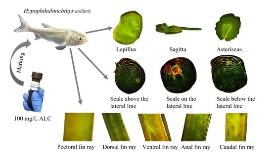
2.1. Experimental Fish

Juvenile silver carp (~2 months old) (Figure 1) were purchased from the Changzhou Professional Nursery Breeding Base, Changzhou City, Jiangsu Province, China, and maintained in a glass aquarium (45 cm width × 100 cm length × 50 cm height) filled with aerated tap water in a laboratory. Every day during the temporary culture process, food residues and feces were cleaned from the aquarium bottom, and approximately one-third of the aquarium’s volume was replaced with clean water.

2.2. Marking Procedure

The experiment was conducted at the Nanquan Base of the Freshwater Fisheries Research Center of the Chinese Academy of Fishery Sciences in Wuxi City, Jiangsu Province, China. After approximately three weeks of temporary culture, juvenile fish (five individuals per group) were transferred to marking solutions (0 and 100 mg/L ALC; Bioengineering Co., Ltd., Shanghai, China), which had been prepared using tap water that had been previously aerated for 48 h. After adding 1 g ALC powder into 10 L of aerated tap water, the mixture was slowly stirred with a glass rod for 30 min to accelerate its dissolution and then left for one day to dissolve completely before starting the experiment. After the fish were maintained in the marking solutions for 48 h, the treatment groups were individually transferred to a plastic basin, where they were rinsed in water for 10 min and then subjected to another clean-water-rinsing step. This step was repeated three times to ensure that there was no solution residue on the surface of the fish. The two treatment groups were then separately transferred to two clean glass aquaria (50 cm width × 50 cm length × 40 cm height) for post-marking culture. After 10 days, the fish were sampled, sealed in plastic bags, and frozen (−20 °C) until processing.

2.3. Hard Tissue Sampling

Hard tissues (Figure 1) were collected from the fish using a dissecting microscope (NV10, Shanghai Precision Instruments Co., Ltd., Shanghai, China). Lateral line scales and non-lateral line scales (≥5 each) were collected from each fish using forceps, washed in deionized water to remove the majority of the surface impurities and films, and then gently flattened on glass slides using coverslips. The first pectoral, dorsal, ventral, anal, and caudal fin rays were sampled from each fish using dissecting needles and scissors, and all three otolith types were extracted. Both fin rays and otoliths were washed in deionized water to remove the majority of surface impurities and films, dehydrated using anhydrous ethanol, dried, and then embedded in clear nail polish, without grinding or polishing. All samples were stored in the dark and were maintained at temperatures of < 25 °C to prevent the degradation of the chemical markings [19].

2.4. Marking Evaluation

Within two weeks of sampling, all hard tissue samples, without any additional grinding, polishing, or sectioning preparation, were visualized using an Olympus BX51 fluorescence microscope (Olympus Co., Tokyo, Japan) (Table 1), which was equipped with an Olympus XC10 digital camera and the “Stream Start” software included with the device (Olympus Co., Tokyo, Japan). The exposure time and gain were set to 20 ms and 5.0 dB, respectively.
Referring to the studies by Liu et al. [7] and Taylor et al. [20], marking effectiveness in the present study was categorized into four grades, based on observation under brightfield (BF; i.e., transmitted light), blue (WBS), and green (WGS) excitation lights: 0, no marking (unacceptable); 1, faint marking (unacceptable); 2, moderate marking (acceptable); or 3, strong marking (acceptable). The mode values of marking grades were used as the quality scores of hard tissues from each fish specimen.

2.5. Statistical Analysis

Body length and wet mass data are presented as mean ± standard error (SE) values. The significance of differences in the mean body length and wet mass values of the control and ALC-marked groups were evaluated using one-way analysis of variance (ANOVA) in SPSS 23.0 (IBM, Armonk, NY, USA). Statistical significance was set at p < 0.05.

3. Results

3.1. Effects of ALC on Fish Survival and Growth

No mortality was observed during the experiment. After the 10-day growth experiment, there were no significant differences observed in either the average body length of the ALC-marked and control groups (64.94 ± 5.51 mm and 67.13 ± 5.27 mm, respectively; N = 5 per group) or their body weights (3.58 ± 0.93 g and 4.14 ± 1.26 g, respectively; N = 5 per group) based on the results of one-way ANOVAs (p > 0.05).

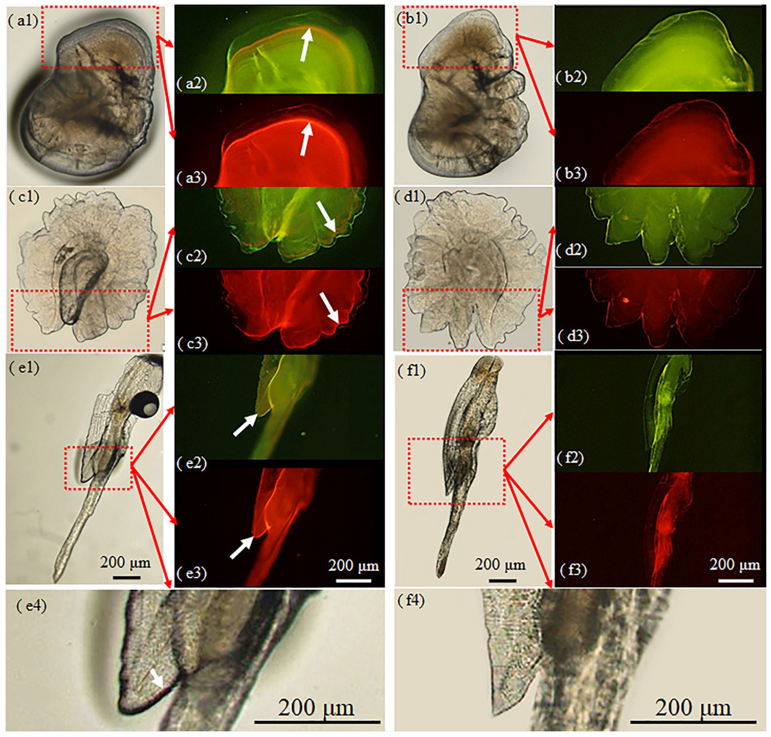
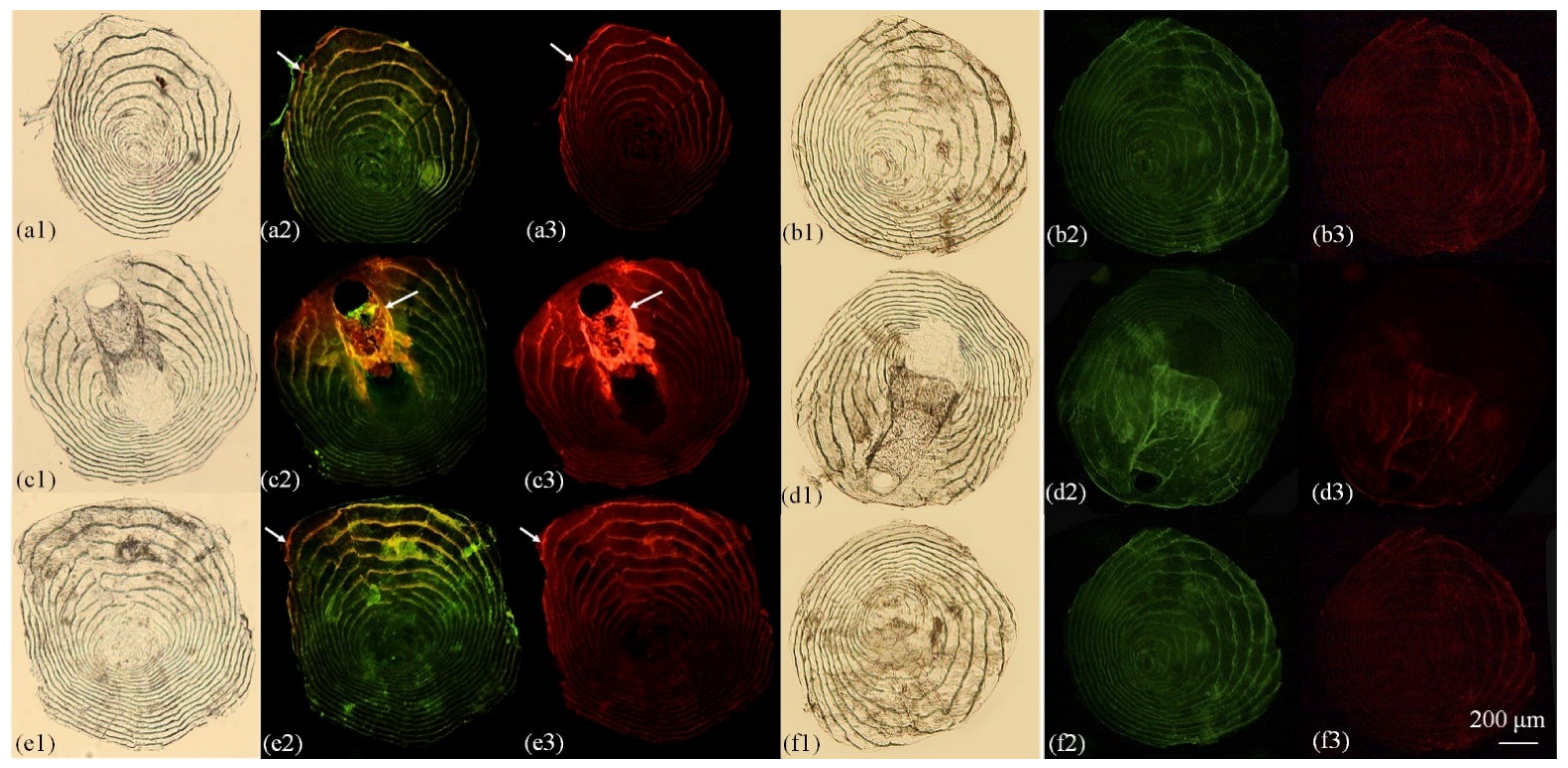
3.2. Otolith Marking

Unlike all the otoliths of the five fish in the control group without any marker rings, all three otolith types of the five juvenile silver carp in the marked group exhibited clear marker rings under visible, blue excitation, and green excitation light, with a marking rate of 100% (Figure 2, Table 2). However, the marking rings were more complete in the lapilli and asterisci than in the sagittae, and marker ring color varied, depending on the light source. For example, under visual light, the marking rings appeared red, with those of the lapilli and sagittae being clearer than those of the asterisci. Meanwhile, under the blue excitation light, the marking rings appeared orange-red, which strongly contrasted with the green coloration of unmarked areas, while under the green excitation light, the marking rings were bright red, whereas the unmarked areas were a less bright red.

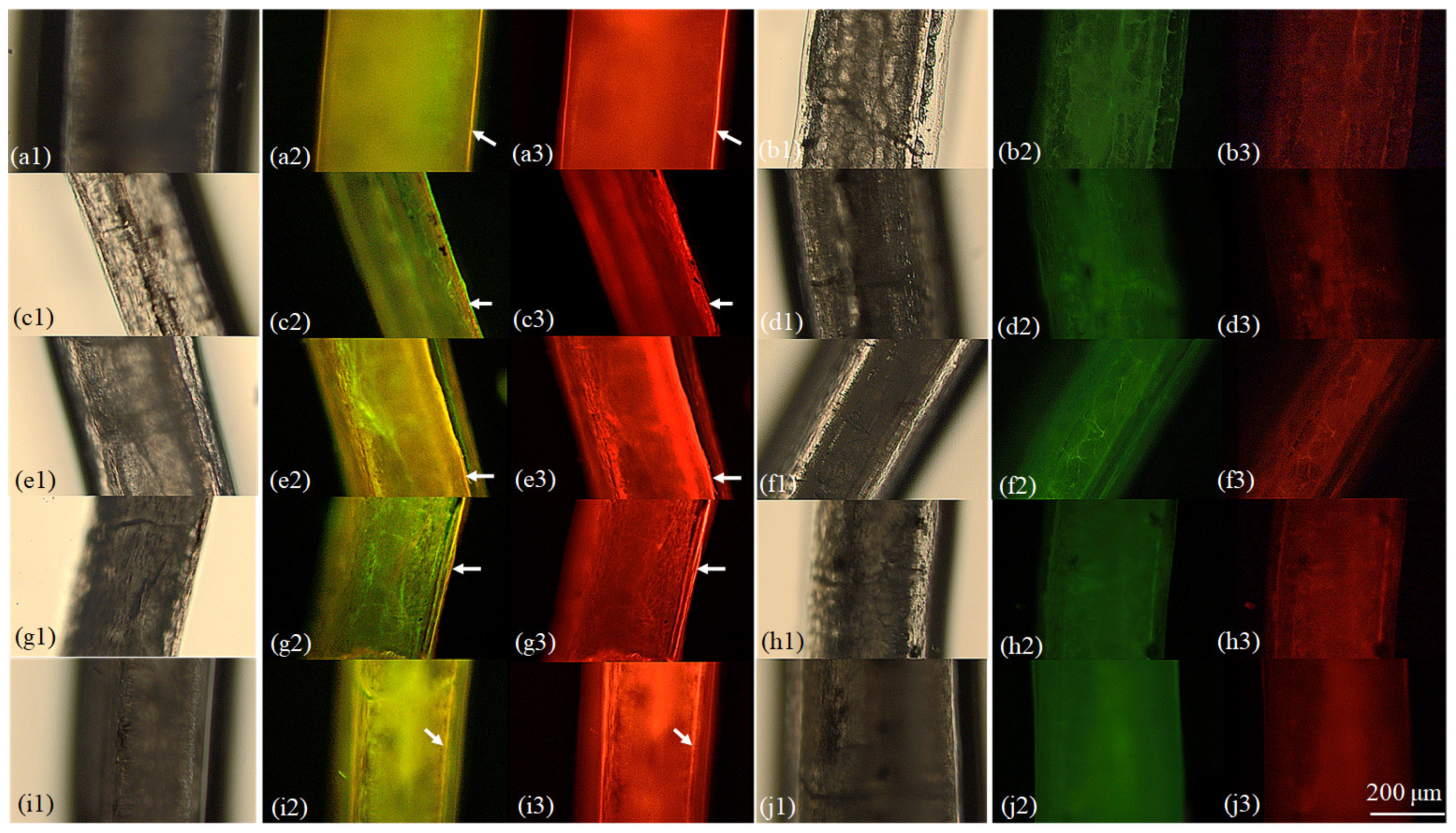
3.3. Fin Ray Marking

Under different light sources and using the fluorescence microscope, fin ray marking efficiency varied (Figure 3, Table 2). Fin ray marking was poorly detectable under visible light but was more evident under the blue and green excitation lights. Under the blue excitation light, the marked areas appeared a faint red, whereas the unmarked areas appeared green, while under the green excitation light, the entire ray appeared red, with brighter fluorescence in the marked areas. There were no obvious differences between the marking efficiencies of the five types of fin rays.

3.4. Marking of Scales

The marking efficiency of the scales also varied under different light sources using the fluorescence microscope (Figure 4, Table 2). Fin ray marking was unapparent under visible light but was more evident under the blue and green excitation lights. Under the blue excitation light, the marked areas exhibited a weak red fluorescence, whereas the unmarked areas appeared green, and under green excitation light, the entire scale appeared red, with brighter fluorescence in the marked areas. The marking efficiency of the lateral line scales was greater than that of scales above and below the lateral line.

4. Discussion

4.1. Variation of Marking Effectiveness among Hard Structures

Otoliths grow in all directions and build concentric layers from the core [21,22]. Fin rays grow in an additive, incremental manner, with the oldest layers at the core and the newest layers at the outer edge [23]. Scales consist of a distinct osseous upper layer and an underlying proteinaceous basal layer. After initial deposition from the focus (similar to the core), scales grow incrementally to create bony scale circuli [23]. As mentioned above, all hard tissue samples in the present study were not ground, polished, or sectioned. Therefore, in Figure 2, Figure 3 and Figure 4 of this study, the whole surface of the growth layer marked by ALC during the immersion process of fish is shown, and the boundary lines of the layer in the hard structures (especially the otolith and fin rays) were the brightest in the fluorescent images. Nevertheless, due to differences in the chemical compositions of hard structures in fish, marking effectiveness was expected to vary within and between the hard structures sampled in the present study, namely, otoliths (sagittae, lapilli, and asterisci), scales (below, on, and above the lateral line), and rays from the pectoral, dorsal, ventral, anal, and caudal fins. As expected, under visible light, ALC markings were more visible in the otoliths than in the fin rays or scales, and the effectiveness of ALC marking also varied among the three otolith types. Asterisci are the largest and thinnest otoliths in cyprinid fish and are more transparent; age could not be determined based on the arrangement of the rings [24]. Under both visible and excitation lights, markings were more apparent in the lapilli and sagittae than in the asterisci, which is why asterisci are usually not selected as the focus of research in marking experiments. However, for the sagitta, the entire otolith shape is arrow-type, which is susceptible to external forces and fractures during sampling. Due to the shape of the sagitta, the marked and external rings of the otoliths were close and, thus, difficult to discriminate during microscopy. In contrast, in terms of the morphology of the otolith and the difficulty of the sampling operation, lapilli are oval-shaped, with a more regular external morphology, a clearer ring arrangement, and more distinguishable marking areas. Thus, the lapilli may be the most appropriate otolith type for marking detection, which agrees with the findings of previous studies [24,25,26,27].
The effectiveness of fluorescence marking has also been reported to vary among tissue types when using other fluorescent dyes. For example, Elle et al. [28] used calcein to mark rainbow trout (Oncorhynchus mykiss Walbaum 1792) and reported that the fluorescence retention of otoliths was greater than that of fin rays. In some cases, such as in the measurement of fish age using oxytetracycline-marked recaptured goliath groupers (Epinephelus itajara Lichtenstein 1822), the results obtained from both otoliths and dorsal spines are consistent [29] and, therefore, external tissues (i.e., dorsal spines) can be used for non-lethal age determination. In contrast, dorsal ray sections were so small that readers tended to have lower confidence in age-reading. For scales, as observed under visible light in the present study, studies on other species [30] (e.g., Aristichthys nobilis Richardson 1845) marked with calcein and alizarin red S have also reported that marker chemicals are ineffective for marking scales.
The results of the present study demonstrated the intra- and inter-hard structure differences in fluorescent marking, i.e., there were no obvious differences in the marking efficiency of rays from the pectoral, dorsal, ventral, anal, and caudal fins, but the lapilli and lateral line scales of the sampled otolith and scale types, respectively, were marked most effectively. Notably, although otoliths are considered optimal marking structures as they consist of calcium carbonate minerals that are metabolically inert and are generally not resorbed [31], lethal sampling is a major disadvantage. Fin rays and scales can offer a non-lethal alternative to otoliths [32]. Nevertheless, the use of both calcium phosphate structures limits resorption [16,23] and loss of marks by sunlight [16]. Furthermore, scales are well known for being easily peeled off and regenerated [23]. Therefore, the aforementioned advantages and disadvantages among these hard tissues are of critical concern for future applications.
It is noteworthy that the marked areas in all three types of hard structures in ALC-immersed carp were all brighter than those of unmarked structures in the control carp when observed under blue excitation light and, especially, green excitation light (Figure 2, Figure 3 and Figure 4). Similar cases have been found in other species with other fluorescent markers [7,30,33]. Moreover, Katakura et al. [34] demonstrated that otolith, scale, and dorsal fin ray marks were identified in captive juvenile walleye pollock (Theragra chalcogramma Pallas 1814) one year after they were marked with ALC. This evidence suggests that this ALC marking method could be used in the future for identification between marked fish and unmarked nature fish during stock enhancement. Unexpectedly, in this study, almost all the scale circuli appeared to be marked by ALC (Figure 4). The mechanism of this phenomenon is still unknown and should be explored in the future. Nevertheless, it could be assumed that ALC dye may be deposited into the circuli directly from the immersion solution (as in the case reported by Able et al. [35]) and indirectly from the skin layer through scale development (as in the case reported by Katakura et al. [34]).

4.2. Optimum Marker Concentration

The ideal chemical marker concentration for fluorescent marking by immersion varies among fish types and even among life-history stages. For example, ALC has been reported to significantly affect hatching. Long-lasting, distinct otolith marks were produced from the immersion of Baltic cod (Gadus morhua Linnaeus 1758) yolk-sac larvae in ≥ 50 mg/L ALC for 24 h. The mortality of eggs and larvae was low during the marking procedure. The hatching success of ALC-marked embryos was significantly reduced, and hatching was delayed with increasing ALC concentrations [36]. Other fluorescent dye markers may also affect fish health [37]. Indeed, results similar to those in the present study reported the immersion of bighead carp (Aristichthys nobilis Richardson 1845), grass carp (Ctenopharyngodon idella Valenciennes 1844), and black rockfish (Sebastes schlegelii Girard 1856) in calcein and alizarin red S solution, and the optimum concentrations for detection of the marker rings in each tissue were not the same [30,33,38].
In the present study, an ALC concentration of 100 mg/L resulted in the successful marking of hard structures in juvenile silver carp. However, under visible light, the marking was more effective in otoliths than in either fin rays or scales. This indicated that the optimal ALC concentrations for marking otoliths, fin rays, and scales were different. Similarly, Lü et al. [8] reported that the immersion of juvenile silver carp in calcein and alizarin red S at concentrations of 50–200 mg/L and 150–300 mg/L, respectively, was ineffective for marking non-lateral line scales. In the present study, the same level of detection was achieved using a lower concentration (100 mg/L) of ALC. Therefore, ALC might be more suitable than calcein or alizarin red S for the fluorescent marking of hard structures in juvenile silver carp. However, if using higher concentrations can improve marking, particularly in fin rays and scales when observed under visible light, while still ensuring fish safety, then the use of higher concentrations should be considered.

5. Conclusions

The findings of the present study demonstrate that immersion in ALC (100 mg/L) can be used for the fluorescent marking of otoliths (lapilli, sagittae, and asterisci), scales (lateral and non-lateral lines), and rays from the pectoral, dorsal, ventral, anal, and caudal fins in juvenile silver carp. Marking effectiveness varied when evaluated using different light sources. However, except in the case of fin rays and scales (the marking of which was not evident when observed under visible light), the marking of all three hard structure types could be observed under visible light, blue excitation light, and green excitation light. The lapilli were the most successful target among the three otolith types, and the marking of lateral line scales was more effective than that of the non-lateral line scales. In contrast, no obvious differences were observed in the marking of rays among the five fin types. Future research should focus more on the practical applications of non-lateral fin ray and scale structures and the development of strategies to avoid their limitations, e.g., material turnover, resorption, loss, or regeneration. The present study provides a method for marking the hard structures of juvenile silver carp and advances the current understanding of ALC marking targets and efficiency, which is invaluable for assessing the success of fish restocking programs involving teleost fishes.

Author Contributions

Conceptualization, J.Y., Y.Z., T.J. and Q.P.; methodology, Y.Z., T.J., X.C., H.L. and J.Y.; investigation, Y.Z., T.J., X.C., H.L. and J.Y.; resources, Y.Z., T.J. and X.C.; funding acquisition, J.Y.; writing—original draft, Y.Z., T.J., X.C., H.L. and Q.P.; writing—review and editing, J.Y.; supervision, J.Y. All authors have read and agreed to the published version of the manuscript.

Funding

This work was supported by the Central Public-Interest Scientific Institution Basal Research Fund, FFRC, CAFS (Grant code 2019JBFM06), the China Central Governmental Research Institutional Basic Special Research Project from Public Welfare Funds, CAFS (Grant code 2021GH08).

Institutional Review Board Statement

The study was conducted according to the guidelines of the Declaration of Helsinki, and approved by the Ethics Committee of Animal Care and Use Committee of the Freshwater Fisheries Research Center at the Chinese Academy of Fishery Sciences. The analysis was carried out following the Guidelines for the Care and Use of Laboratory Animals set by the Animal Care and Use Committee of the Freshwater Fisheries Research Center (2003WXEP61). All operations were carried out with field permit no. 20181AC1128.

Data Availability Statement

Data that support the findings of this study are available from the corresponding author upon reasonable request (Y.J.: [email protected]).

Acknowledgments

We thank Liangcai Pan, Ajun Sun, and Guanming He for their technical support in silver carp aquaculture.

Conflicts of Interest

The authors declare no conflict of interest.

References

  1. Warren-Myers, F.; Dempster, T.; Swearer, S.E. Otolith mass marking techniques for aquaculture and restocking: Benefits and limitations. Rev. Fish Biol. Fish. 2018, 28, 485–501. [Google Scholar] [CrossRef]
  2. Xia, Y.; Li, X.; Yang, J.; Zhu, S.; Wu, Z.; Li, J.; Li, Y. Elevated temperatures shorten the spawning period of silver carp (Hypophthalmichthys molitrix) in a large subtropical river in China. Front. Mar. Sci. 2021, 8, 708109. [Google Scholar] [CrossRef]
  3. Lu, G.; Wang, C.; Zhao, J.; Liao, X.; Wang, J.; Luo, M.; Zhu, L.; Bernatzhez, L.; Li, S. Evolution and genetics of bighead and silver carps: Native population conservation versus invasive species control. Evol. Appl. 2020, 13, 1351–1362. [Google Scholar] [CrossRef] [PubMed] [Green Version]
  4. Curtis, J.M.R. Visible implant elastomer color determination, tag visibility, and tag loss: Potential sources of error for mark-recapture studies. N. Am. J. Fish. Manag. 2006, 26, 327–337. [Google Scholar] [CrossRef]
  5. Phinney, D.E.; Miller, D.M.; Dahlberg, M.L. Mass-marking young salmonids with fluorescent pigment. Trans. Am. Fish. Soc. 1967, 96, 157–162. [Google Scholar] [CrossRef]
  6. Lejk, A.M.; Radtke, G. Effect of marking salmo Trutta lacustris L. Larvae with alizarin red S on their subsequent growth, condition, and distribution as juveniles in a natural stream. Fish. Res. 2021, 234, 105786. [Google Scholar] [CrossRef]
  7. Liu, Q.; Zhang, X.M.; Zhang, P.D.; Nwafili, S.A. The use of alizarin red S and alizarin complexone for immersion marking Japanese flounder Paralichthys olivaceus (T.). Fish. Res. 2009, 98, 67–74. [Google Scholar] [CrossRef]
  8. Lü, H.; Fu, M.; Dai, S.; Xi, D.; Zhang, Z.-X. Experimental evaluation of calcein and alizarin red S for immersion marking of silver carp Hypophthalmichthys molitrix (Valenciennes, 1844). J. Appl. Ichthyol. 2016, 32, 83–91. [Google Scholar] [CrossRef]
  9. Bangs, B.L.; Falcy, M.R.; Scheerer, P.D.; Clements, S. Comparison of three methods for marking a small floodplain minnow. Anim. Biotelem. 2013, 1, 18. [Google Scholar] [CrossRef] [Green Version]
  10. Partridge, G.J.; Ginbey, B.M.; Woolley, L.D.; Fairclough, D.V.; Crisafulli, B.; Chaplin, J.; Prokop, N.; Dias, J.; Bertram, A.; Jenkins, G.I. Development of techniques for the collection and culture of wild-caught fertilised snapper (Chrysophrys auratus) eggs for stock enhancement purposes. Fish. Res. 2017, 186, 524–530. [Google Scholar] [CrossRef]
  11. Partridge, G.J.; Jenkins, G.I.; Doupé, R.G.; De Lestang, S.; Ginbey, B.M.; French, D. Factors affecting mark quality of alizarin complexone-stained otoliths in juvenile black bream Acanthopagrus butcheri and a prescription for dosage. J. Fish Biol. 2009, 75, 1518–1523. [Google Scholar] [CrossRef] [PubMed]
  12. Gaines, P.C.; Martin, C.D. Feasibility of dual-marking age-0 Chinook salmon for mark–recapture studies. N. Am. J. Fish. Manag. 2004, 24, 1456–1459. [Google Scholar] [CrossRef]
  13. Schumann, D.A.; Koupal, K.D.; Hoback, W.W.; Schoenebeck, C.W. Evaluation of sprayed fluorescent pigment as a method to mass-mark fish species. Open Fish Sci. J. 2013, 6, 41–47. [Google Scholar] [CrossRef]
  14. Caraguel, J.-M.; Barreau, T.; Brown-Vuillemin, S.; Iglésias, S.P. In vivo staining with alizarin for ageing studies on Chondrichthyan fishes. Aquat. Living Resour. 2020, 33, 1. [Google Scholar] [CrossRef]
  15. Watanabe, K.-I.; Ohta, K. Efficacy of a fluorescent tagging technique on otolith and scales using alizarin complexone in artificial seawater for Red Sea bream and Japanese flounder juveniles. Aquac. Sci. 2009, 57, 627–630. [Google Scholar] [CrossRef]
  16. Lü, H.; Fu, M.; Zhang, Z.; Su, S.; Yao, W. Marking fish with fluorochrome dyes. Rev. Fish. Sci. Aquac. 2020, 28, 117–135. [Google Scholar] [CrossRef]
  17. Mansur, L.; Catalán, D.; Plaza, G.; Landaeta, M.F.; Ojeda, F.P. Validations of the daily periodicity of increment deposition in rocky intertidal fish otoliths of the South-Eastern Pacific Ocean. Rev. Biol. Mar. Oceanogr. 2013, 48, 629–633. [Google Scholar] [CrossRef] [Green Version]
  18. Cottingham, A.; Hall, N.G.; Loneragan, N.R.; Jenkins, G.I.; Potter, I.C. Efficacy of restocking an estuarine-resident species demonstrated by long-term monitoring of cultured fish with alizarin complexone-stained otoliths. A case study. Fish. Res. 2020, 227, 105556. [Google Scholar] [CrossRef]
  19. Stötera, S.; Degen-Smyrek, A.K.; Krumme, U.; Stepputtis, D.; Bauer, R.; Limmer, B.; Hammer, C. Marking otoliths of Baltic cod (Gadus morhua Linnaeus, 1758) with tetracycline and strontium chloride. J. Appl. Ichthyol. 2019, 35, 427–435. [Google Scholar] [CrossRef]
  20. Taylor, M.D.; Fielder, D.S.; Suthers, I.M. Batch marking of otoliths and fin spines to assess the stock enhancement of Argyrosomus japonicus. J. Fish Biol. 2005, 66, 1149–1162. [Google Scholar] [CrossRef]
  21. Romanek, C.S.; Gauldie, R.W. A Predictive model of otolith growth in fish based on the chemistry of the endolymph. Comp. Biochem. Physiol. 1996, 114, 71–79. [Google Scholar] [CrossRef]
  22. Fisher, M.; Hunter, E. Digital imaging techniques in otolith data capture, analysis and interpretation. Mar. Ecol. Prog. Ser. 2018, 598, 213–231. [Google Scholar] [CrossRef] [Green Version]
  23. Tzadik, O.E.; Curtis, J.S.; Granneman, J.E.; Kurth, B.N.; Pusack, T.J.; Wallace, A.A.; Hollander, D.J.; Peebles, E.B.; Stallings, C.D. Chemical archives in fishes beyond otoliths: A review on the use of other body parts as chronological recorders of microchemical constituents for expanding interpretations of environmental, ecological, and life-history changes. Limnol. Oceanogr. 2017, 15, 238–263. [Google Scholar] [CrossRef]
  24. Seibert, J.R.; Phelps, Q.E. Evaluation of aging structures for silver carp from midwestern U.S. rivers. N. Am. J. Fish. Manag. 2013, 33, 839–844. [Google Scholar] [CrossRef]
  25. Ding, C.; He, D.; Chen, Y.; Jia, Y.; Tao, J. Otolith microstructure analysis based on wild young fish and its application in confirming the first annual increment in Tibetan Gymnocypris selincuoensis. Fish. Res. 2020, 221, 105386. [Google Scholar] [CrossRef]
  26. Durham, B.W.; Wilde, G.R. Validation of daily growth increment formation in the otoliths of juvenile Cyprinid fishes from the Brazos River, Texas. N. Am. J. Fish Manag. 2008, 28, 442–446. [Google Scholar] [CrossRef]
  27. Gao, M.H.; Wu, Z.Q.; Huang, L.L.; Tan, X.C.; Liu, H.; Rad, S. Otolith shape analysis and growth characteristics in larval and juvenile Squalidus argentatus. Environ. Biol. Fishes 2021, 104, 937–945. [Google Scholar] [CrossRef]
  28. Elle, S.F.; Koenig, M.K.; Meyer, K.A. Evaluation of calcein as a mass mark for rainbow trout raised in outdoor hatchery raceways. N. Am. J. Fish. Manag. 2011, 30, 1408–1412. [Google Scholar] [CrossRef]
  29. Brusher, J.H.; Schull, J. Non-lethal age determination for juvenile goliath grouper Epinephelus itajara from Southwest Florida. Endang. Species Res. 2009, 7, 205–212. [Google Scholar] [CrossRef]
  30. Lü, H.; Fu, M.; Xi, D.; Yao, W.-Z.; Su, S.-Q.; Wu, Z.-L. Experimental evaluation of using calcein and alizarin red S for immersion marking of bighead carp Aristichthys nobilis (Richardson, 1845) to assess growth and identification of marks in otoliths, scales and fin rays. J. Appl. Ichthyol. 2015, 31, 665–674. [Google Scholar] [CrossRef]
  31. Wood, R.S.; Chakoumakos, B.C.; Fortner, A.M.; Gillies-Rector, K.; Frontzek, M.D.; Ivanov, I.N.; Kah, L.C.; Kennedy, B.; Pracheil, B.M. Quantifying fish otolith mineralogy for trace-element chemistry studies. Sci. Rep. 2022, 12, 2727. [Google Scholar] [CrossRef] [PubMed]
  32. Clarke, A.D.; Telmer, K.H.; Shrimpton, J.M. Elemental analysis of otoliths, fin rays and scales: A comparison of bony structures to provide population and life-history information for the Arctic grayling (Thymallus arcticus). Ecol. Freshw. Fish 2010, 16, 354–361. [Google Scholar] [CrossRef]
  33. Lü, H.; Zhang, X.; Xi, D.; Gao, T. Use of calcein and alizarin red S for immersion marking of black rockfish Sebastes schlegelii Juveniles. Chin. J. Oceanol. Limnol. 2014, 32, 88–98. [Google Scholar] [CrossRef]
  34. Katakura, S.; Ohta, M.; Jin, M.; Sakurai, Y. Otolith-marking experiments of juvenile walleye pollock Theragra chalcogramma using oxytetracycline, alizarin complexone, and alizarin red S. Aquac. Sci. 2003, 51, 327–335. [Google Scholar] [CrossRef]
  35. Able, K.W.; Sakowicz, G.P.; Lamonaca, J.C. Scale formation in selected fundulid and cyprinodontid fishes. Ichthyol. Res. 2009, 56, 1–9. [Google Scholar] [CrossRef]
  36. Meyer, S.; Sørensen, S.R.; Peck, M.A.; Støttrup, J.G. Sublethal effects of alizarin complexone marking on Baltic cod (Gadus morhua) eggs and larvae. Aquaculture 2012, 324, 158–164. [Google Scholar] [CrossRef]
  37. Jurgelėnė, Ž.; Montvydienė, D.; Stakėnas, S.; Poviliūnas, J.; Račkauskas, S.; Taraškevičius, R.; Skrodenytė-Arbačiauskienė, V.; Kazlauskienė, N. Impact evaluation of marking Salmo trutta with alizarin red S produced by different manufacturers. Aquat. Toxicol. 2022, 242, 106051. [Google Scholar] [CrossRef]
  38. Lü, H.; Chen, H.; Fu, M.; Peng, X.; Xi, D.; Zhang, Z. Experimental evaluation of calcein and alizarin red S for immersion marking grass carp Ctenopharyngodon idellus. Fish. Sci. 2015, 81, 653–662. [Google Scholar] [CrossRef]
Figure 1. Location of targeted hard structures in the juvenile silver carp (Hypophthalmichthys molitrix).
Fishes 07 00066 g001
Figure 2. The typical patterns of marking when alizarin complexone (ALC) is used on otoliths (lapillus, sagitta, and asteriscus) from juvenile silver carp, Hypophthalmichthys molitrix. Note: (a1), ALC-marked lapilli under transmitted light; (a2), ALC-marked lapilli under blue excitation light; (a3), ALC-marked lapilli under green excitation light; (b1), control lapilli under transmitted light; (b2), control lapilli under blue excitation light; (b3), control lapilli under green excitation light; (c1), ALC-marked asterisci under transmitted light; (c2), ALC-marked asterisci under blue excitation light; (c3), ALC-marked asterisci under green excitation light; (d1), control asterisci under transmitted light; (d2), control asterisci under blue excitation light; (d3), control asterisci under green excitation light; (e1), (e4) (enlarged view), ALC-marked sagittae under transmitted light; (e2), ALC-marked sagittae under blue excitation light; (e3), ALC-marked sagittae under green excitation light; (f1), (f4) (enlarged view), control sagittae under transmitted light; (f2), control sagittae under blue excitation light; (f3), control sagittae under green excitation light. The white arrows point to the fluorescent marks.
Fishes 07 00066 g002
Figure 3. The typical patterns of marking when alizarin complexone (ALC) is used for marking fin rays in juvenile silver carp, Hypophthalmichthys molitrix. Note: (a1), ALC-marked pectoral fin rays under transmitted light; (a2), ALC-marked pectoral fin rays under blue excitation light; (a3), ALC-marked pectoral fin rays under green excitation light; (b1), control pectoral fin rays under transmitted light; (b2), control pectoral fin rays under blue excitation light; (b3), control pectoral fin rays under green excitation light; (c1), ALC-marked dorsal fin rays under transmitted light; (c2), ALC-marked dorsal fin rays under blue excitation light; (c3), ALC-marked dorsal fin rays under green excitation light; (d1), control dorsal fin rays under transmitted light; (d2), control dorsal fin rays under blue excitation light; (d3), control dorsal fin rays under green excitation light; (e1), ALC-marked ventral fin rays under transmitted light; (e2), ALC-marked ventral fin rays under blue excitation light; (e3), ALC-marked ventral fin rays under green excitation light; (f1, control ventral fin rays under transmitted light; (f2), control ventral fin rays under blue excitation light; (f3), control ventral fin rays under green excitation light; (g1), ALC-marked anal fin rays under transmitted light; (g2), ALC-marked anal fin rays under blue excitation light; (g3), ALC-marked anal fin rays under green excitation light; (h1), control anal fin rays under transmitted light; (h2), control anal fin rays under blue excitation light; (h3), control anal fin rays under green excitation light; (i1), ALC-marked caudal fin rays under transmitted light; (i2), ALC-marked caudal fin rays under blue excitation light; (i3), ALC-marked caudal fin rays under green excitation light; (j1), control caudal fin rays under transmitted light; (j2), control caudal fin rays under blue excitation light; (j3), control caudal fin rays under green excitation light. The white arrows point to the fluorescent marks.
Fishes 07 00066 g003
Figure 4. The typical patterns of marking when alizarin complexone (ALC) is used for marking scales in juvenile silver carp Hypophthalmichthys molitrix. Note: (a1), ALC-marked scales above the lateral line under transmitted light; (a2), ALC-marked scales above the lateral line under blue excitation light; (a3), ALC-marked scales above the lateral line under green excitation light; (b1), control scales above the lateral line under transmitted light; (b2), control scales above the lateral line under blue excitation light; (b3), control scales above the lateral line under green excitation light; (c1), ALC-marked scales on the lateral line under transmitted light; (c2), ALC-marked scales on the lateral line under blue excitation light; (c3), ALC-marked scales on the lateral line under green excitation light; (d1), control scales on the lateral line under transmitted light; (d2), control scales on the lateral line under blue excitation light; (d3), control scales on the lateral line under green excitation light; (e1), ALC-marked scales below the lateral line under transmitted light; (e2), ALC-marked scales below the lateral line under blue excitation light; (e3), ALC-marked scales below the lateral line under green excitation light; (f1), control scales below the lateral line under transmitted light; (f2), control scales below the lateral line under blue excitation light; (f3), control scales below the lateral line under green excitation light. The white arrows point to the fluorescent marks.
Fishes 07 00066 g004
Table 1. The combined wavelength of the dichromatic mirror and filter used for visual ALC marker detection.
Light SourceWavelength (nm)
Excitation FilterSuppression FilterDichromatic Mirror
Brightfield light
Blue excitation light460–495510–550505
Green excitation light530–550575–625570
Table 2. Effect of a light source on the detection (mode marking grade) of alizarin complexone marking in hard tissues from five juvenile silver carps in the marked group.
Hard Tissue TypeSample SizeMarking Grade
BFWBSWGS
Otolith (sagitta)5333
Otolith (lapillus)5333
Otolith (asteriscus)5222
Fin ray (pectoral)5133
Fin ray (dorsal)5133
Fin ray (ventral)5133
Fin ray (anal)5133
Fin ray (caudal)5133
Scales (above lateral line)25022
Scales (on lateral line)25033
Scales (below lateral line)25022
BF: brightfield/transmitted light; WBS: blue excitation light; WGS: green excitation light.
Publisher’s Note: MDPI stays neutral with regard to jurisdictional claims in published maps and institutional affiliations.

