Article

Fish Diversity Monitored by Environmental DNA in the Yangtze River Mainstream

1
CAS Key Laboratory of Marine Ecology and Environmental Sciences, Institute of Oceanology, Chinese Academy of Sciences, Qingdao 266071, China
2
Laboratory for Marine Ecology and Environmental Science, Qingdao National Laboratory for Marine Science and Technology, Qingdao 266237, China
3
Center for Ocean Mega-Science, Chinese Academy of Sciences, Qingdao 266071, China
4
School of Marine Science and Engineering, Qingdao Agricultural University, Qingdao 266109, China
5
University of Chinese Academy of Sciences, Beijing 100049, China
*
Authors to whom correspondence should be addressed.
This author is the lead correspondence.
Submission received: 18 October 2021 / Revised: 17 December 2021 / Accepted: 17 December 2021 / Published: 22 December 2021
(This article belongs to the Special Issue Sustainable Aquaculture and Fisheries)

Abstract

:
Surveys and assessments based on environmental DNA are not only efficient and time-saving, but also cause less harm to monitoring targets. Environmental DNA has become a common tool for the assessment and monitoring of aquatic organisms. In this study, we investigated fish resources in the Yangtze River mainstream using environmental DNA, and the variations in fish during two seasons (spring and autumn) were compared. The results showed that 13 species were identified in spring, and nine species of fish were identified in autumn. The fish with higher eDNA detection were Sinibotia superciliaris, Tachysurus fulvidraco, Cyprinus carpio, Ctenopharyngodon Idella, Monopterus albus, Acanthogobius hasta, Saurogobio dabryi, Oncorhynchus mykiss, Mugil cephalus, Odontamblyopus rubicundus. Seasonal variation between spring and autumn was not significant, and the environmental factors had different effects on fish assemblages during the two seasons. Our study used the eDNA technique to monitor the composition of fish in the spring and autumn in the Yangtze River mainstream, providing a new technology for the long-term management and protection of fishery resources in the region. Of course, problems such as pollution and insufficient databases are the current shortcomings of environmental DNA, which will be the focus of our future research and study.

1. Introduction

As the largest river in Asia, and the third largest river in the world, the Yangtze River is rich in resources and has many tributaries and lakes, forming an important economic link connecting the east and the west of China [1]. The variety and output of Yangtze River fish rank first in China [1]. According to statistics, the fishery resources in the Yangtze River mainstream have gradually declined in the past half century [2]. In recent years, the government has gradually implemented relevant fishing bans in key waters such as the mainstream of the Yangtze River and important tributaries. In addition, an accurate understanding of its biodiversity is required to address the current challenges [3]. To appropriately monitor, manage and protect these key regions, it is essential to develop better monitoring approaches for biodiversity [4].
First explicitly proposed by Ogram et al. (1987) for microbial DNA from sediment, the environmental DNA (eDNA) technique introduced new opportunities in the study of microbial diversity [5]. Later, Rondon et al. (2000) used eDNA to assess the genetic and functional diversity of uncultured microorganisms in soil [6]. The eDNA technique is used to identify the target species presented by exploiting DNA molecules shed in the environment (such as water, soil, air, feces or snow) [7]. Due to its limited cost and versatility in numerous contexts, the eDNA technique is expected to become an effective monitoring method [8]. The eDNA technique, with high probability of detection, is especially suitable for fish, and water is the most common sample [9]. eDNA monitoring of aquatic creatures was first used in 2008 to detect the presence of the American bullfrog Rana Catesbeiana in freshwater, and it has since been used extensively to monitor fish species [10]. Since then, eDNA has been used to explore the trajectories of invasive organisms such as American bullfrog Rana Catesbeiana [11], silver carp (Hypophthalmichthys molitrix) and bighead carp (Hypophthalmichthys mobilis) in USA area canals and waterways [12], New Zealand mudsnails Potamopyrgus antipodarum [13], the crayfish Procambarus clarkia [14], quagga Dreissena bugensis and zebra D. polymorpha mussels [15] and golden mussel Limnoperna fortune [16], and to protect the endangered largetooth sawfish Pristis pristis [17], Maugean skate Zearaja maugeana [18] and Plecoptera species Isogenus nubecula [19], as well as to monitor rare species such as the round goby Neogobius melanostomus from the Black Sea [20].
Additionally, eDNA has gradually become more widely used, mainly including the following three aspects. Firstly, it is used for monitoring single fish distribution, including determining habitat connectivity for the migration of fish between the sea and rivers [21]. Sigsgaard et al. (2016) demonstrated that the high-throughput sequencing of seawater eDNA could provide useful estimates of genetic diversity in a whale shark (Rhincodon typus) aggregation [22]. Bakker et al. (2017) used an eDNA metabarcoding approach specifically targeted to infer shark presence, diversity and eDNA read abundance in tropical habitats [23]. In addition, Boussarie et al. (2018) confirmed that eDNA can be used to detect shark diversity by comparison with traditional underwater visual censuses and baited videos [24]. Thalinger et al. (2019) monitored the spawning migrations of potamodromous fish species including Danube bleak Alburnus mento and Vimba bream Vimba vimba via eDNA, which found a strong correlation between daily visual fish counts and downstream eDNA signals [25].
Secondly, eDNA technology is used for multi-species monitoring, including surveys of vertebrate and invertebrate populations [26]. Port et al. (2015) successfully surveyed the vertebrate fauna present along a gradation of diverse marine habitats associated with a kelp forest ecosystem using eDNA [27]. Forsström et al. (2016) demonstrated for the first time that eDNA from an introduced mud crab Rhithropanopeus harrisii could be successfully amplified in aquarium water samples and detected in a brackish water environment [28]. Lanzén et al. (2016) applied DNA metabarcoding of eukaryotic communities to successfully identify changes in sediment communities surrounding oil platforms [29]. O’Donnell et al. (2017) investigated the potential of eDNA to provide information about the breadth of biodiversity present in a tropical marine environment, which illustrated that eDNA can be used to explore diversity beyond taxon identifications [30]. DiBattista et al. (2017) assessed the utility of eDNA as a tool to survey reef fish communities in the Red Sea [31]. Later, DiBattista et al. (2019) also investigated the potential for rapid universal metabarcoding surveys (RUMS) of eDNA in sediment samples to provide snapshots of eukaryotic subtropical biodiversity [32].
Moreover, eDNA approaches have made great achievements in ancient and contemporary ecosystems for the description of past biodiversity and practical present-day conservation [33]. Jeunen et al. (2019) determined the ability of eDNA metabarcoding surveys to distinguish localized signals, adding the evidence that eDNA in marine environments can detect a broad range of taxa that are spatially discrete [34]. With the development of large-scale parallel sequencing, eDNA has been used to conduct quantitative analyses of monitoring targets in a large number of studies [35]. Kimmerling et al. (2018) used high-throughput metabarcoding to quantify the species-level ecology of reef fish larvae, which could assist in coral reef conservation and fishery management efforts [36]. West et al. (2020) also demonstrated the utility of a multimarker metabarcoding approach in capturing multitrophic biodiversity across an entire coral reef atoll, which sets an important baseline for ongoing monitoring and management [37]. As an eco-friendly tool, eDNA technology can not only be used for fishery monitoring, but might also importantly assist in marine resource conservation and the sustainable exploitation of fisheries [38].
The purpose of this study was to use eDNA techniques to investigate fish resources in the Yangtze River mainstream, as they could provide a new monitoring technology for fish resources in this region. Additionally, we compared the changing seasonal trends between spring and autumn. The results provide a data reference for future studies on the monitoring and protection of fish resources in the Yangtze River. Our research confirms the utility of eDNA technology in resource assessment.

2. Materials and Methods

2.1. Sample Sites and Collection

Sampling points were set up in the upper reaches of the Yangtze River from Panzhihua to the lower reaches of Nanjing, which covered the Yangtze River mainstream (Figure 1). We collected 10 eDNA samples in spring and 11 eDNA samples in autumn at the same coordinate position (Table 1). Due to the distance between the sampling points, the sample collection of the two quarters was completed in around 8 days. The specific eDNA samples and corresponding sampling site information are shown in Table 1. Before sampling, we sterilized all instruments with ultrapure water and 75% alcohol and used the water near the sampling point to rinse the sampling supplies again; then, we discarded them downstream of the sampling to reduce field sampling pollution. A 1 L water sample from a single site was collected from the surface water layers and mixed in a sterile brown plastic bottle with Urine DNA Locker (Beijing, China). Then, the 1 L water samples were filtered through a 0.22 μm Sterivextex TM-GP filter unit (Millipore, Burlington, MA, USA), and each water sample bottle was filtered twice to ensure adequate collection.
In addition, 1 L of water was collected to measure environmental factors. First, 500 mL of water was filtered with a 0.65 μm pore-size polycarbonate microporous membrane. After the membrane was collected, the chlorophyll (Chl) was measured by fluorescence extraction, and the filtered water was used to measure nutrients. A QuAAtro Nutrient Salt Automatic Analyzer (Germany SEAL, Norderstedt, Germany) was used to analyze the nutrient salt, including total nitrogen (TN), total phosphorus (TP), SiO3, NH4, NO3, NO2. In the laboratory, we wore gloves to handle samples, in strict accordance with the experimental specifications, and to perform disinfection; in addition, all experimental materials were disinfected and sterilized to prevent contamination. To prevent contamination of the experimental operation, the filter bottles and anti-corrosion peristaltic filter tubes were rinsed with 75% alcohol and ultrapure water after each sample was filtered. We then added DNA preservation buffer (Tiandz Inc., Beijing, China) to the filter unit and filtered three equal volumes of deionized water as the negative control.

2.2. DNA Extraction and PCR

We used a DNeasy Blood and Tissue Kit (Qiagen, Shanghai, China) to extract total eDNA, following the manufacturer’s protocol. At the same time, Nanodrop was used to quantify the DNA, and the DNA extraction quality was detected by 1.2% agarose gel electrophoresis. The conserved region of 12S rRNA was amplified by PCR using the primers MiFish-F (5′-ACACTCTTTCCCTACACGACGCTCTTCCGATCTNNNNNNGTCGGTAAAACTCGTGCCAGC-3′) and MiFish-R (5′-GTGACTGGAGTTCAGACGTGTGCTCTTCCGATCTNNNNNNCATAGTGGGGTATCTAATCCCAGTTTG-3′) [39]. Three PCR replicates were performed for each sample, and the details of library construction were as follows: all three reaction volumes were 25 µL, including 5 µL 5 × buffer, 2 µL dNTP (2.5 mM), 1 µL (10 uM) forwardprimer, 1 µL (10 uM) reverse primer, 1 µL DNA Template, 14.75 µL ddH2O and 0.25 µL fast pfu DNA polymerase. The thermal-cycle profile was for 12S: 98 °C for 2 min; 35 cycles of 98 °C for 15 s, 55 °C for 30 s and 72 °C for 30 s; and 72 °C for 5 min. The negative control was set, which was used to detect microbial contamination from sources such as the environment and reagents, and any sample group with bands amplified by the negative control could not be used for subsequent experiments. The PCR products were quantified by a microplate reader (BioTek, FLx800, Invitorgen, San Diego, CA, USA) as the quantification instrument. The fluorescence reagent was the Quant-iT PicoGreen dsDNA Assay Kit (Invitorgen, San Diego, CA, USA).

2.3. Library Preparation and Sequencing

The sequencing library was prepared using the TruSeq Nano DNA LT Library Prep Kit (Illumina, San Diego, CA, USA). Firstly, the sequence ends of the amplified products were repaired by the End Repair Mix 2 in the kit. We added an “A” base to the 3′ end of the DNA sequence to prevent the DNA fragment from self-linking and ensure that the target sequence could be connected to the sequencing adapter. A sequencing adapter containing a library-specific tag (index sequence) was added to the 5′ end, so that the DNA molecule could be immobilized on the Flow Cell. Secondly, the BECKMAN AMPure-XP Beads were used to purify the library system after adding adapters; then, we performed PCR amplification on the above-mentioned DNA fragments with adapters, and used BECKMAN AMPure XP Beads to purify the library-enriched products again. Finally, the library was subjected to final fragment selection and purification by 2% agarose gel electrophoresis. The sequencing platform was Illumina Novaseq 6000. Before sequencing, we used the Agilent High Sensitivity DNA Kit to perform quality inspection on the library on the Agilent Bioanalyzer. The qualified library had only a single peak and no linker before proceeding to the next step. The NovaSeq sequencer was used for pair-end sequencing, and the corresponding reagent was the NovaSeq 6000 SP Reagent Kit (500 cycles).

2.4. Bioinformatic Statistics and Fish Identification

The raw sequencing data were saved in FASTQ format. The sequence clustering statistics were mainly performed with QIIME 2 2019.4, while the OTU clustering procedure followed the Vsearch (v2.13.4) pipeline described previously (https://github.com/torognes/vsearch/wiki/VSEARCH-pipeline, 8 August 2021) [40,41]. Briefly, raw sequence data were demultiplexed using the demux plugin, followed by primer cutting with the cutadapt plugin. Sequences were then merged, filtered and dereplicated using the fastq_mergepairs, fastq_filter and derep_fulllength functions in Vsearch [42]. All the unique sequences were then clustered at 98% (via cluster_size), followed by chimera removal (via uchime_denovo). Lastly, the non-chimera sequences were re-clustered at 95% to generate out representative sequences and an OTU table. Through the statistics of the flattened OTU table, the specific composition table of the fish communities in each sample at each classification level was obtained. Then, alpha diversity metrics (Chao1) [43], observed species, Shannon [44], Simpson [45], Faith’s PD [46], Pielou’s evenness [47], Good’s coverage [48], beta diversity metrics (weighted UniFrac) [49], unweighted UniFrac, Jaccard distance and Bray–Curtis dissimilarity were estimated using the diversity plugin, with samples rarefied to sequences [50]. We used NCBI Organelle Genome Resources for the BLAST search. The information from the databases was merged to build a new database by using the Blastn tool. We obtained the specific composition table of the fish community in each sample at each classification level by using the statistics of the flattened OTU table, and we then used the R package (v3.3.2) script to construct the data from the table as a histogram (Figure 2). In the following process, we showed the composition of fish, and the related analyses were based on the level of species classification. Finally, we took into account the conditions of the Yangtze River to exclude the species that should not have appeared; we used Fishbase (https://www.fishbase.se/search.php, 9 October 2021) and also consulted books about fishery resources in the Yangtze River for localization certification [51].

2.5. Seasonal Difference Comparison

We compared the results of eDNA surveys in spring and autumn and analyzed the variation in fish resources in the mainstream of the Yangtze River. Nonmetric multidimensional scaling (NMDS) was used to reveal the seasonal differences [52]. NMDS analysis was performed on each distance matrix separately using the Vegan package under the R script, and the structure distribution of community samples was described by a two-dimensional sorting diagram. A thermal image was used to show the difference in species composition based on average abundance ranking or the degree of similarity between the two seasons. In addition, canonical correspondence analysis (CCA) was applied to analyze the correlation between seven environmental factors (Chl, TP, TN, SiO3, NH4, NO3, NO2) and the distribution pattern of fish assemblages between the two seasons; the fish composition was based on the analysis of the relative abundance of the obtained sequence [38]. Seven environmental factor parameters are presented in Supplementary Material Table S1.

3. Results

3.1. Fish Resource Results by eDNA

The results of the three negative controls proved that there was no cross-contamination during eDNA extraction. After the quality filtering procedure, the total readings of high-quality sequences detected in spring were 1227540, while there were 1317686 in autumn, from 21 samples (Supplementary Material Table S2). Samples were collected from 10 sampling sites in the spring and 11 sampling sites in autumn (Table 1). In spring, the domain species were a number of small and economic species known to inhabit shallow waters, such as Sinibotia superciliaris, Asian swamp eel Monopterus albus and the Chinese lizard gudgeon Saurogobio dabryi. In addition, some deep-water fish, such as the flathead grey mullet Mugil cephalus and the yellow catfish Tachysurus fulvidraco, were found, which displayed significantly greater relative sequence abundances in shoreline samples compared to interior samples (Figure 2, indicated as “SE”). In autumn, several medium-sized and large-sized fish species were typically found, such as the grass carp Ctenopharyngodon idella, Common carp Cyprinus carpio, yellow catfish Tachysurus fulvidraco and Rainbow trout Oncorhynchus mykiss (Figure 2, indicated as “AE”).

3.2. Diversity of Fish Communities

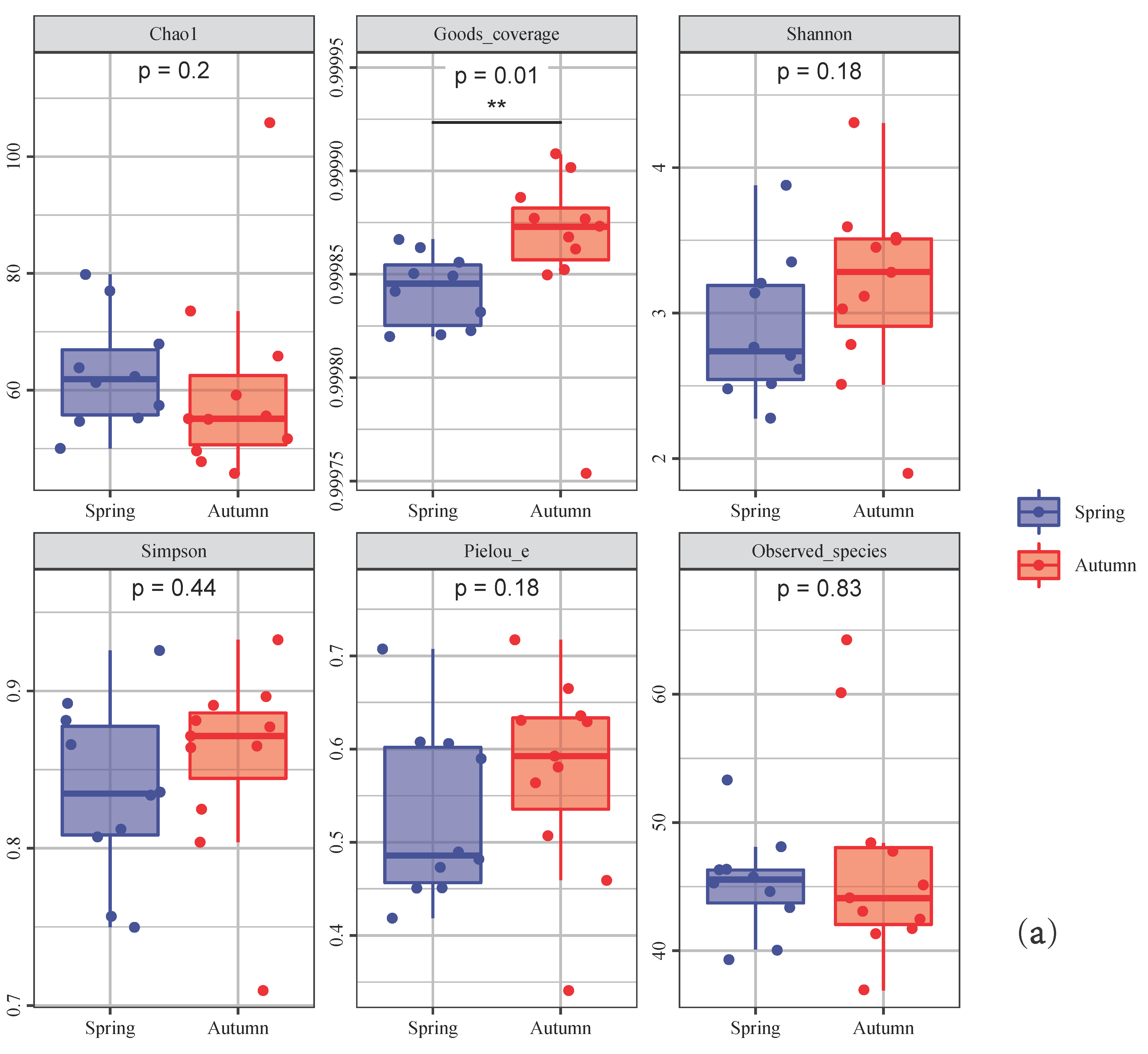
The spring and autumn samples were divided into two groups and were represented by different colors. In the figures, each panel corresponds to an alpha diversity index, marked in gray at the top. The box plot area of the “spring” Simpson and Shannon index plate was larger than that for “autumn”, which indicated that the community diversity in spring was greater than in autumn (Figure 3a). In addition, the box plot area of observed species index analysis for “autumn” was larger than that in the “spring”, indicating that the abundance in spring was higher than that in autumn (Figure 3a). In the two-dimensional sorting graph of the NMDS analysis, the points of the “spring” and “autumn” groups were closer, indicating that the difference in fish communities in spring and autumn was smaller, i.e., the composition of fish species in the Yangtze River mainstream in 2019 was similar in spring and autumn (Figure 3b).

3.3. Seasonal Variation Analysis

The heatmap of the relative sequence abundances of individual fish taxa revealed disparities in spatial distributions among species in each season (Figure 4). A total of 13 fish species were identified from the two seasons in the mainstream of the Yangtze River in 2019; they were common carp Cyprinus carpio, the Chinese lizard gudgeon Saurogobio dabryi, Saurogobio dumerili, yellow catfish Tachysurus fulvidraco, Acanthogobius hasta, Sinibotia superciliaris, amur sturgeon Acipenser schrenckii, grass carp Ctenopharyngodon idella, Odontamblyopus rubicundus, flathead grey mullet Mugil cephalus, stone flounder Kareius bicoloratus, the Asian swamp eel Monopterus albus and rainbow trout Oncorhynchus mykiss.

3.4. Relationship with Environmental Factors

The relationships between the environmental factors and the detected species were illustrated in a CCA analysis diagram using data from the 13 species detected in the two seasons and the set of seven environmental factors (Figure 5). As shown in the CCA plot, Chlorophyll-a was the key environmental factor affecting the fish assemblages, as the first axis was strongly correlated with TN, NO2 and Chlorophyll-a. The correlation between environmental factors and the distributions of the different species was inconsistent, and Chlorophyll-a had the strongest effect. The fish detected in spring and autumn, which are marked in purple in Figure 5, were not significantly affected by environmental factors.

4. Discussion

4.1. Fish Diversity in the Mainstream of the Yangtze River Based on eDNA

Our study investigated the composition of fish diversity in the spring and autumn in the Yangtze River mainstream, showing that eDNA technology can be used to investigate fish diversity. However, some common Yangtze fish, such as silver carp, bighead carp, grass carp and herring, were not detected in this investigation [51]. This may be attributed to the following aspects.
(1)
Insufficient sample collection: The fish activities in the Yangtze River mainstream are relatively large; in the present study, we only set 11 sampling points in the Yangtze River mainstream, which may have led to insufficient sampling. Ficetola et al. (2015) pointed out that replication, i.e., the number of water samples collected as opposed to the number of technical replicates run from a single sample, can be adjusted to compensate for false negatives [53]. Therefore, adding sampling points according to the study area would increase the possibility of detecting fish.
(2)
Less eDNA preservation: The amount of eDNA secreted by fish is related to itself; studies have shown that different species secrete DNA at different rates and quantities [54]. Moreover, the preservation of eDNA is also very important, as the long transportation after sampling, temperature, time and other factors will affect the preservation effect of the eDNA experiment. In addition, delayed extraction from the Sterivex filter may also result in less DNA being extracted. Thus, special attention should be paid to the preservation of samples, and timely filtration after sampling will reduce the degradation rate of eDNA.
(3)
Lack of local reference database information: This study did not establish a more accurate local reference database for fish in the Yangtze River mainstream, which may have reduced the number of native species identified. The effectiveness of eDNA depends on the quality of the reference sequence database and the classification parameters employed [55]. In future investigations and studies, we will consider establishing a local database. At the same time, the results of this survey will also serve as a reference for future surveys of fish in the Yangtze River mainstream.
(4)
Inapplicability of 12S primer to freshwater fish: The primers that we used may not be suitable for the identification of fish in the mainstream of the Yangtze River, which could explain the small number of identification results. Many studies have used other primers, such as the mitochondrial cytochrome c oxidase subunit I (COI) sequence and the 16S primer for the fish amplification [56]. In future studies, different primers can be designed to explore primers that are more suitable for Yangtze River fish. In the non-closed fishing season, fish body muscle tissue can be used for primer design, which will be our next research direction.
Shen et al. (2019) gathered a DNA barcode reference library of 2830 sequences from 238 species, with the objective of re-examining Yangtze River ichthyofauna diversity through a DNA-based method of species delimitation [1]. In order to obtain as many individuals per species as possible, various gears were used to capture specimens at different locations [1]. This will cause harm to the monitored species itself, and the eDNA technology only needs to collect water samples to detect the species. Therefore, it is very important to use the eDNA technique to monitor species and implement ecological protection in the future [38]. In addition, we detected some bottom-dwelling fish such as the Asian swamp eel Monopterus albus, which is difficult to be observed during normal activities due to their hidden living environment; this also demonstrates the sensitivity of our eDNA monitoring.

4.2. Seasonal Variation between Spring and Autumn

In the present study, the thermal imaging, through NMDS, showed that the composition of fish communities detected by eDNA technology was similar in spring and autumn. Nine species of fish were detected in both seasons; the research sampling was conducted in April and October in the Yangtze River. This result might be related to the similar temperature of the two seasons, as well as the living habits of the species themselves [57]. In addition, studies have shown that some water systems lack seasonal patterns regarding the fish species composition [38]. Alpha diversity analysis showed higher diversity in autumn, which may be related to the higher content of eDNA during the breeding period, which is consistent with the results of other previous investigations [38,57]. The current results suggest that continuous investigation would be of great help to fully understand the biodiversity of the mainstream of the Yangtze River.

4.3. Relationship between Fish Assemblage and Environmental Factors

Environmental DNA has been investigated in many waters in China since its inception [3]. Recently, there has been research on the construction of a database of fish in the Yangtze River mainstream through DNA barcoding technology, which is the key to the successful application of eDNA technology [1]. Chlorophyll-a had the greatest effect on fish species distribution, which is consistent with the findings of fish communities in other studies [38,55,57]. Our survey results also provide a reference for future surveys on the Yangtze River mainstream, and the eDNA technology used in species detection in this area is worthy of popularization and application. In addition, the impact of environmental factors on the detection of eDNA technology is also worthy of in-depth exploration, and more investigation and verification are required here [27].

5. Conclusions

In this study, we investigated the fish composition in the spring and autumn in the Yangtze River mainstream in 2019 through eDNA technology, which provides a reference for future surveys on the Yangtze River mainstream. Currently, there are few studies on the use of eDNA technology to investigate the seasonal composition of fish in the mainstream of the Yangtze River. It is worth noting that eDNA can represent a tool to assess composition in fish resource surveys, especially in the context of the ten-year ban on fishing in the Yangtze River. Although there are still some shortcomings caused by technical operations, pollution and insufficient databases, etc., this area needs to be studied in depth in the future. Therefore, we suggest that relevant research should continue to be carried out in the future to monitor the fish resources in the Yangtze River mainstream, so as to provide a scientific basis for the development and utilization of fishery resources in the region.

Supplementary Materials

The following are available online at https://www.mdpi.com/article/10.3390/fishes7010001/s1, Table S1: Seven environmental factors of two seasons, Table S2: Sequencing statistics for each sample in two seasons.

Author Contributions

Conceptualization, H.J., W.X. and H.Z.; methodology, H.J., W.X. and H.Z.; software, H.J.; validation, W.X. and H.Z.; formal analysis, H.J.; investigation, H.J.; resources, W.X. and H.Z.; data curation, W.X. and H.Z.; writing—original draft preparation, H.J.; writing—review and editing, H.J., W.X. and H.Z.; visualization, H.J., W.X. and H.Z.; supervision, H.Z. and W.X. All authors have read and agreed to the published version of the manuscript.

Funding

This research was funded by the National Natural Science Foundation of China (No. 41976094 and 31872568); Natural Science Foundation of Jiangsu Province (No. BK20201212); Key Deployment Program of Center for Ocean Mega-Science CAS (No. COMS2019Q14); Youth Innovation Promotion Association CAS (No.2020211).

Institutional Review Board Statement

Not applicable.

Informed Consent Statement

Informed consent was obtained from all subjects involved in the study.

Data Availability Statement

The original data file for the article can be found online at https://www.ncbi.nlm.nih.gov/sra/PRJNA783738, accessed on 11 December 2021.

Acknowledgments

We would like to thank the editor and the reviewers for their help with our article.

Conflicts of Interest

The authors declare no conflict of interest.

References

  1. Shen, Y.; Hubert, N.; Huang, Y.; Wang, X.; Gan, X.; Peng, Z.; He, S. DNA barcoding the ichthyofauna of the Yangtze River: Insights from the molecular inventory of a mega-diverse temperate fauna. Mol. Ecol. Resour. 2019, 19, 1278–1291. [Google Scholar] [CrossRef]
  2. Qu, C.; Stewart, K.A.; Clemente-Carvalho, R.; Zheng, J.; Wang, Y.; Gong, C.; Ma, L.; Zhao, J.; Lougheed, S.C. Comparing fish prey diversity for a critically endangered aquatic mammal in a reserve and the wild using eDNA metabarcoding. Sci. Rep. 2020, 10, 16715. [Google Scholar] [CrossRef] [PubMed]
  3. Eble, J.A.; Daly-Engel, T.S.; DiBattista, J.D.; Koziol, A.; Gaither, M.R. Marine environmental DNA: Approaches, applications, and opportunities. Adv. Mar. Biol. 2020, 86, 141–169. [Google Scholar] [CrossRef]
  4. Zou, K.; Chen, J.; Ruan, H.; Li, Z.; Guo, W.; Li, M.; Liu, L. eDNA metabarcoding as a promising conservation tool for monitoring fish diversity in a coastal wetland of the Pearl River Estuary compared to bottom trawling. Sci. Total Environ. 2020, 702, 134704. [Google Scholar] [CrossRef] [PubMed]
  5. Ogram, A.; Sayler, G.S.; Barkay, T. The extraction and purification of microbial DNA from sediments. J. Microbiol. Methods 1987, 7, 57–66. [Google Scholar] [CrossRef]
  6. Rondon, M.R.; August, P.R.; Bettermann, A.D.; Brady, S.F.; Grossman, T.H.; Liles, M.R.; Loiacono, K.A.; Lynch, B.A.; MacNeil, I.A.; Minor, C.; et al. Cloning the soil metagenome: A strategy for accessing the genetic and functional diversity of uncultured microorganisms. Appl. Environ. Microbiol. 2000, 66, 2541–2547. [Google Scholar] [CrossRef] [Green Version]
  7. Barnes, M.A.; Turner, C.R. The ecology of environmental DNA and implications for conservation genetics. Conserv. Genet. 2016, 17, 1–17. [Google Scholar] [CrossRef] [Green Version]
  8. Castañeda, R.A.; Van Nynatten, A.; Crookes, S.; Ellender, B.R.; Heath, D.D.; MacIsaac, H.J.; Mandrak, N.E.; Weyl, O.L.F. Detecting Native Freshwater Fishes Using Novel Non-invasive Methods. Front. Environ. Sci. 2020, 8, 29. [Google Scholar] [CrossRef]
  9. Hering, D.; Borja, A.; Jones, J.I.; Pont, D.; Boets, P.; Bouchez, A.; Bruce, K.; Drakare, S.; Hänfling, B.; Kahlert, M.; et al. Implementation options for DNA-based identification into ecological status assessment under the European Water Framework Directive. Water Res. 2018, 138, 192–205. [Google Scholar] [CrossRef]
  10. Ficetola, G.F.; Miaud, C.; Pompanon, F.; Taberlet, P. Species detection using environmental DNA from water samples. Biol. Lett. 2008, 4, 423–425. [Google Scholar] [CrossRef] [Green Version]
  11. Dejean, T.; Valentini, A.; Miquel, C.; Taberlet, P.; Bellemain, E.; Miaud, C. Improved detection of an alien invasive species through environmental DNA barcoding: The example of the American bullfrogLithobates catesbeianus. J. Appl. Ecol. 2012, 49, 953–959. [Google Scholar] [CrossRef]
  12. Jerde, C.L.; Mahon, A.R.; Chadderton, W.L.; Lodge, D.M. “Sight-unseen” detection of rare aquatic species using environmental DNA. Conserv. Lett. 2011, 4, 150–157. [Google Scholar] [CrossRef]
  13. Goldberg, C.S.; Sepulveda, A.; Ray, A.; Baumgardt, J.; Waits, L.P. Environmental DNA as a new method for early detection of New Zealand mudsnails (Potamopyrgus antipodarum). Freshw. Sci. 2013, 32, 792–800. [Google Scholar] [CrossRef] [Green Version]
  14. Geerts, A.N.; Boets, P.; Van den Heede, S.; Goethals, P.; Van der heyden, C. A search for standardized protocols to detect alien invasive crayfish based on environmental DNA (eDNA): A lab and field evaluation. Ecol. Indic. 2018, 84, 564–572. [Google Scholar] [CrossRef]
  15. Egan, S.P.; Grey, E.; Olds, B.; Feder, J.L.; Ruggiero, S.T.; Tanner, C.E.; Lodge, D.M. Rapid Molecular Detection of Invasive Species in Ballast and Harbor Water by Integrating Environmental DNA and Light Transmission Spectroscopy. Environ. Sci. Technol. 2015, 49, 4113–4121. [Google Scholar] [CrossRef] [Green Version]
  16. Xia, Z.; Zhan, A.; Gao, Y.; Zhang, L.; Haffner, G.D.; MacIsaac, H.J. Early detection of a highly invasive bivalve based on environmental DNA (eDNA). Biol. Invasions 2017, 20, 437–447. [Google Scholar] [CrossRef]
  17. Simpfendorfer, C.A.; Kyne, P.M.; Noble, T.H.; Goldsbury, J.; Basiita, R.K.; Lindsay, R.; Jerry, D.R. Environmental DNA detects critically endangered largetooth sawfish in the wild. Endanger. Species Res. 2016, 30, 109–116. [Google Scholar] [CrossRef] [Green Version]
  18. Weltz, K.; Lyle, J.M.; Ovenden, J.; Morgan, J.A.T.; Moreno, D.A.; Semmens, J.M. Application of environmental DNA to detect an endangered marine skate species in the wild. PLoS ONE 2017, 12, e0178124. [Google Scholar] [CrossRef]
  19. Mauvisseau, Q.; Davy-Bowker, J.; Bulling, M.; Brys, R.; Neyrinck, S.; Troth, C.; Sweet, M. Combining ddPCR and environmental DNA to improve detection capabilities of a critically endangered freshwater invertebrate. Sci. Rep. 2019, 9, 14064. [Google Scholar] [CrossRef] [Green Version]
  20. Nevers, M.B.; Byappanahalli, M.N.; Morris, C.C.; Shively, D.; Przybyla-Kelly, K.; Spoljaric, A.M.; Dickey, J.; Roseman, E.F. Environmental DNA (eDNA): A tool for quantifying the abundant but elusive round goby (Neogobius melanostomus). PLoS ONE 2018, 13, e0191720. [Google Scholar] [CrossRef]
  21. Yamanaka, H.; Minamoto, T. The use of environmental DNA of fishes as an efficient method of determining habitat connectivity. Ecol. Indic. 2016, 62, 147–153. [Google Scholar] [CrossRef] [Green Version]
  22. Sigsgaard, E.E.; Nielsen, I.B.; Bach, S.S.; Lorenzen, E.D.; Robinson, D.P.; Knudsen, S.W.; Pedersen, M.W.; Jaidah, M.A.; Orlando, L.; Willerslev, E.; et al. Population characteristics of a large whale shark aggregation inferred from seawater environmental DNA. Nat. Ecol. Evol. 2016, 1, 4. [Google Scholar] [CrossRef]
  23. Bakker, J.; Wangensteen, O.S.; Chapman, D.D.; Boussarie, G.; Buddo, D.; Guttridge, T.L.; Hertler, H.; Mouillot, D.; Vigliola, L.; Mariani, S. Environmental DNA reveals tropical shark diversity in contrasting levels of anthropogenic impact. Sci. Rep. 2017, 7, 16886. [Google Scholar] [CrossRef]
  24. Boussarie, G.; Bakker, J.; Wangensteen, O.S.; Mariani, S.; Bonnin, L.; Juhel, J.B.; Kiszka, J.J.; Kulbicki, M.; Manel, S.; Robbins, W.D.; et al. Environmental DNA illuminates the dark diversity of sharks. Sci. Adv. 2018, 4, eaap9661. [Google Scholar] [CrossRef] [Green Version]
  25. Thalinger, B.; Wolf, E.; Traugott, M.; Wanzenböck, J. Monitoring spawning migrations of potamodromous fish species via eDNA. Sci. Rep. 2019, 9, 15388. [Google Scholar] [CrossRef] [Green Version]
  26. Cowart, D.A.; Breedveld, K.G.H.; Ellis, M.J.; Hull, J.M.; Larson, E.R. Environmental DNA (eDNA) applications for the conservation of imperiled crayfish (Decapoda: Astacidea) through monitoring of invasive species barriers and relocated populations. J. Crustacean Biol. 2018, 38, 257–266. [Google Scholar] [CrossRef]
  27. Port, J.A.; O’Donnell, J.L.; Romero-Maraccini, O.C.; Leary, P.R.; Litvin, S.Y.; Nickols, K.J.; Kelly, R.P. Assessing vertebrate biodiversity in a kelp forest ecosystem using environmental DNA. Mol. Ecol. 2015, 25, 527–541. [Google Scholar] [CrossRef]
  28. Forsström, T.; Vasemägi, A. Can environmental DNA (eDNA) be used for detection and monitoring of introduced crab species in the Baltic Sea? Mar. Pollut. Bull. 2016, 109, 350–355. [Google Scholar] [CrossRef]
  29. Lanzén, A.; Lekang, K.; Jonassen, I.; Thompson, E.M.; Troedsson, C. High-throughput metabarcoding of eukaryotic diversity for environmental monitoring of offshore oil-drilling activities. Mol. Ecol. 2016, 25, 4392–4406. [Google Scholar] [CrossRef]
  30. Stat, M.; Huggett, M.J.; Bernasconi, R.; DiBattista, J.D.; Berry, T.E.; Newman, S.J.; Harvey, E.S.; Bunce, M. Ecosystem biomonitoring with eDNA: Metabarcoding across the tree of life in a tropical marine environment. Sci. Rep. 2017, 7, 12240. [Google Scholar] [CrossRef]
  31. DiBattista, J.D.; Coker, D.J.; Sinclair-Taylor, T.H.; Stat, M.; Berumen, M.L.; Bunce, M. Assessing the utility of eDNA as a tool to survey reef-fish communities in the Red Sea. Coral Reefs 2017, 36, 1245–1252. [Google Scholar] [CrossRef]
  32. DiBattista, J.D.; Reimer, J.D.; Stat, M.; Masucci, G.D.; Biondi, P.; De Brauwer, M.; Bunce, M. Digging for DNA at depth: Rapid universal metabarcoding surveys (RUMS) as a tool to detect coral reef biodiversity across a depth gradient. PeerJ 2019, 7, e6379. [Google Scholar] [CrossRef] [PubMed]
  33. Thomsen, P.F.; Willerslev, E. Environmental DNA–An emerging tool in conservation for monitoring past and present biodiversity. Biol. Conserv. 2015, 183, 4–18. [Google Scholar] [CrossRef]
  34. Jeunen, G.J.; Knapp, M.; Spencer, H.G.; Lamare, M.D.; Taylor, H.R.; Stat, M.; Gemmell, N.J. Environmental DNA (eDNA) metabarcoding reveals strong discrimination among diverse marine habitats connected by water movement. Mol. Ecol. Resour. 2019, 19, 426–438. [Google Scholar] [CrossRef] [PubMed]
  35. Lacoursière-Roussel, A.; Côté, G.; Leclerc, V.; Bernatchez, L. Quantifying relative fish abundance with eDNA: A promising tool for fisheries management. J. Appl. Ecol. 2016, 53, 1148–1157. [Google Scholar] [CrossRef]
  36. Kimmerling, N.; Zuqert, O.; Amitai, G.; Gurevich, T.; Armoza-Zvuloni, R.; Kolesnikov, I.; Berenshtein, I.; Melamed, S.; Gilad, S.; Benjamin, S.; et al. Quantitative species-level ecology of reef fish larvae via metabarcoding. Nat. Ecol. Evol. 2018, 2, 306–316. [Google Scholar] [CrossRef] [PubMed]
  37. West, K.M.; Stat, M.; Harvey, E.S.; Skepper, C.L.; DiBattista, J.D.; Richards, Z.T.; Travers, M.J.; Newman, S.J.; Bunce, M. eDNA metabarcoding survey reveals fine-scale coral reef community variation across a remote, tropical island ecosystem. Mol. Ecol. 2020, 29, 1069–1086. [Google Scholar] [CrossRef]
  38. Zhang, H.; Yoshizawa, S.; Iwasaki, W.; Xian, W.W. Seasonal Fish Assemblage Structure Using Environmental DNA in the Yangtze Estuary and Its Adjacent Waters. Front. Mar. Sci. 2019, 6, 515. [Google Scholar] [CrossRef]
  39. Miya, M.; Sato, Y.; Fukunaga, T.; Sado, T.; Poulsen, J.Y.; Sato, K.; Minamoto, T.; Yamamoto, S.; Yamanaka, H.; Araki, H.; et al. MiFish, a set of universal PCR primers for metabarcoding environmental DNA from fishes: Detection of more than 230 subtropical marine species. R. Soc. Open Sci. 2015, 2, 150088. [Google Scholar] [CrossRef] [PubMed] [Green Version]
  40. Bolyen, E.; Rideout, J.R.; Dillon, M.R.; Bokulich, N.A.; Abnet, C.C.; Al-Ghalith, G.A.; Alexander, H.; Alm, E.J.; Arumugam, M.; Asnicar, F.; et al. Reproducible, interactive, scalable and extensible microbiome data science using QIIME 2. Nat. Biotechnol. 2019, 37, 852–857. [Google Scholar] [CrossRef]
  41. Rognes, T.; Flouri, T.; Nichols, B.; Quince, C.; Mahé, F. VSEARCH: A versatile open source tool for metagenomics. PeerJ 2016, 4, e2584. [Google Scholar] [CrossRef]
  42. Martin, M. Cutadapt Removes Adapter Sequences from High-Throughput Sequencing Reads. EMBnet J. 2011, 17, 10–12. [Google Scholar] [CrossRef]
  43. Chao, A. Nonparametric Estimation of the Number of Classes in a Population. Scand. J. Stat. 1984, 11, 265–270. [Google Scholar]
  44. Shannon, C.E. A mathematical theory of communication. Bell Syst. Tech. J. 1948, 27, 379–656. [Google Scholar] [CrossRef] [Green Version]
  45. Legovic, T. Statistical ecology. A primer on methods and computing. Ecol. Model. 1991, 54, 143–144. [Google Scholar] [CrossRef]
  46. Faith, D.P. Conservation evaluation and phylogenetic diversity. Biol. Conserv. 1992, 61, 1–10. [Google Scholar] [CrossRef]
  47. Pielou, E.C. The measurement of diversity in different types of biological collections. J. Theor. Biol. 1966, 13, 131–144. [Google Scholar] [CrossRef]
  48. Good, I.J. The population frequency of species and theestimation of the population parameters. Biometrics 1958, 40, 237–246. [Google Scholar] [CrossRef]
  49. Lozupone, C.A.; Hamady, M.; Kelley, S.T.; Knight, R. Quantitative and qualitative beta diversity measures lead to different insights into factors that structure microbial communities. Appl. Environ. Microbiol 2007, 73, 1576–1585. [Google Scholar] [CrossRef] [Green Version]
  50. Lozupone, C.; Knight, R. UniFrac: A new phylogenetic method for comparing microbial communities. Appl. Environ. Microbiol. 2005, 71, 8228–8235. [Google Scholar] [CrossRef] [Green Version]
  51. Fish Laboratory, Institute of Hydrobiology, Chinese Academy of Sciences. Fishes of Yangtze River; Science Press: Beijing, China, 1976. [Google Scholar]
  52. Anderson, M.J.; Ellingsen, K.E.; McArdle, B.H. Multivariate dispersion as a measure of beta diversity. Ecol. Lett. 2006, 9, 683–693. [Google Scholar] [CrossRef]
  53. Ficetola, G.F.; Pansu, J.; Bonin, A.; Coissac, E.; Giguet-Covex, C.; De Barba, M.; Gielly, L.; Lopes, C.M.; Boyer, F.; Pompanon, F.; et al. Replication levels, false presences and the estimation of the presence/absence from eDNA metabarcoding data. Mol. Ecol Resour. 2015, 15, 543–556. [Google Scholar] [CrossRef]
  54. Russo, T.; Maiello, G.; Talarico, L.; Baillie, C.; Colosimo, G.; D’Andrea, L.; Di Maio, F.; Fiorentino, F.; Franceschini, S.; Garofalo, G.; et al. All is fish that comes to the net: Metabarcoding for rapid fisheries catch assessment. Ecol. Appl. 2021, 31, e02273. [Google Scholar] [CrossRef]
  55. Gold, Z.; Curd, E.E.; Goodwin, K.D.; Choi, E.S.; Frable, B.W.; Thompson, A.R.; Walker, H.J.; Burton, R.S.; Kacev, D.; Martz, L.D.; et al. Improving metabarcoding taxonomic assignment: A case study of fishes in a large marine ecosystem. Mol. Ecol. Resour. 2021, 21, 2546–2564. [Google Scholar] [CrossRef]
  56. Zhang, S.; Zhao, J.; Yao, M. A comprehensive and comparative evaluation of primers for metabarcoding eDNA from fish. Methods Ecol. Evol. 2020, 11, 1609–1625. [Google Scholar] [CrossRef]
  57. Jia, H.; Wang, Y.; Yoshizawa, S.; Iwasaki, W.; Li, Y.; Xian, W.; Zhang, H. Seasonal Variation and Assessment of Fish Resources in the Yangtze Estuary Based on Environmental DNA. Water 2020, 12, 2874. [Google Scholar] [CrossRef]
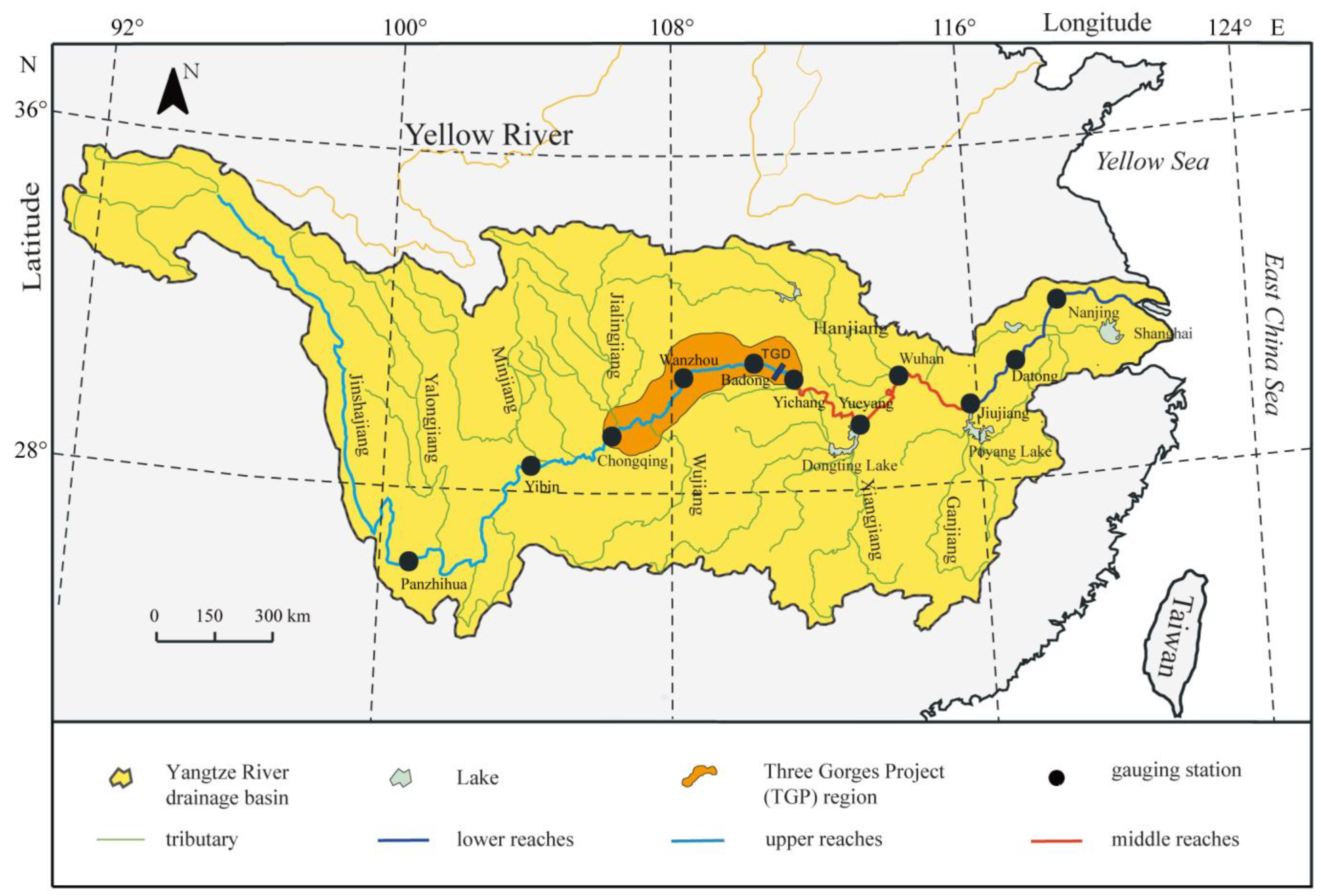
Figure 1. Sampling sites from the Yangtze River mainstream.
Fishes 07 00001 g001
Figure 2. Species distribution in spring and autumn samples. Different colors indicate different species and are marked on the right. “SE” represents the 2019 Yangtze River spring samples; “AE” represents the 2019 Yangtze River autumn samples.
Fishes 07 00001 g002
Figure 3. Seasonal difference based on two-index analysis. The alpha index (a): The blue group represents spring eDNA; the red group is autumn eDNA; the number under the label of diversity index is the p value of the Kruskal–Wallis test. In each panel, the abscissa is the grouping label, and the ordinate is the value of the corresponding alpha diversity index. “**” is p < 0.01 The beta index (b): The blue triangle group represents the spring samples; the red circle group represents autumn samples. The closer (farther) the distance between the two points, the smaller (greater) the fish community difference between the two samples.
Fishes 07 00001 g003aFishes 07 00001 g003b
Figure 4. Species distribution in two seasons. The samples are clustered by UPGMA according to the Euclidean distance of the relative abundance composition data of the species, and are arranged according to the clustering results by default. “SE19” represents the samples in spring 2019 in the Yangtze River; “AE19” represents the samples in autumn 2019 in the Yangtze River.
Fishes 07 00001 g004
Figure 5. The CCA diagram of the relationship between fish and seven environmental factors for two seasons. Fish marked in purple are the common species detected in both spring and autumn.
Fishes 07 00001 g005
Table 1. Details of sampling stations.
NumberStationLongitudeLatitudeSpringAutumn
SamplingTimeSamplingTime
1Panzhihua101.7026.56C19SE0112 April 2019C19AE0113 October 2019
2Yibin104.6628.77C19SE0214 April 2019C19AE0215 October 2019
3Chongqing106.5729.55 C19AE0315 October 2019
4Wanzhou108.4330.76C19SE0315 April 2019C19AE0416 October 2019
5Badong110.3431.05C19SE0416 April 2019C19AE0517 October 2019
6Yichang111.4030.57C19SE0517 April 2019C19AE0618 October 2019
7Yuyang113.1229.40C19SE0617 April 2019C19AE0718 October 2019
8Wuhan114.3030.55C19SE0718 April 2019C19AE0819 October 2019
9Jiujiang116.0329.75C19SE0819 April 2019C19AE0920 October 2019
10Datong117.7430.86C19SE0920 April 2019C19AE1021 October 2019
11Nanjing118.7532.12C19SE1020 April 2019C19AE1122 October 2019
Publisher’s Note: MDPI stays neutral with regard to jurisdictional claims in published maps and institutional affiliations.