Review

Structures Associated with Oogenesis and Embryonic Development during Intraovarian Gestation in Viviparous Teleosts (Poeciliidae)

by
Mari Carmen Uribe
*,
Gabino De la Rosa Cruz
,
Adriana García Alarcón
,
Juan Carlos Campuzano Caballero
and
María Guadalupe Guzmán Bárcenas
Laboratorio de Biología de la Reproducción Animal Departamento de Biología Comparada, Facultad de Ciencias, Universidad Nacional Autónoma de México, Ciudad Universitaria, 04510 Ciudad de México, México
*
Author to whom correspondence should be addressed.
Fishes 2019, 4(2), 35; https://doi.org/10.3390/fishes4020035
Submission received: 29 March 2019 / Revised: 4 June 2019 / Accepted: 4 June 2019 / Published: 19 June 2019
(This article belongs to the Special Issue Fish Reproductive Physiology and Aquaculture)

Abstract

:
Viviparity in teleosts involves, invariably, the ovary in a gestational role. This type of viviparity is due to the combination of unique aspects, different from those found in the rest of vertebrates. These aspects are: The ovary has a saccular structure; the germinal epithelium lines the ovarian lumen; the absence of oviducts; and the intraovarian insemination, fertilization, and gestation. The communication of the germinal zone of the ovary to the exterior is via the caudal zone of the ovary—the gonoduct. The germinal epithelium is composed of oogonia and oocytes scattered individually or in cell nests among somatic epithelial cells. In the ovarian stroma the follicles are included which are formed by the oocyte, which is surrounded by follicular cells and the vascularized theca. The oogenesis comprises three stages: chromatin-nucleolus, previtellogenesis, and vitellogenesis. There is no ovulation, as the oocyte is retained in the follicle. During the insemination, the spermatozoa enter into the ovarian lumen and the intrafollicular fertilization occurs, followed by intrafollicular gestation. The intraovarian gestation of poeciliids involves morphological characteristics associated with the intrafollicular embryogenesis and types of nutrition, such as lecithotrophy and matrotrophy. In lecithotrophy, the nutrients come from the yolk reserves stored during oogenesis, whereas in matrotrophy the nutrients are provided by supplies from maternal tissues to the embryo during gestation. The maternal–embryonic metabolic interchanges converge through the development of the association of maternal and embryonic blood vessels, establishing a follicular placenta.

1. Ovarian Adaptations of Viviparous Teleosts

Among more than 32,672 species of teleost fishes [1], approximately 510 are viviparous [2,3,4]. Viviparity in teleosts has unique aspects compared with the rest of viviparous vertebrates. To compare viviparity of teleosts with other viviparous vertebrates is very useful for defining the morpho-physiological adaptations of reproductive structures, which are unique elements associated with teleost viviparity [5,6,7]. In the evolution of viviparity from oviparity there is a change in the site of embryonic development from the external environment to the internal female reproductive system. Viviparity in fishes is a complex process, which involves several modifications in the reproductive systems of both males and females [8,9]. Some of these aspects are related with the structure of the ovary for gestation, because the viviparity in teleosts involves, invariably, the ovary in a gestational role.
During the adult life of non-teleost vertebrates, the ovarian cortex, which is lined by the germinal epithelium with oogonia for oogenesis, remains in contact with the coelomic cavity where ovulation occurs. In contrast, in the cystovarian type of teleosts, the cortex of the embryonic ovary, also lined by the germinal epithelium, undergoes an invagination that fuses internally becoming the saccular structure of the ovary. This saccular structure has an internal space—the ovarian lumen. As result of this unique development of the ovary of teleosts, the surface of the ovarian lumen contains the germinal epithelium situated internally [10]. Consequently, in teleosts, at ovulation, the eggs are shed into the lumen of the ovary (internal ovulation), in contrast to the rest of vertebrates where ovulation occurs toward the coelomic cavity [11].
Additionally, a distinctive feature of almost all viviparous teleosts occurs when, during the early embryonic development, the right and left ovaries fuse, forming a single ovary [8,11,12]. This fusion establishes a single and saccular ovary, with the germinal epithelium lining the internal lumen.
Another unique characteristic of teleosts among vertebrates is that teleosts do not develop Müllerian ducts during the embryogenesis, as occurs in the rest of vertebrates; consequently, teleosts do not have oviducts. Then, the communication of the germinal zone of the ovary to the exterior occurs via the caudal zone of the ovary—which is called the gonoduct—an ovarian zone that lacks germinal cells [8,13,14]. Therefore, the lumen of the ovarian germinal zone is continuous with that of the gonoduct, where the development of numerous folds of the mucosa forms a limit, similar to a cervix, displayed at the border of the germinal portion of the ovary and the gonoduct [15].
Because of the lack of oviducts in viviparous teleosts, the gestation takes place in the ovary, in contrast to the rest of viviparous vertebrates where the gestation is in the uterus. That is, exclusively in viviparous teleosts, it occurs an intraovarian gestation. Therefore, the ovary of viviparous teleosts is not only the structure where oogenesis occurs, but, also, it receives the spermatozoa during the insemination, maintains the spermatozoa, and it allows the fertilization of oocytes and the development of offspring until birth [16]. The sequence of these processes makes the ovary of viviparous teleosts a very complex organ, developing morpho-physiological adaptations for the insemination, the entrance and movements of spermatozoa into the ovary [15], the storage of spermatozoa until fertilization [17], the fertilization of oocytes [16], and the intraovarian gestation [18].
According to the nutrients used by the embryos during their gestation, there are two nutritional patterns in viviparous teleosts, both involving ovarian structure and egg morphology. These patterns are lecithotrophy and matrotrophy [7,9,19,20]. In lecithotrophy, the nutrients for the embryo come from the yolk reserves stored in the egg during oogenesis, prior to fertilization, similar to that which occurs in oviparous fishes. In matrotrophy the nutrients are provided not only by those stored in the egg during oogenesis, but also by supplies from the maternal tissues during gestation, consequently, after fertilization [5,6,9,13,14,18]. Pires and Reznick [21] mention that matrotrophic species invest fewer resources prior to fertilization and, instead, provide resources to offspring throughout development. Thus, the nutritional pattern in species with small eggs involves the reduction of lecithotrophy and the increase of matrotrophy. Therefore, the reproductive strategy of viviparous species is significantly connected with the nutrition of the embryos during gestation. Consequently, in accordance with the amount of yolk deposited in the oocyte during oogenesis, the nutrition of the embryo requires the transfer of nutrients from the maternal tissue, developing different levels of matrotrophy: incipient, middle, or high [19]. The understanding of this complex process of nutrition during gestation requires the analysis of the specialized structures related with this process. Then, as it is considered by Blackburn and Starck [20], morphology has a central role in helping to explain the function and evolution of patterns of fetal nutrition in viviparous teleosts.

2. The Family Poeciliidae

The family Poeciliidae includes species of freshwater teleosts of the order Cyprinodontiformes. Poeciliid reproduction involves viviparity. This family contains the most numerous species of viviparous teleosts, with approximately 337 species [2]. In the context of viviparity of this family, there is an interesting species long considered essential in understanding the evolution of viviparity, this species is Tomeurus gracilis, which is the unique zygoparous or embryoparous poecilid. characterized because, after the intraovarian fertilization, the females let the zygotes into the exterior prematurely, this is fertilized eggs or embryos in early stage of development which are attached to plants where the development continues [22].
Poeciliids occur from the southeastern United States to South America, Africa, including Madagascar [3,23,24,25], with a wide distribution in lands of Central Mexico, the Caribbean islands, and around the Gulf of Mexico. Actually, due to the release of specimens from home aquaria (ornamental fishes), poeciliids can be found in all tropical and subtropical freshwater ecosystems [24,26]. Poeciliids have been used in evolutionary and ecological studies analyzing important questions in the field of conservation biology. Related to the viviparous reproduction in teleosts, poeciliids have been of great importance in the definition and understanding of this unique type of gestation [7,9].

3. The Ovary of Poeciliids

The ovary of poeciliids is of cystovarian type, as a single and saccular structure with a central lumen (Figure 1A,B,C). Histologically, the ovarian wall consists of four tissue layers. From the interior to the exterior, these layers are: (1) Germinal epithelium integrated by germ cells, such as oogonia, and oocytes scattered individually or in cell nests among somatic epithelial cells (Figure 2A). The germinal epithelium borders the ovarian lumen and it is separated from the stroma by a basement membrane [10]; (2) stroma, formed by loose and vascularized connective tissue, enclosing the ovarian follicles. The follicles are integrated by oocytes in different stages of development as previtellogenesis or vitellogenesis; (3) smooth muscle layers; and (4) serosa, formed by thin connective tissue and externally lined by mesothelium [7,11,27,28]. The caudal zone of the ovary forms the gonoduct, which lacks germinal cells (Figure 1C and Figure 2A).
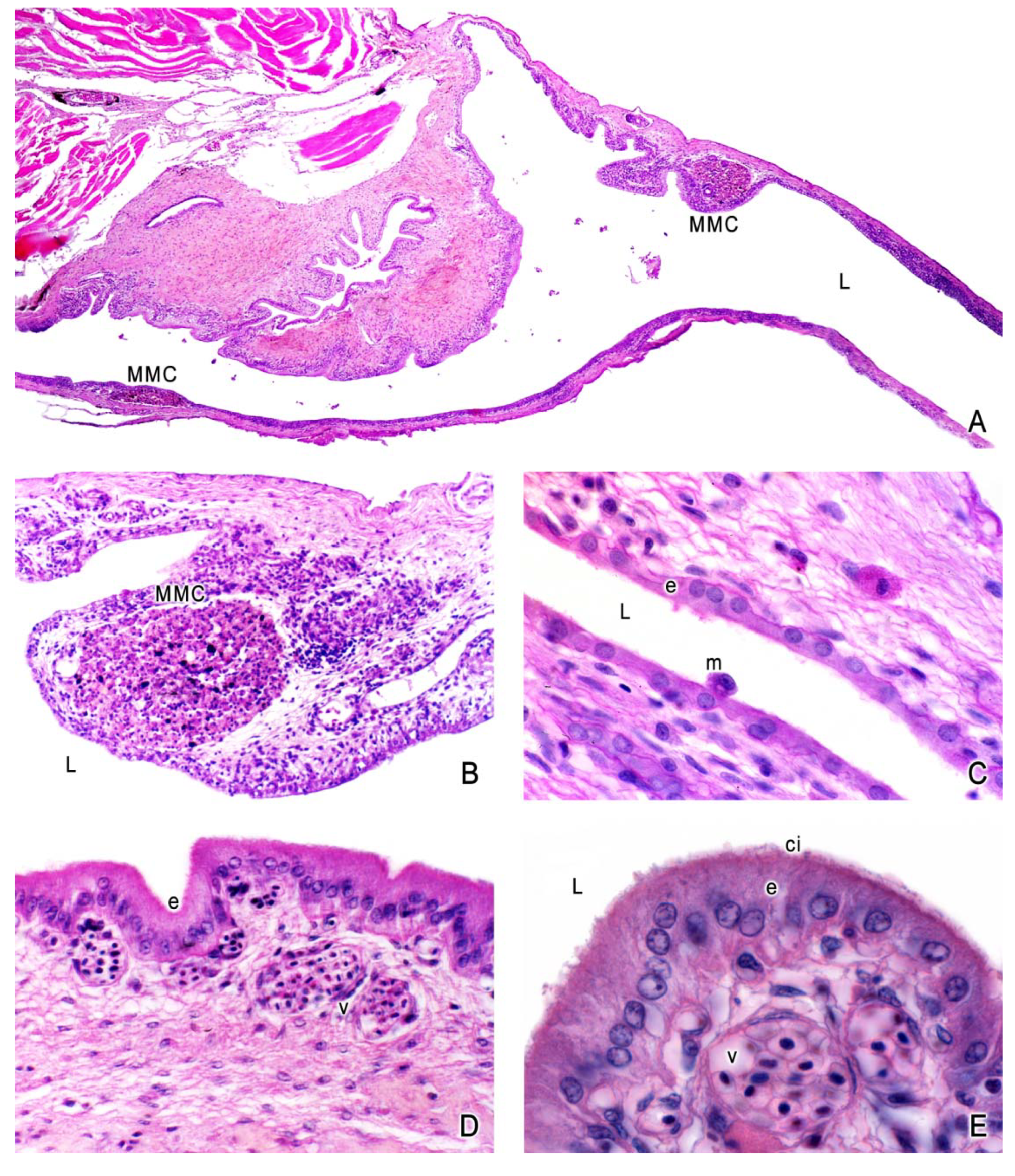
The gonoduct acquires special interest in viviparous species, where during birth the offspring goes to the exterior through the gonoduct. Consequently, the gonoduct forms a barrier between the germinal zone of the ovary and the exterior. Campuzano-Caballero and Uribe [15,29] analyzed the gonoduct of Poecilia reticulata [15] and Poeciliopsis gracilis [29], describing this structure as a muscular tube that includes multiple longitudinal folds of the mucosa extended into the gonoductal lumen. The gonoduct is lined internally by single cuboidal or columnar epithelium with ciliated and non-ciliated cells, plus stroma of connective tissue, smooth muscle, and serosa (Figure 2A–E). Melano-macrophage centers are irregularly located in the connective tissue, adjacent to the epithelium; they are round or oval aggregates of cells such as macrophages, lymphocytes, and melanocytes (Figure 2A,B). The presence of these centers suggests its involvement in the protection of the ovary and embryos by immunological functions. Macrophages and lymphocytes may be also located subjacent to the epithelium or in the lumen of the gonoduct (Figure 2C). The presence of ciliated and non-ciliated epithelial cells (Figure 2D,E) may facilitate the transport of spermatozoa during the insemination and embryos during birth.

4. Oogenesis

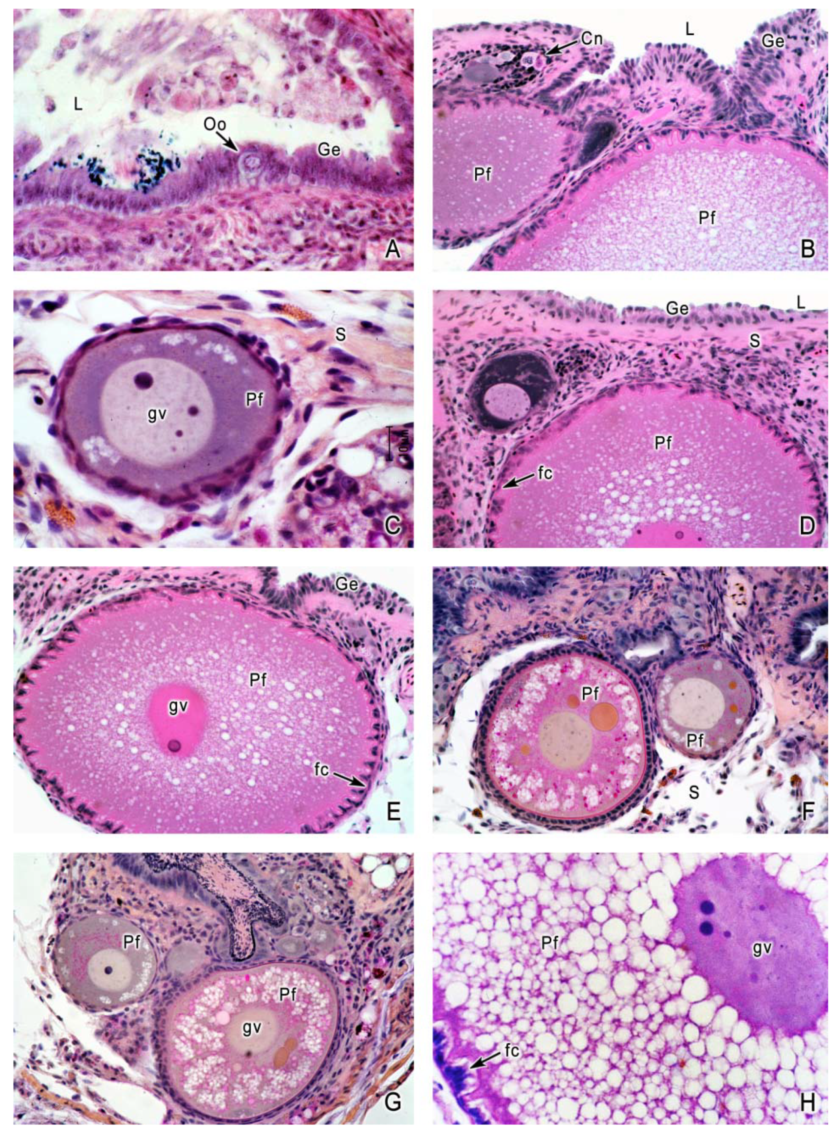
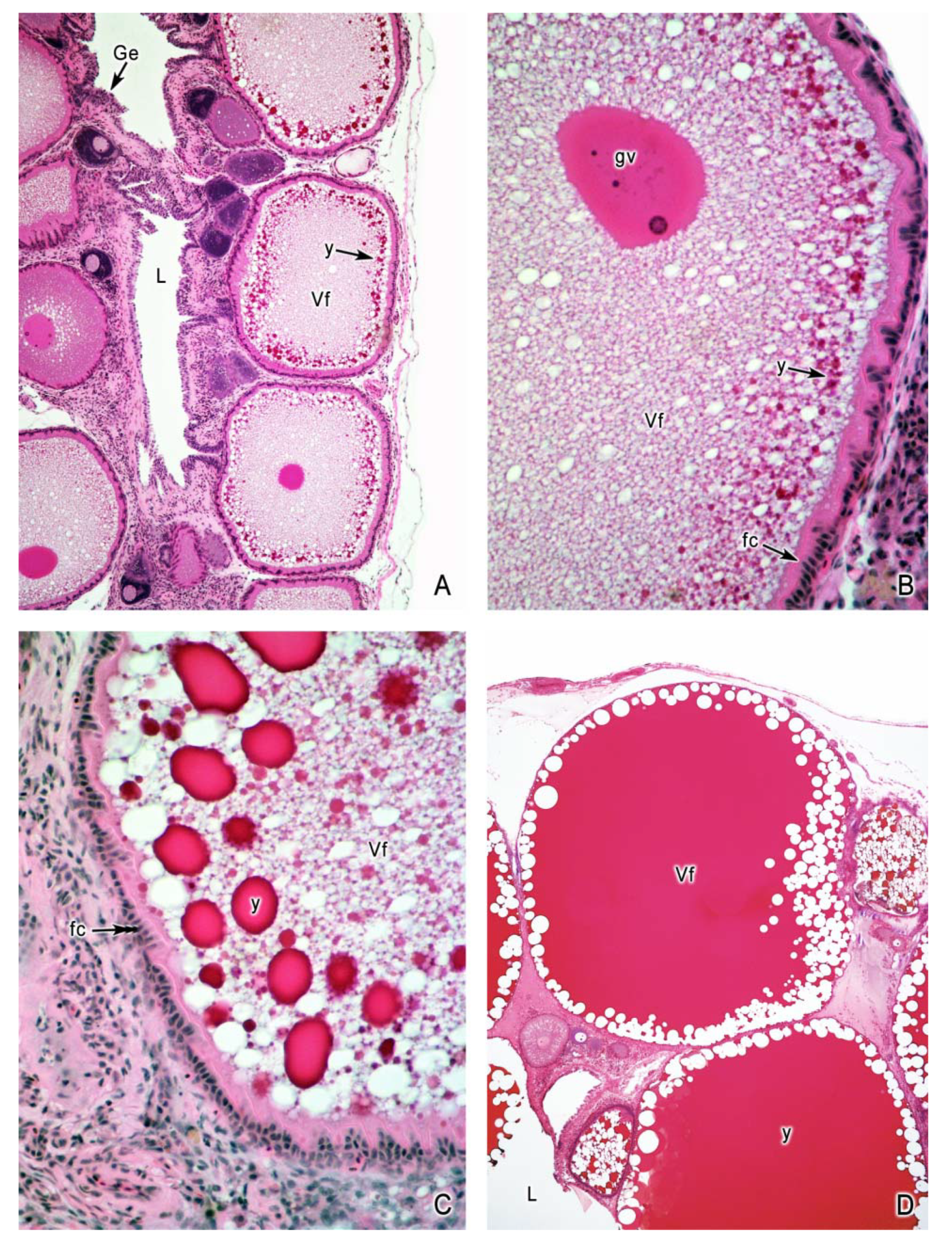
The oocyte matures during the process of oogenesis in the follicle (Figure 3A–H). The follicle is composed of the oocyte surrounded by a single layer of follicular cells supported by a thin vascularized theca [28]. The oogenesis includes three stages: chromatin-nucleolus, previtellogenesis, and vitellogenesis [28]. When oogonia initiate meiosis, they form oocytes in the chromatin-nucleolus stage (Figure 3B), where the germinal vesicle (nucleus) displays the lampbrush chromosomes. During previtellogenesis the oocyte acquires the ooplasm components and organelles. During vitellogenesis the oocyte acquires the diverse and complex nutrients stored in the yolk [4,7,23,28,30,31]. Previtellogenesis is marked by several features: The ooplasm is basophilic (Figure 3C), the germinal vesicle (nucleus) has a single nucleolus that proliferates to multiple nucleoli (Figure 3C), numerous lipid droplets (Figure 3C–H) and cortical alveoli appear in the ooplasm, and the oocyte diameter increases. Vitellogenesis is seen when yolk globules are deposited in the ooplasm (Figure 4A–D); the yolk becomes fluid and some lipid droplets may be seen around the oocyte periphery (Figure 4D). The follicular epithelium becomes columnar, indicating intense secretion of nutrients for the oocyte [32]. At the end of vitellogenesis, the oocyte reaches its maximum size and the germinal vesicle migrates to the periphery of the ooplasm at the animal pole [28,32].
Most poeciliids develop large oocytes with abundant yolk. Then, oogenesis forms large oocytes which grow from a diameter of 10 µm of the oogonial stage to 2.0 mm of mature eggs. Some examples of poecilid species and the mean diameter of their mature oocytes are: Girardinus denticulatus, 2.6 mm; Girardinus uninotatus, 2.3 mm [33]; Poecilia reticulata, 2.1 mm [34]; Gambusia puncticulata, 2.6 mm [33]; Poeciliopsis occidentalis, 2.02 mm [34], Poeciliopsis monacha, 2.0 mm; Limia vittata, 2.3 mm [31]; but other species develop smaller oocytes containing less amount of yolk such as Gambusia affinis, 1.7 mm [31], Quintana atrizona, 1.6 mm [33]; Poeciliopsis lucida, 1.4 mm [35] and species with even smaller oocytes, such as Poeciliopsis turneri, 1.0 mm and Poeciliopsis prolifica, 0.8 mm [35]. However, the species with the smallest oocyte, considered a microlecithal oocyte, with a mean diameter of 400 µm, is Heterandria formosa [36]. Turner [37] considered that the diminution of yolk is an important and specialized feature involved in teleost viviparity. Later, Turner [13] commented that the most advanced type of viviparity occurred in species where the yolk has become greatly reduced, as in H. formosa. This is an evolutionary change involved from lecithotrophy to matrotrophy.

5. The Intraovarian Gestation in Poeciliids

The intraovarian gestation in poeciliids is initiated when the oocytes are fertilized in the ovarian follicle. The embryos remain into the follicle throughout all their development, until birth. That is, fertilization and gestation in poeciliids are intrafollicular [7,27,31]. All the morpho-physiological adaptations of the ovary are features essentially related with this reproductive strategy of viviparity: the intraovarian and intrafollicular embryogenesis.
It is common in poeciliids that, after insemination, the ovary stores spermatozoa. In species in which sperm storage occurs, the germinal epithelium is involved in relationships with the spermatozoa as observed in Heterandria formosa [13,16], Poecilia reticulata [38], and Xiphophorus maculatus [17].
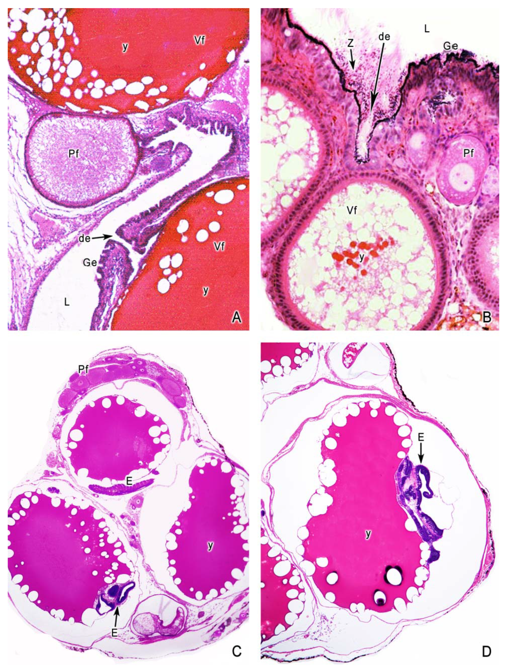
The intrafollicular fertilization is possible by a specific structure at the periphery of each oocyte where the spermatozoa make contact with the oocyte. This structure penetrates into each oocyte as a funnel-like invagination of the ovarian lining to the follicular epithelium, opening a duct from the ovarian lumen to the oocyte membrane [16]. This invagination is called delle by Stuhlmann and Philippi [30,31]. Therefore, the delle is the only possible way where the spermatozoa may have access to the oocyte (Figure 5A,B). When mature oocytes are fertilized by the spermatozoa into the follicle, the intrafollicular embryonic development is initiated.
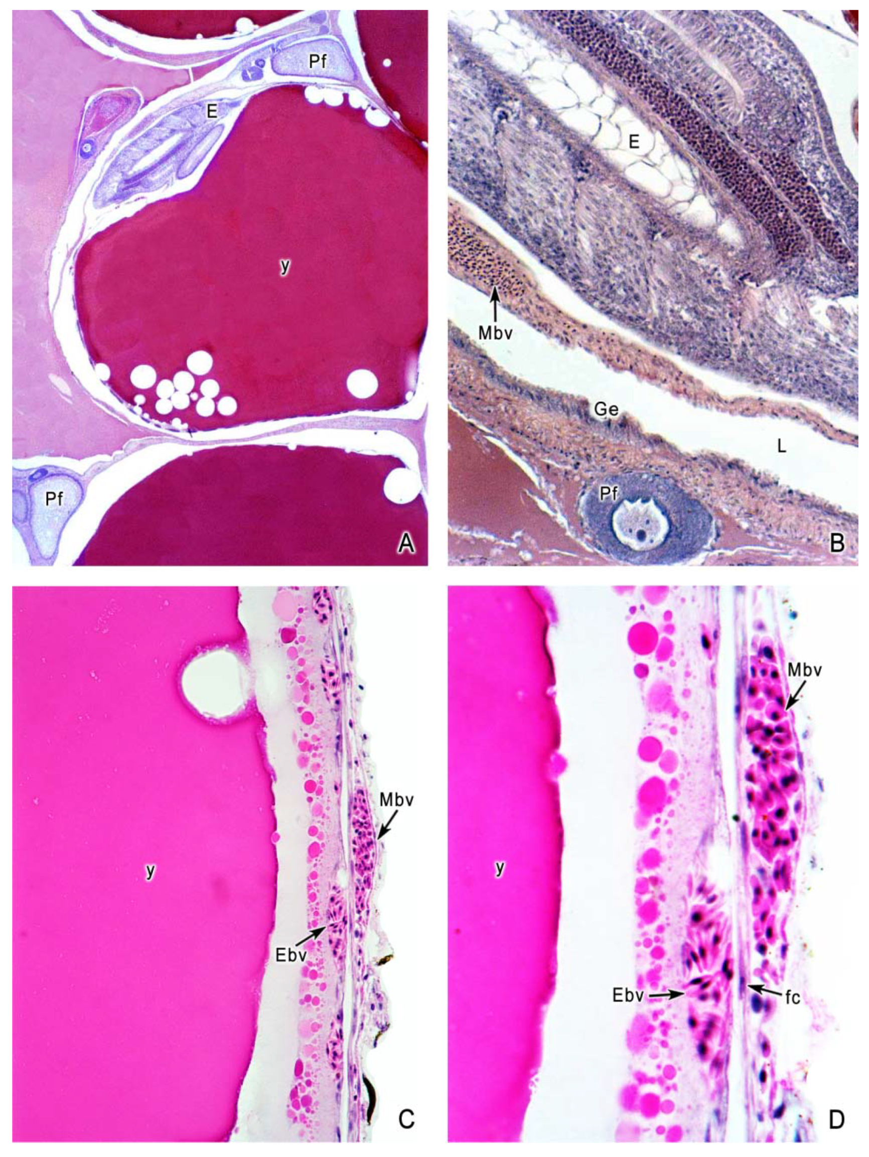
The intrafollicular embryogenesis in poeciliids continues (Figure 5C,D and Figure 6A,B) through the development of peripheral blood vessels, adjacent to the maternal tissue. These vessels permit the exchange of essential supplies for the embryos, such as the transfer of nutrients and gas for respiration, elimination of waste products of the metabolism, and fulfilling hormonal and immunological requirements. For these functionfs, the development of adjacent embryonic and maternal vascularization is essential (Figure 6C,D). The follicular cells become squamous, simplifying the pass of metabolites between maternal and offspring blood vessels.
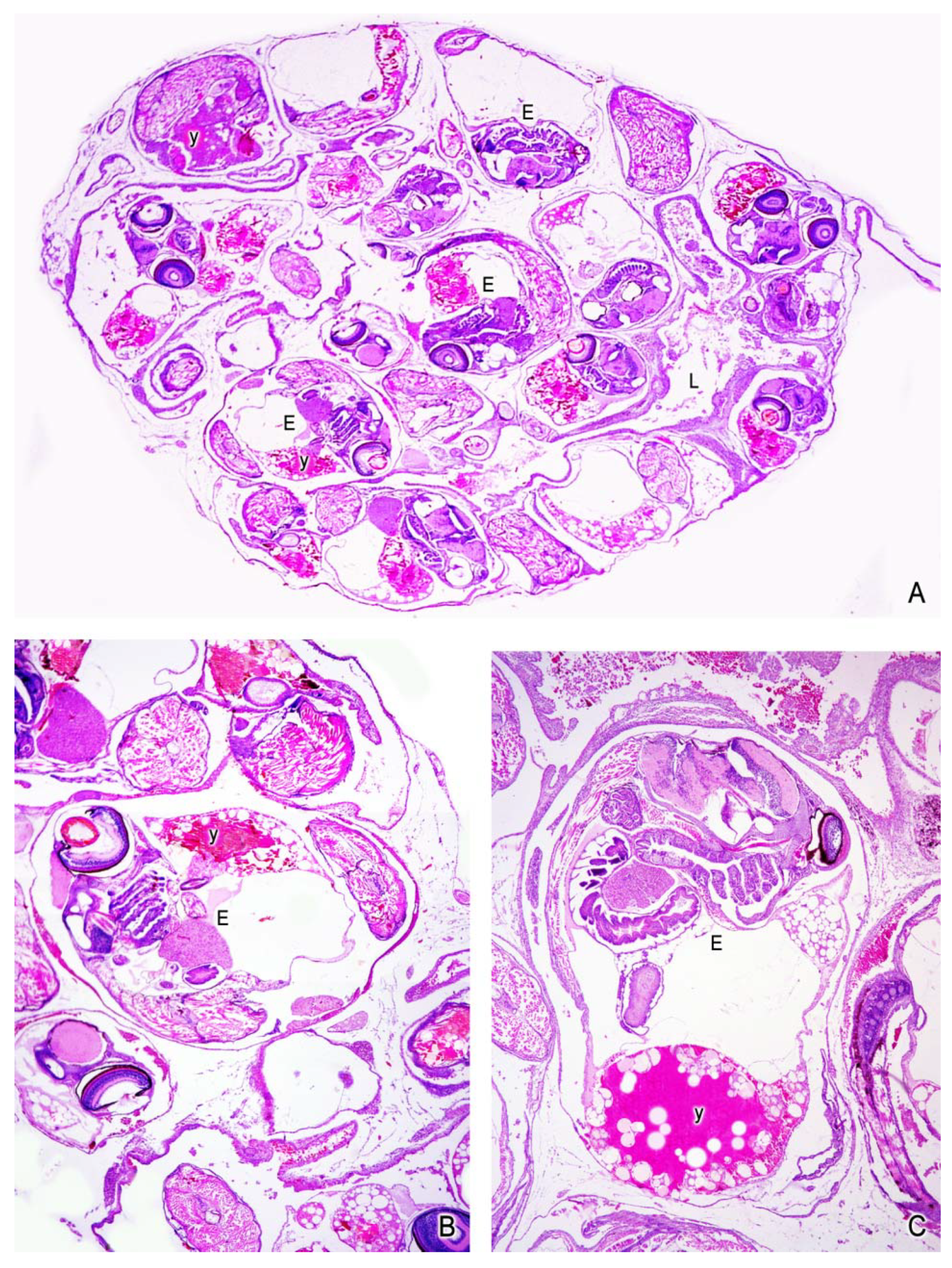
In early embryogenesis, the amount of yolk is abundant, but it diminishes progressively as the embryo takes the nutrients during the advance of gestation (Figure 7A–C).
According to the differences in the amount of yolk of the oocytes, the species having oocytes with abundant yolk have been considered lecithotrophic, even though some transfer of maternal nutrients may also occur. This is the case of Gambusia geiseri [39], a species with large eggs, which has a high level of lecithotrophy but also demonstrates transfer of nutrients from the mother to the embryo as a small matrotrophic contribution. Similar observations were described in other species: Gambusia clarkhubbsi, Gambusia gaigei, Gambusia holbrooki, Gambusia nobilis, Poecilia formosa, Poecilia latipinna, and Poecilia mexicana [19]. The incipient matrotrophy has potential significance in helping us understand the evolution of matrotrophy. Blackburn [6] considered that ancestral features that function in gas exchange may have been adapted for nutrient transfer, through minor modifications of their components.
The follicular epithelium, the maternal layer surrounding the embryo, is very active in allowing the passage of metabolites in species with matrotrophic nutrition. The follicular cells select, digest, and transport nutrients from the blood vessels of the maternal tissue to the embryo [40]. The proximity of embryonic and maternal tissues also offers conditions favoring the evolution of matrotrophy [5,20]. This nutrient transfer involves the association of embryonic and maternal tissues, which form the follicular placenta [14,41].
The components of the follicular placenta are: endothelium of maternal capillaries, follicular epithelium, embryonic surface epithelium, and endothelium of embryonic capillaries. Consequently, a placenta allows the mother to feed embryos during gestation by matrotrophic nutrition, instead of the deposition of abundant nutrients into the oocyte during oogenesis. Extensive matrotrophy is associated with more placental complexity. Analysis of the structure of the placenta in several species of poeciliids allowed the identification of specialized morphological characteristics of the follicular cells that increased the active transfer of nutrients between mother and embryos, such as increase in the number of microvilli and microvilli length [41,42,43]. Thus, extreme matrotrophy is associated with placental characteristics that allow the transference of nutrients between the mother and the embryo. The development and function of the placenta in matrotrophic species are associated with stable environmental conditions, because it requires a constant transfer of resources from the mother to the embryo during gestation [35,41,44,45]. In contrast to this situation, the lecithotrophy of species such as Poecilia vivipara may be related to environmental conditions of a semiarid region, with intermittent rivers, and where food supply is variable [32].
In this diversity of embryonic nutrition, there are several species with extreme matrotrophy such as: Heterandria formosa [13]; Poeciliopsis prolifica, Poeciliopsis turneri, Poeciliopsis retropinna [21,46]; Xenodexia ctenolepis [21]; Poecilia branneri, Poecilia bifurca [45]; Phalloptychus januarius [42]. Consequently, in the context of the analysis of the evolution of poecilid placenta, species with extreme matrotrophic nutrition may reveal essential aspects, taking into account that the evolution of the placenta is one mechanism of matrotrophy [43,47]. As it is considered by Pollux et al. [43], viviparity in fishes have evolved the development of the placenta, when the reduction in pre-fertilization nutrients are associated with trophic specializations for the post-fertilization transfer of nutrients from the mother to the developing young.
Wourms [18] suggested that the trophic relationships in the evolution of viviparity involve the transition from lecithotrophy to matrotrophy, a shift from embryonic nutritional autonomy to maternal nutritional dependency. Blackburn [5,6], comparing lecithotrophy and matrotrophy, discussed the evolution from lecithotrophic oviparity to lecithotrophic viviparity to matrotrophic viviparity. In this context, the small oocytes developed in H. formosa represent an extreme in the reduction of yolk deposition, developing the smallest egg described in poeciliids [36], along with the high level of matrotrophy. H. formosa is considered the species with extreme matrotrophy, with very little yolk available at fertilization and young increasing in dry mass during development [44]. Therefore, this species is an excellent model among poeciliids for the study of the formation of microlecithal oocytes during oogenesis, as well as the structural complexity of the placenta and the complementary evolution to matrotrophy [43,47].

6. Conclusions

Viviparity of teleosts is a specialized and derived mode of reproduction that has evolved from oviparity. The type of viviparity in teleosts is due to the combination of unique reproductive aspects, different from the rest of vertebrates: The saccular ovary, the internal germinal epithelium lining the ovarian lumen, the absence of oviducts, the development of intraovarian fertilization, and the gestation in the ovary. This strategy of reproduction has morphological variations as the result of adaptations associated with viviparity. The intraovarian gestation of poeciliids involves several morphological characteristics: The intrafollicular fertilization, the intrafollicular embryogenesis, and diverse levels of matrotrophy that converge in maternal–embryo interchanges developing a follicular placenta. In order to attain a better understanding of the biology of reproduction in viviparous teleosts, several fields of investigation need to be integrated with the morpho-physiological adaptations, such as ontogenesis, endocrinology, reproductive cycles, and evolutionary trends [5,6]. Additionally, comparative studies at species, genus, and family levels [3,4] will reveal essential aspects related to the viviparity, this unique and extraordinary reproductive strategy developed in viviparous teleosts.

Author Contributions

Conceptualization, M.C.U.; investigation, M.C.U., G.D.l.R.C., A.G.A., J.C.C.C., M.G.G.B.; original draft preparation, M.C.U.; methodology; G.D.l.R.C., A.G.A., J.C.C.C., M.G.G.B., review, M.C.U., G.D.l.R.C., A.G.A., J.C.C.C., M.G.G.B.; editing, M.C.U., G.D.l.R.C., A.G.A., J.C.C.C., M.G.G.B.

Funding

This research received no external funding.

Conflicts of Interest

The authors declare no conflict of interest.

References

  1. Froese, R.; Pauly, D. Fish Base, (10/2018) version; World Wide Web Electronic Publication, Naturalis, Leiden, the Netherlands. 2018. Available online: http://www.catalogueoflife.org/annual-checklist/2018/ (accessed on 19 June 2019).
  2. Rosen, D.E.; Bailey, R.M. The poeciliid fishes (Cyprinodontiformes), their structure, zoogeography, and systematics. Bull. Am. Museum Nat. Hist. 1963, 126, 11–76. [Google Scholar]
  3. Parenti, L.R. A phylogenetic and biogeographic analysis of cyprinodontiform fishes (Teleostei, Atherinomorpha). Bull. Am. Museum Nat. Hist. 1981, 168, 335–557. [Google Scholar]
  4. Parenti, L.R.; Grier, H.J. Evolution and phylogeny of gonad morphology in bony fishes. Integr. Comp. Biol. 2004, 44, 333–348. [Google Scholar] [CrossRef] [PubMed]
  5. Blackburn, D.G. Convergent evolution of viviparity, matrotrophy and specializations for fetal nutrition in reptiles and other vertebrates. Am. Zool. 1992, 32, 313–321. [Google Scholar] [CrossRef]
  6. Blackburn, D.G. Evolution of vertebrate viviparity and specializations for fetal nutrition: A quantitative and qualitative analysis. J. Morphol. 2015, 276, 961–990. [Google Scholar] [CrossRef] [PubMed]
  7. Uribe, M.C.; Grier, H.J.; De la Rosa Cruz, G.; García Alarcón, A. Modifications in ovarian and testicular morphology associated with viviparity in teleosts. In Reproductive Biology and Phylogeny of Fish (Agnatha and Bony Fishes), 1st ed.; Jamieson, B., Ed.; Science Publishers, Inc.: Enfield, NH, USA; Plymouth, UK, 2009; pp. 85–117. [Google Scholar]
  8. Wourms, J.P. Viviparity: The maternal-fetal relationship in fishes. Am. Zool. 1981, 21, 473–515. [Google Scholar] [CrossRef]
  9. Uribe, M.C.; Aguilar-Morales, M.; De la Rosa-Cruz, G.; García-Alarcón, A.; Campuzano-Caballero, J.C.; Guerrero-Estévez, S.M. Ovarian structure and embryonic traits associated with viviparity in poeciliids and goodeids. In Viviparous Fishes II, 1st ed.; Uribe, M.C., Grier, H.J., Eds.; New Life Publications: Homestead, FL, USA, 2010; pp. 211–229. [Google Scholar]
  10. Grier, H.J.; Uribe, M.C.; Parenti, L.R.; Lo Nostro, F.L.; Mims, S.D. Constancy of the germinal epithelium through 500 million years of vertebrate evolution. J. Morphol. 2016, 277, 1014–1044. [Google Scholar] [CrossRef]
  11. Dodd, J.M. The structure of the ovary of non-mammalian vertebrates. In The Ovary, 1st ed.; Zuckerman, S., Weir, B.J., Eds.; Ac. Press: New York, NY, USA, 1977; Volume 1, pp. 219–263. [Google Scholar]
  12. Hoar, W.S. Reproduction. In Fish Physiology, 1st ed.; Hoar, W.S., Randall, D.J., Eds.; Ac. Press: New York, NY, USA; London, UK, 1969; Volume 3, pp. 1–72. [Google Scholar]
  13. Turner, C.L. Viviparity in teleost fishes. Sci. Mon. 1947, 65, 508–518. [Google Scholar]
  14. Wourms, P.J.; Grove, B.D.; Lombardi, J. The maternal-embryonic relationship in viviparous fishes. In Fish Physiology, 1st ed.; Hoar, W.S., Randal, D.J., Eds.; Ac Press, Inc.: New York, NY, USA, 1988; pp. 1–134. [Google Scholar]
  15. Campuzano-Caballero, J.C.; Uribe, M.C. Structure of the female gonoduct of the viviparous teleost Poecilia reticulata (Poeciliidae) during non-gestation and gestation stages. J. Morphol. 2014, 275, 247–257. [Google Scholar] [CrossRef]
  16. Uribe, M.C.; Grier, H.J. Insemination, intrafollicular fertilization and development of the fertilization plug during gestation in Heterandria formosa (Poeciliidae). J. Morphol. 2018, 279, 970–980. [Google Scholar] [CrossRef]
  17. Potter, H.; Kramer, C.R. Ultrastructural observations on sperm storage in the ovary of the platyfish, Xiphophorus maculatus (Teleostei: Poeciliidae): The role of the duct epithelium. J. Morphol. 2000, 245, 110–129. [Google Scholar] [CrossRef]
  18. Wourms, J.P. Functional morphology, development and evolution of trophotaeniae. In Viviparous Fishes, 1st ed.; Uribe, M.C., Grier, H.J., Eds.; New Life Publications: Homestead, FL, USA, 2005; pp. 217–242. [Google Scholar]
  19. Marsh-Matthews, E.; Deaton, R.; Brooks, M. Survey of matrotrophy in lecithotrophic poeciliids. In Viviparous Fishes II, 1st ed.; Uribe, M.C., Grier, H.J., Eds.; New Life Publications: Homestead, FL, USA, 2010; pp. 255–258. [Google Scholar]
  20. Blackburn, D.G.; Starck, J.M. Morphological specializations for fetal maintenance in viviparous vertebrates: An introduction and historical retrospective. J. Morphol. 2015, 276, E1–E16. [Google Scholar] [CrossRef] [PubMed]
  21. Pires, M.N.; Reznick, D.N. Life-history evolution in the fish genus Poecilia (Poeciliidae: Cyprinodontiformes: Subgenus Pamphorichthys): An evolutionary origin of extensive matrotrophy decoupled from superfetation. Biol. J. Linnean Soc. 2018, 125, 547–560. [Google Scholar] [CrossRef]
  22. Parenti, L.R.; Lo Nostro, F.L.; Grier, H.J. Reproductive histology of Tomeurus gracilis Eigenmann, 1909 (Teleostei: Atherinomorpha: Poeciliidae) with comments on evolution of viviparity in Atherinomorph fishes. J. Morphol. 2010, 271, 1399–1406. [Google Scholar] [CrossRef] [PubMed]
  23. Lorier, E.; Berois, N. Reproducción y nutrición embrionaria en Cnesterodon decimmaculatus (Teleostei: Poeciliidae). Rev. Brasil. Biol. 1993, 55, 27–44. [Google Scholar]
  24. Stockwell, C.A.; Henkanaththegedara, S. Evolutionary conservation biology, Chapter 12. In The Evolutionary Ecology of the Livebearing Fishes, 1st ed.; Evans, J.P., Pilastro, A., Schlupp, I., Eds.; University of Chicago Press: Chicago, IL, USA, 2011; pp. 128–141. [Google Scholar]
  25. Nelson, J.S.; Grande, T.C.; Wilson, M.V.H. Fishes of the World, 5th ed.; John Wiley & Sons: New York, NY, USA, 2016; pp. 378–380. [Google Scholar]
  26. Jourdan, J.; Wilhelm Miesen, F.; Zimmer, C.; Gasch, K.; Herder, F.; Schleucher, E.; Plath, M.; Bierbach, D. On the natural history of an introduced population of guppies (Poecilia reticulata Peters, 1859) in Germany. BioInvasions Rec. 2014, 3, 175–184. [Google Scholar] [CrossRef]
  27. Grier, H.J.; Uribe, M.C.; Parenti, L.R.; De la Rosa-Cruz, G. Fecundity, the germinal epithelium, and folliculogenesis in viviparous fishes. In Viviparous Fishes, 1st ed.; Uribe, M.C., Grier, H.J., Eds.; New Life Publications: Homestead, FL, USA, 2005; pp. 126–191. [Google Scholar]
  28. Grier, H.J.; Uribe, M.C.; Patiño, R. Chapter 2. The ovary, folliculogenesis and oogenesis in teleosts. In Reproductive Biology and Phylogeny of Fish (Agnatha and Bony Fishes), 1st ed.; Jamieson, B., Ed.; Science Publishers, Inc.: Enfield, NH, USA; Plymouth, UK, 2009; pp. 25–84. [Google Scholar]
  29. Campuzano-Caballero, J.C.; Uribe, M.C. Functional morphology of the gonoduct of the viviparous teleost Poeciliopsis gracilis (Heckel, 1848) (Poeciliidae). J. Morphol. 2017, 278, 1647–1655. [Google Scholar] [CrossRef] [PubMed]
  30. Bailey, R.J. The ovarian cycle in the viviparous teleost Xiphophorus helleri. Biol. Bull. 1933, 64, 206–225. [Google Scholar] [CrossRef]
  31. Koya, Y.; Inoue, M.; Naruse, T.; Sawaguchi, S. Dynamics of oocyte and embryonic development during ovarian cycle of the viviparous mosquitofish Gambusia affinis. Fish. Sci. 2000, 66, 63–70. [Google Scholar] [CrossRef]
  32. Arcanjo, R.B.; de Souza, L.P.; Rezende, C.F.; Silva, J.R.F. Embryonic development and nourishment in the viviparous fish Poecilia vivipara (Cyprinodontiformes: Poeciliidae). Acta Zool. (Stockh.) 2014, 95, 493–500. [Google Scholar] [CrossRef]
  33. Ponce de León, J.L.P.; Rodríguez, R.; Acosta, M.; Uribe, M.C. Egg size and its relationship with fecundity, newborn length and female size in Cuban poeciliid fishes (Teleostei: Cyprinodontiformes). Ecol. Freshw. Fish. 2011, 20, 243–250. [Google Scholar] [CrossRef]
  34. Scrimshaw, N.S. 1946. Egg size in poeciliid fishes. Copeia 1963, 1, 20–23. [Google Scholar]
  35. Thibault, R.E.; Schultz, R.J. Reproductive adaptations among viviparous fishes (Cyprinodontiformes: Poeciliidae). Evolution 1978, 32, 320–333. [Google Scholar] [CrossRef] [PubMed]
  36. Uribe, M.C.; Grier, H.J. Oogenesis of microlecithal oocytes in the viviparous teleost Heterandria formosa. J. Morphol. 2011, 272, 241–257. [Google Scholar] [CrossRef] [PubMed]
  37. Turner, C.L. Pseudoamnion, pseudochorion and follicular pseudoplacenta in poeciliid fishes. J. Morphol. 1940, 67, 59–89. [Google Scholar] [CrossRef]
  38. Constanz, J. Reproductive biology of the poeciliid fishes. In Ecology and Evolution of Live Bearing Fishes (Poeciliidae), 1st ed.; Meffe, G.K., Snelson, F.F., Eds.; Prentice Hall: New York, NY, USA, 1989; pp. 33–50. [Google Scholar]
  39. Marsh-Matthews, E.; Skierkowski, P.; DeMarais, A. Direct evidence for mother-to-embryo transfer of nutrients in the livebearing fish Gambusia geiseri. Copeia 2001, 1, 1–6. [Google Scholar] [CrossRef]
  40. Jollie, W.P.; Jollie, L.G. The fine structure of the ovarian follicle of the ovoviviparous poeciliid fish Lebistes reticulatus. I. Maturation of follicular epithelium. J. Morphol. 1964, 114, 479–501. [Google Scholar] [CrossRef] [PubMed]
  41. Olivera-Tlahuel, C.; Moreno-Mendoza, N.A.; Villagrán-Santa Cruz, M.; Zúñiga-Vega, J.J. Placental structures and their association with matrotrophy and superfetation in poeciliid fishes. Acta Zool. 2019, 100, 167–181. [Google Scholar] [CrossRef]
  42. Pollux, B.J.A.; Reznick, D.N. Matrotrophy limits a female’s ability to adaptively adjust offspring size and fecundity in fluctuating environments. Funct. Ecol. 2011, 25, 747–756. [Google Scholar] [CrossRef]
  43. Pollux, B.J.A.; Pires, M.N.; Banet, A.I.; Reznick, D.N. Evolution of placentas in the fish family Poeciliidae: An empirical study of macroevolution. Annu. Rev. Ecol. Evol. Syst. 2009, 40, 271–289. [Google Scholar] [CrossRef]
  44. Reznick, D.N.; Callahan, H.; Llauredo, R. Maternal effects on offspring quality in poeciliid fishes. Am. Zool. 1996, 36, 147–156. [Google Scholar] [CrossRef]
  45. Riesch, R.; Plath, M.; Schlupp, I.; Marsh-Matthews, E. Matrotrophy in the cave molly: An unexpected provisioning strategy in an extreme environment. Evol. Ecol. 2010, 24, 789–801. [Google Scholar] [CrossRef]
  46. Kwan, L.; Fris, M.; Rodd, F.H.; Rowe, L.; Tuhela, L.; Panhuis, T.M. An examination of the variation in maternal placentae across the genus Poeciliopsis (Poeciliidae). J. Morphol. 2015, 276, 707–720. [Google Scholar] [CrossRef] [PubMed]
  47. Pires, M.N.; Arendt, J.; Reznick, D.N. The evolution of placentas and superfetation in the fish genus Poecilia (Cyprinodontiformes: Poeciliidae: Subgenera Micropoeciliia and Acanthophacelus). Biol. J. Linnean Soc. 2010, 99, 784–796. [Google Scholar] [CrossRef]
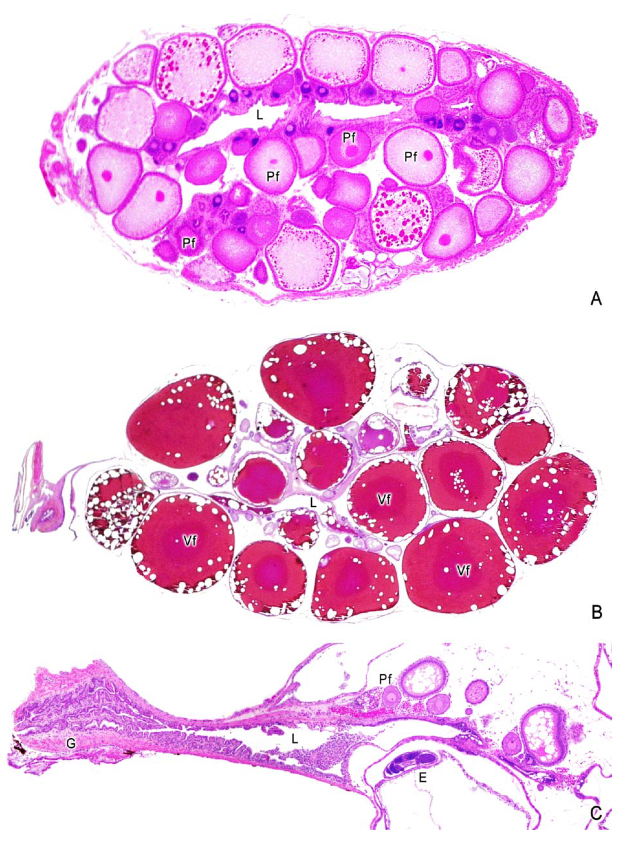
Figure 1. Ovaries during previtellogenesis and vitellogenesis, and the gonoduct. Saccular structure of the ovary with a central lumen (L). (A) Ovary of Poecilia reticulata during non-gestation. Numerous previtellogenic follicles (Pf), containing oocytes of different size; smallest with dense and basophilic ooplasm; larger with abundant oil droplets in the ooplasm. Optical magnification: 20×. (B) Ovary of Poecilia reticulata during non-gestation. Numerous vitellogenic follicles (Vf), having different sizes of oocytes, the larger follicles having reached the full-grown size. The yolk is fluid and homogeneous. 12×. (C) Ovary of Heterandria formosa during gestation. The anterior part of the ovary is the germinal zone which contains previtellogenic follicles (Pf) and one embryo (E). The posterior part of the ovary is the gonoduct (G) with several folds of the wall displayed to the lumen (L). 12×.
Fishes 04 00035 g001
Figure 2. Histological components of the gonoduct. (A) The gonoduct of Poecilia reticulata forms the caudal portion of the saccular ovary with a central lumen (L). Melano-macrophage centers (MMC) are adjacent to the luminal epithelium. 32×. (B) Melano-macrophage center (MMC) surrounded by connective tissue. 78×. (C) The luminal epithelium (e) is formed by cubic cells. A macrophage (m) is seen in the lumen (L). 200×. (D,E) Epithelium formed by columnar cells (e) with cilia (ci) bordering the lumen (L). The connective tissue contains abundant blood vessels (v). 500×, 800×.
Fishes 04 00035 g002
Figure 3. Morphology of the germinal cells during oogenesis. Germinal cells from oogonia to previtellogenic follicles. (A) Oogonium (Oo) of Heterandria formosa observed in the germinal epithelium (Ge) among somatic cells. The germinal epithelium borders the ovarian lumen (L). The oogonium is spherical and possesses light ooplasm. 500×. (B) Oocyte of Poecilia reticulata in chromatin-nucleolus stage (Cn) at the initiation of meiosis. Germinal epithelium (Ge), previtellogenic follicles (Pf), and ovarian lumen (L) are seen. 78×. (C) Previtellogenic follicle (Pf) of Heterandria formosa with basophilic ooplasm, the germinal vesicle (nucleus) (gv) contains several nucleoli. Stroma (S) surrounds the follicle. 500×. (D) Previtellogenic follicle (Pf) of Poecilia reticulata containing lipid droplets around the germinal vesicle. Follicular cells (fc) are seen around the oocyte. Stroma (S) surrounds the follicle. The germinal epithelium (Ge) borders the ovarian wall (L). 78×. (E) Previtellogenic follicle (Pf) of Poecilia reticulata, with abundant lipid droplets in the ooplasm. The germinal vesicle (gv) and follicular cells (fc) are seen. Germinal epithelium (Ge). 78×. (F,G) Previtellogenic follicles (Pf) of Heterandria formosa with lipid droplets. The ooplasm also contains cortical alveoli seen as small red granules. The germinal vesicle (gv) is seen. Stroma (S). 78×. (H) Detail of previtellogenic follicle (Pf) of Xiphophorus hellerii with abundant lipid droplets. The germinal vesicle (gv) contains several nucleoli. The follicular cells (fc) are seen around the oocyte. 200×.
Fishes 04 00035 g003
Figure 4. Morphology of the germinal cells during oogenesis. Follicles during vitellogenesis (Vf). (A) Oocytes of Poecilia reticulata at the initiation of vitellogenesis (Vf), with the deposition of yolk globules (y) in the ooplasm. The germinal epithelium (Ge) borders the ovarian lumen (L). 32×. (B) Detail of the figure A, where the fine yolk globules (y) are seen at the ooplasmic periphery. The follicular cells (fc) are seen around the oocyte. 200×. (C) Peripheral ooplasm of an oocyte of Poecilia reticulata at the advance of vitellogenesis (Vf), with larger yolk globules (y) than those seen in the previous figure. 500×. (D) Maximum size of an oocyte of Gambusia affinis; the yolk (y) is fluid and homogeneous; some lipid droplets may be seen around the oocyte periphery. Ovarian lumen (L). 20×.
Fishes 04 00035 g004
Figure 5. The delle and early intrafollicular embryogenesis. (A) Ovary of Poecilia latipinna presenting the delle (de) structure as a funnel-like invagination of the ovarian lining at the periphery of a vitellogenic oocyte (Vf). The delle is opened to the ovarian lumen (L). The germinal epithelium (Ge) borders the ovarian lumen. The yolk (y) is fluid. A previtellogenic oocyte (Pf) with lipid droplets in the ooplasm is also seen. 32×. (B) Ovary of Heterandria formosa with a vitellogenic oocyte (Vf) presenting the delle (de), which contains abundant spermatozoa (Z). The delle is opened to the ovarian lumen (L). This species has scarce yolk (y) developing microlecithal eggs. A previtellogenic oocyte (Pf) with basophilic ooplasm is also seen. The germinal epithelium (Ge) borders the ovarian lumen. 78×. (C) Ovary of Gambusia affinis with embryos (E) during early development; the fluid yolk (y) is abundant. Previtellogenic oocytes (Pf) are seen. 20×. (Poecilia latipinna) Detail of the previous figure with one embryo (E) during early development; and the fluid yolk (y) at the ventral side of the embryo. 32×.
Fishes 04 00035 g005
Figure 6. Ovaries during early intrafollicular embryogenesis and follicular placenta. (A) Ovary of Xiphophorus helleri with an embryo (E) during early development; the fluid yolk (y) is abundant. Previtellogenic oocytes (Pf) are also seen. 200×. (B) Detail of the previous figure with the embryo (E). A maternal blood vessel (Mbv) near the embryo is seen. The ovarian wall shows a previtellogenic follicle (Pf) with basophilic ooplasm. The germinal epithelium (Ge) borders the lumen (L). 500×. (C) Periphery of an embryo of Gambusia affinis at the ventral side where the yolk is seen (y). The essential components of the follicular placenta, the maternal (Mbv) and embryonic (Ebv) blood vessels, are closely positioned. 200×. (D) Detail of the previous figure with the evident close position of the maternal (Mbv) and embryonic (Ebv) blood vessels; follicular cells (fc) between them form a squamous epithelium. 500×.
Fishes 04 00035 g006
Figure 7. Ovaries during late intrafollicular embryogenesis. (A) Ovary of Poecilia reticulata with embryos (E) during late gestation. The development of embryos is more advanced compared with that seen in the embryos of the Figure 4 and Figure 5; consequently, the reduction of yolk is also seen. 12×. (B,C) Details of embryos (E) in late stage of development and reduction of yolk. 78×.
Fishes 04 00035 g007