Article

Transcriptomic Changes during Previtellogenic and Vitellogenic Stages of Ovarian Development in Wreckfish (Hāpuku), Polyprion oxygeneios (Perciformes)

1
Department of Zoology, University of Otago, PO Box 56, Dunedin 9054, New Zealand
2
National Institute of Water and Atmospheric Research, Northland Marine Research Centre, PO Box 147, Ruakaka 0151, New Zealand
3
Department of Anatomy, University of Otago, 270 Great King Street, Dunedin 9016, New Zealand
4
GeneCology Research Centre, University of the Sunshine Coast, Queensland 4556, Australia
*
Author to whom correspondence should be addressed.
Present address: Matthew J Wylie, The New Zealand Institute for Plant and Food Research, Seafood Production Unit, 293-297 Port Nelson, Nelson 7010, New Zealand.
Present address: Jane E Symonds, Cawthron Institute, Private Bag 2, Nelson 7042, New Zealand.
Fishes 2019, 4(1), 16; https://doi.org/10.3390/fishes4010016
Submission received: 10 October 2018 / Revised: 5 February 2019 / Accepted: 19 February 2019 / Published: 28 February 2019
(This article belongs to the Special Issue Fish Reproductive Physiology and Aquaculture)

Abstract

:
‘Wreckfish’ a collective of species belonging to the family Polyprionidae, are an important commercial fishery and have significant aquaculture potential. Until now, genomic or transcriptomic information for any species within the genus Polyprion has either remained unpublished or is non-existent. Using Illumina HiSeq, we compared the transcriptomes of hāpuku (Polyprion oxygeneios) ovaries to explore developmental stage-specific variations underlying their reproductive physiology. We sought to identify differentially expressed genes and the associated shifts in biological pathways between previtellogenic and early vitellogenic ovaries. Ovarian tissue was repeatedly biopsied by gonopore cannulation from the same females (n = 3) throughout oogenesis. Reproductive status of initial biopsies was confirmed as being previtellogenic and that in biopsies collected eight weeks later as early vitellogenic. A de novo hāpuku transcriptome was assembled (146,189 transcripts) from RNA-Seq data without a reference genome. On average, each tissue sample contained 17.5 million trimmed reads. Gene annotation was 80% when using BLASTX against Genbank Non Redundant database. Fifty-three transcripts were differentially expressed within the FDR of 0.05 when previtellogenic and early vitellogenic ovaries were compared; this reduced to 35 differentially expressed genes when transcript duplications were pooled. Among these were genes tentatively associated with the electron transport chain, lipid metabolism, steroidogenesis and mineral/solute transportation. These data provide a snap-shot into stage-specific physiological events during oogenesis in the ovary of a teleost and an extensive molecular resource for research on species in the Genus Polyprion.

1. Introduction

A critical component of aquaculture is the requirement to supply high quality gametes and fingerlings that can be raised to satisfy market demands [1,2]. The events leading to the formation of gametes are complex and dynamic [3]. In order to gain insight into the factors affecting egg quality and fertilisation, an understanding of the mechanisms and coordinated processes that occur during oogenesis is required [4]. Such insights include the composition and roles of maternal transcripts (DNA and RNA) and other molecular cargo produced and stored during oocyte formation as these are essential for early embryonic development and the support of core cellular functions until the genome of the zygote is activated [2,5,6,7]).
During oogenesis, the application of transcriptomic tools allows researchers to examine the entire population of mRNA within developing oocytes, thus providing insight into the genes expressed at the time they were harvested to reflect a particular physiological state [8]. The relative abundance of each transcript can be quantified by RNA sequencing (RNA-Seq), permitting the researcher to explore whether particular genes or pathways of biological processes differ between two RNA populations originally harvested from different subject cells or tissues [9,10,11].
The development of an aquaculture industry for an emerging species demands an understanding of the reproductive biology at different phases of the life cycle. Under intensive culture conditions, these phases include pubertal onset [12], secondary oocyte growth [1,2] and the establishment of methods to control gamete maturation and spawning [13,14]. With a considerable commercial fishery and potential aquaculture value of hāpuku (Polyprion oxygeneios), a wreckfish species [15,16,17], the aim of this study was to explore the stage-specific variations underlying their ovarian reproductive development at the level of the transcriptome. This long-lived fish is gonochoristic and spawns repeatedly during a spawning season, reflected in the group-synchronous ovaries. Recently, ovarian development was described in captive-reared broodstock during their first reproductive cycle while subjected to constant or varying temperature regimes [17]. To address our aim, we sought to identify differentially expressed genes and the associated shifts in biological pathways between previtellogenic and early vitellogenic ovaries. To do so, ovarian tissue was repeatedly biopsied by gonopore cannulation from the same first filial (F1) females throughout oogenesis as a strategy to reduce variability since transcriptomes are inherently prone to temporal and spatial variation [8].

2. Results

2.1. Stages of Ovarian Development

Stages of reproductive development between previtellogenic females sampled in March and early vitellogenic females sampled in May were confirmed on the basis of oocyte diameter, ovarian histology, and plasma levels of estradiol-17β (E2). The mean oocyte diameter of the three previtellogenic females was 0.22 mm in March and increased to 0.37 mm eight weeks later (Table 1). Likewise, increases in plasma E2 concentrations were also observed in females (0.08 ± 0.003 ng/mL to 0.63 ± 0.076 ng/mL; Table 1) between the March and May sampling points. The appearance of yolk platelets in histological sections (Figure 1) coincided with increases in mean oocyte diameter and plasma E2 concentrations. Additional details on ovarian developmental stages of hāpuku throughout a reproductive cycle have been outlined in a previous study [17].

2.2. Illumina Sequencing and De Novo Assembly

The hāpuku transcriptome consisted of 146,189 transcripts (Table 2). On average, each tissue sample contained 17.5 million trimmed reads. Sequencing data were of high quality as the trimming steps only excluded 24% of the data (data not shown).

2.3. Identification of Differentially Expressed Genes and Transcriptome Annotation

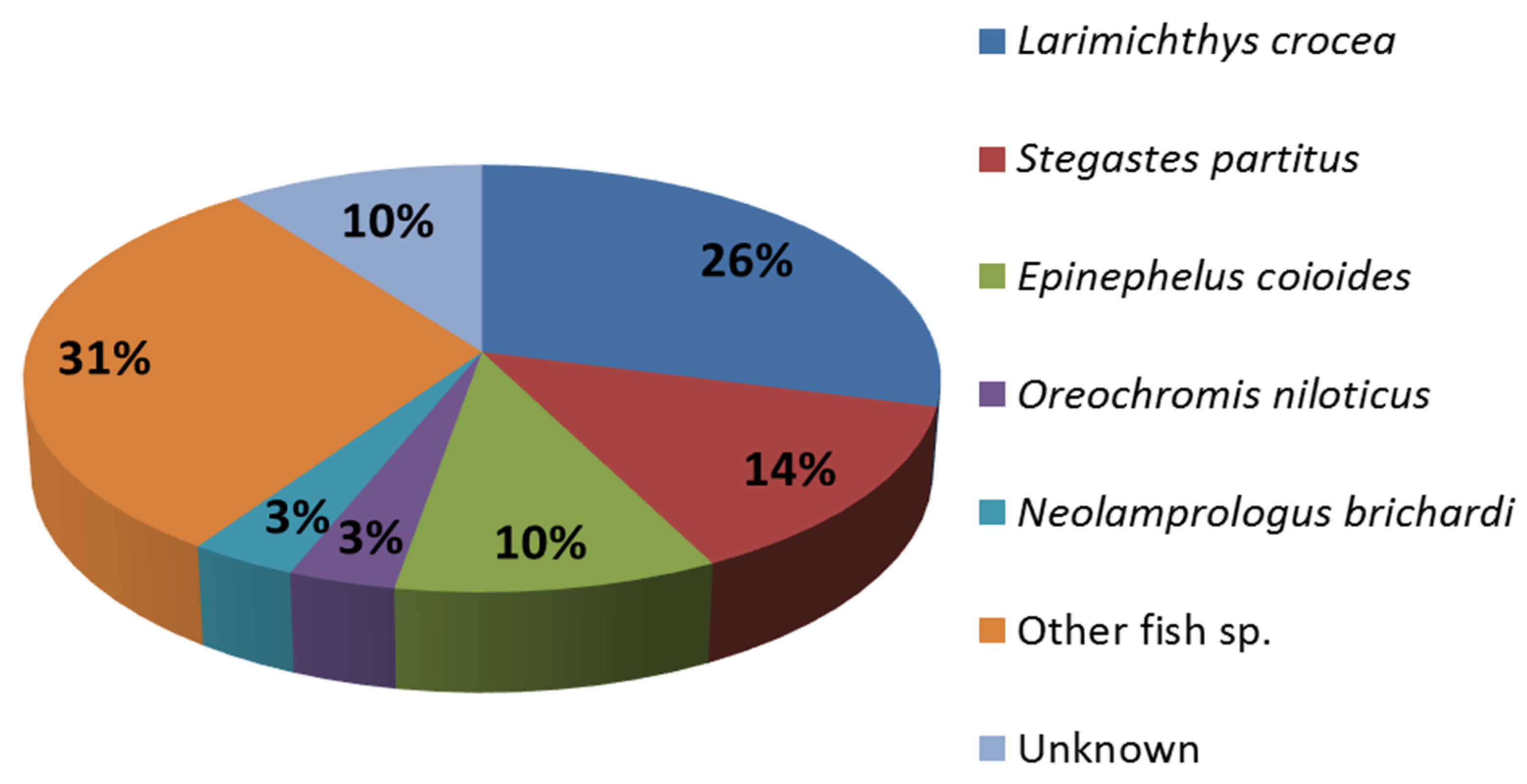
Fifty-three transcripts were differentially expressed within the false detection rate (FDR) of 0.05 when previtellogenic and early vitellogenic ovaries were compared. However, when transcripts that annotated to the same gene were pooled, expression analysis revealed only 35 differentially expressed genes between the two reproductive stages (Table 3). Of the 35 differentially expressed genes identified between previtellogenic and early vitellogenic stages of ovarian development in hāpuku, 80% were annotated. Species composition of annotated transcripts were predominantly from two perciform species, the yellow croaker (Larimichthys crocea) and damselfish (Stegastes partitus). A notable number of annotations was further obtained from perciform members in the genus Epinephelus (groupers) and the Family Cichlidae, e.g., Birchardi cichlid (Neolamprologus brichardi) and Nile tilapia (Oreochromis niloticus) (Figure 2).
Of the 35 genes differentially expressed between previtellogenic and early vitellogenic stages, only one gene could be annotated (uncharacterized protein LOC100536992; Table 3) against the zebrafish (Danio rerio) transcriptome. Results from the gene ontology (GO) enrichment are, therefore, not discussed further.

2.4. Hierarchical Cluster and Gene Ontology Enrichment/Pathway Analyses

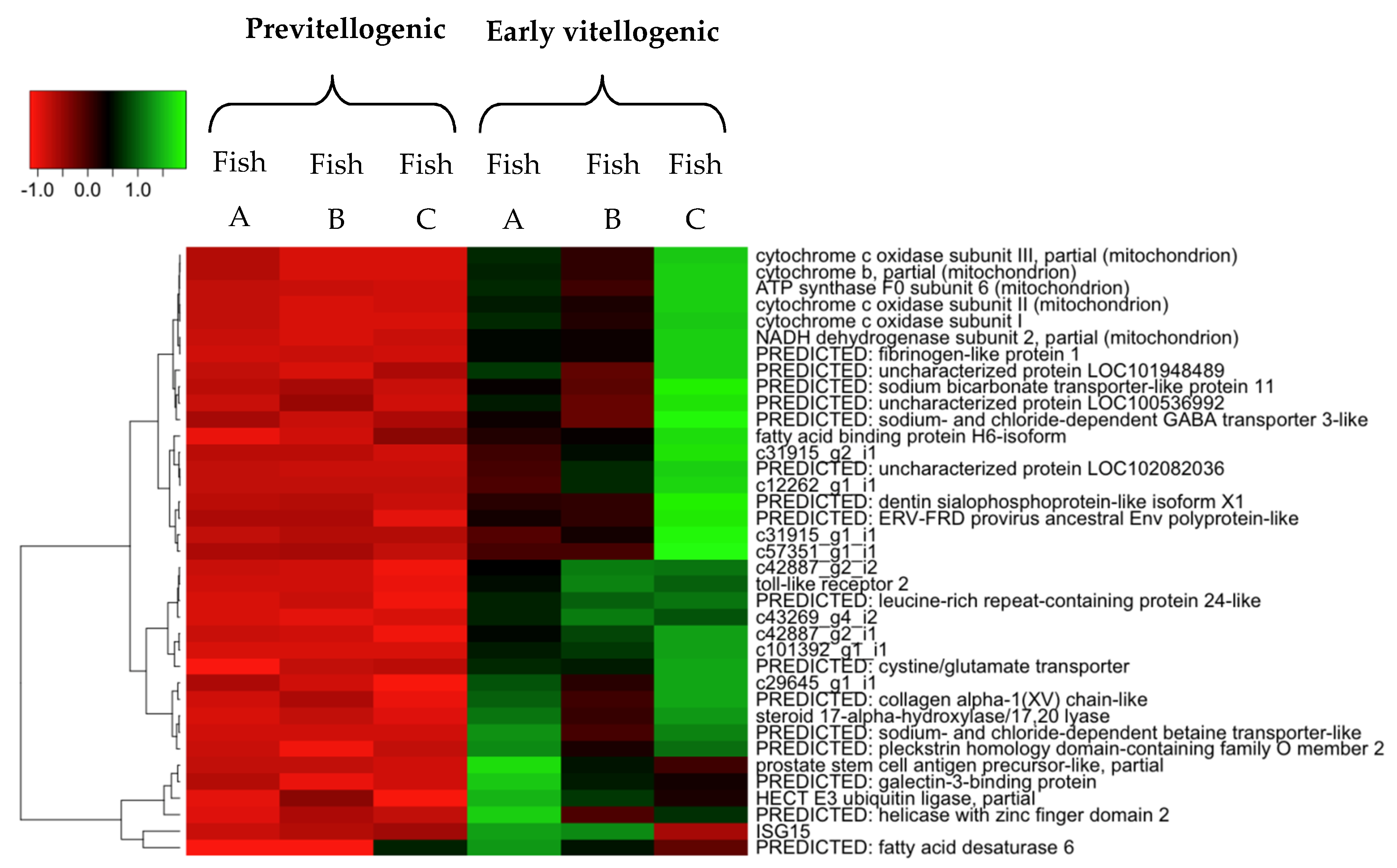
The heat-map generated by the hierarchical cluster analysis tended to identify groups of genes with comparable expression patterns (Figure 3). A key grouping comprised of genes associated with the electron transport chain which were all significantly upregulated as ovarian development progressed to early vitellogenesis. While groupings did not seem so clear-cut with regard to biological functions for other genes, those associated with steroid biosynthesis and lipid metabolism and mineral/solute transportation were significantly upregulated in early vitellogenic ovaries.

3. Discussion

Understanding the molecular events associated with the production, packaging and storage of cellular contents during oogenesis can lead to advances in applied aquaculture settings [2,5,6,7]. Such information is a necessary foundation for advances in fundamental studies exploring ovarian development, its endocrine regulation and egg quality in fish as described for striped bass [20,21]. Until now, genomic or transcriptomic information for hāpuku or any other wreckfish species within the genus Polyprion was unpublished or non-existent. Using HiSeq Illumina sequencing technology, this study produced and compared the transciptomes of previtellogenic and vitellogenic ovaries from hāpuku. Findings from this study provide a snap-shot of the genes being differentially expressed in the ovary as oogenesis progresses from previtellogenic to early vitellogenic stages of development.
Thirty-five differentially expressed genes were identified during early vitellogenesis, several of which are discussed in light of their tentative functions. While the categorisation of oogenesis into specific stages of development is somewhat arbitrary due to its continuous and dynamic nature [3], a hallmark event associated with entry into vitellogenesis is the acquisition of local and blood-derived factors (e.g., carbohydrates, neutral or polar lipids, yolk proteins, and vitamins) that are vital for the development of the embryo subsequent to fertilisation [4,22]. Hence, it is not surprising that genes associated with the electron transport chain, lipid metabolism, steroidogenesis, and mineral/solute transportation were differentially expressed and upregulated as oocyte development progressed in hāpuku undergoing their maiden spawning cycle.
A suite of electron transport chain-related genes were differentially expressed and upregulated during early vitellogenesis. Such genes included all three mitochondrial cytochrome c oxidase subunits I, II and III (mt-c01, mt-c02 and mt-c03 respectively) and cytochrome b (myt-cb). Increases in ovarian myt-cb transcript abundance were previously documented in wild New Zealand longfinned eel (Anguilla dieffenbachii) undergoing oogenesis [23]. More specifically, increases in cytochrome b signals determined by Northern blot and in situ hybridisation were highest during mid-vitellogenesis when compared to immature ‘yellow’ females or those that had entered early vitellogenesis. Such findings led the authors to propose that increases in myt-cb transcript abundance were a response to increased demands for adenosine triphosphate (ATP) during oogenesis and/or post-fertilisation, during early zygote development [23].
Comparable findings to those of hāpuku in this study were also observed in largemouth bass (Micropterus salmoides) by transcriptomic analysis of ovarian tissues throughout oogenesis [24]. Specifically, as ovarian development progressed from the primary growth phase in largemouth bass, transcript abundance of myt-cb, cytochrome c (cycs) and NADH dehydrogenase subunit 5 (mt-nd5) also increased. Again, authors proposed this to be a response to an increase in mitochondrial activity and the associated increased potential for production of ATP [24].
A significant increase in expression of lipid metabolism-related genes was observed between previtellogenic and early vitellogenic stages of development in hāpuku, possibly reflecting the notable increase in mean oocyte diameter which virtually doubled from ~0.2 mm to ~0.4 mm in the space of eight weeks. During this eight week timeframe, putative steroid-secreting granulosa and theca cells that surround developing oocytes became structurally obvious in histological sections of early vitellogenic oocytes. Once formed, theca cells synthesise androgens which are then converted to E2 in the granulosa cells by the steroidogenic enzyme P450 aromatase (cyp19a) [25,26]. While the cyp19a gene was not differentially expressed in this present study, mRNA encoding the steroidogenic enzyme 17α-hydroxylase (cyp17a1) was upregulated during early vitellogenesis.
17α-hydroxylase is involved in several steroidogenic pathways where it can either convert progesterone to 17α-hydroxyprogesterone or pregnenolone to 17α-hydroxypregnenolone through hydroxylase activity. The same enzyme can also perform 17-20-lyase activity to transform 17α-hydroxyprogesterone to androstenedione or 17α-hydroxypregnenolone to dehydroepiandrosterone, both of which are precursors for the synthesis of androgens and estrogens [27,28]. Hence, increased cyp17a expression possibly reflects increased steroidogenic potential and/or activity of the ovary as oogenesis progresses and given the involvement of steroids in regulating nutrient accumulation in the oocyte [3,29], this is in keeping with expectation. The latter may also explain why significant increases in plasma levels of E2 were also observed as oogenesis progressed (discussed below), yet significant increases in cyp19a expression were not detected.
Increases in cyp17a1 expression were also observed during early gonadal recrudescence and early vitellogenesis in channel catfish (Ictalurus punctatus). However, transcript levels dropped abruptly at the completion of vitellogenesis [30]. Similarly, cyp17a1 transcript levels were highest during the ‘developing’ phase of oogenesis rather than at spawning, during regression or in resting ovaries of Atlantic croaker (Micropogonias undulatus) [31]. The “developing” phase of Atlantic croaker was defined by [32] as an ovary containing only primary growth, cortical alveoli and a few partially yolked oocytes. Contrary to transcript levels of cyp17a1 in channel catfish and Atlantic croaker, which peaked around vitellogenesis, cyp17a1 mRNA levels in the Japanese eel (Anguilla japonica) (subsequent to induced maturation using chum salmon (Oncorhynchus keta) pituitary homogenate treatment) continued to increase throughout vitellogenesis until the migratory nucleus stage [33].
Another notable structural change evident in histological sections from ovaries between previtellogenic and early vitellogenic stages of hāpuku was the increase in lipid (oil droplets) consisting, presumably, of neutral lipids like triglycerides and wax or steryl esters [3]. Several genes associated with lipid metabolism were upregulated during early vitellogenic ovarian development. Among these genes was fatty-acid binding protein (FABP) H6-isoform (h6-fabp). Fatty acid-binding proteins are members of a broad multigene family of intracellular lipid-binding proteins [34,35]. The particular FABP gene identified in this study was originally reported in the heart of long-fingered ice fish (Cryodraco antarticus) [36] but is nowadays more commonly referred to as teleost-specific fatty-acid binding protein-11 (fabp11) [37]. Interestingly, duplicate genes (fabp11a and fabp11b) have been identified in zebrafish, three-spined stickleback (Gasterosteus aculeatus), fugu (Takifugu rubripes), spotted green pufferfish (Tetraodon nigroviridis), medaka (Oryzias latipes) [38], and rainbow trout (Oncorhynchus mykiss) [35].
Despite the limited information about both membrane-bound and cytoplasmic FABPs in the ovary of fish [2], these proteins are thought to act as regulators of fatty-acid metabolism and of bioavailability by facilitating the transport of long-chain fatty acids through the cytoplasm to the site of their oxidation. FABPs are also thought to facilitate fatty acid transport for further lipid-signalling pathways as well as support overall lipid homeostasis [2,35,38,39,40].
Recently, three FABP genes (fabp1, fabp3, and fabp11) were identified in ovary of the cutthroat trout (Oncorhynchus clarkii) [41]. However, explicit details on their expression were not further discussed apart from fabp1, which was highly expressed during the ‘lipidic’ and yolk vesical stages of ovarian development. From these observations the authors advocated for roles of FABPs (namely fabp1 and fatty acid transfer protein) in the transport of free fatty acids across the plasma membrane of the oocyte or within the cytosol [41]. The authors further proposed that such proteins may play a critical role in the de novo synthesis of neutral lipids by the oocyte for formation of lipid droplets. Some years earlier, the same researchers documented fabp11 transcripts in somatic cells and vitellogenic oocytes of cutthroat trout. While fabp11 transcripts were abundant in somatic cells but not in oocytes, transcripts were notably abundant in the ooplasm of atretic oocytes [42]. Similar findings were observed in the flatfish (Solea senegalensis) where fabp11 transcripts were highly expressed during oocyte atresia [37].
If indeed the upregulation of fabp11 transcripts are an indication of increased lipid trafficking in hāpuku as ovarian development progresses, then it is interesting to note that other genes associated with lipidation and the release of free fatty-acids are not also significantly upregulated (or detected to be differentially expressed). Amongst others, genes associated with ‘lipidation’ include ovarian lipoprotein lipase (lpl) that encodes anenzyme involved in the processing of very-low-density lipoproteins into low-density lipoproteins [43,44]. Ovarian lpl transcript abundance has been documented to increase as oogenesis progresses in a number of teleosts including New Zealand shortfinned eel (Anguilla australis) [45] and European sea bass (Dicentrarchus labrax) [46].
In addition to the accumulation of neutral lipids in the form of oil droplets, yolk platelets were clearly evident in histological sections of early vitellogenic follicles of hāpuku. At the time when yolk platelets become apparent in this study, mean plasma levels of E2 also increased relative to those eight weeks earlier (0.08 ± 0.003 ng/mL to 0.63 ± 0.076 ng/mL). Hence, what is interesting is the general lack of expression of genes associated with vitellogenesis such as cathepsin D (ctsd), an important enzyme involved in the conversion of vitellogenin into its constituent yolk proteins lipovitellin, phosvitin, and β’component [3,47,48]. A possible reason for the absence of such vitellogenesis-related genes in the dataset may be related to the stage of ovarian development when samples were collected. Alternatively, the low number of transcripts related to vitellogenesis may be attributable to the large abundance of previtellogenic oocytes in early vitellogenic ovaries; their molecular fingerprint dilutes that of oocytes in the most advanced stage of development [17]. Hence, sampling ovarian follicles in a size- or stage-separated manner may be of value for future studies c.f. [17].
Prostate stem cell antigen precursor referred to as the PSCA gene in humans encodes for a glycosylphosphatidylinositol-anchored cell membrane glycoprotein (a cell surface antigen) [49]. The function of psca in fish remains largely unknown and appears to be diverse. For example, psca has been reported as an ‘immune related gene’ in gill tissue of rock bream (Oplegnathus fasciatus) [50] and been identified by microarray analysis as a viral-repressor gene in orange-spotted grouper (Epinephelus coioides) larvae that survived a betanodavirus outbreak [51]. Aside from immune-related functions, psca was most recently identified as a sex-related gene in the brain of male yellow catfish (Pelteobagrus fulvidraco) [52]. However, without further investigation, the possible roles of psca in hāpuku ovary remain speculative.
The same functional speculations hold true for some of the genes encoding for transmembrane proteins that were differentially expressed and upregulated in early vitellogenic hāpuku ovaries that belong to the solute carrier 6 (SLC6) family (e.g., sodium- and chloride-dependent γ-aminobutyric acid (GABA), transporter 3-like (slc6a11) and sodium- and chloride-dependent betaine transporter-like (slc6a12)). While interesting, this study is not the first report of neurotransmitter-like activity in the ovary of a teleost as GABA receptor subunits (gabrb2, gabrb3, gabrg2) have been identified in largemouth bass ovary by transcriptomic analysis [24]. GABA and its receptors are also found in the membranes of the human ovary [53]. However, these transport proteins are more commonly associated with the brain and central nervous system of vertebrates as recently described in zebrafish [54].
Despite the limitations of this study regarding low biological replication and the large abundance of previtellogenic oocytes in early vitellogenic ovaries, this study is the first to report any transcriptomic information for hāpuku and provides a snap-shot of the genes being differentially expressed in ovary as oogenesis progresses from previtellogenic to early vitellogenic stages of development. Thirty-five differentially expressed genes were detected. Among these were genes tentatively associated with the electron transport chain, lipid metabolism, steroidogenesis, and mineral/solute transportation. These data provide a platform for future studies aimed at exploring and understanding stage-specific physiological events during oogenesis in the ovary of teleosts and an extensive molecular resource for future research on species in the genus Polyprion.

4. Materials and Methods

4.1. Broodstock and Experimental Design

Three five-year-old F1 females (brood year 2008; initial mean body weight 8.5 ± 1.2 kg; mean fork length 76 ± 2.6 cm) were selected from a group of co-housed fish (11 females and 7 males). The three fish were sampled to describe ovarian development and estimate plasma levels of E2 during their maiden spawning cycle, as previously detailed by [17]. In brief, fish were housed in a 20 m3 semi-recirculating tank at NIWA’s Northland Marine Research Centre, Ruakaka, New Zealand. Their husbandry temperature regime cycled from cool temperatures in the winter months (10–12.5 °C) to warmer temperatures (17 °C) in the summer months, prior to being held at 13.5 °C during the predicted spawning season. Culture conditions are detailed in [17]. This study was completed in accordance with the Animal Welfare Act 1999 through the NIWA Animal Ethics Committee.
Ovarian tissues were repeatedly collected by gonopore cannulation (Section 4.2) from the three females at each sampling point; March (previtellogenic), May (early vitellogenic) and July (vitellogenic). Only previtellogenic and early vitellogenic ovarian samples were subjected to differential expression analyses (Section 4.5.1) as these were the developmental stages of interest; vitellogenic samples were only used for contiguous (contig) sequence assembly. Only three fish were selected at each sampling point due to financial constraints.

4.2. Sample Collection

4.2.1. Anaesthesia

Sedation of hāpuku for blood sample and ovarian biopsy collection was a two-step process. Firstly, fish were starved for 48 h and mass-sedated in 0.01 mL/L Aqui-S (Aqui-S New Zealand Ltd, Lower Hutt, New Zealand) in the tank. Once sufficiently sedated to handle, fish were subjected to complete sedation in a second anaesthetic bath (300 L) containing 0.025 mL/L Aqui-S on a one-by-one basis.

4.2.2. Blood Sampling and Measurement of E2 Levels by Radioimmunoassay

A 2 mL blood sample was collected from the caudal vasculature by needle and syringe, transferred to a microcentrifuge tube containing 10 μL of anticoagulant (200 mg/mL ethylenediaminetetraacetic acid) and mixed thoroughly by inverting. Blood samples were placed on ice and within 2 h of collection, centrifuged at 16,000× g for 5 min at 4 °C. Blood plasma was aspirated, aliquoted and stored frozen at −80 °C until measurement of E2 levels by radioimmunoassay using the reagents and protocol previously detailed in [17].

4.2.3. Ovarian Biopsy Collection

Ovarian tissue collected by gonopore cannulation, as previously described by Wylie et al. (2018b), was roughly divided into three portions. One portion was added to Ringer’s solution (180 mM NaCl; 4 mM KCl; 1.5 mM CaCl2; 1.2 mM MgSO4; 3 mM NaH2PO4; 12.5 NaHCO3—pH 7.5) for oocyte measurement by light microscopy while the second portion of tissue was fixed in 4% paraformaldehyde for gonadal staging c.f. [17]. The third portion for transcriptomic analysis was immediately snap-frozen in liquid nitrogen upon collection and then transferred to −80 °C until preparation for RNA-Seq (Section 4.3).

4.3. RNA Extraction and Preparation for RNA-Seq

Total RNA was extracted from ovarian tissue samples using the NucleoSpin® RNA kit (Macherey-Nagel, Düren, Germany) following the manufacturer’s instructions which included an on-column DNase digestion. RNA integrity was checked using the Agilent 2100 Bioanalyzer (Agilent Technologies, Palo Alto, CA, USA), while RNA quantity was analysed by fluorometry (Quant-iT RiboGreen® RNA) (Invitrogen, Auckland, New Zealand)) and spectrophotometry (Nanodrop® ND-1000, NanoDrop Technologies Inc., Wilmington, DE, USA). When total RNA yield, ratio of absorbance at 260 and 280 nm in water, and RIN (RNA integrity number) were deemed acceptable (RIN 5.4–9.7; Table 1), 20 µL volumes (100 ng/µL in nuclease-free water) from all samples were sent for next-generation sequencing (Section 4.4).

4.4. Illumina Sequencing

RNA samples were sent to New Zealand Genomics Limited (Dunedin, New Zealand) for cDNA library construction, next generation sequencing and bioinformatics. Sequencing was completed using the Illumina HiSeq 2500 platform equipped with dual flow technology which enabled generation of up to 1 terabase of sequence per run (2 × 125 bp paired end).

Library Construction

Samples were demultiplexed using bcl2fastq script from the Illumina sequencer. Prior to de novo assembly, Illumina adaptors were trimmed using fastq-mcf according to [55]. Subsequently, reads were quality-trimmed at a probability of 0.001 (Phred score of Q 30). Only full-length paired-end reads were kept using the SolexaQA Package [56], which were further subjected to quality checking using fastqc tools (http://bioinformatics.bbsrc.ac.uk/projects/fastqc). In order to quantify and differentiate expression, only reads with a Phred quality score (Q score) of 20 or above were kept with a sequence size of ≥50 nucleotides on both paired reads.

4.5. De Novo Assembly and Annotation

The Trinity platform was used to complete de novo transcriptome assembly from RNA-Seq data without a reference genome as described by [57]. In order to generate raw count data for differential expression and abundance analysis, adaptor-trimmed reads were mapped back to the de novo assembled transcripts using bowtie as described by [58]. For each of the samples, transcript abundance was estimated using the RSEM package according to [59].

4.5.1. Normalisation and Differential Expression Analysis

As a measure account for both biological and technical variability attributed to limited sample sizes, the differential expression analyses were completed using the edgeR package [60,61] to compare previtellogenic and early vitellogenic stages of ovarian development. Initially, raw read counts were normalised with a trimmed of mean of M value (TMM) approach. This was followed by differential expression analysis using a quantile adjusted conditional maximum likelihood (qCML). In order to remove low abundance transcripts, only those that had >90 read counts in the sum of all six samples were used. Bonferroni correction for p-value adjustment and a cut off adjusted p-value at <0.1 was employed to score differentially expressed transcripts between the different stages of ovarian development. Differentially expressed transcripts were further defined as those within the FDR of 0.05.
Transcripts for different contigs that annotated to the same gene within the 0.05 FDR were pooled for hierarchical cluster analyses (Section 4.6). Relative expression levels were measured in reads per kilobase of transcript per million mapped reads (RPKM) from normalised values. The annotation of transcripts was completed using BLASTX [19] against the Genbank non-redundant database. For each transcript, the relevant information of the first hit was received and compiled into a single file.

4.6. Hierarchical Cluster and Gene Ontology (GO) Enrichment/Pathway Analyses

A hierarchical cluster analysis of differentially expressed genes was constructed using RPKM from normalised values using heatmap3 and gplots packages [62]. In order to run a GO enrichment/pathway analysis to identify the molecular function, biological process and cellular components of differentially expressed genes, the hāpuku transcriptome was aligned against the most complete fish RNA assemblies from Genbank (Danio rerio version 104) using BLASTN [63]. Gene ontology enrichment analysis was undertaken using WebGestalt tools. A hypergeometric test with a correction for multiple testing using the Benjamini Hochberg method was used with the zebrafish transcripts as the background.

Author Contributions

M.J.W., A.N.S., and G.W.I. undertook animal husbandry and sample collection. The study was conceptualized by M.J.W., P.M.L., A.E., and J.E.S. Laboratory analysis was done by M.J.W. Data analysis and visualization was done by New Zealand Genomics Limited (Dunedin, New Zealand), H.L., and M.J.W. Original draft preparation was undertaken by M.J.W. and all co-authors assisted with review and editing. Funding was obtained by J.E.S.

Funding

This research was supported by NIWA’s Research Aquaculture Program 3 (2013/2014; 2014/15 SCI) funding and a University of Otago Māori Doctoral Scholarship to M.J.W.

Acknowledgments

We would like to acknowledge the contribution by the technical staff at New Zealand’s National Institute of Water and Atmospheric Research (NIWA) for their roles in fish husbandry and for their assistance with sampling.

Conflicts of Interest

The authors declare no conflict of interest.

References

  1. Migaud, H.; Bell, G.; Cabrita, E.; McAndrew, B.; Davie, A.; Bobe, J.; Herraez, M.P.; Carrillo, M. Gamete quality and broodstock management in temperate fish. Rev. Aquac. 2013, 5, S194–S223. [Google Scholar] [CrossRef]
  2. Lubzens, E.; Bobe, J.; Young, G.; Sullivan, C.V. Maternal investment in fish oocytes and eggs: The molecular cargo and its contributions to fertility and early development. Aquaculture 2017, 472, 107–143. [Google Scholar] [CrossRef]
  3. Patiño, R.; Sullivan, C.V. Ovarian follicle growth, maturation, and ovulation in teleost fish. Fish Physiol. Biochem. 2002, 26, 57–70. [Google Scholar] [CrossRef]
  4. Lubzens, E.; Young, G.; Bobe, J.; Cerdà, J. Oogenesis in teleosts: How fish eggs are formed. Gen. Comp. Endocrinol. 2010, 165, 367–389. [Google Scholar] [CrossRef] [PubMed]
  5. Pelegri, F. Maternal factors in zebrafish development. Dev. Dyn. 2003, 228, 535–554. [Google Scholar] [CrossRef] [PubMed] [Green Version]
  6. Lanes, C.F.C.; Bizuayehu, T.T.; de Oliveira Fernandes, J.M.; Kiron, V.; Babiak, I. Transcriptome of Atlantic cod (Gadus morhua L.) early embryos from farmed and wild broodstocks. Mar. Biotechnol. 2013, 15, 677–694. [Google Scholar] [CrossRef] [PubMed]
  7. Bobe, J. Egg quality in fish: Present and future challenges. Anim. Front. 2015, 5, 66–72. [Google Scholar] [CrossRef] [Green Version]
  8. Johansen, S.D.; Karlsen, B.O.; Furmanek, T.; Andreassen, M.; Jørgensen, T.E.; Bizuayehu, T.T.; Breines, R.; Emblem, Å.; Kettunen, P.; Luukko, K.; et al. RNA deep sequencing of the Atlantic cod transcriptome. Comp. Biochem. Physiol. D 2011, 6, 18–22. [Google Scholar] [CrossRef] [PubMed]
  9. Wheat, C.W. Rapidly developing functional genomics in ecological model systems via 454 transcriptome sequencing. Genetica 2010, 138, 433–451. [Google Scholar] [CrossRef] [PubMed]
  10. Xuan, J.; Yu, Y.; Qing, T.; Guo, L.; Shi, L. Next-generation sequencing in the clinic: Promises and challenges. Cancer Lett. 2013, 340, 284–295. [Google Scholar] [CrossRef] [PubMed] [Green Version]
  11. Lokman, P.M.; Symonds, J.E. Molecular and biochemical tricks of the research trade:-omics approaches in finfish aquaculture. N. Z. J. Mar. Freshw. Res. 2014, 48, 492–505. [Google Scholar] [CrossRef]
  12. Taranger, G.L.; Carrillo, M.; Schulz, R.W.; Fontaine, P.; Zanuy, S.; Felip, A.; Weltzien, F.A.; Dufour, S.; Karlsen, Ø.; Norberg, B.; et al. Control of puberty in farmed fish. Gen. Comp. Endocrinol. 2010, 165, 483–515. [Google Scholar] [CrossRef] [PubMed] [Green Version]
  13. Mylonas, C.C.; Zohar, Y. Use of GnRHa-delivery systems for the control of reproduction in fish. Rev. Fish Biol. Fish. 2000, 10, 463–491. [Google Scholar] [CrossRef]
  14. Zohar, Y.; Mylonas, C.C. Endocrine manipulations of spawning in cultured fish: From hormones to genes. In Reproductive Biotechnology in Finfish Aquaculture; Donaldson, E.M., Lee, C.S., Eds.; Elsevier: Amsterdam, The Netherlands, 2001; pp. 99–136. [Google Scholar]
  15. Symonds, J.E.; Walker, S.P.; Pether, S.; Gublin, Y.; McQueen, D.; King, A.; Irvine, G.W.; Setiawan, A.N.; Forsythe, J.A.; Bruce, M. Developing yellowtail kingfish (Seriola lalandi) and hāpuku (Polyprion oxygeneios) for New Zealand aquaculture. N. Z. J. Mar. Freshw. Res. 2014, 48, 371–384. [Google Scholar] [CrossRef]
  16. Wylie, M.J.; Setiawan, A.N.; Irvine, G.W.; Symonds, J.E.; Elizur, A.; Lokman, P.M. Effects of neuropeptides and sex steroids on the pituitary-gonadal axis of pre-pubertal F1 wreckfish (hāpuku) Polyprion oxygeneios in vivo: Evidence of inhibitory effects of androgens. Gen. Comp. Endocrinol. 2018, 257, 113–121. [Google Scholar] [CrossRef] [PubMed]
  17. Wylie, M.J.; Setiawan, A.N.; Irvine, G.W.; Symonds, J.E.; Elizur, A.; Dos Santos, M.; Lokman, P.M. Ovarian development of captive F1 wreckfish (hāpuku) Polyprion oxygeneios under constant and varying temperature regimes—Implications for broodstock management. Gen. Comp. Endocrinol. 2018, 257, 86–96. [Google Scholar] [CrossRef] [PubMed]
  18. Diaz de Cerio, O.; Rojo-Bartolomeé, I.; Bizarro, C.; Ortiz-Zarragoitia, M.; Cancio, I. 5S rRNA and accompanying proteins in gonads: Powerful markers to identify sex and reproductive endocrine disruption in fish. Environ. Sci. Technol. 2012, 46, 7763–7771. [Google Scholar] [CrossRef] [PubMed]
  19. Altschul, S.F.; Gish, W.; Miller, W.; Myers, E.W.; Lipman, D.J. Basic local alignment search tool. J. Mol. Biol. 1990, 215, 403–410. [Google Scholar] [CrossRef]
  20. Reading, B.J.; Chapman, R.W.; Schaff, J.E.; Scholl, E.H.; Opperman, C.H.; Sullivan, C.V. An ovary transcriptome for all maturational stages of the striped bass (Morone saxatilis), a highly advanced perciform fish. BMC Res. Notes 2012, 5, 111. [Google Scholar] [CrossRef] [PubMed]
  21. Chapman, R.W.; Reading, B.J.; Sullivan, C.V. Ovary transcriptome profiling via artificial intelligence reveals a transcriptomic fingerprint predicting egg quality in striped bass, Morone saxatilis. PLoS ONE 2014, 9, e96818. [Google Scholar] [CrossRef] [PubMed]
  22. Tyler, C.R.; Sumpter, J.P. Oocyte growth and development in teleosts. Rev. Fish Biol. Fish. 1996, 6, 287–318. [Google Scholar] [CrossRef]
  23. Lokman, P.; Kazeto, Y.; Ijiri, S.; Young, G.; Miura, T.; Adachi, S.; Yamauchi, K. Ovarian mitochondrial cytochrome b mRNA levels increase with sexual maturity in freshwater eels (Anguilla spp.). J. Comp. Physiol. B 2003, 173, 11–19. [Google Scholar] [PubMed]
  24. Martyniuk, C.J.; Prucha, M.S.; Doperalski, N.J.; Antczak, P.; Kroll, K.J.; Falciani, F.; Barber, D.S.; Denslow, N.D. Gene expression networks underlying ovarian development in wild largemouth bass (Micropterus salmoides). PLoS ONE 2013, 8, e59093. [Google Scholar] [CrossRef] [PubMed]
  25. Kagawa, H.; Young, G.; Adachi, S.; Nagahama, Y. Estradiol-17β production in amago salmon (Oncorhynchus rhodurus) ovarian follicles: Role of the thecal and granulosa cells. Gen. Comp. Endocrinol. 1982, 47, 440–448. [Google Scholar] [CrossRef]
  26. Uno, T.; Ishizuka, M.; Itakura, T. Cytochrome P450 (CYP) in fish. Environ. Toxicol. Pharmacol. 2012, 34, 1–13. [Google Scholar] [CrossRef] [PubMed]
  27. Gohin, M.; Bobe, J.; Chesnel, F. Comparative transcriptomic analysis of follicle-enclosed oocyte maturational and developmental competence acquisition in two non-mammalian vertebrates. BMC Genom. 2010, 11, 18. [Google Scholar] [CrossRef] [PubMed]
  28. Su, T.; Ijiri, S.; Kanbara, H.; Hagihara, S.; Wang, D.S.; Adachi, S. Characterization and expression of cDNAs encoding P450c17-II (cyp17a2) in Japanese eel during induced ovarian development. Gen. Comp. Endocrinol. 2015, 221, 134–143. [Google Scholar] [CrossRef] [PubMed]
  29. Kagawa, H. Oogenesis in teleost fish. Aqua BioSci. Monogr. 2013, 6, 99–127. [Google Scholar] [CrossRef]
  30. Sampath Kumar, R.; Ijiri, S.; Trant, J.M. Changes in the expression of genes encoding steroidogenic enzymes in the channel catfish (Ictalurus punctatus) ovary throughout a reproductive cycle. Biol. Reprod. 2000, 63, 1676–1682. [Google Scholar] [CrossRef] [PubMed]
  31. Nunez, B.S.; Applebaum, S.L. Tissue-and sex-specific regulation of CYP19A1 expression in the Atlantic croaker (Micropogonias undulatus). Gen. Comp. Endocrinol. 2006, 149, 205–216. [Google Scholar] [CrossRef] [PubMed]
  32. Barbieri, L.R.; Chittenden, M.E., Jr.; Lowerre-Barbieri, S.K. Maturity, spawning, and ovarian cycle of Atlantic croaker, Micropogonias undulatus, in the Chesapeake Bay and adjacent coastal waters. Fish. Bull. 1994, 92, 671–685. [Google Scholar]
  33. Matsubara, H.; Kazeto, Y.; Ijiri, S.; Hirai, T.; Adachi, S.; Yamauchi, K. Changes in mRNA levels of ovarian steroidogenic enzymes during artificial maturation of Japanese eel Anguilla japonica. Fish. Sci. 2003, 69, 979–988. [Google Scholar] [CrossRef]
  34. Thirumaran, A.; Wright, J.M. Fatty acid-binding protein (fabp) genes of spotted green pufferfish (Tetraodon nigroviridis): Comparative genomics and spatial transcriptional regulation. Genome 2014, 57, 289–301. [Google Scholar] [CrossRef] [PubMed]
  35. Bayır, M.; Bayır, A.; Wright, J.M. Divergent spatial regulation of duplicated fatty acid-binding protein (fabp) genes in rainbow trout (Oncorhynchus mykiss). Comp. Biochem. Physiol. D 2015, 14, 26–32. [Google Scholar] [CrossRef] [PubMed]
  36. Vayda, E.M.; Londraville, L.R.; Cashon, E.R.; Costello, L.; Sidell, D.B. Two distinct types of fatty acid-binding protein are expressed in heart ventricle of Antarctic teleost fishes. Biochem. J. 1998, 330, 375–382. [Google Scholar] [CrossRef] [PubMed] [Green Version]
  37. Agulleiro, M.J.; André, M.; Morais, S.; Cerda, J.; Babin, P.J. High transcript level of fatty acid-binding protein 11 but not of very low-density lipoprotein receptor is correlated to ovarian follicle atresia in a teleost fish (Solea senegalensis). Biol. Reprod. 2007, 77, 504–516. [Google Scholar] [CrossRef] [PubMed]
  38. Parmar, M.B.; Venkatachalam, A.B.; Wright, J.M. The evolutionary relationship of the transcriptionally active fabp11a (intronless) and fabp11b genes of medaka with fabp11 genes of other teleost fishes. FEBS J. 2012, 279, 2310–2321. [Google Scholar] [CrossRef] [PubMed]
  39. Bonen, A.; Chabowski, A.; Luiken, J.J.P.; Glatz, J.F. Mechanisms and regulation of protein-mediated cellular fatty acid uptake: Molecular, biochemical, and physiological evidence. Physiology 2007, 22, 15–28. [Google Scholar] [CrossRef] [PubMed]
  40. Glatz, J.F. Lipids and lipid binding proteins: A perfect match. Prostaglandins Leukot. Essent. Fat. Acids 2015, 93, 45–49. [Google Scholar] [CrossRef] [PubMed]
  41. Hiramatsu, N.; Todo, T.; Sullivan, C.V.; Schilling, J.; Reading, B.J.; Matsubara, T.; Ryu, Y.W.; Mizuta, H.; Luo, W.; Nishimiya, O.; et al. Ovarian yolk formation in fishes: Molecular mechanisms underlying formation of lipid droplets and vitellogenin-derived yolk proteins. Gen. Comp. Endocrinol. 2015, 221, 9–15. [Google Scholar] [CrossRef] [PubMed] [Green Version]
  42. Ryu, Y.W.; Tanaka, R.; Kasahara, A.; Saito, K.; Kanno, K.; Ito, Y.; Hiramatsu, N.; Todo, T.; Sullivan, C.V.; Hara, A. Expression of genes involved in oocyte lipidation in cutthroat trout, Oncorhynchus clarki. Indian J. Sci. Technol. 2011, 4, 203–204. [Google Scholar]
  43. Yeong Kwon, J.; Prat, F.; Randall, C.R.; Tyler, C. Molecular characterization of putative yolk processing enzymes and their expression during oogenesis and embryogenesis in rainbow trout (Oncorhynchus mykiss). Biol. Reprod. 2001, 65, 1701–1709. [Google Scholar] [CrossRef]
  44. Hiramatsu, N.; Luo, W.; Reading, B.J.; Sullivan, C.V.; Mizuta, H.; Ryu, Y.W.; Nishimiya, O.; Todo, T.; Hara, A. Multiple ovarian lipoprotein receptors in teleosts. Fish Physiol. Biochem. 2013, 39, 29–32. [Google Scholar] [CrossRef] [PubMed]
  45. Divers, S.L.; McQuillan, H.J.; Matsubara, H.; Todo, T.; Lokman, P.M. Effects of reproductive stage and 11-ketotestosterone on LPL mRNA levels in the ovary of the shortfinned eel. J. Lipid Res. 2010, 51, 3250–3258. [Google Scholar] [CrossRef] [PubMed] [Green Version]
  46. Ibáñez, A.J.; Peinado-Onsurbe, J.; Sánchez, E.; Cerdá-Reverter, J.M.; Prat, F. Lipoprotein lipase (LPL) is highly expressed and active in the ovary of European sea bass (Dicentrarchus labrax L.), during gonadal development. Comp. Biochem. Physiol. A 2008, 150, 347–354. [Google Scholar]
  47. Carnevali, O.; Cionna, C.; Tosti, L.; Lubzens, E.; Maradonna, F. Role of cathepsins in ovarian follicle growth and maturation. Gen. Comp. Endocrinol. 2006, 146, 195–203. [Google Scholar] [CrossRef] [PubMed]
  48. Carnevali, O.; Cionna, C.; Tosti, L.; Cerdà, J.; Gioacchini, G. Changes in cathepsin gene expression and relative enzymatic activity during gilthead sea bream oogenesis. Mol. Reprod. Dev. 2008, 75, 97–104. [Google Scholar] [CrossRef] [PubMed]
  49. Lu, Y.; Chen, J.; Ding, Y.; Jin, G.; Wu, J.; Huang, H.; Deng, B.; Hua, Z.; Zhou, Y.; Shu, Y.; et al. Genetic variation of PSCA gene is associated with the risk of both diffuse-and intestinal-type gastric cancer in a Chinese population. Int. J. Cancer 2010, 127, 2183–2189. [Google Scholar] [CrossRef] [PubMed]
  50. Lee, J.H.; Noh, J.K.; Kim, H.C.; Park, C.J.; Min, B.H.; Choi, S.J.; Myeong, J.I.; Park, H.J.; Park, C.I. Expressed sequence tags analysis of immune-relevant genes in rock bream Oplegnathus fasciatus peripheral leukocytes stimulated with LPS. J. Fish Pathol. 2009, 22, 353–366. [Google Scholar]
  51. Wu, M.S.; Chen, C.W.; Lin, C.H.; Tzeng, C.S.; Chang, C.Y. Differential expression profiling of orange-spotted grouper larvae, Epinephelus coioides (Hamilton), that survived a betanodavirus outbreak. J. Fish Dis. 2012, 35, 215–225. [Google Scholar] [CrossRef] [PubMed]
  52. Lu, J.; Zheng, M.; Zheng, J.; Liu, J.; Liu, Y.; Peng, L.; Wang, P.; Zhang, X.; Wang, Q.; Luan, P.; et al. Transcriptomic analyses reveal novel genes with sexually dimorphic expression in yellow catfish (Pelteobagrus fulvidraco) brain. Mar. Biotechnol. 2015, 17, 613–623. [Google Scholar] [CrossRef] [PubMed]
  53. Erdö, S.L.; László, Á. High specific γ-aminobutyric acid binding to membranes of the human ovary. J. Neurochem. 1984, 42, 1464–1467. [Google Scholar] [CrossRef] [PubMed]
  54. Cocco, A.; Rönnberg, A.C.; Jin, Z.; André, G.I.; Vossen, L.E.; Bhandage, A.K.; Thörnqvist, P.O.; Birnir, B.; Winberg, S. Characterization of the γ-aminobutyric acid signaling system in the zebrafish (Danio rerio Hamilton) central nervous system by reverse transcription-quantitative polymerase chain reaction. Neuroscience 2017, 343, 300–321. [Google Scholar] [CrossRef] [PubMed]
  55. Aronesty, E. ea-Utils: “Command-Line Tools for Processing Biological Sequencing Data”. 2011. Available online: http://code.google.com/p/ea-utils (accessed on 6 September 2014).
  56. Cox, M.P.; Peterson, D.A.; Biggs, P.J. SolexaQA: At-a-glance quality assessment of Illumina second-generation sequencing data. BMC Bioinform. 2010, 11, 485. [Google Scholar] [CrossRef] [PubMed]
  57. Grabherr, M.G.; Haas, B.J.; Yassour, M.; Levin, J.Z.; Thompson, D.A.; Amit, I.; Adiconis, X.; Fan, L.; Raychowdhury, R.; Zeng, Q.; et al. Full-length transcriptome assembly from RNA-Seq data without a reference genome. Nat. Biotechnol. 2011, 29, 644–652. [Google Scholar] [CrossRef] [PubMed] [Green Version]
  58. Langmead, B.; Salzberg, S.L. Fast gapped-read alignment with Bowtie 2. Nat. Methods 2012, 9, 357–359. [Google Scholar] [CrossRef] [PubMed] [Green Version]
  59. Li, B.; Dewey, C.N. RSEM: Accurate transcript quantification from RNA-Seq data with or without a reference genome. BMC Bioinform. 2011, 12, 323. [Google Scholar] [CrossRef] [PubMed]
  60. Robinson, M.D.; Oshlack, A. A scaling normalization method for differential expression analysis of RNA-seq data. Genome Biol. 2010, 11, R25. [Google Scholar] [CrossRef] [PubMed]
  61. Robinson, M.D.; McCarthy, D.J.; Smyth, G.K. edgeR: A Bioconductor package for differential expression analysis of digital gene expression data. Bioinformatics 2010, 26, 139–140. [Google Scholar] [CrossRef] [PubMed]
  62. Zhao, S.; Guo, Y.; Sheng, Q.; Shyr, Y. Advanced heat map and clustering analysis using heatmap3. BioMed Res. Int. 2014. [Google Scholar] [CrossRef] [PubMed]
  63. Altschul, S.F.; Madden, T.L.; Schäffer, A.A.; Zhang, J.; Zhang, Z.; Miller, W.; Lipman, D.J. Gapped BLAST and PSI-BLAST: A new generation of protein database search programs. Nucleic Acids Res. 1997, 25, 3389–3402. [Google Scholar] [CrossRef] [PubMed]
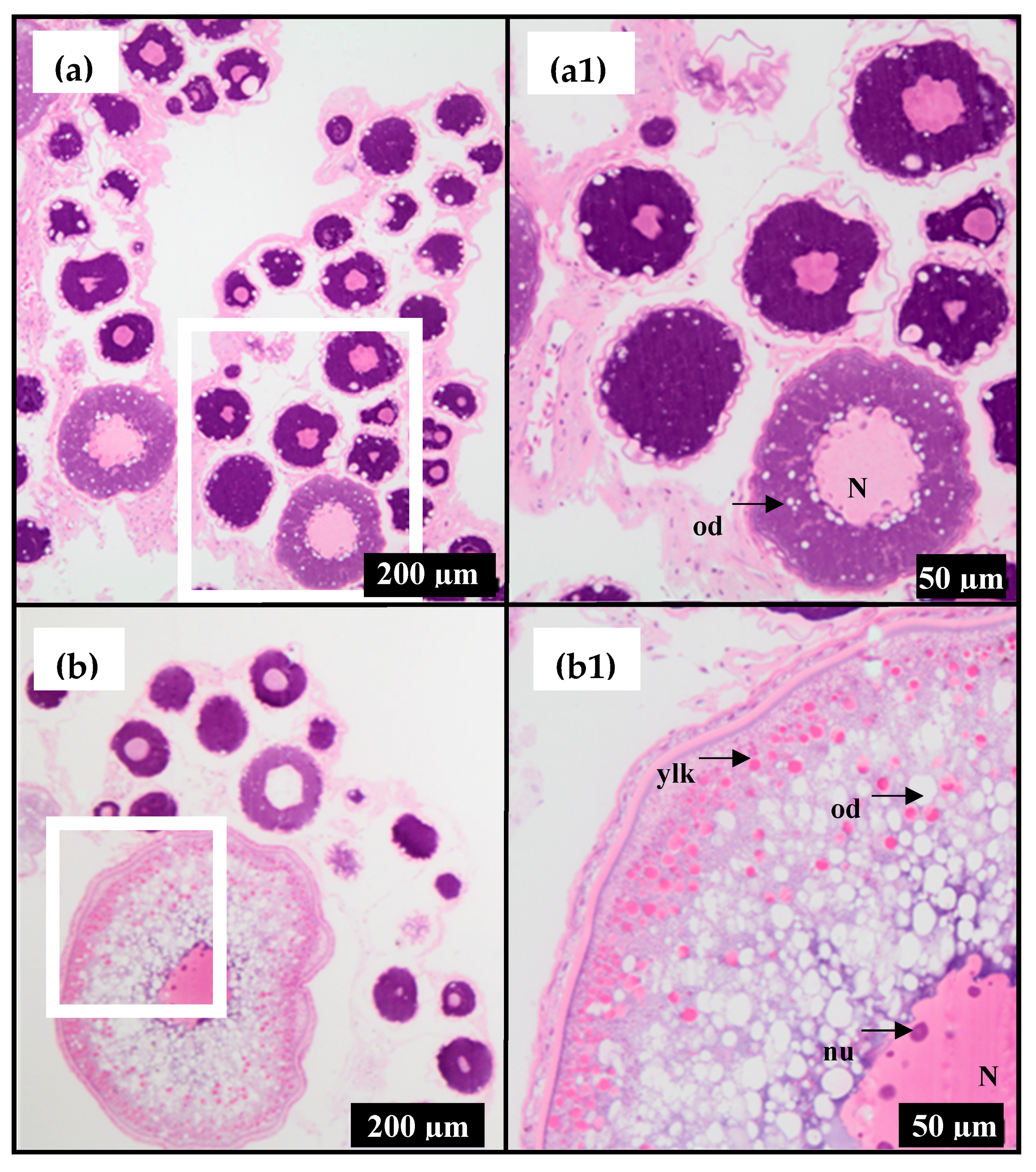
Figure 1. Light micrographs of ovarian tissue from F1 hāpuku (Polyprion oxygeneios) representing the two stages of development used for transcriptome sequencing. Images are from Fish A (Table 1) but are representative of all fish used. White boxes represent areas at higher magnification in adjacent images e.g., a and a1, respectively. (a) Pre-vitellogenic ‘early oil droplet’ (mean oocyte diameter of 0.22 ± 0.004 mm); (b) early vitellogenic (mean oocyte diameter of 0.40 ± 0.009 mm). Abbreviations: N = nucleus; nu = nucleoli; ylk = yolk platelets; od = oil droplets.
Fishes 04 00016 g001
Figure 2. Annotation of differentially expressed genes using BLASTX [19] against the Genbank Non-Redundant database detailing the species distribution of annotations. The group ‘Other fish sp’ combined fish species that had only one hit in the data set.
Fishes 04 00016 g002
Figure 3. Heat-map of differentially expressed genes in the ovary of hāpuku (Polyprion oxygeneios) during previtellogenic (n = 3 fish) and early vitellogenic (n = 3 fish) stages of oogenesis. Expression levels were measured in reads per kilobase of transcript per million mapped reads (RPKM) from normalised values (red = lower expression and green = higher expression as oogenesis progresses from previtellogenic to early vitellogenic stages of development).
Fishes 04 00016 g003
Table 1. Reproductive status (ovarian stage, mean oocyte diameter and plasma level of estradiol-17β (E2) (mean ± SE) of the three F1 female hāpuku (Polyprion oxygeneios) (fish A–C) repeatedly sampled in March 2013 and May 2013 to collect ovarian tissue (with respective RNA integrity numbers (RIN) of samples) for transcriptomic analysis.
FishMonthOvarian StageRIN *Oocyte Diameter (mm)Plasma E2 (ng/mL)
AMarchPrevitellogenic5.40.220.09
BMarchPrevitellogenic6.30.230.09
CMarchPrevitellogenic6.90.22  x ¯ = 0.22 ± 0.0030.08  x ¯ = 0.08 ± 0.003
AMayEarly vitellogenic9.20.400.78
BMayEarly vitellogenic8.90.340.58
CMayEarly vitellogenic9.70.38  x ¯ = 0.37 ± 0.0170.53  x ¯ = 0.63 ± 0.076
* Relatively low RIN are often encountered for fish ovaries with previtellogenic oocytes due to the high abundance of 5S RNA that overshadows the amounts of 18S and 28S RNA in this tissue type [18].
Table 2. De novo transcriptome assembly by Trinity Assembler of ovarian sequencing reads from hāpuku (Polyprion oxygeneios).
ConditionsTranscriptsComponentsN50 *Average LengthNumber of Bases
PE Illumina Q30 and l100146,189120,6881901916.89134,039,555
* N50 is defined as the shortest sequence length at 50% of the genome.
Table 3. Identification and annotation of differentially expressed genes (n = 35 within the FDR of 0.05) in the ovary of hāpuku (Polyprion oxygeneios) during previtellogenic (March) and early vitellogenic (May) stages of oogenesis.
No.Contig NameHit DescriptionGene NameAssession No.SpeciesIdentity (%)E-Value
1c35045_g1_i1prostate stem cell antigen precursor-likepscagb|ADG29182Epinephelus coioides685.0E-28
2c42367_g1_i4PREDICTED: helicase with zinc finger domain 2helz2ref|XP_010750041Larimichthys crocea770.0E+00
3c39480_g2_i2PREDICTED: sodium bicarbonate transporter-like protein 11slc4a11ref|XP_010740414Larimichthys crocea930.0E+00
4c42970_g3_i2PREDICTED: sodium- and chloride-dependent GABA transporter 3-likeslc6a11ref|XP_006782336Neolamprologus brichardi745.0E-144
5c67236_g1_i1PREDICTED: sodium- and chloride-dependent betaine transporter-likeslc6a12ref|XP_008286219Stegastes partitus841.0E-157
6c551_g1_i2cytochrome bmyt-cbgb|AAF06983Perca fluviatilis923.0E-64
7c19980_g1_i1cytochrome c oxidase subunit Imt-c01ref|YP_163831Anguilla australis950.0E+00
8c11536_g1_i1cytochrome c oxidase subunit IImt-c02ref|YP_009058427Howella brodiei923.0E-118
9c13004_g1_i1cytochrome c oxidase subunit IIImt-c03gb|AFQ94066Lutjanus lunulatus913.0E-49
10c26582_g1_i1PREDICTED: fibrinogen-like protein 1fgl1ref|XP_010747488Larimichthys crocea850.0E+00
11c24576_g1_i1PREDICTED: collagen α-1(XV) chain-likecol15a1ref|XP_008277067Stegastes partitus752.0E-129
12c70752_g1_i1PREDICTED: galectin-3-binding proteinlgals3bpref|XP_010736908Larimichthys crocea729.0E-140
13c68469_g1_i1steroid 17-α-hydroxylase/17,20 lyasecyp17a1gb|AEL31248Lateolabrax japonicus940.0E+00
14c43269_g4_i4toll-like receptor 2tlr2gb|AEB32453Epinephelus coioides961.0E-05
15c33557_g1_i3HECT E3 ubiquitin ligasegb|AER42668Epinephelus coioides803.0E-122
16c61987_g1_i1PREDICTED: dentin sialophosphoprotein-like isoform X1dsppref|XP_010745298Larimichthys crocea648.0E-03
17c1460_g1_i1PREDICTED: leucine-rich repeat-containing protein 24-likelrrc24ref|XP_008297789Stegastes partitus952.0E-96
18c90876_g1_i1fatty-acid binding protein H6-isoformh6-fabpgb|AAC60352Notothenia coriiceps935.0E-86
19c32685_g1_i1PREDICTED: fatty-acid desaturase 6fads6ref|XP_010740675Larimichthys crocea900.0E+00
20c43378_g5_i1PREDICTED: ubiquitin-like protein ISG15igs15gb|ADJ57326Sciaenops ocellatus798.0E-83
21c43475_g1_i3PREDICTED: ERV-FRD provirus ancestral Env polyprotein-likeervfrd-1ref|XP_004920373Xenopus (Silurana) tropicalis346.0E-16
22c40949_g1_i2PREDICTED: cystine/glutamate transporterslc7a11ref|XP_008290185Stegastes partitus910.0E+00
23c35826_g2_i1PREDICTED: pleckstrin homology domain-containing family O member 2plekho2ref|XP_010746837Larimichthys crocea730.0E+00
24c83938_g1_i1NADH dehydrogenase subunit 2nad2gb|AFN88677Percina shumardi882.0E-28
25c80759_g1_i1ATP synthase F0 subunit 6mt-atp6ref|YP_007317144Pseudopentaceros richardsoni928.0E-34
26c19734_g1_i1PREDICTED: uncharacterized protein LOC102082036ref|XP_005478141Oreochromis niloticus786.0E-97
27c43363_g4_i1PREDICTED: uncharacterized protein LOC100536992ref|XP_009301840Danio rerio393.0E-52
28c43475_g1_i1PREDICTED: uncharacterized protein LOC101948489ref|XP_008168575Chrysemys picta bellii460.0E+00
29c43269_g4_i2NO HIT
30c12262_g1_i1NO HIT
31c31915_g1_i1NO HIT
32c29645_g1_i1NO HIT
33c57351_g1_i1NO HIT
34c101392_g1_i1NO HIT
35c42887_g2_i1NO HIT