Article

Toxicity and Microecological Responses of Daphnia magna and Danio rerio to PVC and PHA Microplastics: Focusing on Trophic Pathogen Enrichment


1
College of Life Sciences, Hebei University, Baoding 071002, China
2
Hebei Innovation Center for Bioengineering and Biotechnology, Hebei University, Baoding 071002, China
3
Engineering Research Center of Ecological Safety and Conservation in Beijing-Tianjin-Hebei (Xiong’an New Area) of MOE, Baoding 071002, China
*
Authors to whom correspondence should be addressed.
Fishes 2026, 11(9), 503; https://doi.org/10.3390/fishes11090503
Submission received: 27 July 2026 / Revised: 23 August 2026 / Accepted: 26 August 2026 / Published: 27 August 2026

Abstract

Microplastic pollution severely threatens aquatic ecosystems, yet the trophic microecological risks of petroleum-based polyvinyl chloride (PVC) and biodegradable polyhydroxyalkanoate (PHA) microplastics remain insufficiently compared. This study established a two-trophic food chain model with Daphnia magna and zebrafish to explore the toxic effects and microbial disturbances of environmentally relevant 0.1 mg/L PVC and PHA. A 21-day chronic exposure induced no physiological damage to the survival, growth, reproduction, and locomotion of both organisms, suggesting conventional phenotypic indicators fail to identify early microplastic stress. However, trophic microplastic exposure triggered prominent zebrafish gut dysbiosis with distinct pathogen enrichment patterns. PHA trophic exposure enriched plant pathogenic genera (Acidovorax, Clavibacter, Rhizobium), whereas PVC trophic exposure accumulated zoonotic and clinical pathogens (Acinetobacter, Neisseria, Mycobacterium, Aeromonas). This work verifies that PHA is not entirely eco-safe, and PVC poses greater ecotoxicological risks via trophic pathogen transmission, providing genus-level evidence for differentiated microplastics risk assessment.
Key Contribution: Environmental PVC and PHA microplastics cause microbial dysbiosis and pathogen enrichment via trophic transfer in Daphnia magna and zebrafish, with no overt physiological toxicity. Biodegradable PHA is not fully ecologically safe, while PVC presents greater hidden ecological risks through enriching zoonotic and clinical opportunistic pathogens, supporting differentiated microplastic risk assessment.

1. Introduction

Microplastics are a class of persistent pollutants that have garnered considerable global attention in recent years. Despite the lack of a generally accepted definition of microplastics, particles with sizes ranging from 100 nm to 5 mm are defined as microplastics [1,2]. Microplastics are ubiquitously distributed across global freshwater and marine ecosystems and exert persistent ecological pressure on aquatic environments [3]. Polyvinyl chloride (PVC) microplastics are a prevalent polymer type frequently detected in aquatic habitats [4]. As a petroleum-derived plastic, PVC features strong environmental persistence, wide distribution, and substantial ecotoxicity. Its adverse biological effects have been extensively documented in a broad range of aquatic organisms [5,6]. Polyhydroxyalkanoate (PHA) is a biodegradable polymer widely promoted as an environmentally friendly alternative to conventional petroleum plastics [7]. Nevertheless, accumulating evidence demonstrates that degradation intermediates released from PHA and microbial biofilms colonized on PHA surfaces can trigger severe toxic stress in aquatic biota, which challenges the prevailing assumption that biodegradable plastics are environmentally safe [8,9,10].
The particle size and food-like morphology of microplastics (from 100 nm to 5 mm) and nanoplastics (<100 nm) render them readily ingestible by zooplankton [11]. Accordingly, microplastic ingestion has become a widespread ecological phenomenon across all trophic levels within aquatic food webs [12]. Aquatic organisms spanning low-trophic zooplankton to high-trophic predatory fish can take up microplastics via filter-feeding and predation behaviors [13]. Once internalized, microplastics trigger a cascade of adverse biological reactions, including physical abrasion of intestinal epithelia and abnormal biofilm formation [14,15]. These microplastic-derived disturbances disrupt intestinal microbial homeostasis, impair nutrient absorption and metabolic functions, and ultimately weaken the physiological fitness and overall health of aquatic organisms [16]. For filter-feeding invertebrates such as mussels, ingested microplastics can translocate from digestive tracts to multiple somatic tissues, further amplifying biological risks [17]. Consistent field surveys have continuously detected microplastic residues in the digestive tracts of wild fish populations, confirming pervasive microplastic contamination in natural aquatic environments [18]. Complementary laboratory tests using standard aquatic model organisms further verify that microplastics accumulate in zebrafish tissues and trigger physiological and biochemical disorders, highlighting hidden toxic risks associated with microplastic bioaccumulation [19]. Widespread ingestion and bioaccumulation enable microplastics to propagate continuously along aquatic food webs, threatening aquatic ecological stability and posing potential health hazards to humans through dietary intake [12,20,21].
Prior studies have validated the trophic transfer and bioaccumulation of microplastics in freshwater food chains using multiple paired aquatic model species, including Oryzias sinensis and Zacco temminckii [22], as well as Artemia franciscana and Macquaria novemaculeata [23]. Microplastics have also been confirmed to transfer sequentially through the plankton–primary consumer–fish trophic cascade and accumulate in key tissues of high-trophic aquatic organisms, such as the intestine, liver and muscle [24]. Despite these fundamental findings, current research on microplastic trophic transmission bears notable limitations. Most existing studies primarily focus on microplastic ingestion patterns, transfer pathways and tissue distribution in aquatic biota [16,25,26]. Critically, how microplastic trophic transfer from invertebrates to fish reshapes host intestinal microbial communities and the corresponding microecological risks remains poorly understood. Furthermore, comparative research exploring divergent intestinal microbiota disruptions induced by traditional petroleum plastics and biodegradable plastics during trophic transmission remains scarce, creating a critical research gap in comprehensive ecological risk assessments for different plastic types.
Daphnia magna Straus, 1820 and Danio rerio (Hamilton, 1822) are two classic, widely recognized model organisms for freshwater ecotoxicology research [27,28]. The stable prey–predator trophic relationship between D. magna and D. rerio (zebrafish) allows the construction of a simplified, controllable freshwater food chain model that effectively simulates real-world cross-trophic microplastic transmission in natural aquatic environments. Against this research backdrop, the present study first compares the chronic toxic effects of petroleum-based PVC microplastics and biodegradable PHA microplastics on D. magna and zebrafish. We then establish a standardized two-trophic-level freshwater food chain model based on D. magna and zebrafish to characterize and compare the trophic transfer behaviors of the two microplastic types. Specifically, this study aims to explore divergent impacts of PVC and PHA microplastics on intestinal microbial community structure in aquatic organisms at two trophic levels, and clarify distinct microecological risks induced by conventional and biodegradable microplastics during food web transmission.

2. Materials and Methods

2.1. Microplastic and Experimental Organisms

In the present study, 500 nm spherical PVC and PHA microplastics were purchased from Huachuang Plasticisation Co. (Dongguan, China) and Shunjie Plastic Technology Co., Ltd. (Dongguan, China), respectively. D. magna and zebrafish were selected as test organisms to evaluate microplastic toxicity. The D. magna strain originated from Baiyangdian Lake, China, and has been maintained in laboratory cultures for nearly seven years; detailed protocols for organism collection, identification, and laboratory acclimation were described in our previous research [29]. Wild-type zebrafish (Danio rerio (Hamilton, 1822)), AB strain, were used in the present toxicological experiment. Juvenile zebrafish were commercially purchased from YiXiYue Biotechnology Co., Ltd. (Qingdao, China). Then, the zebrafish were raised in a recirculating aquaculture system (Zhongkehai, YG01, Qingdao, China) under standardized laboratory conditions: water temperature maintained at 26 ± 1 °C, with a 14 h light/10 h dark photoperiod. Individuals of zebrafish were fed juvenile shrimp twice daily and acclimated for 7 days prior to formal experiments. All experimental procedures complied with institutional guidelines for laboratory animal care and use.

2.2. Chronic Microplastic Toxicity Assays for D. magna and Zebrafish

A 21-day chronic toxicity test for D. magna was conducted in accordance with the standard experimental protocol issued by the Organisation for Economic Co-operation and Development [30], aiming to clarify the toxic effects of PVC and PHA microplastics. The experiment adopted a semi-static culture system, with M4 culture medium (with or without microplastics) renewed every 48 h throughout the exposure period. D. magna individuals were fed Chlorella pyrenoidosa algae during cultivation. Exposure systems were prepared by adding PVC or PHA microplastics to the M4 medium at a uniform environmentally relevant concentration of 0.1 mg/L. Three groups were employed in the 21-day chronic toxicity test for D. magna: control group (DControl group), PHA microplastic exposure group (DPHA group), and PVC microplastic exposure group (DPVC group). Multiple biological endpoints covering survival, growth, and reproduction were measured to quantify the toxic response of D. magna. Daily observations were recorded over the 21-day trial to collect reproductive indicators, including the molting frequency, time to first spawning, individual number of the first brood, total brood quantity, and cumulative offspring number per daphnid. For all survival, growth, and reproductive detection assays, each microplastic treatment group contained ten biological replicates, with one individual D. magna cultured per 50 mL beaker.
Zebrafish chronic toxicity tests followed the Organization for Economic Cooperation and Development [31]. Commercially purchased zebrafish were acclimated for 7 days in 72 h pre-aerated dechlorinated tap water. Water quality parameters were stabilized at 25 ± 1 °C, pH 7.2, under a 12 h light: 12 h dark photoperiod. In total, 90 10-week post-hatching zebrafish were randomly assigned to three groups: zebrafish control group (Zcontrol group), PHA microplastic exposure group (ZPHA group), and PVC microplastic exposure group (ZPVC group). Each treatment included six biological replicates with five fish per replicate, housed in separate 5 L pre-aerated dechlorinated tap water aquaria. Both PHA and PVC microplastics were administered at an identical nominal concentration of 0.1 mg/L for a 21-day semi-static chronic exposure test. Zebrafish were fed newly hatched Artemia nauplii twice daily at a feeding rate of 0.01 g per individual. Fish feces and residual feed were regularly siphoned, and full medium renewal was performed every 48 h to sustain stable microplastic concentrations. Initial body weight and body length of all zebrafish were recorded before exposure. Post-exposure measurements quantified multiple physiological and behavioral endpoints, including survival rate, body weight growth ratio (abbreviated as BW), body length growth ratio (abbreviated as BL), and locomotor activity. BW and BL were calculated via Equations (1) and (2), respectively:
BW = ( W 0 W t   )   ×   100 % / W 0
BL   =   ( L 0 L t   )   ×   100 % / L 0
where W0 (g) and L0 (mm) represent initial body weight and length; Wt (g) and Lt (mm) denote body weight and length after 21 days of microplastic exposure.

2.3. Locomotor Assay

Swimming speed assays were designed for D. magna and zebrafish following the methods described in our previous studies [32,33]. In brief, each individual of D. magna was transferred into a single well (35 mm diameter, 10 mm depth) of a 6-well plate filled with 2 mL M4 medium. For zebrafish swimming speed tests, each individual was placed in a 7 L aquarium containing 5 L culture water. Swimming behaviors of D. magna or zebrafish were recorded for 1 min using a 38MP-HDMI industrial microscopic camera (Deep-photo, Shenzhen, China). The recorded videos were first analyzed with Kinovea software version 2025.2.0 (https://www.kinovea.org/ (accessed on 22 May 2026)) to acquire swimming trajectory data. The data were further processed using Ctrax software version 0.5.13 (http://ctrax.sourceforge.net/ (accessed on 2 February 2026)) to generate swimming trajectories and calculate speed for both species. For either D. magna or zebrafish, swimming speed assays included ten biological replicates with one organism per replicate.

2.4. Trophic Transfer Experimental Design

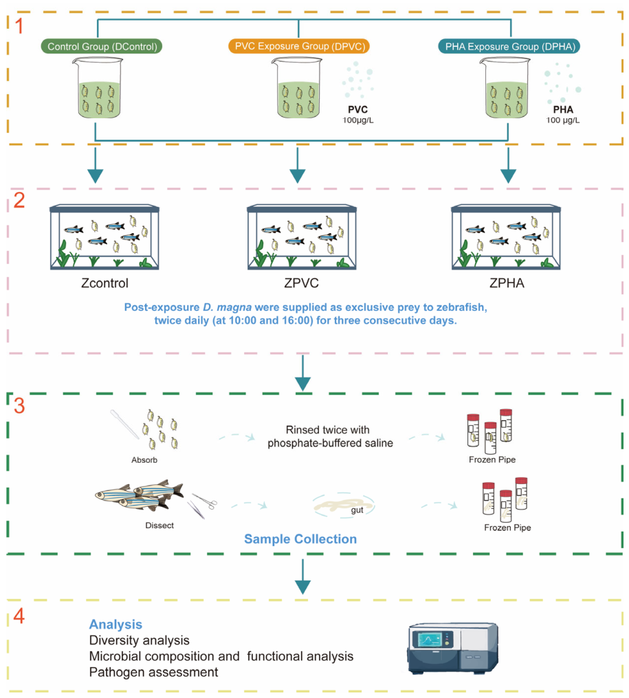
We established a two-trophic-level food chain model to investigate microplastic toxic effects across trophic levels (Figure 1). D. magna were assigned to three groups: the control group (abbreviated as Dcontrol), the PVC microplastic exposure group (abbreviated as DPVC), and the PHA microplastic exposure group (abbreviated as DPHA). Pre-exposed D. magna from each group were used as prey to feed zebrafish, generating three corresponding fish treatments: ZControl (zebrafish fed Dcontrol D. magna), ZPVC (zebrafish fed DPVC D. magna), and ZPHA (zebrafish fed DPHA D. magna).
For D. magna pre-exposure, 20 same-age individuals (21 days post-hatching) were cultured in 1 L M4 medium beakers supplemented with 0.1 mg/L PVC or PHA microplastics for 7 days. Post-exposure, D. magna were then supplied as exclusive prey to zebrafish twice daily for three consecutive days. During this feeding phase, adult zebrafish (5 months old) were exposed to microplastics solely via dietary ingestion, with no direct aqueous microplastic exposure. Zebrafish were housed in 7 L aquaria with 5 L culture water at a density of ten individuals per tank.
Microbiota collected from whole D. magna and zebrafish samples were characterized via 16S rRNA gene amplicon sequencing. Zebrafish sampling was conducted on day 4 (24 h after the 3-day dietary exposure): five individuals were randomly selected per group for dissection and intestinal tissue collection. Previous studies report that the carapax, body cavities, gut, and feeding apparatus of D. magna are colonized by a variety of bacteria, collectively referred to as D. magna-associated microbiota [34,35]. Therefore, the term D. magna-associated microbiota was used in the present study. D. magna sampling occurred on day 7 of microplastic exposure: 20 individuals were randomly picked per group, rinsed twice with phosphate-buffered saline (PBS) to remove surface contaminants, and the whole D. magna samples were prepared to sequence the diversity of D. magna-associated microbiota. Each treatment in the trophic transfer experiment contained five biological replicates; accordingly, 16S rRNA sequencing was performed with five replicates per group for both D. magna and zebrafish.

2.5. DNA Extraction, 16S rRNA Amplicon Sequencing and Bioinformatics Analysis

Total microbial genomic DNA was extracted from D. magna-associated microbiota and zebrafish gut microbiota samples using the MagBeads FastDNA Kit for Soil (MP Biomedicals, Irvine, CA, USA) following manufacturer standard protocols. Purified DNA was temporarily stored at −20 °C for subsequent molecular analysis. DNA concentration and purity were quantified with a NanoDrop NC2000 spectrophotometer (Thermo Fisher Scientific, Waltham, MA, USA), and DNA integrity was verified via agarose gel electrophoresis.
The hypervariable V3–V4 region of the bacterial 16S rRNA gene was amplified by polymerase chain reaction (PCR) with a pair of universal primers: forward primer 338F (5′-ACTCCTACGGGAGGCAGCA-3′) and reverse primer 806R (5′-GGACTACHVGGGTWTCTAAT-3′). Unique 7-base pair (bp) barcodes were integrated into primer sequences to distinguish samples during multiplex high-throughput sequencing. Each 25 μL PCR reaction system contained: 5 μL 5× reaction buffer, 0.25 μL FastPfu DNA Polymerase (5 U/μL), 2 μL 2.5 mM dNTP mixture, 1 μL 10 μM forward primer, 1 μL 10 μM reverse primer, 1 μL template DNA, and 14.75 μL double-distilled water. The thermal cycling program was set as: initial pre-denaturation at 98 °C for 5 min; 25 amplification cycles (98 °C for 30 s, 53 °C for 30 s, 72 °C for 45 s); final extension at 72 °C for 5 min.
Subsequently, qualified PCR products were purified using VAHTS™ DNA Clean Beads (Vazyme Biotech Co., Ltd., Nanjing, China). The concentration of purified amplicons was accurately quantified using the Quant-iT PicoGreen dsDNA Assay Kit (Invitrogen, Carlsbad, CA, USA). All qualified amplicon samples were mixed at equal molar concentrations and subjected to paired-end sequencing (2 × 250 bp) on the Illumina NovaSeq 6000 platform. Library construction and sequencing procedures were performed with the NovaSeq 6000 SP Reagent Kit (500 cycles) at Shanghai Personal Biotechnology Co., Ltd. (Shanghai, China).

2.6. Sequence Data Processing and Microbial Community Analysis

Raw sequencing data were processed primarily with QIIME2 and R software (v4.3.3). Alpha diversity metrics at the amplicon sequence variant (ASV) level, including Chao1, Shannon, and Simpson indices, were calculated via QIIME2 and visualized as boxplots. ASV-level rank-abundance curves were plotted to compare species richness and evenness across samples. Microbial taxonomic composition and relative abundance were visualized using MEGAN6 (metagenome analyzer version 6.25.10, http://software-ab.cs.uni-tuebingen.de/download/megan6/welcome.html, assessed on 5 May 2026) [36].
To evaluate potential pathogenic bacteria, high-quality ASV sequences obtained after quality filtering, DADA2 denoising, and chimera removal were aligned against the reference sequences in the multi-bacterial pathogen detection database (MBPD) [37,38]. Taxonomic classification of ASVs was first performed using the SILVA reference database with a naïve Bayesian classifier [39,40], and potential pathogenic taxa were further screened using the MBPD pipeline. ASVs were annotated as putative pathogens only when they showed high-confidence matches to MBPD reference sequences, with an E-value < 1 × 10⁻¹⁰ and sequence identity > 99%. These stringent criteria have been used in previous studies [38,41] and can improve the reliability of pathogen-related screening by reducing ambiguous or low-confidence assignments. Taxa consistently supported by MBPD annotation criteria were retained for downstream comparative analyses among experimental groups, while ambiguous matches and low-confidence annotations were removed.

2.7. Statistical Analysis

One-way analysis of variance (ANOVA) was applied to test intergroup differences in alpha diversity metrics and endpoints of chronic toxicity tests among experimental groups. Normality and homogeneity of variance were validated prior to statistical testing. Where significant overall treatment effects were detected, post-hoc Tukey’s HSD multiple comparison tests were performed. Statistical significance was defined at p < 0.05. Distinct lowercase letters (a, b, ab) above boxplots denote statistically significant differences between groups; groups sharing identical letters show no significant differences.

3. Results and Discussion

3.1. Chronic Toxic Assay of Microplastics on D. magna and Zebrafish

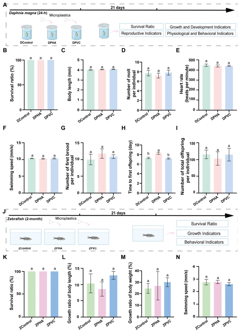
Chronic toxicity bioassays represent standard frameworks for evaluating chemical hazards in D. magna and zebrafish [30,31]. In this study, 21-day chronic exposure tests were conducted to investigate toxic effects of environmentally relevant concentrations (0.1 mg/L) of PHA and PVC microplastics on both species. Figure 2A illustrates the schematic workflow for D. magna chronic exposure. No significant adverse impacts on D. magna growth and development were observed for either microplastic type. Measured endpoints including survival ratio (Figure 2B), body length (Figure 2C), molting frequency (Figure 2D), heart rate (Figure 2E), and swimming speed (Figure 2F) showed no statistical differences between control and microplastic exposure groups. Although PHA exposure slightly delayed the time to first brood release (Figure 2H), no significant alterations were detected in the number of first offspring (Figure 2G) or number of total offspring (Figure 2I) over the 21-day trial. Similarly, PVC microplastic exposure exerted no significant inhibitory effects on D. magna reproductive capacity. Subsequent 21-day chronic toxicity tests evaluated microplastic hazards to zebrafish, with experimental procedures outlined in Figure 2J. Consistent with the observations in D. magna, neither PHA nor PVC microplastics induced overt toxic responses in zebrafish. Survival ratio (Figure 2K), growth ratio of body length (Figure 2L), growth ratio of body weight (Figure 2M), and swimming speed (Figure 2N) remained unaltered across all treatment groups.
Collectively, our 21-day chronic exposure results demonstrate that microplastics at the environmentally relevant concentration tested did not induce statistically significant organism-level toxic effects in D. magna or zebrafish. These findings are consistent with prior studies showing that environmentally realistic microplastic concentrations do not impair survival, development, or reproduction in D. magna over a 21-day exposure period and that, although substantial microplastic accumulation in zebrafish intestines following chronic exposure, it does not cause significant changes to standard physiological or behavioral toxicity indicators [42,43,44].

3.2. Microbial Community Alpha Diversity Analysis

As a typical filter-feeding zooplankton, D. magna continuously ingests ambient bacteria, algae, and suspended particulate matter from aquatic environments, rendering the D. magna-associated microbiota highly sensitive to shifts in water chemical conditions [34,45,46]. Accordingly, this study compared microbiota perturbations induced by PVC and PHA microplastics across two trophic levels (D. magna to zebrafish).
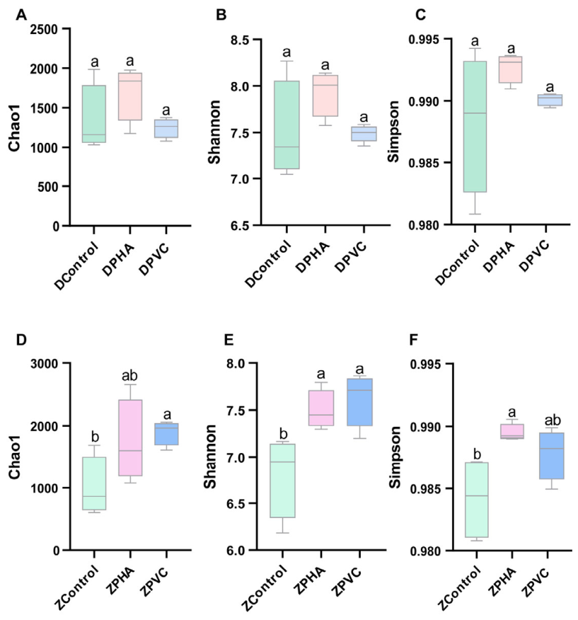
As shown in Figure 3A–C, no significant intergroup differences were detected in D. magna-associated microbial alpha diversity indices (Chao1, Shannon, Simpson) across Dcontrol, DPVC and DPHA groups. As vertebrates, zebrafish possess more complex gut morphology and more diverse indigenous gut microbial communities than cladocerans [47]. Previous research has confirmed that microplastic exposure triggers gut dysbiosis in zebrafish, accompanied by pronounced changes in microbial diversity and nutrient metabolism [48,49]. In the present trophic transfer experiment, dietary ingestion of microplastic-contaminated D. magna significantly altered zebrafish gut microbial alpha diversity indices (Chao1, Shannon, Simpson; Figure 3D–F). This evidence indicates that microplastics can propagate through aquatic food chains and disrupt host intestinal microbial homeostasis at higher trophic levels.

3.3. Microbial Community Composition Analysis

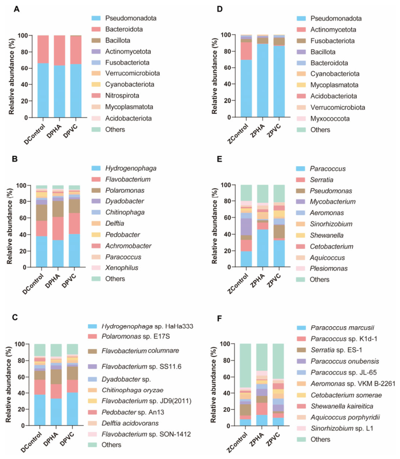
At the phylum level, Pseudomonadota and Bacteroidota dominated the D. magna-associated microbiota across all control and exposure groups (Figure 4A and Table S1 in Supplementary Material). Multiple prior studies have identified Pseudomonadota and Bacteroidota as core functional taxa in the D. magna-associated microbiota [50,51,52]. Pseudomonadota participate in metabolite absorption and mediate host gut responses to exogenous contaminants in aquatic invertebrates [53,54,55]. Bacteroidota primarily degrade refractory macromolecular organics such as polysaccharides and chitin, including intermediate metabolites generated during microplastic degradation [45,56].
At the genus level (Figure 4B and Table S2 in Supplementary Material), PHA and PVC microplastic exposure pronouncedly modified the relative abundances of Flavobacterium, Dyadobacter, Achromobacter, and Paracoccus in D. magna-associated microbiota. Flavobacterium was sharply enriched in the DPHA groups, while PVC exposure markedly elevated Hydrogenophaga abundance. A previous freshwater microplastic study reported Hydrogenophaga as an early dominant colonizer on PVC surfaces, which regulates extracellular polymeric substance production within plastisphere microhabitats [57]. Consistent with this report, elevated Hydrogenophaga abundance in PVC-exposed D. magna is hypothesized to derive from biofilm assembly on PVC microplastic particles retained within the gut lumen. Species-level taxonomic profiling further revealed divergent microbial restructuring between treatments (Figure 4C and Table S3 in Supplementary Material). Hydrogenophaga sp. HaHa333 dominated DControl samples but declined drastically in DPHA individuals, concurrent with striking proliferation of Flavobacterium columnare. Likewise, PVC exposure also increased the relative abundance of F. columnare in D. magna.
Compared with D. magna, zebrafish exposed to trophic microplastics exhibited far more dramatic shifts in gut microbial community composition. At the phylum level (Figure 4D and Table S4 in Supplementary Material), Pseudomonadota dominated all zebrafish gut samples, and microplastic trophic exposure further upregulated its relative abundance. At the genus level (Figure 4E and Table S5 in Supplementary Material), Paracoccus was significantly enriched in zebrafish of ZPHA groups, alongside elevated Shewanella and Cetobacterium abundances. In contrast, zebrafish of ZPVC groups displayed reduced Paracoccus abundance coupled with higher proportions of Mycobacterium, Aeromonas and Plesiomonas. Species-level analysis (Figure 4F and Table S6 in Supplementary Material) revealed Paracoccus marcusii and Paracoccus sp. Kd-1 as dominant taxa in zebrafish gut microbiota of ZPHA groups, while ZPVC groups showed prominent abundance shifts in Aeromonas sp. VKM B-2261, Cetobacterium somerae, Shewanella kaireitica and Sinorhizobium sp. L1.

3.4. Pathogenic Taxa Profiling of Microbiota

3.4.1. Putative Pathogens in D. magna-Associated Microbiota

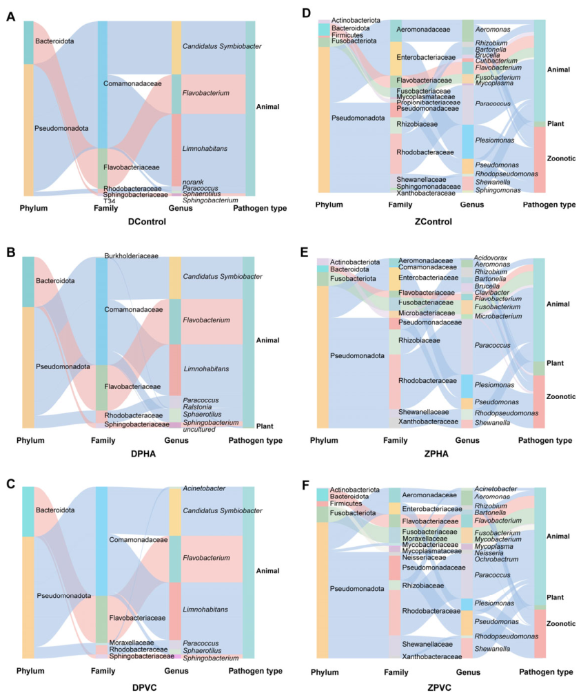
As illustrated in Figure 5A and Table S7 (Supplementary Material), putative pathogenic taxa detected in D. magna-associated microbiota predominantly belonged to two phyla: Pseudomonadota and Bacteroidota, with dominant families including Comamonadaceae, Flavobacteriaceae, Rhodobacteraceae, and Sphingobacteriaceae. Taxonomic pathogenicity profiling further revealed that these enriched bacteria were classified as animal-associated pathogens.
Relative to DControl groups, PHA exposure induced marked shifts in pathogenic community composition at the family level (Figure 5B and Table S8 in Supplementary Material). Flavobacteriaceae abundance increased markedly in DPHA groups, while Burkholderiaceae was uniquely detected in DPHA groups and absent from DControl groups. Pathotype classification further confirmed that PHA-exposed D. magna hosted plant-associated pathogens undetectable in DControl groups.
At the genus level, PHA microplastic exposure elevated multiple putative pathogenic genera in D. magna, including Candidatus Symbiobacter, Flavobacterium, Paracoccus, Sphaerotilus and Limnohabitans. Ubiquitous in aquatic habitats, Flavobacterium readily colonizes biofilms and the intestinal tracts and encodes metabolic pathways for complex organic degradation; multiple Flavobacterium species act as opportunistic aquatic pathogens [58,59]. The pronounced enrichment of Flavobacterium under PHA exposure is likely linked to the biodegradable properties of PHA microplastics, which may further facilitate accumulation of opportunistic pathogens along aquatic food webs. Additional genera frequently isolated from microplastic-polluted water—Ralstonia, Hydrogenophaga, Dyadobacter, and Achromobacter [60]—were also enriched in PHA-exposed D. magna. Notably, Ralstonia was absent from DControl samples, and multiple Ralstonia species are recognized clinical opportunistic pathogens, such as Ralstonia mannitolilytica [61], Ralstonia pickettii [62], and Ralstonia insidiosa [63].
PVC microplastic exposure also drove substantial restructuring of putative pathogenic bacteria within the D. magna-associated microbiota compared with DControl groups (Figure 5C and Table S9 in Supplementary Material). Moraxellaceae, an opportunistic pathogenic family absent from DControl samples, was significantly enriched in DPVC individuals. Flavobacteriaceae abundance rose sharply under PVC exposure, whereas Sphingobacteriaceae declined dramatically relative to DControl samples. At the genus level, PVC exposure markedly elevated Flavobacterium abundance while depleting Sphaerotilus, Candidatus Symbiobacter, Paracoccus, and Sphingobacterium. Flavobacterium columnare, a major pathogenic bacterium causing freshwater fish disease [64], accumulated prominently in both DPHA and DPVC groups, demonstrating that microplastic exposure exacerbates pathogen-related ecological risks in aquatic systems. Acinetobacter, absent from DControl groups, uniquely proliferated in DPVC groups; this genus contains multiple clinically relevant human pathogens, including Acinetobacter baumannii, Acinetobacter pittii, and Acinetobacter nosocomialis [65,66].
Notably, PHA and PVC microplastics induced distinct pathogenic community profiles in D. magna. Flavobacterium, Candidatus Symbiobacter and Sphaerotilus were far more abundant in DPHA than DPVC groups. The opportunistic pathogen Ralstonia was exclusively detected in DPHA groups, whereas Acinetobacter served as a signature pathogenic genus enriched specifically under PVC exposure.

3.4.2. Putative Pathogens in Zebrafish Gut Microbiota

Zebrafish hosted a more diverse pool of putative pathogenic taxa than D. magna (Figure 5D and Table S10 in Supplementary Material). Pathogenic communities were dominated by Pseudomonadota, accompanied by Bacteroidota, Fusobacteriota, Actinobacteriota and Firmicutes. Rhodobacteraceae, Enterobacteriaceae and Aeromonadaceae constituted the dominant pathogenic families, while Paracoccus, Plesiomonas and Aeromonas represented core pathogenic genera. Pathotype classification revealed baseline pathogens in ZControl groups were predominantly animal-associated genera (Paracoccus, Shewanella, Flavobacterium), alongside zoonotic taxa (Plesiomonas, Aeromonas, Pseudomonas); plant-related pathogens (Rhizobium) accounted for only a minor fraction.
Trophic PHA exposure induced profound remodeling of pathogenic communities in zebrafish relative to ZControl groups (Figure 5E and Table S11 in Supplementary Material). Animal-associated and zoonotic pathogens dominated the gut microbiota of Zcontrol zebrafish, while PHA exposure further elevated animal-associated pathogen loads and triggered a striking expansion of plant-related pathogenic populations. At the family level, Rhodobacteraceae and Rhizobiaceae were significantly enriched in zebrafish of ZPHA groups, whereas the abundance of Enterobacteriaceae, Aeromonadaceae, Flavobacteriaceae and Shewanellaceae declined. Two families undetectable in ZControl samples—Comamonadaceae and Microbacteriaceae—emerged prominently in zebrafish of ZPHA groups. At the genus level, trophic PHA exposure increased Paracoccus, Rhizobium and Fusobacterium abundances, while depleting Plesiomonas and Aeromonas. Several genera absent from ZControl zebrafish exclusively proliferated under PHA stress, including Acidovorax, Microbacterium and Clavibacter. Acidovorax and Clavibacter are well-documented plant pathogens, exemplified by Acidovorax citrulli [67], Acidovorax sacchari [68], and Clavibacter michiganensis [69]. Additionally, the isolation of Microbacterium aurum from patient blood samples suggests its potential clinical relevance [70].
Trophic PVC microplastic exposure induced minimal shifts in the phylum-level pathogenic profile of zebrafish gut microbiota versus ZControl, yet drove substantial alterations at the family level (Figure 5F and Table S12 in Supplementary Material). Families Pseudomonadaceae, Rhodobacteraceae, Shewanellaceae, Flavobacteriaceae and Fusobacteriaceae increased, while Enterobacteriaceae and Aeromonadaceae decreased. Pathotype analysis indicated that zebrafish in ZPVC groups carried higher loads of animal-associated pathogens than ZControl counterparts. At the genus level, PVC-treated zebrafish possessed higher abundances of potential pathogens belonging to Paracoccus, Shewanella, Fusobacterium and Flavobacterium, accompanied by a depletion of Plesiomonas. Furthermore, PVC exposure enriched multiple pathogenic genera absent from ZControl groups, namely Neisseria, Acinetobacter, Mycobacterium and Ochrobactrum. These results align with prior research linking PVC microplastic exposure to Acinetobacter enrichment in carp gut microbiota [71]. Mycobacterium, a common biofilm colonizer on PVC surfaces, also acts as a reservoir for diverse antimicrobial resistance genes [72].
Distinct pathogenic assemblages were observed in zebrafish following trophic exposure to PHA versus PVC microplastics (Figure 5E,F). Paracoccus, Plesiomonas and Rhodopseudomonas were more abundant in ZPHA groups, while zebrafish in ZPVC groups harbored higher levels of Shewanella, Aeromonas, Flavobacterium and Pseudomonas. Unique pathogenic taxa distinguished the two treatments: Acidovorax, Clavibacter and Microbacterium exclusively bloomed in ZPHA groups, whereas Neisseria, Acinetobacter, Mycobacterium and Ochrobactrum represented PVC-specific enriched pathogens.

3.4.3. Summary of Key Findings, Study Limitations, and Future Perspectives

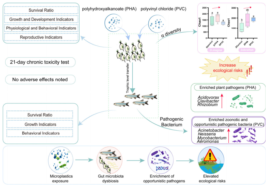
As shown in Figure 6, this study systematically compared the chronic toxic and microecological effects of petroleum-based PVC and biodegradable PHA microplastics on two typical freshwater model organisms (D. magna and zebrafish) through direct individual exposure and two-trophic food chain exposure experiments. The 21-day chronic toxicity test demonstrated that microplastics at environmentally relevant concentrations caused no significant physiological damage to the survival, growth, reproduction and locomotion of D. magna and zebrafish, indicating that conventional individual phenotypic indicators are insensitive to early microplastic stress. However, microplastic exposure induced severe microbiota dysbiosis in both aquatic organisms, with more pronounced adverse microecological effects in zebrafish exposed to microplastics via trophic transfer. PVC and PHA microplastics distinctly reshaped microbial communities and differentially enriched potential pathogens. Specifically, PHA microplastics promoted the proliferation of plant-associated pathogens along the food chain, while PVC microplastics selectively enriched clinical and zoonotic opportunistic pathogens, posing latent risks to aquatic ecosystems and human health through food web transmission.
This study reveals that microplastic pollution can trigger cryptic ecological risks, mainly including microecological imbalance and pathogen proliferation, even without observable physiological impairment in aquatic organisms. Accordingly, biodegradable PHA microplastics cannot be regarded as completely safe alternatives to traditional petroleum-based PVC microplastics. These findings suggest that future ecological risk assessment of microplastics should incorporate microbial community characteristics and pathogen profiles, rather than relying merely on individual physiological endpoints.
Despite the above findings, this study has several limitations that need to be addressed in future research. First, further studies are required to explore the alterations of gut microenvironments induced by PVC and PHA microplastics and their association with pathogen enrichment in aquatic animals. Second, it is necessary to clarify the differences and underlying mechanisms of gut microbial variations in zebrafish under direct waterborne microplastic exposure and indirect trophic exposure via D. magna. Third, in the present study, relatively high intra-group variation in alpha-diversity indices was detected among control samples. Such large baseline variability in microbiota may reduce statistical power and mask subtle microbial responses to pollutant exposure. Individual-specific differences in the native microbiome and minor stochastic fluctuations during animal culture may account for this variation. Future investigations should use an increased number of biological replicates to reduce the influence of natural inter-individual heterogeneity. Furthermore, future research based on microcosm systems is needed to evaluate the long-term effects of microplastic exposure on the physiological health and stability of D. magna-associated microbiota and zebrafish gut microbiota. Overall, this study supplements the theoretical basis of microplastic trophic toxicity and provides a scientific reference for differentiated risk management of freshwater microplastic pollution.

4. Conclusions

This study systematically clarified the differential toxic and microecological effects of PVC and PHA microplastics on D. magna and zebrafish under environmentally realistic exposure conditions. Microplastic exposure caused no observable physiological damage to either test organism, but induced obvious microbial community dysbiosis, with more severe adverse effects in zebrafish through trophic transfer. Most critically, PVC and PHA induced completely divergent genus-level pathogenic enrichment patterns in the gut of high-trophic zebrafish. Trophic PHA exposure specifically enriched typical plant pathogenic genera (Acidovorax, Clavibacter, Rhizobium), facilitating the persistent spread and accumulation of plant pathogens in aquatic food chains. By comparison, trophic PVC exposure selectively enriched a series of zoonotic and clinical opportunistic pathogenic genera, including Acinetobacter, Neisseria, Mycobacterium and Aeromonas, substantially elevating the cross-species health risks from aquatic environments to humans. Collectively, biodegradable PHA cannot serve as a fully safe alternative to petroleum-based PVC due to its non-negligible plant-pathogen ecological risks, while PVC presents more threatening hidden zoonotic risks via specific pathogenic genus enrichment during trophic transmission. This work highlights that microbial community variation and genus-level pathogenic taxon profiling are essential complementary indicators for accurate early risk evaluation of microplastics pollution, rather than relying solely on traditional physiological endpoints.

Supplementary Materials

The following supporting information can be downloaded at: https://www.mdpi.com/article/10.3390/fishes11090503/s1, Table S1: Relative abundance of top 10 bacterial genera at phylum level in D. magna-associated microbiota; Table S2: Relative abundance of top 10 bacterial genera at genus level in D. magna-associated microbiota; Table S3: Relative abundance of top 10 bacterial genera at species level in D. magna-associated microbiota; Table S4: Relative abundance of top 10 bacterial genera at phylum level in gut microbiota of zebrafish; Table S5: Relative abundance of top 10 bacterial genera at genus level in gut microbiota of zebrafish; Table S6: Relative abundance of top 10 bacterial genera at species level in gut microbiota of zebrafish; Table S7: Pathogen abundance in D. magna-associated microbiota from DControl group; Table S8: Pathogen abundance in D. magna-associated microbiota from DPHA group; Table S9: Pathogen abundance in D. magna-associated microbiota from DPVC group; Table S10: Pathogen abundance in gut microbiota of zebrafish from ZControl group; Table S11: Pathogen abundance in gut microbiota of zebrafish from ZPHA group; Table S12: Pathogen abundance in in gut microbiota of zebrafish from ZPVC group.

Author Contributions

M.C.: conceptualization, validation, and writing (original draft). M.W.: conceptualization, formal analysis, validation, and methodology. S.W.: conceptualization, formal analysis, validation, and methodology. Z.W.: conceptualization, validation, and methodology. J.G.: conceptualization, data validation, methodology, and funding acquisition. Z.N.: conceptualization of the study, methodology, data validation, and writing (review and editing). Y.Z.: conceptualization of the study, writing (review and editing), and Funding acquisition. All authors have read and agreed to the published version of the manuscript.

Funding

This work was supported by the National Natural Science Foundation of China (No. 42207336); the Natural Science Foundation of Hebei Province, China (No. B2023201017); and the Natural Science Joint Foundation of Biological and Agriculture, Hebei Province, China (No. C2022204227).

Institutional Review Board Statement

The research presented in this manuscript was conducted under the supervision of the Institutional Animal Care and Use Committee of Hebei University (approval code: SYXK-2022-009; approval date: 18 March 2022).

Data Availability Statement

The original contributions presented in this study are included in the article. Further inquiries can be directed to the corresponding author.

Conflicts of Interest

The authors declare no conflicts of interest.

References

  1. Gigault, J.; El Hadri, H.; Nguyen, B.; Grassl, B.; Rowenczyk, L.; Tufenkji, N.; Feng, S.; Wiesner, M. Nanoplastics are neither microplastics nor engineered nanoparticles. Nat. Nanotechnol. 2021, 16, 501–507. [Google Scholar] [CrossRef] [Scilit]
  2. Ompala, C.; Renault, J.-P.; Taché, O.; Cournède, É.; Devineau, S.; Chivas-Joly, C. Stability and dispersibility of microplastics in experimental exposure medium and their dimensional characterization by SMLS, SAXS, Raman microscopy, and SEM. J. Hazard. Mater. 2024, 469, 134083. [Google Scholar] [CrossRef] [Scilit] [PubMed]
  3. Ali, S.S.; Elsamahy, T.; Al-Tohamy, R.; Sun, J. A critical review of microplastics in aquatic ecosystems: Degradation mechanisms and removing strategies. Environ. Sci. Ecotechnol. 2024, 21, 100427. [Google Scholar] [CrossRef] [Scilit] [PubMed]
  4. Turner, A.; Filella, M. Polyvinyl chloride in consumer and environmental plastics, with a particular focus on metal-based additives. Environ. Sci. Process. Impacts 2021, 23, 1376–1384. [Google Scholar] [CrossRef] [Scilit] [PubMed]
  5. Liu, Y.; Zhang, J.; Zhao, H.; Cai, J.; Sultan, Y.; Fang, H.; Zhang, B.; Ma, J. Effects of polyvinyl chloride microplastics on reproduction, oxidative stress and reproduction and detoxification-related genes in Daphnia magna. Comp. Biochem. Physiol. C Toxicol. Pharmacol. 2022, 254, 109269. [Google Scholar] [CrossRef] [Scilit] [PubMed]
  6. Boyle, D.; Catarino, A.I.; Clark, N.J.; Henry, T.B. Polyvinyl chloride (PVC) plastic fragments release Pb additives that are bioavailable in zebrafish. Environ. Pollut. 2020, 263, 114422. [Google Scholar] [CrossRef] [Scilit] [PubMed]
  7. Li, Z.; Yang, J.; Loh, X.J. Polyhydroxyalkanoates: Opening doors for a sustainable future. NPG Asia Mater. 2016, 8, e265. [Google Scholar] [CrossRef] [Scilit]
  8. Shi, C.; Zhang, Y.; Shao, Y.; Ray, S.S.; Wang, B.; Zhao, Z.; Yu, B.; Zhang, X.; Li, W.; Ding, J.; et al. A review on the occurrence, detection methods, and ecotoxicity of biodegradable microplastics in the aquatic environment: New cause for concern. Trends Anal. Chem. 2024, 178, 117832. [Google Scholar] [CrossRef] [Scilit]
  9. Nik Mut, N.N.; Na, J.; Jung, J. A review on fate and ecotoxicity of biodegradable microplastics in aquatic system: Are biodegradable plastics truly safe for the environment? Environ. Pollut. 2024, 344, 123399. [Google Scholar] [CrossRef] [Scilit] [PubMed]
  10. Morohoshi, T.; Oi, T.; Aiso, H.; Suzuki, T.; Okura, T.; Sato, S. Biofilm formation and degradation of commercially available biodegradable plastic films by bacterial consortiums in freshwater environments. Microbes Environ. 2018, 33, 332–335. [Google Scholar] [CrossRef] [Scilit] [PubMed]
  11. Botterell, Z.L.R.; Beaumont, N.; Dorrington, T.; Steinke, M.; Thompson, R.C.; Lindeque, P.K. Bioavailability and effects of microplastics on marine zooplankton: A review. Environ. Pollut. 2019, 245, 98–110. [Google Scholar] [CrossRef] [Scilit] [PubMed]
  12. Gouin, T. Toward an improved understanding of the ingestion and trophic transfer of microplastic particles: Critical review and implications for future research. Environ. Toxicol. Chem. 2020, 39, 1119–1137. [Google Scholar] [CrossRef] [Scilit] [PubMed]
  13. Wang, F.; Wu, H.; Wu, W.; Wang, L.; Liu, J.; An, L.; Xu, Q. Microplastic characteristics in organisms of different trophic levels from Liaohe Estuary, China. Sci. Total Environ. 2021, 789, 148027. [Google Scholar] [CrossRef] [Scilit] [PubMed]
  14. Qiao, R.; Deng, Y.; Zhang, S.; Wolosker, M.B.; Zhu, Q.; Ren, H.; Zhang, Y. Accumulation of different shapes of microplastics initiates intestinal injury and gut microbiota dysbiosis in the gut of zebrafish. Chemosphere 2019, 236, 124334. [Google Scholar] [CrossRef] [Scilit] [PubMed]
  15. Yu, Z.; Qiu, D.; Zhou, T.; Zeng, L.; Yan, C. Biofilm enhances the interactive effects of microplastics and oxytetracycline on zebrafish intestine. Aquat. Toxicol. 2024, 270, 106905. [Google Scholar] [CrossRef] [Scilit] [PubMed]
  16. Li, W.; Chen, X.; Li, M.; Cai, Z.; Gong, H.; Yan, M. Microplastics as an aquatic pollutant affect gut microbiota within aquatic animals. J. Hazard. Mater. 2022, 423, 127094. [Google Scholar] [CrossRef] [Scilit] [PubMed]
  17. Woods, M.N.; Stack, M.E.; Fields, D.M.; Shaw, S.D.; Matrai, P.A. Microplastic fiber uptake, ingestion, and egestion rates in the blue mussel (Mytilus edulis). Mar. Pollut. Bull. 2018, 137, 638–645. [Google Scholar] [CrossRef] [Scilit] [PubMed]
  18. Campbell, S.H.; Williamson, P.R.; Hall, B.D. Microplastics in the gastrointestinal tracts of fish and the water from an urban prairie creek. Facets 2017, 2, 395–409. [Google Scholar] [CrossRef] [Scilit]
  19. Martínez-Álvarez, I.; Le Menach, K.; Cajaraville, M.P.; Budzinski, H.; Orbea, A. Effects of polystyrene nano- and microplastics and of microplastics with sorbed polycyclic aromatic hydrocarbons in adult zebrafish. Sci. Total Environ. 2024, 927, 172380. [Google Scholar] [CrossRef] [Scilit] [PubMed]
  20. Provencher, J.; Ammendolia, J.; Rochman, C.M.; Mallory, M. Assessing plastic debris in aquatic food webs: What we know and don’t know about uptake and trophic transfer. Environ. Rev. 2018, 27, 304–317. [Google Scholar] [CrossRef] [Scilit]
  21. Bhutto, S.U.A.; Ma, Y.; Akram, M.; You, X. Microplastics in Tai lake food web: Trophic transfer and human health risk assessment. Environ. Toxicol. Pharmacol. 2023, 101, 104206. [Google Scholar] [CrossRef] [Scilit] [PubMed]
  22. Chae, Y.; Kim, D.; Kim, S.W.; An, Y.-J. Trophic transfer and individual impact of nano-sized polystyrene in a four-species freshwater food chain. Sci. Rep. 2018, 8, 284. [Google Scholar] [CrossRef] [Scilit] [PubMed]
  23. Afrose, S.; Tran, T.K.A.; O’Connor, W.; Pannerselvan, L.; Carbery, M.; Fielder, S.; Subhaschandrabose, S.; Palanisami, T. Organ-specific distribution and size-dependent toxicity of polystyrene nanoplastics in Australian bass (Macquaria novemaculeata). Environ. Pollut. 2024, 341, 122996. [Google Scholar] [CrossRef] [Scilit] [PubMed]
  24. Abdolahpur Monikh, F.; Holm, S.; Kortet, R.; Bandekar, M.; Kekäläinen, J.; Koistinen, A.; Leskinen, J.T.T.; Akkanen, J.; Huuskonen, H.; Valtonen, A.; et al. Quantifying the trophic transfer of sub-micron plastics in an assembled food chain. Nano Today 2022, 46, 101611. [Google Scholar] [CrossRef] [Scilit]
  25. Huang, X.; Lu, B.; Liu, H.; Wu, X.; Liu, Y. Ecotoxicological impacts of microplastics to gut microbiota: Response mechanism, challenges and environmental sustainability-A review. Ecotoxicol. Environ. Saf. 2025, 302, 118748. [Google Scholar] [CrossRef] [Scilit] [PubMed]
  26. Evariste, L.; Barret, M.; Mottier, A.; Mouchet, F.; Gauthier, L.; Pinelli, E. Gut microbiota of aquatic organisms: A key endpoint for ecotoxicological studies. Environ. Pollut. 2019, 248, 989–999. [Google Scholar] [CrossRef] [Scilit] [PubMed]
  27. Tkaczyk, A.; Bownik, A.; Dudka, J.; Kowal, K.; Ślaska, B. Daphnia magna model in the toxicity assessment of pharmaceuticals: A review. Sci. Total Environ. 2021, 763, 143038. [Google Scholar] [CrossRef] [Scilit] [PubMed]
  28. Li, H.; Liu, Y.; Chen, Q.; Jin, L.; Peng, R. Research progress of zebrafish model in aquatic ecotoxicology. Water 2023, 15, 1735. [Google Scholar] [CrossRef] [Scilit]
  29. Li, J.; Li, H.; Lin, D.; Li, M.; Wang, Q.; Xie, S.; Zhang, Y.; Liu, F. Effects of butyl benzyl phthalate exposure on Daphnia magna growth, reproduction, embryonic development and transcriptomic responses. J. Hazard. Mater. 2021, 404, 124030. [Google Scholar] [CrossRef] [Scilit] [PubMed]
  30. OECD. Daphnia magna Reproduction Test (OECD Test No. 211); OECD: Paris, France, 2012. [Google Scholar]
  31. OECD. Fish, Juvenile Growth Test (OECD Test No. 215); OECD: Paris, France, 2000. [Google Scholar]
  32. Ni, Z.; Tian, X.; Zhao, W.; Hu, W.; Lv, J.; Sun, X.; Zhang, Y.; Zhang, Y.; Zhang, Y.; Li, B.; et al. The detrimental effects and mechanisms of Orlistat in disrupting energy homeostasis and reproduction in Daphnia magna. Aquat. Toxicol. 2025, 279, 107201. [Google Scholar] [CrossRef] [Scilit] [PubMed]
  33. Zhang, Y.; Tian, X.; Li, H.; Zhang, Y.; Zhang, Y.; Chen, M.; Ni, Z.; Gu, J.; Tang, T.; Xie, S.; et al. Mechanisms underlying the chronic toxicity of ofloxacin to Daphnia magna: Oxidative stress, mitochondrial dysfunction, and energy allocation. Environ. Pollut. 2025, 382, 126764. [Google Scholar] [CrossRef] [Scilit] [PubMed]
  34. Krzynowek, A.; Van de Moortel, B.; Pichler, N.; Vanoverberghe, I.; Lapere, J.; Jenisch, L.M.; Deloof, D.; Thielemans, W.; Muylaert, K.; Dusselier, M.; et al. Effects of microplastics on Daphnia-associated microbiomes in situ and in vitro. ISME J. 2025, 19, wrae234. [Google Scholar] [CrossRef] [Scilit] [PubMed]
  35. Liu, Q.; Chen, H.; Akbar, S.; Gu, L.; Sun, Y.; Yang, Z. Fish predation risk alters the microbiota of Daphnia in the process of inducing its life-history defence traits. Freshw. Biol. 2024, 69, 591–605. [Google Scholar] [CrossRef] [Scilit]
  36. Huson, D.H.; Mitra, S.; Ruscheweyh, H.J.; Weber, N.; Schuster, S.C. Integrative analysis of environmental sequences using MEGAN4. Genome Res. 2011, 21, 1552–1560. [Google Scholar] [CrossRef] [Scilit] [PubMed]
  37. Callahan, B.J.; McMurdie, P.J.; Rosen, M.J.; Han, A.W.; Johnson, A.J.A.; Holmes, S.P. DADA2: High-resolution sample inference from Illumina amplicon data. Nat. Methods 2016, 13, 581–583. [Google Scholar] [CrossRef] [Scilit] [PubMed]
  38. Yang, X.; Jiang, G.; Zhang, Y.; Wang, N.; Zhang, Y.; Wang, X.; Zhao, F.; Xu, Y.; Shen, Q.; Wei, Z. MBPD: A multiple bacterial pathogen detection pipeline for One Health practices. iMeta 2023, 2, e82. [Google Scholar] [CrossRef] [Scilit] [PubMed]
  39. Quast, C.; Pruesse, E.; Yilmaz, P.; Gerken, J.; Schweer, T.; Yarza, P.; Peplies, J.; Glöckner, F.O. The SILVA ribosomal RNA gene database project: Improved data processing and web-based tools. Nucleic Acids Res. 2013, 41, D590–D596. [Google Scholar] [CrossRef] [Scilit] [PubMed]
  40. Yilmaz, P.; Parfrey, L.W.; Yarza, P.; Gerken, J.; Pruesse, E.; Quast, C.; Schweer, T.; Peplies, J.; Ludwig, W.; Glöckner, F.O. The SILVA and “All-species Living Tree Project (LTP)” taxonomic frameworks. Nucleic Acids Res. 2014, 42, D643–D648. [Google Scholar] [CrossRef] [Scilit] [PubMed]
  41. Zhu, D.; Ma, J.; Li, G.; Rillig, M.C.; Zhu, Y.G. Soil plastispheres as hotspots of antibiotic resistance genes and potential pathogens. ISME J. 2022, 16, 521–532. [Google Scholar] [CrossRef] [Scilit] [PubMed]
  42. Jemec Kokalj, A.; Dolar, A.; Titova, J.; Visnapuu, M.; Škrlep, L.; Drobne, D.; Vija, H.; Kisand, V.; Heinlaan, M. Long-term exposure to virgin and recycled LDPE microplastics induced minor effects in the freshwater and terrestrial crustaceans Daphnia magna and Porcellio scaber. Polymers 2021, 13, 771. [Google Scholar] [CrossRef] [Scilit] [PubMed]
  43. Khosrovyan, A.; Kahru, A. Virgin and UV-weathered polyamide microplastics posed no effect on the survival and reproduction of Daphnia magna. PeerJ 2022, 10, e13533. [Google Scholar] [CrossRef] [Scilit] [PubMed]
  44. Batel, A.; Baumann, L.; Carteny, C.C.; Cormier, B.; Keiter, S.H.; Braunbeck, T. Histological, enzymatic and chemical analyses of the potential effects of differently sized microplastic particles upon long-term ingestion in zebrafish (Danio rerio). Mar. Pollut. Bull. 2020, 153, 111022. [Google Scholar] [CrossRef] [Scilit] [PubMed]
  45. Maszczyk, P.; Kiersztyn, B.; Gozzo, S.; Kowalczyk, G.; Jimenez-Lamana, J.; Szpunar, J.; Pijanowska, J.; Jines-Muñoz, C.; Zebrowski, M.L.; Babkiewicz, E. Combined effects of polystyrene nanoplastics and enrofloxacin on the life histories and gut microbiota of Daphnia magna. Water 2022, 14, 3403. [Google Scholar] [CrossRef] [Scilit]
  46. Polhill, L.; de Bruijn, R.; Amaral-Zettler, L.; Praetorius, A.; van Wezel, A. Daphnia magna’s favorite snack: Biofouled plastics. Environ. Toxicol. Chem. 2022, 41, 1977–1981. [Google Scholar] [CrossRef] [Scilit] [PubMed]
  47. Zhao, Y.; Qin, Z.; Huang, Z.; Bao, Z.; Luo, T.; Jin, Y. Effects of polyethylene microplastics on the microbiome and metabolism in larval zebrafish. Environ. Pollut. 2021, 282, 117039. [Google Scholar] [CrossRef] [Scilit] [PubMed]
  48. Jin, Y.; Xia, J.; Pan, Z.; Yang, J.; Wang, W.; Fu, Z. Polystyrene microplastics induce microbiota dysbiosis and inflammation in the gut of adult zebrafish. Environ. Pollut. 2018, 235, 322–329. [Google Scholar] [CrossRef] [Scilit] [PubMed]
  49. Pei, X.; Heng, X.; Chu, W. Polystyrene nano/microplastics induce microbiota dysbiosis, oxidative damage, and innate immune disruption in zebrafish. Microb. Pathog. 2022, 163, 105387. [Google Scholar] [CrossRef] [Scilit] [PubMed]
  50. Freese, H.M.; Schink, B. Composition and stability of the microbial community inside the digestive tract of the aquatic crustacean Daphnia magna. Microb. Ecol. 2011, 62, 882–894. [Google Scholar] [CrossRef] [Scilit] [PubMed]
  51. Motiei, A.; Brindefalk, B.; Ogonowski, M.; El-Shehawy, R.; Pastuszek, P.; Ek, K.; Liewenborg, B.; Udekwu, K.; Gorokhova, E. Disparate effects of antibiotic-induced microbiome change and enhanced fitness in Daphnia magna. PLoS ONE 2020, 15, e0214833. [Google Scholar] [CrossRef] [Scilit] [PubMed]
  52. You, M.; Yang, W. Environmental changes driving shifts in the structure and functional properties of the symbiotic microbiota of Daphnia. Microorganisms 2024, 12, 2492. [Google Scholar] [CrossRef] [Scilit] [PubMed]
  53. Carrillo, M.P.; Vila-Costa, M.; Barata, C. Micro-bioplastic impact on gut microbiome, cephalic transcription and cognitive function in the aquatic invertebrate Daphnia magna. Environ. Pollut. 2025, 382, 126690. [Google Scholar] [CrossRef] [Scilit] [PubMed]
  54. Cooper, R.O.; Cressler, C.E. Characterization of key bacterial species in the Daphnia magna microbiota using shotgun metagenomics. Sci. Rep. 2020, 10, 652. [Google Scholar] [CrossRef] [Scilit] [PubMed]
  55. Li, Y.; Wang, W.; Liu, H. Gut-microbial adaptation and transformation of silver nanoparticles mediated the detoxification of Daphnia magna and their offspring. Environ. Sci. Nano 2022, 9, 361–374. [Google Scholar] [CrossRef] [Scilit]
  56. Parras-Moltó, M.; Lund, D.; Ebmeyer, S.; Larsson, D.G.J.; Johnning, A.; Kristiansson, E. The transfer of antibiotic resistance genes between evolutionarily distant bacteria. mSphere 2025, 10, e00114-25. [Google Scholar] [CrossRef] [Scilit] [PubMed]
  57. Feng, C.; Liang, Z.; Liao, X.; Lin, K.; Zhai, Y.; Liu, G.; Malpei, F.; Hu, A. Microbial dynamics on different microplastics in coastal urban aquatic ecosystems: The critical roles of extracellular polymeric substances. Environ. Sci. Technol. 2025, 59, 10554–10566. [Google Scholar] [CrossRef] [Scilit] [PubMed]
  58. Mann, A.J.; Hahnke, R.L.; Huang, S.; Werner, J.; Xing, P.; Barbeyron, T.; Huettel, B.; Stüber, K.; Reinhardt, R.; Harder, J.; et al. The genome of the alga-associated marine flavobacterium Formosa agariphila KMM 3901T reveals a broad potential for degradation of algal polysaccharides. Appl. Environ. Microbiol. 2013, 79, 6813–6822. [Google Scholar] [CrossRef] [Scilit] [PubMed]
  59. Loch, T.P.; Faisal, M. Emerging flavobacterial infections in fish: A review. J. Adv. Res. 2015, 6, 283–300. [Google Scholar] [CrossRef] [Scilit] [PubMed]
  60. Roager, L.; Sonnenschein, E.C. Bacterial candidates for colonization and degradation of marine plastic debris. Environ. Sci. Technol. 2019, 53, 11636–11643. [Google Scholar] [CrossRef] [Scilit] [PubMed]
  61. Alnimr, A. Bloodstream infections caused by drug resistant Ralstonia species: A case series during the COVID-19 pandemic. Infect. Drug Resist. 2023, 16, 1339–1344. [Google Scholar] [CrossRef] [Scilit] [PubMed]
  62. Sheng, Z.; Li, J.; Han, G.; Fan, R.; Zhu, P.; Fang, X. Molecular epidemiological and clinical infection characteristics analysis of Ralstonia. Eur. J. Clin. Microbiol. Infect. Dis. 2024, 43, 1161–1170. [Google Scholar] [CrossRef] [Scilit] [PubMed]
  63. Ryan, M.P.; Adley, C.C. Ralstonia spp.: Emerging global opportunistic pathogens. Eur. J. Clin. Microbiol. Infect. Dis. 2014, 33, 291–304. [Google Scholar] [CrossRef] [Scilit] [PubMed]
  64. Declercq, A.M.; Haesebrouck, F.; Van den Broeck, W.; Bossier, P.; Decostere, A. Columnaris disease in fish: A review with emphasis on bacterium-host interactions. Vet. Res. 2013, 44, 27. [Google Scholar] [CrossRef] [Scilit] [PubMed]
  65. Morris, F.C.; Dexter, C.; Kostoulias, X.; Uddin, M.I.; Peleg, A.Y. The mechanisms of disease caused by Acinetobacter baumannii. Front. Microbiol. 2019, 10, 1601. [Google Scholar] [CrossRef] [Scilit] [PubMed]
  66. Richards, G.A.; Perovic, O.; Brink, A.J. The challenges of difficult-to-treat Acinetobacter infections. Clin. Microbiol. Rev. 2024, 37, e0009324. [Google Scholar] [CrossRef] [Scilit] [PubMed]
  67. Burdman, S.; Walcott, R. Acidovorax citrulli: Generating basic and applied knowledge to tackle a global threat to the cucurbit industry. Mol. Plant Pathol. 2012, 13, 805–815. [Google Scholar] [CrossRef] [Scilit] [PubMed]
  68. Sawada, H.; Shinohara, H.; Takashima, Y.; Naito, K.; Satou, M. Acidovorax sacchari sp. nov., a pathogen causing red stripe of sugarcane in Japan. Int. J. Syst. Evol. Microbiol. 2025, 75, 006575. [Google Scholar] [CrossRef] [Scilit] [PubMed]
  69. Eichenlaub, R.; Gartemann, K.H. The Clavibacter michiganensis subspecies: Molecular investigation of gram-positive bacterial plant pathogens. Annu. Rev. Phytopathol. 2011, 49, 445–464. [Google Scholar] [CrossRef] [Scilit] [PubMed]
  70. Varghese, G.; Sarawat, D.; Jamwal, A.; Patel, S.S.; Tejan, N.; Sahu, C. Case series: Microbacterium aurum bacteremia in immunosuppressed patients—An emerging threat. Am. J. Trop. Med. Hyg. 2024, 111, 132–135. [Google Scholar] [CrossRef] [Scilit] [PubMed]
  71. Li, W.; Zeng, J.; Zheng, N.; Ge, C.; Li, Y.; Yao, H. Polyvinyl chloride microplastics in the aquatic environment enrich potential pathogenic bacteria and spread antibiotic resistance genes in the fish gut. J. Hazard. Mater. 2024, 475, 134817. [Google Scholar] [CrossRef] [Scilit] [PubMed]
  72. Zhang, X.; Dong, Z.; Zhang, S.; Ma, J.; Liu, S. Microplastic biofilm as hotspots of antibiotic resistance genes and potential pathogens. NPJ Biofilms Microbiomes 2026, 12, 24. [Google Scholar] [CrossRef] [Scilit] [PubMed]
Figure 1. Schematic diagram of the two-trophic-level food web experiment for assessing microplastic-induced adverse effects on D. magna-associated microbiota and zebrafish gut microbiota. D. magna individuals were pre-exposed to PVC or PHA microplastics and subsequently provided as prey to zebrafish. Neither species received microplastic exposure in the control group. D. magna without microplastic exposure served as the DControl group, while individuals exposed to PHA and PVC microplastics were designated DPHA and DPVC groups, respectively. Similarly, unexposed zebrafish were defined as the ZControl group, and zebrafish subjected to PHA or PVC microplastics via the food chain were denoted ZPHA and ZPVC groups, respectively.
Fishes 11 00503 g001
Figure 2. Schematic design and physiological endpoints of the 21-day chronic microplastic toxicity tests with D. magna and zebrafish. (A) Schematic illustration of the 21-day chronic exposure assay for D. magna. (B) Survival ratio of D. magna; (C) Body length of D. magna; (D) number of molt per individual D. magna; (E) heart rate of D. magna; (F) swimming speed of D. magna; (G) number of first brood per individual D. magna; (H) time to first offspring of D. magna; (I) number of total offspring per individual D. magna; (J) schematic diagram of 21-day chronic assays of microplastics towards zebrafish; (K) survival ratio of zebrafish; (L) growth ratio of body length; (M) growth ratio of body weight; (N) swimming speed of zebrafish. D. magna without microplastic exposure served as the DControl group, while individuals exposed to PHA and PVC microplastics were designated DPHA and DPVC groups, respectively. Similarly, unexposed zebrafish were defined as the ZControl group, and zebrafish subjected to PHA or PVC microplastics were denoted ZPHA and ZPVC groups, respectively. Different lowercase letters (a, b) indicate statistically significant differences among treatments at p < 0.05.
Fishes 11 00503 g002
Figure 3. Alpha diversity indices of D. magna-associated microbiota and zebrafish gut microbiota following exposure to PHA or PVC microplastics. D. magna without microplastic exposure served as the DControl group, while individuals exposed to PHA and PVC microplastics were designated DPHA and DPVC groups, respectively. Similarly, unexposed zebrafish were defined as the ZControl group, and zebrafish subjected to PHA or PVC microplastics via the food chain were denoted ZPHA and ZPVC groups, respectively. D. magna-associated microbial alpha diversity indices of Chao1, Shannon, and Simpson are illustrated in (A), (B), and (C), respectively. Meanwhile, zebrafish gut microbial alpha diversity indices of Chao1, Shannon, and Simpson are illustrated in (D), (E), and (F), respectively. Different lowercase letters atop each bar represent statistically significant differences between experimental groups (p < 0.05).
Fishes 11 00503 g003
Figure 4. Relative abundance of the D. magna-associated microbiota and zebrafish gut microbiota at phylum, genus, and species levels. D. magna without microplastic exposure served as the DControl group, while individuals exposed to PHA and PVC microplastics were designated DPHA and DPVC groups, respectively. Similarly, unexposed zebrafish were defined as the ZControl group, and zebrafish subjected to PHA or PVC microplastics via the food chain were denoted ZPHA and ZPVC groups, respectively. Relative abundance of D. magna-associated microbiota at phylum, genus, and species levels is displayed in (A), (B), and (C), respectively. Meanwhile, relative abundance of zebrafish gut microbiota at phylum, genus, and species levels is displayed in (D), (E), and (F), respectively.
Fishes 11 00503 g004
Figure 5. Sankey diagram illustrating the taxonomic classification of putative pathogens identified in the D. magna-associated microbiota (AC) and zebrafish gut microbiota (DF). D. magna without microplastic exposure served as the DControl group, while individuals exposed to PHA and PVC microplastics were designated DPHA and DPVC groups, respectively. Similarly, unexposed zebrafish were defined as the ZControl group, and zebrafish subjected to PHA or PVC microplastics via the food chain were denoted ZPHA and ZPVC groups, respectively.
Fishes 11 00503 g005
Figure 6. Schematic diagram of toxicity and microecological responses of D. magna and zebrafish to PVC or PHA microplastics.
Fishes 11 00503 g006
Disclaimer/Publisher’s Note: The statements, opinions and data contained in all publications are solely those of the individual author(s) and contributor(s) and not of MDPI and/or the editor(s). MDPI and/or the editor(s) disclaim responsibility for any injury to people or property resulting from any ideas, methods, instructions or products referred to in the content.